determining the possible etiology of hospital-acquired ... · specimens were obtained from...
TRANSCRIPT

Causative Pathogens in HAP Patients 9Tohoku J. Exp. Med., 2017, 242, 9-17
9
Received March 13, 2017; revised and accepted April 13, 2017. Published online May 10, 2017; doi: 10.1620/tjem.242.9.Correspondence: Kazuhiro Yatera, M.D., Ph.D., Department of Respiratory Medicine, University of Occupational and Environmental
Health, 1-1 Iseigaoka, Yahatanishiku, Kitakyushu City, Fukuoka 807-8555, Japan.e-mail: [email protected]
Determining the Possible Etiology of Hospital-Acquired Pneumonia Using a Clone Library Analysis in Japan
Kazuhiro Yatera,1 Shingo Noguchi,1,2 Kei Yamasaki,1 Toshinori Kawanami,1 Kazumasa Fukuda,3 Keisuke Naito,1 Kentaro Akata,1 Takashi Kido,1 Hiroshi Ishimoto,4 Noriho Sakamoto,4 Hatsumi Taniguchi3 and Hiroshi Mukae4
1Department of Respiratory Medicine, University of Occupational and Environmental Health, Japan, Kitakyusyu, Fukuoka, Japan
2Department of Respiratory Medicine, Wakamatsu Hospital of the University of Occupational and Environmental Health, Japan, Kitakyusyu, Fukuoka, Japan
3Department of Microbiology, University of Occupational and Environmental Health, Japan, Kitakyusyu, Fukuoka, Japan
4Department of Respiratory Medicine, Unit of Translational Medicine, Nagasaki University Graduate School of Biomedical Sciences, Nagasaki, Nagasaki, Japan
Obtaining precise etiological information regarding causative bacteria is important for the proper use of antimicrobials in hospital-acquired pneumonia (HAP), which is associated with a high rate of mortality. The aim of this study was to comparatively investigate the bacterial diversity in bronchoalveolar lavage fluid (BALF) in Japanese patients with HAP by the clone library method using the 16S rRNA gene. This study included Japanese patients with HAP who were treated at our hospital and referring hospitals. BALF specimens were obtained from pneumonia lesions identified on chest radiographs and/or computed tomography. Sputum specimens were also evaluated in patients with sputum production. Sixty-eight patients were ultimately enrolled. BALF cultivation revealed bacterial positivity in 53 of 68 (77.9%) patients, and Staphylococcus aureus (30.9%) was the most frequently isolated, followed by Pseudomonas aeruginosa (16.2%), and Escherichia coli (10.3%). In contrast, the clone library analysis identified the presence of some bacterial phenotype in 65 of 68 (95.6%) patients, and streptococci (16.2%), Corynebacterium species (11.8%), anaerobes (10.3%) were frequently detected as the predominant phylotypes. Both methods tended to detect S. aureus, Klebsiella pneumoniae, and E. coli in patients with late-onset pneumonia. In addition, the cases that phylotypes of S. aureus and P. aeruginosa were found to account for > 5% of the bacterial flora of each case were 42.9% and 72.7%, respectively. These results indicate that attention should be paid to the roles of gram-positive bacilli such as streptococci, Corynebacterium species and anaerobes, in addition to Gram-negative bacilli, in the pathogenesis of HAP.
Keywords: bronchoalveolar lavage fluid; hospital-acquired pneumonia; methicillin resistant staphylococcus aureus; pseudomonas aeruginosa; 16S ribosomal RNATohoku J. Exp. Med., 2017 May, 242 (1), 9-17. © 2017 Tohoku University Medical Press
IntroductionNosocomial pneumonia is one of major nosocomial
infections; the reported incidence ranges from 1.6 to 18.8 cases per 1,000 admissions (Gómez et al. 1995; Sopena et al. 2005, 2014; Cakir Edis et al. 2009); the reported mortal-ity rates range from 30 to 70%; however, these rates are influenced by the underlying conditions (American Thoracic Society and Infectious Diseases Society of America 2005).
Etiologically, Enterococcus faecium, Staphylococcus aureus, Klebsiella pneumoniae, Acinetobacter baumannii,
Pseudomonas aeruginosa and Enterobacter species are commonly reported as causative pathogens in nosocomial pneumonia (Sandiumenge and Rello 2012). Recent clinical investigations of the bacteriological etiology of nosocomial pneumonia indicated that approximately 30-70% of cases had unknown etiology (Bahrani-Mougeot et al. 2007; Chung et al. 2011; Sopena et al. 2014), but the most of them came from ventilator-associated pneumonia (VAP) patients and the data on hospital-acquired pneumonia (HAP) patients is relatively rare (Sopena et al. 2014). The precise understanding of bacteriological etiology is important in HAP patients (American Thoracic Society and Infectious

K. Yatera et al.10
Diseases Society of America 2005) for appropriate empiri-cal antimicrobial therapy that is crucial to decrease the mor-tality and complication rates and the length of hospital stay (Ferrer et al. 2010; Jones 2010).
Sequence-based molecular methods are useful for bac-teriological identification, and there are several reports showing the etiological data of nosocomial pneumonia patients, mostly VAP patients (Bahrani-Mougeot et al. 2007; Bousbia et al. 2012; Lu et al. 2014). Using 16S rRNA gene sequencing, we have previously reported the microbiota in community-acquired pneumonia (CAP) (Yamasaki et al. 2013), healthcare-associated pneumonia (HCAP) (Noguchi et al. 2015) in bronchoalveolar lavage fluid (BALF), and bacterial pleurisy using pleural effusion (Kawanami et al. 2011), and these results indicated the important roles of oral streptococci and anaerobes.
We herein investigated the bacterial diversity in BALF specimens from Japanese HAP patients using the clone library analysis and cultivation methods.
Patients and MethodsStudy population
Patients with suspected HAP who underwent bronchofiberscopy at the University of Occupational and Environmental Health, Japan and referring community hospitals in Japan between April 2010 and December 2014 were prospectively enrolled, with prior approval of the Ethical Committee of our university (No.09-118, UMIN000011839). All patients provided written informed consent. A part of the data of the participants in this study was applied in our previous reports (Kawanami et al. 2016).
Definitions of hospital-acquired pneumoniaHAP was defined as pneumonia acquired in the hospital ≥ 48 h
after admission according to the American Thoracic Society (ATS)/Infectious Disease Society of America (IDSA) guidelines (American Thoracic Society and Infectious Diseases Society of America 2005). HAP patients were categorized into two groups according to the days before the onset of pneumonia, early-onset (2-4 days) and late-onset (≥ 5 days).
Specimen collectionBALF specimens were obtained from radiologically-identified
pneumonia lesions using 40 ml saline via bronchofiberscopy, as pre-viously described (Yamasaki et al. 2013; Noguchi et al. 2015). Sputum specimens were evaluated in patients with sputum produc-tion.
Microbiological evaluation using conventional cultivation methodsBacterial cultivation of BALF and sputum specimens was per-
formed using the Vitek 2 apparatus (bioMérieux) with or without the associated API identification strip (bioMérieux) by a semiquantitative method (Yamasaki et al. 2013; Mukae et al. 2015; Noguchi et al. 2015).
Bacteriological assessment using a clone library analysisDNA extraction from BALF and the 16S rRNA gene amplifica-
tion were performed as previously described (Yamasaki et al. 2013; Noguchi et al. 2015). The PCR products were cloned and the DNA
sequences of 96 randomly selected colonies from each clone library were determined. The sequences were then compared with an in-house database using the basic local alignment search tool (BLAST) algorithm with the 16S rRNA gene sequences of type strains obtained from the DNA Data Bank of Japan (http://www.ddbj.nig.ac.jp/) and the Ribosomal Database Project (http://rdp.cme.msu.edu/) (Yamasaki et al. 2013; Noguchi et al. 2015). Using epifluorescent microscopy, the total bacterial cell counts and the efficiency of cell lysis were also evaluated (Yamasaki et al. 2013; Noguchi et al. 2015).
Definitions of mono- and mixed-bacterial-dominant according to the clone library analysis
According to the results of the clone library analysis, patients in whom the most predominant phylotype comprised > 80% of the detected bacterial phylotypes in each specimen were defined as the “mono-bacterial-dominant group,” all other patients were assigned to the “mixed-bacterial-dominant group” (Yamasaki et al. 2013; Noguchi et al. 2015).
ResultsPatient characteristics
Eighty-three patients who underwent bronchoscopy during the study periods were enrolled; eventually 68 patients with HAP were evaluated because 15 patients with non-HAP were excluded. The baseline characteristics of these 68 patients are shown in Table 1. The average age was 70.9 years and male/female ratio was 53/15. The rates of early-onset (2-4 days) and late-onset (≥ 5 days) were 9 (13.2%) and 59 (86.8%), respectively. In-hospital mortality was 26.5%.
Total bacterial cell countThe clone library analysis using BALF detected some
bacterial phylotypes in 95.6% (64/68). Bacterial cell counts ranged from 1.2 × 10⁴ to 7.4 × 10⁸ (median: 5.6 × 10⁷) cells/mL, and all patients with negative PCR results showed < 104 cells/mL. The cell lysis efficiency was maintained at ≥ 80% in all specimens.
Conventional culture methodsThe results of conventional culture methods and the
predominant phylotypes according to the clone library anal-ysis in BALF are shown in Table 2. The culture method identified some microbes in 77.9% (53/68) of BALF sam-ples and 42.6% (29/68) of the sputum specimens, respec-tively. The BALF culture demonstrated that 22 patients had polymicrobial infection (2 pathogens in 19 patients, and > 3 pathogens in 3 patients). S. aureus (30.9%) was most fre-quently isolated, followed by P. aeruginosa (16.2%), E. coli (10.3%). The sputum culture similarly demonstrated that S. aureus (14.7%) was most frequently isolated, followed by P. aeruginosa (11.8%), K. pneumoniae (5.9%) and E. coli (5.9%). Among multidrug-resistant bacteria, MRSA were cultured in 19 cases, while ESBL-producing K. pneumoniae and E. coli were cultured in 3 and 2 cases, respectively.
Causative Pathogens in HAP Patients 11
Determination of the predominant phylotypes using the clone library analysis
Streptococci (16.2%) (S. oralis 5, S. salivarius 2, S. pseudopneumonia 2, S. sinensis 1, and S. intermedius 1) were the most frequently detected predominant phylotype, followed by Corynebacterium species (11.8%), anaerobes (10.3%) (Prevotella melaninogenica, Fusobacterium cani-felium, Veillonella dispar, Clostridium indolis, Actino-bacillus rossii, and Actinomyces meyeri), H. influenzae (8.8%), and Neisseria species (8.8%) (Table 2). The clone library analysis detected bacterial phylotypes in 12/15 (80.0%) of the patients in whom BALF culture identified no bacteria; namely, streptococci (n = 1), Neisseria species (n = 2), anaerobes (n = 5), and others (n = 4).
Comparison of the culture and clone library analysis results in patients with early- and late-onset HAP
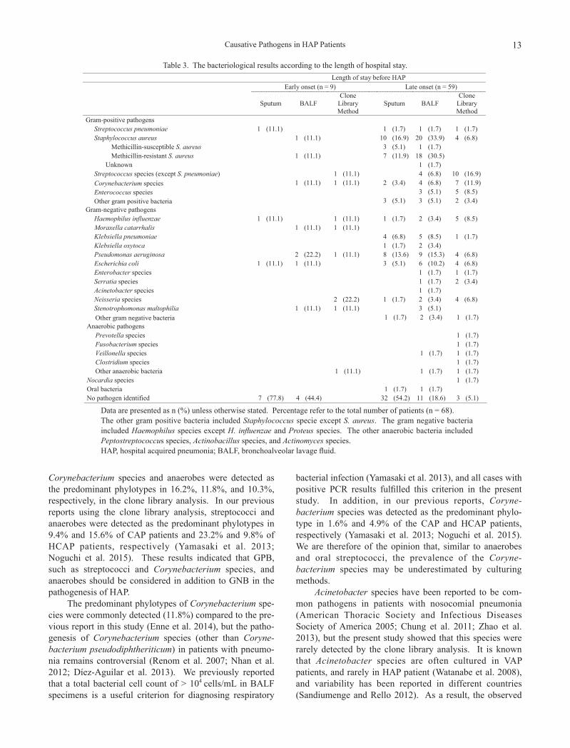
The detected bacteria in the BALF using the culture and the clone library analysis depend on the length of stay before the development of HAP (early- or late-onset) are shown in Table 3. Nine (13.2%) and 59 (86.8%) patients had early- and late-onset pneumonia, respectively. Most of the patients in whom S. aureus (mostly MRSA), K. pneu-moniae, and E. coli were cultured had a longer length of stay (late-onset) before the development of HAP. A similar tendency was found by the clone library analysis; however, the clone library analysis that we performed could not dif-
ferentiate MRSA from MSSA.
Comparison of bacterial phylotypes by conventional culture methods and the clone library analysis
The comparison of the bacterial phylotypes in BALF by the clone library analysis and culture methods is shown in Table 4. The concordance rate of the first or second pre-dominant phylotypes of the BALF culture with the bacterial floral analysis was ≥ 75% for S. pneumoniae, S. epidermi-dis, streptococci, Enterococcus species, H. influenzae, Moraxella catarrhalis, and Enterobacter species, Neisseria species, and S. maltophilia; 50-74% for Corynebacterium species, Pseudomonas aeruginosa and E. coli; and 25-49% for S. aureus and K. pneumoniae.
Evaluation of the proportion of microbiota by the clone library analysis
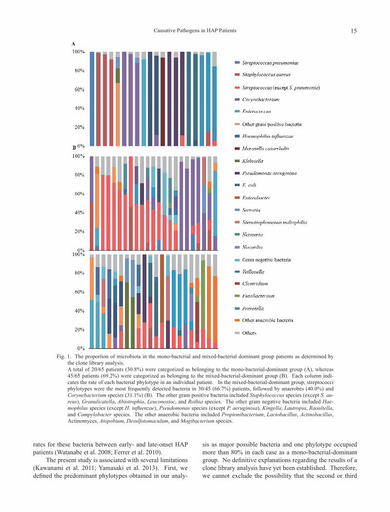
Among the 65 patients in whom bacterial phylotypes were detected by the clone library analysis, 20 (30.8%) and 45 (69.2%) were categorized as belonging to the mono-bac-terial-dominant and mixed-bacterial-dominant groups, respectively. In the mixed-bacterial-dominant group, strep-tococci were the most frequently detected phylotypes (30/45; 66.7%), followed by anaerobes (18/45; 40.0%) and the Corynebacterium species (14/45; 31.1%) (Fig. 1).
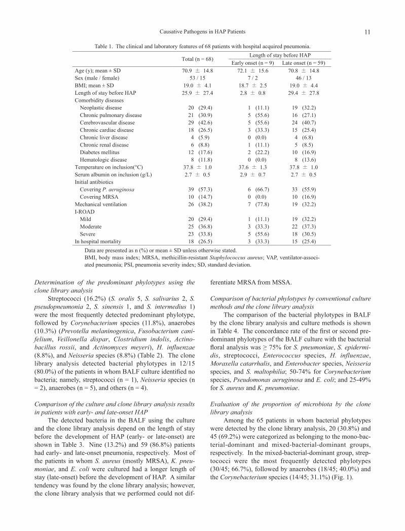
Total (n = 68) Length of stay before HAP
Early onset (n = 9) Late onset (n = 59) Age (y); mean ± SD 70.9 ± 14.8 72.1 ± 15.6 70.8 ± 14.8 Sex (male / female) 53 / 15 7 / 2 46 / 13 BMI; mean ± SD 19.0 ± 4.1 18.7 ± 2.5 19.0 ± 4.4 Length of stay before HAP 25.9 ± 27.4 2.8 ± 0.8 29.4 ± 27.8 Comorbidity diseases
Neoplastic disease 20 (29.4) 1 (11.1) 19 (32.2) Chronic pulmonary disease 21 (30.9) 5 (55.6) 16 (27.1) Cerebrovascular disease 29 (42.6) 5 (55.6) 24 (40.7) Chronic cardiac disease 18 (26.5) 3 (33.3) 15 (25.4) Chronic liver disease 4 (5.9) 0 (0.0) 4 (6.8) Chronic renal disease 6 (8.8) 1 (11.1) 5 (8.5) Diabetes mellitus 12 (17.6) 2 (22.2) 10 (16.9) Hematologic disease 8 (11.8) 0 (0.0) 8 (13.6)
Temperature on inclusion(°C) 37.8 ± 1.0 37.6 ± 1.3 37.8 ± 1.0 Serum albumin on inclusion (g/L) 2.7 ± 0.5 2.9 ± 0.7 2.7 ± 0.5 Initial antibiotics
Covering P. aeruginosa 39 (57.3) 6 (66.7) 33 (55.9) Covering MRSA 10 (14.7) 0 (0.0) 10 (16.9)
Mechanical ventilation 26 (38.2) 7 (77.8) 19 (32.2) I-ROAD
Mild 20 (29.4) 1 (11.1) 19 (32.2) Moderate 25 (36.8) 3 (33.3) 22 (37.3) Severe 23 (33.8) 5 (55.6) 18 (30.5)
In hospital mortality 18 (26.5) 3 (33.3) 15 (25.4)
Table 1. The clinical and laboratory features of 68 patients with hospital acquired pneumonia.
Data are presented as n (%) or mean ± SD unless otherwise stated.BMI, body mass index; MRSA, methicillin-resistant Staphylococcus aureus; VAP, ventilator-associ-ated pneumonia; PSI, pneumonia severity index; SD, standard deviation.

K. Yatera et al.12
DiscussionIn this study, we performed a clone library analysis to
investigate the BALF specimens of HAP patients. Our results suggested that, in addition to Gram-negative bacilli (GNB), aerobic Gram-positive bacilli (GPB), such as strep-tococci and Corynebacterium species and anaerobes might have important roles in the etiology of HAP. This is the first report of an evaluation of the microbiota in patients with HAP using a clone library analysis, and this analysis may add useful information in addition to the data obtained with ordinary cultivation methods, especially in detecting difficult-to-culture bacteria and bacteria that tend to be underestimated, such as streptococci.
It is more difficult to evaluate the etiology in HAP patients because the extraction of good sputum and/or spec-imens that are taken by invasive methods can be difficult due to the poor general condition of the patient and because of the presence of comorbidities. Indeed, bacterial etiolo-
gies are only known in approximately one-third of HAP patients, although the bacterial etiology in patients with HAP have been reported previously (Gómez et al. 1995; Jones 2010; Chung et al. 2011; Piskin et al. 2012; Quartin et al. 2013; Sopena et al. 2014). The present study demon-strated that the clone library analysis achieved a higher bac-terial phylotype detection rate (95.6%) in comparison to culture methods of BALF (77.9%), which was similar to our previous reports (Yamasaki et al. 2013; Noguchi et al. 2015). We therefore believe that the results of the present study provided precise information that cannot be obtained from ordinary culture methods alone.
In the present study, some GNB were detected in 39 of the 53 (73.6%) cases in which some bacteria were cultured in ordinary BALF cultures, and we re-identified the impor-tance of GNB in HAP using culture methods. However, the detection of some GNB phylotypes were only found to be the predominant phylotypes in 28 of 65 cases (43.1%) by the clone library analysis. In contrast, streptococci,
Conventional culture method Clone Library Method in BALF
Sputum BALF Case detected as the predominant phylotype
Gram-positive pathogens Streptococcus pneumoniae 2 (2.9) 1 (1.5) 1 (1.5) Staphylococcus aureus 10 (14.7) 21 (30.9) 4 (5.9)
Methicillin-susceptible S. aureus 3 (4.4) 1 (1.5) 0 (0.0) Methicillin-resistant S. aureus 7 (10.3) 19 (27.9) 0 (0.0) Unknown 0 (0.0) 1 (1.5) 0 (0.0)
Streptococcus species (except S. pneumoniae) 0 (0.0) 4 (5.9) 11 (16.2) Corynebacterium species 2 (2.9) 5 (7.4) 8 (11.8) Enterococcus species 0 (0.0) 3 (4.4) 5 (7.4) Other gram positive bacteria 3 (4.4) 3 (4.4) 2 (2.9)
Gram-negative pathogens Haemophilus influenzae 2 (2.9) 2 (2.9) 6 (8.8) Moraxella catarrhalis 0 (0.0) 1 (1.5) 1 (1.5) Klebsiella pneumoniae 4 (5.9) 5 (7.4) 1 (1.5) Klebsiella oxytoca 1 (1.5) 2 (2.9) 0 (0.0) Pseudomonas aeruginosa 8 (11.8) 11 (16.2) 5 (7.4) Escherichia coli 4 (5.9) 7 (10.3) 4 (5.9) Enterobacter species 0 (0.0) 1 (1.5) 1 (1.5) Serratia species 0 (0.0) 1 (1.5) 2 (2.9) Acinetobacter species 0 (0.0) 1 (1.5) 0 (0.0) Neisseria species 1 (1.5) 2 (2.9) 6 (8.8) Stenotrophomonas maltophilia 0 (0.0) 4 (5.9) 1 (1.5) Other gram negative bacteria 1 (1.5) 2 (2.9) 1 (1.5)
Anaerobic pathogens Prevotella species 0 (0.0) 0 (0.0) 1 (1.5) Fusobacterium species 0 (0.0) 0 (0.0) 1 (1.5) Veillonella species 0 (0.0) 1 (1.5) 1 (1.5) Clostridium species 0 (0.0) 0 (0.0) 1 (1.5) Other anaerobic bacteria 0 (0.0) 1 (1.5) 2 (2.9)
Nocardia species 0 (0.0) 0 (0.0) 1 (1.5) Oral bacteria 1 (1.5) 1 (1.5) 0 (0.0) No pathogen identified 39 (57.4) 15 (22.1) 3 (4.4)
Table 2. The results of bacteria detected using the conventional culture method and the predominant phy-lotypes according to the clone library analysis.
Data are presented as n (%) unless otherwise stated. Percentage refer to the total number of patients (n = 68).The other gram positive bacteria included Staphylococcus specie except S. aureus. The gram negative bacteria included Haemophilus species except H. influenzae and Proteus species. The other anaerobic bacteria included Peptostreptococcus species, Actinobacillus species, and Actinomyces species.BALF, bronchoalveolar lavage fluid.
Causative Pathogens in HAP Patients 13
Corynebacterium species and anaerobes were detected as the predominant phylotypes in 16.2%, 11.8%, and 10.3%, respectively, in the clone library analysis. In our previous reports using the clone library analysis, streptococci and anaerobes were detected as the predominant phylotypes in 9.4% and 15.6% of CAP patients and 23.2% and 9.8% of HCAP patients, respectively (Yamasaki et al. 2013; Noguchi et al. 2015). These results indicated that GPB, such as streptococci and Corynebacterium species, and anaerobes should be considered in addition to GNB in the pathogenesis of HAP.
The predominant phylotypes of Corynebacterium spe-cies were commonly detected (11.8%) compared to the pre-vious report in this study (Enne et al. 2014), but the patho-genesis of Corynebacterium species (other than Coryne- bacterium pseudodiphtheriticum) in patients with pneumo-nia remains controversial (Renom et al. 2007; Nhan et al. 2012; Díez-Aguilar et al. 2013). We previously reported that a total bacterial cell count of > 104 cells/mL in BALF specimens is a useful criterion for diagnosing respiratory
bacterial infection (Yamasaki et al. 2013), and all cases with positive PCR results fulfilled this criterion in the present study. In addition, in our previous reports, Coryne-bacterium species was detected as the predominant phylo-type in 1.6% and 4.9% of the CAP and HCAP patients, respectively (Yamasaki et al. 2013; Noguchi et al. 2015). We are therefore of the opinion that, similar to anaerobes and oral streptococci, the prevalence of the Coryne-bacterium species may be underestimated by culturing methods.
Acinetobacter species have been reported to be com-mon pathogens in patients with nosocomial pneumonia (American Thoracic Society and Infectious Diseases Society of America 2005; Chung et al. 2011; Zhao et al. 2013), but the present study showed that this species were rarely detected by the clone library analysis. It is known that Acinetobacter species are often cultured in VAP patients, and rarely in HAP patient (Watanabe et al. 2008), and variability has been reported in different countries (Sandiumenge and Rello 2012). As a result, the observed
Length of stay before HAP Early onset (n = 9) Late onset (n = 59)
Sputum BALF Clone
Library Method
Sputum BALF Clone
Library Method
Gram-positive pathogens Streptococcus pneumoniae 1 (11.1) 1 (1.7) 1 (1.7) 1 (1.7) Staphylococcus aureus 1 (11.1) 10 (16.9) 20 (33.9) 4 (6.8) Methicillin-susceptible S. aureus 3 (5.1) 1 (1.7) Methicillin-resistant S. aureus 1 (11.1) 7 (11.9) 18 (30.5)
Unknown 1 (1.7) Streptococcus species (except S. pneumoniae) 1 (11.1) 4 (6.8) 10 (16.9) Corynebacterium species 1 (11.1) 1 (11.1) 2 (3.4) 4 (6.8) 7 (11.9) Enterococcus species 3 (5.1) 5 (8.5) Other gram positive bacteria 3 (5.1) 3 (5.1) 2 (3.4)
Gram-negative pathogens Haemophilus influenzae 1 (11.1) 1 (11.1) 1 (1.7) 2 (3.4) 5 (8.5) Moraxella catarrhalis 1 (11.1) 1 (11.1) Klebsiella pneumoniae 4 (6.8) 5 (8.5) 1 (1.7) Klebsiella oxytoca 1 (1.7) 2 (3.4) Pseudomonas aeruginosa 2 (22.2) 1 (11.1) 8 (13.6) 9 (15.3) 4 (6.8) Escherichia coli 1 (11.1) 1 (11.1) 3 (5.1) 6 (10.2) 4 (6.8) Enterobacter species 1 (1.7) 1 (1.7) Serratia species 1 (1.7) 2 (3.4) Acinetobacter species 1 (1.7) Neisseria species 2 (22.2) 1 (1.7) 2 (3.4) 4 (6.8) Stenotrophomonas maltophilia 1 (11.1) 1 (11.1) 3 (5.1) Other gram negative bacteria 1 (1.7) 2 (3.4) 1 (1.7)
Anaerobic pathogens Prevotella species 1 (1.7) Fusobacterium species 1 (1.7) Veillonella species 1 (1.7) 1 (1.7) Clostridium species 1 (1.7) Other anaerobic bacteria 1 (11.1) 1 (1.7) 1 (1.7)
Nocardia species 1 (1.7) Oral bacteria 1 (1.7) 1 (1.7) No pathogen identified 7 (77.8) 4 (44.4) 32 (54.2) 11 (18.6) 3 (5.1)
Table 3. The bacteriological results according to the length of hospital stay.
Data are presented as n (%) unless otherwise stated. Percentage refer to the total number of patients (n = 68).The other gram positive bacteria included Staphylococcus specie except S. aureus. The gram negative bacteria included Haemophilus species except H. influenzae and Proteus species. The other anaerobic bacteria included Peptostreptococcus species, Actinobacillus species, and Actinomyces species.HAP, hospital acquired pneumonia; BALF, bronchoalveolar lavage fluid.

K. Yatera et al.14
differences may be partly due to these reasons.Although MRSA and P. aeruginosa were highly
(approximately 50%) detected in BALF culture, discrepan-cies were observed between the results of culture and the clone library analysis in detecting these bacteria. Similar discrepancies were observed in HCAP patients in our previ-ous study (Noguchi et al. 2015). These discrepancies in the results between the two methods might be due to an under-estimation of the oral bacteria, including streptococci, dur-ing culture, as the colonies macroscopically recognized as normal bacteria were commonly reported to be normal flo-ral bacteria, and the culture of anaerobes was also generally difficult. In the clone library analysis, the cases that phylo-types of S. aureus and P. aeruginosa were found to account for > 5% of the bacterial flora of each case were only 25% (4/16) and 50% (7/14) of the HCAP patients, whose BALF cultures were positive for S. aureus or P. aeruginosa, respectively (Noguchi et al. 2015). In contrast, the rates for S. aureus or P. aeruginosa occupied 42.9% (9/21) and 72.7% (8/11) in HAP patients, respectively. We recently reported the efficacy of the clone library analysis in patients with MRSA pneumonia (Kawanami et al. 2016), although it is difficult to clinically distinguish whether or not MRSA and P. aeruginosa are true causative bacteria based solely on culture results. Thus, these results may indicate the pos-
sibility that these bacteria contribute to HAP rather than HCAP.
The ATS/IDSA guidelines classified patients with HAP into early- and late-onset groups based on the onset of pneumonia because multidrug-resistant pathogens or poly-clonal pathogens are reported to be more common in late-onset HAP patients than in early-onset patients (American Thoracic Society and Infectious Diseases Society of America 2005; Enne et al. 2014). Contrarily, a few reports have shown similar detection rates of MDR pathogens (including MRSA), in early-onset and late-onset patients (Ferrer et al. 2010; Uvizl et al. 2011; Restrepo et al. 2013). In this study, MRSA was similarly detected mostly in the late-onset period, although the number of early-onset HAP patients was relatively small. In addition, K. pneumoniae and E. coli, of which approximately 30% of the detected specimens were ESBL-producing bacteria, were also far more frequently detected in late-onset HAP patients than in early-onset ones. Uvizl et al. (2011) similarly reported that ESBL-non producing K. pneumoniae and E. coli were detected in the many cases with late-onset pneumonia, although half of these bacteria were ESBL-producing bac-teria. Therefore, potential infection with these GNB should be considered in late-onset HAP pneumonia. However, other reports found no marked differences in the detection
BALF The
number detected
in cultivation
Clone Library Method
The first predominant
phylotype
The second predominant
phylotype
The third (or less) predominant
phylotype (excluding others§)
Others§
Gram-positive pathogens Streptococcus pneumoniae 1 1 (100) Staphylococcus aureus 21 4 (19.0) 2 (9.5) 3 (14.3) 12 (57.1)
Methicillin-susceptible S. aureus 1 1 (100) Methicillin-resistant S. aureus 19 4 (21.1) 2 (10.5) 2 (10.5) 11 (57.9) Unknown 1 1 (100)
Streptococcus species (except S. pneumoniae) 4 3 (75.0) 1 (25.0) Corynebacterium species 5 2 (40.0) 1 (20.0) 1 (20.0) 1 (20.0) Enterococcus species 3 2 (66.7) 1 (33.3) Other gram positive bacteria 3 2 (66.7) 1 (33.3)
Gram-negative pathogens 3 2 (66.7) 1 (33.3) Haemophilus influenzae 2 2 (100) Moraxella catarrhalis 1 1 (100) Klebsiella pneumoniae 5 1 (20.0) 1 (20.0) 3 (60.0) Klebsiella oxytoca 2 2 (100) Pseudomonas aeruginosa 11 5 (45.5) 2 (18.2) 1 (9.1) 3 (27.3) Escherichia coli 7 3 (42.9) 1 (14.3) 1 (14.3) 2 (28.6) Enterobacter species 1 1 (100) Serratia species 1 1 (100) Acinetobacter species 1 1 (100) Proteus species 2 2 (100) Neisseria species 2 1 (50.0) 1 (50.0)
4 2 (50.0) 1 (25.0) 1 (25.0) Stenotrophomonas maltophilia Anaerobic pathogens
1 1 (100) Veillonella species Peptostreptococcus species 1 1 (100)
Table 4. Comparison of bacterial results obtained by the clone library analysis and cultivation methods using BALF specimens.
The other gram positive bacteria included Staphylococcus species except S. aureus.§The phylotypes that dominated < 5% in each clone library were classified as “others”.BALF, bronchoalveolar lavage fluid.
Causative Pathogens in HAP Patients 15
rates for these bacteria between early- and late-onset HAP patients (Watanabe et al. 2008; Ferrer et al. 2010).
The present study is associated with several limitations (Kawanami et al. 2011; Yamasaki et al. 2013). First, we defined the predominant phylotypes obtained in our analy-
sis as major possible bacteria and one phylotype occupied more than 80% in each case as a mono-bacterial-dominant group. No definitive explanations regarding the results of a clone library analysis have yet been established. Therefore, we cannot exclude the possibility that the second or third
Fig. 1. The proportion of microbiota in the mono-bacterial and mixed-bacterial dominant group patients as determined by the clone library analysis.
A total of 20/65 patients (30.8%) were categorized as belonging to the mono-bacterial-dominant group (A), whereas 45/65 patients (69.2%) were categorized as belonging to the mixed-bacterial-dominant group (B). Each column indi-cates the rate of each bacterial phylotype in an individual patient. In the mixed-bacterial-dominant group, streptococci phylotypes were the most frequently detected bacteria in 30/45 (66.7%) patients, followed by anaerobes (40.0%) and Corynebacterium species (31.1%) (B). The other gram positive bacteria included Staphylococcus species (except S. au-reus), Granulicatella, Abiotrophia, Leuconostoc, and Rothia species. The other gram negative bacteria included Hae-mophilus species (except H. influenzae), Pseudomonas species (except P. aeruginosa), Kingella, Lautropia, Raoultella, and Campylobacter species. The other anaerobic bacteria included Propionibacterium, Lactobacillus, Actinobacillus, Actinomyces, Atopobium, Desulfotomaculum, and Mogibacterium species.

K. Yatera et al.16
most-predominant phylotypes may play a major role in the pathogenesis of pneumonia. In addition, the definitions of mono- or mixed-bacterial-dominant groups require further investigation to clarify the clinical significance of the per-centages of the bacterial phylotypes that are detected in the clone library analysis. Second, the universal primers we used in this study could not amplify all of the bacterial 16S rRNA genes; however, there were no reported human caus-ative pathogens among the bacterial species that were unde-tectable with these primers. Third, only approximately 100 clones were analyzed per specimen in this study, which might make minor populations undetectable. Fourth, because broad-spectrum antibiotics were frequently admin-istered to the patients in this study, the correlation between the results of the clone library analysis and the choice of antibiotics was uncertain.
In conclusion, we reported the bacterial phylotypes in BALF specimens obtained from HAP patients according to the clone library analysis and compared the results with those obtained by the conventional cultivation method. The results indicate that, in addition to GNB, GPB, such as streptococci, Corynebacterium species, and anaerobes may have important roles in the pathogenesis of HAP. In addi-tion, our results demonstrate that MRSA and P. aeruginosa might be considered as possible bacteria in HAP more fre-quently than they are in HCAP; however, this decision should be made by physician based on the clinical charac-teristics and the findings of examinations. Further investi-gations in which molecular methods are compared to cul-ture methods may better reveal the etiology of pneumonia and lead to the selection of more appropriate antibiotics for the treatment of pneumonia in the clinical setting.
AcknowledgmentsThe authors thank Drs. Chiharu Yoshii, Hideto Obata,
Yusuke Taura, Yukiko Kawanami, Yugo Yoshida, Takeshi Orihashi, Chinatsu Nishida, Naoyuki Inoue, Takaaki Ogoshi, Yu Suzuki, Susumu Tokuyama and Keishi Oda for collecting the samples, and Ms. Yoshiko Yamazaki, Kumiko Matsuyama and Michiyo Taguchi for their valuable assistance.
Author ContributionsConception and study design: Kazuhiro Yatera, Shingo
Noguchi, Toshinori Kawanami, Kazumasa Fukuda and Hiroshi Mukae. Data analysis: Shingo Noguchi, Kei Yamasaki, Keisuke Naito and Kentarou Akata. Data interpretation: Kazuhiro Yatera, Shingo Noguchi, Kei Yamasaki, Hiroshi Ishimoto, Hatsumi Taniguchi and Hiroshi Mukae. Manuscript drafting: Kazuhiro Yatera and Shingo Noguchi. Critical manuscript revision; Toshinori Kawanami, Kazumasa Fukuda, Kei Yamasaki, Keisuke Naito, Kentarou Akata, Hiroshi Ishimoto, Hatsumi Taniguchi and Hiroshi Mukae. Study supervision/final approval: Hiroshi Mukae.
FundingThis study was partially supported by a High Altitude
Research Grant from the University of Occupational and Envi-ronmental Health, Japan, a Ministry of Education, Science, Sports and Culture Grant-in-Aid for Scientific Research (C),
23591173, 2011 and a Ministry of Education, Science, Sports and Culture Grant-in-Aid for Young Scientists (B), 24790828, 2012.
Conflict of InterestThe authors declare no conflict of interest.
ReferencesAmerican Thoracic Society and Infectious Diseases Society of
America (2005) Guidelines for the management of adults with hospital-acquired, ventilator-associated, and healthcare-associated pneumonia. Am. J. Respir. Crit. Care Med., 171, 388-416.
Bahrani-Mougeot, F.K., Paster, B.J., Coleman, S., Barbuto, S., Brennan, M.T., Noll, J., Kennedy, T., Fox, P.C. & Lockhart, P.B. (2007) Molecular analysis of oral and respiratory bacte-rial species associated with ventilator-associated pneumonia. J. Clin. Microbiol., 45, 1588-1593.
Bousbia, S., Papazian, L., Saux, P., Forel, J.M., Auffray, J.P., Martin, C., Raoult, D. & La Scola, B. (2012) Repertoire of intensive care unit pneumonia microbiota. PLoS One, 7, e32486.
Cakir Edis, E., Hatipoglu, O.N., Yilmam, I., Eker, A., Tansel, O. & Sut, N. (2009) Hospital-acquired pneumonia developed in non-intensive care units. Respiration, 78, 416-422.
Chung, D.R., Song, J.H., Kim, S.H., Thamlikitkul, V., Huang, S.G., Wang, H., So, T.M., Yasin, R.M., Hsueh, P.R., Carlos, C.C., Hsu, L.Y., Buntaran, L., Lalitha, M.K., Kim, M.J., Choi, J.Y., et al. (2011) High prevalence of multidrug-resistant nonfer-menters in hospital-acquired pneumonia in Asia. Am. J. Respir. Crit. Care Med., 184, 1409-1417.
Díez-Aguilar, M., Ruiz-Garbajosa, P., Fernández-Olmos, A., Guisado, P., Del Campo, R., Quereda, C., Cantón, R. & Meseguer, M.A. (2013) Non-diphtheriae Corynebacterium species: an emerging respiratory pathogen. Eur. J. Clin. Microbiol. Infect. Dis., 32, 769-772.
Enne, V.I., Personne, Y., Grgic, L., Gant, V. & Zumla, A. (2014) Aetiology of hospital-acquired pneumonia and trends in anti-microbial resistance. Curr. Opin. Pulm. Med., 20, 252-258.
Ferrer, M., Liapikou, A., Valencia, M., Esperatti, M., Theessen, A., Antonio Martinez, J., Mensa, J. & Torres, A. (2010) Valida-tion of the American Thoracic Society-Infectious Diseases Society of America guidelines for hospital-acquired pneu-monia in the intensive care unit. Clin. Infect. Dis., 50, 945-952.
Gómez, J., Esquinas, A., Agudo, M.D., Sánchez Nieto, J.M., Núñez, M.L., Baños, V., Canteras, M. & Valdes, M. (1995) Retrospective analysis of risk factors and prognosis in non-ventilated patients with nosocomial pneumonia. Eur. J. Clin. Microbiol. Infect. Dis., 14, 176-181.
Jones, R.N. (2010) Microbial etiologies of hospital-acquired bacterial pneumonia and ventilator-associated bacterial pneu-monia. Clin. Infect. Dis., 51 Suppl 1, S81-87.
Kawanami, T., Fukuda, K., Yatera, K., Kido, M., Mukae, H. & Taniguchi, H. (2011) A higher significance of anaerobes: the clone library analysis of bacterial pleurisy. Chest, 139, 600-608.
Kawanami, T., Yatera, K., Yamasaki, K., Noguchi, S., Fukuda, K., Akata, K., Naito, K., Kido, T., Ishimoto, H., Taniguchi, H. & Mukae, H. (2016) Clinical impact of methicillin-resistant staphylococcus aureus on bacterial pneumonia: cultivation and 16S ribosomal RNA gene analysis of bronchoalveolar lavage fluid. BMC Infect. Dis., 16, 155.
Lu, W., Yu, J., Ai, Q., Liu, D., Song, C. & Li, L. (2014) Increased constituent ratios of Klebsiella sp., Acinetobacter sp., and Streptococcus sp. and a decrease in microflora diversity may be indicators of ventilator-associated pneumonia: a prospec-
Causative Pathogens in HAP Patients 17
tive study in the respiratory tracts of neonates. PLoS One, 9, e87504.
Mukae, H., Yatera, K., Noguchi, S., Kawanami, T., Yamasaki, K., Tokuyama, S., Inoue, N., Nishida, C., Kawanami, Y., Ogoshi, T., Orihashi, T., Yoshii, C. & Ishimoto, H. (2015) Evaluation of a rapid immunochromatographic ODK0501 assay for detecting Streptococcus pneumoniae antigens in the sputum of pneumonia patients with positive S. pneumoniae urinary anti-gens. J. Infect. Chemother., 21, 176-181.
Nhan, T.X., Parienti, J.J., Badiou, G., Leclercq, R., & Cattoir, V. (2012) Microbiological investigation and clinical significance of Corynebacterium spp. in respiratory specimens. Diagn. Microbiol. Infect. Dis., 74, 236-241.
Noguchi, S., Mukae, H., Kawanami, T., Yamasaki, K., Fukuda, K., Akata, K., Ishimoto, H., Taniguchi, H. & Yatera, K. (2015) Bacteriological assessment of healthcare-associated pneu-monia using a clone library analysis. PLoS One, 10, e0124697.
Piskin, N., Aydemir, H., Oztoprak, N., Akduman, D., Comert, F., Kokturk, F. & Celebi, G. (2012) Inadequate treatment of ventilator-associated and hospital-acquired pneumonia: risk factors and impact on outcomes. BMC Infect. Dis., 12, 268.
Quartin, A.A., Scerpella, E.G., Puttagunta, S. & Kett, D.H. (2013) A comparison of microbiology and demographics among patients with healthcare-associated, hospital-acquired, and ventilator-associated pneumonia: a retrospective analysis of 1184 patients from a large, international study. BMC Infect. Dis., 13, 561.
Renom, F., Garau, M., Rubí, M., Ramis, F., Galmés, A. & Soriano, J.B. (2007) Nosocomial outbreak of Corynebacterium stri-atum infection in patients with chronic obstructive pulmonary disease. J. Clin. Microbiol., 45, 2064-2067.
Restrepo, M.I., Peterson, J., Fernandez, J.F., Qin, Z., Fisher, A.C.
& Nicholson, S.C. (2013) Comparison of the bacterial etiology of early-onset and late-onset ventilator-associated pneumonia in subjects enrolled in 2 large clinical studies. Respir. Care, 58, 1220-1225.
Sandiumenge, A. & Rello, J. (2012) Ventilator-associated pneu-monia caused by ESKAPE organisms: cause, clinical features, and management. Curr. Opin. Pulm. Med., 18, 187-193.
Sopena, N., Heras, E., Casas, I., Bechini, J., Guasch, I., Pedro-Botet, M.L., Roure, S. & Sabrià, M. (2014) Risk factors for hospital-acquired pneumonia outside the intensive care unit: a case-control study. Am. J. Infect. Control, 42, 38-42.
Sopena, N. & Sabrià, M.; Neunos 2000 Study Group (2005) Multicenter study of hospital-acquired pneumonia in non-ICU patients. Chest, 127, 213-219.
Uvizl, R., Hanulik, V., Husickova, V., Sedlakova, M.H., Adamus, M. & Kolar, M. (2011) Hospital-acquired pneumonia in ICU patients. Biomed. Pap. Med. Fac. Univ. Palacky Olomouc Czech Repub., 155, 373-378.
Watanabe, A., Yanagihara, K., Kohno, S. & Matsushima, T.; HAP study group (2008) Multicenter survey on hospital-acquired pneumonia and the clinical efficacy of first-line antibiotics in Japan. Intern. Med., 47, 245-254.
Yamasaki, K., Kawanami, T., Yatera, K., Fukuda, K., Noguchi, S., Nagata, S., Nishida, C., Kido, T., Ishimoto, H., Taniguchi, H. & Mukae, H. (2013) Significance of anaerobes and oral bacteria in community-acquired pneumonia. PLoS One, 8, e63103.
Zhao, T., Liu, Y., Cao, B., Wang, H., Chen, L., She, D., Liang, Z., Sun, T., Li, Y., Tong, Z., Wang, Z., He, B., Yang, W., Qu, J. & Li, X. (2013) Prospective multicenter study of pathogen distributions in early-onset and late-onset hospital-acquired pneumonia in china. Antimicrob. Agents Chemother., 57, 6404-6405.