determination of the minimum inhibitory concentration of ... · medicalmycology,2014,52,90–98...
TRANSCRIPT
Medical Mycology, 2014, 52, 90–98doi: 10.3109/13693786.2013.806827
Advance Access Publication Date: 1 July 2013Original Article
Original Article
Determination of the minimum inhibitory
concentration of Cryptococcus neoformans and
Cryptococcus gattii against fluconazole by flow
cytometry
Bernardina Penarrieta Morales1,∗, Ivan Neves Junior1, Luciana Trilles1,
Alvaro Luiz Bertho2, Raquel De Vasconcellos Carvalhaes De Oliveira1,
Marilia Martins Nishikawa3, Monica Dos Santos Elias1, Bodo Wanke1
and Marcia Dos Santos Lazera1
1Evandro Chagas Clinical Research Institute (IPEC), Oswaldo Cruz Foundation (FIOCRUZ), Rio de Janeiro,2Immunoparasitology Laboratory, Oswaldo Cruz Foundation, Rio de Janeiro and 3National Institute forQuality in Health, Oswaldo Cruz Foundation, Rio de Janeiro, Brazil
*To whom correspondence should be addressed. E-mail: [email protected]
Received 30 November 2012; Revised 28 February 2013; Accepted 15 May 2013
Abstract
Recent studies have used flow cytometry (FCM) as an important alternative method todetermine the antifungal susceptibility of yeasts compared to the broth microdilutionClinical and Laboratory Standards Institute (CLSI) reference procedure. We present acomparative study of the broth microdilution method and flow cytometry to assessthe in vitro antifungal susceptibility of Cryptococcus neoformans (n = 16) and C. gattii(n = 24) to fluconazole. The minimum inhibitory concentration (MIC) assays by flowcytometry were defined as the lowest drug concentration that showed ∼50% of the countof acridine orange negative cells compared to that of the growth control. Categoricalclassification showed all C. neoformans isolates were susceptible to fluconazole. Threeisolates of C. gattii were susceptible dose-dependent and the remaining 21 isolates wereclassified as susceptible. MICs comparison of both methodologies demonstrated 100%categorical agreement of the results obtained for C. neoformans and C. gattii. The MICsobtained with the CLSI-approved method and flow cytometry were compared by theSpearman correlation test and a significant Pv = 0.001. The flow cytometric methodhas the advantage of analyzing a large and constant number of cells in less time, i.e.,9 h incubation for fluconazole using acridine orange versus 72 h for broth microdilutionmethod. In conclusion, the two methods were comparable and flow cytometry methodcan expedite and improve the results of in vitro susceptibility tests of C. neoformans and
90 C© The International Society for Human and Animal Mycology 2013. All rights reserved. For permissions,please e-mail: [email protected].
at FundaçÃ
£o Osw
aldo Cruz-B
iblioteca de Manguinhos/O
swaldo C
ruz Foundation-Ma on M
arch 25, 2015http://m
my.oxfordjournals.org/
Dow
nloaded from
Morales et al. 91
C. gattii against fluconazole and also allows comparative studies in vitro/in vivo morerapidly, which along with clinical data, could assist in selecting the most appropriatetreatment choice.
Key words: flow cytometry, antifungal susceptibility, Cryptococcus.
Introduction
Cryptococcus neoformans is a pathogenic fungus, the cos-mopolitan agent of fungal meningoencephalitis in immune-compromised patients [1] including a high number of oc-currences in acquired immunodeficiency syndrome (AIDS)-related deaths in sub-Saharan Africa [2], as well as beingrecognized as an important infectious agent throughout theworld [3]. The mortality rate in infections caused by thisfungus is still high, especially when diagnosis is delayed andthe access to highly active antiretroviral therapy (HAART)in AIDS patients is limited [4].
Cryptococcus gattii, a close sibling species of C. neofor-mans, is endemic in tropical and subtropical climates [5],causing meningoencephalitis with high death rates in hostswith normal immunity, especially in the emerging countries[6–9]. In the humid Brazilian Amazon (North) and in thesemi-arid Northeast regions, C. gattii is the causative agentof meningitis in children and adolescents with high mortal-ity in these groups, despite the use of colloidal amphotericinB (AMB) [10,11]. It has been speculated that C. gattii ismore virulent and less susceptible to some antifungal drugsthan C. neoformans based on in vitro investigations [12].
High doses of AMB associated with 5-fluorocytosineare indicated for induction therapy for cryptococcosis, fol-lowed by fluconazole (FCZ) in the consolidation phase [3].FCZ is also indicated for the primary treatment of local-ized infections without evidence of dissemination [13,14].Due to the increased significance of fungal infections andthe introduction of new antifungal drugs, the Clinical andLaboratory Standards Institute (CLSI, formerly NCCLS)in the USA has developed (since 1985) protocols for invitro susceptibility testing to aid physicians in their thera-peutic choices for treating cryptococcosis. The standardiza-tion and reproductibility of broth microdilution method-ology for in vitro susceptibility testing for yeasts, currentCLSI M27-A3 [15,16], allows greater comparability of re-sults among different laboratories. However, this method isconsidered laborious and time-consuming because it takes24/48 h for incubation of Candida spp. and 72 h for Cryp-tococcus spp. and training of personnel, mainly to appro-priately obtain the data from the testing. This encouragedthe search for new methods to reduce incubation time andautomate the collection of results. Recent studies have com-bined the use flow cytometry (FCM) with the conventional
method CLSI M27-A3/M27-S3 and found it to be an im-portant alternative procedure to establish the in vitro an-tifungal susceptibility of yeasts. FCM is a powerful tech-nique for the diagnosis of hematological diseases and iswidely used in clinical laboratories [17,18]. This techniquehas shown great potential in the classification of tumorsof hematopoietic and lymphoid tissue using morphologicalphenotypic and genotypic parameters, as recommended byBethesda International Consensus in 2006 to study the Lin-focitosis in FCM [18]. In addition, FCM combined withspecific fluorochromes, like propidium iodide (PI), FUN-1 [2-choro-4-(2,3-dihydro-3-methyl-(benzo-1,3-thiazol-2-yl)-methylidene)-1 phenylquinolinium iodide] and acredineorange (AO), has been used by many authors to determinethe in vitro susceptibility of yeasts, especially Candida spp.The MIC values generated through the use of FCM arebased on the analysis of large numbers of individual yeastcells which does not require protracted incubation periods[19–22]. However, there have been very few studies whichhave examined the application of this method in assessingthe antifungal susceptibility of C. neoformans and C. gattii[22].
Therefore, this work aims to optimize the in vitro an-tifungal susceptibility assay to obtain results in a shorterperiod of time for C. neoformans and C. gattii against flu-conazole, the most common antifungal drug used in thetreatment of cryptococcosis. For this we performed a com-parison of MICs obtained by flow cytometry and the mi-crodilution method (CLSI-M27-A3/M27-S3).
Material and methods
Strains
A total of 16 isolates of C. neoformans and 24 of C. gat-tii strains stored at the Culture Collection of PathogenicFungi in Evandro Chagas Clinical Research Institute atOswaldo Cruz Foundation (IPEC/FIOCRUZ) at −70◦C in15% glycerol were employed in the studies. Purity, viabil-ity and genus identity of the strains were confirmed throughthe use of Niger seed agar medium (NSA) and biochemicaltests (Vitek YBC, bioMerieux, Inc., Durham, NC, USA).The species were identified with CGB (canavanine-glycinebromothymol blue) medium test [23]. Candida parapsilosisINCQS 40038 (ATCC 22019) and Candida krusei INCQS
at FundaçÃ
£o Osw
aldo Cruz-B
iblioteca de Manguinhos/O
swaldo C
ruz Foundation-Ma on M
arch 25, 2015http://m
my.oxfordjournals.org/
Dow
nloaded from

92 Medical Mycology, 2014, Vol. 52, No. 1
40147 (ATCC 6258) were included in the study as qualitycontrols.
Antifungal agents
Fluconazole was obtained in powder form (Sigma-AldrichInc., St Louis, MO, USA). Stock solutions were prepared indimethylsulfoxide-DMSO P.A. 99.70% (Vetec Fine Chemi-cals Ltd, Duque de Caxias, Brazil) at 5120 µg/ml and main-tained at −70◦C for up to 12 months [16].
Antifungal susceptibility testing CLSI
The microdilution tests were performed following theCLSI M27-A3 and M-27S3 broth microdilution guidelines[15,16]. Fluconazole dilutions ranging from 0.12–64 µg/mlwere prepared in flat-bottomed 96-well microplates usingRPMI-1640 broth medium with L-glutamine without bicar-bonate (Gibco
TM, New York, USA) buffered at pH 7.0 with
(3-[N-Morpholino]- propanesulfonic acid) buffer >99%(Vetec Fine Chemicals Ltd, Duque de Caxias, Brazil),and supplemented with D-(+)-Glucose 2% (Sigma-AldrichInc., St Louis, MO, USA), modification accepted by theCLSI document [16,24]. The inocula were obtained from48-h-old cultures of each test strain grown on drug-freeSabouraud dextrose Agar at 35◦C. A pilot study showedthat the growth of Cryptococcus cells was far better us-ing 0.45% saline solution rather than 0.85% in a shortincubation time (2–12 h), so that FCM results could be re-producible. Therefore, the suspensions were prepared with0.45% sterile saline and adjusted using Bio-Merieux Den-sichek instrument to match the turbidity of a 0.5 McFar-land standard. The final concentration of the inoculum inthe microplates was 0.5 × 10³ to 2.5 × 10³ CFU ml−1 usingRPMI1640 broth medium as described above.
Antifungal susceptibility testing by flow cytometry(FCM)
The minimum inhibitory concentration (MIC) of flucona-zole was determined by a flow cytometric approach usingacridine orange as fluorochrome (AO) (Sigma-Aldrich Inc.,St Louis, MO, USA).
Fluconazole solutions were prepared following the brothmicrodilution guidelines [15] using RPMI 1640 media asdescribed above. In order to reduce the dilution range of flu-conazole we used final concentrations of from 1–64 µg/ml.Three hundred microliters were distributed in capped ster-ile tubes specific to flow cytometry and kept at −70◦C upto 6 months or until used in the study.
To prepare the inoculums suspensions, strains weregrown in Petri dishes containing drug-free Sabouraud dex-
trose agar (Difco Laboratories, Sparks, MD, USA) at 35◦Cfor 48 h. Five to 10 colonies of approximately 1 mm indiameter each were suspended in sterile 0.45% saline, theturbidity was adjusted using Bio-Merieux Densichek instru-ment at a wevelength of 530 nm to 1 McFarland standard(approximatly 300 × 106 CFU ml−1). Portions of this in-oulum suspension (300 µl) were distributed in tubes con-taining 300 µl of one of the drug dilutions (2–128 µg/ml).
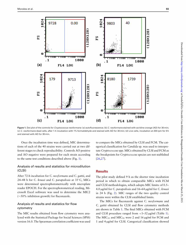
To evaluated the action of AO at a final concentrationof 11 mg/l as described in tests involving Candida spp. [25],cells of C. neoformans and C. krusei were analyzed withfluorescence microscopy (Olympus B × 40: 1). Cells sus-pension in sterile 0.45% saline matching turbidity of 0.5McFarland were incubated for 30 min with AO in the darkand were defined as negative control or AO negative (viablecells) if green cells were observed. In contrast, cells treatedwith formaldehyde at a final concentration 1% incubatedfor 1 h prior to addition of the AO for 30 min were de-fined as positive control or AO positive (non-viable cells)when orange-red cells were found (data not shown). Todetermine the MIC obtained with flow cytometry for C. ne-ofomans and C. gattii, we performed a pilot study using AOat 11 mg/l as an indicator of viability. The MIC of FCZ wasdefined as the lowest concentration that showed ∼50% ofviable cells, corresponding to the left quadrant of dot plot(Fig. 1).
Inoculum dilution acquisition was performed in Beck-man Coulter XL-MCL flow cytometer and studies wereconducted with a flow cytometric protocol in which sizeforward scatter (FSC) and granularity side scatter (SSC) at675 nm fluorescence emission (FL4) 620V were measured.Data analysis was performed by a dual-parameter dot plot– FSC vs. AO fluorescence intensity, which was divided intotwo quadrants, i.e., left comprehending AO negative cells(viable cells), and right comprehending AO positive cells(non-viable cells). A gate was created (for Cryptococcusand Candida) in order to exclude debris and define the cellpopulations of interest.
In order to define the best incubation time for an appro-priate reading in flow cytometry, we tested six representa-tive strains of C. neoformans and C. gattii, and the qualitycontrol strains of Candida krusei and C. parapsilosis. Thestrains were incubated with continuous shaking at 200 rpmusing Tecnal TE-420 incubator for 4, 8, 9, 12, 19 and 24h at 35◦C. After 30-min incubation with AO at 200 rpm,10,000 cells were acquired in triplicate in flow cytometry.The lowest concentration which showed ∼50% (MIC2) vi-able cells after incubation of the inoculum cells for 4, 8, 9,12, 19 and 24 h were compared to the concentrations rangeof the quality control strains (C. krusei and C. parapsilosis)from the CLSI guidelines [15].
at FundaçÃ
£o Osw
aldo Cruz-B
iblioteca de Manguinhos/O
swaldo C
ruz Foundation-Ma on M
arch 25, 2015http://m
my.oxfordjournals.org/
Dow
nloaded from
Morales et al. 93
Figure 1. Dot plot of the controls for Cryptococcus neoformans: (a) autofluorescence; (b) C. neoformans stained with acridine orange (AO) for 30 min;(c) C. neoformans dead cells, after 1-h incubation with 1% formaldehyde and stained with AO for 30 min; (d) Live cells, incubation at 200 rpm for 9 hand stained with AO for 30 min.
Once the incubation time was defined, MIC determina-tions of each of the 40 strains were carried out at two dif-ferent stages to check reproducibility. Controls AO positiveand AO negative were prepared for each strain accordingto the same test conditions described above (Fig. 1).
Analysis of results and statistics for microdilution(CLSI)
After 72-h incubation for C. neoformans and C. gattii, and24–48 h for C. krusei and C. parapsilosis at 35◦C, MICswere determined spectrophotometrically with microplatereader EPOCH. For the spectrophotometrical reading, Mi-crosoft Excel software was used to determine the MIC2(∼50% inhibition growth) for fluconazole.
Analysis of results and statistics for flowcytometry
The MIC results obtained from flow cytometry were ana-lyzed with the Statistical Package for Social Sciences (SPSS)version 16.0. The Spearman correlation coefficient was used
to compare the MICs obtained by CLSI and FCM. The cat-egorical classification for Candida sp. was used to interpre-tate Cryptococcus spp. MICs obtained by CLSI and FCM asthe breakpoints for Cryptococcus species are not stablished[26,27].
Results
The pilot study defined 9 h as the shorter time incubationperiod in which to obtain comparable MICs with FCMand CLSI methodologies, which adopts MIC limits: of 0.5–4.0 µg/ml for C. parapsilosis and 16–64 µg/ml for C. kruseiin 24 h (Fig. 2). MIC ranges of the two quality controlstrains were within the CLSI established limits.
The MICs for fluconazole against C. neoformans andC. gattii obtained by CLSI and flow cytometry methodsare shown in Table 1. The final MICs obtained with FCMand CLSI procedure ranged from <1–32 µg/ml (Table 1).The MIC50 and MIC90 were 2 and 16 µg/ml for FCM and1 and 4 µg/ml for CLSI. Categorical classification showed
at FundaçÃ
£o Osw
aldo Cruz-B
iblioteca de Manguinhos/O
swaldo C
ruz Foundation-Ma on M
arch 25, 2015http://m
my.oxfordjournals.org/
Dow
nloaded from

94 Medical Mycology, 2014, Vol. 52, No. 1
Figure 2. Dot plot of the corresponding MIC and the previous dilution obtained from the quality control strains, Candida krusei INCQS 40147 (ATCC6258) and Candida parapsilosis INCQS 40038 (ATCC 22019): (a) previous dilution of MIC C. krusei; (b) MIC of C.krusei; (c) previous dilution of MIC C.parapsilosis; (d) MIC of C. parapsilosisi.
all C. neoformans isolates were susceptible to fluconazole,whereas three isolates of C. gattii were susceptible dose-dependent and the remaining 21 were classified as suscep-tible (Table 1). MICs comparison of both methodologiesdemonstrated 100% categorical agreement for C. neofor-mans and C. gattii. The MICs for C. gattii in CLSI methodranged from 0.12–32 µg/ml and those found with FCMranged from 1–32 µg/ml, whereas MICs for C. neoformansin the CLSI methods ranged from 0.25–4.0 µg/ml and FCMranged from 1–8 µg/ml (Fig. 3). In general, lower MICswere obtained with the CLSI method compared to FCM,especially for C. neoformans, despite eight isolates showingMICs <1 µg/ml using the CLSI method (minimum dilutionused for FCM) (Table 1).
The Spearman Correlation coefficient was used tocompare the MICs obtained by the two methodologiesand revealed a significant positive correlation coefficient,Pv = 0.001 obtained at the 0.01 level.
Discussion
Flow cytometry has been employed over the past few yearsfor in vitro antifungal susceptibility testing (AST) in orderto shorten the incubation time for slow growing molds [28]and providing greater accuracy in less time as the methodanalyzes larger number of cells allowing the detection ofcell damage [20–22,25].
The AST methods used for Cryptococcus were stan-dadized with Candida species that grow faster than mem-bers of the former species [15]. Therefore, FCM is of interestas it may overcome such limitations [20] of conventionalprocedures for C. neoformans and C. gattii. The currenthigh price of the FCM equipment can be a limiting fac-tor in applying the methodology in clinical laboratories.However, in the laboratories which routinely used FCMfor diagnostic and other purposes, AST could be performedwith a positive cost/benefit ratio, especially when comparedto the commercial methods that can be fiscally prohibitive.
at FundaçÃ
£o Osw
aldo Cruz-B
iblioteca de Manguinhos/O
swaldo C
ruz Foundation-Ma on M
arch 25, 2015http://m
my.oxfordjournals.org/
Dow
nloaded from

Morales et al. 95
Table 1. Minimum inhibitory concentration (MIC) of flucona-
zole obtained by Clinical and Laboratory Standards Institute
(CLSI) and flow cytometry and categorical correlation of Cryp-
tococcus neoformans and Cryptococcus gattii.
CLSI‡ FCM† CLSI and FCMIsolates MIC2§ (µg/ml) MIC2 (µg/ml) Correlation
categorical∗∗
C. neoformansLMM∗1426 1.00 8.00 SLMM1494 <1 2.00 SLMM1433 4.00 4.00 SLMM1434 1.00 <1.00 SLMM1443 4.00 4.00 SLMM1446 1.00 4.00 SLMM1445 <1 1.00 SLMM1450 <1 2.00 SLMM1452 4.00 4.00 SLMM1455 1.00 4.00 SLMM1457 1.00 2.00 SLMM1468 1.00 1.00 SLMM1473 1.00 2.00 SLMM1475 1.00 2.00 SLMM1496 2.00 2.00 SLMM1498 2.00 4.00 S
C. gattiiLMM201 <1 1.00 SLMM202 2.00 1.00 SLMM244 <1 1.00 SLMM253 <1 1.00 SLMM272 1.00 1.00 SLMM326 2.00 1.00 SLMM330 <1 <1.00 SLMM347 1.00 1.00 SLMM362 2.00 1.00 SLMM378 <1 <1.00 SLMM384 1.00 1.00 SLMM1430 4.00 1.00 SLMM1436 2.00 2.00 SLMM1422 2.00 4.00 SLMM1425 32.00 32.00 SDDLMM1429 32.00 32.00 SDDLMM1431 2.00 2.00 SLMM1432 2.00 8.00 SLMM1435 32.00 32.00 SDDLMM1437 4.00 4.00 SLMM1438 4.00 8.00 SLMM1441 1.00 8.00 SLMM1463 1.00 8.00 SLMM1493 1.00 8.00 S
S, susceptible; SDD, susceptible dose-dependent; ∗Laboratory Medical My-cology; ‡broth microdilution guidelines; §∼50% optical density value of thegrowth control; †FCM, flow cytometry. ∗∗The Categorical classification forCandida sp. was used to interpret Cryptococcus spp. MICs obtained by CLSIand FCM as the breakpoints for Cryptococcus species are not established.
Different fluorochromes have been used in antifun-gal susceptibility tests with FCM, especially for Candidaspecies. For the analysis of Cryptococcus spp., the fluo-rocromes propidium iodide (PI) and FUN-1 were used byRamani et al. [29], Chaturvedi et al. [30] and Joung et al.[19]. So far, variable results have been obtained with thefew strains of C. neoformans that have been analyzed withFCM using PI and FUN-1 [22,31,32]. Green et al. [31] ob-tained comparable results between microdilution methodand FCM using PI, but the authors tested only one strain ofC. neoformans among several isolates of Candida spp. andSaccharomyces cerevisiae. Pina-Vaz et al. [22] also com-pared both methodologies, analysing three Cryptococcusand 63 Candida isolates, and obtained comparable resultswith FCM using FUN-1 after 1 h of incubation in testsinvolving itraconazole, voriconazole and caspofungin. Ra-mani and Chaturvedi [32] tested the in vtro AST of 16strains of C. neoformans to amphotericin B and FCZ withFCM using PI and obtained results comparable to thosefound with the CLSI procedures after 4 and 6 h of incuba-tion for amphotericin B and FCZ, respectively. However,FCM required the use of one more step, i.e., the addition ofsodium deoxycholate at the end of incubation to improvethe permeability for PI. According to Rudensky et al. [25],sodium deoxycholate results in gelling of the yeast suspen-sion during cytometer reading, so they proposed the use ofacridine orange (AO) as the viability indicator of Candidaspp.
Most antifungal susceptibilities studies with FCM arebased on the mean channel fluorescence of the dead cells,as used by Ramani et al., with 50% increasing mean channelfluorescence for Candida albicans using PI [29]. However,another study demonstrated that a 30% increase in meanchannel fluorescence was best correlated to the MICs ofCandida spp. to FCZ [33].
In this study the MICs found with FCM were obtainedby ∼50% of AO negative cells count, comparing with theAO negative of the growth control for each isolate. Thus,MICs results were based on living cells, as used in the CLSImethod. We obtained comparable results to those obtainedwith the CLSI method in our pilot study of C. neoformansand C. gattii susceptibility against FCZ with 9-h incuba-tion. A considerable number of living cells was lost in thegrowth control (drug-free) during 9-h incubation at 200rpm. Approximately 1700 positive AO cells were detectedcompared to the control prepared 30 min prior to acqui-sitions by FCM. Thus, the fast death of cryptococcal cellsobserved in 9 h was one of the reasons to choose countingAO negative cells to determine the MICs by FCM. More-over, the use of AO eliminated the need for the additionalsodium deoxycholate [25].
at FundaçÃ
£o Osw
aldo Cruz-B
iblioteca de Manguinhos/O
swaldo C
ruz Foundation-Ma on M
arch 25, 2015http://m
my.oxfordjournals.org/
Dow
nloaded from

96 Medical Mycology, 2014, Vol. 52, No. 1
Figure 3. Dot plot of the corresponding MIC and the previous dilution for representative strains of Cryptococcus neoformans (LMM1433) and C. gattii(LMM1493): (a) previous dilution of MIC C. neoformans; (b) MIC of C. neoformans; (c) previous dilution of MIC C. gattii; (d) MIC of C. gattii.
Usually the FCM method to evaluate the in vitro flucona-zole susceptibility using FUN-1 and PI provided rapid andreproducible results in 6–8 h for Candida species [32,33]. Incontrast, our study determined 9 h was shortest incubationtime for C. neoformans and C. gattiii, but 12, 19 and 24 hpresented a variation of one dilution.
The MICs ranges obtained by CLSI and FCM methodswere similar, although 0.12 µg/ml or 1 µg/ml is the lowestdilution in CLSI and FCM, respectively. This difference mayhave influenced the statistical analysis since eight strains hadMIC <1 µg/ml with the CLSI method, but a significant pos-itive correlation coefficient was still obtained. The rangeconcentration of 1–64 µg/ml for FCM was used becausein the categorical classification MICs ≤ 8 µg/ml indicatesusceptible (S), 16–32 µg/ml susceptible dose-dependent (S-DD) and ≥64 resistant (R) strains [15]. Thus, when con-sidered categorical classification, 100% agreement betweenboth methodologies was observed for C. neoformans andC. gattii strains.
CLSI inoculum is small and variable (500–2,500CFU/ml), while FCM method has the advantage of allow-ing the analysis of a large and constant number of cells,such as 10,000 cells (in this study), or 30,000 by Pina-Vazet al. [22], yielding a more representative result. Thus, weconsider inoculum size to be a critical factor for achievingreproducible results with isolates containing heterogeneoussub-populations of cells with different fluconazole suscepti-bilities, such as heteroresistance of C. neoformans describedby Yamazumi et al. [34].
In conclusion, we present a rapid flow cytometry as-say using acridine orange for C. neoformans and C. gat-tii susceptibility testing. Flow cytometry proved to be re-producible, with the additional advantage of analyzing alarge and a constant number of cells, allowing comparativestudies carried out on Cryptococcus isolates in less time(9 hours) than the standard CLSI M27-A3 method (72 h).The FCM method can expedite and improve the results ofin vitro susceptibility tests of C. neoformans and C. gattii.
at FundaçÃ
£o Osw
aldo Cruz-B
iblioteca de Manguinhos/O
swaldo C
ruz Foundation-Ma on M
arch 25, 2015http://m
my.oxfordjournals.org/
Dow
nloaded from
Morales et al. 97
Acknowledgments
This work was financially supported by Clinical Research Institute /Oswaldo Cruz Foundation (IPEC/FIOCRUZ) Rio de Janeiro, Brasil.The authors thank Roland Mortimer and Rodrigo de Almeida Paes.
Declaration of interest
The authors report no conflicts of interest. The authors alone areresponsible for the content and the writing of the paper.
References
1. Davis JA, Horn DL, Marr KA, Fishman JA. Central nervoussystem involvement in cryptococcal infection in individuals aftersolid organ transplantation or with AIDS. Transpl Infect Dis2009; 11: 432–437.
2. Park BJ, Wannemuehler KA, Marston BJ et al. Estimation of thecurrent global burden of cryptococcal meningitis among personsliving with HIV/AIDS. AIDS 2009; 23: 525–530.
3. Perfect JR, Dismukes WE, Dromer F et al. Clinical practice guide-lines for the management of cryptococcal disease: 2010 updateby the infectious diseases society of america. Clin Infect Dis2010; 50: 291–322.
4. Hakim JG, Gangaidzo IT, Heyderman RS et al. Impact of HIV in-fection on meningitis in Harare, Zimbabwe: a prospective studyof 406 predominantly adult patients. AIDS 2000; 14: 1401–1407.
5. Dixit A, Carroll SF, Qureshi ST. Cryptococcus gattii: an emerg-ing cause of fungal disease in North America. Interdiscip Per-spect Infect Dis 2009; 2009: 840452.
6. Kidd SE, Hagen F, Tscharke RL et al. A rare genotype of Crypto-coccus gattii caused the cryptococcosis outbreak on VancouverIsland (British Columbia, Canada). Proc Natl Acad Sci USA2004; 101: 17258–17263.
7. Carriconde F, Gilgado F, Arthur I et al. Clonality and α-a recom-bination in the Australian Cryptococcus gattii VGII population –an emerging outbreak in Australia. PLoS ONE 2011; 6: 16936.
8. Ngamskulrungroj P, Serena C, Gilgado F, Malik R, Meyer W.Global VGIIa isolates are of comparable virulence to the majorfatal Cryptococcus gattii Vancouver Island outbreak genotype.Clin Microbiol Infect 2011; 17: 251–258.
9. Fraser JA, Giles SS, Wenink EC et al. Same-sex mating and theorigin of the Vancouver Island Cryptococcus gattii outbreak.Nature 2005; 437: 1360–1364.
10. Trilles L, Lazera MS, Wanke B et al. Regional pattern of themolecular types of Cryptococcus neoformans and Cryptococ-cus gattii in Brazil. Mem Inst Oswaldo Cruz 2008; 103: 455–462.
11. Santos WRA dos, Meyer W, Wanke B et al. Primary endemiccryptococcosis gattii by molecular type VGII in the state of Para,Brazil. Mem Inst Oswaldo Cruz 2008; 103: 813–818.
12. Trilles L, Meyer W, Wanke B, Guarro J, Lazera M. Correla-tion of antifungal susceptibility and molecular type within theCryptococcus neoformans/ C. gattii species complex. Med Mycol2011; 50: 328–332.
13. Jarvis JN, Harrison TS. Pulmonary cryptococcosis. Semin RespirCrit Care Med 2008; 29: 141–150.
14. Brouwer AE, Teparrukkul P, Rajanuwong A et al. Cerebrospinalfluid HIV-1 viral load during treatment of cryptococcal menin-gitis. J Acquir Immune Defic Syndr 2010; 53: 668–669.
15. Clinical and Laboratory Standards Institute. Reference Methodfor Broth Dilution Antifungal Susceptibility Testing of Yeast,Approved standard, 3rd ed., document M27-A3 (ISBN 1-56238-666-2). Wayne, PA: Clinical and Laboratory StandardsInstitute, 2008.
16. Clinical and Laboratory Standards Institute. Reference Methodfor Broth Dilution Antifungal Susceptibility Testing of Yeast, 3rdInformational Suplement, document M27-S3 (ISBN 1-56238-667-0). Wayne, PA: Clinical and Laboratory Standards Institute,2008.
17. Stetler-Stevenson M, Braylan RC. Flow cytometric analysis oflymphomas and lymphoproliferative disorders. Semin Hematol2001; 38: 111–123.
18. Davis BH, Holden JT, Bene MC et al. 2006 Bethesda interna-tional consensus recommendations on the flow cytometric im-munophenotypic analysis of hematolymphoid neoplasia: med-ical indications. Cytometry B Clin Cytom 2007; 72(Suppl. 1):S5–13.
19. Joung YH, Kim HR, Lee MK, Park AJ. Fluconazole susceptibilitytesting of Candida species by flow cytometry. J Infect 2007; 54:504–508.
20. Vale-Silva LA, Buchta V. Antifungal susceptibility testing by flowcytometry: is it the future?Mycoses 2006; 49: 261–273.
21. Ramani R, Chaturvedi V. Flow cytometry antifungal suscepti-bility testing of pathogenic yeasts other than Candida albicansand comparison with the NCCLS broth microdilution test. An-timicrob Agents Chemother 2000; 44: 2752–2758.
22. Pina-Vaz C, Costa-de-Oliveira S, Rodrigues AG, Espinel-IngroffA. Comparison of two probes for testing susceptibilities ofpathogenic yeasts to voriconazole, itraconazole, and caspofun-gin by flow cytometry. J Clin Microbiol 2005; 43: 4674–4679.
23. Kwon-Chung KJ, Polacheck I, Bennett JE. Improved diagnosticmedium for separation of Cryptococcus neoformans var. neo-formans (serotypes A and D) and Cryptococcus neoformans var.gattii (serotypes B and C). J Clin Microbiol 1982; 15: 535–537.
24. Aller AI, Martın-Mazuelos E, Gutierrez MJ et al. Comparison ofthe Etest and microdilution method for antifungal susceptibilitytesting of Cryptococcus neoformans to four antifungal agents. JAntimicrob Chemother 2000; 46: 997–1000.
25. Rudensky B, Broidie E, Yinnon AM et al. Rapid flow-cytometric susceptibility testing of Candida species. J AntimicrobChemother 2005; 55: 106–109.
26. Favalessa OC, Ribeiro LC, Tadano T et al. First description ofphenotypic profile and in vitro drug susceptibility of Crypto-coccus spp yeast isolated from HIV-positive and HIV-negativepatients in State of Mato Grosso. Rev Soc Bras Med Trop 2009;42: 661–665.
27. Nguyen MH, Yu CY. In vitro comparative efficacy of voricona-zole and itraconazole against fluconazole-susceptible and -resistant Cryptococcus neoformans isolates. Antimicrob AgentsChemother 1998; 42: 471–472.
28. Alvarez-Barrientos A, Arroyo J, Canton R, Nombela C, Sanchez-Perez M. Applications of flow cytometry to clinical microbiol-ogy. Clin Microbiol Rev 2000; 13: 167–195.
at FundaçÃ
£o Osw
aldo Cruz-B
iblioteca de Manguinhos/O
swaldo C
ruz Foundation-Ma on M
arch 25, 2015http://m
my.oxfordjournals.org/
Dow
nloaded from
98 Medical Mycology, 2014, Vol. 52, No. 1
29. Ramani R, Ramani A, Wong SJ. Rapid flow cytometric suscep-tibility testing of Candida albicans. J Clin Microbiol 1997; 35:2320–2324.
30. Chaturvedi V, Ramani R, Pfaller MA. Collaborative study of theNCCLS and flow cytometry methods for antifungal susceptibilitytesting of Candida albicans. J Clin Microbiol 2004; 42: 2249–2251.
31. Green L, Petersen B, Steimel L, Haeber P, Current W. Rapiddetermination of antifungal activity by flow cytometry. J ClinMicrobiol 1994; 32: 1088–1091.
32. Ramani R, Chaturvedi V. Flow cytometry antifungal suscepti-bility testing of pathogenic yeasts other than Candida albicansand comparison with the NCCLS broth microdilution test. JAntimicrob Chemother 2000; 44: 2752–2758.
33. Joung YH, Kim HR, Lee MK, Park AJ. Fluconazole susceptibilitytesting of Candida species by flow cytometry. J Infect 2007; 54:504–508.
34. Yamazumi T, Pfaller MA, Messer SA et al. Characterization ofheteroresistance to fluconazole among clinical isolates of Cryp-tococcus neoformans. J Clin Microbiol 2003; 41: 267–272.
at FundaçÃ
£o Osw
aldo Cruz-B
iblioteca de Manguinhos/O
swaldo C
ruz Foundation-Ma on M
arch 25, 2015http://m
my.oxfordjournals.org/
Dow
nloaded from