determinants of competitive antagonist sensitivity on neuronal
TRANSCRIPT

The Journal of Neuroscience, June 15, 1996, 76(12):3798-3806
Determinants of Competitive Antagonist Sensitivity on Neuronal Nicotinic Receptor p Subunits
Scott C. Harvey and Charles W. Luetje
Department of Molecular and Cellular Pharmacology, University of Miami School of Medicine, Miami, Florida 33 10 1
We constructed a series of chimeric and mutant neuronal nic- otinic acetylcholine receptor p subunits to map amino acid residues that determine sensitivity to competitive antagonists. The p2 and p4 subunits form pharmacologically distinct recep- tors when expressed in combination with the a3 subunit in Xenopus oocytes. At equipotent acetylcholine concentrations, ot3p2 is 56-fold more sensitive to blockade by dihydro-P- erythroidine than is a3P4. The c~3/32 combination is also sen- sitive to long-term blockade by neuronal bungarotoxin, whereas (r3p4 is not. Pharmacological analysis of receptors formed by chimeric p subunits reveals that amino acid residues that determine both dihydro-P-erythroidine and neuronal bun- garotoxin sensitivity are located within several sequence seg- ments. The major determinant of sensitivity to both competitive
antagonists is located between residues 54 and 63. A minor determinant of sensitivity to both antagonists lies between residues 1 and 54, whereas a minor determinant of NBT sen- sitivity lies between residues 74 and 80. Within region 54-63 of p2, mutant p2 subunits were used to identify threonine 59 as a residue critical in determining competitive antagonist sensitiv- ity. Changing threonine 59 to lysine, as occurs in 84, causes a g-fold decrease in dihydro-P-erythroidine sensitivity and a 71- fold decrease in neuronal bungarotoxin sensitivity. Changing polar threonine 59 to negatively charged aspartate causes a 2.5-fold increase in neuronal bungarotoxin sensitivity and has no effect on dihydro-P-erythroidine sensitivity.
Key words: nicotinic receptor; neuronal; antagonists; mutant; chimera; neuronal bungarotoxin; dihydro-/3-etythroidine
Nicotinic acetylcholine receptors (nAChRs) are found throughout the central and peripheral nervous systems, with 11 distinct genes encoding neuronal nAChR subunits (a2-c~9, p2+4) currently identified (Sargent, 1993; Elgoyhen et al., 1994). Functional neu- ronal nAChRs can be formed in Xenopus oocytes by expression of various combinations of these subunits (Duvoisin et al., 1989; Papke et al., 1989; Luetje et al., 1990b; Luetje and Patrick, 1991). Although these neuronal nAChR subunits are homologous with one another, each functional subunit combination is pharmaco- logically distinct. This may account for the diversity of neuronal nAChRs observed in viva (Luetje et al., 1990a; Role, 1992; Sar- gent, 1993).
Identification of amino acid residues that are involved in form- ing the ligand-binding sites of nAChRs is essential to understand- ing how these receptors function. Affinity labeling experiments have identified several critical amino acid residues of the muscle- type cy subunit (Kao et al., 1984; Dennis et al., 1988; Abramson et al., 1989; Galzi et al., 1990; Middleton and Cohen, 1992). Non-cl subunits are also involved in forming the ligand-binding site. The ligand-binding sites of muscle nAChRs appear to be located at the interface between the 01 and y subunits and between the LY and 6 subunits (Blount and Merlie, 1989; Pederson and Cohen, 1990; Czajkowski and Karlin, 1991; Middleton and Cohen, 1991). The ligand-binding sites of neuronal nAChRs appear to be formed in
Received Nov. 6. 1995; rcvisrd Feb. 15, 1996; accepted April 2, 1996. This work was suppwlcd by g,rants to C.W.L. from the National Institute on Drug
Ahuar (DAO8102). the American Heart Association Florida Atfiliate, and the Phar- maccutical Rcscarch and Manufacturers of America Foundation. C.W.L. is an Initial Invcstigntor of the American Heart Association. Florida Alfiliate. WC thank Floyd Maddox for technical assistance.
Correspondrncr should be addressed to Dr. Charles W. Luetje, Department of Molecular and Cellular Pharmacology (R-189). University of Miami School of Mcdicinc. P.O. Box 0161x9, Miami, FL 33101. Copyright 0 1996 Society for Neuroscience 0270.6474/9h/l63798-09$05.00/0
a similar manner, because both cy and p subunits influence the pharmacological properties of these receptors (Luetje and Patrick, 1991). The residues identified by affinity labeling experi- ments, using Torpedo electric organ nAChRs, are highly con- served among muscle and neuronal nAChR subunits. Thus, these residues may form parts of the ligand-binding site common to all nAChRs, but cannot be responsible for the pharmacological di- versity observed among nAChR subtypes.
An approach to identification of the amino acid residues of receptor subunits that confer differential pharmacological prop- erties is to construct chimeras of pharmacologically distinct sub- units. This approach has been used to identify several sequence segments of neuronal nAChR cy subunits that affect sensitivity to agonists and the competitive antagonist neuronal bungarotoxin (NBT) (Luetje et al., 1993). Chimeric subunits have been used to identify regions of p2 and /34 that determine sensitivity to agonists (Fig1 et al., 1992; Cohen et al., 1995). This technique has also been used to localize the /3 subunit contribution to NBT sensitivity to the N-terminal 119 (Papke et al., 1993) or 80 (Wheeler et al., 1993) residues of /32.
We constructed a series of chimeric p subunits to more pre- cisely identify regions of /3 subunits that determine competitive antagonist sensitivity. We used the structurally distinct competi- tive antagonists dihydro-/3-erythroidine (DHPE) and NBT, which can distinguish between the ot3fl2 and 013/34 subunit combinations. Having identified residues 54-63 of p2 as containing the major determinant of competitive antagonist sensitivity, we then used a series of mutant /3 subunits to identify threonine 59 as the critical residue within this region.
MATERIALS AND METHODS Materiu1.s. Xenoy~s luevis frogs were purchased from Nasco. RNA tran- scription kits were from Ambion. ACh, atropine, and 3-aminobenzoic acid ethyl ester were from Sigma (St. Louis, MO). Collagenase B was

Harvey and Luetje l Chimeric and Mutant Neuronal nAChR p Subunits
from Boehringer Mannheim (Indianapolis, IN). Sequenase 2.0 kits were from United States Biochemicals (Cleveland, OH). NBT was from Bio- toxins. CloneAmp kits were from Gibco (Gaithersburg, MD). DH/3E was a gift from Merck (Rahway, NJ).
Mutagenesis and construction of chimeric receptors. Chimeric and mu- tant subunits were constructed using PCR (Higuchi, 1990). Our notation for these subunits is to list the source of the N-terminal portion, followed by the residue number in the amino acid sequence in which the chimeric joint is made (numbering taken from the mature p2 subunit sequence), followed by the source of the C-terminal portion. For example, the chimeric subunit 04-204-02 is composed of 84 sequence from the N terminus until residue 204, after which it is composed of /32 sequence. The p2 and p4 cDNAs in the Bluescript SK- vector were used as templates for PCR reactions. PCR products were subcloned into the pAMP1 vector using a CloneAmp kit (Gibco) or into the pCR-Script SK+ vector (Stratagene, La Jolla, CA). To minimize the amount of PCR product in the final construct that would have to be sequenced, as much PCR product as possible was replaced with wild-type p2 or /34 sequence using existing restriction sites. Remaining sequence derived from PCR product was sequenced using Sequenase 2.0 (United States Biochemicals).
Injection of in vitro synthesized RNA into Xenopus oocytes. m7G(5’)ppp(5’)G-capped cRNA was synthesized in vitro from linearized template DNA encoding the cu3, p2, and p4 subunits, as well as the various chimeric and mutant subunits, using an Ambion mMessage mMa- chine kit. Mature X. laevis frogs were anesthetized by submersion in 0.1% 3-aminobenzoic acid ethyl cstcr, and oocytes were surgically removed. Follicle cells were removed by treatment with collagenase B for 2 hr at room temperature. Each oocytc was injected with 5-50 ng of cRNA in 50 nl of water and incubated at 19°C in modified Barth’s saline (88 IIIM NaCl, 1 IIIM KCl, 2.4 mM NaHCO,, 0.3 mM CaNO,, 0.41 mM CaCl,, 0.X2 mM MgSO,, 100 &ml gentamicin, and 15 mM HEPES, pH 7.6) for 2-7 d. RNA transcripts encoding each subunit were injected into oocytes at a molar ratio of 1: 1.
Electrophysiological recordings. Oocytes were perfused at room temper- ature (ZO-ZS’C), in a 300 ~1 chamber with perfusion solution (115 mM NaCl, 1.8 mM CaCl,, 2.5 ITIM KCl, 10 mM HEPES, pH 7.2, and 1.0 FM
atropine). Perfusion was continuous at a rate of -20 mlimin. ACh was diluted in perfusion solution, and the oocytes were exposed to ACh for -10 set using a solenoid valve. NBT sensitivity was tested by comparing ACh-induced current responses before and after the oocytes were incu- bated for 30 min in perfusion solution containing various concentrations of NBT and 100 &ml bovine serum albumin. Preincubation with NBT results in a slowly reversible competitive blockade of c~3p2 but not a3p4 (Boulter et al., 1987; Duvoisin et al., 1989; Luetje et al., 1990b). DHPE sensitivity was tested by measuring the reduction of ACh-induced current responses when DHpE was coapplied with ACh. The response to ACh alone, before treatment with either NBT or DHPE, is taken as the control response. The ACh-induced response, after treatment with NBT or during coapplication with DHPE, is reported as a percent of the control response.
Current responses to agonist application were measured under two- electrode voltage clamp, at a holding potential of -70 mV, using a Knight Industrial Technologies voltage clamp unit. Micropipettes were filled with 3 M KCI and had resistances of 0.5-1.0 MdZ. Agonist-induced responses were captured, stored, and analyzed on a Macintosh IIci computer using a data acquisition program written with LabVIEW (Na- tional Instruments) and LIB1 (University of Arizona) software (Luetje et al., 1993).
Dose-response and dose-inhibition data were fit with Passage II soft- ware by the nonlinear lcast-squares method. For dose-response data, we used the equation: current = maximum current/[1 + (EC,,J[agonist])“], where n and EC,,, represent the Hill coefficient and the agonist concen- tration producing half-maximal response, respectively. Rapid desensiti- zation of these receptors can affect the accuracy of dose-response curves. However, this was found to account for only a small fraction of the difference in EC,,, between cu3p2 and cu3p4 (Cohen ct al., 1995). Rapid desensitization also makes the maximal response an unreliable standard with which to normalize data. For this reason, we normalized each response to the response to a low concentration of agonist. To compare and display results for diiferent receptors, we then renormalized each value to the fit maximal response. For DHPE dose-inhibition data, we used the equation: current = maximum current/[1 + ([antagonist]/ IC,,)“], whcrc n and IC,,, represent the Hill coefficient and the antagonist concentration producing half-maximal inhibition, respectively. Fold dif-
J. Neurosci.
a3
‘I
a3
June 15, 1996, Y6(12):3798-3806 3799
\_i
NBT
L-
Figure 1. a3P2 and a3P4 are pharmacologically distinct. Top traces, Current responses of an a3P2-expressing oocyte to 10 FM ACh alone and in combination with 3 pM DHPE (lefr), and current responses of a different a3P2-expressing oocyte to 1 pM ACh before and after 30 min incubation with 100 nM NBT (right). Bottom traces, Current responses of an (u3p4- expressing oocyte to 100 FM ACh alone or in combination with 3 FM
DH/3E (left), and current responses of a different a3P4-expressing oocyte to 10 PM ACh before and after 30 min incubation with 100 nM NBT (right). ACh application of -10 set is indicated by arrowheads. Scale bars: 150 nA, 10 sec.
ferences between NBT dose-inhibition data for various receptors were determined by visual inspection of Figure 5B. Statistical significance was determined by using a two-sample t test after an F test to ensure equality of variance. For samples with unequal variance @ > 0.05), statistical significance was determined by using a two-sample t test for samples with unequal variance (Cochran’s method).
RESULTS
The p2 and p4 subunits form receptors that are differentially sensitive to competitive antagonists The /32 and p4 subunits each can form functional neuronal nAChRs when expressed in Xenopus oocytes in combination with 013 (Fig. 1). These two subunit combinations differ in their sensi- tivity to both DHPE and NBT. At equipotent agonist concentra- tions (see below), the cu3/32 receptor is almost completely blocked by coapplication of 3 pM DHPE, whereas the a3P4 receptor is blocked only slightly. In addition, the ot3/32 combination is com- pletely blocked by 100 nM NBT, whereas the a3/34 is insensitive to this concentration of NBT. Because these competitive antagonists distinguish between the receptors based on the identity of the p subunits, they are useful probes to identify the structural basis for the contribution of the p subunit to competitive antagonist sensitivity.
Amino acid residues involved in determining competitive an-

3800 J. Neurosci., June 15, 1996, 76(12):3798-3806 Harvey and Luetje l Chimeric and Mutant Neuronal nAChR p Subunits
Table 1. DHPE antagonism can be overcome by increasing the acetylcboline concentration
Receptor PWEI (FM) PChl (PM) Percent of control
cu3p2 I 10 21.5 + 5.0
a3p2 1 1000 96.1 t 1.1 a3p4 30 100 40.4 2 9.9
013p4 30 10,000 95.9 2 1.9
a302,TS9K 10 30 17.2 2 5.8
ol3P2,T59K 10 3000 85.5 ? 1.8
ACh-induced current in the presence of DHpE is presented as a percentage of the control response to ACh alone (mean 2 SD of 3-h oocytes).
loo A 1
80 -
60 -
tagonist sensitivity are of particular interest because the compet- itive antagonist-binding sites of receptors are thought to overlap, at least partially, with the agonist-binding sites. DHPE has been shown to act competitively with ACh in ligand-binding experi- ments on rat brain homogenates (Williams and Robinson, IY84), and essential atomic groups of DHPE can superimpose with those of the agonist nicotine, suggesting that the two compounds can share a similar conformation (Sheridan et al., 1986). In addition, DHPE has been shown to be a purely competitive antagonist of the ot7 neuronal nAChR exogenously expressed in Xenopus oo-
cytes (Bertrand et al., 1992). We find that DHPE antagonism of both a3P2 and a3/34 can be overcome by increasing the ACh concentration, a result indicative of competitive antagonism (Ta- ble 1). NBT has also been shown to antagonize nAChRs in a competitive manner (Halvorsen and Berg, 1987).
MCHI, M
100 -B I ’ “““‘I ’ ’ “““I ’ ’ “““I ’
80
60 To evaluate accurately the degree of blockade by a competitive
antagonist such as DHPE on (~3p2 and a3P4, it is necessary that the ACh concentrations used be equipotent (i.e., at the same point on the dose-response curve) (Craig et al., 1993). For this reason, full dose-response curves for each subunit combination were constructed (Fig. 24). The EC,, values for a3P2 and a3P4 were 70.8 -C 19.6 and 209.7 ? 40.7 PM, respectively (Table 2). The EC,, was chosen as an ACh test dose, because this is high enough to reliably yield useful current responses, but low enough to avoid extensive desensitization. In contrast to DHPE, incubation with NBT results in pseudoirreversible blockade; NBT slowly dissoci- ates over a period of several hours. Because the postincubation ACh application only lasts for 10 set, the ACh and NBT are not in direct competition. Determination of the percent blockade by NBT, therefore, is unrelated to the level of receptor activation by ACh.
40
20
0
lo-’ 1o-6 1o’5 1o’4 [DHBE], M
At the EC,,, (10 IJ,M for ot3p2, 100 PM for (r3p4) for each receptor, responses were measured in the presence of increasing DHpE concentrations (Fig. 2B). The IC,, values for a3P2 and a3P4 were 0.41 ? 0.17 and 23.1 t 10.2 PM, respectively. These inhibition curves were used to select 3 PM DHpE as a concentra- tion that differentiates between the receptors. At 3 PM DHPE, there is relatively little blockade of the 013/34 receptor (86.9 ? 5.1% of control), whereas most of the (~3/32 response is eliminated (10.0 ? 4.6% of control). An NBT concentration of 100 nM was chosen to differentiate between the receptors, because this con- centration blocks (~3/32 almost completely (3.4 -+ 1.6% of control) and has little effect of a3/34 (96.3 ? 8.4% of control).
Figure 2. A, Acetylcholine dose-response curves for (u3@2 (cbrles) and 013p4 (squares). Symbols are the mean normalized responses rt_ SEM of three separate sets of oocytes, each set consisting of three to four separate oocytes. The lines are fits to a Hill equation (see Materials and Methods). EC,,, and n values are 70.8 + 19.6 PM and 0.74 -+ 0.11 for a3P2, respectively, and 209.7 + 40.7 PM and 1.56 t 0.02 for cu3p4, respectively. B, DH PE inhibition curves for a3P2 (circles) and n3P4 (squaws). Increas- ing DH/3E concentrations were coapplied with an EC,,, ACh concentra- tion of 100 PM for cu3p4 and 10 PM for u3p2. The response in the presence of DHPE is reported as a pcrccnt of the rcsponsc to ACh alone (mean ? SD of 3 oocytes). The lines arc fits to a Hill equation (set Materials and Methods). IC,,,values are 0.41 f 0.17 FM for a3/32 and 23.1 + 10.2 PM for cu3p4. Some error bars are obscured by symbols.
Sequence segment 54-63 of p subunits contains a major determinant of DH/3E and NBT sensitivity We constructed a series of chimeric p subunits to determine which sequence segments are responsible for differences in competitive antagonist sensitivity. Sections of one /3 subunit were substituted
with homologous sections from the other p subunit. In Figure 3, A and B, the chimeras contain an N-terminal section of /34 connected to a C-terminal section of /32. In Figure 3, C and D, the chimeras contain an N-terminal section of p2 connected to a C-terminal section of p4. Each of these chimeras was then ex- pressed in Xenopus oocytes, in combination with cu3, and full dose-response curves were obtained to determine the EC?,, for ACh. The degree of blockade of an EC,, ACh response by 3 PM
Harvey and Luetje . Chimeric and Mutant Neuronal nAChR p Subunlts J. Neurosci., June 15, 1996, 76(12):3798-3806 3801
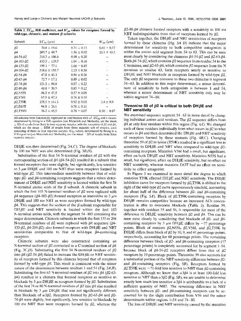
Table 2. EC,,, Hill coefficient, and IC,, values for receptors formed by wild-type, chimeric, and mutant /3 subunits
Subunit
P-2 P4 p4-54.p2
p4-103~p2
p4-133+2
p4-204+?2
p2-54-p4
/32-63-p4
p2-14-p4
p2-80+?4
p2,NSSS
p2,V561
/32,T59K
/32,E63T
/32,T59D
70.8 t 19.6
209.7 k 40.7
314.3 2 108.8
433.3 2 139.3
199.1 t 72.4
518.6 + 155.7
47.8 + 45.3
72.7 2 17.1
124.3 k 96.8
40.0 5 30.5
86.3 t 47.5
67.9 t 47.3
158.5 t 114.1
94.8 t 26.1
16.2 2 45.5
0.74 k 0.11 0.41 ? 0.17
1.56 k 0.02 23.1 i 10.2 0.90 2 0.20 -
1.04 k 0.16 -
1.66 ? 0.45 -
2.23 2 0.30 -
0.84 k 0.26 -
0.89 t 0.02 -
0.87 2 0.22 -
0.85 2 0.12 -
0.57 2 0.05 -
0.76 2 0.10 -
0.92 2 0.03 3.8 t 0.9 0.76 t 0.11 -
0.78 5 0.07 0.30 + 0.07
All subunits were functionally expressed in combination with 013. EC,, and n values, determined by fitting to a Hill equation (see Materials and Methods), are the mean + SD of results from three to four separate oocytes, with the exception of 02 and p4, which are the mean -t SEM of results from three separate sets of oocytcs, each consisting of three to four separate oocytes. IC,,, values, determined by fitting to a Hill equation (see Materials and Methods), are the mean i SD of results from three separate oocytes.
DHPE was then determined (Fig. 3A,C). The degree of blockade by 100 nM NBT was also determined (Fig. 3B,D).
Substitution of the first 54 N-terminal residues of p2 with the corresponding section of /34 @4-54-/32) resulted in a subunit that formed receptors that were slightly, but significantly, less sensitive to 3 PM DHPE and 100 nM NBT than were receptors formed by wild-type p2. This intermediate sensitivity between that of wild- type /32- and P4-containing receptors suggests that a minor deter- minant of DHPE and NBT sensitivity is located within the first 54 N-terminal amino acids of the p subunit. A chimeric subunit in which the first 103 N-terminal residues of /32 were replaced with /34 sequence (/34-103~/32) formed receptors as insensitive to 3 PM
DHPE and 100 nM NBT as were receptors formed by wild-type p4. This suggests that the section of the /3 subunit responsible for DHpE and NBT sensitivity is located within the first 103 N-terminal amino acids, with the segment 54-103 containing the major determinant. Chimeric subunits in which the first 133 or 204 N-terminal residues of p2 were replaced with p4 sequence (p4- 133$2, p4-204~/32) also formed receptors with DHPE and NBT sensitivities comparable to that of wild-type /34-containing receptors.
Chimeric subunits were also constructed containing an N-terminal section of /32 connected to a C-terminal section of /34 (Fig. 3C,D). Substituting the first 54 N-terminal residues of p2 into p4 @2-54-/34) failed to increase the DHpE or NBT sensitiv- ity of receptors formed by this chimera beyond that of receptors formed by wild-type p4. This result is consistent with the minor nature of the determinant between residues 1 and 54 (Fig. 3A,B). Substituting the first 63 N-terminal residues of p2 into /34 @2-63- /34) resulted in a chimera that formed receptors as sensitive to blockade by 3 PM DHPE as receptors formed by p2. Substitution of the first 74 or 80 N-terminal residues of p2 into /34 also resulted in blockade by 3 PM DHpE that was not significantly different than blockade of a3/32. Receptors formed by /32-63-/34 and p2- 74.p4 were slightly, but significantly, less sensitive to blockade by 100 nM NBT than were receptors formed by 02, whereas the
p2-80-p4 chimera formed receptors with a sensitivity to 100 nM NBT indistinguishable from that of receptors formed by /32.
Taken together, the DHPE and NBT sensitivities of receptors formed by these chimeras (Fig. 3A-D) indicate that the major determinant for sensitivity to both competitive antagonists is within the amino acid segment from 54 to 63. This can be seen most clearly by considering the chimeras p4-54-/32 and /32-63-p4. Both p4-54-/32, which contains /32 sequence from residue 54 to the C terminus, and /32-63+4, which contains p2 sequence from the N terminus to residue 63, form receptors nearly as sensitive to DHpE and NBT blockade as receptors formed by wild-type p2. The only p2 sequence common to these two chimeras is segment 54-63. In addition to this major determinant, a minor determi- nant of sensitivity to both antagonists is between 1 and 54, whereas a minor determinant of NBT sensitivity only may be within segment 74-80.
Threonine 59 of p2 is critical to both DHpE and NBT sensitivity We examined sequence segment 54-63 in more detail by chang- ing individual amino acid residues. The p2 sequence differs from p4 at only four residues within this region (Fig. 4A). We changed each of these residues individually from what occurs in p2 to what occurs in p4 and then determined the DH PE and NBT sensitivity of receptors formed by these mutants (Fig. 4B,C). Changing threonine 59 of p2 to lysine (T59K) resulted in a significant loss in sensitivity to DHpE and NBT when compared to wild-type p2- containing receptors. Mutation V561 had a small, but significant, effect on both DH/3E and NBT sensitivity. Mutation N55S had a small, but significant, effect on DH PE sensitivity, but no effect on NBT sensitivity, whereas mutation E63T had no effect on sensi- tivity to either antagonist.
In Figure 5 we examined in more detail the degree to which mutation T59K affected DHpE and NBT sensitivity. The DHPE inhibition curve for receptors formed by P2,T59K is shifted to the right of the wild-type /32 curve approximately ninefold, accounting for about half of the difference between p2- and /34-containing receptors (Fig. 54). Block of P2,T59K-containing receptors by DHpE remains competitive because an increased ACh concen- tration is able to overcome blockade (Table 1). Residue 59, together with residues 55 and 56, is responsible for most of the difference in DHPE sensitivity between /32 and p4. This can be seen most clearly by considering that blockade of p2- and /34- containing receptors by 3 PM DH/3E differs by -77 percentage points. Block of mutants /32,N55S, p2,V561, and /32,T59K by DHPE differs from block of p2 by 10,9, and 41 percentage points, respectively, accounting for 60 percentage points. The remaining difference between block of /32- and fl4-containing receptors (17 percentage points) is completely accounted for by segment l-54, because block of /34-54-/32 receptors differs from that of /?2 receptors by 19 percentage points. Threonine 59 also accounts for a substantial portion of the NBT sensitivity difference between p2- and P4-containing receptors (Fig. 5B). Receptors formed by P2,T59K were -7l-fold less sensitive to NBT than P2-containing receptors. Although we know that (~3p4 is at least loo-fold less sensitive to NBT than 013p2 (Fig. 5B), we are unable to determine exactly how much less sensitive cu3p4 is attributable to a lack of a sufficient quantity of NBT. The remaining difference in NBT sensitivity between p2- and P4-containing receptors can be ac- counted for by the slight contribution from V56 and the minor determinants within regions l-54 and 74-80.
The loss of DHPE and NBT sensitivity caused by the mutation

3802 J. Neurosci., June 15, 1996, 16(12):3798-3806 Harvey and Luetje . Chimeric and Mutant Neuronal nAChR p Subunits
DHPE
0 20 40 60 80 100 I I I I l-
A
p4-54+2 m **
-I ttt
f34-103~p2 1 **3
-I
%%* p4-133+2 -I
*** p4-204+2
*** P4 -I
I I I I I I
I I
P4 C
p2-54+4- x-x-* i
NBT
0 20 40 60 80 100 I I I I I r
ttt B *** ttt
;” ** -I
p2-63-/34- l Ittt
P2-74-P4mitt;
P2-8O-P4 I ttt
I I I I I
0 20 40 60 80 100
Percent of Control
I I I I I I
0 20 40 60 80 100
Percent of Control
Figure 3. DHPE and NBT sensitivity of receptors formed by chimeric p subunits. A, DHPE sensitivity of receptors formed by each of a series of chimeric subunits in which increasingly larger portions of the N-terminal end of p2 were replaced by the corresponding portion of p4. Current in response to an EC,,, concentration of ACh in the presence of 3 WM DHPE is presented as a percent of the response to ACh alone (mean + SD of 3-4 separate oocytes). B, NBT sensitivity of receptors formed by the chimeras in A. Current in response to an ACh concentration at or below the EC,,, after 30 min incubation with 100 nM NBT is presented as a percentage of the response to ACh alone (mean I! SD of 3-4 separate oocytcs, except for p4, which is mean ? SEM of 3 separate sets of oocytes, each set consisting of 3-4 separate oocytes). C, DHpE sensitivity of receptors formed by each of a series of chimeric subunits in which increasingly larger portions of p4 were replaced by the corresponding portion of p2. Current in response to an EC_ ,,, concentration of ACh in the presence of 3 PM DH PE is presented as a percent of the response to ACh alone (mean 2 SD of 3-4 separate oocytes). D, NBT sensitivity of receptors formed by the chimeras in C. Current in response to an ACh concentration at or below the EC,,, after 30 min incubation with 100 nM NBT is presented as a percentage of the response to ACh alone (mean % SD of 3-4 separate oocytcs, cxccpt for p2-54-p4, which is mean t SEM of 3 scparatc sets of oocytes, each set consisting of 3-4 separate oocytes). Significant differences from /32 are denoted by asterisk? (*p < 0.05; **I, < 0.01; ***p < 0.001). Significant differcnccs from /34 are denoted by daggers (‘Jo < 0.05; ‘“+p < 0.001). Some error bars are too small to appear.
T59K could be attributable to the change from the polar side chain of threonine to the positively charged side chain of lysine, or it could be attributable to the change in side chain volume. The identification of arginine 34 of NBT as a critical residue for neuronal nAChR blockade (Dewan et al., 1994; Fiordalisi et al.,
1994) suggests that it is the introduction of the positively charged lysine that interferes NBT sensitivity. To explore this idea, we introduced a negative charge by changing this residue from threo- nine to aspartate (T59D). The DHPE and NBT sensitivity of receptors formed by P2,T59D is shown in Figure 5, A and B. The

J. Neurosci., June 15, 1996, 16(12):3798-3806 3803 Harvey and Luetje l Chimeric and Mutant Neuronal nAChR p Subunits
A 54 63
P2 TNVWLTQEWE
PdTSIWLKQEWT l l * 0 l
B DHPE c NBT
I I I I I I I I I I
I
P2,N55S --I ytt i ttt
p2,V561 m Y **
ttt ttt
/32,T59K m -I;;
***
+tt
PW63T --I tt.+ -I ttt
04 -I + I I I I I I
0 20 40 60 80 100 0 20 40 60 80 100
Percent of Control Percent of Control
Figure 4. Threonine 59 of 02 is critical to both DHPE and NBT sensitivity.A, Alignment of p2 and p4 sequences within segment 54-63. Residues that differ arc denoted by solid circles. Tryptophan 57 is starred. B, DHpE sensitivity of receptors formed by each of a series of mutant p2 subunits. Current in response to an EC,,, concentration of ACh in the presence of 3 pM DHPE is presented as a percent of the response to ACh alone (mean + SD of 3 separate oocytes). C, NBT sensitivity of receptors formed by the p2 mutants in B. Current in response to an ACh concentration at or below the EC,,, after 30 min incubation with 100 nM NBT is presented as a percentage of the response to ACh alone (mean 2 SD of 3 separate oocytes, except for p4, which is mean + SEM of three separate sets of oocytes, each set consisting of 3-4 separate oocytes). Significant differences from p2 are denoted by asterisks (*p < 0.05; **p < 0.01; ***p < 0.001). Significant differences from p4 are denoted by daggers (“‘p < 0.01; ‘+‘p < 0.001). Some error bars are too small to appear.
T59D mutation resulted in an increase in NBT sensitivity of -2.5 fold. The T59D mutation had no effect on DHPE sensitivity.
DISCUSSION The neuronal nAChRs a3/32 and 013/34 differ in their sensitivity to the antagonists DHPE and NBT. Pharmacological analysis of a series of chimeric p subunits has allowed us to identify areas of the /!l subunits that determine sensitivity to these competitive antag- onists. The major determinant of both DHPE and NBT sensitivity lies in sequence segment 54-63, with a minor determinant of sensitivity to both antagonists in region 1-54 and a minor deter- minant of NBT sensitivity in region 74-80. Within sequence segment 54-63, we identified threonine 59 of /32 as the critical residue. Changing this residue to lysine, as in p4, results in a 9-fold loss in DHpE sensitivity and a 71-fold loss in NBT sensitivity. Changing threonine 59 of p2 to aspartate, thus introducing a negative charge, caused a 2.5-fold increase in NBT sensitivity.
It has become clear recently that non-a subunits are involved in
determining both the physical structure and pharmacological properties of the ligand-binding sites of nAChRs (Blount and Merlie, 1989; Duvoisin et al., 1989; Pederson and Cohen, 1990; Czajkowski and Karlin, 1991; Luetje and Patrick, 1991; Middleton and Cohen, 1991). Affinity labeling and mutagenesis studies of Torpedo electric organ and mammalian muscle nAChRs have identified amino acid residues of the y and 6 subunits that are associated with ligand binding (Cohen et al., 1992; Czajkowski et al., 1993; Sine, 1993; Fu and Sine, 1994). In covalent labeling experiments involving the competitive antagonist d-tubocurarine, Cohen et al. (1992) demonstrated incorporation of label onto a tryptophan residue of the y and 6 subunits (residue 55 and 57, respectively). This residue is conserved in the rat neuronal p2 and /34 subunits (position 57, Fig. 44), and thus appears to be a common feature of the ligand-binding sites of nAChRs. Interest- ingly, the homologous residue of neuronal ot7 (tryptophan 54) is involved in determining sensitivity to both agonists and antago-

3804 J. Neurosci., June 15, 1996, 16(12):3798-3806 Harvey and Luetje . Chimeric and Mutant Neuronal nAChR p Subunits
60
100
80
60
40
20
0
lo-' 1o'6 1o-5 1o-4 [DHBE], M
1o’g 1o'8 10" NW, M
Figure 5. Effect of mutations of threonine 59 on DHPE and NBT sensitivity. A, DHpE sensitivity of ot3P2,T59K @led squares) and (~3P2,T59D (filled circles). Current in response to an EC,, concentration of ACh in the presence of various concentrations of DHPE is presented as a percent of the response to ACh alone (mean 2 SD of 3-6 oocytes). The lines arc fits to a Hill equation (see Materials and Methods). IC,, values are 3.8 + 0.9 pM for (~3P2,T59K and 0.30 2 0.07 p,M for a3P2,T59D. Inhibition curves for 013p2 and a3P4 from Figure 2B are shown as clashed lines for reference. B, NBT sensitivity of o3P2,T59K (filled squares), cx3P2,T59D (filled circles), CY 3/32 (open circles), and 01304 (open squares). Current in response to an ACh concentration at or below the EC,,, after 30 min incubation with various concentrations of NBT is presented as a percentage of the response to ACh alone (mean i SD of 3 separate oocytes). Significant differences from /32 are denoted by asfer- isks (*p < 0.02; ***p < 0.001). Significant differences from 04 are denoted by daggers (“‘p i 0.01). Some error bars are obscured by symbols.
nists, leading to the proposal that (r7 contributes both an “CX component” and a “non-a component” when forming homo- oligomeric receptors (Corringer et al., 1995).
The conservation of this tryptophan among muscle and neuro- nal nAChRs means that this residue cannot be responsible for
pharmacological differences between nAChR subtypes. It is the amino acid residues that differ among subunits that must be responsible for this diversity. We have identified such a residue, separated by only one residue from the conserved tryptophan, as the major determinant of differences in competitive antagonist sensitivity between /32- and PCcontaining receptors. Changing this residue in /32 from threonine to what occurs in /34 (lysine) results in a substantial loss of both DHPE and NBT sensitivity. The change from threonine to lysine is a change in both the character (polar to negatively charged) and the size (55.7-101.5 A’) of the side chain. Either or both of these properties might be responsible for the effect of changing this residue. Considering that arginine 34 of NBT has been identified as a critical residue involved in neuronal nAChR blockade (Dewan et al., 1994) we hypothesized that insertion of lysine at position 59 in p2 might be decreasing NBT sensitivity by electrostatic repulsion. If this were true, then introduction of a negative charge at this position might be expected to increase NBT sensitivity. Changing threonine 59 to aspartate does result, in fact, in an increase in NBT sensitivity (Fig. 5B).
Construction and functional analysis of chimeric receptor sub- units allows identification of structural differences that confer unique pharmacological properties. This methodology has been used to map determinants of both agonist and antagonist sensi- tivity on neuronal nAChR /3 subunits. Chimeric subunits have been used to identify the general region on p subunits that contributes to NBT sensitivity. Papke et al. (1993) showed that substitution of the first 119 amino acids of the /34 subunit with the corresponding section of /32 can confer NBT sensitivity onto the fi4 subunit. The identity of the p subunit also influences the NBT sensitivity of the receptors (~4p2 and a4P4. Wheeler et al. (1993) showed that a chimeric subunit composed of the N-terminal 80 residues of p2 followed by p4 sequence, formed receptors with a4 that were sensitive to NBT blockade. A series of p subunit chimeras, expressed in combination with the a3 subunit, has been used to identify the sequence segment 104-120 of /32 as important in determining sensitivity to the agonist cytisine (Fig1 et al., 1992; Cohen et al., 1995). Consistent with these reports, we find that receptors formed by 013 and the chimera p4-103-p2 have a cytisine sensitivity similar to that of receptors formed by wild-type ot3p2 (data not shown). Cohen et al. (1995) also show that region 104-120 is responsible for part of the difference in EC,,, for ACh, as well as for part of the difference in Hill slope, between a3/32 and a3P4.
Differential sensitivity to agonists may result from differences in affinity or efficacy, making it difficult to infer conclusions about the functional role of the sequence segment being mapped. Compet- itive antagonists are ideal probes, because they compete with agonist for a common binding site but do not activate the recep- tors; they have no efficacy. Because our determination of the DHPE sensitivity of each receptor is dependent on use of equi- potent concentrations of ACh, it is important that differences in DHPE sensitivity not be an artifact of differences in ACh dose- response curve characteristics. The cu3/32 and (u3/34 ACh dose- response curves clearly differ in both EC,, and apparent Hill coefficient (Table 2). However, these differences can be taken into consideration when DHpE dissociation constants (Ki) for each receptor are calculated (Leff and Dougall, 1993). The resulting DHPE K, values of 0.21 and 32.4 pM (for cr3p2 and 013/34, respectively) differ by -154-fold. Additional arguments against differences in DHPE sensitivity being artifactual are that DHPE sensitivity maps differently than the ACh EC,, and the apparent

Harvey and Luetje . Chimeric and Mutant Neuronal nAChR p Subunits J. Neurosci., June 15, 1996, 76(12):3798-3806 3805
Hill coefficient (compare Fig. 3 and Table 2), and that use of both DHPE and NBT as probes has identified the same residue as a major determinant of competitive antagonist sensitivity. I f DHPE antagonism is competitive, why do determinants of DH /3E sensi- tivity and ACh ECS,, differ? Although competitive antagonists do compete for a common binding site with agonists, their interac- tions with the binding site would not necessarily be coextensive with those of agonists. In fact, this would not be expected, because competitive antagonists differ from agonists by lacking efficacy.
Although the pseudoirreversibility of NBT blockade makes concerns over ACh dose-response curves irrelevant, NBT is a large peptide toxin and may block receptor activation by adven- titiously occluding the ligand-binding site after binding elsewhere on the receptor. Therefore, mapping the areas of the receptor responsible for differential sensitivity to NBT may identify regions of uncertain significance. However, both DHPE and NBT sensi- tivity map similarly, identifying threonine 59 as a major determi- nant and region l-54 as containing a minor determinant, support- ing the view that NBT is a useful probe. The significance of region 74-80, containing an additional minor determinant of NBT sen- sitivity, is unclear. Another example of determinants of NBT sensitivity overlapping with those of small ligand sensitivity occurs on (Y subunits. The cy2 subunit forms receptors with p2 that are insensitive to NBT and are more sensitive to nicotine than to ACh, whereas (~3p2 receptors are blocked by NBT and arc much less sensitive to nicotine than to ACh. Region 195-21.5 contains determinants of both properties. Within this region, the glutamine residue at position 198 of a3 (proline in ot2) was shown to be an important determinant of both NBT and nicotine sensitivity (Lu- etje et al., 1993). Similar results regarding the role of glutamine 198 in determining nicotine sensitivity have been obtained re- cently using chicken neuronal nAChR (Y subunits (Hussy et al., 1994).
The exact physical role of residues identified in this and previ- ous studies, that confer pharmacological differences among nAChR subtypes, remains unclear. In this study, we provide data consistent with a direct interaction between residue 59 of pZ/fl4 and NBT. Thus, these residues may be structural features of the binding site and may participate in the binding of ligand. Alter- natively, these residues may not actually participate in binding of ligand, but impinge upon those that do, reshaping the site enough to alter ligand sensitivity. More extensive mutagenesis of identi- fied residues will be required to distinguish between these possi- bilities. Particularly promising is the potential for incorporating unnatural amino acids at these sites (Nowak et al., 1995).
REFERENCES Abramson SN, Li Y, Culver P, Taylor P (1989) An analog of lophotoxin
reacts covalently with Tyr “” in the a-subunit of the nicotinic acetylcho- line receptor. J Biol Chem 264:12666-12672.
Bertrand D, Bertrand S, Ballivet M (1992) Pharmacological properties of the homomeric (~7 receptor. Neurosci Lett 146:87-90.
Blount P, Merlie J (1989) Molecular basis of the two nonequivalent ligand binding sites of the muscle nicotinic acetylcholine receptor. Neuron 31349-357.
Boulter J, Connolly J, Deneris E, Goldman D, Heinemann S, Patrick J (1987) Functional expression of two neuronal nicotinic acetylcholine receptors from cDNA clones identifies a gene family. Proc Nat1 Acad Sci USA 84~7763-7767.
Cohen JB, Blanton MP, Chiara DC, Sharp SD, White BH (1992) Struc- tural organization of functional domains of the nicotinic acetylcholine receptor. J Cell Biochem [Suppl] 16E:217.
Cohen BN, Fig1 A, Quick MW, Labarca C, Davidson N, Lester HA (1995) Regions of p2 and /34 responsible for differences between the steady
state dose-response relationships of the a3P2 and a3P4 neuronal nicotinic receptors. J Gen Physiol 105:745-764.
Corringer PJ, Galzi JL, Eisele JL, Bertrand S, Changeux JP, Bertrand D (1995) Identification of a new component of the agonist binding site of the nicotinic alpha 7 homooligomeric receptor. J Biol Chcm 270:11749-11752.
Craig DA (1993) The Cheng-Prusoff relationship: something lost in the translation. Trends Pharmacol Sci 14:89-93.
Czajkowski C, Karlin A (1991) Agonist binding site of Torpedo electric tissue nicotinic acetylcholine receptor: a negatively charged region of the 6 subunit within 0.9 nm of the 01 subunit binding site disulfide. J Biol Chem 266:22603-22612.
Czajkowski C, Kaufmann C, Karlin A (1993) Negatively charged amino acid residues in the nicotinic receptor y subunit that contribute to the binding of acetylchoiine. Proc Nat1 Acad Sci USA 90:6285-6289.
Dennis M, Giraudat J, Kotzyba-Hibert F, Goeldner M, Hirth C, Chang J-Y, Lazure C, Chretien M, Changeux J-P (1988) Amino acids of the Topdo marmorata acetylcholine receptor 01 subunit labelled by pho- toaffinity ligand for acetylcholine binding site. Biochemistry 2712346-2357.
Dewan JC, Grant GA, Sacchcttini JC (lY94) Crystal structure of K-bungarotoxin at 2.3-A resolution. Biochemistry 33:13147-13154.
Duvoisin RM, Deneris ES, Boulter J, Patrick J, Heincmnnn S (1989) The functional diversity of the neuronal nicotinic acctylcholine rcccptors is increased by a 11ovcI subunit: p4. Neuron 3:4X7-406.
Elgoyhcn AB, Johnso~~ DS, Boultcr J, Vcttcr DE, Hcincmann S (lYY4) cu9: an acctylcholinc rcccptor with novel pharmacological properties cxprcsscd in rat cochlcar hair cells. Ccl1 79:705-71.5.
Fig1 A, Cohen BN, Quick MW, Davidson N, Lcstcr HA (lYY2) Regions of /34-p2 subunit chimeras that contribute to the agonist selectivity of neuronal nicotinic rcccptors. FEBS Lctt 308:245-248.
Fiordalisi JJ, Al-Rabiee R, Chiappinelli VA, Grant GA (lY94) Site- directed mutagenesis of K-bungarotoxin: implications for neuronal re- ceptor specificity. Biochemistry 33:3872-3877.
Fu DX, Sine SM (1994) Competitive antagonists bridge the a-y subunit interface of the acetylcholine receptor through quaternary ammonium- aromatic interactions. J Biol Chem 269:26152-26157.
Galzi J, Revah F, Black D, Goeldner M, Hirth C, Changeux J-P (1990) Identification of a novel amino acid a-tyrosine 93 within the cholinergic ligands-binding sites of the acetylcholine receptor by photoaffinity la- beling. J Biol Chem 265:10430-10437.
Halvorsen SW, Berg DK (1987) Affinity labeling of neuronal acetylcho- line receptor subunits with an oc-neurotoxin that blocks receptor func- tion. J Neurosci 7:2547-2555.
Higuchi R (1990) Recombinant PCR. In: PCR protocols, a guide to methods and applications (Innis MA, Gelfand GH, Sninsky JJ, White TJ, eds). San Diego: Academic.
Hussy N, Ballivet M, Bertrand D (1994) Agonist and antagonist effects of nicotine on chick neuronal nicotinic receptors are defined by 01 and p subunits. J Neurophys 72:1317-1326.
Kao PN, Dwork AJ, Kaldany R-RJ, Silver MS, Wideman J, Stein S, Karlin A (1984) Identification of the LY subunit half-cystinc spccitically labclcd by an affinity reagent for the acetylcholine receptor binding site. J Biol Chem 259:11662-l 1665.
Leff P, Dougall IG (19Y3) Further concerns over Cheng-Prusoft’ analysis. Trends Pharmacol Sci 14:110-l 12.
Luetje CW, Patrick J (1991) Both 01- and P-subunits contribute to the agonist sensitivity of ncuronal nicotinic acetylcholine receptors. J Neu- rosci 11:837-845.
Luetje CW, Patrick J, SCguela P (1990a) Nicotine receptors in the mam- malian brain. FASEB J 4:2753-2760.
Luetje CW, Wada K, Rogers S, Abramson SN, Tsuji K, Heinemann S, Patrick J (1990b) Ncurotoxins distinguish between different neuronal nicotinic acetylcholine receptor subunit combinations. J Neurochem 55:632-640.
Luetje CW, Piattoni M, Patrick J (1993) Mapping of ligand binding sites of neuronal nicotinic acetylcholine receptors using chimeric (Y subunits. Mol Pharmacol 441657-666.
Middleton RE, Cohen JB (1991) Mapping of the acetylcholine binding site of the nicotinic acetylcholine receptor: [‘HInicotine as an agonist photoaffinity label. Biochemistry 30:6987-6997.
Nowak MW, Kearney PC, Sampson JR, Saks ME, Ldbarca CG, Silverman SK, Zhong W, Thorson J, Abelson JN, Davidson N, Schulz PG, Dough- erty DA, Lester HA (1995) Nicotinic receptor binding site probed with

3806 J. Neurosci., June 15, 1996, 16(12):3798-3806 Harvey and Luetje l Chimeric and Mutant Neuronal nAChR p Subunits
unnatural amino acid incorporation in intact cells. Science 268:439-442.
Papke RL, Boulter J, Patrick J, Heinemann S (1989) Single-channel currents of rat neuronal nicotinic acetylcholine receptors expressed in Xenopus oocytes. Neuron 3:589-596.
Papke RL, Duvoisin RM, Heinemann S (1993) The amino terminal half of the nicotinic P-subunit extracellular domain regulates the kinetics of inhibition by neuronal bungarotoxin. Proc R Sot Lond [Biol] 252:141-148.
Pedersen SE, Cohen JB (1990) d-Tubocurarine binding sites are located at the (Y-Y and cu.6 subunit interfaces of the nicotinic acetylcholine receptor. Proc Nat1 Acad Sci USA 87:2785-2789.
Role LW (1992) Diversity in primary structure and function of neuronal nicotinic acetylcholine receptor channels. Curr Opin Neurobiol 21254-262.
Sargent PB (1993) The diversity of neuronal nicotinic acetylcholine re- ceptors. Annu Rev Neurosci 16:403-443.
Sheridan RP, Nilakantan R, Dixon JS, Venkataraghavan R (1986) The ensemble approach to distance geometry: application to the nicotinic pharmacophore. J Med Chem 29:899-906.
Sine SM (1993) Molecular dissection of subunit interfaces in the acetyl- choline receptor: identification of residues that determine curare selec- tivity. Proc Nat1 Acad Sci USA 90:9436-9440.
Wheeler SV, Chad JE, Foreman R (1993) Residues 1 to 80 of the N-terminal domain of the p subunit confer neuronal bungarotoxin sensitivity and agonist sensitivity on neuronal nicotinic receptors. FEBS Lett 332:139-142.
Williams M, Robinson JL (1984) Binding of the nicotinic cholinergic antagonist dihydro-P-erythroidine to rat brain tissue. J Neurosi 4:2906- 2911.