detection of telomerase activity in high concentration of ... · the trap assay with a post-pcr...
TRANSCRIPT

Detection of Telomerase Activity in High Concentration of CellLysates Using Primer-Modified Gold Nanoparticles
Yi Xiao,†,‡,§ Karen Y. Dane,| Takanori Uzawa,‡ Andrew Csordas,⊥ Jiangrong Qian,§
H. Tom Soh,§ Patrick S. Daugherty,| Eric T. Lagally,# Alan J. Heeger,† andKevin W. Plaxco*,‡,∇
Department of Physics, Materials Department, and Institute for Polymers and Organic Solids,Department of Chemistry and Biochemistry, Department of Mechanical Engineering, Department
of Chemical Engineering, Institute for CollaboratiVe Biotechnologies, and Program inBioMolecular Science and Engineering, UniVersity of California, Santa Barbara,
California 93106, United States, and Michael Smith Laboratories and Department of Chemicaland Biological Engineering, 301 - 2185 East Mall, The UniVersity of British Columbia,
VancouVer, BC, Canada V6T 1Z4
Received July 22, 2010; E-mail: [email protected]
Abstract: Although the telomeric repeat amplification protocol (TRAP) has served as a powerful assay fordetecting telomerase activity, its use has been significantly limited when performed directly in complex,interferant-laced samples. In this work, we report a modification of the TRAP assay that allows the detectionof high-fidelity amplification of telomerase products directly from concentrated cell lysates. Briefly, wecovalently attached 12 nm gold nanoparticles (AuNPs) to the telomere strand (TS) primer, which is usedas a substrate for telomerase elongation. These TS-modified AuNPs significantly reduce polymerase chainreaction (PCR) artifacts (such as primer dimers) and improve the yield of amplified telomerase productsrelative to the traditional TRAP assay when amplification is performed in concentrated cell lysates.Specifically, because the TS-modified AuNPs eliminate most of the primer-dimer artifacts normally visibleat the same position as the shortest amplified telomerase PCR product apparent on agarose gels, theAuNP-modified TRAP assay exhibits excellent sensitivity. Consequently, we observed a 10-fold increasein sensitivity for cancer cells diluted 1000-fold with somatic cells. It thus appears that the use of AuNP-modified primers significantly improves the sensitivity and specificity of the traditional TRAP assay andmay be an effective method by which PCR can be performed directly in concentrated cell lysates.
1. Introduction
Telomeres, which are specific nucleotide sequences occurringat the ends of linear chromosomes, are indispensable for themaintenance of chromosome integrity during replication.1-4
Telomerase, the ribonucleoprotein enzyme5 that recognizes andelongates the G-rich tips of existing telomere DNA repeats, isnot expressed in most healthy tissues.4,6,7 Re-expression of
telomerase is often seen, however, in immortal phenotypes suchas cancer.8 Specifically, although it is repressed in most somaticcells, telomerase is active in more than 80% of all humancancers.8,9 This renders this enzyme an important therapeutictarget and a valuable marker of malignancy and tumor progres-sion.10
To detect the activity of cellular telomerase, a polymerasechain reaction (PCR)-based method known as the telomererepeat amplification protocol (TRAP)11 has been developed asa gold-standard assay. The conventional TRAP assay is a single-tube reaction in which telomerase extracted from cells or tissuesynthesizes telomeric products (telomeres) first from an exog-enously added telomere strand (TS) primer. These elongatedproducts then serve as the template for PCR amplification, whichis followed by polyacrylamide gel electrophoresis (PAGE)characterization and, finally, quantification via densitometry.11
Since the invention of the TRAP assay, various modificationsto improve quantification and simplify the time-consuming post-PCR steps have been reported. Examples include combining
† Department of Physics, Materials Department, and Institute forPolymers and Organic Solids, UCSB.
‡ Department of Chemistry and Biochemistry, UCSB.§ Department of Mechanical Engineering, UCSB.| Department of Chemical Engineering, UCSB.⊥ Institute for Collaborative Biotechnologies, UCSB.# The University of British Columbia.∇ Program in BioMolecular Science and Engineering, UCSB.
(1) Blackburn, E. H. Nature 1991, 350, 569–573.(2) Greider, C. W. Curr. Opin. Genet. DeV. 1994, 4, 203–211.(3) Allsopp, R. C.; Chang, E.; Kashefi-Aazam, M.; Rogaev, E. I.;
Piatyszek, M. A.; Shay, J. W.; Harley, C. B. Exp. Cell Res. 1995,220, 194–200.
(4) Shay, J. W. Mol. Med. Today 1995, 1, 378–384.(5) Counter, C. M.; Avilion, A. A.; LeFeuvre, C. E.; Stewart, N. G.;
Greider, C. W.; Harley, C. B.; Bacchetti, S. EMBO J. 1992, 11, 1921–1929.
(6) Greider, C. W.; Blackburn, E. H. Cell 1985, 43, 405–413.(7) Blackburn, E. H.; Szostak, J. W. Annu. ReV. Biochem. 1984, 53, 163–
194.
(8) Shay, J. W.; Bacchetti, S. Eur. J. Cancer 1997, 33, 787–791.(9) Shay, J. W.; Wright, W. E. Curr. Opin. Oncol. 1996, 8, 66–71.
(10) Davis, A. J.; Siu, L. L. Cancer InVest. 2000, 18, 269–277.(11) Kim, N. W.; Piatyszek, M. A.; Prowse, K. R.; Harley, C. B.; West,
M. D.; Ho, P. L. C.; Coviello, G. M.; Wright, W. E.; Weinrich, S. L.;Shay, J. W. Science 1994, 266, 2011–2015.
Published on Web 10/08/2010
10.1021/ja106513f 2010 American Chemical Society J. AM. CHEM. SOC. 2010, 132, 15299–15307 9 15299

the TRAP assay with a post-PCR hybridization protocolemploying chemiluminescent probes,12 fluorescent dyes,13 energy-transfer primers,14 or biotinylated primers15 to measure theamount of PCR-amplified double-stranded DNA.
When combined with the above-described post-PCR modi-fications, the TRAP assay is perhaps the most effective assayreported to date for the detection of telomerase activity.However, the limited specificity and sensitivity of the currentTRAP assay remain significant challenges and make the assaydifficult to use in complex samples. For example, primer-dimerproducts resulting from the staggered annealing of PCR primersoften plague the TRAP assay,16 and this problem has proven tobe recalcitrant even to careful primer design.17 Additionally,the TRAP assay appears to be inhibited by poorly definedinterferants present in crude, highly concentrated cell extractsand other samples,18,19 including those taken from solid tumors(e.g., tumor biopsies20) and bodily fluids (e.g., coloniceffluents21-23). In response, several methods have been devel-oped in which these inhibitors are removed or diluted prior tothe PCR via phenol/chloroform extraction,19 biotin affinitylabeling and extraction,24 or sample dilution.21 Alternatively,Kim and co-workers have designed a 36 base pair (bp) double-stranded internal standard that allows the operator to “correct”signal for the presence of inhibitors.25 These modified methods,however, complicate the elegant simplicity of the underlyingTRAP assay.
Here we report a modified TRAP assay that leads tosignificantly enhanced performance in complex samples, suchas concentrated, crude cellular extracts, without significantlycomplicating the method. The inspiration for our approachstemmed from previous studies demonstrating that the additionof gold nanoparticles (AuNPs) improves the yield of PCRreactions and reduces nonspecific amplification artifacts. Forexample, several groups have shown that 12 nm citrate-capped
AuNPs26,27 or primer-modified AuNPs28 enhance the yield ofPCR products obtained with a single purified template26-28 orgenomes extracted from culture medium or whole blood.29 Todate, however, there have been no reports of AuNP-based PCRassays working directly in highly concentrated cell extracts,presumably because citrate-capped AuNPs aggregate at con-centrations above 0.9 nM at the standard ionic strengths requiredfor PCR reactions. To circumvent this problem, we havemodified citrate-capped AuNPs with a PCR primer (modifiedTS primer). Using the conventional TRAP assay as a test bed,we have shown that such primer-modified AuNPs used at highconcentrations significantly improve the specificity and sensitiv-ity of PCR performed directly in complex, interferant-lacedsamples. Our AuNP-modified TRAP assay directly detects lowlevels of cancer-indicative telomerase activity in cancer cellextracts that have been doped with a 1000-fold higher concen-tration of somatic cell lysates.
2. Experimental Section
2.1. Preparation and Characterization of TS-Primer-Modi-fied AuNPs. Citrate-capped AuNPs (12 ( 1 nm) were preparedaccording to literature procedures,30 and the AuNP concentrationwas determined by measurement of the absorbance at 519 nm.Oligonucleotides 1 and 2 (modified TS primers) were synthesizedby Integrated DNA Technologies, Inc. (Coralville, IA) and purifiedby PAGE. The sequences of modified TS primers were thefollowing:1:5′-HS(CH2)6TTTTTTTTTTAATCCGTCGAGCAGAGTT-3′2:5′-HS(CH2)6TTTTTTTTTTTTTTTTTTAATCCGTCGAGCAG-AGTT-3′
The TS-modified AuNPs were prepared as reported previously.31
The DNA/AuNP ratio was calculated by the following method:31
The absorbance spectra of the AuNPs before and after DNAmodification were measured. Both spectra were normalized to anabsorbance of 1 at the 519 nm plasmon peak of the AuNPs. Themolar ratio was calculated using the known extinction coefficientsof DNA strands at 260 nm and the NPs at 519 nm. The calculatedloading corresponded to ∼50 TS primers per AuNP.
2.2. Preparation of Telomerase Extracts. Human breast car-cinoma cell lines MDA-MB-231, MCF-7, and T47-D were obtainedfrom American Type Culture Collection (ATCC, Manassas, VA).T47-D was cultured in RPMI-1640 medium, MDA-MB-231 inDMEM, and MCF-7 in EMEM supplemented with 10% fetal bovineserum and 1× pen/strep (Invitrogen, Carlsbad, CA). Humanmammary epithelial cells (HMEC) were cultured in mammaryepithelial cell medium obtained from Lonza (Walkersville, MD).After the cells were harvested with trypsin, 1 million cells werecollected into microcentrifuge tubes and centrifuged at 2000 rpmfor 10 min at 4 °C. Cells were washed once in phosphate bufferedsaline (pH 7.4), centrifuged again, and frozen at -80 °C. The cellswere resuspended at a concentration of 1 × 10⁶ cells/mL in cold1× CHAPS lysis buffer [10 mM Tris-HCl (pH 7.4), 1 mM MgCl2,1 mM EGTA, 0.1 mM PMSF, 0.5% CHAPS (Sigma), and 10%glycerol] plus 2 mM DTT and 200 units/mL RNase inhibitor. Theresuspended cells were then incubated for 30 min on ice before
(12) (a) Hirose, M.; Abe-Hashimoto, J.; Ogura, K.; Tahara, H.; Ide, T.;Yoshimura, T. J. Cancer Res. Clin. Oncol. 1997, 123, 337–344. (b)Hou, M.; Xu, D. W.; Bjorkholm, M.; Gruber, A. Clin. Chem. 2001,47, 519–524. (c) Herbert, B. S.; Hochreiter, A. E.; Wright, W. E.;Shay, J. W. Nat. Protoc. 2006, 1, 1583–1590.
(13) (a) Gelmini, S.; Caldini, A.; Becherini, L.; Capaccioli, S.; Pazzagli,M.; Orlando, C. Clin. Chem. 1998, 44, 2133–2138. (b) Liu, Y.; Wu,B. Q.; Zhong, H. H.; Xu, M. L.; Fang, W. G. Pathol. Int. 2010, 60,386–394.
(14) Uehara, H.; Nardone, G.; Nazarenko, I.; Hohman, R. J. Biotechniques1999, 26, 552–558.
(15) Wu, Y. Y.; Hruszkewycz, A. M.; Delgado, R. M.; Yang, A.;Vortmeyer, A. O.; Moon, Y. W.; Weil, R. J.; Zhuang, Z.; Remaley,A. T. Clin. Chim. Acta 2000, 293, 199–212.
(16) Goessens, W. H.; Mouton, J. W.; van der Meijden, W. I.; Deelen, S.;van Rijsoort-Vos, T. H.; Toom, N. L.; Verbrugh, H. A.; Verkooyen,R. P. J. Clin. Microbiol. 1997, 35, 2628–2633.
(17) Bauwens, J. E.; Clark, A. M.; Stamm, W. E. J. Clin. Microbiol. 1993,31, 3023–3027.
(18) Yoshida, K.; Sugino, T.; Tahara, H.; Woodman, A.; Bolodeoku, J.;Nargund, V.; Fellows, G.; Goodison, S.; Tahara, E.; Tarin, D. Cancer1997, 79, 362–369.
(19) Kavaler, E.; Landman, J.; Chang, Y.; Droller, M. J.; Liu, B. C. Cancer1998, 82, 708–714.
(20) Morales, C. P.; Burdick, J. S.; Saboorian, M. H.; Wright, W. E.; Shay,J. W. Gastrointest. Endosc. 1998, 48, 402–405.
(21) Sugino, T.; Yoshida, K.; Bolodeoku, J.; Tarin, D.; Goodison, S.J. Pathol. 1997, 183, 57–61.
(22) Gollahon, L. S.; Holt, S. E. Cancer Lett. 2000, 159, 141–149.(23) Kim, N. W.; Wu, F. Nucleic Acids Res. 1997, 25, 2595–2597.(24) Grabar, K. C.; Freeman, R. G.; Hommer, M. B.; Natan, M. J. Anal.
Chem. 1995, 67, 735–743.(25) Krupp, G.; Kuhne, K.; Tamm, S.; Klapper, W.; Heidorn, K.; Rott,
A.; Parwaresch, R. Nucleic Acids Res. 1997, 25, 919–921.
(26) Li, H.; Huang, J.; Lv, J.; An, H.; Zhang, X.; Zhang, Z.; Fan, C.; Hu,J. Angew. Chem., Int. Ed. 2005, 44, 5100–5103.
(27) Li, M.; Lin, Y. C.; Wu, C. C.; Liu, H. S. Nucleic Acids Res. 2005, 33,e184.
(28) Shen, H. B.; Hu, M.; Wang, Y. B.; Zhou, H. Q. Biophys. Chem. 2005,115, 63–66.
(29) Huang, S. H.; Yang, T. C.; Tsai, M. H.; Tsai, I. S.; Lu, H. C.; Chuang,P. H.; Wan, L.; Lin, Y. J.; Lai, C. H.; Lin, C. W. Nanotechnology2008, 19, 405101.
(30) Xiao, Y.; Ju, H. X.; Chen, H. Y. Anal. Chim. Acta 1999, 391, 73–82.(31) Pavlov, V.; Xiao, Y.; Shlyahovsky, B.; Willner, I. J. Am. Chem. Soc.
2004, 126, 11768–11769.
15300 J. AM. CHEM. SOC. 9 VOL. 132, NO. 43, 2010
A R T I C L E S Xiao et al.

being centrifuged for 20 min. (12 000 rpm, 4 °C). After centrifuga-tion, 160 µL of the supernatant was transferred into a fresh tube,flash-frozen, and stored at -80 °C before use.
2.3. AuNP-Modified TRAP Assay. A TRAPEZE telomerasedetection kit (S7700) was purchased from Chemicon International,Inc. (Billerica, MA). The composition of the 25 µL PCR reactionmixture was 16 µL of PCR water, 2.5 µL of 10× TRAP reactionbuffer, 0.5 µL of 50× dNTP mix, 3 µL of AuNP-TS primer (TS-modified AuNPs were added to the PCR reaction system to anappropriate concentration), 0.5 µL of TRAP primer mix, 0.5 µL ofTaq polymerase (5 units/µL), and 2 µL of cell extract. These tubeswere then incubated in a thermocycler at 30 °C for 30 min to allowtelomerase elongation. Afterward, a three-step PCR (94 °C/30 s,62.9 °C/30 s, 72 °C/1 min) was performed for 33 cycles. The PCRproducts were analyzed by agarose gel electrophoresis [4.0% low-melting agarose (Fisher Scientific), 100 V for 60 min]. The gelwas stained for 20 min with SYBR Green (Molecular Probes) andimaged using with a Gel Logic EDAS 290 digital imaging system(Kodak, Rochester, NY).
2.4. Real-Time PCR of the TRAP Assays. Real-time PCR wasperformed with an iQ5 instrument (Bio-Rad, Hercules, CA). A PCRmixture containing 2.5 µL of 10× TRAP reaction buffer, 0.5 µLof 50× dNTP mix, 3 µL of TS primer (1/6 dilution) or AuNP-TSprimer (TS-modified AuNPs were diluted to an appropriateconcentration), 0.5 µL of TRAP primer mix, 0.5 µL of Taqpolymerase (5 units/µL), and 0.15 µL of SYBR Green (1/100dilution from a stock solution) was combined with 2 µL of cellextract and nuclease-free water to bring the total volume to 25 µL.The telomerase elongation step took place at 30 °C for 30 min,and a three-step PCR (94 °C/30 s, 62.9 °C/30 s, 72 °C/1 min) wasperformed for 33 cycles. Amplification results were evaluated byplotting threshold cycle (CT) values as a function of cell numberand by performing a dissociation analysis.
3. Results and Discussion
3.1. Design of the AuNP-Modified TRAP Assay. The com-mercial TRAP assay (Chemicon kit S7700) is a one-buffer, two-enzyme system utilizing telomerase elongation and PCR am-plification. The assay involves the use of cellular telomerase toelongate exogenously added oligonucleotide primers (TS prim-ers). The elongated TS primers serve as templates and are thenamplified to detectable levels using PCR. Recently, Fan26 andLiu27 showed that when they are deployed in the amplificationof purified single-DNA sequence, 0.2-0.8 nM citrate-cappedAuNPs enhance the PCR yield and reduce nonspecific ampli-fication of byproducts. However, the suitability of this methodfor direct deployment in complex samples such as crude cellularextracts has not been previously reported. To explore whetherthe addition of citrate-capped AuNPs would enhance theefficiency of the TRAP assay, we tested 0.2-0.8 nM citrate-capped AuNPs with the commercial telomerase detection kit(Chemicon kit S7700). However, we did not observe anyenhancement in the PCR yield and specificity (data not shown).Our attempts to obtain this enhancement by performing theTRAP assay in the presence of higher concentrations ofunmodified AuNPs also failed: under the employed conditions,citrate-capped AuNPs aggregated at concentrations above 0.9nM, and the precipitate did not enhance the PCR yield (datanot shown).
In contrast to unmodified AuNPs, oligonucleotide-modifiedAuNPs are stable in the solution, which contained high-concentration salts because of the densely packed monolayerof polyanionic DNA on their surface,32 allowing us to raise theconcentration of AuNPs in the reaction. We developed amodified TRAP assay using modified TS primers covalently
attached to gold nanoparticles (TS-AuNPs) in place of thetraditional TS primers. To do so, we functionalized citrate-capped AuNPs with freshly reduced, thiolated TS primers 1 (28bases) and purified the resulting TS-AuNPs using Centriconcolumns (10 000 Da nominal molecular weight cutoff). Thecalculated surface coverage31 of the TS-AuNPs indicated anaverage loading of ∼50 TS primers per AuNP. The negativelycharged TS primers on the AuNPs inhibit aggregation, and thus,the TS-AuNPs remained quite soluble and separated even atthe relatively high salt concentrations required for PCR. Thisallowed us to use the TS-AuNPs directly in the TRAP assayat concentrations of greater than 1.2 nM.
3.2. Performance of the AuNP-Modified TRAP Assay withCancer or Somatic Cells. In the first step of the commercialTRAP reaction, telomerase adds a number of telomeric repeats(GGTTAG) onto the 3′ end of its TS oligonucleotide (18 basesin length), which acts as the substrate. In the second step, theseextended products are PCR-amplified using their TS and reverse(RP) primers, generating a ladder of products with six-baseincrements, typically starting at 50 nucleotides (50, 56, 62, etc).When we replaced the traditional TS primers with our TS-AuNPs(modified TS primer 1 is 28 bases in length), we observed thatthese AuNP-attached TS primers were able to serve as bothsubstrates for telomerase-induced elongation and templates forsubsequent PCR amplification in a single tube, generating aladder of products with six-base increments starting at 60nucleotides (60, 66, 72, etc.) (Scheme 1).
The AuNP-modified TRAP assay showed significantly im-proved performance relative to that of the standard, com-mercially available assay. For example, using the commercialtelomerase detection assay, we observed a significant 50-baseprimer-dimer artifact23 when the assay was performed usingextracts of somatic cells [human mammary epithelial cells(HMEC)], presumably lacking telomerase activity, as a negativecontrol. This artifact has the same length (lane 12 in Figure S1in the Supporting Information) as the shortest amplified telom-erase product (50 bases, arising from templates consisting offour telomeric repeats added to a TS primer)33 observed whenextracts of human breast carcinoma (MCF7) cells were em-ployed (Figure S1, lane 11), and thus, it degrades the specificityof the traditional TRAP assay.
(32) Hurst, S. J.; Lytton-Jean, A. K. R.; Mirkin, C. A. Anal. Chem. 2006,78, 8313–8318.
(33) Manufacturer’s instructions for TRAPEZE telomerase detection kit(S7700) (Chemicon International).
Scheme 1. Schematic Illustration of the AuNP-Modified TRAPAssay, in Which TS Primers Covalently Attached to GoldNanoparticles Serve Both as Substrates for Telomerase-InducedElongation and Templates for Subsequent PCR Amplification inthe Same Tube
J. AM. CHEM. SOC. 9 VOL. 132, NO. 43, 2010 15301
Detection of Telomerase Activity in Cell Lysates Using AuNPs A R T I C L E S

In contrast, when we employed AuNP-modified TS primersin place of the traditional TS primers, we observed a highlyamplified signal from the shortest telomerase product (60 bp,arising from elongation of the 28-base TS primer with fourtelomeric repeats) and a smear of amplified telomerase productslarger than 60 bp (due to elongation by the addition of morethan four repeats) when MCF-7 cancer cell extracts were tested(Figure 1, lanes 1, 3, 5, and 7). This observation is consistentwith the literature, which suggests that the addition of AuNPsfavors the amplification of shorter products over longer oneswhen PCR amplification of variable-length templates is per-formed.34 We did not observe any 60 bp product when extractsof HMEC somatic cells were employed; only weak primer-dimerartifacts were clearly observed at 56, 86, and 107 bp (Figure 1,lanes 2, 4, 6, and 8). Control experiments with the AuNP-modified assay employing distilled water or CHAPS bufferblanks in place of cell extracts did not produce any observablePCR product bands (Figure 1, lanes 9, 10, 11, and 12).
To optimize the AuNP-modified TRAP assay, we testeddifferent concentrations of TS-AuNPs in the reaction. Weobserved that the amplification of the 60 bp telomerase productwas enhanced at TS-AuNP concentrations ranging from 0.9to 2.5 nM (Figure S1, lanes 9, 7, 5, and 3) and strongly inhibitedat TS-AuNP concentrations above 4.9 nM, where no productbands were observed (Figure S1, lanes 1 and 2). This observa-tion is in agreement with the work of Fan et al.,26,35 whoreported that excess AuNPs inhibit PCR. This inhibition maybe due to the adsorption of polymerase on the AuNPs.34-36 Theintensity of primer-dimer artifacts also varied with the initialTS-AuNP concentration. For example, the 86 and 107 bp dimerartifacts weakened or even vanished as the TS-AuNP concen-tration was reduced below 1.2 nM (Figure S1, lanes 4, 6, 8,and 10).
3.3. Coupling Real-Time PCR with the TRAP Assay. Tofurther demonstrate the advantage of the AuNP-modified TRAPassay when it is performed in complex, protein-rich samples,we carried out a real-time PCR employing the fluorescence ofSYBR Green to quantify the yield of amplified double-stranded
PCR products. Using this approach and our modified assay, wecharacterized extracts of cancer cells, somatic cells, or a mixtureof the two either with or without TS-modified AuNPs. Asexpected, the fluorescence change produced by the TS-modifiedAuNPs was very small relative to the change in fluorescenceproduced by the amplified PCR products (data not shown),which is consistent with the previous report by Fan and co-workers,37 who noted that despite the potential quenching effectsof added AuNPs, absolute quantification in real-time PCR wasnot obviously affected by the addition of unmodified AuNPs.This said, the addition of TS-AuNPs retarded somewhat theupturn in product formation used to monitor real-time PCR. Forexample, the threshold cycle (CT) values of the extract of 100cancer cells were 25.22 ( 0.06 when 1.2 nM TS-modifiedAuNPs were employed (Figure 2A) and 21.67 ( 0.04 in thetraditional TRAP assay (Figure 2B). Nevertheless, when thesame cancer cell extracts were doped into an extract of 10 000normal somatic cells, the AuNP-modified TRAP assay signifi-cantly outperformed the traditional TRAP assay. Specifically,whereas the TS-AuNP-modified TRAP assay performed nearlyidentically in the presence (CT ) 25.81 ( 0.04) and absence(25.22 ( 0.06) of a 100-fold excess of somatic cells, theperformance of the traditional TRAP assay was negativelyaffected by the addition of the somatic cell background (CT
changed from 21.67 ( 0.04 to 23.82 ( 0.13) (Figure 2). Thepoor PCR performance of the traditional TRAP assay in thepresence of a high concentration of somatic cell extracts couldbe due to significant contamination of the PCR inhibitors fromthe cell extract.29,33,38 We also noted that the PCR byproductfrom CHAPS buffer blanks in the AuNP-modified TRAP assaywere greatly inhibited (no CT value was observed, as theamplification fluorescence signal did not exceed the fluorescencethreshold) in comparison with the large amount of PCRbyproduct produced from CHAPS buffer blanks in the traditionalTRAP assay (a CT value of 25.20 ( 0.12 was observed) (Figure2).
In order to verify that the apparently spurious amplificationobserved from somatic cell samples differs from that
(34) Vu, B. V.; Litvinov, D.; Willson, R. C. Anal. Chem. 2008, 80, 5462–5467.
(35) Wan, W. J.; Yeow, J. T. W. Nanotechnology 2009, 20, 325702.(36) Li, H. X.; Rothberg, L. J. J. Am. Chem. Soc. 2004, 126, 10958–10961.
(37) Yang, W. C.; Mi, L. J.; Cao, X. Y.; Zhang, X. D.; Fan, C. H.; Hu, J.Nanotechnology 2008, 19, 255101.
(38) Pearson, A. S.; Gollahon, L. S.; O’Neal, N. C.; Saboorian, H.; Shay,J. W.; Fahey, T. J. Ann. Surg. Oncol. 1998, 5, 186–193.
Figure 1. The AuNP-modified TRAP assay exhibits sufficient sensitivity to detect 10 cancer cells. Shown are assays performed using either telomerase-positive human breast carcinoma cells (MCF7) or telomerase-negative human mammary epithelial cells (HMEC). Samples derived from 500, 100, 50, and10 MCF7 cells (lanes 1, 3, 5, and 7) exhibited a strong band at 60 bp indicative of telomerase activity. In contrast, samples derived from similar numbersof HMEC cells (lanes 2, 4, 6, and 8) and control samples containing only CHAPS buffer blanks (lanes 9 and 10) or distilled water (lanes 11 and 12) did notexhibit this telomerase-specific band.
15302 J. AM. CHEM. SOC. 9 VOL. 132, NO. 43, 2010
A R T I C L E S Xiao et al.

produced from cancer cells, we also performed a DNAdissociation analysis on our amplification products. This isan essential step when using nonspecific double-strandedDNA binding dyes such as SYBR Green in real-time PCR,as these can produce false positives due to nonspecificamplification. Melting curve analysis of the PCR productsgenerated using the AuNP-modified TRAP assay showed asingle peak at 81.5 °C for the cancer samples, peaks at 77.5°C and 80.2 °C for somatic cells, and no peak from CHAPSbuffer blanks (Figure S2 in the Supporting Information). The
presence of distinct peaks for somatic and cancer cell samplesindicates that different PCR products were formed from thetwo types of cells. For the PCR products generated via thetraditional TRAP assay, we observed a single peak at 82 °Cfor cancer cell samples, double peaks at 77.5 and 81 °C forsomatic cell samples, and a single peak at 77.5 °C for CHAPSbuffer blanks (Figure S3 in the Supporting Information),indicating that the same byproduct was generated in the blankand somatic cell samples.
Figure 2. Real-time PCR DNA amplification curves of the extract of cancer cells only, somatic cells only, a mixture of the two, and buffer-only blanksusing (A) the AuNP-modified TRAP method or the (B) the traditional TRAP assay.
Figure 3. The AuNP-modified TRAP assay produces a six-base periodic pattern of products. Using either primer, the AuNP-modified TRAP assay readilydetected the telomerase activity of 50 MCF7 cancer cells in isolation (lanes 1 and 5) or when doped in 5000 telomerase-negative HMEC cells (lanes 2 and6), and no detectable telomerase activity was observed from 5000 HMEC cells alone (lanes 3 and 7). In contrast, with the traditional TRAP assay, only weaktelomerase activity was detected from 50 MCF7 cells (lane 9), and no telomerase activity was detected from 50 MCF7 cells doped in the extract of 5000HMEC cells (lane 10) or from 5000 HMEC cells alone (lane 11).
J. AM. CHEM. SOC. 9 VOL. 132, NO. 43, 2010 15303
Detection of Telomerase Activity in Cell Lysates Using AuNPs A R T I C L E S
3.4. Effect of Different TS-Primer-Modified AuNPs onTRAP Performance. When we employed agarose gel electro-phoresis to monitor our products, we observed only a singleclear band at 60 bp and a smear of >60 bp amplified telomeraseproducts. When we employed higher-resolution PAGE instead,we were able to visualize the expected 6 bp ladder of products.Indeed, using AuNPs modified with 28- or 36-base telomeraseTS primers, we saw the expected ladders offset by the six-basedifference in the lengths of these two primers (Figure 3, lanes1 and 5). This also held true when our AuNP-modified assaywas used to detect telomerase activity in extracts of 25 cancercells doped into 5000 somatic cells (Figure 3, lanes 1 and 2 forthe 28-base TS primer and 5 and 6 for the 36-base TS primer),where somatic cell extracts produced only a 56 bp primer-dimerartifact and a smeared background (Figure 3, lanes 3 and 7).We did not observe any telomerase products in CHAPS bufferblanks (Figure 3, lanes 4 and 8), which is consistent with theobtained real-time PCR results (Figure 2). In contrast, in thetraditional TRAP assay, the expected ladder of telomeraseproducts was observed only for extracts lacking added somaticcells (Figure 3, lane 9); the addition of 5000 somatic cells almostcompletely inhibited the amplification of the elongated telom-erase products (Figure 3, lane 10), and significant primer-dimerartifacts were obtained in the extract of 5000 somatic cells andCHAPS buffer blanks (Figure 3, lanes 11 and 12). These resultsfurther indicate that all of the cancer-related samples in ourAuNP-modified TRAP assay (Figure 3, lanes 1, 2, 5, and 6)demonstrate not only the typical feature, the 6 bp periodic patternof products, but also the significantly amplified shortest telom-erase product (60 bp), allowing us to directly use the 60 bpproduct band on agarose to identify the cancer-related samples.
3.5. Performance of the AuNP-Modified TRAP Assay inCrude Cellular Extracts of Cancer Cells Doped with SomaticCells. The traditional TRAP assay does not function effectivelyin protein-rich clinical samples such as extracts containing amixture of telomerase-positive cells within a large backgroundof telomerase-negative cells (i.e., high levels of contaminating
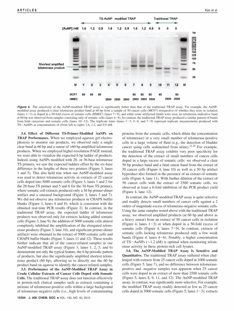
proteins from the somatic cells, which dilute the concentrationof telomerase) or a very small number of telomerase-positivecells in a large volume of fluid (e.g., the detection of bladdercancer using cells sedimented from urine).22,38 For example,the traditional TRAP assay exhibits very poor specificity forthe detection of the extract of small numbers of cancer cellsdoped in a large excess of somatic cells: we observed a clear50 bp product band and a faint smear band from the extract of50 cancer cells (Figure 4, lane 10) as well as a 50 bp artifactbyproduct also formed in the presence of an extract of somaticcells (Figure 4, lane 11). With further dilution of the extract of50 cancer cells with the extract of 2500 somatic cells, weobserved at least a 6-fold inhibition of the PCR product yield(Figure 4, lane 12).
In contrast, the AuNP-modified TRAP assay is quite selectiveand readily detects small numbers of cancer cells against a 2orders of magnitude excess of telomerase-negative somatic cells.Using the same samples tested above with the traditional TRAPassay, we observed amplified products (at 60 bp and above asa heavy smear) from an extract of 50 cancer cells in isolation(Figure 4, lanes 1-3) or when doped in a 50-fold excess ofsomatic cells (Figure 4, lanes 7-9). In contrast, extracts ofsomatic cells lacking telomerase produced only a few weakbands (Figure 4, lanes 4-6). Notably, a higher concentrationof TS-AuNPs (∼1.2 nM) is optimal when monitoring telom-erase activity in these protein-rich cell lysates.
3.6. The AuNP-Modified TRAP Assay Is Sensitive andQuantitative. The traditional TRAP assay suffered when chal-lenged with extracts from 25 cancer cells doped in 1000 somaticcells (Figure 5, lane 7), and no difference between telomerase-positive and -negative samples was apparent when 25 cancercells were doped in an extract of more than 2500 somatic cells(Figure 5, lanes 8, 9, 11, and 12). The AuNP-modified TRAPassay, in contrast, was significantly more selective. For example,the modified TRAP assay readily detected as few as 25 cancercells doped in 5000 somatic cells (Figure 5, lanes 1-3); extracts
Figure 4. The selectivity of the AuNP-modified TRAP assay is significantly better than that of the traditional TRAP assay. For example, the AuNP-modified assay produced a clear telomerase product band at 60 bp from a sample of 50 cancer cells (MCF7) irrespective of whether they were in isolation(lanes 1-3) or doped in a 50-fold excess of somatic cells (HMEC) (lanes 7-9), and while some artifactual bands were seen, no telomerase-indicative bandat 60 bp was observed from samples consisting only of somatic cells (lanes 4-6). In contrast, the traditional TRAP assay produced a similar pattern of bandsfrom both cancerous and somatic cells (lanes 10-12). The triplicate lanes (lanes 1-3, 4-6, and 7-9) represent replicate measurements produced withTS-AuNPs at concentrations of (from left to right) 1.6, 1.2, and 0.9 nM.
15304 J. AM. CHEM. SOC. 9 VOL. 132, NO. 43, 2010
A R T I C L E S Xiao et al.

from only somatic cells produced only a primer-dimer artifactat or below 60 bp (Figure 5, lanes 4-6). A heavily smearedband was also well-correlated with extract samples that includedtelomerase-positive cancer cells (Figure 5, lanes 1-3 and 7).
Even when deployed in complex mixtures, the AuNP-modified TRAP assay can quantitatiVely detect cancer cells overa wide dynamic range. For example, extracts of 10 to 500 MCF7cancer cells in a background of 1000 somatic HMEC cells
produced a reproducible, monotonically increasing band at 60bp amplified product (Figure 6, top panel, lanes 1-5), thedensity of which was strongly correlated with the number ofcancer cells in each sample (Figure 6, bottom panel). In contrast,the traditional TRAP assay produced dramatically less telom-erase PCR product (smeared bands) when employed againstextracts consisting of 100 to 500 cancer cells within 1000somatic cells (Figure 6, top panel, lanes 7 and 8), and no clear
Figure 5. The AuNP-modified TRAP assay is sufficiently selective to ensure the detection of cancer cells (here MCF7 cells) even when they are dilutedwith a large (up to 200-fold) excess of somatic cells (here HMEC cells) (lanes 1-3). The traditional TRAP assay, in contrast, is significantly less selective(lanes 7-9).
Figure 6. The yield of telomerase PCR product increases monotonically with increasing number of cancer cells in the AuNP-modified TRAP assay. Forexample, against a background of 1000 somatic HMEC cells, the intensity of the telomerase-indicative 60 bp band produced by the AuNP-modified assay(top panel) was closely correlated with the number of MCF7 cells in the sample (bottom panel).
J. AM. CHEM. SOC. 9 VOL. 132, NO. 43, 2010 15305
Detection of Telomerase Activity in Cell Lysates Using AuNPs A R T I C L E S
products were observed when the sample contained fewer than100 cancer cells (Figure 6, top panel, lanes 9-11).
The detection limit of the AuNP-modified TRAP assay wasfurther improved when employed in real-time PCR. Thetraditional TRAP assay performed only marginally when 50cancer cells were doped in 5000 somatic cells (CT ) 25.92 (0.11) and produced CT values of 26.65 ( 0.09 and 29.20 (0.21 with CHAPS buffer blanks and extracts of 5000 somatic
cells, respectively (Figure 7A). In contrast, the AuNP-modifiedTRAP assay exhibited at least 10-fold higher sensitivity thanthe traditional TRAP assay: it easily detected the presence of 5cancer cells doped into 5000 somatic cells (CT ) 30.25 ( 0.32)(Figure 7B) and produced no amplification upturn with eitherCHAPS buffer blanks or extracts of 5000 somatic cells. Themodified real-time PCR TRAP assay was also highly quantita-tive, producing a strong correlation between the observed CT
values and the number of cancer cells in a sample (Figure S4in the Supporting Information). In addition, we found that bothTRAP assays displayed atypically high real-time PCR efficien-cies in the presence of a high concentration of somatic cellextract. For example, the slopes for the standard curves of CT
versus the logarithm of the number of cancer cells were -2.78and -2.48 for the AuNP-modified and traditional TRAP assays,respectively, which might be due to the interaction of AuNPswith proteins and PCR inhibitors from the cell extract.29
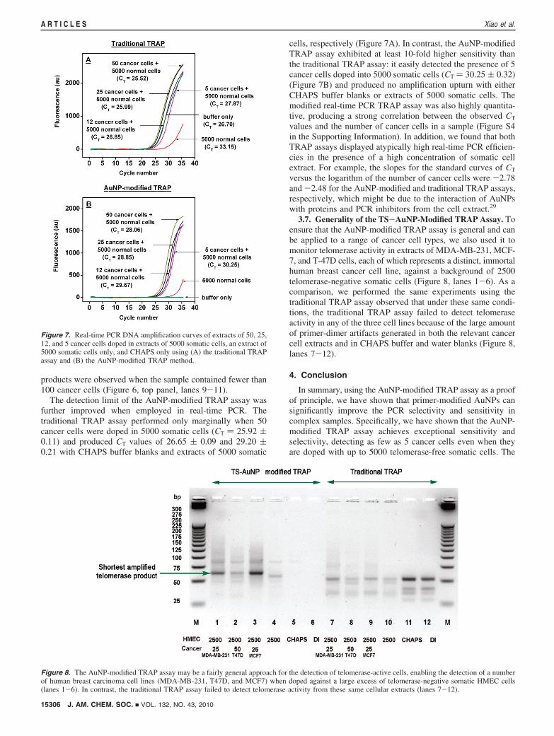
3.7. Generality of the TS-AuNP-Modified TRAP Assay. Toensure that the AuNP-modified TRAP assay is general and canbe applied to a range of cancer cell types, we also used it tomonitor telomerase activity in extracts of MDA-MB-231, MCF-7, and T-47D cells, each of which represents a distinct, immortalhuman breast cancer cell line, against a background of 2500telomerase-negative somatic cells (Figure 8, lanes 1-6). As acomparison, we performed the same experiments using thetraditional TRAP assay observed that under these same condi-tions, the traditional TRAP assay failed to detect telomeraseactivity in any of the three cell lines because of the large amountof primer-dimer artifacts generated in both the relevant cancercell extracts and in CHAPS buffer and water blanks (Figure 8,lanes 7-12).
4. Conclusion
In summary, using the AuNP-modified TRAP assay as a proofof principle, we have shown that primer-modified AuNPs cansignificantly improve the PCR selectivity and sensitivity incomplex samples. Specifically, we have shown that the AuNP-modified TRAP assay achieves exceptional sensitivity andselectivity, detecting as few as 5 cancer cells even when theyare doped with up to 5000 telomerase-free somatic cells. The
Figure 7. Real-time PCR DNA amplification curves of extracts of 50, 25,12, and 5 cancer cells doped in extracts of 5000 somatic cells, an extract of5000 somatic cells only, and CHAPS only using (A) the traditional TRAPassay and (B) the AuNP-modified TRAP method.
Figure 8. The AuNP-modified TRAP assay may be a fairly general approach for the detection of telomerase-active cells, enabling the detection of a numberof human breast carcinoma cell lines (MDA-MB-231, T47D, and MCF7) when doped against a large excess of telomerase-negative somatic HMEC cells(lanes 1-6). In contrast, the traditional TRAP assay failed to detect telomerase activity from these same cellular extracts (lanes 7-12).
15306 J. AM. CHEM. SOC. 9 VOL. 132, NO. 43, 2010
A R T I C L E S Xiao et al.
modified assay is thus vastly improved relative to the traditionalTRAP assay, for which the very best reported sensitivity andselectivity is 10 cancer cells diluted only 50-fold by somaticcells.13 The origins of the enhanced selectivity and sensitivityof the AuNP-modified TRAP assay are unclear, but theenhancements may arise from the large surface-to-volume ratioof the AuNPs,39 which would enhance the adsorption of proteinsfrom the cell lysate onto the particle surface.39 Such proteinadsorption indirectly, but potentially significantly, dilutes po-tential interferants from the cell extracts, thereby reducing theinhibition of the elongation reaction of telomerase andthe subsequent PCR. The use of AuNP-modified primers in theTRAP assay reduces PCR artifacts such as primer-dimers andalso reduces the inhibition often observed when TRAP isperformed on crude cell lysates. We believe that the use ofAuNP-modified primers may prove to be of utility for a widerange of PCR-based assays, including real-time PCR, and can
be a potential solution for performing PCR directly in protein-rich solutions.
Acknowledgment. This work was supported in part by LawrenceLivermore National Laboratory (URP-06-019) and by the U.S.Army (Grant W81XWH-08-1-0304). We thank Dr. Qiang Gongfor providing helpful discussions.
Supporting Information Available: Experimental results forthe concentration of TS-AuNPs employed in the AuNP-modified TRAP assay; melting curves of extracts of cancer cells,somatic cells, mixtures of cancer cells and somatic cells, andCHAPS using the AuNP-modified and traditional TRAP assays;and PCR efficiency of extract mixtures of different cancer cellsand 5000 somatic cells using the AuNP-modified and traditionalTRAP assays. This material is available free of charge via theInternet at http://pubs.acs.org.
JA106513F(39) Klein, J. Proc. Natl. Acad. Sci. U.S.A. 2007, 104, 2029–2030.
J. AM. CHEM. SOC. 9 VOL. 132, NO. 43, 2010 15307
Detection of Telomerase Activity in Cell Lysates Using AuNPs A R T I C L E S