detection of replication competent retrovirus and lentivirus · screening vector products for rcr...
TRANSCRIPT

Chapter 17
Detection of Replication Competent Retrovirus and Lentivirus
Lakshmi Sastry and Kenneth Cornetta
Summary
Retroviral vectors based on murine leukemia viruses (MuLV) have been used in clinical investigations for over a decade. Alternative retroviruses, most notably vectors based on HIV-1 and other lentiviruses, are now entering into clinical trials. Although vectors are designed to be replication defective, recombina-tion events during vector production could lead to the generation of replication competent retroviruses (RCR) or replication competent lentiviruses (RCL). Careful screening of vector prior to human use must insure that patients are not inadvertently exposed to RCR or RCL. We describe methods capable of detecting low levels of virus contamination and discuss the current regulatory guidelines for screening gene therapy products intended for human use.
Key words: Retroviral vectors , Lentiviral vectors , Gene therapy, Replication competent retrovirus , Replication competent lentivirus .
1. Introduction
1.1. Risks Associated with RCR and RCL
Gammaretrovirus-based retroviral vectors (subsequently referred to as “retroviral vectors”) are membrane bound RNA viruses ini-tially based on murine leukemia viruses (1–3). These were the first viral vectors to enter clinical trials (4) and remain an attrac-tive gene delivery tool when integration of the transgene into the target cells is sought. Unfortunately, integration is also asso-ciated with insertional mutagenesis whereby the risk of malig-nant transformation occurs due to dysregulation of cellular gene expression (reviewed in (5)). Insertional mutagenesis is a rare but known occurrence after insertion of a replication defective vec-tor. Integration has led to activation of the Evi-1 oncogene with
Christopher Baum (ed.), Methods in Molecular Biology, Methods and Protocols, vol. 506 © Humana Press, a part of Springer Science + Business Media, LLC 2009 DOI: 10.1007/978-1-59745-409-4_17
243

244 Sastry and Cornetta
subsequent development of myeloid leukemia in a murine bone marrow transplantation model (6). Most concerning, integra-tion of a retroviral vector led to the development of leukemias in subjects treated for X-linked severe combined immunodeficiency (SCID) (7–10). Why these children have developed leukemia is complex, but preliminary evidence suggests the possibility that unregulated expression of the vector transgene (the common gamma chain cytokine receptor) may add to vector-induced dys-regulation of the LMO-2 gene (i.e., a second “hit”) (11, 12).
While replication defective vector carry risk, the risk of inser-tional mutagenesis is believed to be greatest if RCR is present, since ongoing viral infection is likely to result in a much greater number of insertional events. RCR contaminating retroviral vec-tor preparations have been shown to cause malignancy in both mice and nonhuman primates (13, 14).
The risks associated with lentiviral vectors are currently unknown. The MLVs are known to cause leukemia related to regulatory regions with the MLV long-terminal repeats (LTR). HIV-1 is not known to cause leukemia, and third-generation len-tiviral vectors have deleted the regulatory regions within the LTR (SIN, or self-inactivating vectors). Nevertheless, cellular gene dysregulation mediated by vector promoters remain a theoretical concern. Whether RCL can produce a HIV-1 like syndrome is unknown, although HIV-1 accessory proteins normally required for virulence have been deleted from vector constructs to improve their safety profile.
1.2. Technical Consid-erations
Retroviral and lentiviral vectors are generated by deletion of the viral protein coding sequences ( gag, pol, and env) and substitu-tion of this region with an exogenous gene(s) of interest. Vector particles must be generated by coexpressing the vector sequences along with the viral genes. Traditionally, retroviral vectors are generated in packaging cell lines that stably express the gag, pol, and env genes along with the vector plasmid (see Miller (15) for review). With packaging cells, RCR arises by recombination between the vector and viral genes, and was frequently detected in early versions of vector packaging cell lines in which all the viral genes ( gag/pol/env) were expressed from a single plasmid (16–19). By segregating gag-pol and env genes onto separate plasmids and minimizing homology between vector and pack-aging sequences, the rate of RCR development has been sub-stantially decreased, but not eliminated (20–25). In addition, the marked decrease in RCR generation resulting from decreased homologous recombination has resulted in rare recombinations between vector and cellular sequences leading to RCR, or rescue of endogenous viruses ( see Note 1) especially when murine-based packaging cell lines are utilized (26–28).

1.3. Testing Methodology
Detection of Replication Competent Retrovirus and Lentivirus 245
While murine oncoretroviruses have been known to develop recombinations leading to RCR, a number of factors compli-cate the detection of vector-associated RCL. First, RCL has not been reported with the commonly used lentiviral vector systems. Therefore, the RCL detection system must anticipate a currently theoretical virus, which will presumably arise through recombina-tion between transfer vector and packaging construct sequences. Lentiviral vectors are generally produced by transient transfec-tion of three or more plasmids: a plasmid expressing the trans-fer vector (containing the gene of interest in a lentiviral vector backbone), the packaging plasmid(s) (containing gag and pol), and an envelope-expressing plasmid (e.g., vesicular stomatitis virus glycoprotein (VSV-G)) (29–32). Although areas of homol-ogy between the transfer and the packaging plasmids will greatly increase the likelihood of such recombination, nonhomologous recombination is also possible. Another concern is recombination with human endogenous retroviral (HERV) sequences (33). RCL testing is also complicated by the transient transfection methods of lentiviral vector generation, which result in substantial con-tamination of supernatants with packaging plasmid DNA that contain the same viral sequences likely to be present in a RCL (34, 35). Finally, a question that is still debated is whether vec-tor product should be screened for a true RCL, or should signs of recombination events between vector and packaging plasmids (without complete generation of RCL) be grounds for rejecting a vector product.
Screening vector products for RCR has generally utilized biologic assays, while monitoring of patients after gene transfer rely on molecular or serologic testing (36, 37). Molecular and serologic testing is rapid and less resource intensive than biologic assays but is prone to false positives. Particular care must be taken when performing PCR shortly after vector exposure since contamina-ting plasmids carried over from transient production meth-ods or DNA from producer cell line can yield a false positive result suggesting RCR (38). Therefore, screening of vector prior to clinical use has generally relied on biologic assays aimed at documenting a replication competent virus, rather than surrogate markers of virus.
The biologic assays most commonly used are the extended S +/L − assay (Fig. 1) and the marker rescue assay (13, 39–41). In these assays, test material is placed on a permissive cell line, and the cells are passaged for a minim um of 3 weeks (amplification phase). In the marker rescue assay, the permissive cell line con-tains a retroviral vector with an easily identifiable transgene (for example, a “marker” gene such as the neomycin phosphotrans-ferase gene). After three weeks, the cell media from the amplifica-tion phase is collected. If RCR is present, it will package both the

246 Sastry and Cornetta
RCR genome and “rescue” the marker vector. Cell free media from the amplification phase cells is then used to transduce a naïve cell line, which is then subjected to drug selection. Dem-onstration of drug resistance in the transduced cell population is indicative of RCR. In the extended S +/L − assay, the amplification cells do not contain vector. Instead, virus is detected using indi-cator cell lines, such as the cat cell line PG-4 (42). The PG-4 cell line is referred to as a S +/L − cell line, as it contains the murine sarcoma virus genome (S +) but lacks the murine leukemia virus genome (L −). Cells that express the murine sarcoma virus induce a transformed phenotype but only in cells coexpressing a murine leukemia virus. In this assay, media from the amplification phase is placed on PG-4 cells and transformation indicates the present of RCR. The S +/L − assay can also be performed without the ampli-fication phase (a direct S +/L − assay), which allows one to titer the number of RCR (expressed as focus forming units per mL).
Fig. 1. The detection of replication competent retrovirus (rcr) using biologic assays. Amplifica-tion Phase: Test material depicts retroviral vector supernatant in which a small portion of the rep-lication defective vector material ( open ovals ) is contaminated with RCR ( filled ovals ). Biologic assays often utilize a 3-week amplification phase in which a permissive cell line is used to increase the titer of any RCR present in the test material. In the Marker Rescue Assay, the cell lines used in the amplification phase contains an integrated retrovi-ral vector that expresses a marker gene (such as a drug resistance gene). If RCR is present, it will “rescue” the marker vector and the cell superna-tant will contain RCR along with virions capable of conferring drug resistance to naïve cells in the Indicator Phase. The S + /L − assay also has an ampli-fication step, but in this case the indicator cell line detects RCR directly. The indicator cell is termed an S + /L − cells since it contains the murine sarcoma virus (MSV), which will transform the cell pheno-type but only in the presence of a murine leukemia virus (MSV) RCR.
Detection of Replication Competent Retrovirus and Lentivirus 247
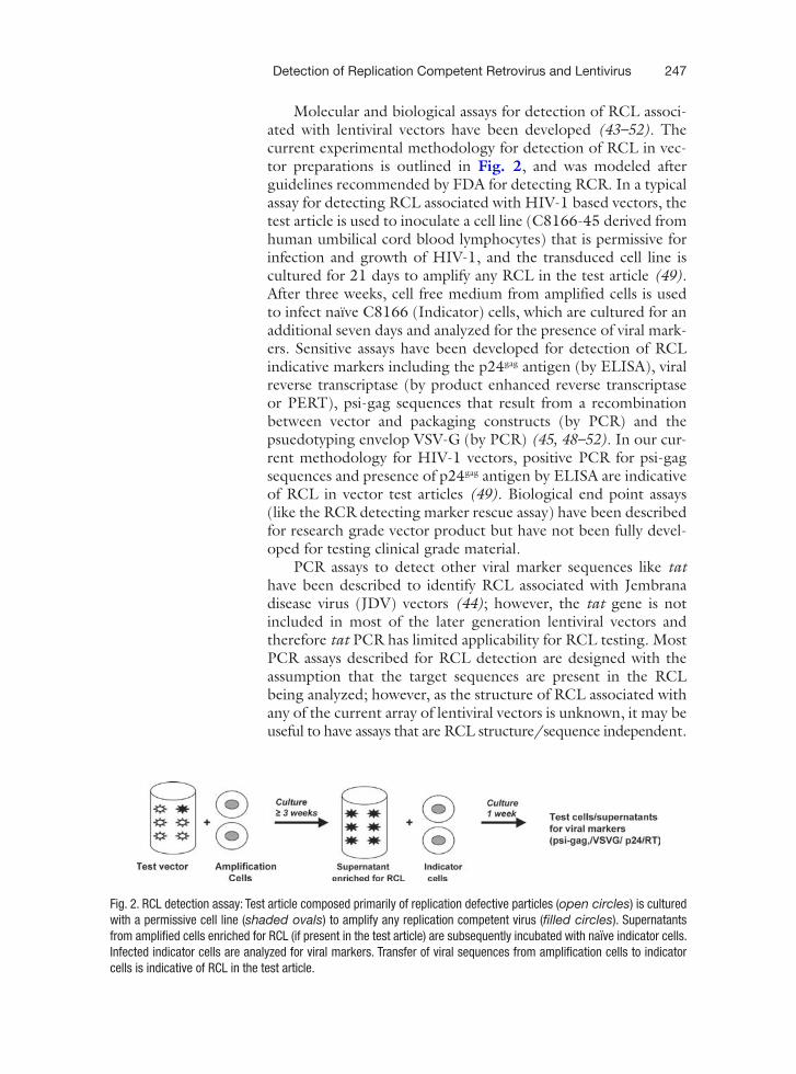
Molecular and biological assays for detection of RCL associ-ated with lentiviral vectors have been developed (43–52). The current experimental methodology for detection of RCL in vec-tor preparations is outlined in Fig. 2, and was modeled after guidelines recommended by FDA for detecting RCR. In a typical assay for detecting RCL associated with HIV-1 based vectors, the test article is used to inoculate a cell line (C8166-45 derived from human umbilical cord blood lymphocytes) that is permissive for infection and growth of HIV-1, and the transduced cell line is cultured for 21 days to amplify any RCL in the test article (49). After three weeks, cell free medium from amplified cells is used to infect naïve C8166 (Indicator) cells, which are cultured for an additional seven days and analyzed for the presence of viral mark-ers. Sensitive assays have been developed for detection of RCL indicative markers including the p24 gag antigen (by ELISA), viral reverse transcriptase (by product enhanced reverse transcriptase or PERT), psi-gag sequences that result from a recombination between vector and packaging constructs (by PCR) and the psuedotyping envelop VSV-G (by PCR) (45, 48–52). In our cur-rent methodology for HIV-1 vectors, positive PCR for psi-gag sequences and presence of p24 gag antigen by ELISA are indicative of RCL in vector test articles (49). Biological end point assays (like the RCR detecting marker rescue assay) have been described for research grade vector product but have not been fully devel-oped for testing clinical grade material.
PCR assays to detect other viral marker sequences like tat have been described to identify RCL associated with Jembrana disease virus (JDV) vectors (44); however, the tat gene is not included in most of the later generation lentiviral vectors and therefore tat PCR has limited applicability for RCL testing. Most PCR assays described for RCL detection are designed with the assumption that the target sequences are present in the RCL being analyzed; however, as the structure of RCL associated with any of the current array of lentiviral vectors is unknown, it may be useful to have assays that are RCL structure/sequence independent.
Fig. 2. RCL detection assay: Test article composed primarily of replication defective particles ( open circles ) is cultured with a permissive cell line ( shaded ovals ) to amplify any replication competent virus ( filled circles ). Supernatants from amplified cells enriched for RCL (if present in the test article) are subsequently incubated with naïve indicator cells. Infected indicator cells are analyzed for viral markers. Transfer of viral sequences from amplification cells to indicator cells is indicative of RCL in the test article.

248 Sastry and Cornetta
The PERT assay detects reverse transcriptase activity associated with retroviruses and has shown to be as sensitive as the p24 gag
ELISA and psi-gag PCR for RCL detection when the RCL has been amplified (52). The disadvantage of PERT is a high back-ground found in certain cell types, but a major advantage is the enzymatic function (of RT) it detects is crucial for replication of any RCL, i.e., it can be used for detection of RCL associated with structurally distinct retroviruses. However, due to the hypo-thetical nature of RCL, it may be advisable to use at least two assays that detect different viral genes/functions for conclusive evidence of RCL in vector products. Ideally a molecular assay (PCR for viral sequence) can be used in conjunction with a func-tional (marker rescue) or protein expression (RT/p24 ELISA) assay for RCL testing.
For subject monitoring for evidence of RCR infection, positive results obtained by PCR or serologic methods should be confirmed by biologic assays to exclude false positives. Also, one must consider that complex recombinations may generate a RCR that lacks the sequence targeted in these assays. Clinical suspicion of RCR with a negative PCR or serologic result should prompt further testing for other target sequences, as well as testing in biologic assays. Subjects receiving lentiviral gene therapy should be monitored similarly for RCL infection by molecular and serological methods. Initial posi-tive reactivity in any of these assays should be followed by biological amplification assays for vector derived RCL.
1.4. Regulatory Issues The concern surrounding RCR and RCL has led US regulators to develop recommendations relevant to the clinical use of these vectors. As retroviral vectors generally utilized packaging cell lines the US FDA requires RCR testing on the Master Cell Bank, any Working Cell Banks, and the post-production cells (1% or a maxi-mum of 10 8 cells). Also, 5% of the final product volume must also be screened. In addition, vectors used in ex vivo transduction protocols in which the cells are cultured for greater than 4 days must be screened for RCR. A new guidance also defines screen-ing that must occur for subjects treated with retroviral vectors. Patients treated with retroviral vectors must also be monitored for RCR pre-treatment, then at 3, 6, and 12 months. If all sam-ples are negative for RCR the subject can be followed by yearly assessment of clinical status and archiving of blood or relevant tis-sue. Any suggestion of RCR exposure mandates extensive evalua-tion and close follow-up. There are similar expectations for RCL testing and follow-up.
To date, there have been no documented exposures of patients treated with gene therapy to RCR or RCL. Although this suggests current screening methods are sensitive, the known risk of insertional mutagenesis mandates continued vigilance and

Detection of Replication Competent Retrovirus and Lentivirus 249
continued monitoring clinical trial subjects, especially given the limited experience to date with lentiviral vectors.
2. Materials
2.1. RCR Testing
2.1.1. Media and Cell Culture Supplies
2.1.2. Positive Control for RCR Testing
2.2. RCL Testing 2.2.1. Media and Cell Culture Supplies
1. McCoys 5A Medium + 1% penicillin and streptomycin (all from GIBCO) containing 10% fetal bovine serum (FBS, Hyclone) (McCoys10); Trypsin-EDTA (GIBCO) and DPBS (Cambrex) for subculturing cells.
2. For cell expansion 75, 175, and 300, 450 cm 2 flasks, calibrated pipettes of various sizes, aspirating pipettes (1 and 10 mL). For dilution of samples 12 × 75 and 17 × 100 mm polypro-pylene tubes and 6-well plates for the assay. 1–200 μL sterile pipette tips for all standard procedures and 0.45 μm cellulose acetate syringe filters for filtering cell supernatants.
3. Mus dunni (CRL-2017) and PG4 (CRL-2032) cells avail-able from the American Tissue Culture Collection (ATCC) at http://www.atcc.org.
4. Polybrene (Sigma). Final concentration will be 8 μg/mL throughout the procedure, it is helpful to prepare an 8 mg/ mL stock.
1. 4070A positive control (VR-1450) available from the Ameri-can Tissue Culture Collection (ATCC) at http://www.atcc. org. We infect cells with virus then grow up a high titer stock which is aliquoted into vials and used as a positive control for multiple assays. The stock is characterized for titer and the TCID 50 (determined by using infectious titer and Reed Muench formula) (49). Stocks of virus are stored at −70°C.
1. RPMI 1640 + 1% penicillin and streptomycin (all from GIBCO) containing 10% fetal bovine serum (FBS, Hyclone) (subsequently referred to as RPMI 10); DMEM supplemented with 2 mM l-glutamine and 1 mM sodium pyruvate and con-taining 10% FBS (subsequently referred to as D10).
2. For cell expansion 75, 175, and 300 cm 2 flasks, calibrated pipettes of various sizes, aspirating pipettes (1 and 10 mL), centrifuge tubes (15 and 50 mL) from BD Falcon or equiva-lent; 10 cm 2–50 mL flat bottom culture tubes for small scale transductions.
3. For RCL testing of HIV-1 vectors, C8166 cells are available from the NIH AIDS Research and Reference Reagents Program.
4. Polybrene (Sigma).

250 Sastry and Cornetta
2.2.2. Test Articles for RCL Testing
Test articles can consist of lentiviral vector preparation or cell media from transduced cell populations.
2.2.3. Positive Control Virus for RCL Testing
For HIV-1 vectors, an attenuated HIV-1 virus lacking the acces-sory genes is used as a positive control in cGMP RCL assays (ex. R8.71) (45, 48). Stocks of virus are stored at −70°C. The TCID 50 of the positive control virus is determined by using infectious titer and Reed Muench formula (49).
2.2.4. Reagents for RCL Detection Assays
1. HIV-1 p24 gag antigen ELISA kit (RETROtek or Perkin Elmer) for measuring p24 gag protein in indicator cultures.
2. Puregene DNA extraction kit (Gentra) for isolating genomic DNA from indicator cells.
3. PCR: 1. PCR primers for amplifying psi-gag sequences: GrecF1
(5-CAGGACTCGGCTTGCTGAA-3) and GrecR2 (5-TGTCTTATGTCCAGAATGCT-3), GrecP (5-AAGATTTAAACACCATGCTA-3)
2. PCR reagents: HotStarTaq DNA polymerase, dNTPs, water (molecular biology grade).
3. PCR reaction analysis: Agarose, 1 M Tris pH 7.6 (Sigma), ethidium bromide (Sigma), 1× TAE, Blue-orange loading dye (Promega), molecular standards (GIBCO).
4. Southern blotting and probing PCR products:
Blotting: Nytran supercharged membrane (Midwest), NaOH Probe labeling: Probe GrecP, 10× phosphorylation buffer
(Roche), Gamma 32P dATP (Perkin Elmer), 1× STE (0.1 M sodium chloride, 0.01 M Tris-HCl, 0.001 M EDTA), polynucle-otide kinase (Roche), Scintillation counting fluid.
Hybridization: 5 M NaCl, 1 M Tris, pH 7.6, 0.5 M EDTA (pH 8.0), 10% SDS, ExpressHyb hybridization solution, 20× SSC.
2.3. Equipment 1. Tissue Culture Incubator, Pipette Aid, Biological Safety Cabi-net, Inverted Microscope.
2. Centrifuge (with aerosol protective cover). 3. Adjustable Pipettor: 1–200 μL. 4. Hybridization oven, Scintillation counter and protective bar-
riers for radioactive work. 5. ELISA reader. 6. >−70°C Freezer.
Detection of Replication Competent Retrovirus and Lentivirus 251
3. Methods
3.1. RCR Detection 3.1.1. Overview
3.1.2. Amplification Phase
The Extended S +/L − Assay is composed of two stages (Fig. 1). The first stage is the amplification stage in which a test article/ sample is inoculated into a culture of a cell line permissive for viral replication (for this assay Mus dunni cells). The second stage is the detection phase where media is obtained from the ampli-fication cells at the three-week time point and analyzed on a cell line capable of detecting replication competent retrovirus. For this assay, the PG-4 S +/L − cells are used. Foci of transformed cells indicate the presence of replication competent retrovirus. The assay described here is for detection of amphotropic virus, the assay may need to be modified for other viral envelopes ( see Note 2).
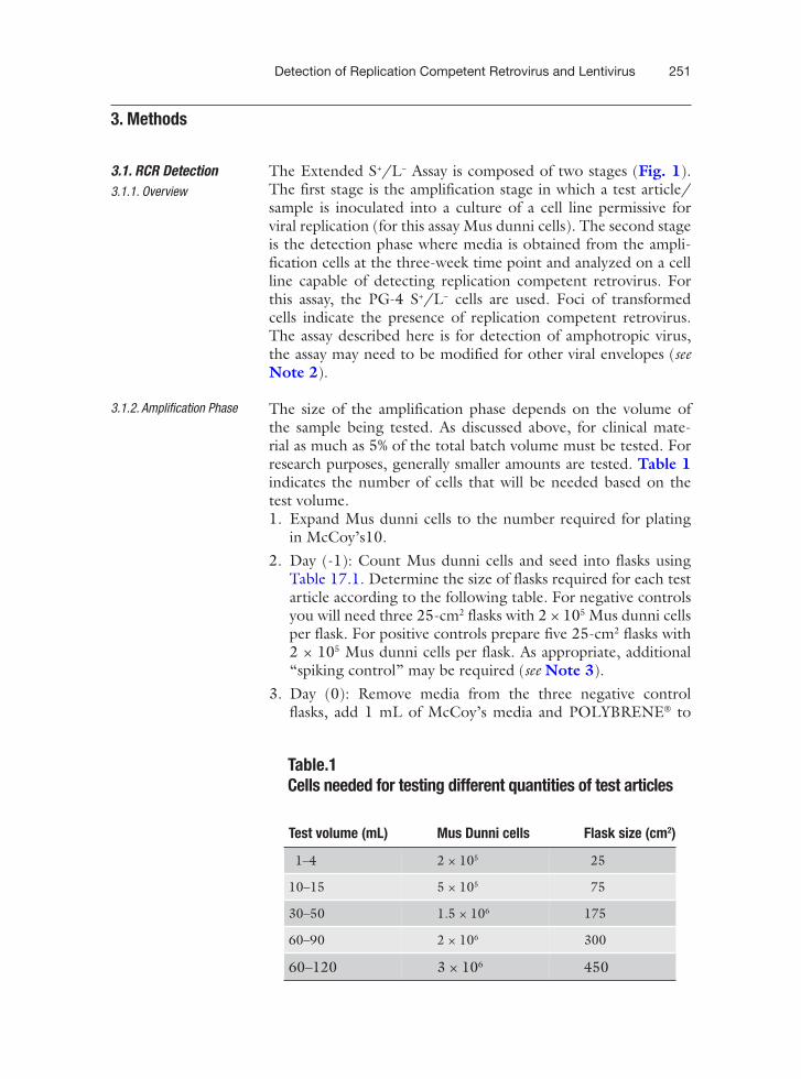
The size of the amplification phase depends on the volume of the sample being tested. As discussed above, for clinical mate-rial as much as 5% of the total batch volume must be tested. For research purposes, generally smaller amounts are tested. Table 1 indicates the number of cells that will be needed based on the test volume. 1. Expand Mus dunni cells to the number required for plating
in McCoy’s10. 2. Day (-1): Count Mus dunni cells and seed into flasks using
Table 17.1. Determine the size of flasks required for each test article according to the following table. For negative controls you will need three 25-cm 2 flasks with 2 × 10 5 Mus dunni cells per flask. For positive controls prepare five 25-cm 2 flasks with 2 × 10 5 Mus dunni cells per flask. As appropriate, additional “spiking control” may be required ( see Note 3).
3. Day (0): Remove media from the three negative control flasks, add 1 mL of McCoy’s media and POLYBRENE ® to
Table.1 Cells needed for testing different quantities of test articles
Test volume (mL) Mus Dunni cells Flask size (cm 2 )
1–4 2 × 10 5 25
10–15 5 × 10 5 75
30–50 1.5 × 10 6 175
60–90 2 × 10 6 300
60–120 3 × 10 6 450
252 Sastry and Cornetta
a final concentration of 8 μg/mL. Remove media from the test article flasks, and add the test article and POLYBRENE ®. Return flasks to incubator. Then remove media from the five 25-cm 2 flasks to be used as the positive control. Add 1 mL of 4070A virus stock that has been diluted to the TCID 50. Add POLYBRENE ®. Incubate at 37°C, 5% CO 2 for 2–4 h. After infection remove media from all flasks and replace with the appropriate volume of fresh McCoy’s medium. Return flasks to incubator.
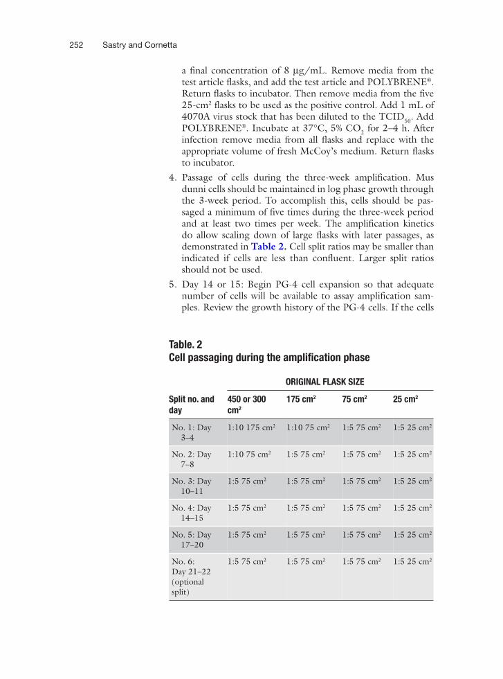
4. Passage of cells during the three-week amplification. Mus dunni cells should be maintained in log phase growth through the 3-week period. To accomplish this, cells should be pas-saged a minimum of five times during the three-week period and at least two times per week. The amplification kinetics do allow scaling down of large flasks with later passages, as demonstrated in Table 2. Cell split ratios may be smaller than indicated if cells are less than confluent. Larger split ratios should not be used.
5. Day 14 or 15: Begin PG-4 cell expansion so that adequate number of cells will be available to assay amplification sam-ples. Review the growth history of the PG-4 cells. If the cells
Table. 2 Cell passaging during the amplification phase
ORIGINAL FLASK SIZE
Split no. and 450 or 300 175 cm 2 75 cm 2 25 cm 2
day cm 2
No. 1: Day 3–4
1:10 175 cm 2 1:10 75 cm 2 1:5 75 cm 2 1:5 25 cm 2
No. 2: Day 7–8
1:10 75 cm 2 1:5 75 cm 2 1:5 75 cm 2 1:5 25 cm 2
No. 3: Day 10–11
1:5 75 cm 2 1:5 75 cm 2 1:5 75 cm 2 1:5 25 cm 2
No. 4: Day 14–15
1:5 75 cm 2 1:5 75 cm 2 1:5 75 cm 2 1:5 25 cm 2
No. 5: Day 17–20
1:5 75 cm 2 1:5 75 cm 2 1:5 75 cm 2 1:5 25 cm 2
No. 6: Day 21–22 (optional split)
1:5 75 cm 2 1:5 75 cm 2 1:5 75 cm 2 1:5 25 cm 2

3.1.3. Pg-4 Focus Forming Assay
Detection of Replication Competent Retrovirus and Lentivirus 253
have been passaged greater than 15 times, discard and thaw new vial of PG-4 cells.
6. Harvest amplification media: Media can be harvested from amplification cultures 3≥21 days from culture initiation. Cultures should be confluent (usually 2–4 days after 5th or 6th split), then change media. Approximately 24 h after media change, collect supernatant and filter through a 0.45 μm filter. Super-natant may be inoculated directly onto PG-4 cells (see below) or frozen for later assay (store at −70°C). Do not leave super-natant out at room temperature for more than 2 h.
1. Determine the total number of 6-well plates required for the assay. Three wells will be needed for each test article. For the control samples 30 wells (6 plates) will be needed.
2. Day (-1): Plate 1 × 10 5 cells per well in 6-well plates. Add 4 mL of McCoy’s10. Incubate overnight at 37°C, 5% CO 2.
3. Day (0): Prepare direct (nonamplified) negative control for the PG-4 Assay. Remove media from 3 wells and add 1 mL of McCoy’s10 and add POLYBRENE ®.
4. Day (0): Prepare amplification negative controls. Remove medium from each well of one plate. Add 1 mL of filtered supernatant from each of the three negative control flasks from the amplification phase to 2 wells and add POLYBRENE ®.
5. Day (0): Prepare test articles. From each flask of the test arti-cles, remove and filter supernatant; material will be tested undi-luted and at 10 −2 dilution (dilute in McCoys10, see Note 4). Remove media from designated wells. Add 1 mL of undiluted supernatant to two wells and 1 mL of supernatant diluted to 10 −2 to the other well. Add POLYBRENE ® to each well.
6. Day (0): Prepare amplification positive controls. From each of the 5 flasks of amplification positive control, remove and fil-ter supernatant. Prepare the following dilutions for each flask: Flask #1: undiluted, 10 −2, 10 −4, 10 −5, 10 −6, and 10 −7 Flasks #2–5: 10 −2, 10 −4, and 10 −6. Remove media from designated wells and add 1 mL of the appropriate dilution, add POLY-BRENE ®.
7. Day (0): Prepare direct (nonamplified) positive control for the PG-4 assay. Thaw a vial of stock 4070A positive control virus. Infect cells with three dilutions that are more concentrated than the TCID 50, at the TCID 50 dilution, and two dilutions that are less concentrated than the TCID (Example: If TCID = 10 7
50. 50 test dilutions will be 10 −4, 10 −5, 10 −6, the TCID (10 −7) and two dilutions less (10 −8 and 10 −9). Prepare two wells per dilution, and then add POLYBRENE ® to each well. Incubate for 2–4 h then remove medium from all wells and replace with 4 mL of McCoy’s medium. Return plates to incubator.

254 Sastry and Cornetta
3.1.4. Score Foci
3.2. RCL Detection in HIV-1 Vectors
3.2.1. Determine P24 Content of Test Articles
8. Day (2): Remove medium and add 4 mL of fresh McCoy’s medium. Return to incubator.
1. Day (4): Examine negative control wells for confluence using an inverted microscope with a 4× objective. If negative controls are not confluent, remove medium from all wells and replace with 4 mL of McCoy’s medium. Check daily until negative controls show a confluent lawn of cells. If >2 additional days are required for cells to become confluent, suspect complications.
2. Each well should be examined for the presence of foci using a 4× objective. When the number of foci is between 0 and 20 in a well, record the actual number observed. If there are greater than 20 foci, record a qualitative assessment (+ if foci are grown together but normal cells observed, ++ if near or total ablation of the cell lawn is observed).
3. Examine the negative control wells. The assay is acceptable if no foci are observed.
4. Next examine the amplified positive controls. Foci should be present in at least one of the dilutions from the five amplified positive controls.
5. Examine the direct positive control. Foci should be present in the dilution which is two log more concentrated than the TCID 50 (Example: if TCID 50 = 10 7, foci should be present in the 10 −5 dilution). Foci in less concentrated dilutions are acceptable, if foci are not seen in dilutions 2 log greater than the TCID 50 consider complications. Count and record the number of foci in each well.
6. Examine the test article wells and record the number of foci. For cGMP work, we have two trained technicians read each well independently.
In this section, a detailed protocol for detecting RCL in HIV-1 vectors is described.
1. If the test article is a lentiviral vector, the concentration of p24 must be determined as concentrated lentiviral vectors can inhibit the growth of the C8166 cell line. Determine the p24 value of test vector using p24 gag ELISA as per manufacturers instructions.
2. For test articles with p24 values ≤1,000 ng/mL, use the mate-rial undiluted. For materials that exceed this amount, the test article should be diluted to final concentration of 1,000 ng/ mL. Calculate the final volume of diluted test article and determine the amount of C8166 cells that will be required according to Table. 3. Propagate the required number of C8166 cells in RPMI10.

3.2.2. Amplification Phase
Detection of Replication Competent Retrovirus and Lentivirus 255
1. Day (-1) Preparation of C8166 cells ( see Note 5): Harvest C8166 cells and count. Label six 50 mL culture tubes (3 for negative control and 3 for positive control). Add 12 mL RPMI to each culture tube. Aliquot 5 × 10 6 C8166 cells/ culture tube. Label appropriate number of flasks for the test article. Add RPMI10 to each flask (12 mL/75 cm 2 flask or 50 mL culture tube, 30 mL/175 cm 2 flask, 60 mL/300 cm 2
flask). Add appropriate number of C8166 cells for the test article according to calculations from Table. 3. Place all cul-ture tubes/flasks in the incubator (37°C, 5% CO 2).
2. Day (0) Inoculation of cultures: Centrifuge negative control tubes at 660 × g for 3 min. Remove the media and add 1 mL of RPMI10. Add POLYBRENE ® to a final concentration of 8 μg/mL.
3. Day (0) Prepare test article for inoculation into culture. If starting p24 concentration is >1,000 ng/mL dilute test arti-cle in RPMI medium to obtain 1,000 ng of p24. Centrifuge C8166 cells at 660 × g for 3 min then add the appropriate amount of test article using the ratios defined in Table. 1. Add POLYBRENE ®.
4. Day (0) Preparation of positive control cultures ( see Note 6): Centrifuge C8166 cells as described above for negative control, but replacing media with replication competent virus (R8.71). The level of sensitivity will be based on the dilution of the virus. For example, use of virus at the TCID 50 would suggest maximal sensitivity of the assay. To insure that the test article is not inhibitory, additional positive controls may be added in which the test article is spiked with R8.71 ( see Note 3). Add POLYBRENE ®. Mix all cultures and return them to the (37°C, 5% CO 2). Incubate for 4 h.
Table. 3 Cells needed for testing different amounts of test vector for RCL
Test volume (after appropri-ate dilution to obtain p24 C8166 amplification levels of £ 1,000 ng/mL) cells Flask (tube) size
1–5 mL 5 × 10 6 50 mL flat bottom culture tubes
6–30 mL 3 × 10 7 75 cm 2
31–60 mL 6 × 10 7 175 cm 2
61–100 mL 1 × 10 8 300 cm 2

256 Sastry and Cornetta
5. 5 Day (0). After incubation, centrifuge the cells in all cul-tures, remove media and add appropriate amount ofRPMI10 (Table. 4) and transfer to appropriate sized flasks and trans-fer to the incubator. The positive and negative control sam-ples are resuspended in 12 mL RPMI and transferred 75 cm 2
flasks. 6. Passage of cells during the 3-week amplification. Amplifica-
tion cells should be maintained in log phase growth through the 3-week period by passing (ratio 1:5 to 1:10) a minimum of five times during the three-week period (least two times per week).
7. Day 17–19. Prepare for the Indicator Phase by expanding naïve C8166 cells 17–19 days after the start of the amplifica-tion phase.
8. Three to four days after 5th split and 24 h before collecting the amplification supernatants, transfer all cultures to conical tubes and spin at 660 × g for 3 min. Remove media and add 12 mL of fresh RPMI medium. Transfer to appropriate sized flasks and return cultures to the incubator.
9. Twenty-four hours after media change, transfer all cultures to 15 mL conical tubes, centrifuge and harvest supernatants and cells. Filter supernatants through a 0.45 μm filter, reserve 1 mL for p24 gag determination. Use 2 mL of test article, and 2 mL from each of the negative and positive control flasks for the indicator phase testing. Reserve unused supernatant and store at −70°C.
10. OPTIONAL – Collect approximately 6 × 10 6 cells from each test article and controls for further analysis if RCL is detected after the indicator phase.
Table. 4 Processing of cultures after transduction
Original sample Resuspension volume Flask size after volume (mL) after transduction (mL) transduction (cm 2 )
1–5 12 75
6–30 30 75
31–60 60 175
61–100 100 300

3.2.3. Indicator Phase (Begin at Least 21 Days after Initiation of the Amplification Phase)
3.2.4. RCL Detection Assays
Detection of Replication Competent Retrovirus and Lentivirus 257
1. On the basis of the amplification phase, 2 × 50 mL culture tubes will be required for each test article, and 6 for the neg-ative control and 6 for the positive controls. Plate 1 × 10 6
C8166 cells per 50 mL culture tube with 4 mL RPMI10 and incubate overnight.
2. After the overnight incubation, centrifuge cells. There will be three negative control supernatants: add 1 mL from each to duplicate culture tubes. For each test article, duplicate culture tubes should be inoculated with 1 mL. The three positive controls should be tested by adding 1 mL of each into duplicate culture tubes. Polybrene ® should be added to each tube. Incubate for 4 h, centrifuge at 660 × g for 3 min. Remove medium, and replace with fresh RPMI10, then return to the incubator and culture for two days.
3. Day (2): Transfer all cultures from tubes to a 75 cm 2 flask with 12 mL of fresh RPMI10 and return to incubator.
4. Day (5): Remove cells from flask, centrifuge and resuspend cell pellet in fresh RPMI10. Return to incubator.
5. Day (6): At least 24 h after media change, supernatant and cells are ready to be harvested. For each sample, transfer media to a 15 mL conical tubes, centrifuge at 660 × g, for 3 min and filter separately through a 0.45 μm filter. Transfer an aliqout of filtered supernatant to a labeled tube for p24 determina-tion. Then collect approximately 6 × 10 6 cells for DNA isola-tion in a 2.0 mL microcentrifuge tube and isolate as directed by manufacturer using a commercial kit (ex. Puregene DNA extraction kit, Qiagen,Valencia, CA). Freeze unused superna-tants as a reserve samples at ≤ −70°C.
RCL can be detected by a variety of methods and the stringency will relate to the level of assurance required. The easiest method is detection of p24 gag antigen by ELISA (43, 48–49). Additional tests can include psi-gag PCR and PCR for the VSV-G enve-lope (49). The ELISA method is rapid and reproducible but the kits are expensive. The PCR methods assume that the RCL has generated from vector and packaging sequence (psi-gag), or that the RCL has not incorporated another envelope besides VSV-G. For clinical grade material, we perform both the p24 ELISA and psi-gag. For nonclinical material, we utilize the p24 ELISA and reserve the psi-gag if there is concerns about false positive ELISA results. 1. p24 gag antigen estimation: Perform p24 ELISA on amplifica-
tion and indicator phase cultures using a commercially avail-able ELISA kit according to manufacturers specifications. The kit positive control standards (purified p24 gag protein) are used to establish a standard curve for the assay; a cut off value is established using buffer and negative controls from the RCL

258 Sastry and Cornetta
assay to set a lower detection limit. All samples are analyzed in duplicate and are scored positive if they are above the cut off value of the assay.
2. Psi-gag PCR: Extract DNA from indicator cell samples accord-ing to manufacturer’s specifications (Puregene Kit). Amplify 1 μg of DNA from indicator cell samples in a standard 50 μL PCR assay containing 0.5 μM psi-gag specific primers (Grec F1/ GrecR2), 200 μM dNTPs, 1× HotStart buffer, and 0.05 U of HotStart Taq. Amplification conditions are 95°C, 10 min and 40 cycles of 95°C for 1 min; 55°C, 1 min; 72°C, 1 min and a final extension of 72°C for 7 min. As a positive control for PCR use 100 copies of plasmid DNA containing recombinant HIV-1 psi-gag sequences (R8.71) in a background of C8166 genomic DNA; no template control is included as a negative control for the PCR assay. All samples are run in duplicate in the PCR assay and are analyzed by agarose gel electrophoresis followed by transfer of PCR products to a nylon membrane and probing with a radio-labeled probe (GrecP). Test arti-cles are scored as positive if they exhibit a 953 bp band in the Southern analysis; negatives from the RCL assay should exhibit no signal and positive samples from the RCL assay should be positive. The PCR positive control (R8.71) should show the expected sized band and the assay negatives should show no signal for the assay to be valid.
3.2.5. Interpretation of Assays
1. Although p24 is normally below the detectable limit at the end of the amplification phase, the high concentration of p24 in lentiviral vectors occasionally results in low levels still detectable at 21 days. Also, low level transfer of sequences (such as psi-gag or VSV-G) can occur in amplification phase cells (possibly due to plasmid contamination) in the absence of RCL. Therefore, we consider a true RCL to be one that can be transferred to the indicator phase.
2. Positive controls from the indicator phase (at least 1/3) should be positive for p24 antigen (and psi-gag sequences if tested) if vector was inoculated at the TCID 50.
3. Negative controls from the indicator phase should be negative for p24 antigen (and psi-gag sequences if tested).
4. If test articles are negative for both p24 and psi-gag sequences at the indicator phases, no RCL is present in the test arti-cle. If any test articles from the indicator phase tested positive for p24 antigen alone or psi-gag sequences alone, repeat p24 ELISA and psi-gag PCR to confirm results. Consider analyz-ing saved material from the amplification phase.
5. If test articles are positive for both p24 antigen and psi-gag they may contain RCL.

Detection of Replication Competent Retrovirus and Lentivirus 259
4. Notes
1. As an example, Miller and colleagues identified a novel ret-rovirus, the Mus dunni endogenous virus (MDEV), which was present in the Mus dunni cell line used in a marker res-cue assay. The virus was activated by hydrocortisone in the medium being tested for RCR (41, 53). Therefore, cell lines used for amplification and assay must be considered as a pos-sible source of RCR unrelated to the vector product.
2. The selection of the amplification phase cell line and the indi-cator cell assay will depend in part on the vector pseudotype. Pseudotype refers to the viral envelope selected for expression on the surface of the vector particle. The MoMLV from which many vectors are derived normally expressed the ecotropic envelope. As the ecotropic receptor is limited to rodent cells, vectors for human cell transduction were initially pseudotyped with the envelope from the amphotropic 4070A virus whose receptor is present on most mammalian cells. Methods detect-ing RCR pseudotyped with the 4070A envelope were devel-oped, and the Mus dunni cell line is commonly used in the amplification phase due to the susceptibility to and amplifica-tion of a wide variety of murine leukemia viruses (54, 55). Although Mus dunni propagates many RCRs, viruses envel-oped with the ecotropic MoMLV glycoprotein are a notable exception and alternate cell lines must be used for their detec-tion (56). Also, the recent use of non-murine retroviral enve-lopes to pseudotype retroviral vectors has complicated the screening for RCRs. One that has now been used in a variety of clinical applications is the envelope derived from the Gib-bon Ape Leukemia Virus (GALV) (25, 57). The GALV enve-lope has demonstrated improved transduction efficiency in a number of target cells, in part, due to the increased expression of the GALV receptor on many target cells (58–61). Although GALV acts as a xenotropic virus in that it infects primate, and other mammalian cells, it cannot infect murine cells (25). Therefore, Mus dunni cells are not suitable for GALV ampli-fication. To address this, 293 cells have been substituted for Mus dunni during the amplification phase of the extended S +/ L − assay with similar levels of virus detection (62). Another retroviral envelope being developed for clinical trial use was cloned from the RD114 virus, which also displays properties of xenotropic viruses (63–65). The 293 cell line is also useful for amplifying RD114 pseudotyped RCR in an extended S +/ L − assay (66).
3. For testing clinical samples “spiking” or mixing of the test article with a known concentration of the positive control is

260 Sastry and Cornetta
prudent to determine whether the test article is inhibitory. In this situation, use a concentration of virus above the TCID 50 since some inhibition is expected when the vector and RCR/ RCL contain the same envelope (due to receptor interfer-ence). At present the FDA has not set guidelines as to the level of inhibitory effect that is allowable.
4. For RCR testing on PG-4 cells we test both undiluted and a 10 −2 dilution. Generally foci can be seen only at low (<100 infectious virus) per well. Above this level, the majority of cells will be transformed and the lawn will be disrupted (gener-ally when we titer virus after amplification it is >10 4 infec-tious viruses per mL). We use the 10 −2 dilution because we have occasionally seen very high concentration of virus inhibit transformation (possibly through receptor interference by defective particles). In this case, the lawn of PG-4 cells will appear abnormal but not disrupted. At the 10 −2 dilution the cells will be clearly transformed and the lawn will be disrupted, confirming the presence of RCR.
5. The choice of amplification cell line for RCL may vary depend-ing on the type of lentiviral vector being tested, the pseudo-typing envelop and on the positive control virus being used for the assay. For HIV-1 vectors, the C8166 cell line was used as it has high transduction efficiency for these vectors, can be infected efficiently by the positive control virus with a natural HIV-1 envelope, and can amplify the positive control over 100-fold (49).
6. The choice of a positive control for the RCL assay is challeng-ing as the structure of a RCL is not known. As the vectors are VSV-G pseudotyped, it may be argued that an HIV-1 virus pseudotyped with VSV-G is a better positive control for the RCL assay than an attenuated HIV-1 virus carrying its natu-ral envelop. We chose the attenuated virus as a VSV-G pseu-dotyped HIV-1 presents an unacceptable risk to laboratory workers (49). For RCL detection in EIAV vectors, 4070A amphotropic MLV has been used as a positive control (51); however, whether an EIAV-RCL behaves like 4070A MLV in experimental situations is debatable.
References
1 . Miller, A.D. , Eckner, R.J. , Jolly, D.J. , Fried- into cells in culture and into murine hemat-man , T., Verma , I.M. (1984) Expression of opoietic cells in vivo . Proc Natl Acad Sci USA a retrovirus encoding human HPRT in mice . 83 , 2566 – 2570 . Science 225 , 630 – 632 . 3 . Eglitis , M.A. , Kantoff , P., Gilboa , E. , and
2 . Williams , D.A. , Orkin , S.H. , and Mulligan , Anderson , W.F. (1985) Gene expression in R.C. (1986) Retrovirus-mediated transfer of mice after high efficiency retroviral-mediated human adenosine deaminase gene sequences gene transfer. Science 230 , 1395 – 1398 .

Detection of Replication Competent Retrovirus and Lentivirus 261
4 . Rosenberg , S.A. , Aebersold , P.M. , Cornetta , K. , Kasid , A. , Morgan , R.A. , Moen , E.M. , et al. (1990) Gene transfer into humans-immunotherapy of patients with advanced melanoma, using tumor infiltrating lym-phocytes modified by retroviral gene trans-duction . N Engl J Med 323 , 570 – 578 .
5 . Rosenberg , N. and Joelicoer, P. (1997) Ret-roviral Pathogenesis , in Retroviruses ( Coffin , J.M. , Hughes , S.H. , and Varmus , H.E. , ed.), Cold Spring Harbor Laboratory Press , Cold Spring Harbor, NY. pp. 475 – 586 .
6 . Li , Z. , Dullman , J. , Schiedlmeier, B. , Schmidt , M. , von Kalle , C. , Meyer, J. , et al. (2002) Murine leukemia induced by retroviral gene marking . Science 296 , 497 .
7 . Cavazzana-Calvo , M. , Hacein-Bey, S. , de Saint Basile , G. , Gross , F., Yvon , E. , Nusbaum , P., et al. (2000) Gene therapy of human severe combined immunodeficiency (SCID)-X1 dis-ease . Science 288 , 669 – 672 .
8 . Hacein-Bey-Abina , S. , von Kalle , C. , Schmidt , M. , Le Deist , F., Wulffraat , N. , McIntyre , E. , et al. (2003) A serious adverse event after suc-cessful gene therapy for X-linked severe com-bined immunodeficiency. N Engl J Med 348 , 255 – 6 .
9 . Hacein-Bey-Abina , S. , von Kalle , C. , Schmidt , M. , McCormack , M. , Wulffraat , N. , Leb-oulch , P., et al. (2003) LMO2-associated clonal T cell proliferation in two patients after gene therapy for SCID-X1 . Science 302 , 415 – 419 [erratum appears in Science, 302 , 568] .
10 . Hacein-Bey-Abina , S. , Le Deist , F., Carlier, F., Bouneaud , C. , Hue , C. , De Villartay, J.P. , et-al. (2002) Sustained correction of X-linked severe combined immunodeficiency by ex vivo gene therapy. N Engl J Med 346 , 1185 – 1193 .
11 . Berns , A. (2004) Good news for gene therapy. N Engl J Med 350 , 1679 – 1680 .
12 . Dave , U.P. , Jenkins , N.A. , and Copeland , N.G. (2004) Gene therapy insertional muta-genesis insights . Science 303 , 333 .
13 . Cornetta , K. , Nguyen , N. , Morgan , R.A. , Muenchau , D.D. , Hartley, J. , and Anderson , W.F. (1993) Infection of human cells with murine amphotropic replication-competent retroviruses . Hum Gene Ther 4 , 579 – 588 .
14 . Donahue , R.E. , Kessler, S.W. , Bodine , D. , McDonagh , K. , Dunbar, C. , Goodman , D. , et-al. (1992) Helper virus induction T cell lymphoma in nonhuman primates after retro-viral mediated gene transfer. J Exp Med 176 , 1125 – 1135 .
15 . Miller, A.D. (1990) Retrovirus packaging cells . Hum Gene Ther 1 , 5 – 14 .
16 . Muenchau , D.D. , Freeman , S.M. , Cornetta , K. , Zwiebel , J.A. , and Anderson , W.F. (1990) Analysis of retroviral packaging lines for gen-erationof replication-competent virus . Virol-ogy 176 , 262 – 265 .
17 . Bodine , D.M. , McDonagh , K.T. , Brandt , S.J. , Ney, P.A. , Agricola , B. , Byrne , E. , et al. (1990) Development of a high-titer retrovi-rus producer cell line capable of gene transfer into rhesus monkey hematopoietic stem cells . Proc Natl Acad Sci USA 87 , 3738 – 3742 .
18 . Scarpa , M. , Cournoyer, D. , Muzny, D.M. , Moore , K.A. , Belmont , J.W. , and Caskey, C.T. (1991) Characterization of recombinant helper retroviruses from Moloney-based vec-tors in ecotropic and amphotropic packaging cell lines . Virology 180 , 849 – 852 .
19 . Otto , E. , Jones-Trower, A. , Vanin , E.F. , Stambaugh , K. , Mueller, S.N. , Anderson , A.W. , et al. (1994) Characterization of a replication-competent retrovirus resulting from recombination of packaging and vector sequences . Hum Gene Ther 5 , 567 – 575 .
20 . Bosselman , R.A. , Hsu , R.-Y. , Bruszewski , J. , Hu , S. , Martin , F., and Nicolson , M. (1987) Replication-defective chimeric helper provi-ruses and factors affecting generation of com-petent virus: expression of Moloney Leukemia Virus structural genes via the metallothionein promoter. Mol Cell Biol 7 , 1797 – 1806 .
21 . Markowitz , D. , Goff , S. , and Bank , A. (1988) A safe packaging line for gene transfer: seper-ating viral genes on two different plasmids . J Virol 62 , 1120 – 1124 .
22 . Markowitz , D. , Goff , S. , and Bank , A. (1988) Construction and use of a safe and efficient amphotropic packaging line . Virology 167 , 400 – 406 .
23 . Danos , O. and Mulligan , R.C. (1988) Safe and efficient generation of recombinant retroviruses with amphotroic and ecotropic host ranges . Proc Natl Acad Sci USA 85 , 6460 – 6464 .
24 . Miller, A.D. and Rosman , G.J. (1989) Improved retroviral vectors for gene transfer and exprssion . BioTechniques 7 , 980 – 990 .
25 . Miller, A.D. , Garcia , J.V. , Von Suhr, N. , Lynch , C.M. , Wilson , C. , and Eiden , M.V. (1991) Construction and properties of ret-rovirus packaging cells based on gibbon ape leukemia virus . J Virol 65 , 2220 – 2224 .
26 . Chong , H. , Starkey, W., and Vile , R.G. (1998) A replication-competent retrovirus arising from a split-function packaging cell line was generated by recombination events between the vector, one of the packaging constructs, and endogenous retroviral sequences . J Virol 72 , 2663 – 2670 .
262 Sastry and Cornetta
27 . Garrett , E. , Miller, A.R.-M. , Goldman , J. , Apperley, J.F. , and Melo , J.V. (2000) Charac-terization of recombinant events leading to the production of an ecotropic replication-compe-tent retrovirus in a GP + envAM12-derived pro-ducer cell line . Virology 266 , 170 – 179 .
28 . Patience , C. , Takeuch , Y., Cosset , F.L. , and Weiss , R.A. (2001) MuLV packaging systems as models for estimating/measuring retrovi-rus recombination frequency. Dev Biol 106 , 169 – 179 .
29 . Kim , V.N. , Mitrophanous , K. , Kingsman , S.M. , and Kingsman , A.J. (1998) Minimal requirement for a lentivirus vector based on human immunodeficiency virus type 1 . J Virol 72 , 811 – 6 .
30 . Mochizuki , H. , Schwartz , J.P. , Tanaka , K. , Brady, R.O. , and Reiser, J. (1998) High-titer human immunodeficiency virus type 1-based vector systems for gene delivery into nondi-viding cells . J Virol 72 , 8873 – 83 .
31 . Gasmi , M. , Glynn , J. , Jin , M.J. , Jolly, D.J. , Yee , J.K. , and Chen , S. (1999) Requirements for efficient production and transduction of human immunodeficiency virus type 1-based vectors . J Virol 73 , 1828 – 34 .
32 . Dull , T., Zufferey, R. , Kelly, M. , Mandel , R.J. , Nguyen , M. , Trono , D. , et al. (1998) A third-generation lentivirus vector with a conditional packaging system . J Virol 72 , 8463 – 71 .
33 . Urnovitz , H.B. , and Murphy, W.H. (1996) Human endogenous retroviruses: nature, occurrence, and clinical implications in human disease . Clin Microbiol Rev 9 , 72 – 99 . Review.
34 . Naldini , L. , Blomer, U. , Gallay, P., Ory, D. , Mulligan , R. , Gage , F.H. , et-al. (1996) In vivo gene delivery and stable transduction of nondividing cells by a lentiviral vector. Science 12 , 263 – 7 .
35 . Sastry, L. , Johnson , T., Hobson , M.J. , Smucker, B. , and Cornetta , K. (2002) Titer-ing lentiviral vectors: comparison of DNA, RNA and marker expression methods . Gene Ther 9 , 1155 – 62 .
36 . Long , Z. , Li , L.-P. , Grooms , T., Lockey, C. , Nader, K. , Mychkovsky, I. , et al. (1998) Biosafety monitoring of patients receiving intracerebral injections of murine retroviral vector producer cells . Hum Gene Ther 9 , 1165 – 1172 .
37 . Martineau , D. , Klump , W.M. , McCor-mack , J.E. , DePolo , N.J. , Kamantigue , E. , Petrowski , M. , et al. (1997) Evaluation of PCR and ELISA Assays for screening clinical trial subjects for replication-competent retro-virus . Hum Gene Ther 8 , 1231 – 1241 .
38 . Chen , J. , Reeves , L. , Sanburn , N. , Croop , J. , Williams , D.A. , and Cornetta , K. (2001) Pack-
aging cell line DNA contamination of vector supernatants: Implication for laboratory and clinical research . Virology 282 , 186 – 197 .
39 . Printz , M. , Reynolds , J. , Mento , S.J. , Jolly, D. , Kowal , K. , and Sajjadi , N. (1995) Recom-binant retroviral vector interferes with the detection of amphotropic replication com-petent retrovirus in standard culture assays . Gene Ther 2 , 143 – 150 .
40 . Forestell , S.P. , Dando , J.S. , Bohnlein , E. , and Rigg , R.J. (1996) Improved detection of rep-lication-competent retrovirus . J Virol Methods 60 , 171 – 178 .
41 . Miller, A.D. , Bonham , L. , Alfano , J. , Kiem , H.P. , Reynolds , T., and Wolgamot , G. (1996) A novel murine retrovirus identified during testing for helper virus in human gene transfer trials . J Virol 70 , 1804 – 1809 .
42 . Bassin , R.H. , Ruscetti , S. , Ali , I. , Haapala , D. , and Rein , A. (1982) Normal DBA/2 mouse cells synthesize a glycoprotein which inter-feres with MCF virus infection . Virology 123 , 139 – 151 .
43 . Chang , L.J. , Urlacher, V., Iwakuma , T., Cui , Y., and Zukali , J. (1999) Efficacy and safety analyses of a recombinant human immuno-deficiency virus type 1 derived vector system . Gene Ther 6 , 715 – 728 .
44 . Metharom , P., Takyar, S. , Xia , H.H. , Ellem , K.A. , Macmillan , J. , Shepherd , R.W. , et al. (2000) Novel bovine lentiviral vectors based on Jembrana disease virus . J Gene Med 2 , 176 – 185 .
45 . Farson , D. , Witt , R. , McGuinness , R. , Dull , T., Kelly, M. , Song , J. , et al. (2001) A new-generation stable inducible packaging cell line for lentiviral vectors . Hum Gene Ther 12 , 981 – 997 .
46 . Kappes , J.C. , and Wu, X. (2001) Safety con-siderations in vector development . Somat Cell Mol Genet 26 , 147 – 58 . Review.
47 . Delenda , C. , Audit , M. , and Danos , O. (2002) Biosafety issues in lentivector produc-tion . Curr Top Microbiol Immunol 261 , 123 – 141 . Review.
48 . Escarpe , P., Zayek , N. , Chin , P., Borellini , F., Zufferey, R. , Veres , G. , et al. (2003) Devel-opment of a sensitive assay for detection of replication-competent recombinant lentivirus in large-scale HIV-based vector preparations . Mol Ther 8 , 332 – 341 .
49 . Sastry, L. , Xu , Y., Johnson , T., Desai , K. , Riss-ing , D. , Marsh , J. , et al. (2003) Certification assays for HIV-1-based vectors: frequent pas-sage of gag sequences without evidence of replication-competent viruses . Mol Ther 8 , 830 – 839 .
Detection of Replication Competent Retrovirus and Lentivirus 263
50 . Segall , H.I. , Yoo , E. , and Sutton , R.E. (2003) Characterization and detection of artificial replication-competent lentivirus of altered host range . Mol Ther 8 , 118 – 29 .
51 . Miskin , J. , Chipchase , D. , Rohll , J. , Beard , G. , Wardell , T., Angell , D. , et al. (2006) A repli-cation competent lentivirus (RCL) assay for equine infectious anaemia virus (EIAV)-based lentiviral vectors . Gene Ther 13 , 196 – 205 .
52 . Sastry, L. , Xu , Y., Duffy, L. , Koop , S. , Jasti , A. , Roehl , H. , et al. (2005) Product Enhanced Reverse Transcriptase Assay for Replication-Competent Retrovirus and Lentivirus Detec-tion . Hum Gene Ther 16 , 1227 – 1236 .
53 . Bonham , L. , Wolgamot , G. , and Miller, A.D. (1997) Molecular cloning of Mus dunni Endogenous Virus: an unusual retrovirus in a new murine viral interference group with a wide host range . J Virol 71 , 4663 – 4670 .
54 . Lander, M.R. , and Chattopadhyay, S.K. (1984) A Mus dunni cell line that lacks sequences closely related to endogenous murine leukemia viruses and can be infected by ecotropic, amphotropic, xenotropic and mink cell focus-forming viruses . J Virol 52 , 695 – 696 .
55 . Wilson , C.A. , Ng , T., and Miller, A.E. (1997) Evaluation of recommendations for replica-tion-competent retrovirus testing associated with use of retroviral vectors . Hum Gene Ther 8 , 869 – 874 .
56 . Reeves , L. , Duffy, L. , Koop , S. , Fyffe , J. , and Cornetta , K. (2002) Detection of ecotropic replication-competent retroviruses: Compari-son of S + /L − and marker rescue assays . Hum Gene Ther 13 , 1783 – 1790 .
57 . Wilson , C. , Reitz , M.S. , Okayama , H. , and Eiden , M.V. (1989) Formation of infectious hybrid virions with gibbon ape leukemia virus and human T-cell leukemia virus retroviral envelope glycoproteins and the gag and pol proteins of Molony Murine Leukemia Virus . J Virol 63 , 2374 – 2378 .
58 . Bayle , J.Y. , Johnson , L.G. , St. George , J.A. , Boucher, R.C. , and Olsen , J.C. (1993) High-efficiency gene transfer to primary monkey airway epithelial cells with retrovirus vectors using the gibbon ape leukemia virus receptor. Hum Gene Ther 4 , 161 – 170 .
59 . von Kalle , C. , Kiem , H.P. , Goehle , S. , Darovsky, B. , Heimfeld , S. , Torok-Storb , B. et al. (1994) Increased gene transfer into human hematopoietic progenitor cells by extended in vitro exposure to a pseudotyped retroviral vector. Blood 84 , 2890 – 2897 .
60 . Bauer, T.R. Jr. , Miller, A.D. , and Hickstein , D.D. (1995) Improved transfer of the leu-kocyte integrin CD18 subunit into hemat-opoietic cell lines by using retroviral vectors having a Gibbon Ape Leukemia Virus enve-lope . Blood 86 , 2379 – 2387 .
61 . Bunnell , B.A. , Muul , L.M. , Donahue , R.E. , Blaese , R.M. , and Morgan , R.A. (1995) High-efficiency retroviral-mediated gene transfer into human and non-human primate periph-eral blood lymphocytes . Proc Natl Acad Sci USA 92 , 7739 – 7743 .
62 . Chen , J. , Reeves , L. , and Cornetta , K. (2001) Safety testing for replication-competent retrovi-rus (RCR) associated with Gibbon Ape Leuke-mia Virus pseudotyped retroviral vectors . Hum Gene Ther 12 , 61 – 70 .
63 . Cosset , F., Takeuchi , Y., Battini , J. , Weiss , R.A. , and Collins , M.K.L. (1995) High-titer packaging cells producing recombinant retro-viruses resistant to human serum . J Virol 69 , 7430 – 7436 .
64 . Goerner, M. , Horn , P.A. , Peterson , L. , Kurre, P., Storb , R. , Rasko , J.E.J. et al. (2001) Sus-tained multilineage gene persistence and expression in dogs transplanted with CD34(+) marrow cells transduced by RD114-pseu-dotyped oncoretrovirus vectors . Blood 98 , 2065 – 2070 .
65 . Kelly, P.F., Vandergriff , J. , Nathwani , A. , Nien-huis , A.W. , and Vanin , E.F. (2000) Highly effi-cient gene transfer into cord blood nonobese diabetic/severe combined immunodeficiency repopulating cells by oncoretroviral vector par-ticles pseudotyped with the feline endogenous retrovirus (RD114) envelope protein . Blood 96 , 1206 – 1214 .
66. Duffy, L., Koop, S., Fyffe, J., and Cornetta, K. (2003) Extended S + /L − assay for detecting replication competent retroviruses (RCR) pseu-dotyped with the RD114 viral envelope. Precli-nica 53–59.