detection of microglial activation in an acute model of...
TRANSCRIPT
Detection of Microglial Activation in an Acute Modelof Neuroinflammation Using PET and Radiotracers11C-(R)-PK11195 and 18F-GE-180
Alex M. Dickens1,2, Susanne Vainio2, Päivi Marjamäki2, Jarkko Johansson3, Paula Lehtiniemi4, Johanna Rokka4,Juha Rinne3, Olof Solin4, Merja Haaparanta-Solin2, Paul A. Jones5, William Trigg5, Daniel C. Anthony6, and Laura Airas7
1Department of Pharmacology, Drug Development and Therapeutics, University of Turku, Turku, Finland; 2MediCity/PET PreclinicalLaboratory, University of Turku, Turku, Finland; 3Turku PET Centre, University of Turku, Turku, Finland; 4RadiopharmaceuticalChemistry Laboratory, University of Turku, Turku PET Centre, Turku, Finland; 5GE Healthcare Ltd., Amersham, United Kingdom;6Department of Pharmacology, University of Oxford, Oxford, United Kingdom; and 7Department of Neurology, Turku UniversityHospital, Turku, Finland
It remains unclear how different translocator protein (TSPO) ligands
reflect the spatial extent of astrocyte or microglial activation invarious neuroinflammatory conditions. Here, we use a reproducible
lipopolysaccharide (LPS)-induced model of acute central nervous
system inflammation to compare the binding performance of a new
TSPO ligand 18F-GE-180 with 11C-(R)-PK11195. Using immunohis-tochemistry, we also explore the ability of the TSPO ligands to de-
tect activated microglial cells and astrocytes. Methods: Lewis rats
(n5 30) were microinjected with LPS (1 or 10 μg) or saline (1 μL) intothe left striatum. The animals were imaged in vivo at 16 h after theinjection using PET radiotracers 18F-GE-180 or 11C-(R)-PK11195
(n 5 3 in each group) and were killed afterward for autoradiography
of the brain. Immunohistochemical assessment of OX-42 and glial
fibrillary acidic protein (GFAP) was performed to identify activatedmicroglial cells and reactive astrocytes. Results: In vivo PET imag-
ing revealed an increase in the ipsilateral TSPO binding, compared
with binding in the contralateral hemisphere, after the microinjectionof 10 μg of LPS. No increase was observed with vehicle. By auto-
radiography, the TSPO radiotracer binding potential in the injected
hemisphere was increased after striatal injection of 1 or 10 μg of
LPS. However, the significant increase was observed only whenusing 18F-GE-180. The area of CD11b-expressing microglial cells
extended beyond that of enhanced GFAP staining and mapped
more closely to the extent of 18F-GE-180 binding than to 11C-(R)-
PK11195 binding. The signal from either PET ligand was signifi-cantly increased in regions of increased GFAP immunoreactivity
and OX-42 colocalization, meaning that the presence of both acti-
vated microglia and astrocytes in a given area leads to increasedbinding of the TSPO radiotracers. Conclusion: 18F-GE-180 is able
to reveal sites of activated microglia in both gray and white matter.
However, the signal is increased by the presence of activated astro-
cytes. Therefore, 18F-GE-180 is a promising new fluorinated longer-half-life tracer that reveals the presence of activated microglia in
a manner that is superior to 11C-(R)-PK11195 due to the higher
binding potential observed for this ligand.
Key Words: neuroinflammation; positron emission tomography;second-generation TSPO ligand; brain; astrocyte
J Nucl Med 2014; 55:466–472DOI: 10.2967/jnumed.113.125625
There is an unmet need for imaging agents that reveal thespatial extent of inflammation in the brain (1,2). The peripheralbenzodiazepine receptor, now known as the 18-kDa translocatorprotein (TSPO), is a cholesterol-transporter protein expressed inthe membrane of mitochondria of cells throughout the body (3).The basal expression of TSPO within the brain is low; however, itsexpression on microglial cells and on astrocytes increases after braininjury and inflammation (4). Because of this expression profile, TSPOhas been suggested as a surrogate marker for neuroinflammation(5). The first TSPO PET radiotracer developed was the 11C-labeledtracer 11C-(R)-PK11195 (11C-N-methyl-N-[1-methylpropyl]-1-[2-chlorophenyl]-isoquinoline-3-carboxamide, Fig. 1), which has beenused to image the increase in TSPO expression in vivo (6). How-ever, there are several problems with 11C-(R)-PK11195 that haveprevented it from being adopted widely in the clinic. The compoundis highly lipophilic (logD7.4 [octanol-water distribution constant atpH 7.4] 5 4.58), leading to binding to fatty structures within thebrain. Thus, it has a poor signal-to-noise ratio and is not selective(7,8). The requirement of an on-site cyclotron also limits widespreaduse. To overcome these shortcomings, novel TSPO ligands have beendeveloped (9). 18F-GE-180 (S-N,N-diethyl-9-[2-18F-fluoroethyl]-5-methoxy 2,3,4,9-tetrahydro-1H-carbazole-4-carboxamide, Fig. 1)is the lead compound from a new series of tricyclic indoles, whichhave been shown to have a high affinity for TSPO (10). Accordingto a preliminary in vitro and biodistribution study, 18F-GE-180 hasshown higher selectivity for TSPO than 11C-PK11195 (10). How-ever, no direct comparison has been made of the relative sensitiv-ities of these 2 different TSPO radiotracers in a proven model ofneuroinflammation.Here, we sought to compare the binding of 18F-GE-180 with
11C-PK11195 in a model of neuroinflammation that involves theactivation of both the microglia (11) and the astrocytes (12). Al-though the binding properties of several novel TSPO radiotracershave been reported in the literature (2,9), it is still unclear which
Received May 31, 2013; revision accepted Oct. 24, 2013.For correspondence or reprints contact: Alex Dickens, Department of
Pharmacology, Itäinen Pitkäkatu 4 B, University of Turku, 20520 Turku,Finland.E-mail: [email protected] online Feb. 10, 2014.COPYRIGHT © 2014 by the Society of Nuclear Medicine and Molecular
Imaging, Inc.
466 THE JOURNAL OF NUCLEAR MEDICINE • Vol. 55 • No. 3 • March 2014
by on February 2, 2019. For personal use only. jnm.snmjournals.org Downloaded from

cell populations are responsible for the TSPO binding in responseto brain injury. Historically, the increase in TSPO expression wasattributed to the activation of the microglia within the centralnervous system (13). However, there is now growing evidence thatreactive astrocytes also show an increase in TSPO binding afterbrain insult (2,14). The specificity of TSPO tracers needs to be ex-plored to determine their suitability as surrogate markers for neuro-inflammation. In this article, we have performed in vivo PET imagingusing both 11C-(R)-PK11195 and 18F-GE-180 in conjunction withex vivo autoradiography and immunohistochemistry. As such, wedemonstrate the relationship between the extent of activation of bothmicroglial cells and astrocytes assessed by immunohistochemistryand the area of binding of each TSPO ligand after autoradiography.
MATERIALS AND METHODS
Tracer Production11C-(R)-PK11195 and 18F-GE-180 were synthesized according to
methods first described by Hashimoto (15), Shah et al. (16), andWadsworthet al. (10), with some modifications (the supplemental methods provide
details; supplemental materials are available at http://jnm.snmjournals.org).
Animals
Adult male Lewis rats (3–4 mo, n5 30) were obtained from Harlanand housed in accordance with the Amsterdam protocol for animal experi-
ments (17). The animals were divided randomly into 2 sets. Animalsin set A (n 5 18) were used for the ex vivo and in vitro autoradiography
experiments. The animals in set B (n 5 12) were used in the in vivo
experiments (Supplemental Fig. 1). All animal experiments were per-formed with ethical approval from the Finnish Animal Experiment
Board, application number ESAVI/6360/04.10.03/2011. Animals wereanesthetized using isoflurane (Baxter Medical AB) at 3% in air or oxygen,
and body temperature was maintained using an electronic heating blanket.During the procedure, anesthesia was maintained at 2.5% in air.
Intrastriatal Injection
The animals were injected intrastriatally with lipopolysaccharide(LPS) using the method previously described by Serres et al. (18) (the
supplemental methods provide more details).
Digital Autoradiography
Rats from set A were killed for autoradiography analysis 16 h afterthe injection of LPS (n5 3 per group, groups consisted of rats injected
with LPS [10 or 1 mg/mL] or saline). The digital autoradiography wasperformed as previously described by Forsback et al. (19) (the sup-
plemental methods provide more details).
In Vitro Blocking Studies
Coronal brain sections (20 or 40 mm thick) from rats in set A werecryocut and preincubated for 30 min at room temperature in 50 mM
Tris-HCl (pH 7.4 at 25�C) before incubation for 30 min with 11C-(R)-PK11195 (0.67 MBq, ;50 pM) or 18F-GE-180 (0.42 MBq, ;50 pM).
The binding specificity for 11C-(R)-PK11195 or 18F-GE-180 was de-termined by the addition of the unlabeled GE-180 or PK11195 (90 nM),
respectively. The sections were washed with ice-cold Tris-buffer andrinsed in ice-cold distilled water to remove buffer salts. The slides
were dried under a stream of air at room temperature, exposed to theimaging plate and imaged as described above.
Autoradiography Data Analyses
Regions of interest (ROIs) were drawn in the left and right striatal
and cortical areas on the images obtained from the autoradiography.The digital images were analyzed for count densities (photostimulated
luminescence per unit area, PSL/mm2) with Aida 2D Analysis (RaytestIsotopenmessgeräte GmbH). To quantify the binding potential (BPex vivo)
of the radiotracer, the following calculation was performed:
BPexvivo 5��
PSL=mm2�ðLesionÞ 2
�PSL=mm2
�ðContralateralÞ
�.�PSL=mm2
�ðContralateralÞ
Each result was averaged across the whole striatum to get an overall
BPex vivo for each injected animal. To validate the use of the contra-lateral hemisphere as an appropriate reference region, the binding
ratio (BR) of this area was calculated in each animal using an ROIdrawn in the cerebellum as a reference region.
In Vivo Imaging
For the comparison between the 2 different radiotracers, the animals
in set B (n 5 3 for each group, 10 mg of LPS, 1 mg of LPS, and saline)were imaged 16 h after the intracerebral injection of LPS (the supple-
mental methods provide more details).
In Vivo Data Analysis
The PET data were analyzed as previously described by Farde et al.(20) with some modifications (supplemental methods). The following cal-
culation was performed to determine the binding potential of the ligands:
BPinvivo 5�TACðLesionÞ 2 TACðContralateralÞ
��TACðContralateralÞ
Pixelwise Modeling of In Vivo Images
The dynamic datasets were aligned to a standard T2 MR imaging
template with PMOD (PMOD Technologies Ltd.) using the inbuilt rigidbody-matching tool in the fusion package. Spheric volumes of interest
were once again placed in the contralateral hemisphere. These volumesof interest were used to perform the pixelwise calculation of the binding
potential during the period of maximal binding (25–50 min). These cal-culations were performed in Matlab (R2011a; The MathWorks). Further
manipulations of the resultant images (averaging and subtraction) wereperformed in Statistical Parametric Mapping version 8 (SPM 8; Wellcome
Trust Centre for Neuroimaging).
Immunohistochemical Analysis
Fresh tissue from animals in set Awere stained for OX-42 and glialfibrillary acidic protein (GFAP) as previously described (18) (supple-
mental methods).To assess the extent of the microglial activation, the number of OX-
42–positive cells in both the striatum and the cortex was counted.Three photomicrographs (area, 399.7 · 258.2 mm) from randomly
chosen fields of view from bregma 11.0AP were obtained. The totalcell count per mm2 was calculated for each field of view in a masked
fashion and then averaged to obtain an average cell count per unit area.
FIGURE 1. Chemical structures of the 2 radiotracers, 11C-(R)-PK11195
and 18F-GE-180.
DETECTION OF MICROGLIAL ACTIVATION • Dickens et al. 467
by on February 2, 2019. For personal use only. jnm.snmjournals.org Downloaded from
This average was then multiplied by the slice thickness for each brain
region. This analysis was performed on animals imaged with 11C-(R)-PK11195 (10 mg of LPS, n5 3; 1 mg of LPS, n5 3; saline, n5 3) and18F-GE-180 (10 mg of LPS, n 5 3; 1 mg of LPS, n 5 3; saline, n 5 3).
Statistical Analysis
Prism (5.01; Graph Pad Software Inc.) was used for any statisticalanalysis and the outcome was considered significant if the P value was
less than 0.05. Autoradiography and immunohistochemistry imageswere manually coregistered with Photoshop (CS4, Adobe) to resize
and overlay the images using neuroanatomic landmarks such as theventricles.
RESULTS
PK11195 and GE-180 were successfully labeled with the short-lived radioactive isotopes 11C and 18F, respectively, and this labelingwas confirmed by radio–high-performance liquid chromatography(Fig. 1; more information is provided in the supplemental results).
Ex Vivo Autoradiography
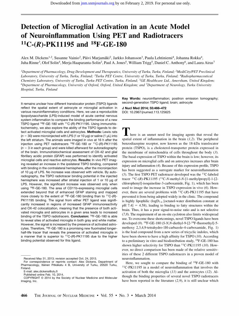
The intrastriatal injection of LPS in set A animals caused wide-spread neuroinflammation in the injected hemisphere and activa-tion of the microglia. This activation was observed by the uptakeof both 11C-(R)-PK11195 and 18F-GE-180 in the images obtainedfrom the digital autoradiography (Fig. 2A). Increased radiotracerbinding was observed in animals injected with either 10 mg of LPS(Fig. 2A, BPex vivo 5 0.76 6 0.31 for 11C-PK11195 and 1.32 60.13 for 18F-GE-180) or 1 mg of LPS (Fig. 2A, BPex vivo 5 0.67 60.30 for 11C-PK11195 and 1.33 6 0.17 for 18F-GE-180) but not inanimals injected with saline (Fig. 2A, BPex vivo 5 0.05 6 0.02 for11C-PK11195 and 0.05 6 0.01 for 18F-GE-180). In the animalsinjected with 10 mg of LPS, a halo of increased binding was ob-served around the injection site, which was not observed in ani-mals injected with 1 mg of LPS.The repeated-measures ANOVA on the binding potentials ob-
tained from the ROI analysis performed on the autoradiographyimages showed that there was a significant main effect of LPStreatment (F2,12 5 16.40, P 5 0.0004). In addition, there was asignificant main effect (F2,12 5 6.45, P 5 0.0260) in which thebinding potential of 18F-GE-180 was greater than that of 11C-(R)-PK11195 (Fig. 2B). When Bonferroni post hoc analysis was used,only the animals injected with LPS at either concentration, andimaged with 18F-GE-180, showed a significant increase in radio-tracer binding in the ipsilateral hemisphere (P , 0.01), comparedwith the saline-injected group (Fig. 2B). To determine the use ofthe contralateral hemisphere as a reference region, we comparedthe BR between the contralateral striatum and the cerebellumbetween the different groups. There was no significant difference(P 5 0.106) between brain regions distal to the LPS injection inthe binding of 11C-(R)-PK11195 (10 mg, BR5 0.796 0.04; 1 mg,BR 5 0.92 6 0.12) when compared with the saline control group(BR 5 0.74 6 0.04) (Fig. 2C).
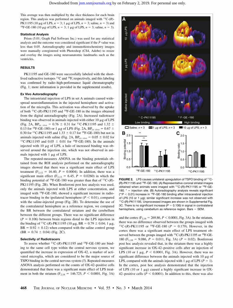
Selectivity of Radiotracers
To assess whether 11C-(R)-PK11195 and 18F-GE-180 are bind-ing to the same cell type within the central nervous system, wequantified the increase in expression of OX-42, a marker of acti-vated microglia, which are considered to be the major source ofTSPO binding in the central nervous system (5). Repeated-measuresANOVA analysis performed on the counts of OX-42–positive cellsdemonstrated that there was a significant main effect of LPS treat-ment in both the striatum (F2,18 5 146.725, P , 0.0001, Fig. 3A)
and the cortex (F2,18 5 269.86, P, 0.0001, Fig. 3A). In the striatum,there was no difference observed between the groups imaged with11C-(R)-PK11195 or 18F-GE-180 (P 5 0.179). However, in thecortex there was a significant main effect of LPS treatment ob-served between the groups imaged with 11C-(R)-PK11195 or 18F-GE-180 (F1,18 5 9.086, P 5 0.011, Fig. 3A) (P 5 0.02). Bonferronipost hoc analysis revealed that, in the striatum there was a highlysignificant increase in OX-42–positive cells after an injection ofLPS (10 or 1 mg, P , 0.0001, Fig. 3A). However, there was nosignificant difference between the animals injected with 10 mg ofLPS, compared with the animals injected with 1 mg of LPS (P5 1).In the cortex, post hoc analysis demonstrated that the injectionof LPS (10 or 1 mg) caused a highly significant increase in OX-42–positive cells (P , 0.0001). In addition to this, there was also
FIGURE 2. LPS causes unilateral upregulation of TSPO binding of 11C-
(R)-PK11195 and 18F-GE-180. (A) Representative coronal striatal images
obtained when animals were imaged with 11C-(R)-PK11195 or 18F-GE-
180. * 5 injection site. (B) Autoradiography analysis reveals significant
(**P , 0.01) increase in 18F-GE-180 binding after intracerebral injection
of LPS (10 or 1 μg); similar significant increase was not observed with11C-(R)-PK11195. Unprocessed images are shown in Supplemental Fig.
3C. There is no significant increase (P 5 0.106) in signal in contralateral
hemisphere, using cerebellum as reference region. Bars 5 SEM.
468 THE JOURNAL OF NUCLEAR MEDICINE • Vol. 55 • No. 3 • March 2014
by on February 2, 2019. For personal use only. jnm.snmjournals.org Downloaded from
a significant increase in OX-42–positive cells after an injection of10 mg of LPS, compared with an injection of 1 mg of LPS (P ,0.0001). After an injection of LPS into the left striatum, there wasalso a statistically significant difference between the number ofOX-42–positive cells in the injected hemisphere, compared withthe contralateral hemisphere (data not shown, P , 0.0001). Invitro blocking studies revealed a reduction in specific binding ofboth 11C-(R)-PK11195 and 18F-GE-180 when either was blockedby the corresponding nonradioactive tracer (Fig. 3B). Analysis ofthe ROI means showed a significant reduction (Student t test, P ,0.0001) in the binding potential when 11C-(R)-PK11195 was blocked
with nonradioactive GE-180 (Fig. 3C). Asimilar significant reduction (Student t test,P , 0.0005) was observed in the bindingpotential when 18F-GE-180 was blockedwith nonradioactive PK11195 (Fig. 3C).To confirm that the administration of tracerhad no impact on the histopathology, wecounted the number of OX-42–positivecells. In the striatum, there was no signif-icant difference in the number of OX-42–positive cells in the animals imaged with18F-GE-180 or 11C-(R)-PK11195. There wasa small increase in OX-42–positive cells inthe cortex in the 11C-(R)-PK11195 group,compared with animals imaged with 18F-GE-180 (Fig. 3A), but the binding poten-tial was decreased in the 11C-(R)-PK11195group, further highlighting the improvedimaging with 18F-GE-180.
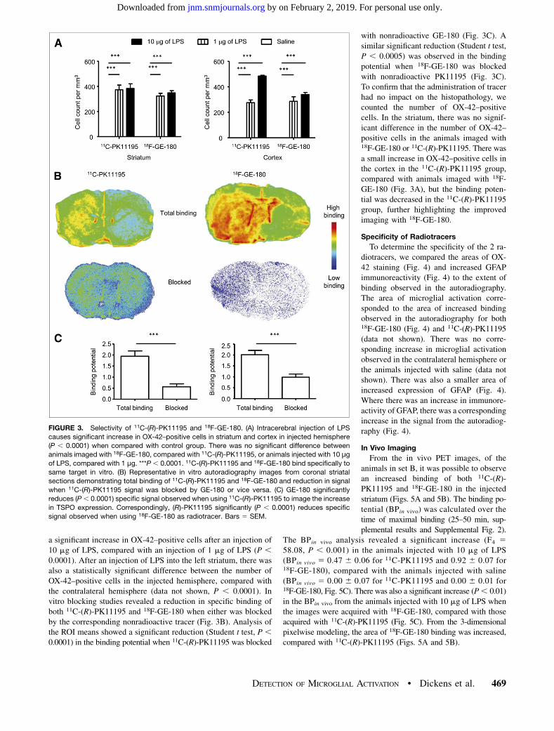
Specificity of Radiotracers
To determine the specificity of the 2 ra-diotracers, we compared the areas of OX-42 staining (Fig. 4) and increased GFAPimmunoreactivity (Fig. 4) to the extent ofbinding observed in the autoradiography.The area of microglial activation corre-sponded to the area of increased bindingobserved in the autoradiography for both18F-GE-180 (Fig. 4) and 11C-(R)-PK11195(data not shown). There was no corre-sponding increase in microglial activationobserved in the contralateral hemisphere orthe animals injected with saline (data notshown). There was also a smaller area ofincreased expression of GFAP (Fig. 4).Where there was an increase in immunore-activity of GFAP, there was a correspondingincrease in the signal from the autoradiog-raphy (Fig. 4).
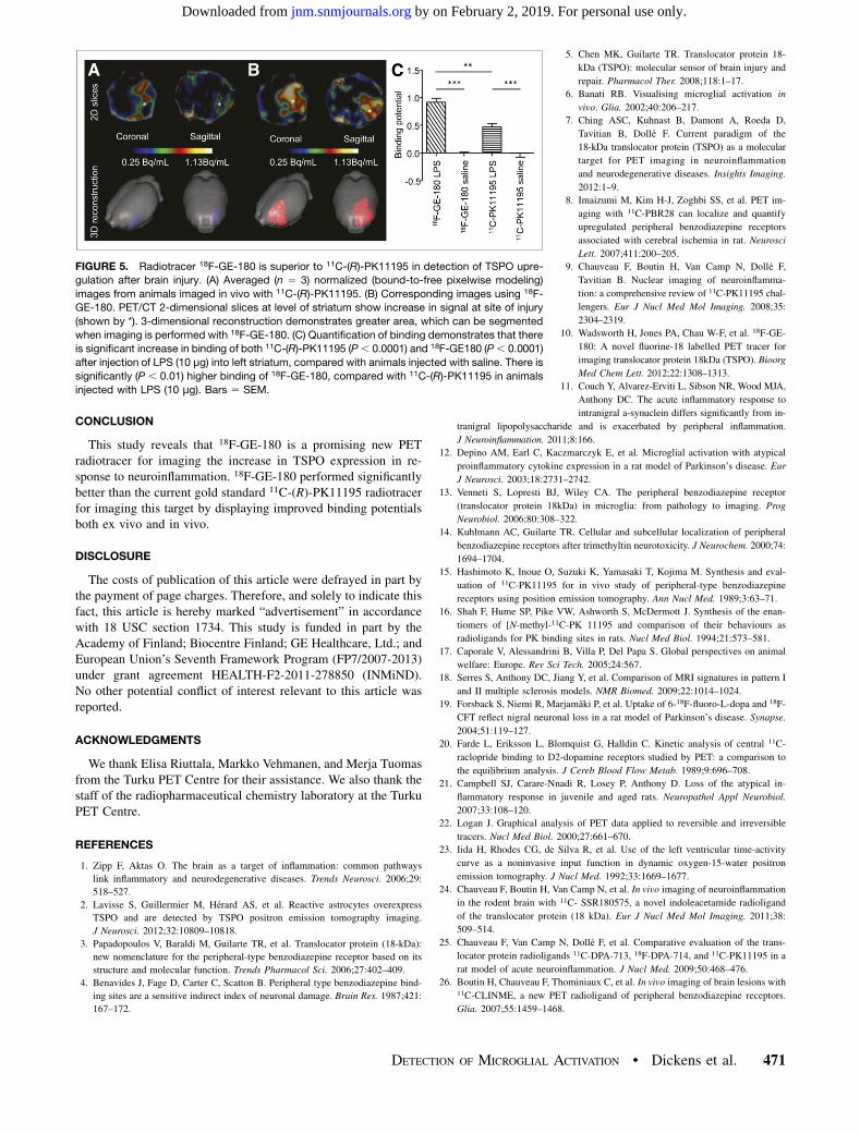
In Vivo Imaging
From the in vivo PET images, of theanimals in set B, it was possible to observean increased binding of both 11C-(R)-PK11195 and 18F-GE-180 in the injectedstriatum (Figs. 5A and 5B). The binding po-tential (BPin vivo) was calculated over thetime of maximal binding (25–50 min, sup-plemental results and Supplemental Fig. 2).
The BPin vivo analysis revealed a significant increase (F4 558.08, P , 0.001) in the animals injected with 10 mg of LPS(BPin vivo 5 0.47 6 0.06 for 11C-PK11195 and 0.92 6 0.07 for18F-GE-180), compared with the animals injected with saline(BPin vivo 5 0.00 6 0.07 for 11C-PK11195 and 0.00 6 0.01 for18F-GE-180, Fig. 5C). There was also a significant increase (P, 0.01)in the BPin vivo from the animals injected with 10 mg of LPS whenthe images were acquired with 18F-GE-180, compared with thoseacquired with 11C-(R)-PK11195 (Fig. 5C). From the 3-dimensionalpixelwise modeling, the area of 18F-GE-180 binding was increased,compared with 11C-(R)-PK11195 (Figs. 5A and 5B).
FIGURE 3. Selectivity of 11C-(R)-PK11195 and 18F-GE-180. (A) Intracerebral injection of LPS
causes significant increase in OX-42–positive cells in striatum and cortex in injected hemisphere
(P , 0.0001) when compared with control group. There was no significant difference between
animals imaged with 18F-GE-180, compared with 11C-(R)-PK11195, or animals injected with 10 μgof LPS, compared with 1 μg. ***P , 0.0001. 11C-(R)-PK11195 and 18F-GE-180 bind specifically to
same target in vitro. (B) Representative in vitro autoradiography images from coronal striatal
sections demonstrating total binding of 11C-(R)-PK11195 and 18F-GE-180 and reduction in signal
when 11C-(R)-PK11195 signal was blocked by GE-180 or vice versa. (C) GE-180 significantly
reduces (P, 0.0001) specific signal observed when using 11C-(R)-PK11195 to image the increase
in TSPO expression. Correspondingly, (R)-PK11195 significantly (P , 0.0001) reduces specific
signal observed when using 18F-GE-180 as radiotracer. Bars 5 SEM.
DETECTION OF MICROGLIAL ACTIVATION • Dickens et al. 469
by on February 2, 2019. For personal use only. jnm.snmjournals.org Downloaded from

DISCUSSION
Data presented here demonstrate for the first time that the novel
radiotracer 18F-GE-180 is superior to 11C-(R)-PK11195 in detect-
ing in vivo glial activation in our rodent model of neuroinflamma-
tion. The superiority of the tracer was apparent both in ex vivo and
in vivo imaging techniques, for which 18F-GE-180 had a statisti-
cally significant higher binding potential when compared with11C-(R)-PK11195. The area of TSPO binding colocalized with
the area of microglial activation after an intrastriatal injection of
LPS. However, the intensity of the TSPO signal increased in the
presence of both activated microglial cells and reactive astrocytes.We used an intrastriatal injection of LPS to induce unilateral
acute inflammation and glial activation within the rodent brain,
which provides a reliable and reproducible inflammatory response
throughout adulthood (21). The main benefit of the model used in
this paper is that it causes a unilateral inflammatory response,
permitting an internal control for the PET modeling and avoiding
the need for serial blood sampling, which would be required if no
reference region was available (22). In addition to this, high bind-
ing of both tracers within the myocardium prevents the use of the
left ventricle as an internal blood input function (23).
Several other models have been used tocompare TSPO ligands (9). The intrastriatalinjection of a-amino-3-hydroxy-5-methyl-4-isoxazolepropionic has been used inseveral studies (24–26) and was used pri-marily to study the effect of excitotoxity onglial activation (27). The pathology is as-sociated with considerable cell death (28),which could be considered a confound, andmaximal activation of the microglia andastrocytes occurs after 7 d (26). The LPSinjection does not cause any significant celldeath (11). LPS, on binding to LPS bindingprotein and CD14, quickly activates theimmune system via toll-like receptor 4 (29)and the NFkB pathway (30) to induce an M1phenotype (31). This model has also beenused in the past to show the different bind-ing characteristics of TSPO ligands (32)and has the advantage over the a-amino-3-hydroxy-5-methyl-4-isoxazolepropionicmodel that the animals can be imaged at amuch earlier time point and it does not in-duce overt clinical signs.We also observed an area of increased
GFAP immunoreactivity after the LPS in-jection. The area of increased immunore-activity of GFAP varied in the model butwas always considerably smaller than thearea of OX-42 activation. It has been sug-gested that it takes longer for astrocytes tobecome fully activated after brain injury,but TSPO ligands do display increased bind-ing to activated astrocytes (33), and to fullyprobe the interaction of astrocytes with theTSPO ligands imaging at a later time point,such as at 3–7 d, might be useful. However,the results presented here suggest that theTSPO signal is the sum total of the in-
crease in TSPO expression in both the microglia and the astrocyteswhere the binding coincides. This does not diminish the use ofTSPO as a surrogate marker of neuroinflammation where the ac-tivation of both populations is usual. However, attributing thesignal to one specific cell type must be done with caution.In addition to the increase in TSPO expression observed in the
presence of both activated microglia and astrocytes, there was bindingof the radiotracers in the ventricular area and the olfactory bulbs, evenin the saline-injected animals, suggesting that the tracer is bindingto further cell types in these areas. This fact has previously beenused to validate TSPO tracers in healthy animals (10). Previously,it has been reported that the TSPO protein is also expressed in theependymal cells (34) and cells within the choroid plexus (9,35).Additionally, the binding in the olfactory bulbs in rodents has pre-viously been observed (36). The causes for this binding remain unclear,with several reports suggesting that neuronal cells in this brain regionexpress TSPO in neo-natal rats (35) and humans (37). It has beensuggested that there is an activation of the microglial cells thoughtto be caused by the replacement of the granular and periglomerularneurons from the subventricular zone (38). It has been shown thatexperimental autoimmune encephalomyelitis enhances this cellularmigration and can be seen by an increase in TSPO binding (39).
FIGURE 4. Specificity studies; histologic comparison to autoradiography results. (Left) Three-
dimensional surface plots demonstrate area of increased signal observed in autoradiography
from animals imaged with 18F-GE-180 and increase in OX-42 and GFAP immunoreactivity in
sequential sections. (Right) Shown are corresponding images or high powered photomicrographs
from injection site obtained from 18F-GE-180 autoradiography, OX-42 (activated microglia), and
GFAP (astrocytes) staining. $ marks increase in autoradiography signal where there is corre-
sponding area of OX-42–positive cells and increased GFAP immunoreactivity. Scale bar5 50 μm.
470 THE JOURNAL OF NUCLEAR MEDICINE • Vol. 55 • No. 3 • March 2014
by on February 2, 2019. For personal use only. jnm.snmjournals.org Downloaded from
CONCLUSION
This study reveals that 18F-GE-180 is a promising new PETradiotracer for imaging the increase in TSPO expression in re-sponse to neuroinflammation. 18F-GE-180 performed significantlybetter than the current gold standard 11C-(R)-PK11195 radiotracerfor imaging this target by displaying improved binding potentialsboth ex vivo and in vivo.
DISCLOSURE
The costs of publication of this article were defrayed in part bythe payment of page charges. Therefore, and solely to indicate thisfact, this article is hereby marked “advertisement” in accordancewith 18 USC section 1734. This study is funded in part by theAcademy of Finland; Biocentre Finland; GE Healthcare, Ltd.; andEuropean Union’s Seventh Framework Program (FP7/2007-2013)under grant agreement HEALTH-F2-2011-278850 (INMiND).No other potential conflict of interest relevant to this article wasreported.
ACKNOWLEDGMENTS
We thank Elisa Riuttala, Markko Vehmanen, and Merja Tuomasfrom the Turku PET Centre for their assistance. We also thank thestaff of the radiopharmaceutical chemistry laboratory at the TurkuPET Centre.
REFERENCES
1. Zipp F, Aktas O. The brain as a target of inflammation: common pathways
link inflammatory and neurodegenerative diseases. Trends Neurosci. 2006;29:
518–527.
2. Lavisse S, Guillermier M, Hérard AS, et al. Reactive astrocytes overexpress
TSPO and are detected by TSPO positron emission tomography imaging.
J Neurosci. 2012;32:10809–10818.
3. Papadopoulos V, Baraldi M, Guilarte TR, et al. Translocator protein (18-kDa):
new nomenclature for the peripheral-type benzodiazepine receptor based on its
structure and molecular function. Trends Pharmacol Sci. 2006;27:402–409.
4. Benavides J, Fage D, Carter C, Scatton B. Peripheral type benzodiazepine bind-
ing sites are a sensitive indirect index of neuronal damage. Brain Res. 1987;421:
167–172.
5. Chen MK, Guilarte TR. Translocator protein 18-
kDa (TSPO): molecular sensor of brain injury and
repair. Pharmacol Ther. 2008;118:1–17.
6. Banati RB. Visualising microglial activation in
vivo. Glia. 2002;40:206–217.
7. Ching ASC, Kuhnast B, Damont A, Roeda D,
Tavitian B, Dollé F. Current paradigm of the
18-kDa translocator protein (TSPO) as a molecular
target for PET imaging in neuroinflammation
and neurodegenerative diseases. Insights Imaging.
2012:1–9.
8. Imaizumi M, Kim H-J, Zoghbi SS, et al. PET im-
aging with 11C-PBR28 can localize and quantify
upregulated peripheral benzodiazepine receptors
associated with cerebral ischemia in rat. Neurosci
Lett. 2007;411:200–205.
9. Chauveau F, Boutin H, Van Camp N, Dollé F,
Tavitian B. Nuclear imaging of neuroinflamma-
tion: a comprehensive review of 11C-PK11195 chal-
lengers. Eur J Nucl Med Mol Imaging. 2008;35:
2304–2319.
10. Wadsworth H, Jones PA, Chau W-F, et al. 18F-GE-
180: A novel fluorine-18 labelled PET tracer for
imaging translocator protein 18kDa (TSPO). Bioorg
Med Chem Lett. 2012;22:1308–1313.
11. Couch Y, Alvarez-Erviti L, Sibson NR, Wood MJA,
Anthony DC. The acute inflammatory response to
intranigral a-synuclein differs significantly from in-
tranigral lipopolysaccharide and is exacerbated by peripheral inflammation.
J Neuroinflammation. 2011;8:166.
12. Depino AM, Earl C, Kaczmarczyk E, et al. Microglial activation with atypical
proinflammatory cytokine expression in a rat model of Parkinson’s disease. Eur
J Neurosci. 2003;18:2731–2742.
13. Venneti S, Lopresti BJ, Wiley CA. The peripheral benzodiazepine receptor
(translocator protein 18kDa) in microglia: from pathology to imaging. Prog
Neurobiol. 2006;80:308–322.
14. Kuhlmann AC, Guilarte TR. Cellular and subcellular localization of peripheral
benzodiazepine receptors after trimethyltin neurotoxicity. J Neurochem. 2000;74:
1694–1704.
15. Hashimoto K, Inoue O, Suzuki K, Yamasaki T, Kojima M. Synthesis and eval-
uation of 11C-PK11195 for in vivo study of peripheral-type benzodiazepine
receptors using position emission tomography. Ann Nucl Med. 1989;3:63–71.
16. Shah F, Hume SP, Pike VW, Ashworth S, McDermott J. Synthesis of the enan-
tiomers of [N-methyl-11C-PK 11195 and comparison of their behaviours as
radioligands for PK binding sites in rats. Nucl Med Biol. 1994;21:573–581.
17. Caporale V, Alessandrini B, Villa P, Del Papa S. Global perspectives on animal
welfare: Europe. Rev Sci Tech. 2005;24:567.
18. Serres S, Anthony DC, Jiang Y, et al. Comparison of MRI signatures in pattern I
and II multiple sclerosis models. NMR Biomed. 2009;22:1014–1024.
19. Forsback S, Niemi R, Marjamäki P, et al. Uptake of 6-18F-fluoro-L-dopa and 18F-
CFT reflect nigral neuronal loss in a rat model of Parkinson’s disease. Synapse.
2004;51:119–127.
20. Farde L, Eriksson L, Blomquist G, Halldin C. Kinetic analysis of central 11C-
raclopride binding to D2-dopamine receptors studied by PET: a comparison to
the equilibrium analysis. J Cereb Blood Flow Metab. 1989;9:696–708.
21. Campbell SJ, Carare-Nnadi R, Losey P, Anthony D. Loss of the atypical in-
flammatory response in juvenile and aged rats. Neuropathol Appl Neurobiol.
2007;33:108–120.
22. Logan J. Graphical analysis of PET data applied to reversible and irreversible
tracers. Nucl Med Biol. 2000;27:661–670.
23. Iida H, Rhodes CG, de Silva R, et al. Use of the left ventricular time-activity
curve as a noninvasive input function in dynamic oxygen-15-water positron
emission tomography. J Nucl Med. 1992;33:1669–1677.
24. Chauveau F, Boutin H, Van Camp N, et al. In vivo imaging of neuroinflammation
in the rodent brain with 11C- SSR180575, a novel indoleacetamide radioligand
of the translocator protein (18 kDa). Eur J Nucl Med Mol Imaging. 2011;38:
509–514.
25. Chauveau F, Van Camp N, Dollé F, et al. Comparative evaluation of the trans-
locator protein radioligands 11C-DPA-713, 18F-DPA-714, and 11C-PK11195 in a
rat model of acute neuroinflammation. J Nucl Med. 2009;50:468–476.
26. Boutin H, Chauveau F, Thominiaux C, et al. In vivo imaging of brain lesions with11C-CLINME, a new PET radioligand of peripheral benzodiazepine receptors.
Glia. 2007;55:1459–1468.
FIGURE 5. Radiotracer 18F-GE-180 is superior to 11C-(R)-PK11195 in detection of TSPO upre-
gulation after brain injury. (A) Averaged (n 5 3) normalized (bound-to-free pixelwise modeling)
images from animals imaged in vivo with 11C-(R)-PK11195. (B) Corresponding images using 18F-
GE-180. PET/CT 2-dimensional slices at level of striatum show increase in signal at site of injury
(shown by *). 3-dimensional reconstruction demonstrates greater area, which can be segmented
when imaging is performed with 18F-GE-180. (C) Quantification of binding demonstrates that there
is significant increase in binding of both 11C-(R)-PK11195 (P, 0.0001) and 18F-GE180 (P, 0.0001)
after injection of LPS (10 μg) into left striatum, compared with animals injected with saline. There is
significantly (P , 0.01) higher binding of 18F-GE-180, compared with 11C-(R)-PK11195 in animals
injected with LPS (10 μg). Bars 5 SEM.
DETECTION OF MICROGLIAL ACTIVATION • Dickens et al. 471
by on February 2, 2019. For personal use only. jnm.snmjournals.org Downloaded from
27. Cuthill DJ, Fowler JH, McCulloch J, Dewar D. Different patterns of axonal
damage after intracerebral injection of malonate or AMPA. Exp Neurol. 2006;
200:509–520.
28. Parfenova H, Tcheranova D, Basuroy S, Fedinec AL, Liu J, Leffler CW. Func-
tional role of astrocyte glutamate receptors and carbon monoxide in cerebral
vasodilation response to glutamate. Am J Physiol Heart Circ Physiol. 2012;302:
H2257–H2266.
29. Bsibsi M, Ravid R, Gveric D, Van Noort JM. Broad expression of toll-like
receptors in the human central nervous system. J Neuropathol Exp Neurol. 2002;
61:1013.
30. Palsson-McDermott EM, O’Neill LAJ. Signal transduction by the lipopolysac-
charide receptor, toll-like receptor-4. Immunology. 2004;113:153–162.
31. Cunningham C. Microglia and neurodegeneration: the role of systemic inflam-
mation. Glia. 2013;61:71–90.
32. Venneti S, Lopresti BJ, Wang G, et al. A comparison of the high-affinity periph-
eral benzodiazepine receptor ligands DAA1106 and (R)-PK11195 in rat models
of neuroinflammation: implications for PET imaging of microglial activation.
J Neurochem. 2007;102:2118–2131.
33. Seneca N. Recent advances in positron emission tomography imaging of brain.
Drugs Future. 2011;36:601.
34. Cosenza-Nashat M, Zhao ML, Suh HS, et al. Expression of the translocator
protein of 18-kDa by microglia, macrophages and astrocytes based on im-
munohistochemical localization in abnormal human brain. Neuropathol Appl
Neurobiol. 2009;35:306–328.
35. Anholt RR, De Souza EB, Oster-Granite ML, Snyder SH. Peripheral-type ben-
zodiazepine receptors: autoradiographic localization in whole-body sections of
neonatal rats. J Pharmacol Exp Ther. 1985;233:517–526.
36. Zhang M-R, Kumata K, Maeda J, et al. 11C-AC-5216: a novel PET ligand for
peripheral benzodiazepine receptors in the primate brain. J Nucl Med. 2007;48:
1853–1861.
37. Rupprecht R, Papadopoulos V, Rammes G, et al. Translocator protein (18-kDa)
(TSPO) as a therapeutic target for neurological and psychiatric disorders. Nat
Rev Drug Discov. 2010;9:971–988.
38. Mattner F, Staykova M, Berghofer P, et al. Central nervous system expression
and PET imaging of the translocator protein in relapsing: remitting experimental
autoimmune encephalomyelitis. J Nucl Med. 2013;54:291–298.
39. Picard-Riera N, Decker L, Delarasse C, et al. Experimental autoimmune enceph-
alomyelitis mobilizes neural progenitors from the subventricular zone to undergo
oligodendrogenesis in adult mice. Proc Natl Acad Sci USA. 2002;99:13211–
13216.
472 THE JOURNAL OF NUCLEAR MEDICINE • Vol. 55 • No. 3 • March 2014
by on February 2, 2019. For personal use only. jnm.snmjournals.org Downloaded from

Doi: 10.2967/jnumed.113.125625Published online: February 10, 2014.
2014;55:466-472.J Nucl Med. Olof Solin, Merja Haaparanta-Solin, Paul A. Jones, William Trigg, Daniel C. Anthony and Laura AirasAlex M. Dickens, Susanne Vainio, Päivi Marjamäki, Jarkko Johansson, Paula Lehtiniemi, Johanna Rokka, Juha Rinne,
F-GE-18018)-PK11195 and RC-(11PET and Radiotracers Detection of Microglial Activation in an Acute Model of Neuroinflammation Using
http://jnm.snmjournals.org/content/55/3/466This article and updated information are available at:
http://jnm.snmjournals.org/site/subscriptions/online.xhtml
Information about subscriptions to JNM can be found at:
http://jnm.snmjournals.org/site/misc/permission.xhtmlInformation about reproducing figures, tables, or other portions of this article can be found online at:
(Print ISSN: 0161-5505, Online ISSN: 2159-662X)1850 Samuel Morse Drive, Reston, VA 20190.SNMMI | Society of Nuclear Medicine and Molecular Imaging
is published monthly.The Journal of Nuclear Medicine
© Copyright 2014 SNMMI; all rights reserved.
by on February 2, 2019. For personal use only. jnm.snmjournals.org Downloaded from