detection of changes in the nuclear phase and evaluation ... · pdf fileintroduction . the...
TRANSCRIPT

Instructions for use
Title Detection of changes in the nuclear phase and evaluation of male germ units by flow cytometry during in vitro pollentube growth in Alstroemeria aurea
Author(s) Hirano, Tomonari; Hoshino, Yoichiro
Citation Journal of Plant Research, 122(2): 225-234
Issue Date 2009-03
Doc URL http://hdl.handle.net/2115/36650
Rights The original publication is available at www.springerllink.com.
Type article (author version)
Additional Information There are other files related to this item in HUSCAP. Check the above URL.
File Information hoshino.pdf
Hokkaido University Collection of Scholarly and Academic Papers : HUSCAP

1
Corresponding author: Yoichiro Hoshino
Address: Field Science Center for Northern Biosphere, Hokkaido University, Kita 11, Nishi
10, Kita–ku, Sapporo 060–0811, Japan
E-mail: [email protected]
Tel/Fax: +81–11–706–2857
Whether the corresponding author is a member or non-member of the Botanical
Society of Japan: non-member
Subject area: (5) Physiology/Biochemistry/Molecular and Cellular Biology
Number of tables, black-and-white figures, and color figures:
Three tables, 4 black-and-white figures, and 1 color figure

2
Title: Detection of changes in the nuclear phase and evaluation of male germ units by flow
cytometry during in vitro pollen tube growth in Alstroemeria aurea.
Authors: Tomonari Hirano1 and Yoichiro Hoshino
1, 2
Affiliations: 1Division of Innovative Research, Creative Research Initiative ‘Sousei’ (CRIS),
Hokkaido University, Kita 21, Nishi 10, Kita–ku, Sapporo 001–0021, Japan
2Field Science Center for Northern Biosphere, Hokkaido University, Kita 11, Nishi 10,
Kita–ku, Sapporo 060–0811, Japan

3
Abstract
This study aimed to analyze male gamete behavior from mature pollen to pollen tube growth
in the bicellular pollen species Alstroemeria aurea. For mature pollen, pollen protoplasts
were examined using flow cytometry. The protoplasts showed two peaks of DNA content at
1C and 1.90C. Flow cytometry of pollen tubes at different developmental stages that were
cultured in vitro revealed changes in the nuclear phase at 9 h and 18 h after culture. Sperm
cell formation occurred at 6–9 h after culture, indicating that the first change was due to the
division of the generative cells into sperm cells. After sperm cell formation, the number of
vegetative nucleus associations with sperm cell showed a tendency to increase. The
association was suggested as male germ unit. When sperm cells, vegetative nuclei, and
partial male germ units were separately collected from pollen tubes cultured for 18 h and
analyzed using a flow cytometer, the sperm cells and vegetative nuclei contained 1C DNA,
while the DNA content of partial male germ units was counted as 2C. Therefore, the second
change in the nuclear phase, which resulted in an increase in 2C nuclei, was possibly related
to the formation of male germ units.

4
Keywords: flow cytometry · generative cell · male germ unit · pollen protoplast · sperm cell
Introduction
The genus Alstroemeria (Alstroemeriaceae) is native to South America and is found from
Venezuela to Chile and Argentina. Alstroemeria is a cut flower of increasing commercial
importance, and many hybrids that produce large and beautiful flowers have been bred by
using interspecific crosses (Hoshino 2008). Cross incompatibility between distantly related
species has been observed (Buitendijk et al. 1995) and is one of the problems limiting further
progress in Alstroemeria breeding.
The in vitro fertilization (IVF) technique, in which isolated male and female gametes are
directly fused under artificial conditions, has been developed for higher plants (Kranz and
Lörz 1993; Kranz et al. 1998; Uchiumi et al. 2007). IVF is considered a useful approach for
overcoming cross incompatibility and additionally has potential as an experimental tool for
the analysis of fertilization processes or early embryogenesis (Sauter et al. 1998; Scholten et

5
al. 2002; Faure et al. 2003; Hoshino et al. 2004; Kranz and Scholten 2008). IVF requires the
isolation of living male and female gametes. At present, the manipulation of female
gametophytes is difficult because these are usually deeply embedded in the ovule tissue. In
Alstroemeria, however, an efficient method for the isolation of living egg cells and zygotes
was recently developed (Hoshino et al. 2006). Thus, an isolation procedure for sperm cells
should be developed in order to perform IVF in Alstroemeria. Sperm cells are formed by
mitosis of generative cells in the pollen or pollen tube. In the latter case, which occurs in
bicellular pollen species, including Alstroemeria, pollen germination and pollen tube
elongation are required for sperm cell isolation, and two methods have been used to isolate
sperm cells: those utilizing in vitro culture of pollen grains (reviewed in Russell, 1991) and
those utilizing a semi-in vivo technique (Shivanna et al. 1988).
Understanding the dynamics of male gametes during pollen tube growth is also
important for IVF and the elucidation of the double fertilization process. In Plumbago
zeylanica L., which has tricellular pollen, two sperm cells are joined by common transverse
cell wall and enclosed by inner vegetative cell membrane, and one of the two sperm cells is

6
associated with vegetative nucleus (Russell 1984). On the other hand, bicellular pollen
species, such as Hippeastrum vitatum Hreb., Rhododendron spp, and Petunia hybrida Vilm.,
generative cell and sperm cell were physically associated with vegetative nucleus (Mogensen
1986; Kaul et al. 1987; Wagner and Mogensen 1988). This association was termed the male
germ unit (MGU) and was proposed to functional unit as a vehicle for transmission and to
participate in fusion with the female target cells during fertilization (Dumas et al. 1984).
Sperm dimorphism, which showed difference of sperm cells in size, shape, and organelle
content, is also observed in the pollen tube (Russell 1991; Tian et al. 2001; Chen et al. 2006).
Moreover, in Arabidopsis thaliana (L.) Heynh, sperm cells are in the S phase of the cell
cycle and continue to synthesize DNA during pollen tube growth. By the time the pollen
tubes reach the ovary and emerge from the septum, the sperm nuclei contain approximately
1.75C DNA (Friedman 1999). Up to now, there is no simple and quick method to evaluate
the general tendency of male gamete behavior such as MGU formation or DNA synthesis of
sperm cell during pollen tube growth. For analysis of the male gamete behavior, we focused
on flow cytometry (FCM) analysis. Since FCM analysis is a very simple procedure that can

7
rapidly measure DNA in large cell populations, it is widely used to estimate the DNA
contents in plant cell nuclei (Doležel and Bartoš 2005). Several ploidy-based studies have
been conducted on pollen by using FCM (reviewed in Suda et al. 2007). FCM was only used
to study germinated gymnosperm pollen of Cupressus dupreziana A. Camus (Pichot and El
Maâtaoui 2000), and no study has used FCM to analyze male gamete during pollen tube
growth.
In the present study, we established an in vitro pollen culture method in a liquid medium
for Alstoemeria aurea Graham toward isolation of sperm cell from pollen tube and analysis
of male gamete. We performed DNA analysis of male gamete during pollen tube growth by
novel methods which combine the germination technique with FCM, and attempted to detect
changes in the nuclear phase during pollen tube growth in A. aurea.
Materials and methods
Plant materials and pollen culture
A. aurea Graham was grown under field conditions in Hokkaido University, and anthers that

8
had undergone dehiscence were collected from the flowers. To overview the procedures for
analysis of pollen grains and pollen tubes, protocols developed in this study were
summarized in Fig. 1. Detailed procedures were described as followings.
Pollen grains from an anther were sown in 2 ml liquid culture medium that contained
0.01% (w/v) CaCl2, 0.01% (w/v) H3BO3, 0.0007% (w/v) KH2PO4, 10% (w/v) sucrose, and
0.01% (w/v) yeast extract at pH 5.8; the culture medium was sterilized by autoclaving at
121°C for 15 min. The pollen grains were then cultured at 25°C under dark condition. After
culture in the medium for 3–18 h, 4′, 6-diamidino-2-phenylindole (DAPI; final concentration,
1 μg ml–1) and Triton X-100 (final concentration, 0.5%) were directly added to the culture
medium. After staining for 15 min, the nuclei in the pollen tubes cultured 3-18 h were
observed through an epifluorescence microscope (Axiovert 200, Carl Zeiss, Oberkochen,
Germany). The nuclei in the pollen tubes were observed for evaluating the rates of sperm
cell formation or MGU formation by following conditions after DAPI staining: vegetative
nucleus and generative cell entered the pollen tube at 3h, and the pollen tubes elongated over
approximately 0.7 mm and 1.5 mm at 6 h and after 9 h culture, respectively.

9
Isolation and FCM analysis of pollen protoplasts
For the isolation of pollen protoplasts, pollen grains from an anther were directly suspended
in 2 ml enzyme solution which contained 2% (w/v) Cellulase Onozuka R-10, 1% (w/v)
Macerozyme R-10, 20 mM morpholineethansulphonic acid, and 1 M mannitol in the
presence of the following salts: KH2PO4 (27.2 mg/l), KNO3 (100 mg/l), CaCl2 (150mg/l),
MgSO4 (250 mg/l), Fe2(SO4)3·6H2O (2.5 mg/l), KI (0.16 mg/l) and CuSO4 (0.00025 mg/l),
pH 5.8 (Frearson et al. 1973). After incubation for 1 h in the enzyme solution at 25°C, the
solution was removed by centrifugation at 400 × g for 3 min. For FCM, the protoplasts were
suspended in 100 μl extraction buffer of CyStain UV precise P (Partec, Münster, Germany),
and then 400 μl staining buffer of CyStain UV precise P was added. The suspension was
filtered through a 30 μm nylon mesh and then analysed using a flow cytometer (Ploidy
Analyzer PA, Partec). Partec FloMax software was used for the analysis of FCM data. 2C
DNA value in A. aurea was determined by leaf, and then used it to estimate the 1C value.
The smallest DNA value of pollen protoplast, which showed half of fluorescence value in

10
leaf 2C peak, was regarded as the 1C DNA value in the FCM histogram. The experiment
was repeated 4 times.
FCM analysis for pollen tubes
The nuclear ploidy level in pollen tubes was determined using a flow cytometer. For
collecting pollen tubes at different developmental stages, nylon meshes were utilized. Based
on the pollen sizes, 65 µm in length and 45 µm in width approximately, the pore sizes of
meshes were selected. For FCM analysis, pollen tubes cultured for 3 h were collected from
the culture medium using a 30 μm mesh. By this filtration, pollen tubes and pollen grains
were recovered and provided for FCM analysis after nucleus extraction and staining
processes as described in next paragraph. In this protocol, nuclei from pollen grains were not
extracted and only nuclei from pollen tubes could be measured. After culture for 6–18 h,
pollen grains and pollen tubes which had burst in the early developmental stage were
removed by filtering the culture medium through a 77 μm mesh, and only elongated pollen
tubes on the mesh were collected.

11
The collected pollen tubes were transfered into 200 μl extraction buffer were chopped
with a sharp razor blade, and 800 μl staining buffer was added. The suspension was filtered
through a 30 μm nylon mesh and immediately analyzed. At least 200 nuclei were counted
for each sample. The experiment was repeated 4 times.
FCM and fluorescence intensity measurement for separately collected cells and nuclei
For detailed FCM analysis, 30 generative cells, 30 sperm cells, 30 vegetative nuclei, 20
paired sperm cells, and 20 MGUs, which were isolated from pollen protoplasts or pollen
tubes in the extraction buffer with staining buffer, were separately collected using a
microcapillary connected to a micropump (Nano Spuit, Ikeda Scientific Co., Ltd, Tokyo,
Japan) under an epifluorescence microscope. Collected cells and nuclei were transferred to
fresh 100 μl extraction buffer, and 400 μl staining buffer was added. The solution was
analyzed using a flow cytometer. The collected cells and nuclei were also observed and
photographed through an epifluorescence microscope with a variable relief contrast
(VAREL, Carl Zeiss, Oberkochen, Germany) function. In order to estimate the fluorescence

12
intensity of nuclei in each cell collected from pollen tubes cultured for 18 h, area and mean
of fluorescence intensity of DAPI-labeled nuclei were measured using ImageJ (The National
Institute of Health, USA), and then total fluorescence of each sample was calculated. A net
fluorescence intensity of nuclei was subtracted the background total fluorescence of same
area near the nucleus from total fluorescence of each nucleus. Fluorescence intensity was
standardized by average of fluorescence intensity of 5 generative nucleus regarded as 200
relative fluorescence intensity.
Statistical analysis
The proportions of nuclear DNA C-values in the pollen tube culture were subjected to
analysis of variance after arcsin transformation, and the means were compared using the
least significant difference test.
Results
When the pollen grains of A. aurea were cultured in liquid medium, approximately 70% of

13
the pollen grains germinated, and pollen tubes elongated until 18 h of culture. The pollen
tubes were used for male gamete analysis by using a procedure developed in this study (Fig.
1).
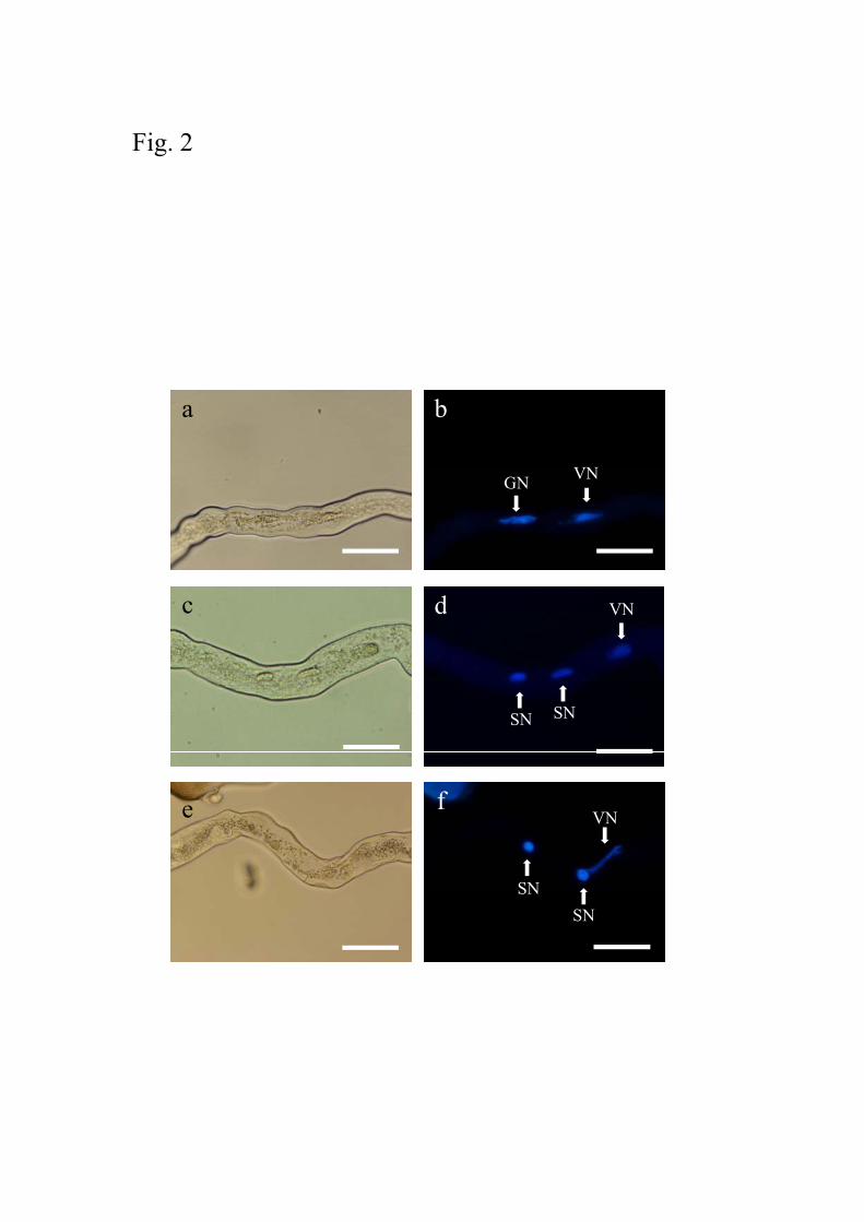
DAPI staining identified a vegetative nucleus and generative cell in a pollen tube
cultured for 6 h (Fig. 2a, b). Sperm cells were observed in pollen tubes cultured for 9 h (Fig.
2c, d). When pollen tubes were stained with DAPI after culture for 18 h, it was observed that
one of the two sperm cells was occasionally associated with vegetative nuclei (Fig. 2e, f).
This suggested that sperm cells and vegetative nuclei formed MGUs in the pollen tubes. For
analyzing the timings and frequencies of generative cell division and MGU formation, sperm
cell formation and proportion of male gametes associated with vegetative nucleus were
counted at each developmental stage. Sperm cells were observed in pollen tubes cultured
after 6 h, and proportion of the pollen tubes contained sperm cells were drastically increased
from 32% at 6 h to 79% at 9 h (Table 1). Vegetative nuclei were associated with generative
cells or sperm cells in the pollen tubes, and the number of vegetative nucleus associations
with sperm cell showed a tendency to increase during pollen culture periods from 12 h to 18

14
h (Table 1).
For analyzing the DNA value of mature pollen, we isolated protoplasts from pollen
grains by enzyme treatment. When pollen protoplasts were transferred to the extraction
buffer for FCM, the pollen protoplasts burst, and the vegetative nuclei and generative cells
were released into the buffer (Fig. 3a, b). FCM analysis of the suspension revealed peaks of
1C and 1.90C nuclei (Fig. 3c). For the confirmation of the peak sources, vegetative nuclei,
which showed filamentous or irregular shape, and generative cells, which had cylindrical
shape nuclei, in the suspension were separately collected with a microcapillary connected to
a micropump. It was observed that the relative DNA content of the separated vegetative
nuclei (Fig. 3d) and generative cells (Fig. 3e) corresponded to the 1C and 1.90C peaks of
pollen protoplasts, respectively. This confirmed that the vegetative nuclei were responsible
for the 1C DNA content, and the generative cells, for the 1.90C DNA content. The DNA
content value of generative cells in pollen grains was regarded as 2C in following pollen tube
analysis.
During in vitro culture of pollen grains of A. aurea, it was observed that the germination

15
of some pollen grains was delayed. Moreover, some pollen tubes burst during the early
developmental stage (data not shown). For the analysis of the male gametes during pollen
tube growth, a protocol for removal of contaminated cells and nuclei from bursting pollen
grains and tubes is required. Therefore, we filtered the tubes during 6–18 h culture through a
77 μm mesh and collected only elongated pollen tubes (Fig. 1). For the evaluation of male
gamete behavior, the filtered pollen tubes at different developmental stages were subjected to
FCM analysis. When pollen tubes cultured for 3 h and 6 h were analyzed using FCM, the
proportion of 1C and 2C nuclei was the same (Table 2). In pollen tubes cultured for 9 h, the
quantity of 1C nuclei increased from 48% to 72%. Further, the proportion of 1C and 2C
nuclei did not change between 9 h and 15 h after culture. Extended culture period from 15 h
to 18 h significantly increased 2C nuclei from 24% to 32% (P = 0.05). The proportion of 3C
nuclei remained almost constant during pollen tube growth.
The cells and nuclei isolated from pollen tubes in extraction buffer by chopping were
observed under an epifluorescence microscope after DAPI staining. The suspension from
pollen tubes cultured 3 h contained vegetative nuclei and generative cells, and it was also

16
observed that a small number of the vegetative nucleus and generative cell formed the MGU
(Fig. 4a, b). In the suspension from pollen tubes cultured 18 h, vegetative nuclei, sperm cells,
undivided generative cells, and paired sperm cells (Fig. 4c, d) were observed. Moreover, two
types of association between sperm cells and vegetative nuclei were observed in the
suspension: those with one of the two sperm cells and one vegetative nucleus (partial MGU;
Fig. 4e, f), and those with two sperms cells and one vegetative nucleus (MGU; Fig. 4g, h).
In order to determine the change in the nuclear phase detected with FCM analysis
between 15 and 18 h of culture, the sperm cells, vegetative nuclei, paired sperm cells, and
partial MGUs from pollen tubes cultured 18 h were separated with a microcapillary
connected to micropunp and then analyzed using FCM. Relative DNA content of the sperm
cells and vegetative nuclei were same as 1C of pollen tubes cultured 18 h (Fig. 5a, b, c),
while the paired sperm cells and partial MGUs showed 2C value (Fig. 5d, e). Relative
fluorescence intensities of the separated cells and nuclei were also measured. When
fluorescence intensity of generative nucleus (2C) was used as DNA standard corresponding
to 200 relative fluorescence intensity, average of the 30 sperm nuclei and 30 vegetative

17
nuclei showed 96.4 ± 13.5 and 84.6 ± 14.9 relative fluorescence intensity, respectively
(Table 3). Relative fluorescence values of the 20 paired sperm cells and 20 partial MGUs
were averaged 190.2 ± 13.5 and 184.0 ± 16.5, respectively (Table 3), and the results are
consistent with prospect value from the sperm nuclei and vegetative nuclei. These results
indicate that 2C nuclei, which were counted in the FCM analysis of pollen tubes grown for
18 h, contained paired sperm cells and partial MGUs.
Discussion
The isolation of nuclei from tissues or organs is indispensable for FCM analysis. Extracting
sufficient numbers of intact nuclei from pollen grains is often difficult, and the effectiveness
of different methods varies from species to species (Suda et al. 2007). Some methods have
successfully used for nuclei isolation of pollen grains, such as chopping method, which was
used for pollen tubes in the present study (Bino et al. 1990; Sugiura et al. 1998; Sugiura et al.
2000; van Tuyl et al. 1989), bursting in hypotonic solution (Zhang et al. 1992), crushing or
squashing (Jacob et al. 2001; Pichot and El Maâtaoui 2000), and sonication (Pan et al. 2004).

18
In the case of A. aurea, it is difficult to isolate nuclei and cells from pollen grains by using
the chopping method (data not shown). We used pollen protoplasts and isolated a sufficient
number of vegetative nuclei and generative cells from A. aurea pollen grains; this indicated
that the new method developed in this study is effective for FCM of pollen.
FCM of generative cells of A. aurea pollen stained with DAPI revealed a DNA content
value of 1.90C (Fig. 3c). Vegetative, generative, and sperm nuclei can be quite different
structurally and morphologically, and as a result, may take up nuclear stains differently (de
Paepe et al. 1990). In Chamerion angustifolium (L.) Holub, 2n generative nuclei show
approximately 1.7 times the fluorescence of 1n vegetative nuclei when stained with
propidium iodide (Suda et al. 2007). Propidium iodide is sensitive to the chromatin structure,
whereas DAPI is less influenced by it (Doležel and Bartoš 2005). Therefore, it is suggested
that nuclear staining in vegetative or generative nuclei of A. aurea with DAPI may be only
slightly affected by the structural or morphological state such as chromatin structure. FCM
of pollen revealed few 3C nuclei (Fig. 3c), and the association of the vegetative nucleus and
generative cell was not observed in suspension (Fig. 3a, b). This suggested that MGUs

19
consisting of 1 vegetative nucleus and 1 generative cell might be not formed in A. aurea
pollen grains and formed after pollen germination.
To investigate male gamete behavior, pollen tubes at different developmental stages
were subjected to FCM analysis, and the chopping method for nuclei isolation was used.
FCM analysis of pollen tubes cultured for 3 and 6 h showed the same proportion of 1C and
2C DNA content in the nuclei (Table 2), suggesting that the 1C content in vegetative nuclei
and the 2C content in generative cells were isolated efficiently from pollen tubes. The
proportion of 3C nuclei was almost constant during pollen tube growth (Table 2). In pollen
tubes, generative cells and sperm cells were associated with vegetative nuclei (Table 1).
Moreover, by using isolation technique for male gametes from pollen tubes, MGUs
consisting of one vegetative nucleus and one generative cell were observed before sperm cell
formation (Fig. 4a, b), and those consisting of two sperm cells and 1 vegetative nucleus were
formed after generative cell mitosis (Fig. 4g, h). This suggested that the 3C nuclei detected
by FCM were derived from MGUs.
The FCM analysis revealed two changes in the nuclear phase between culture periods 6

20
h and 9 h and between culture periods 15 h and 18 h (Table 2). Sperm cells were found to
form in pollen tubes cultured for 6 h, and most of generative cells divided into sperm cells
until 9 h of culture (Fig. 2c, d and Table 1). These results suggest that sperm cell formation
was begun at 6 h approximately and completed by 9 h under the culture conditions used in
the present study. The first change observed at 6–9 h of culture in FCM analysis was a
decrease in 2C nuclei. Further, this period was consistent with the period of sperm formation
observed using DAPI staining of the pollen tubes, indicating that the first change in the
nuclear phase is due to the division of the generative cell (2C) into sperm cells (1C).
After the second change in the nuclear phase, the number of 2C nuclei in pollen tubes
cultured for 18 h was significantly higher than the corresponding number after culture for 15
h. It has reported that sperm cells of A. thaliana are in the S phase of the cell cycle and
continue to synthesize DNA during pollen tube growth (Friedman 1999) and sperm cells in
Nicotiana tabacum L. are in the S phase after deposition in the degenerated synergid (Tian et
al. 2005). Therefore, one possibility of the cause of the second change in the nuclear phase
might be DNA replication in sperm cells. However, sperm cells isolated from pollen tubes

21
cultured for 18 h contained 1C DNA (Fig. 5b) and paired sperm cells were counted as 2C in
FCM analysis (Fig. 5d). Relative fluorescence intensity of sperm nuclei showed about half of
generative nuclei, and that of paired sperm nuclei was also confirmed equivalent value to
generative nuclei (Table 3). These results suggest the possibility that the sperm cells of A.
aurea might not synthesize DNA in the pollen tubes at least when cultured in our condition.
Population of 2C nuclei counted by the FCM analysis of pollen tubes grown for 18 h were
thought to be consisted of undivided generative cells, paired sperm cells, and partial MGUs
(Fig. 5d, e and Table 3). Among them, partial MGUs might be increased at the second
nuclear phase change as following speculations. In N. tabacum, it has been reported that the
vegetative nucleus and generative cell form the MGU in the pollen tube (Yu and Russell
1994a). The MGU association loosens when the generative cell undergoes mitosis and enters
the prophase; the generative cell separates from the vegetative nucleus in the metaphase, and
the MGU is reestablished after sperm cells enter the interphase (Yu and Russell 1994b). In A.
aurea, although proportion of MGU formation in pollen tube is estimated to approximately
same just before and after generative cell mitosis, the association rate between sperm cells

22
and vegetative nuclei in pollen tube increased from 35% at 12 h to 44% at 18 h (Table 1).
These results imply that sperm cells were newly associated with the vegetative nucleus after
12 h culture rather than reestablishment of MGU observed in N. tabacum. As sperm cells
and vegetative nuclei progressively move toward the tip of the pollen tubes as the culture
period increases, the opportunity of MGU formation might continuously increase. In A.
aurea, isolated sperm cells from pollen tubes cultured 18 h contained paired sperm cells (Fig.
4c, d), indicating that two sperm cells in the pollen tube enclosed by inner vegetative cell
membrane and/or connected with each other. If it is assumed that paired sperm cells newly
associated with vegetative nucleus, proportion of 3C nuclei increases between culture
periods 15 h and 18 h. However, proportion of 3C nuclei was not changed in the culture
periods (Table 2). Isolated paired sperm cells generally become spherical shape and loosely
connected in the buffer (reviewed in Russell 1991; Theunis et al. 1991), suggesting that one
sperm cell came off MGU during isolation procedure or in extraction buffer. One possibility
might be that the inner vegetative cell membrane is partly broken but still remains as
enclosing one sperm cell. It is also conjectured that some physiological and/or morphological

23
changes occur in connected sperm cells between culture periods 12 h and 18 h. To clarify the
phenomenon involved in the pollen tubes, we are now attempting to analyze in detail the
sperm cell changes by comparison of gene expression and cytoskeleton.
The MGU is thought to be related to double fertilization (Dumas et al. 1984). Regarding
the MGU of P. zeylanica, sperm cells that were not associated with vegetative nuclei
preferentially fused with egg cells, and sperm cells associated with vegetative nuclei fused
with central cells (Russell 1985). It was also reported that the expression of germ-line
specific polyubiquitin gene differed between sperm cells in the MGUs of P. zeylanica (Singh
et al. 2002). In A. aurea, the second change in the nuclear phase occurred just before pollen
tube growth stopped, suggesting that the change may be related to sperm cell maturation for
the double fertilization process.
In the present study, we developed an in vitro pollen culture system for A. aurea. The
manipulation of pollen tubes for nuclei and cell isolation, DAPI staining, and FCM analysis
is facilitated by this method because of the use of a liquid medium. FCM was very useful for
grasping male gamete behavior in pollen tube because large populations were rapidly

24
analyzed, and some changes which occurred during pollen tube growth, such as sperm cell
formation and MGU formation, were detected. FCM could also be applied to a small number
of targeted sperm cells and MGUs isolated using a microcapillary controlled by a
micropump, and confirmed MGU formation and whether DNA synthesis of sperm cells
occurs or not. These results of the present study indicate that the FCM based method
combined with observation using fluorescence microscopy has potential as a simple and
quick tool for analyzing male gamete behavior during pollen tube growth. The time course
study provides the necessary time frame for successful isolation of sperm cell that could be
potentially used in IVF experiments. Clarifying the reasons of second change in the nuclear
phase, which detected in A. aurea pollen tubes, are expected to lead to successful IVF in
Alstroemeria.
Acknowledgements
We thank Dr. K. Shinoda (National Agricultural Research Center for Hokkaido Region) for
providing plant materials. This work was supported in part by grants from The Akiyama

25
Foundation, The Inamori Foundation, and a Grant-in-Aid for Scientific Research from the
Ministry of Education, Culture, Sports, Science and Technology, Japan.
References
Bino RJ, van Tuyl JM, de Vries JN (1990) Flow cytometric determination of relative nuclear
DNA contents in bicellulate and tricellulate pollen. Ann Bot 65: 3-8
Buitendijk JH, Pinsonneaux N, van Donk AC, Ramanna MS, van Lammeren AAM (1995)
Embryo rescue by half-ovule culture for the production of interspecific hybrids in
Alstroemeria. Scientia Hort 64:65-75
Chen SH, Liao JP, Kuang AX, Tian HQ (2006) Isolation of two populations of sperm cells
from the pollen tube of Torenia fournieri. Plant Cell Rep 25:1138-1142
de Paepe R, Koulou A, Pham JL, Brown SC (1990) Nuclear DNA content and separation of
Nicotiana sylvestris vegetative and generative nuclei at various stages of male
gametogenesis. Plant Sci 70:255-265
Doležel J and Bartoš J (2005) Plant DNA flow cytometry and estimation of nuclear genome

26
size. Ann Bot 95:99-110
Dumas C, Knox RB, McConchie CA, Russell SD (1984) Emerging physiological concepts
in fertilization. What's new in Plant Physiol 15:17-20
Faure JE, Rusche ML, Thomas A, Keim P, Dumas C, Mogensen HL, Rougier M, Chaboud
A (2003) Double fertilization in maize: the two male gametes from a pollen grain have
the ability to fuse with egg cells. Plant J 33:1051-1062
Frearson EM, Power JB, Cocking EC (1973) The isolation, culture and regeneration of
Petunia leaf protoplasts. Dev Biol 33:130-137
Friedman WE (1999) Expression of the cell cycle in sperm of Arabidopsis: implications for
understanding patterns of gametogenesis and fertilization in plants and other eukaryotes.
Development 126:1065-1075
Hoshino Y, Scholten S, von Wiegen P, Lörz H, Kranz E (2004) Fertilization-induced
changes in the microtubular architecture of the maize egg cell and zygote - an
immunocytochemical approach adapted to single cells. Sex Plant Reprod 17:89-95
Hoshino Y, Murata N, Shinoda K (2006) Isolation of individual egg cells and zygotes in

27
Alstroemeria followed by manual selection with a microcapillary-connected micropump.
Ann Bot 97:1139-1144
Hoshino Y (2008) Advances in Alstroemeria biotechnology. In: Teixeira da Silva JA (ed)
Floriculture, Ornamental and Plant Biotechnology: advances and topical issues, vol. 5.
Global Science Book, London, UK, pp 540-547
Jacob Y, Priol V, Ferrero F, Coudret A, Sallanon H (2001) Fluorescent staining of roses
pollen tubes and nuclei by microscopy and flow cytometry analysis. Acta Horticult
547:383-385
Kaul V, Theunis CH, Palser BF, Knox RB, Williams GW (1987) Association of the
generative cell and vegetative nucleus in pollen tubes of Rhododendron. Ann Bot 59:
227-235
Kranz E, Lörz H (1993) In vitro fertilization with isolated, single gametes results in zygotic
embryogenesis and fertile maize plant. Plant Cell 5:739-746
Kranz E, von Wiegen P, Quader H, Lörz H (1998) Endosperm development after fusion of
isolated, single maize sperm and central cells in vitro. Plant Cell 10:511-524

28
Kranz E, Scholten S (2008) In vitro fertilization: analysis of early post-fertilization
development using cytological and molecular techniqus. Sex Plant Reprod 21:67-77
Mogensen HL (1986) Juxtaposition of the generative cell and vegetative nucleus in the
mature pollen grain of Amarylis (Hippeastrum vitatum). Protoplasma 134: 67-72
Pan G, Zhou Y, Fowke LC, Wang H (2004) An efficient method for flow cytometric analysis
of pollen and detection of 2n nuclei in Brassica napus pollen. Plant Cell Rep
23:196-202
Pichot C, El Maâtaoui M (2000) Unreduced diploid nuclei in Cupressus dupreziana A.
Camus pollen. Theor Appl Genet 101:574-579
Russell SD (1985) Preferential fertilization in Plumbago: ultrastructual evidence for
gamete-level recognition in an angiosperm. Proc Natl Acad Sci USA 82:6129-6132
Russell SD (1991) Isolation and characterization of sperm cells in flowering plants. Annu
Rev Plant Physiol Plant Mol Biol 42:189-204
Sauter M, von Wiegen P, Lörz H, Kranz E (1998) Cell cycle regulatory genes from maize
are differentially controlled during fertilization and first embryonic cell division. Sex

29
Plant Reprod 11:41-48
Shivanna KR, Xu H, Taylor P, Knox RB (1988) Isolation of sperm cells from the pollen
tubes of flowering plants during fertilization. Plant Physiol 87:647-650
Scholten S, Lörz H, Kranz E (2002) Paternal mRNA and protein synthesis coincides with
male chromatin decondensation in maize zygotes. Plant J 32:221-231
Singh MB, Xu H, Bhalla PL, Zhang Z, Swoboda I, Russell SD (2002) Developmental
expression of polyubiquitin genes and distribution of ubiquitinated proteins in
generative and sperm cells. Sex Plant Reprod 14:325-329
Suda J, Kron P, Husband BC, Travnicek P (2007) Flow Cytometry and Ploidy: Application
in plant systematic, ecology and evolutionary biology. In: Doležel J, Greilhuber J, Suda
J (eds) Flow cytometry with plant cells. WILEY-VCH Verlag GmbH & Co., KGaA,
Weinheim, pp 103-130
Sugiura A, Tao R, Ohkuma T, Tamura M (1998) Pollen nuclear number in four Diospyros
species. HortScience 33:149-150
Sugiura A, Ohkuma T, Choi YA, Tao R, Tamura M (2000) Production of nonaploid (2n = 9x)

30
Japanese persimmons (Diospyros kaki) by pollination with unreduced (2n = 6x) pollen
and embryo rescue culture. J Am Soc Hort Sci 125:609-614
Theunis CH, PiersonES, Cresti M (1991) Isolation of male and female gametes in higher
plants. Sex Plant Reprod 4: 145-154
Tian HQ, Zhang Z, Russell SD (2001) Sperm dimorphism in Nicotiana tabacum L. Sex
Plant Reprod 14:123-125
Tian HQ, Yuan T, Russell SD (2005) Relationship between fertilization and the cell cycle in
male and female gametes of tobacco. Sex Plant Reprod 17:243-252
Uchiumi T, Uemura I, Okamoto T (2007) Establishment of an in vitro fertilization system in
rice (Oryza sativa L.). Planta 226:581-589
van Tuyl JM, de Vries JN, Bino RJ, Kwakkenbos TAM (1989) Identification of 2n-pollen
producing interspecific hybrids of Lilium using flow cytometry. Cytologia 54:737-745
Wanger VT, Mogensen HL (1988) The male germ unit in the pollen and pollen tubes of
Petunia hybrida: Ultrastructual, quantitative and three-dimensional feature. Protoplasma
143: 101-110

31
Yu H-S, Russell SD (1994a) Populations of plastids and mitochondoria during male
reproductive cell maturation in Nicotiana tabacum L.: A cytological basis for occasional
biparental inheritance. Planta 193: 115-122
Yu H-S, Russell SD (1994b) Male reproductive cell development in Nicotiana tabacum:
male germ unit associations and quantitative cytology during sperm maturation. Sex
Plant Reprod 7:324-332
Zhang G, Campenot MK, McGann LE, Cass DD (1992) Flow cytometric characteristics of
sperm cells isolated from pollen of Zea mays L. Plant Physiol 99:54-59

32
Table 1 Changes of male gametes during pollen tube growth.
culture
periods
(h)
pollen tube containing generative cell
pollen tube containing sperm cells
number of
pollen tubes
number of generative cell
associated with the
vegetative nucleus
number of
pollen tubes
number of sperm cell
associated with the
vegetative nucleus
3 100 (100%)
31 (31.0%)
0 (0%)
0 (0.0%)
6 68 (68%)
25 (36.8%)
32 (32%)
11 (34.4%)
9 21 (21%)
7 (33.3%)
79 (79%)
27 (34.2%)
12 17 (17%)
4 (23.5%)
83 (83%)
29 (34.9%)
15 15 (15%)
6 (40.0%)
85 (85%)
34 (40.0%)
18 11 (11%)
4 (36.4%) 89 (89%) 39 (43.8%)
Pollen tubes were measured 100 individuals at each culture period.

33
Table 2 Frequency distribution of three C-values of nuclear
DNA in pollen tubes during 18 h of culture in liquid medium.
culture
periods (h)
percentage of nuclei
1C 2C 3C
3
51.6 a 46.4 a 2.0 ns
6
48.4 a 46.4 a 5.2 ns
9
71.8 bc 25.9 bc 2.3 ns
12
73.7 b 23.8 b 2.5 ns
15
72.8 b 24.3 b 2.9 ns
18 64.9 c 32.4 c 2.7 ns
Data represent the mean of 4 replicates. Values in each column
followed by the same letter are not significantly different at 0.05 level.
ns: not significant.

34
Table 3 Relative fluorescence intensity of isolated cells and nucli
from pollen tubes cultured 18 h.
relative fluorescence
n intensity
vegetative nucleus 30 84.6 ± 14.9
sperm cell nucleus
30 96.4 ± 13.5
pair of sperm cell nuclei 20 190.2 ± 13.5
partial MGU* 20 184.0 ± 16.5
Data represent mean ± standard deviations of three replicates.
Relative fluorescence intensities were standardized by average of
fluorescence intensity of generative nucleus regarded as 200.
*MGU was consisted of one sperm cell and one vegetative nucleus.

35
Figure legends
Fig. 1 Schematic flow of the method for male gamete analysis developed in this study.
Mature pollen grains were cultured in liquid medium, and elongated pollen tubes at different
developmental stages were used for observation of male gamete nuclei stained with DAPI
and for FCM analysis. For FCM analysis, liquid medium containing pollen tubes was
filtered to remove ungerminated pollen, pollen tubes that burst at an early developmental
stage, and discharged pollen tube contents. The collected pollen tubes in extraction buffer
were chopped with a sharp razor blade, and the relative DNA content of isolated male
gametes and vegetative nuclei was then analyzed using a flow cytometer. For further detailed
study, only targeted cells or nuclei were separated from the suspension by using a
microcapillary connected to micropump, and small-scale FCM analysis was performed on
the separated cells or nuclei.
Fig. 2 DAPI staining of nuclei in pollen tubes of A. aurea. Pollen tubes after culture for 6 h
(a, b), 9 h (c, d), and 18 h (e, f) were observed under bright field (a, c, e) and fluorescence (b,

36
d, f) followed by DAPI staining. GN: generative nucleus, VN: vegetative nucleus, SN: sperm
nucleus. Bars = 50 μm.
Fig. 3 FCM analysis of pollen protoplasts. Generative cell (arrowhead) and vegetative
nucleus (arrow) isolated from pollen protoplasts were stained with DAPI and then observed
by an epifluorescence microscope with a variable relief contrast function (a) and under
fluorescence (b). Histogram obtained after FCM analysis of pollen protoplasts (c). For
detailed study of pollen protoplasts, vegetative nuclei (d) and generative cells (e) were
separately collected from the suspension and analyzed using FCM. Bars: (a, b) 100 μm.
Fig. 4 Isolated MGUs and sperm cells from pollen tubes. MGU consisting of one vegetative
nucleus and one generative cell were isolated from pollen tubes cultured for 3 h (a, b). In
pollen tubes cultured for 18 h, paired sperm cells (c, d), partial MGUs, which consist of one
sperm cell and one vegetative nucleus (e, f), and MGUs consisted of two sperms cells and
one vegetative nucleus (g, h) were isolated. The MGUs and paired sperm cell isolated in

37
extraction buffer were stained with DAPI, and then observed by an epifluorescence
microscope with a variable relief contrast function (a, c, e, g) and under fluorescence (b, d, f,
h). GN: generative cell, VN: vegetative nucleus, SN: sperm nucleus. Bar = 50 μm.
Fig. 5 Flow cytometric analysis of manually separated cells and nuclei from pollen tubes
cultured for 18 h. Histogram obtained by FCM of pollen tubes cultured for 18 h (a). Sperm
cells (b), vegetative nuclei (c), paired sperm cells (d), and partial MGUs consisting of one
sperm cell and one vegetative nucleus (e) were collected using a microcapillary and then
analyzed using a flow cytometer.

Fig. 1
In vitro liquid culture Collection of pollen tubes
Cytological observationIsolation of cells and nuclei
Cytological observationafter DAPI staining
FCM analysisSeparation withmicrocapillary
cFig. 2
b
VN
a
GNVN
d VNc d VN
SNSN
c
SN
VNfe
SN
SN

Fig. 3
ba
mbe
r of
nuc
lei
c d
mbe
r of
nuc
lei
Num
Relative DNA content
Num
Relative DNA content
e
lei
Num
ber
of n
uc
Relative DNA content

Fig. 4
ba
VNGN
SNSN
dc
fe
h
VNSN
g h
VNSN
SNg

Fig. 5
b
mbe
r of
nuc
lei
1C↓
a
mbe
r of
nuc
lei
1C↓
2C↓
Num
Relative DNA content
d
nucl
ei
Num
Relative DNA content
nucl
ei
c
Num
ber
of n
Relative DNA content
2C↓
e
Num
ber
of n
Relative DNA content
1C↓
e
Num
ber
of n
ucle
i
2C↓
N
Relative DNA content