detection of cell death activity during experimentally induced...
TRANSCRIPT

Proceedings 9th International Coral Reef Symposium Bali, Indonesia 23-27 October 2000
Detection of cell death activity during experimentally induced bleaching of the symbiotic sea anemone Aiptasia sp.
S. R. Dunn1, J. C. Thomason, M. D. A. Le Tissier and J. C. Bythell
ABSTRACT
In this study, methods of identifying and detecting different cell death pathway activity were investigated. Using a suite of techniques consisting of haematoxylin and eosin staining of tissue sections, in situ end-labelling (ISEL) of fragmented DNA, electron microscopy and confocal laser microscopy, different forms of cell death activity were indicated during the bleaching of the symbiotic sea anemone Aiptasia sp. in response to a hyperthermic stress. A hyperthermic stress of 33-34oC was used to ensure bleaching, promote cell death and validate the suite of techniques that can then be applied in ongoing studies into bleaching mechanisms. It is the entire combination of these techniques that avoids ambiguity in cell death pathway detection. Cell necrosis was detected in the host endodermis tissues after 4 days of hyperthermic treatment. The in-situ degradation of zooxanthellae characterised by both cell necrosis and programmed cell death in separate cells within the same tissues was detected after 7 days.
1 Department of Marine Sciences and Coastal Management, University of Newcastle upon Tyne, NE1 7RU, UK E-mail: [email protected]
Keywords Bleaching, Cell death, Apoptosis, Program-med cell death, Necrosis
Introduction
Alterations in the concentration of photosynthetic pigments and loss or in situ degradation of zooxanthellae are mechanisms leading to bleaching and often mortality of hermatypic corals. Different environmental factors may act synergistically to promote these changes (see Brown 1997, Hoegh-Guldberg 1999 for reviews). However, there is still little evidence to associate the host or symbiont with the primary failure of the symbiosis in response to environmental stress. The underlying pathways of tissue degradation and cell death during bleaching are unknown. Cell death pathways occur in two main recognised forms, cell necrosis and programmed cell death (PCD). Cell necrosis is an extrinsically mediated process triggered by direct physical injury, large and rapid changes in environmental conditions, pathogenic activity and / or a collapse of the cell integrity (Wyllie et al. 1980, Koss 1992). Cell necrosis may encompass many cells within a tissue area. Conversely PCD is an intrinsically mediated, genetically determined pathway triggered by intracellular signals operating at a single cell level and not affecting a widespread area of tissue. Two examples of possible trigger mechanisms are firstly, the faciclin (FAS / FAS ligand) mammalian lymphocyte membrane cross-linking mechanism (Nagata and Golstein 1995, Shiratsuchi et al. 1998), for which a cnidarian homologous system was recently described in Anthopleura elegantissima (Reynolds et al.2000) and secondly, oxidative stress (Lesser 1997). PCD has a distinct morphology (Fig. 1), also known as apoptosis, which differentiates this form of cell death from cell necrosis (Wyllie et al. 1980, Kerr et al. 1995). PCD is a necessary process in maintaining multi-cellular organism cell populations and tissue homeostasis, tissue morphogenesis, elimination of damaged or infected cells, removal of self-reactive clones, and structures no longer required for their respective function (Wyllie et al. 1980, Raff 1998, Mire and Venable 1999, Vardi et al. 1999). In response to a physiological stress, both cell
necrosis and PCD may be active within a particular tissue area at any one time.
Many of the stages of necrosis and PCD processes can appear similar when taken out of context. It is therefore important to use a broad suite of techniques to detect the different forms of cell death to avoid ambiguous conclusions (Arends and Harrison 1994, Willingham 1999). The aim of this study, using Aiptasia sp. as a model system, was to establish a suite of techniques that detect cell death pathways in host tissues and zooxanthellae during bleaching.
Fig. 1 Schematic diagram of the morphological characteristics associated with the programmed cell death and necrotic cell death (adapted from Kerr et al. 1995).
Materials and methods
Culture conditions and treatments
Cultures of mixed Aiptasia sp. clones were maintained in aquaria containing 80 L of artificial seawater (Tropic Marine salinity 34-35 pH 8.2) at 26-27oC with a 12:12 hrs light/dark regime. Each aquarium was illuminated by a 150w (5200K) metal halide lamp (Hitlite HIT-DE 150dw) providing 400 μmol m-2 sec-1 PAR at the water surface. Anemones were fed once a week with frozen adult Artemia sp. To induce bleaching, Aiptasia sp. were placed in a hyperthermic treatment aquarium at 33-34oC for 7 days. Water condition and temperature were monitored
twice a day. A random sub-sample of anemones was removed after 4 days in order to assess any intermediate stages of induced stress responses. Colchicine has previously been shown to induce PCD in Hydra vulgaris (Cikala et al. 1999), so to induce PCD as a positive control, whole anemones from control conditions were placed in a 10mM colchicine solution in artificial seawater (salinity 34-35 pH 8.2) at 26-27oC for 8 hrs. Following this, anemones were returned control culture conditions for 18 hrs prior to analysis. To assess the extent of bleaching, 5-7 days treated and 5 control Aiptasia sp. were blotted on absorbent tissue paper and fresh weights recorded. Each specimen was longitudinally bisected and the fresh weight recorded for each. One half was frozen at -80oC for later zooxanthellae population analysis whilst the remaining half was analysed for amount of photosynthetic pigments in accordance with Bythell et al. (1997) using established extinction coefficients for chlorophyll a, c2 and total carotenoids (Jeffrey and Humphrey 1975, Parsons et al. 1984). To assess zooxanthellae density, tissue samples thawed on ice were homogenised in 0.5 ml of artificial seawater (pH8) and centrifuged at 10000 g for 10 min at 4oC. The pellets were re-suspended in 0.5 ml of artificial seawater. Zooxanthellae density was measured from 10x50 μl sub-samples from each sample using an Improved Neubauer haemacytometer (Weber, England) and Olympus BH2 binocular microscope. The number of zooxanthellae per mg of tissue sample was calculated. Detecting cell death activity The following techniques were used to detect characteristic features used in distinguishing the two different forms of cell death activity (Table 1). Histology Three individual anemones were removed from each of the treatment and control aquaria and anaesthetised in a 1:1 0.36M MgCl2.6H2O artificial seawater solution for 2 hrs, then fixed in 4% paraformaldahyde / artificial seawater solution (pH 8) and stored at 4oC overnight. After 16 hrs the anemones were removed from the fixative and washed for 30min in each of a graded series (30-100%) ethanol solutions. After rinsing in 100% ethanol (x3) for 30 min anemones were placed in a series of 4 xylene baths for a total of 4.5 hrs, prior to wax embedding. Whole anemone sections were produced using a Microm HM325 microtome and water bath and mounted on 0.01% poly-l-lysine (Sigma) pre-coated glass slides and used in the following histological techniques. Haematoxylin and Eosin (H/E) staining Glass slides with mounted sections were de-waxed in xylene for 10 min then rinsed twice in 100% ethanol for 2 min before staining with haematoxylin and eosin (H/E)
using the protocol of Pearse (1980). Stained sections were rinsed in dH2O and mounted with DPX. The sections were observed and photographed using a Dialux 20 compound light microscope complete with Wild photoautomat MPS 45 system. In Situ End-Labelling (ISEL) of DNA fragmentation The ISEL protocol was adapted from a method applied to mammalian tissue sections (pers. com. G. Collett). Sections were dewaxed in xylene, briefly air-dried, then digested with proteinase K (8μg/ml in double distilled H2O (ddH2O) optimised from a graded series of concentrations) and incubated at 37oC for 30 min. The sections were then rinsed in ddH2O (x3), then 70% ethanol for 2 min and 100% ethanol for 2 min. Prior to incorporation of labelled nucleotides, positive control sections were treated with DNAse and untreated sections were immersed in phosphate buffer solution (PBS). With the positive control treatment completed, all tissue sections, with exception of the negative control sections, were applied with a Klenow/buffer/biotin-dATP solution [Klenow fragment (Gibco), reaction buffer, 2.5mM of dCTP, dGTP, dTTP (Pharmacia), 0.4mM biotin-dATP (Gibco) and ddH2O], and incubated for 30 min at 37oC. Negative controls were treated with the same solution but with the omission of Klenow fragment polymerase /endonuclease. Following 3 rinses in ddH2O, tissue sections were treated with 0.8% H2O2 in PBS for 15 min at room temperature to block any endogenous peroxidase. After PBS rinses (x4), sections were incubated with StreptABC complex (DACO) (consisting of 5μl streptavidin, 5μl of biotinylated horseradish peroxidase and 0.5ml of PBS), at room temperature for 30 min. The sections were rinsed in PBS (x3) before staining with 3,3'-diaminobenzidine (DAB) solution [5% DAB stock solution (Sigma), PBS and H2O2] for 8 min or until the stain was visible. Rinsing sections in ddH2O halted the enzymatic reaction before counter-staining with haematoxylin for between 1 and 2 min. The sections were rinsed in 70% ethanol for 2 min, followed by 100% ethanol for 2 min and then xylene for 2 min. Sections were mounted with DPX, observed and photographed as previously described Electron microscopy Anemones selected at random were removed from treatment and control aquaria and anaesthetised / fixed as above. Aiptasia sp. tissue was embedded with a mixture of 50% hard / 50% medium resin (TAAB Laboratories Ltd) using the protocol of Le Tissier (1990). Sections were produced on a Reichert-Jung Ultracut-E microtome and mounted on filmed copper grids prior to staining with lead nitrate and uranyl acetate. The sections were viewed and photographed with a Phillips CM100 transmission electron microscope (TEM).
Measurement of zooxanthellae size A total of 500 zooxanthellae were counted from a total of 5 tentacles from each of 5 Aiptasia sp. removed from treatment and control conditions. Tentacles were placed in a 250mM acridine orange/ artificial seawater (salinity 34 -35 pH 8) solution for 2 hrs. Samples were squashed on a glass slide under a glass coverslip and observed under a BioRad MRC 600 krypton argon laser confocal microscope with excitation λ of 488, 568 and 647nm used
in conjunction with a Nikon Optiphot 2 compound microscope and BioRad Comos version 7.1 Beta 2 software. Images were later analysed using BioRad comos version 7.1 Beta 2, Confocal Assistant version 4.02 and Image Tool version 2 (Uthscsa) software to establish the maximum diameter of the first 500 individual zooxanthellae randomly selected within tentacles of anemones from each treatment. Data was represented as size class frequency histograms.
Table 1. Distinguishing features associated with programmed cell death (PCD) / apoptosis and necrosis and the techniques used to identify them (adapted from Wyllie et al. 1980, Arends and Harrison 1994, Willingham 1999). (H /E = haematoxylin and eosin stain, ISEL = In situ end-labelling of DNA fragmentation, EM = electron microscopy and CS = cell size)
Feature PCD Cells Necrotic Cells Technique Application H/E ISEL EM CS
Association with adjacent cells
Detachment from adjacent cells through single cell deletion.
Widespread detachment and rupturing of many cells throughout tissue
� � � �
Cell size Reduced cell size / volume
Increased cell size /volume Swelling, dilated / distended morphology Often ruptured
�
� � �
� � �
Cell surface Loss of specialised structures eg. microvilli Cell may exhibit smooth contour, crenated, spiked, blebbed / blistered morphology.
Cell membrane permeable to vital dyes May display blebbing and /or rupturing
� �
� �
Cytoplasm Overall condensation Possible fragmentation into various sizes
Swelling / dilation of cytoplasm often in conjunction with vacuolisation
� �
� �
Organelles Smooth ER may dilate Otherwise all organelles remain compacted / condensed and intact.
Swelling / dilation of organelles. Rupture of organelles
� �
Nucleus and Chromatin
Crenation of nuclear outline. Nucleus may split into various sizes containing specific fragments of DNA via endonuclease activity
Condensation into toroids or crescentric caps at nuclear periphery
Swelling of nucleus. Marginal pyknotic clumping of nuclear chromatin and random DNA fragmentation Possible rupture may release chromatin into cytoplasm / intercellular space
� �
� � �
Cell debris (disposal)
Formation of extracellular round, smooth, membrane bound 'apoptotic bodies', often as cluster at cell periphery ‘Bodies’ may contain cell debris normally within markedly eosinophilic cytoplasm ‘Bodies’shed from cell surfaces and phagocytosed
Rupture of cell membrane Indiscriminate dispersal of cell contents and debris released into inter / extracellular matrix Possible phagocytosis of debris
� � �
� � �
� � �
� �
Results Bleached status of anemones There was a highly significant difference in the concentration of photosynthetic pigments in homogenised Aiptasia sp. from the 7-day hyperthermic treatment and those from control; Chlorophyll a (one-way ANOVA, F = 76.8, P < 0.001), Chlorophyll c2 (F = 61.04, P < 0.001) and Total Carotenoids (F = 43.01, P < 0.001). There was also a highly significant difference (F = 146.84, P < 0.001) in the number of zooxanthellae per mg of homogenised tissue (fresh weight) between the control (mean = 272115 ± 95% CI: 60246, N = 5) and 7-day hyperthermic treatment (mean = 8238 ± 95% CI: 1833, N = 5).
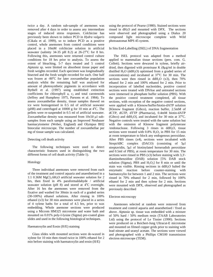
Detection of cell death activity: Histology The 4-day hyperthermic treated anemones (Fig. 2B) showed widespread degradation of endoderm tissue throughout the whole anemone, which was not present in untreated anemones (Fig. 2A) (tentacles shown only). The widespread degradation was characterised by vacuolisation, collapse of the tissue structure and detached degraded tissue in the coelenteron. Zooxanthellae, with a normal morphology, were present in the coelenteron both as isolated cells and as aggregations within degraded endoderm tissue (Fig. 2B). With the 7-day hyperthermic treatment (Fig. 2C) there was increased degradation of the remaining endoderm tissue and zooxanthellae remaining within endoderm
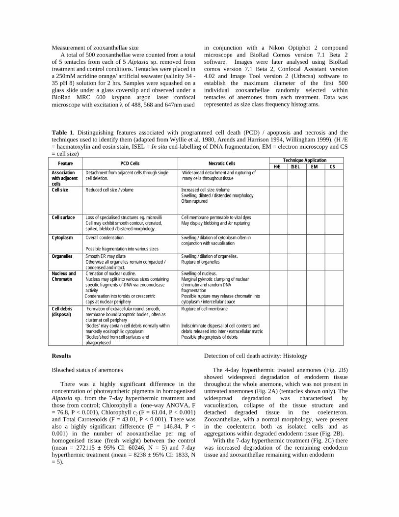
tissue had a degraded morphology. The morphology of zooxanthellae was differentiated from control and 4-day hyperthermic treated samples by the presence of haematoxylin stained peripheral bodies, condensed or dilated cytoplasm, crenated and reduced perimeters or swollen and distended perimeters, which indicated stages of degradation and activity of both forms of cell death (Fig. 2D). An absence of staining of negative control ISEL sections (Fig. 3A) and ubiquitous staining of positive control ISEL sections (Fig. 3B) showed the ISEL DNA fragmentation technique to work for anemone
tissues. ISEL stained cells were present at the periphery of the endoderm of all 3 anemones removed from the 4-day hyperthermic treated tissues (not shown) and throughout the endoderm and ectoderm tissues of the 3 anemones from the 7-day hyperthermic treatment (Fig. 3C). ISEL stained zooxanthellae were only observed within the endoderm from the 7-day hyperthermic treated anemones (Fig. 3C). The peripheral 'bodies' in zooxanthellae from 7-day hyperthermic treated tissue samples, stained by both haematoxylin and ISEL, indicated that they contained fragmented nuclear chromatin.
Fig. 2 Haematoxylin and eosin stained longitudinal sections of tentacle tissue from Aiptasia sp. from control conditions (A), (B) 4-day hyperthermic treatment, (C) 7-day hyperthermic treatment and (D) higher magnification view of 7-day hyperthermic treated tissue. Key: En = endoderm, M = mesoglea, Ec = ectoderm. Arrows = zooxanthellae (scale bars: 15μm).
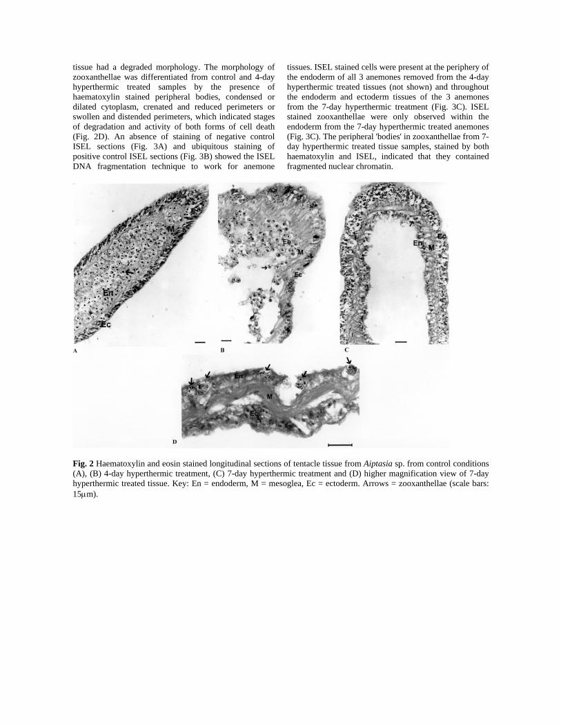
Fig. 3 Longitudinal sections of tentacle tissue showing In Situ End-Labelling (ISEL) of Aiptasia sp. fragmented DNA; (A) negative control (no klenow fragment) (scale bar: 50μm), (B) positive control (DNAse treatment) (scale bar: 50μm) and (C) ISEL stained endoderm and ectoderm cells, in addition to stained zooxanthellae (shown by arrows) tissue from 7-day hyperthermic treatment (scale bar: 20μm). Electron microscopy The morphology of untreated endoderm cells (Fig. 4A) was markedly different to treated samples. In Aiptasia sp. subjected to the colchicine treatment, endoderm cells were reduced in size, condensed in appearance with peripheral blebs on the surface and often membrane-bound bodies would be the only remaining evidence of a cell (Fig. 4B). Endoderm cells of Aiptasia sp. from the 4-day hyperthermic treatment were dilated, swollen and characterised by a ruptured plasma membrane with cell debris released into interstitial spaces. Cells that retained membrane integrity contained ruptured and damaged organelles with a high degree of vacuolation (Fig. 4C). 7-day hyperthermic treated Aiptasia sp. endoderm cells displayed increased cellular degradation with most cells distended. Some were ruptured with little or no cell contents and organelles that were present were often degraded beyond recognition (Fig. 4D).
Zooxanthellae from the 7-day hyperthermic treatment (Fig. 5C and D) displayed a different morphology to zooxanthellae from control conditions (Fig 5A). Some zooxanthellae had morphological characteristics associated with PCD, such as condensation of organelles and cytoplasm, spike or bleb formation at the cell periphery, reduction in cell size and the formation of electron dense, extracellular bodies (Fig. 5C). Whereas some zooxanthellae displayed morphological characteristics associated with cell necrosis, such as a degree of vacuolation, dilation of organelle structure and cytoplasm and cell swelling with or without rupturing (Figs. 5D). Zooxanthellae from Aiptasia sp. subjected to the colchicine treatment had morphological features of PCD only, such as condensation of the cytoplasm and organelles, reduction in cell size and often displayed spikes and blebs at the periphery of the plasma membrane (Fig. 5B).
Fig. 4 Electron micrographs of tentacle endoderm cells (indicated by arrows) and adjacent zooxanthellae of anemones from (A) control culture, (B) colchicine treatment (C) 4-day hyperthermic treatment and (D) 7-day hyperthermic treatment.

Fig. 5 Electron micrographs of zooxanthellae from tentacle tissue sections from Aiptasia sp. subjected to different treatments. (A) control, (B) colchicine and (C and D) 7-Day hyperthermic treatment (morphological characteristics are discussed within results section) Zooxanthellae size Zooxanthellae diameters were segregated into size class frequency histograms (Fig 6A-D). Following significant (P>0.05) Anderson Darling normality tests, the median diameter of the zooxanthellae within tentacles of anemones removed from control, colchicine and hyperthermic treatments were compared using a Kruskal-Wallis test which indicated a highly significant difference (H = 42.22, DF =3, P < 0.001). The size classes were then segregated into subjective 'small' and 'large' size groups on the basis of ± 1 Standard Deviation of the control median (8.46μm ± 0.76). A log-linear G-test used to compare the frequency of small size groups; control and 4-day hyperthermic treatment (Gadj 348.7, P<0.001), control and 7-day hyperthermic treatment (Gadj 284.2, P<0.001), and control and colchicine (Gadj 871.7, P<0.001), and large size groups; control and 4-day hyperthermic treatment (Gadj 91.11, P<0.001), control and 7-day hyperthermic treatment (Gadj 357.4, P<0.001), showed there were highly significant differences. One exception was the large cell size comparison between control and colchicine treatment (Gadj 1.27, P>0.05), which indicated that enlargement of cells did not occur under colchicine treatment. Both enlarged and reduced zooxanthellae sizes were present in tentacles of anemones exposed to hyperthermic treatments. Discussion This study applied a suite of techniques in conjunction with established characteristics (Table 1) to diagnose cell death pathways of zooxanthellae and host endoderm tissues during bleaching of Aiptasia sp. Bleaching was achieved by application of the 7-day hyperthermic treatment and characterised by a highly significant reduction in the concentration of photosynthetic pigments and zooxanthellae population which corresponds to one definition of bleaching (see Brown 1997 and Hoegh-Guldberg 1999 for reviews).
Light microscope analysis of H/E stained sections highlighted widespread degradation of endoderm tissue from the 4-day hyperthermic treatment. The dilated eosin stain within endoderm cells from the 4-day hyperthermic treatment indicated loss of cytoplasm. Reduction in haematoxylin staining within the same cells compared to control, indicated loss of organelle integrity (Stevens and Wilson 1996) which are features associated with cell necrosis. The term ‘bodies’ used within the following text refers to aggregations of cellular material indicative of PCD as present in mammalian apoptosis and should not be confused with the ‘accumulation body’ present within a healthy zooxanthella (Muller-Parker et al. 1996). As haematoxylin has a high affinity with anionic cellular components, such as nuclear chromatin (Stevens and Wilson 1996), the observed prominent haematoxylin stained 'bodies' at the periphery of degraded zooxanthellae remaining within the endoderm tissue from the 7-day hyperthermic treatment most likely contain nuclear chromatin. The presence of stained bodies, often accompanied by an increase in eosin stain indicating condensation of the cytoplasm within the zooxanthellae, are characteristics of a PCD pathway. Within the same tissue sections some zooxanthellae displayed no evidence of 'bodies' and absence of cytoplasm, indicating cell necrosis. This indicates different stages of both cell death pathways were in the observed in situ degradation of zooxanthellae in the H /E stained sections from the 7-day hyperthermic treatment. The difference between controls and treatments indicated that the ISEL technique was successful and that DNA fragmentation occurred during the experimentally induced bleaching of Aiptasia sp. Comparison of ISEL and H /E stained sections of the 4-day hyperthermic treated tissue indicated widespread DNA fragmentation was occurring in the endoderm cells which displayed morphological characteristics associated with cellular necrosis. The peripheral 'bodies' observed in zooxanthellae from 7-day hyperthermic treated tissue samples, containing fragmented nuclear chromatin is a main characteristic of the PCD pathway. Within the PCD

pathway the activation of a cysteine protease or caspase 'cascade' leads to specific endonuclease cleavage of DNA at 180-200 bp intervals (Wyllie 1980, Earnshaw 1995). During necrosis, exonuclease and lysosomal deoxyribonucleases activity may indiscriminately degrade DNA (Ganote et al. 1975, Harmon et al. 1990). When undertaking ISEL analysis it is important to note that positive staining occurs both in cells undergoing PCD and necrosis (Hayashi et al. 1988, Gold et al. 1994, Grasl-Kraupp et al. 1995). To avoid ambiguous conclusions the most beneficial use of ISEL is to observe labelled sections in conjunction with H/E stained sections to associate DNA fragmentation with necrotic and PCD / apoptotic morphology (Gold et al. 1994). Electron micrographs of endoderm cells from Aiptasia sp. subjected to the 4-day hyperthermic treatment displayed morphological characteristics consistent with cellular necrosis (see Table 1) which were accentuated with the additional 3 days of hyperthermic treatment. The use of colchicine, was an invaluable positive control and demonstrated the ability for Aiptasia sp. tissues and zooxanthellae to undergo PCD with the appropriate trigger mechanisms. The morphological characteristics of colchicine-treated endoderm cells were consistent with features associated with PCD (Table 1). However, the contrasting morphology of colchicine treated cells in comparison to hyperthermic treated cells indicated that cell necrosis was the underlying mechanism of host cell degradation during experimentally induced bleaching by hyperthermic stress. Zooxanthellae from the colchicine and 7-day hyperthermic treatments were visibly different to zooxanthellae from the 4-day hyperthermic treatment and control. Colchicine treated zooxanthellae displayed ubiquitous morphological characteristics of degradation associated with PCD (Table 1), and were recently shown in the free living dinoflagellate, Peridinium gatunense in response to reactive oxygen species generation (Vardi et al. 1999). Based on these characteristics the observed difference in morphological characteristics in individual zooxanthellae after the 7-day hyperthermic treatment indicated both cellular necrosis and PCD activity (Table 1). Further evidence of this was provided by the laser confocal microscopy measurement of zooxanthellae diameter from anemones maintained under control and treatment conditions. There was a highly significant increase in the number of smaller zooxanthellae and a reduction in the number of larger zooxanthellae from the colchicine treatment, a result consistent with cell shrinkage as a key feature of PCD. There was a significant increase in the number of both smaller and larger zooxanthellae in the 4-day hyperthermic treatment compared to control conditions indicating that some cells were undergoing cell shrinkage (PCD) whereas others were swelling, a characteristic associated with necrosis. An extended period of hyperthermic stress with the 7-day treatment resulted in further zooxanthellae degradation with significant increases in the frequency of small and large sized zooxanthellae.
A substantial hyperthermic stress (33-34oC) was used to ensure bleaching occurred in Aiptasia sp. in order to establish techniques that would identify cell death pathways involved. A similar temperature regime was also recently used by Jones et al. (1998) to induce bleaching in Stylophora pistillata in order to assess the efficiency of photosynthesis and carboxylation within zooxanthellae during exposure to hyperthermic stress. In this study an intermediate stage (4 days) was required for tissue degradation analysis and to identify key features of the processes involved, since by the completion of the 7-day hyperthermic treatment the density of zooxanthellae was too low to make useful comparisons. Using the suite of techniques developed during this study will enable the detection of cell death pathway activity in bleaching experiments using different temperatures and time scales (work in progress) and can also be used to indicate the roles of PCD and cell necrosis within anemones and corals from the natural environment and during coral bleaching.
Fig. 6. Frequency histograms of zooxanthellae diameter within tentacles of anemones from (A) control untreated conditions, (B) 4-day hyperthermic treatment (C) 7-day hyperthermic treatment and (D) colchicine treatment. Dotted lines indicate +/- 1 S.D of the control median used to segregate between large and small cell size groups. (N = 500 from 5 individual per treatment)

In conclusion, this study established a suite of techniques which offers a combination of evidence necessary to differentiate between the activity of different cell death pathways during experimentally induced bleaching of Aiptasia sp. and indicated cell necrosis of host endoderm cells and both cell necrosis and PCD within individual zooxanthella in response to hyperthermic treatment. Acknowledgements The authors would like to thank the following for their contributions and assistance. Dr M G Bentley, Prof. B E Brown and Dr W J Burnett (Dept of Marine Sciences and Coastal Management, University of Newcastle upon Tyne), Dr G Collett (Dept of Surgery, Royal Victoria Infirmary, Newcastle upon Tyne), Mrs. J Coaker and Dr C A Higgins (Dept of Biological and Nutritional Sciences, University of Newcastle upon Tyne). Dr L Whitehead (University of York), Dr H Hall, B Zimmerman and D McGinnie (Regents Park Zoo, London), Deep Sea World (Edinburgh) and London Aquarium for generously supplying Aiptasia sp. References Arends MJ , Harrison DJ (1994) Apoptosis: molecular
aspects and pathological perspective. In: Crocker J, (ed). Molecular Biology in Histopathology, John Wiley and Sons Ltd, pp 151-170
Brown BE (1997) Coral bleaching: causes and consequences Coral Reefs 16, Suppl: S129-S138
Bythell JC, Douglas AE, Sharp VA, Searle JB , Brown BE (1997) Algal genotype and photoacclimatory responses of the symbiotic alga Symbiodinium in natural populations of the sea anemone Anemonia viridis. Proc R Soc Lond B 264: 1277-1282
Cikala M, Wilm B, Hobmayer E, Böttger A, David CN (1999) Identification of caspases and apoptosis in the simple metazoan Hydra. Curr Biol 9: 959-962
Earnshaw WC (1995) Nuclear changes in apoptosis. Curr Opin Cell Biol 7: 337-343
Ganote CE, Seabra-Gomes R, Nayler WG, and Jennings RB (1975) Irreversible myocardial injury in anoxic perfused rat hearts. Am J Path 80 :419-450
Gold R, Schmied M, Giegerich G, Breitschopf H, Hartung HP, Toyka KV, Lassmann H (1994) Differentiation between cellular apoptosis and necrosis by the combined use of In situ tailing and nick translation techniques. Lab Invest. 71: 219.
Grasl-Kraupp B, Ruttkay-Nedecky B, Koudelka H, Bukowska K, Bursch W, Schulte-Hermann R (1995) In situ detection of fragmented DNA (TUNEL Assay) fails to discriminate among apoptosis, necrosis and autolytic cell death: A cautionary note. Hepatology 21: 1465-1468
Harmon BV, Corder AM, Collins RJ, Gobé GC, Allen J, Allan DJ, Kerr JFR (1990) Cell death induced in a murine mastocytoma by 42-47oC heating in vitro: evidence that the form of death changes from
apoptosis to necrosis above a critical heat load. Int J Rad Biol 58: 845-858
Hayashi R, Ito Y, Matsumoto K, Fujino Y ,Otsuki Y (1998) Quantitative differentiation of both free 3'-OH and 5'-OH DNA ends between heat induced apoptosis and necrosis. J Histochem and Cytochem 46: 1051-1059
Hoegh-Guldberg O (1999) Climate change, coral bleaching and the future of the world's coral reefs. Aust J Mar and Freshwater Res 50: 839-866
Jeffrey SW, Humphrey GF (1975) New Spectrophotometric Equations for Determining Chlorophylls a,b,c
1and c
2 in Higher Plants, Algae and
Natural Phytoplankton. Biochem Physiol Pflanzen 167: 191-194
Jones RJ, Hoegh-Guldberg O, Larkum AWD , Schreiber U (1998) Temperature-induced bleaching of corals begins with impairment of the CO2 fixation mechanism in zooxanthellae. Plant Cell and Environ 21: 1219-1230
Kerr JFR, Gobé GC, Winterford CM, Harmon BV (1995) Anatomical methods in cell death. In: Schwartz LM and Osborne BA (eds) Methods in Cell Biology 46, Academic Press, London, pp 1-27
Koss LG (1992) Diagnostic Cytology and Its Histopathological Bases, JP Lippincott Company, Philadelphia
Lesser MP (1997) Oxidative stress causes coral bleaching during exposure to elevated temperatures. Coral Reefs 16: 187-192
Le Tissier MDA (1990) The ultrastructure of the skeleton and skeletogenic tissues of the temperate coral Caryophyllia smithii. J Mar Biol Ass UK 70: 295-310
Mire P and Venable S (1999) Programmed cell death during longitudinal fission in a sea anemone. Invert Biol 118 (4): 319-331
Muller-Parker G, Lee KW , Cook CB (1996) Changes in the ultrastructure of symbiotic zooxanthellae (Symbiodinium sp., Dinophyceae) in fed and starved sea anemones maintained under high and low light. J Phycol 32: 987-994
Nagata S, Golstein P (1995) The FAS death factor. Science 267: 1449-1456
Parsons TR, Maita Y and Lalli CM (1984) A Manual of Chemical and Biological Methods for Seawater Analysis. Pergamon Press 173 pp
Pearse AGE (1980) Theoretical and Applied Histochemistry, Churchill Livingstone, Edinburgh.
Raff M (1998) Cell suicide for beginners Nature. 396:119-122.
Reynolds WS, Schwarz JA , Weis VM (2000) Symbiosis-enhanced gene expression in cnidarian-algal associations: cloning and characterization of a cDNA, sym 32, encoding a possible cell adhesion protein. Comp Biochem Physiol A 126: 33-44
Shiratsuchi A, Osada S, Kanazawa S, Nakanishi Y (1998) Essential role of phosphatidylserine externalization in apoptosing cell phagocytosis by macrophages. Biochem Biophys Res Comm 246: 549-555.

Stevens A, Wilson I (1996) The haematoxylins and eosin. In: Bancroft JD and Stevens A (eds) Theory and Practice of Histological Techniques, Churchill Livingstone, Edinburgh, pp766
Vardi A, Berman-Frank I, Rozenberg T, Hadas O, Kaplan A and Levine A (1999) Programmed cell death of the dinoflagellate Peridinium gatunense is mediated by CO2 limitation and oxidative stress. Curr Biol 9: 1061-1064
Willingham MC (1999) Cytochemical methods for the detection of apoptosis. J Histochem Cytochem 47: 1101-1109
Wyllie AH (1980) Glucocorticoid-induced thymocyte apoptosis is associated with endogenous endonuclease activation. Nature 284: 555-556
Wyllie AH, Kerr JFK and Currie AR (1980) Cell death: The significance of apoptosis. Int Rev Cytol 68: 251-306