detection of borrelia and ehrlichia in … focus.pdf · rhipicephalus sanguineus, the brown dog...
TRANSCRIPT

DETECTION OF BORRELIA AND EHRLICHIA IN RHIPICEPHALUS SANGUINEUSROSA VASQUEZ-ESPINOZA AND DAVID L. BECK* DEPARTMENT OF BIOLOGY, TENNESSEE TECHNOLOGICAL UNIVERSITY,COOKEVILLE, TN, USA
Copyright 2015, Fine Focus all rights reserved
MANUSCRIPT RECEIVED 30 APRIL, 2015; ACCEPTED 6 JULY, 2015
FinalJournal.indd 109 9/25/15 11:38 AM

Rhipicephalus sanguineus, the brown dog tick, is endemic throughout the world wherever domestic dogs are present. It has been recently reported by some veterinarians in the city of Laredo, Texas, USA, that Lyme disease, the most common tick-borne disease in the Northern United States, is present in local domestic dogs. Fully engorged R. sanguineus ticks were collected and their DNA was purified. The ticks were screened to determine the prevalence of Borrelia, Rickettsia and Ehrlichia species. Sequences related to Borrelia burgdorferi in 9.8% (n=11/112), “Candidatus Borrelia lonestari” in 16.9% (n=19/112) and Ehrlichia canis in 12.5% (n=14/112) were detected by PCR. Sequencing has confirmed the presence of DNA from Ehrlichia canis and “Candidatus B. lonestari”, corroborating that Borrelia and Ehrlichia are present in domestic dogs in South Texas.
ABSTRACTCORRESPONDING AUTHOR
*David L. BeckAssistant Professor of BiologyDepartment of BiologyTennessee Technological UniversityBox 5063Cookeville, TN 38505
Phone – (931) 372-3375Fax – (931) 372-6257Email – [email protected]
KEYWORDS• Borrelia• Ehrlichia • Rhipicephalus sanguineus
• Lyme disease• STARI
Rhipicephalus sanguineus, known as the brown dog tick, is the most widely distributed tick in the world (13). This tick is a known vector of Ehrlichia canis (23), the causative agent of canine ehrlichiosis (1,31). The symptoms of chronic E. canis infection in domestic dogs may include thrombocytopenia, anemia, weight loss, bleeding, fever, inflammation of the eye, and anoxic hepatitis (35). Acute ehrlichiosis in domestic dogs may result in loss of appetite, lethargy, shortness of breath, bruises, joint pain and depression (25). R. sanguineus is also thought to be a vector of Rickettsia rickettsii (13,22), the cause of the Rocky Mountain spotted fever (14). Although R. sanguineus ticks typically prefer to feed on domestic dogs, they have been reported to parasitize humans as well (20,33). The
presence of a disease agent in the domestic dog population can indicate that the disease could also be present in humans (29).
B. burgdorferi sensu stricto (39) has been identified as the sole etiologic agent of Lyme disease in North America (24,38). Lyme disease is the most common vector-borne illness in the Northern United States (8) and is considered an emerging infectious disease (32). The main vectors are Ixodes scapularis (17,18,37) and Ixodes pacificus (34). The agent has also been detected at a lower incidence in R. sanguineus (9,21,42) and Amblyomma inornatum ticks (30). However, the vector potential of these ticks has not yet been characterized. It has been previously reported that B. burgdorferi in domestic dogs may result in arthritis, similar to
INTRODUCTION
110 • FINE FOCUS, VOL. 1 (2)
FinalJournal.indd 110 9/25/15 11:38 AM

humans suffering from Lyme disease (27). Symptoms in domestic dogs from the acute form of Lyme disease may include fever, swelling, pain, lameness, lymphadenopathy and malaise (12). Acute renal failure, myocarditis, cardiac arrhythmia, peripheral edema, neurological syndrome and arthritis have been described as clinical signs found in the chronic form of Lyme disease in domestic dogs (2). A Lyme disease-like illness has also been described in the southern United States since the 1980s (41). This condition is referred to as the Southern Tick Associated Rash Illness (STARI), and is thought to be caused by “Candidatus B. lonestari” (40) after being bitten by Amblyomma americanum (3). The symptoms of “Candidatus B. lonestari” infection in domestic dogs have not been determined.
A veterinarian in Laredo, Texas, USA has reported that Lyme disease is present in local domestic dogs (Dr. Sandra Leyendecker, personal communication). B. burgdorferi has been previously detected in coyotes in Webb County (7), Texas. However, to the extent of our knowledge it has not been detected in domestic dogs. House
pets, including dogs, have an increased exposure to ticks, and can serve as sentinel organisms for some diseases that occur in humans (29). Given the limited information available on vector-borne diseases in South Texas counties, there is a need for tick and pathogen surveillance in the area. This surveillance can help define areas at high risk for transmission (26) of infection to mammals, including humans. Identifying areas at high risk of transmission can increase awareness, potentially leading to the implementation of better diagnosis and prevention methods. In addition, as Laredo is the largest land-based port of entry in the United States, there is the movement of a large number of people and animals to and from this city into the rest of United States (11). For example, truck drivers frequently travel to Laredo, TX with their domestic dogs to warehouses in the city. They may have to wait a day or two before leaving to their destination. The purpose of this study was to determine the prevalence of DNA from tick-borne disease agents in domestic dogs from Laredo, Texas, by investigating the prevalence of Borrelia, Ehrlichia and Rickettsia species in R. sanguineus ticks.
PATHOGENS AND ANTIMICROBIAL FACTORS • 111
TICK COLLECTION AND IDENTIFICATION Fully engorged adult R. sanguineus ticks were collected at multiple sites in Laredo, Texas. The ticks were collected from the walls of dog kennels, or from a CO2 trap placed in the Laredo animal shelter. Ticks that were removed while grooming dogs were also collected from animal caregivers/owners. The researchers had no contact with any animal in the study. The ticks were counted and individually examined
under the microscope to identify them to the species level using a published key (10).
DNA EXTRACTION A total of 124 R. sanguineus ticks (55 males and 69 females) were used for DNA extraction using the E.Z.N.A. Mollusk DNA Isolation Kits (OMEGA Bio-tek, Norcross, GA, USA). A previously reported protocol was followed and modified as previously described (30). Briefly, the tick was homogenized in 300 µl lysis buffer.
MATERIALS AND METHODS
FinalJournal.indd 111 9/25/15 11:38 AM

112 • FINE FOCUS, VOL. 1 (2)
Table 1. Primers and thermal cycler settings used in this study
Primers Amplicon length PCR conditions
Gene Name Sequence (5’ → 3’) Specificity Denaturing Annealing Extension Cycles
12S rRNA 85F TTAAGCTTTTCAGAGGAATTTGCTC 140 bp 95°C, 30sec 45°C, 30sec 72°C, 1min 40
225R TTTWWGCTGCACCTTGACTTAA Not Reported
flaB FlaLS AACAGCTGAAGAGCTTGGAATG 353 bp 95°C, 1min 55°C, 1min 72°C, 1min 36
FlaRS CTTTGATCACTTATCATTCTAATAGC Borrelia genus
BL-Fla522F GGTACATATTCAGATGCAGACAGAGGG 660 bp 95°C, 1min 55°C, 1min 72°C, 1min 46
BL-Fla1182R GCACTTGATTTGCTTGTGCAATCATAGCC “Candidatus B. lonestari”
BL-Fla662F AACTGCTGAAGAGCTTGGAATGC 198 bp 95°C, 1min 55°C, 1min 72°C, 1min 36
dsb BL-Fla860R AGCTGGTTGAACTCCTTCCTGTTGT “Candidatus B. lonestari”
Ehr-DSB-330F GATGATGTCTGAAGATATGAAACAAAT 398 bp 95°C, 1min 55°C, 1min 72°C, 1min 46
16S rRNA Ehr-DSB-728R CTGCTCGTCTATTTTACTTCTTAAAGT Ehrlichia genus
B16S-FL GACTCGTCAAGACTGACGCTAAGTC 131 bp 95°C, 15sec 58°C, 30sec 72°C,30sec 40
B16S-R GCACACTTAACACGTTAGCTTCGGTACTAA Borrelia genus
BL-16S5F CAGTGCGTCTTAAGCATGCAAGTCAGACGG 481 bp 95°C, 1min 60°C, 1min 72°C, 1min 36
BL-16S486R CTGCTGGCACGTAATTAGCCGGGG “Candidatus B. lonestari”
B16S-23S-IGSF GTATGTTTAGTGAGGGGGGTG Variable 94°C, 30sec 56°C, 30sec 74°C, 1min 35
B16S-23S-IGSR GGATCATAGCTCAGGTGGTTAG Borrelia genus
B16S-23S-IGSFn AGGGGGGTGAAGTCGTAACAAG Variable 94°C, 30sec 60°C, 30sec 74°C, 1min 40
B16S-23S-IGSRn GTCTGATAAACCTGAGGTCGGA Borrelia genus
ECAN-F ATTTATAGCCTCTGGCTATAGGA 383 bp 94°C, 30sec 52°C, 30sec 72°C, 1min 36
E. canis
HE1-F CAATTGCTTATAACCTTTTGGTTATAAAT 383 bp 94°C, 30sec 52°C, 30sec 72°C, 1min 36
E. chaffeensis
rompA HE3-R TATAGGTACCGTCATTATCTTCCCTAT Ehrlichia genus 94°C, 30sec 52°C, 30sec 72°C, 1min 36
Rr190 70P ATGGCGAATATTTCTCCAAAA 532 bp 95°C, 1min 55°C, 1min 72°C, 1min 46
Rr190 602N AGTGCAGCATTCGCTCCCCCT Rickettsia genus
The tick was crushed for 5 minutes using a sterile microtube and pestle. After adding proteinase K, the samples were incubated at 55°C for 3h. The sample purification was then completed following the manufacturer’s protocol.
POLYMERASE CHAIN REACTION (PCR) The samples were screened using PCR for the tick 12S rRNA gene as previously described (30). Samples positive for tick rDNA (n=112) were subjected to PCR for amplification of Borrelia, “Candidatus B. lonestari”, Ehrlichia and Rickettsia bacteria species (43,44).
FinalJournal.indd 112 9/25/15 11:38 AM
PATHOGENS AND ANTIMICROBIAL FACTORS • 113
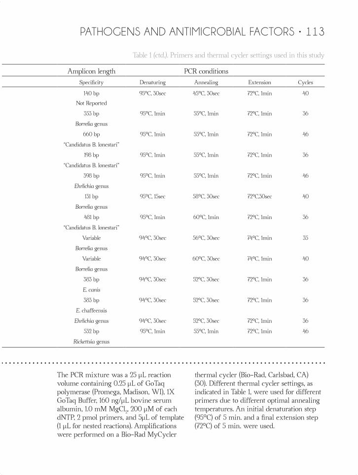
Table 1 (ctd.). Primers and thermal cycler settings used in this study
Primers Amplicon length PCR conditions
Gene Name Sequence (5’ → 3’) Specificity Denaturing Annealing Extension Cycles
12S rRNA 85F TTAAGCTTTTCAGAGGAATTTGCTC 140 bp 95°C, 30sec 45°C, 30sec 72°C, 1min 40
225R TTTWWGCTGCACCTTGACTTAA Not Reported
flaB FlaLS AACAGCTGAAGAGCTTGGAATG 353 bp 95°C, 1min 55°C, 1min 72°C, 1min 36
FlaRS CTTTGATCACTTATCATTCTAATAGC Borrelia genus
BL-Fla522F GGTACATATTCAGATGCAGACAGAGGG 660 bp 95°C, 1min 55°C, 1min 72°C, 1min 46
BL-Fla1182R GCACTTGATTTGCTTGTGCAATCATAGCC “Candidatus B. lonestari”
BL-Fla662F AACTGCTGAAGAGCTTGGAATGC 198 bp 95°C, 1min 55°C, 1min 72°C, 1min 36
dsb BL-Fla860R AGCTGGTTGAACTCCTTCCTGTTGT “Candidatus B. lonestari”
Ehr-DSB-330F GATGATGTCTGAAGATATGAAACAAAT 398 bp 95°C, 1min 55°C, 1min 72°C, 1min 46
16S rRNA Ehr-DSB-728R CTGCTCGTCTATTTTACTTCTTAAAGT Ehrlichia genus
B16S-FL GACTCGTCAAGACTGACGCTAAGTC 131 bp 95°C, 15sec 58°C, 30sec 72°C,30sec 40
B16S-R GCACACTTAACACGTTAGCTTCGGTACTAA Borrelia genus
BL-16S5F CAGTGCGTCTTAAGCATGCAAGTCAGACGG 481 bp 95°C, 1min 60°C, 1min 72°C, 1min 36
BL-16S486R CTGCTGGCACGTAATTAGCCGGGG “Candidatus B. lonestari”
B16S-23S-IGSF GTATGTTTAGTGAGGGGGGTG Variable 94°C, 30sec 56°C, 30sec 74°C, 1min 35
B16S-23S-IGSR GGATCATAGCTCAGGTGGTTAG Borrelia genus
B16S-23S-IGSFn AGGGGGGTGAAGTCGTAACAAG Variable 94°C, 30sec 60°C, 30sec 74°C, 1min 40
B16S-23S-IGSRn GTCTGATAAACCTGAGGTCGGA Borrelia genus
ECAN-F ATTTATAGCCTCTGGCTATAGGA 383 bp 94°C, 30sec 52°C, 30sec 72°C, 1min 36
E. canis
HE1-F CAATTGCTTATAACCTTTTGGTTATAAAT 383 bp 94°C, 30sec 52°C, 30sec 72°C, 1min 36
E. chaffeensis
rompA HE3-R TATAGGTACCGTCATTATCTTCCCTAT Ehrlichia genus 94°C, 30sec 52°C, 30sec 72°C, 1min 36
Rr190 70P ATGGCGAATATTTCTCCAAAA 532 bp 95°C, 1min 55°C, 1min 72°C, 1min 46
Rr190 602N AGTGCAGCATTCGCTCCCCCT Rickettsia genus
The PCR mixture was a 25 µL reaction volume containing 0.25 µL of GoTaq polymerase (Promega, Madison, WI), 1X GoTaq Buffer, 160 ng/µL bovine serum albumin, 1.0 mM MgCl2, 200 µM of each dNTP, 2 pmol primers, and 5µL of template (1 µL for nested reactions). Amplifications were performed on a Bio-Rad MyCycler
thermal cycler (Bio-Rad, Carlsbad, CA) (30). Different thermal cycler settings, as indicated in Table 1, were used for different primers due to different optimal annealing temperatures. An initial denaturation step (95°C) of 5 min. and a final extension step (72°C) of 5 min. were used.
FinalJournal.indd 113 9/25/15 11:38 AM

114 • FINE FOCUS, VOL. 1 (2)
Nested PCR procedures were performed for Borrelia and “Candidatus B. lonestari” using 1µl from the initial reaction as a template. Amplification of target sequences was performed in a Bio-Rad MyCycler thermal cycler (Bio-Rad, Carlsbad, CA) with several denaturing, annealing, and extension times and temperatures (Table 1). For each PCR assay, 5µL of sterile distilled water was used instead of template DNA as a negative control. The positive controls, when used, were added using separate hoods and pipettors to reduce the risk of cross contamination.
VISUALIZATION AND SEQUENCING OF PCR PRODUCTS Five microliters of each PCR reaction was subjected to gel electrophoresis, using 2% agarose gels stained with ethidium bromide in 0.5X TBE (45 mM Tris, 45 mM boric acid, 1 mM disodium ethylene diamine tetraacetic acid) with 0.00005% ethidium bromide. The gels were run at 100 V for 40min.. After electrophoresis, the gels
were examined under UV light. Positive samples were purified using SpinPrep PCR Clean-UP Kits (Novagen, La Jolla CA, USA) following the manufacturer’s protocols. Each purified PCR product was sent for Sanger sequencing at Eurofins (Alabama, USA) or MCLab (San Francisco, CA) using the primers used for PCR.
NUCLEOTIDE SEQUENCE ACCESSION NUMBERS The DNA sequences were visualized using Finch TV, Geospiza, Inc software version 1.5.0 and compared to reported sequences in the NCBI GenBank using BLAST. The assigned GenBank accession numbers are: KR183798-KR183823.
PHYLOGENETIC ANALYSIS Initial alignments for Borrelia and Ehrlichia genes were executed using the MUltiple Sequence Comparison by Log-Expectation (MUSCLE) (16) as performed by the European Bioinformatics Institute’s
Table 2. Detection of bacterial DNA in adult R. sanguineus ticks by PCR
B. burgdorferi-like species
“Candidatus B. lonestari” E. canis Rickettsia
B. burgdorferi-like species
+“Candidatus B. lonestari”
E. canis + B. burgdorferi-like species
Male 12.2% (6/49) 16.3% (8/49) 8.2% (4/49) 0% (0/49) 2.0%(1/49) 4.1% (2/49)
Female 7.9% (5/63) 17.5%(11/63) 15.9% (10/63) 0% (0/63) 1.6%(1/63) 0% (0/63)
Total 9.8% (11/112) 16.9% (19/112) 12.5% (14/112) 0% (0/112) 1.8% (2/112) 1.8% (2/112)
2005 9/85 16/85 11/85 0/85 2/85 2/85
2006 1/7 1/7 0/7 0/7 0/7 0/7
2009 1/11 2/11 1/11 0/11 0/11 0/11
2010 0/4 0/4 1/4 0/4 0/4 0/4
2011 0/5 0/5 1/5 0/5 0/5 0/5
Total 11 of 112 19 of 112 14 of 112 0 of 112 2 of 112 2 of 112
FinalJournal.indd 114 9/25/15 11:38 AM

PATHOGENS AND ANTIMICROBIAL FACTORS • 115
B. turdi D82851
B. garinii KF422823
Borrelia sp. E49Z02-M1
“Candidatus B. lonestari” A25B-M16
“Candidatus B. lonestari” AY442142
B. burgdorferi KF422803
“Candidatus B. lonestari” AY166716
Borrelia sp. A46BB-F1
Borrelia sp. B17Z10-F1
B. tanukii D82848
“Candidatus B. lonestari” A25B-F5
“Candidatus B. lonestari” A32B-F2
Borrelia sp. A25B-M2
Borrelia sp. KM458251
Borrelia sp. A25B-M18
B. parkeri AY604980
Borrelia sp. A46BB-F3
B. miyamotoi JQ926187
“Candidatus B. lonestari” AY237721
“Candidatus B. lonestari” A25B-M11
B. microti JF825472
“Candidatus B. lonestari” A32C-F5
B. burgdorferi CP009656
B. hermsii DQ855534
B. lusitaniae KF836507
B. andersonii D83764
“Candidatus B. lonestari” AY237722
“Candidatus B. lonestari” B17Z09-F2
B. valaisiana JF732880
“Candidatus B. lonestari” AY850063
Borrelia genomosp. 1 KF918619
B. duttonii AB113314
“Candidatus B. californiensis” KF422809
“Candidatus B. lonestari” A25B-M17
B. crocidurae JX292925
Borrelia sp. KM458263
“Candidatus B. lonestari” A35A-M3
B. americana EU081293
B. spielmanii HM802196
B. microti JF708951
B. burgdorferi EU220791
“Candidatus B. californiensis” EU076499
B. turicatae AY604979
“Candidatus B. lonestari” A46BB-F4
B. sinica AB022137
Borrelia sp. KM458264
B. burgdorferi HM345910
“Candidatus B. lonestari” AY237706
B. recurrentis DQ346831
B. japonica D82852
B. americana HM802231
Borrelia genomosp. 2 KF422812
B. anserina DQ849626B. hispanica GU357616
Borrelia sp. A25B-M1
B. afzelii KF422796
Borrelia sp. A25B-M19
50100
99
88
97
100
97
62
82
90
100
100
88
100
100
100
99
92
85
79
100
51
Fig. 1. Baysian inference consensus tree inferred from flaB of Borrelia species. Node support is indicated by the posterior probabilities at the node. The name of the species is followed by the GenBank accession number. A25B-F5, A25B-M1, A25B-M2, A25B-M11, A25B-M16, A25B-M17, A25B-M18, A25B-M19, A32B-F2, A32C-F5, A35A-M3, A46BB-F1, A46BB-F3, A46BB-F4, B17Z09-F2, B17Z10-F1 and E49Z02-M1 represent amplicons from R. sanguineus and are underlined. The scale bar indicates the mean number of nucleotide substitutions per site.
FinalJournal.indd 115 9/25/15 11:38 AM

116 • FINE FOCUS, VOL. 1 (2)
MUSCLE server (http://www.ebi.ac.uk/Tools/muscle/). Default settings were used, with posterior manual adjustments if needed. Bayesian inference phylogenetic analyses were performed using MrBayes v.3.2.5 (36) using two runs, for 10,000,000 generations each, using eight chains and a temperature coefficient of 0.1, and trees sampled every 5,000 generations. Determination of the appropriate model for
each genus was completed via jModelTest 2 (15): GTR + I + Ґ for Borrelia and Ehrlichia. The gamma distribution included six categories for all models obtained. After analysis was completed, the first 25% of trees from each run were discarded as burnin. The consensus trees were observed in FigTree v1.4.2 (http://tree.bio.ed.ac.uk/software/figtree/).
E. species AB024928
E. canis A17S-M1
E. canis A17S-F5
E. ruminantium AF069758
E. canis A17S-F7
E. muris U15527
E. canis KC479022 Spain
E. canis A25B-F22
E. species GU075696
E. canis KP844663 Mexico
E. canis A17S-F3
E. canis KJ659037 China
E. chaffeensis U60476
E. canis A25B-F16
E. species AB196303
E. canis A17S-F1
E. species AF497581
E. canis A25B-F19
“Candidatus Ehrlichia shimanensis”AB074459
E. canis KJ995841 Brazil
E. species AB028319
E. species DQ324547
E. ewingii M73227
E. canis U54805 South Africa
72
100
100
99
93
Fig. 2. Baysian inference consensus tree inferred from 16S rDNA of Erhlichia species. Node support is indicated by the posterior probabilities at the node. The name of the species is followed by the GenBank accession number. A17S-M1, A17S-F1, A17S-F3, A17S-F5, A17S-F7, A25B-F16, A25B-F19 and A25B-F22 represent amplicons from R. sanguineus and are underlined. The scale bar indicates the mean number of nucleotide substitutions per site.
FinalJournal.indd 116 9/25/15 11:38 AM

PATHOGENS AND ANTIMICROBIAL FACTORS • 117
RESULTSPCR DETECTION OF BORRELIA AND EHRLICHIA Of the 112 positive samples for tick rDNA, 44 ticks (18 males and 26 females) were positive for B. burgdorferi-like species, “Candidatus B. lonestari” or Ehrlichia bacteria species (Table 2). “Candidatus B. lonestari”, the most commonly detected tick-borne pathogen, was detected in 16.9% (n=19, 16.3% of males (8/49) and 17.5% (11/63) of females) of all ticks.
B. burgdorferi-like species were detected in 9.8% (n=11, 12.2% (6/49) of males and 7.9% (5/63) of females) of all ticks. E. canis was detected in 12.5% (n= 14, 8.2% (4/49) of males and 15.9% (10/63) of females) of all samples. Two male ticks (4.1%, n=2 of 49) were positive for both E. canis and B. burgdorferi-like species, indicating a 1.8% co-infection rate (2 of 112) of all ticks. One male and one female tick (male: 2.0%, n=1 of 49; female: 1.6%, n=1 of 63) were positive for both “Candidatus B. lonestari” and B. burgdorferi-like species, indicating a 1.8% co-infection
rate (2 of 112) of all ticks. No tick was positive for the spotted fever group rickettsial rompA gene.
PHYLOGENETIC ANALYSIS Samples with strong bands by gel electrophoresis were prepared for sequencing. Sequencing confirmed the PCR results. A Bayesian inference tree including these sequences and other published sequences are shown in Figs. 1 and 2. Phylogenetic analysis showed that “Candidatus B. lonestari” amplicons were in the same clade as known sequences of “Candidatus B. lonestari” and B. burgdorferi-like species amplicons clustered with B. burgdorferi species complex (Fig. 1). “Candidatus B. lonestari” sequences were all identical except A25B-F5, which was polymorphic at one position. Three of the B. burgdorferi-like species sequences (B17Z10-F1, A25B-M1 and E49Z02-M1) were different at one position. Ehrlichia amplicons were in the same clade as known sequences of E. canis (Fig. 2).
DISCUSSIONThe ticks used for this study were fully engorged. Thus, the detection of pathogen DNA either represents the most recent blood meal or potentially a prior infection of the tick. We detected the presence of B. burgdorferi-like species, “Candidatus B. lonestari” and E. canis DNA in R. sanguineus ticks from Laredo, Texas. Our study does not address the issue of vector competency of R. sanguineus ticks in regard to B. burgdorferi-like species and “Candidatus B. lonestari”.
The detection of pathogens in canines can indicate a potential risk for infection
of humans (28,29). We have detected B. burgdorferi-like species DNA in 9.8% of ticks collected from the local animal shelter, as well as ticks submitted by pet caregivers. The main vector for B. burgdorferi in the Northeastern United States is I. scapularis (17,18,37). This tick is present throughout much of Texas and Northern Mexico. B. burgdorferi was previously detected in 45% of tested I. scapularis ticks (19). However, in Webb County TX, no I. scapularis were identified in the combined collection of over 70,000 ticks (5). B. burgdorferi-like
FinalJournal.indd 117 9/25/15 11:38 AM

118 • FINE FOCUS, VOL. 1 (2)
species have been previously reported in A. inornatum from Webb County (30) and A. mixtum from Northeastern Mexico (21). We also detected “Candidatus B. lonestari”, which is thought to be the cause of STARI (40), in 16.9% of ticks. Cohen et al. (1990) reported a 5.5% seroprevalence for Borrelia in domestic dogs from Texas. However, of the dogs in their study 0 of 5 dogs that came from Webb County were seropositive (9). Likewise, Bowman et al. (2009) reported B. burgdorferi in Central and Northern Texas, but did not have any results for South Texas. This is the first report of Borrelia from R. sanguineus ticks and domestic dogs in Webb County, Texas.
R. sanguineus is the only known vector of E. canis (23), and is widely distributed throughout the United States (6). We detected E. canis in 12.5% of ticks. However, all of the ticks that were positive for E. canis were collected from the Laredo animal shelter. Many pet owners acquire their pet from the animal shelter. These dogs are at a high risk for acquiring E. canis at this location. However, domestic dogs in the city of Laredo appear to be at a low risk for
acquiring E. canis. The previously reported seroprevalence for E. canis (2.0%) was higher in Texas than in much of the United States (4). We did not detect E. chaffeensis and E. ewingii in R. sanguineus, even though they are present in the area (30). We also did not detect any spotted fever group Rickettsia. This would suggest that any spotted fever group Rickettsia are either absent from the area or were present at a very low prevalence.
Further research on R. sanguineus distribution and the prevalence of B. burgdorferi-like species, “Candidatus B. lonestari” and Ehrlichia species in South Texas is needed. Further research will help elucidate if R. sanguineus is a vector of a B. burgdorferi-like species. This additional research would allow for better and more accurate diagnosis of tick-borne illnesses, ultimately leading to better treatment and health care for domestic dogs and humans. This research supports the observation of Dr. Sandra Leyendecker and suggests that domestic dogs should be screened for Lyme disease if they present with appropriate symptoms.
Rosa Vasquez-Espinoza was supported by the URECA! Grants Program. David L. Beck was funded for this project by faculty development funds. We thank G.T. Pugh and M. Weems for technical assistance.
ACKNOWLEDGEMENTS
REFERENCES1. Aguiar, D. M., Cavalcante, G. T., Pinter, A., Gennari,
S. M., Camargo, L. M. A., & Labruna, M. B. 2007. Prevalence of Ehrlichia canis (Rickettsiales: Anaplasmataceae) in dogs and Rhipicephalus sanguineus (Acari: Ixodidae) ticks from Brazil. J. Med. Entomol. 44: 126-132. doi:10.1093/jmedent/41.5.126
2. Appel, M. J., Allan, S., Jacobson, R. H., Lauderdale, T. L., Chang, Y. F., Shin, S. J., Thomford, J. W., Todhunter, R. J., & Summers, B. A. 1993. Experimental Lyme disease in dogs produces arthritis and persistent infection. J. Infect. Dis. 167: 651-654.
FinalJournal.indd 118 9/25/15 11:38 AM

PATHOGENS AND ANTIMICROBIAL FACTORS • 119
3. Bacon, R. M., Gilmore, R. D., Quintana, M., Piesman, J., & Johnson, B. J. 2003. DNA evidence of Borrelia lonestari in Amblyomma americanum (Acari: Ixodidae) in southeast Missouri. J. Med. Entomol. 40: 590-592. doi:10.1603/0022-2585-40.4.590
4. Beall, M. J., Alleman, A. R., Breitschwerdt, E. B., Cohn, L. A., Couto, C. G., Dryden, M. W., Guptill, L.C., Iazbik, C., Kania, S. A., Lathan, P., Little, S. E., Roy, A., Sayler, K. A., Stillman, B. A., Welles, E. G., Wolfson, W., & Yabsley, M. J. 2012. Seroprevalence of Ehrlichia canis, Ehrlichia chaffeensis and Ehrlichia ewingii in dogs in North America. Parasit. Vectors. 5: 29.
5. Beck, D.L., Zavala, J., Montalvo, E.O., & Quintana, F.G. 2011. Meteorological Indicators for Amblyomma cajennense Population Dynamics in the Tamaulipan Biotic Province in Texas. J. Vector Ecol. 36: 135-146.
6. Bowman, D., Little, S. E., Lorentzen, L., Shields, J., Sullivan, M. P., & Carlin, E. P. 2009. Prevalence and geographic distribution of Dirofilaria immitis, Borrelia burgdorferi, Ehrlichia canis, and Anaplasma phagocytophilum in dogs in the United States: results of a national clinic-based serologic survey. Vet. Parasitol. 160: 138-148.
7. Burgess, E. C., & Windberg, L. A. 1989. Borrelia sp. infection in coyotes, black-tailed jack rabbits and desert cottontails in southern Texas. J. Wildlife Dis. 25: 47-51.
8. Centers for Disease Control and Prevention. 2014. Summary of notifiable diseases, United States, 2012. MMWR Morb. Mortal. Wkly. Rep., 61 (No. 53): 25.
9. Cohen, N. D., Carter, C. N., Thomas Jr, M. A., Angulo, A. B., & Eugster, A. K. 1990. Clinical and epizootiologic characteristics of dogs seropositive for Borrelia burgdorferi in Texas: 110 cases (1988). J. Am. Vet. Med. Assoc. 197: 893-898. PMID:2228777
10. Cooley, R.A. 1946. The genera Boophilus, Rhipicephalus and Haemaphysalis (Ixodidae) of the New World. Natl. Inst. Health Bull. 187:1-54.
11. Curtis, J. R. 1993. Central Business Districts of The Two Laredos. Geographical Review. 83: 54-65
12. Dambach, D. M., Smith, C. A., Lewis, R. M., & Van Winkle, T. J. 1997. Morphologic, immunohistochemical, and ultrastructural characterization of a distinctive renal lesion in dogs putatively associated with Borrelia burgdorferi infection: 49 cases (1987-1992). Vet. Pathol. Online. 34: 85-96.
13. Dantas-Torres, F. 2010. Biology and ecology of the brown dog tick, Rhipicephalus sanguineus. Parasit.
Vectors. 3:26. doi:10.1186/1756-3305-3-26
14. Dantas-Torres, F., Chomel, B. B., & Otranto, D. 2012. Ticks and tick-borne diseases: a One Health perspective. Trends Parasitol. 28: 437-446. doi:10.1016/j.pt.2012.07.003
15. Darriba, D., Taboada, G.L., Doalla, R. & Posada, D. 2012. jModelTest 2: more models, new heuristics and parallel computing. Nat. Methods. 9: 772.
16. Edgar, R.C. 2004. MUSCLE: multiple sequence alignment with high accuracy and high throughput. Nucleic Acids Res. 32: 1792-1797
17. Falco, R. C. & Fish, D. 1988. Ticks Parasitizing Humans in a Lyme Disease Endemic Area of Southern New York State. Am. J. Epidemiol. 128: 1146-1152.
18. Felz, M. W., Durden, L. A., & Oliver Jr, J. H. 1996. Ticks Parasitizing Humans in Georgia and South Carolina. J. Parasitol. 505-508.
19. Feria-Arroyo, T. P., Castro-Arellano, I., Gordillo-Perez, G., Cavazos, A. L., Vargas-Sandoval, M., Grover, A., Torres, J., Medina, R., Perez de León, A. A. & Esteve-Gassent, M. D. 2014. Implications of climate change on the distribution of the tick vector Ixodes scapularis and risk for Lyme disease in the Texas-Mexico transboundary region. Parasit. Vectors. 7:1, 199.
20. Goddard, J. 1989. Focus of Human Parasitism by the Brown Dog Tick, Rhipicephalus sanguineus (Acari: Ixodidae). J. Med. Entomol. 26: 628-631. doi:10.1093/jmedent/26.6.628
21. Gordillo-Pérez, G., Vargas, M., Solórzano-Santos, F., Rivera, A., Polaco, O. J., Alvarado, L. & Torres, J. 2009. Demonstration of Borrelia burgdorferi sensu stricto infection in ticks from the northeast of Mexico. Clin. Microbiol. Infect. 15: 496-498. doi:10.1111/j.1469-0691.2009.02776.x
22. Gray, J., Dantas-Torres, F., Estrada-Peña, A., & Levin, M. 2013. Systematics and ecology of the brown dog tick, Rhipicephalus sanguineus. Ticks Tick-Borne Dis. 4: 171-180. doi:10.1016/j.ttbdis.2012.12.003
23. Groves, M. G., Dennis, G. L., Amyx, H. L., & Huxsoll, D. L. 1975. Transmission of Ehrlichia canis to dogs by ticks (Rhipicephalus sanguineus). Am. J. Vet. Res. 36:7, 937-940. PMID:1147359
24. Heymann, W. R., & Ellis, D. L. 2012. Borrelia burgdorferi infections in the United States. J. Clin. Aesthetic Derm. 5: 18-28. PMCID: PMC3424593
FinalJournal.indd 119 9/25/15 11:38 AM

120 • FINE FOCUS, VOL. 1 (2)
25. Hildebrandt, P. K., Huxsoll, D. L., Walker, J. S., Nims, R. M., Taylor, R., & Andrews, M. 1973. Pathology of canine ehrlichiosis (tropical canine pancytopenia). Am. J. Vet. Res. 34: 1309-1320.
26. Jongejan, F., & Uilenberg, G. 2004. The Global Importance of Ticks. Parasitology. 129: S3-S14. doi:10.1017/S0031182004005967
27. Kornblatt, A. N., Urband, P. H., & Steere, A. C. 1985. Arthritis caused by Borrelia burgdorferi in dogs. J. Am. Vet. Med. Assoc. 186: 960-964.
28. Lemon, S. M., Sparling, P. F., Hamburg, M. A., Relman, D. A., Choffnes, E. R., Mack, A., & Sparling, F. 2008. Vector-borne diseases: understanding the environmental, human health, and ecological connections. Workshop summary. National Academies Press. ISBN:0-309-10897-7
29. Mead P., Goel R. & Kugeler K. 2011. Canine Serology as Adjunct to Human Lyme Disease Surveillance. Emerg Infect Dis. 17: 1710-1712. doi: http://dx.doi.org/10.3201/1709.110210
30. Medlin, J.S., Cohen, J.I. & Beck, D.L. 2015. Vector potential and population dynamics for Amblyomma inornatum. Ticks Tick-Born Dis. 6:463-472. doi:10.1016/j.ttbdis.2015.03.014
31. Ndip, L. M., Ndip, R. N., Ndive, V. E., Awuh, J. A., Walker, D. H., & McBride, J. W. 2007. Ehrlichia species in Rhipicephalus sanguineus ticks in Cameroon. Vectorborne Zoonotic Dis. 7: 221-227. doi:10.1089/vbz.2006.0608
32. Parola, P., & Raoult, D. 2001. Ticks and Tickborne Bacterial Diseases in Humans: an Emerging Infectious Threat. Clin. Infect. Dis. 32: 897-928. doi: 10.1086/319347
33. Perez, M., Bodor, M., Zhang, C., Xiong, Q., & Rikihisa, Y. 2006. Human infection with Ehrlichia canis accompanied by clinical signs in Venezuela. Ann. Ny. Acad. Sci. 1078: 110-117. doi/10.1196/annals.1374.016
34. Piesman, J. & Sinsky, R. J. 1988. Ability of Ixodes scapularis, Dermacentor variabilis, and Amblyomma americanum (Acari: Ixodidae) to acquire, maintain, and transmit Lyme disease spirochetes (Borrelia burgdorferi). J. Med. Entomol. 25: 336-339. doi:10.1093/jmedent/25.5.336
35. Reardon, M. J., & Pierce, K. R. 1981. Acute experimental canine ehrlichiosis I. Sequential
reaction of the hemic and lymphoreticular systems. Vet. Pathol. Online. 18: 48-61.
36. Ronquist, F. & Huelsenbeck, J.P. 2003. MrBayes 3: Bayesian phylogenetic inference under mixed models. Bioinform. 19: 1572-1574
37. Stafford, K. C., Cartter, M. L., Magnarelli, L. A., Ertel, S. H., & Mshar, P. A. 1998. Temporal Correlations between Tick Abundance and Prevalence of Ticks Infected with Borrelia burgdorferi and Increasing Incidence of Lyme Disease. J. Clin. Microbiol. 36: 1240-1244.
38. Steere, A. C., Coburn, J., & Glickstein, L. 2004. The emergence of Lyme disease. J. Clin. Inv. 113: 1093-1101. doi:10.1172/JCI21681
39. Steere, A. C., & Sikand, V. K. 2003. The Presenting Manifestations of Lyme disease and the Outcomes of Treatment. New England J. Med. 348: 2472-2474. doi:10.1056/NEJM200306123482423
40. Stromdahl, E. Y., Williamson, P. C., Kollars, T. M., Evans, S. R., Barry, R. K., Vince, M. A., & Dobbs, N. A. 2003. Evidence of Borrelia lonestari DNA in Amblyomma americanum (Acari: Ixodidae) Removed From Humans. J. Clin. Microbiol. 41: 5557-5562. doi: 10.1128/JCM.41.12.5557-5562.2003
41. Varela, A. S., Luttrell, M. P., Howerth, E. W., Moore, V. A., Davidson, W. R., Stallknecht, D. E., & Little, S. E. 2004. First Culture Isolation of Borrelia lonestari, Putative Agent of Southern Tick-Associated Rash Illness. J. Clin. Microbiol. 42: 1163-1169. doi: 10.1128/JCM.42.3.1163-1169.2004
42. Whitney, M. S., Schwan, T. G., Sultemeier, K. B., McDonald, P. S., & Brillhart, M. N. 2007. Spirochetemia caused by Borrelia turicatae infection in 3 dogs in Texas. Vet. Clin. Path. 36: 212-216. doi: 10.1111/j.1939-165X.2007.tb00213.x
43. Wilhelmsson, P., Fryland, L., Börjesson, S., Nordgren, J., Bergström, S., Ernerudh, J., Forsberg, P., & Lindgren, P.E. 2010. Prevalence and diversity of Borrelia species in ticks that have bitten humans in Sweden. J. Clin. Microbiol. 48: 4169-4176. doi: 10.1128/JCM.01061-10
44. Williamson, P. C., Billingsley, P. M., Teltow, G. J., Seals, J. P., Turnbough, M. A., & Atkinson, S. F. 2010. Borrelia, Ehrlichia, and Rickettsia in ticks removed from persons, Texas, USA. Emerg. Infect. Dis. 16:3, 441-446. doi: 10.3201/eid1603.091333
FinalJournal.indd 120 9/25/15 11:38 AM