details about the sample preparation. - nature · preparation of cell cultures human monocytes,...
TRANSCRIPT

1
Supplementary Information
Restricted mobility of specific functional groups reduces anti-cancer drug activity in healthy cells Murillo L. Martins1,2, Rosanna Ignazzi1, Juergen Eckert3,4, Benjamin Watts5, Ramon Kaneno2, Willian F. Zambuzzi2, Luke Daemen4, Margarida J. Saeki2 and Heloisa N. Bordallo1,6* 1Niels Bohr Institute, University of Copenhagen, DK-2100 Copenhagen, Denmark; 2Instituto de Biociências - Universidade Estadual Paulista – CP 510, 18618-970 Botucatu – SP, Brazil; 3Department of Chemistry, University of South Florida, 4202 E. Fowler Ave., Tampa, Florida 33620, United States; 4Los Alamos National Laboratory, Los Alamos, New Mexico 87545, United States; 5Swiss Light Source, Paul Scherrer Institute, CH-5232 Villigen, Switzerland; 6European Spallation Source ESS AB, PO Box 176, SE-221 00 Lund, Sweden. *Correspondence and requests for materials should be addressed to: H.N.B ([email protected])
Details about the sample preparation. Mn-Zn ferrites, Mn1.05 Zn0.25Fe1.70O4,
were prepared by the co-precipitation method as described in, [1] using
Mn(NO2)2, Zn(NO2)2 and FeCl3 as starting salts. The salts were diluted in water,
dripped into boiling 0.1M NaOH solution, allowed to react for 120 min and the
resulting precipitate was collected using a magnet and washed with distilled
water. The resulting particles were then dispersed in water and a 40 mg/mL
ferrite suspension was obtained. Then, the encapsulation of the magnetic
nanoparticles together with the paclitaxel (PTX) was performed by a double
emulsion method. To do so, 20 mg of chitosan were dissolved in 2 mL of 4%
acetic solution. 1 ml of the Mn-Zn ferrite nanoparticles dispersion was added to
the chitosan solution together with 0.25mL of the surfactant Tween 80. The
resulting ferrite + chitosan dispersion was vigorously stirred for 30 min.
Meanwhile, 25mg of PTX was dissolved in 0.5mL of dichloromethane. After 30
min, the PTX solution was added to the Mn-Zn ferrite + chitosan dispersion and
stirred for 120 min. The resulting suspension of Mn-Zn ferrite + chitosan + PTX
was added to an organic solution prepared with 50mL of paraffin and 3.17mL of
oleic acid and stirred for 120 min. At this stage, 0.1 mL of glutaraldehyde (25%)
was dripped into the solution to perform the cross-linking reaction of chitosan.
The reactants were stirred for 120 min at room temperature and then kept at

2
70°C for 12h. The resulting material was carefully washed with ethanol, ether and
acetone to ensure that the sample contained no paraffin and surfactants and re-
suspended in 100mL of 0.5M CaCl2 solution and allowed to stir for 120 min in
order to perform the surface modification with apatite by the mimetization
method [1]. Then, 20 mL of H3PO4 0.1% were added to the solution and the pH
was adjusted to 7.4 with NaOH. An additional 60 min of stirring was conducted.
Finally, the sample was separated with a magnet and dried at 60°C for 72h. In
the main text, the sample containing the PTX encapsulated into the bio-NCP was
called “bio-NCP + PTX”. A sample without PTX was also prepared by following all
the steps above and was referred to as “bio-NCP”. All the reagents used in the
procedures described here were purchased from Sigma-Aldrich.
Preparation of cell cultures human monocytes, HCT116 (colon cancer),
3LL (lung cancer) and Balb/c 3T3 fibroblasts. Human monocytes were
isolated from 40 mL of peripheral blood of healthy donors collected with heparin-
containing flasks. Blood samples were subsequently diluted in an equal volume of
phosphate buffered salt solution (PBS) and the mononuclear cells obtained by
centrifugation on a gradient of Ficoll-Isopaque solution (d=1,077 g/mL) for 30
min at centrifugation speed of 900x g. Afterwards, the cell suspension was
washed three times with fresh RPMI 1640 culture medium, suspended in PBS
containing 5% of fetal bovine serum (FBS) and centrifuged on a Percoll 51%
gradient in order to separate most of monocyte (interface) from lymphocytes
(pellet). The resulting monocyte-rich suspension, the HCT116 and the 3LL cells
were then washed three times with RPMI 1640, supplemented with 10% FBS, 1%
nonessential amino acids, 1% sodium pyruvate, 25 mM HEPES, 2mM L-
glutamine, and 1% antibiotic/antimitotic solution (complete culture medium).
Finally, the cells were set to 2 x 10⁵ cells/mL and dispensed on rounded glass
slides (∅13 mm), previously coated with poly-L-lysine.
Regarding the Balb/c 3T3 fibroblasts, the cells were cultured in a DMEM media
containing NaHCO3 (1.2 g/L), ampicillin (0.025 g/L) and streptomycin (0.1 g/L)
3
supplemented with 10% FBS (Cultilab) at 37 ºC in 5% CO2 atmosphere.
Afterwards the cells were dispensed in 96-well plates, each one containing 5x10⁴
cells, and kept in culture for 24h.
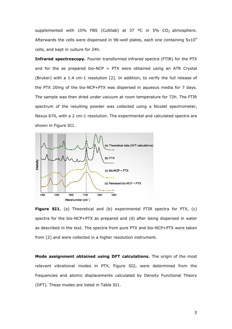
Infrared spectroscopy. Fourier transformed infrared spectra (FTIR) for the PTX
and for the as prepared bio-NCP + PTX were obtained using an ATR Crystal
(Bruker) with a 1.4 cm-1 resolution [2]. In addition, to verify the full release of
the PTX 20mg of the bio-NCP+PTX was dispersed in aqueous media for 7 days.
The sample was then dried under vacuum at room temperature for 72h. The FTIR
spectrum of the resulting powder was collected using a Nicolet spectrometer,
Nexus 670, with a 2 cm-1 resolution. The experimental and calculated spectra are
shown in Figure SI1.
Figure SI1. (a) Theoretical and (b) experimental FTIR spectra for PTX, (c)
spectra for the bio-NCP+PTX as prepared and (d) after being dispersed in water
as described in the text. The spectra from pure PTX and bio-NCP+PTX were taken
from [2] and were collected in a higher resolution instrument.
Mode assignment obtained using DFT calculations. The origin of the most
relevant vibrational modes in PTX, Figure SI2, were determined from the
frequencies and atomic displacements calculated by Density Functional Theory
(DFT). These modes are listed in Table SI1.

4
Figure SI2. Schematic structure of the PTX molecule adapted from [3]. Phenyl
groups (AR1, A2 and AR3) are highlighted in red, methyl groups in green and
carbons bonded to acetyl groups in blue. The dark grey shading highlights the
flexible side chain bonded to C13.
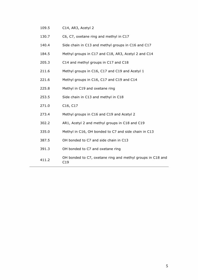
Table SI1. Most relevant contributions from the PTX molecule to the INS vibrations obtained by DFT calculations. AR denotes the Aromatic Rings.
Calculated frequency
(cm-1) Main contributions
6.52 AR1, AR2, AR3
18.70 C13 side chain and AR1
29.24 AR1 and oxetane ring
45.39 AR1 and oxetane ring
52.89 AR2, AR3 and Acetyl 1
57.27 Acetyl 1
67.75 Acetyl 1 and AR1
73.79 Acetyl 1 and AR1
74.50 Acetyl 1, Acetyl 2, oxetane ring
85.48 Oxetane ring, methyl groups in C16, C17 and C19 and C6
95.60 Acetyl 2, methyl groups in C16, C17, C18 and C19, AR1 and C6
5
109.5 C14, AR3, Acetyl 2
130.7 C6, C7, oxetane ring and methyl in C17
140.4 Side chain in C13 and methyl groups in C16 and C17
184.5 Methyl groups in C17 and C18, AR3, Acetyl 2 and C14
205.3 C14 and methyl groups in C17 and C18
211.6 Methyl groups in C16, C17 and C19 and Acetyl 1
221.6 Methyl groups in C16, C17 and C19 and C14
225.8 Methyl in C19 and oxetane ring
253.5 Side chain in C13 and methyl in C18
271.0 C16, C17
273.4 Methyl groups in C16 and C19 and Acetyl 2
302.2 AR1, Acetyl 2 and methyl groups in C18 and C19
335.0 Methyl in C16, OH bonded to C7 and side chain in C13
387.5 OH bonded to C7 and side chain in C13
391.3 OH bonded to C7 and oxetane ring
411.2 OH bonded to C7, oxetane ring and methyl groups in C18 and C19

6
Movie S1. The movie shows the motions for the PTX molecule at 29cm-1 asobtainedbyDFTcalculations.Movie S2. The movie shows the motions for the PTX molecule at 57cm-1 asobtainedbyDFTcalculations.Movie S3. The movie shows the motions for the PTX molecule at 80cm-1 asobtainedbyDFTcalculations.Movie S4. Themovie shows themotions for the PTXmolecule at 109cm-1 asobtainedbyDFTcalculations.Movie S5. Themovie shows themotions for the PTXmolecule at 131cm-1 asobtainedbyDFTcalculations.Movie S6. Themovie shows themotions for the PTXmolecule at 205cm-1 asobtainedbyDFTcalculations.Movie S7. Themovie shows themotions for the PTXmolecule at 271cm-1 asobtainedbyDFTcalculations.Movie S8. Themovie shows themotions for thePTXmolecule at1535cm-1 asobtainedbyDFTcalculations.Movie S9. Themovie shows themotions for thePTXmolecule at1550cm-1 asobtainedbyDFTcalculations. [1] Martins, M. L. et al. Development and characterization of a new bio-nanocomposite (bio-NCP) for diagnosis and treatment of breast cancer. J. Alloys Compd. 584, 514-519 (2014). [2] Martins, M. L. et al. Encapsulation of paclitaxel into a bio-nanocomposite. A study combining inelastic neutron scattering to thermal analysis and infrared spectroscopy. EPJ Web of Conferences. 83, 02011 (2015). [3] Mastropaolo, D., Camerman, A., Luo, Y., Brayer, G. D. & Camerman, N. Crystal and molecular structure of paclitaxel (taxol). Proc. Natl. Acad. Sci. USA 92, 6920-6924 (1995).