detailed model of intersegmental coordination in the
TRANSCRIPT
Detailed Model of Intersegmental Coordination in the Timing Network of theLeech Heartbeat Central Pattern Generator
Sami H. Jezzini, Andrew A. V. Hill, Pavlo Kuzyk, and Ronald L. CalabreseDepartment of Biology, Emory University, Atlanta, Georgia 30322
Submitted 8 July 2003; accepted in final form 21 October 2003
Jezzini, Sami H., Andrew A. V. Hill, Pavlo Kuzyk, and Ronald L.Calabrese. Detailed model of intersegmental coordination in thetiming network of the leech heartbeat central pattern generator. JNeurophysiol 91: 958–977, 2004. First published October 22, 2003;10.1152/jn.00656.2003. To address the general problem of interseg-mental coordination of oscillatory neuronal networks, we have studiedthe leech heartbeat central pattern generator. The core of this patterngenerator is a timing network that consists of two segmental oscilla-tors, each of which comprises two identified, reciprocally inhibitoryoscillator interneurons. Intersegmental coordination between the seg-mental oscillators is mediated by synaptic interactions between theoscillator interneurons and identified coordinating interneurons. Thesmall number of neurons (8) and the distributed structure of the timingnetwork have made the experimental analysis of the segmental oscil-lators as discrete, independent units possible. On the basis of thisexperimental work, we have made conductance-based models to ex-plore how intersegmental phase and cycle period are determined. Weshow that although a previous simple model, which ignored manydetails of the living system, replicated some essential features of theliving system, the incorporation of specific cellular and networkproperties is necessary to capture the behavior of the system seenunder different experimental conditions. For example, spike fre-quency adaptation in the coordinating interneurons and details ofasymmetries in intersegmental connectivity are necessary for repli-cating driving experiments in which one segmental oscillator wasinjected with periodic current pulses to entrain the activity of theentire network. Nevertheless, the basic mechanisms of phase andperiod control demonstrated here appear to be very general and couldbe used by other networks that produce coordinated segmental motoroutflow.
I N T R O D U C T I O N
The generation of many rhythmic movements appears toinvolve the coordination of distributed neural oscillators withinthe nervous system. For example, the motor patterns thatunderlie wave-like behaviors, such as undulatory swimming inleech and in lamprey or the beating of crayfish swimmerets, aregenerated by neuronal networks that can be approximated aschains of coupled segmental oscillators (Friesen and Pearce1993; Grillner et al. 1993; Sigvardt 1993; Skinner and Mul-loney 1998a). Each segmental oscillator consists of a localnetwork of neurons that is capable of generating rudimentaryrhythmic output (Murchison et al. 1993; Sigvardt 1993). Thecoordinated output of the entire chain of oscillators oftenshows phase relationships that are appropriate for the pattern ofmuscle activation in the intact, behaving animal (e.g., forwardswimming) (Wallen and Williams 1984). The appropriatephase relationships between these segmental oscillators arise as
an emergent property of the segmental oscillators and thecoupling between them. Moreover, in many such systems,intersegmental phase is independent of period, which can varywidely (Mulloney 1997; Mulloney et al. 1998; Wallen andWilliams 1984). Although this distributed organization isfound in many different animals, there are large differences interms of the properties of the segmental oscillators, the strengthand symmetry of coupling, and the importance of sensoryfeedback (Hill et al. 2003). Intersegmental coordination resultsprimarily from ascending and descending synaptic connectionsbetween the segmental oscillators, although sensory feedbackalso reinforces and fine tunes the intersegmental phase rela-tionships (Di Prisco et al. 1990; Cang and Friesen 2000).
Nearly from its beginnings, experimental progress in thisfield had been inextricably linked with modeling efforts (Co-hen et al. 1982; Kopell and Ermentrout 1988). Most modelsproduced to date, however, have been either quite abstract orbased on incomplete data about the intrinsic membrane prop-erties or intersegmental connectivity of the network beingstudied (Bem et al. 2003; Buchanan 1992; Cang and Friesen2002; Ekeberg and Grillner 1999; Jones et al. 2003; Skinner etal. 1997, 1998b; Williams et al. 1990). These models havenevertheless been very instructive and have constrained therange of mechanisms that can account for phase and periodcontrol. Two main hypotheses have been proposed to explainthe generation of appropriate phase differences between seg-mental oscillators. The “asymmetric coupling hypothesis”states that phase differences are generated by asymmetries inthe coupling between segmental oscillators (Skinner et al.1997; Williams et al. 1990). For example, ascending anddescending coordinating interneurons may differ in terms ofthe distances that their axons project, the strength and sign oftheir synapses, and their postsynaptic targets (Jones et al. 2003;Skinner and Mulloney 1998a,b). In contrast, the “excitabilitygradient hypothesis” states that phase differences arise fromdifferences in the oscillation periods of the segmental oscilla-tors (Grillner et al. 1993; Ikeda and Wiersma 1964; Matsu-shima and Grillner 1990, 1992). This difference may be basedon either the inherent periods of the segmental oscillators or agradient of excitation along the nerve or spinal cord (Tunstalland Sillar 1993). A more modern view combines these twohypotheses and recognizes that both neuronal intrinsic mem-brane properties (excitability) and intersegmental connectivitycombine to produce proper phasing between segments (Friesenand Cang 2001; Friesen and Pearce 1993; Ullstrom et al. 1998).
We have used the leech heartbeat central pattern generator to
Address reprint requests and other correspondence to: R. L. Calabrese(E-mail: [email protected]).
The costs of publication of this article were defrayed in part by the paymentof page charges. The article must therefore be hereby marked ‘‘advertisement’’in accordance with 18 U.S.C. Section 1734 solely to indicate this fact.
J Neurophysiol 91: 958–977, 2004.First published October 22, 2003; 10.1152/jn.00656.2003.
958 0022-3077/04 $5.00 Copyright © 2004 The American Physiological Society www.jn.org

address the problem of intersegmental coordination of oscilla-tory networks. The timing network of the pattern generatorconsists of two segmental oscillators, each of which is locatedin a separate ganglion and is capable of continuous indepen-
dent oscillation (Fig. 1A). Each oscillator comprises just twomutually inhibitory interneurons that are well characterized interms of their intrinsic membrane properties and their segmen-tal and intersegmental synaptic interactions (Calabrese et al.
A B
C
DE
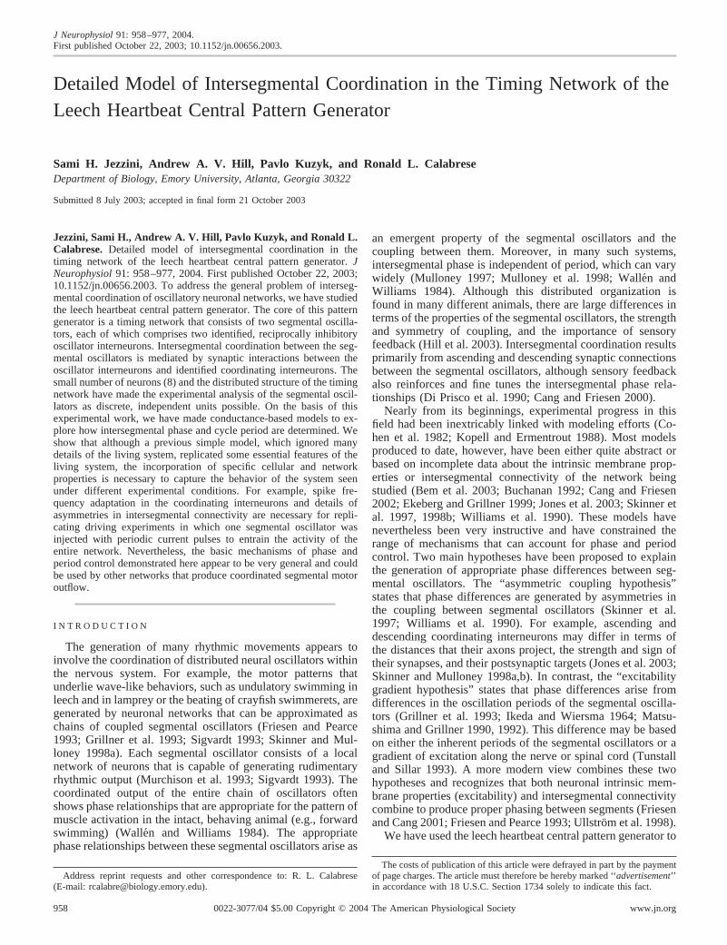
FIG. 1. The timing network of the leech heartbeat central pattern generator: circuit diagram, activity, and the simple symmetricmodel. A: the timing network contains 4 pairs of bilaterally symmetric interneurons that have cell bodies in the first 4 midbodyganglia. There are 2 segmental oscillators located in the 3rd and 4th ganglia. The coordinating interneurons of the first 2 gangliaare functionally equivalent and are, therefore, combined in representation. Open circles represent cell bodies, open squares representsites of spike initiation, and small filled circles represent inhibitory synapses. B: the electrical activity of 3 heart interneuronsrecorded extracellularly from a chain of ganglia (head brain to G4). The heart interneurons are labeled HN and are indexed by bodyside and midbody ganglion number [e.g., HN(L,3)]. Phase (�) of an interneuron X with respect to the G4 oscillator interneuronwas calculated, on a cycle by cycle basis, as the difference in the median spike times (�tX-4) divided by the G4 cycle period (T4),and then multiplied by 100. A positive phase value indicates that the G4 oscillator leads in phase. C: the timing network can beconceptualized as a simple symmetric network made up of 2 segmental oscillators. D: simulated activity of heart interneurons inthe simple symmetric model. The model interneurons are labeled using the same convention as for the living interneurons. Themodel neurons contain Hodgkin-Huxley style voltage-dependent conductances. The maximal conductance of the hyperpolarization-activated current (g�h) was 5.4 nS in the pair of G4 oscillator interneurons and 4.0 nS in the pair of G3 oscillator interneurons.Symbols above each voltage trace indicate the occurrence of the median spike within each burst. The yellow rectangles show thewindows of time in which the coordinating interneurons were active. E: quantification of mutual entrainment in the simplesymmetric model. The period of the mutually entrained system (TC; square symbols) is equal to that of the faster segmentaloscillator. When g�h was reduced below the canonical value (4 ns), the period of the G4 segmental oscillator (T4S) was greater thanthat of the G3 segmental oscillator (T3S). In this range, the period of the coupled system was equal to that of the G3 segmentaloscillator. When g�h was increased above the canonical value, the period of the G4 segmental oscillator was less than that of theG3 segmental oscillator. In this range, the period of the coupled system (TC) closely followed the period of the G4 segmentaloscillator. Regardless of which segmental oscillator was faster, the system could only be sped to the half-center oscillator period(T4H or T3H) of the slower oscillator. Figure adapted from Hill et al. (2002).
959MODEL OF INTERSEGMENTAL COORDINATION
J Neurophysiol • VOL 91 • FEBRUARY 2004 • www.jn.org

1995). Detailed models are available for the oscillator inter-neurons and their interactions with intersegmental coordinatinginterneurons (Hill et al. 2001; Nadim et al. 1995; Olsen et al.1995). The simplicity of a neuronal network containing onlytwo oscillators that reside in separate segmental ganglia hasmade possible the experimental analysis of the oscillators asdiscrete units that can be uncoupled and then re-coupled (Ma-sino and Calabrese 2002a–c).
In this paper, we use simulations to explore how phasedifferences between segmental oscillators and cycle period aredetermined in the timing network of the leech heartbeat centralpattern generator. This work builds on a simple model of thetiming network that ignored some details of the firing pattern ofthe coordinating neurons and their synaptic interactions withthe oscillator interneurons (Hill et al. 2002). This earlier modelshowed behavior that largely matched the behavior of theliving system under conditions of mutual entrainment in whichthe two segmental oscillators feedback on to one another(Masino and Calabrese 2002a,b). However, this model did notmatch the living system’s response during driving experimentsin which current pulses were used to force one segmentaloscillator to oscillate at periods faster and slower than the freerun period (Masino and Calabrese 2002c). Here we explore thebehavior of a model that more realistically represents thecoordinating interneurons as multicompartmental cables,which have properties such as spike adaptation and multiplespike initiation sites and receive asymmetric input from theoscillator interneurons of the two segmental oscillators (Ma-sino and Calabrese 2002a; Peterson 1983a,b). We show herethat these details of the circuit are necessary to capture theproperties of the system observed during driving experiments.Our results suggest that a detailed knowledge of both firingpatterns and intersegmental connectivity are necessary for un-derstanding how the heartbeat timing network functions. How-ever, we have also found simple mechanisms for phase andperiod control that may be easily generalized to other networkswith different intrinsic membrane properties and intersegmen-tal connectivity.
Parts of these results have appeared in abstract form (Jezziniet al. 2000).
M E T H O D S
Modeling methods
Each oscillator heart interneuron (a neuron originating in the 3rd or4th ganglion) was represented as a single isopotential compartmentwith intrinsic and synaptic currents. The coordinating heart interneu-rons (neurons originating in the 1st or 2nd ganglion) were representedas two (1 for each side) multicompartmental cylindrical cables. Thedynamics of membrane potential (V) of each compartment obey
�CdV
dt� ��IIon � �ISyn � IL � Iinject�
where C is total membrane capacitance, Iion is an intrinsic voltage-gated current, IL is the leak current, ISyn is a synaptic current, andIinject is the injected current.
Each oscillator interneuron was modeled as described previously(Hill et al. 2001) and contained eight voltage-dependent intrinsiccurrents. Five inward currents were included: a fast Na� current (INa),a persistent Na� current (IP), a fast low-threshold Ca2� current (ICaF),a slow low-threshold Ca2� current (ICaS), and a hyperpolarization-activated cation current (Ih) (Angstadt and Calabrese 1989, 1991;
Olsen and Calabrese 1996; Opdyke and Calabrese 1994). Three out-ward currents were included: a delayed rectifier-like K� current (IK1),a persistent K� current (IK2), and a fast transient K� current (IKA)(Simon et al. 1992). All currents were characterized in voltage-clampexperiments except for INa. The equations and parameters describingthese intrinsic currents can be found in Hill et al. (2001).
In the living system, there are two types of inhibitory synapsesmade between a pair of oscillator interneurons within the same seg-mental ganglion: graded synapses, which are dependent on the influxof presynaptic Ca2� through low-threshold Ca2� channels (Angstadtand Calabrese 1991; Ivanov and Calabrese 2000), and plastic spike-mediated synapses, which are dependent on the influx of presynapticCa2� through high-threshold Ca2�channels during a spike (Lu et al.1997) and are modulated by slow changes in membrane potential(Ivanov and Calabrese 2003; Nichols and Wallace 1978a,b). There areboth graded and spike-mediated synapses between the model oscilla-tor interneurons. The equations and parameters of these currents aredescribed in Hill et al. (2001). A mutually inhibitory pair of oscillatorinterneurons produces alternating bursting activity and is referred toas a half-center oscillator.
Coordinating heart interneurons originate in the first and secondganglia and function to link the half-center oscillators to form the beattiming network. There are no known differences between coordinatinginterneurons of the first and second ganglia (G1 and G2) with respectto their connectivity and interaction with oscillator interneurons, soipsilateral interneurons were modeled as a single intersegmental cable(fiber) for computational efficiency (Fig. 2C) and are referred to hereas coordinating fibers.
Coordinating fibers [HN(CF)] were modeled as multicompartmen-tal cylindrical cables of realistic dimension (200 � 2 �m) and passiveproperties (Fig. 2C, APPENDIX). Coordinating fibers were divided into150 compartments. In general, each fiber had two spike initiation sites(2-site model) that were each capable of spontaneous activity, one inganglion 3 (G3 site) and one in ganglion 4 (G4 site). The G3 and G4initiation sites, each consisting of a group of five compartments, wereseparated by 50 conduction compartments representing the inter-ganglion portion of the fiber. In some simulations (1-site model), theG3 initiation site was removed by converting its compartments intoconduction compartments. Conduction compartments contained onlythree voltage-dependent currents: INa, IK1, and IK2. Their maximalconductances were adjusted to produce a realistic action potentialpropagation delay, similar to that observed in the living system(Masino and Calabrese 2002a), of �25 ms between ganglionic initi-ation sites (APPENDIX). The ends of the fiber beyond each initiation siteconsisted of an additional 10 conduction compartments followed by35 passive compartments (Fig. 2C). The end conduction compart-ments allowed action potentials to travel unabated through the initi-ation site, whereas the passive end compartments acted as a sink toabsorb axial current and prevented reflection of action potentials. Thepassive voltage response to current injection at initiation sites wasnearly the same as theoretically expected for an infinite cable (Perkeland Mulloney 1978; Rall 1977).
The morphology of the coordinating interneurons has precludeddirect electrophysiological characterization of the intrinsic currents ofthe G3 and G4 initiation sites. The small neuritic processes compris-ing these sites are electrotonically distant from their cell bodies wheremicroelectrode penetration is possible (Masino and Calabrese 2001a).We assumed that the initiation sites of the model coordinating fiberscontain the same currents as measured experimentally in oscillatorinterneurons (INa, IP, ICaF, ICaS, Ih, IK1, IK2, IKA). Activation param-eters of the currents were not altered, but their maximal conductanceswere varied to tune the spiking activity of each site to the desiredcharacteristics. During normal rhythmic activity in the timing net-work, the living coordinating interneurons show spike frequencyadaptation; within each burst the frequency declines from �7 to 2 Hzwith an average of �4 Hz (Masino and Calabrese 2001a). We createdtwo types of model initiation sites: nonadapting and adapting. Non-
960 S. H. JEZZINI, A.A.V. HILL, P. KUZYK, AND R. L. CALABRESE
J Neurophysiol • VOL 91 • FEBRUARY 2004 • www.jn.org

adapting initiation sites were made of compartments containing onlyNa and K currents, and their firing frequency was adjusted by varyingthe leak reversal potential. These compartments were adjusted so thatmodel coordinating fibers fired tonically with a mean spike frequencyof �4 Hz when not inhibited. Nonadapting initiation sites were usedin only a few model experiments that are noted in the text; otherwiseall models contained coordinating fibers with adapting initiationsite(s). Adapting initiation sites (i.e., showing spike frequency adap-tation) were made of compartments containing the full compliment ofactive currents. Variable spike properties between different versionsof these spike initiation sites were made using different maximalconductances for IP, ICaF, ICaS, Ih, (APPENDIX). All two-site modelscontained a canonical adapting G4 site that produced bursts in whichspike frequency declines from �7 to 2 Hz with an average of �4 Hz.In two-site models, adapting G3 sites were given a lower intrinsicspike frequency than the G4 site, leading to dominance of the G4 site.Thus the model coordinating neurons were constrained to conform tothe observed behaviors of the living neurons (Masino and Calabrese2001a; Peterson 1983b). For two-site models, we defined a parameter�f, the difference in the average frequency of the G4 site and the G3site (f�G4 � f�G3).
In two-site models, synaptic connections were made from G3oscillator interneurons onto both ipsilateral G3 and G4 initiation sites(all 5 compartments at each site) of the coordinating fibers, whereasG4 oscillator interneurons made synapses only onto the ipsilateral G4initiation site (all 5 compartments) of the coordinating fibers (Fig.2C). In one-site models, synaptic connections from the G3 and G4oscillator interneurons to the coordinating fibers were made only ontothe active G4 site (all 5 compartments) of the ipsilateral coordinatingfiber (Fig. 2C). In two-site models, synaptic connections were madefrom the central compartment of each initiation site of a coordinatingfiber to the local, ipsilateral G3 and G4 oscillator interneurons. Inone-site models, synaptic connections from the coordinating fiber G4initiation sites to the ipsilateral G4 oscillator interneuron were as inthe two-site model, but connections to the ipsilateral G3 oscillatorinterneuron were made by the conduction compartment that corre-sponded to the previous central compartment of the defunct G3initiation site. Because one model coordinating fiber represented ac-tivity of two coordinating interneurons, each action potential in thesingle fiber was made to produce two inhibitory postsynaptic poten-tials (IPSPs) in each of its postsynaptic targets (ipsilateral oscillatorinterneurons) with the second IPSP occurring with a delay of 135 ms.This delay, which was picked arbitrarily, reduced the synchronousoccurrence of two coordinating fiber-mediated IPSPs in an oscillatorneuron.
Simulations were done with Genesis, software for Hodgkin-and-Huxley-style models (Bower and Beeman 1998; Hodgkin and Huxley1952). The exponential Euler integration method was used with a timestep of 0.1 ms. When a parameter or experimental perturbation wasvaried in a series of trials, each simulation in the series began from thesame initial conditions and was iterated for 100 s of simulation timebefore collecting data for an additional 300 s of simulation time foranalysis. This procedure allowed the model system to settle downfrom the perturbing effects of the parameter change.
Physiological methods
Physiological methods were as described by Masino and Calabrese(2001a). Data were digitized using a digitizing board (Digi-Data 1200Series Interface, Axon Instruments, Foster City, CA) and acquiredusing pCLAMP software (Axon Instruments) on a personal computer(PC).
Data analysis
A spike train analysis program, written in Matlab (Mathworks,Natick, MA), was used to analyze spike train data from simulations
and experiments on a PC. In the subsequent description, we do notdifferentiate between data from heart interneurons and model heartinterneurons. In the analysis program, spikes were detected with adiscrimination window. When voltage crossed a threshold value, aspike event was detected. An upper threshold eliminated transientartifacts in the recording. To prevent multiple detection of the samespike, a refractory period (20 ms), during which spikes could not berecognized, was applied after each detected event. Spikes were thengrouped into bursts as follows. After an interburst interval (1 s) elapsedwithout any spikes detected, the next spike event was identified as thefirst spike of a burst. Subsequent spikes with interspike intervals less thanthe interburst interval were grouped into that burst. The median spike ineach burst was indicated by a symbol above the burst.
The analysis program was also used to determine cycle period (TX),duty cycle (DX), and phase (�X�4) for each interneuron (X). The cycleperiod (TX) was determined for each cell (X) by measuring the intervalfrom median spike to median spike of consecutive bursts. The dutycycle (DX) was defined as the fraction of the cycle period occupied bythe burst duration (Tburst X) and expressed as a percentage: DX �(Tburst X /TX) � 100. The phase of a given heart interneuron wasdetermined cycle-by-cycle as a percentage based on the formula:�X�4 � (�tX�4/T4) � 100, where �tX�4 is the difference between thetime of the median spike (tX) of cell X and the median spike (t4) of thephase reference cell (usually the ipsilateral G4 oscillator interneuron),and T4 is the cycle period of the reference cell. A phase of 0%indicated a cell with no phase difference relative to the reference cell,whereas a 50% phase difference indicated an anti-phasic relationship.A positive phase difference indicated a phase lag with respect to thereference cell. Stable entrainment was indicated, in both mutualentrainment experiments and driving experiments, if all interneuronsin the coupled system were entrained to the same period with acoefficient of variation of �5%.
In all graphs values are expressed as means � SD (n �12 consec-utive bursts). Likewise numerical values given in the text are in somecases reported as means � SD.
R E S U L T S
Modeling strategy
The simulations reported here emulate two types of experi-ments that were aimed at analyzing the control of period andphase in the heartbeat timing network: mutual entrainment anddriving experiments. In the first series of experiments (mutualentrainment experiments), axonal conduction was reversiblyblocked with a sucrose solution flowing across the connectivebetween the third and fourth (G3 and G4) segmental ganglia(Masino and Calabrese 2002b). This arrangement allowed us tomeasure independently the periods of the two segmental oscil-lators, subnetworks of the heartbeat timing network located inindividual segmental ganglia. Because the ganglia were inseparate bathing chambers they could be individually treatedwith agents that either increased or decreased the period ofeach segmental oscillator. Subsequent to attaining the desiredperiod difference between the independent segmental oscilla-tors, the conduction block was relieved, and the effect of themeasured period difference on the period and phase of thecomplete timing network was assessed. In driving experiments,a single oscillator interneuron was driven with rhythmic cur-rent pulses of varying period (50% duty cycle), and entrain-ment and phase relationships in the timing network were as-sessed by recording from other oscillator interneurons and insome cases from coordinating interneurons (Masino and Cala-brese 2002c). The models that we present here build on ourprevious model of intersegmental coordination in the timing
961MODEL OF INTERSEGMENTAL COORDINATION
J Neurophysiol • VOL 91 • FEBRUARY 2004 • www.jn.org

network in which the coordinating interneurons were modeledas single compartments (Fig. 1C) (Hill et al. 2002).
Model half-center oscillator
In our models, rhythmic bursting arises from two mutuallyinhibitory oscillator interneurons, which form a half-centeroscillator (H). These model oscillator interneurons are notinherently bursting but rather fire tonically in isolation. Recentexperiments using extracellular recordings show that the oscil-lator interneurons burst endogenously when synaptic inhibitionis blocked with bicuculline (Cymbalyuk et al. 2002). However,our representation of the system as a half-center oscillator isappropriate because the reciprocal inhibition is strong andcontrols the period and duty cycle of the oscillator interneu-rons. These core half-center oscillators cannot be observedexperimentally with current methods because even in an iso-lated ganglion the processes of the coordinating interneuronsremain active and functional (Fig. 1A). Nevertheless, it isinstructive to assess the characteristics of this oscillatory mod-ule in our simulations. To observe the model half-centers inisolation, we simply silenced the activity of the coordinatingfibers by removing them from the models. To modify theperiod of the half-center oscillator, the maximal conductance(g�h) of Ih in both oscillator neurons was altered from itscanonical value, whereas all other parameters were held con-stant at canonical values. Period is inversely proportional to(g�h) (Hill et al. 2001) (Fig. 1E).
Forming a model-8-cell timing network by couplingsegmental oscillators
The smallest subnetwork that can be assessed experimen-tally is that in an isolated third or fourth ganglion, a segmentaloscillator (S). This subnetwork consists of a mutually inhibi-tory pair of oscillator interneurons (half-center oscillator) andthe local axonal and neuritic processes of the coordinatinginterneurons and their associated synaptic connections (Fig.1A) (Masino and Calabrese 2002a). Even after isolation fromtheir cell bodies, the coordinating interneurons continue tofunction and interact synaptically with the oscillator interneu-rons (Peterson 1983a).
The 8-cell model timing network was formed from twohalf-center oscillators (G3 and G4) and two coordinating fibers(1- or 2-site) and all their associated synaptic connections (Fig.1); essentially we coupled two model segmental oscillators. Inour model, a single coordinating fiber represents two coordi-nating interneurons on the same side of the body (see METH-ODS). Two different configurations of the timing network weremodeled (Fig. 2C). One model contains coordinating fiberswith one spike initiation site in G4 (1-site model) and the othermodel contains coordinating fibers with spike initiation sites inboth G3 and G4 (2-site model). We refer to these 8-cell modelsof the timing network as the “coupled system” (C).
To assess the effects of period differences between thesegmental oscillators on the phase and period of the coupledsystem, it was necessary to observe the segmental oscillatorsindependently. In the one-site model, one segmental oscillatorcould be observed in isolation by simply removing the pair ofoscillator interneurons in the other ganglion (Fig. 2C). Becauseof the functional symmetry inherent in such one-site models,
only one segmental oscillator need be observed and manipu-lated (G4) and the other could be assumed to act identically. Inthe two-site model, an isolated G3 or G4 segmental oscillatorcould be observed by removing the other pair of oscillatorinterneurons and their associated pair of coordinating fiberinitiation sites (Fig. 2C). Because each segmental oscillator insuch two-site models is associated with a different pair ofcoordinating fiber initiation sites, each segmental oscillator(G3 and G4) had to be independently manipulated and as-sessed. To create period differences between the segmentaloscillators, g�h was altered in one pair of oscillator interneuronsas described in the preceding text for the half-center oscillators.
Simulated experiments
MUTUAL ENTRAINMENT. Our experimental paradigm can besummarized as follows: create the appropriate segmental os-cillators, alter the period of one of them (usually the G4oscillator) by varying �gh in both oscillator interneurons [Anincrease in �gh leads to faster oscillations (Hill et al. 2001).], andcouple the segmental oscillators and observe the period andphase relationships of the complete timing network. Mutualentrainment in the coupled system was indicated if all inter-neurons were entrained to the same period (see METHODS).
This paradigm is illustrated for our previous simple sym-metric model in which coordinating interneurons were repre-sented with single compartments (Fig. 1C) (Hill et al. 2002).When a period difference is created between the segmentaloscillators by accelerating the G4 segmental oscillator (�gh isincreased above its canonical value of 4 nS), then the G4oscillator leads in phase (Fig. 1D). In this simple model, theperiod of the coupled system is equal to the period of the fastersegmental oscillator (Fig. 1E). When �gh was reduced below thecanonical value (4 ns), the period of the G4 segmental oscil-lator (T4S) was greater than that of the G3 segmental oscillator(T3S). In this range, the period of the coupled system was equalto that of the G3 segmental oscillator. When �gh was increasedabove the canonical value, the period of the G4 segmentaloscillator was less than that of the G3 segmental oscillator. Inthis range, the period of the coupled system (TC) closelyfollowed the period of the G4 segmental oscillator. Note thatthe range of mutual entrainment extends from the intersectionof the G3 half-center oscillator period (horizontal small-dottedline, T3H) and the T4s dashed line to the point where the G4half-center oscillator period (dotted and dashed curve T4H)intersects the horizontal T3S dotted line. The system works byremoval of inhibition. Phase differences between the oscilla-tors allow the leading oscillator interneurons to truncate thecoordinating interneuron bursts and thus remove inhibition thatwould normally fall late in the inhibited phase of the oscillatorinterneurons of the slower oscillator (Fig. 1D). The sloweroscillator can be accelerated in this way to, at the limit, itshalf-center oscillator period (Hill et al. 2002). The type ofanalysis embodied in Fig. 1E is used repeatedly in the model-ing experiments on mutual entrainment.
DRIVING. Simulated driving experiments were performed onthe 8-cell-coupled system. Pulses of inhibitory conductancewith a duty cycle of 50% were used as external input to driveone segmental oscillator (either G3 or G4). Two square-waves,180° out of phase with each other, each with an amplitude of70 nS and a reversal potential that alternated between –55 and
962 S. H. JEZZINI, A.A.V. HILL, P. KUZYK, AND R. L. CALABRESE
J Neurophysiol • VOL 91 • FEBRUARY 2004 • www.jn.org

–40 mV, were used to control the spiking activity of the twooscillator interneurons in a segmental oscillator. In this way, apair of oscillator interneurons was driven to oscillate at a seriesof specified cycle periods different from the free run period ofthe coupled system. Successful driving of the entire couplednetwork was indicated by the same criteria as for mutualentrainment (see METHODS).
Coordinating fiber model, HN(CF)
We created a coordinating fiber model with two spike-initiation sites (G3 and G4) and tested its properties. In theliving system, coordinating fibers do not burst endogenously(Cymbalyuk et al. 2002), but rather they rebound after receiv-ing inhibitory input from oscillator interneurons with a tran-
A
B
C D
FIG. 2. A and B: interaction between spike initiation sites and spike frequency adaptation in the model coordinating fibers [HN(CF)].The icons at right represent the configuration of each simulation. Each electrode indicates a voltage recording/current injection site. A:1-site coordinating fibers each containing a single initiation site that fires with spike frequency adaptation. Traces are shown from 2different 1-site coordinating fibers, 1 with a canonical G3 site (average spike frequency of 1.9 Hz) and 1 with a canonical G4 site (averagespike frequency of 3.6 Hz). On release from hyperpolarization, the coordinating fiber with the G4 site displays a higher rebound spikefrequency and a higher sustained frequency than the G3 fiber. In each case, spike frequency eventually settles to a constant, tonic rate.B: a 2-site fiber with canonical G3 and G4 sites (as in B), �f �1 .7 Hz. On simultaneous release of both initiation sites fromhyperpolarization, spikes originate at the G4 site and spike initiation at the G3 site is repressed until the G4 site is hyperpolarized. Eachsubsequent hyperpolarization of the G4 site reveals that the spike frequency of the G3 site becomes progressively lower with time. Thisprogression of spike frequency adaptation at the G3 site continues even while its activity is suppressed by the G4 site, therebydemonstrating that adaptation is independent of spiking activity. Toward the end of the record, the G3 site is hyperpolarized along withthe G4 site, restoring excitability so that the G3 site rebounds on release from hyperpolarization with a high spike frequency as seen atthe beginning of the record. Bottom: an initiation site switch on an expanded time scale. CM, current monitor. C: schematic representationsof the 1- and 2-site models of the timing network of the leech heartbeat central pattern generator. The coordinating interneurons of the1st 2 ganglia are functionally equivalent and are, therefore modeled as a single coordinating fiber on each side. Open circles representcell bodies, vertical open bars represent conducting and passive regions of the coordinating fibers, and colored areas in these bars representspike initiations sites. Small filled circles represent inhibitory synapses. D: the effect of coordinating fiber average spike frequency on theperiod of a model segmental oscillator (TS). We constructed a canonical segmental oscillator with a 1-site coordinating fiber that hadcanonical parameters (mean frequency of 3.6 Hz, denoted with an asterisk), which was synaptically coupled to a canonical half-centeroscillator. As the mean spike frequency of the coordinating fiber was reduced from the canonical value to zero, the period of the segmentaloscillator became shorter and eventually reached the half-center oscillator period, TH (filled boxes). A similar effect of coordinating fiberinhibition was seen in a model in which the period of the half-center oscillator was increased by reducing g�h in both oscillator interneuronsto 2 nS (open boxes).
963MODEL OF INTERSEGMENTAL COORDINATION
J Neurophysiol • VOL 91 • FEBRUARY 2004 • www.jn.org

sient burst of spikes that declines in frequency (Masino andCalabrese 2002a). Normally, �75% of the spikes in coordi-nating neuron arise at the G4 site, and in most preparations,the inherent spike frequency of the G3 site is less than that atthe G4 site. We aimed to model the rebound firing with spikefrequency adaptation of the coordinating interneurons and thedominance of the G4 initiation site. We hypothesized thatthe dominance of the G4 site was due to its greater inherentspike frequency. To test this idea, we adjusted the G3 site tospike at a constant frequency of 2.0 Hz and the G4 site to spikeat a constant frequency of 3.8 Hz when not inhibited (data notshown). Both sites were silenced with injected current and thenreleased simultaneously. Spikes arose in the fiber at a constant3.8 Hz, and all spikes could be shown by latency measurementsto arise at the G4 site. The G4 site was then again silenced withinjected current; now spikes arose at a constant 2.0 Hz and allspikes could be shown by latency measurements to have arisenat the G3 site. Even with a frequency difference of as little as0.5 Hz, the faster initiation site suppressed the slower sitewithin one to three spikes, and silencing the faster site withinjected current allowed the suppressed site to resume firingafter a period only slightly longer than its normal interspikeinterval.
We next added spike frequency adaptation and establishedcanonical G3 and G4 initiation sites in our coordinating fibermodel. See APPENDIX for properties of the canonical and mod-ified G3 and G4 sites. The spike frequency within a burst of thecanonical G4 site decreased from �7 to 2 Hz with an averageof 3.6 Hz when it was released from hyperpolarization. In allmodels reported here, the G4 site retained its canonical prop-erties. The canonical G3 site also showed rebound and adap-tation but had an average spike frequency in a burst of 1.9 Hz.Thus the difference in the average frequency of the two ca-nonical sites was 1.7 Hz; we defined this difference as �f. �fwas varied in some model experiments to observe its effects oncoupled system properties; in each case, this change in �f wasaccomplished by changing the average frequency of the G3 site(see APPENDIX). Figure 2A shows the firing pattern of thesecanonical sites after release from hyperpolarizing current in-jection in one-site coordinating fiber models containing each ofthe two canonical sites. In each case, the site was allowed toreach its steady-state firing frequency. Figure 2B shows theinteraction between the two canonical sites in a two-site coor-dinating fiber model as the two sites are released from orsilenced with hyperpolarizing current. The G4 site is dominantwhenever it is not silenced with current, but the G3 sitebecomes active quickly after the G4 site is silenced withcurrent (Fig. 2B, inset). Adaptation continues to build at eachsite whenever that site is not hyperpolarized by injected current(or by synaptic inhibition). Adaptation occurs as the result ofthe inactivation of ICaS (slowly inactivating low-threshold Cacurrent) after release from hyperpolarization and is indepen-dent of spike activity. Rebound at these sites occurs becausehyperpolarization removes this inactivation and thus relievesadaptation.
Period of a segmental oscillator varies with coordinatingfiber input
Coordinating fiber inhibition acts to slow the period of asegmental oscillator. A canonical segmental oscillator con-
structed by adding two canonical G4 One-site (mean spikefrequency of 3.6 Hz) coordinating fibers to a canonical half-center oscillator had a period (TS) of 9.3 � 0.1 s (Fig. 2D). Asthe mean spike frequency of the coordinating fiber was reducedfrom the canonical value to zero, the period became shorter,eventually ending at the period of the canonical half-centeroscillator (TH � 8.6 � 0.2 s). A similar effect of coordinatingfiber inhibition is seen if the period of the half-center oscillatoris increased to 10.1 � 0.2 s by reducing g�h in both oscillatorinterneurons to 2 nS (Fig. 2D). Therefore the slowing effect ofcoordinating fiber inhibition is general and does not depend onthe specific values such as g�h in the model.
One-site model with spike frequency adaptation
A canonical coupled system with one-site coordinating fi-bers showing spike frequency adaptation, hence referred to asa one-site model, has the same functional symmetry as ourprevious simple symmetrical model (Hill et al. 2002). The G3and G4 oscillator interneurons equally inhibit the sole G4 spikeinitiation site of each coordinating fiber (cf. Figs. 1C and 2C).Therefore the one-site model allows us to assess the role ofspike frequency adaptation in the coupled system without anyother substantive changes from our previous simple symmet-rical model. The canonical one-site model had a period of 9.4 s,slightly longer that the period of the segmental oscillators (T3Sand T4S � 9.3 s) from which it was constructed (Fig. 3C). Thisincrease in period was due to increased rebound firing of thecoordinating fibers at the G4 site because of inhibition by boththe G3 and G4 oscillator interneurons. There was no phasedifference between the G3 and G4 oscillators (�3 � �4 � 0%;Fig. 3C). The firing of the coordinating neurons filled theinhibited phase of both (G3 and G4) ipsilateral oscillator in-terneurons, and the high-frequency portion of the coordinatingneuron bursts fell early in the inhibited phases of these oscil-lator interneuron (data not shown). Our previous work with asimple symmetrical model of the timing network (Hill et al.2002) showed that if coordinating neuron inhibition falls earlyin the inhibited phase of oscillator interneuron activity, it isineffective, whereas late inhibition delays the oncoming burstphase and slows the period of the oscillator interneurons.
One-site model
MUTUAL ENTRAINMENT. Creating a period difference betweenthe segmental oscillators leads to phase differences in thecoupled system with the faster oscillator leading (Fig. 3A). Wecreated a period difference by varying �gh in the G4 oscillatorinterneurons. Because of the functional symmetry of the one-site model, equivalent results were obtained by varying �gh inthe G3 oscillator interneurons (data not shown). Phase has anear linear dependence on the period difference of the segmen-tal oscillators and on the underlying period difference of thehalf-center oscillators (Fig. 3, B and C). The period of thecoupled system follows the period of the faster segmentaloscillator more closely than that of the slower segmental os-cillator, but it is always slightly slower than the period of thefaster segmental oscillator (Fig. 3D). The range of mutualentrainment lies between that of the unvaried G3 half-centeroscillator period [T3H; biggest G4 phase lead; highest value ofHN(4) �gh] and the point where the period of the coupled systemmeets the period of the varied G4 half-center oscillator period
964 S. H. JEZZINI, A.A.V. HILL, P. KUZYK, AND R. L. CALABRESE
J Neurophysiol • VOL 91 • FEBRUARY 2004 • www.jn.org

[T4H; biggest G3 phase lead; lowest value of HN(4) �gh]. Plottedin an alternative manner, it can be seen that when the G4segmental oscillator is the faster oscillator, the change inperiod of the coupled system is similar to the change in periodof the G4 segmental oscillator (�T4S), but when the G4 oscil-
lator is the slower oscillator, the coupled system experienceslittle change in period (Fig. 3E).
As in the simple symmetric model, the faster segmentaloscillator cannot accelerate the slower oscillator faster than thehalf-center oscillator period of the slower segmental oscillator.
A
B C
D E
FIG. 3. Mutual entrainment in the one-site model. In this and subsequent figures showing simulated activity from the modeltiming network, the recordings from model oscillator interneurons are indexed by body side and ganglion number [e.g., HN(L,3)]and recordings from model coordinating fibers by body side [e.g., HN(L,CF)]. The icon on the right indicates the model usedcorresponding to the alternatives of Fig. 2C. In color figures, as in this one, the color of the traces of a given neuron correspondsto the color of its icon. For the model oscillator interneurons, recordings are from the solitary compartment, but for the modelcoordinating fibers, recordings are from the anterior-most conduction compartment. The uppermost trace in each record indicatesthe instantaneous frequency of the left model coordinating fiber. The symbol above each burst of spikes indicates the median spiketime. The vertical dashed line indicates where the median spike of the model HN(L,CF) coordinating fiber falls with respect tomodel HN(L,3) and HN(L,4) oscillator interneurons. Phase differences were created between the G3 and G4 oscillators by adjustingg�h in 1 pair of model oscillator interneurons as indicated above each panel, to create period differences between the G3 and G4segmental oscillators (the canonical value of g�h is 4 nS). When the G3 and G4 oscillators are equivalent, the there is no phasedifference between them. A: the G4 oscillator leads and terminates spiking of the ipsilateral coordinating fiber [HN(4) g�h isincreased above the canonical value]. When g�h is changed by the same amount in the G3 oscillator as in the G4 oscillator, a phasedifference of the same absolute magnitude is created because the one-site model is functionally symmetric. B and C: the phasedifference between the G3 and G4 oscillators (�3 � �4) in the coupled system varies with the period difference between the G3and G4 half-center oscillators (B) and segmental oscillators (C). D: the period of the coupled system is close to the period of thefaster segmental oscillator. E: the change in the period of the couple system (�TC) and the G4 segmental oscillator (�T4S) fromtheir canonical periods were plotted against the change in g�h. �TC hovers close to the change in period of the faster segmentaloscillator from its canonical period. Period differences were created between the G3 and G4 segmental and half-center oscillatorsby adjusting g�h of the G4 pair of model oscillator interneurons.
965MODEL OF INTERSEGMENTAL COORDINATION
J Neurophysiol • VOL 91 • FEBRUARY 2004 • www.jn.org

However, unlike the simple symmetric model, in the one-sitemodel, the slower oscillator clearly influences the period of thecoupled system (cf. Figs. 1E and 3D). For example, when �gh isvaried in the G4 oscillator, the limits of mutual entrainmentextend beyond the intersection between the half-center oscil-lator period (TH) of the slower oscillator and the period of thefaster segmental oscillator (in Fig. 3D this equality occurswhere T4H � T3S and where T3H � T4S). The reason the sloweroscillator can slow the faster oscillator is well illustrated in Fig.3A. The slower G3 oscillator lags behind the faster G4 oscil-lator, delaying the onset of the coordinating fiber burst. There-fore the high-frequency portion of the coordinating neuronburst falls later in the inhibited phase of the leading G4oscillator thus slowing that oscillator. A corresponding slowingmechanism is seen (data not shown) when the G3 oscillatorleads. In contrast to the simple symmetric model, which onlyallowed entrainment to occur based on the acceleration of theslower oscillator by the faster oscillator, in the one-site Model,due to spike frequency adaptation in the coordinating fibers,the phase difference between the oscillators creates both ac-celerating and slowing effects that permit mutual entrainment.
To determine whether the period of the coupled system wasmainly determined by the faster or slower segmental oscillatorand to facilitate comparison with data from the living system,we replotted the model data in a different manner (Masino andCalabrese 2002b). For each trial, the faster and slower of thetwo segmental oscillators was determined, and their periodswere plotted separately against the period of the coupled sys-tem. This plot confirms that although TC is longer than theperiod of the faster segmental oscillator, it is closer to theperiod of the faster segmental oscillator than the period of theslower segmental oscillator (Fig. 4C), demonstrating that theprimary mechanism for coordination in the coupled system isacceleration of the slower oscillator.
As in the simple symmetric model, in the one-site model, thecoordinating fibers fire in the window of time between the endof the burst of the trailing ipsilateral oscillator interneuron andthe beginning of the burst of the leading ipsilateral oscillatorinterneuron (cf. Figs. 1D and 3A). Because the G4 and G3oscillator interneurons equally inhibit the coordinating fibers,the coordinating fiber duty cycle decreases symmetrically asthe absolute value of �3 � �4 increases (Fig. 4A), and thephase of the coordinating fiber bursts relative to the oscillatorinterneurons (�CF � �4 and �CF � �3) varies symmetricallywith the phase difference between the G4 and G3 oscillators(�3 � �4); the absolute values of slopes of the regression linesfor these relationships are nearly equal; slopes 0.50 and �0.48,respectively (Fig. 4B). This symmetric relationship arises fromthe equal ability of the G3 and G4 oscillator interneurons to
inhibit the activity of the coordinating fibers. This symmetry inthe phasing of the coordinating fiber bursts is not observed inthe living system, where the slopes of the change in �CF � �4
and �CF � �3 versus �3 � �4 are 0.7 and �0.3 (Masino andCalabrese 2002a), reflecting a dominance of the G3 oscillator
FIG. 4. Mutual entrainment in the one-site model. A: the coordinating fiberduty cycle decreases symmetrically as the absolute value of the phase differ-ence between the G4 and G3 oscillators (��3 � �4�) increases. The duty cycleof the oscillator interneurons does not vary because of their half-center con-figuration. B: the phase of the coordinating fiber bursts relative to the oscillatorinterneurons (�CF � �4 and �CF � �3) varies symmetrically with the phasedifference between the G4 and G3 oscillators (�3� �4); the slopes of theregression lines for these relationships are 0.50 and �0.48, respectively. C: theperiod of the coupled system (TC) is closer to the faster segmental oscillatorperiod (TS) than to the slower segmental oscillator period, although TC isgenerally longer than the period of the faster segmental oscillator. Phasedifferences were created between the G3 and G4 oscillators by systematicallyvarying g�h in the G4 pair of model oscillator interneurons.
966 S. H. JEZZINI, A.A.V. HILL, P. KUZYK, AND R. L. CALABRESE
J Neurophysiol • VOL 91 • FEBRUARY 2004 • www.jn.org

in controlling the phase of the coordinating fibers in the livingsystem.
DRIVING. During driving experiments in the simple symmet-rical model, the undriven segmental oscillator could not beentrained to periods greater than its segmental oscillator periodbecause the only available mechanism for oscillator interactionwas removal of coordinating fiber inhibition (Hill et al. 2002).In other words, in contrast to the living system (Masino andCalabrese 2002c), the driven segmental oscillator may neverslow the system by lagging in phase. In the one-site model,which has the same functional symmetry as the simple sym-metric model, the slowing mechanism based on spike fre-quency adaptation described in the preceding text permits thedriven oscillator to lag in phase and the system to be drivenslower than the segmental oscillator period of the undrivenoscillator (Fig. 5C). When one segmental oscillator is drivenslower than the period of the other, it shifts the high-frequency
portion of coordinating fiber burst to very late in the inhibitedphase of the leading undriven oscillator, effectively slowingthe undriven oscillator (Fig. 5B). Driving to faster periods isaccomplished by removal of late coordinating fiber inhibition(Fig. 5A). Driving produces entrainment over a broad, nearlysymmetric range of periods (�10%) periods and a similarlybroad range of phases (�20%). The phase difference betweenthe oscillators (�3 � �4) is nearly linearly related to the perioddifference between the driven oscillator (TDriven) and the freerun period of the coupled system (TUndriven; Fig. 5C).
The functional symmetry of the one-site model makes theG3 and G4 oscillators perform identically during driving (datanot shown). This equivalence of the G3 and G4 oscillators isnot observed in the living system, where during driving the G3oscillator entrains the coupled system over a broader, moresymmetric period range than the G4 oscillator (Masino andCalabrese 2002c). Therefore the addition of spike frequencyadaptation in the coordinating fibers corrected one flaw in ourprevious simple symmetrical model (Hill et al. 2002) but didnot capture the asymmetries observed when driving the livingcoupled system (Masino and Calabrese 2002c).
Two-site model with spike frequency adaptation
In an attempt to capture the asymmetries observed in theheartbeat timing network, we implemented the two-site modelwith spike frequency adaptation, henceforth referred to as thetwo-site model (Fig. 2C). This model is functionally asymmet-ric because, as in the living system, the G3 oscillator interneu-rons inhibit both coordinating fiber initiation sites, whereas theG4 oscillator interneurons inhibit only the G4 site. For acanonical two-site model, we chose a difference in spike fre-quency between the G4 and G3 sites (�f �1.7 Hz) that reflectsmeasurements made in the living system (Masino and Cala-brese 2002a).
The canonical two-site model had a period (TC � 9.4 s),approximately equal to the period of the G4 segmental oscil-lator (T4S � 9.3 s), but longer than the period of the G3segmental oscillator (T3S �8.9 s) (Figs. 7C and 8C). Eventhough in the canonical two-site model the G3 and G4 half-center oscillators have identical periods, the correspondingsegmental oscillators do not. The G3 segmental oscillator hasa shorter period than the G4 segmental oscillator because theG3 initiation sites of the coordinating fibers have a loweraverage firing frequency than the G4 sites. In the coupledsystem, the dominance of the G4 sites of the coordinatingfibers causes the G3 oscillator to slow to near the period of theG4 oscillator. In this canonical coupled system, the G4 oscil-lator has a slight phase lead (3 � 2%; data not shown). Thephase lead is due to coordinating fiber spikes that arise at theG3 site just after the onset of the G4 burst. These spikes haveno effect on the G4 burst, which has already begun, but slightlydelay the imminent G3 burst. Spikes that arise at the G3 siteand overlap with the G4 bursts are often observed in the livingsystem (Masino and Calabrese 2002a).
Two-site model
MUTUAL ENTRAINMENT. The asymmetries in the two-sitemodel mean that the G3 and G4 oscillators are no longerfunctionally equivalent. Therefore to study the effects ofchanges in segmental oscillator period on phase relations and
A
B
C
FIG. 5. Driving in the 1-site model. A: the G4 oscillator was driven fasterthan the period of the coupled system and led in phase. B: the G4 oscillator wasdriven slower than the period of the coupled system and lagged in phase. C: thephase difference between the G3 and G4 oscillators (�3 � �4) varied with thepercent change in period [(TDriven � TUndriven)/TUndriven] between the drivenoscillator (TDriven) and the undriven coupled system (TUndriven). Electrodes inthe icon to the right indicate that the G4 oscillator that was driven bysquare-wave conductance changes. All model parameters were canonical.
967MODEL OF INTERSEGMENTAL COORDINATION
J Neurophysiol • VOL 91 • FEBRUARY 2004 • www.jn.org

period of the coupled systems, we varied �gh separately in theG4 (Fig. 6A and Fig. 7) and G3 (Fig. 6B and Fig. 8) oscillatorinterneurons. Like in the one-site model, the alterations of �gh inone of the half-center oscillators led to phase differences be-tween the segmental oscillators in the coupled system (Fig. 6).Unlike in the one-site model, these phase differences did notlead to truncation of the coordinating fiber bursts; the coordi-nating fibers never fired during the ipsilateral G3 oscillatorinterneuron bursts but did fire at lower frequency during the G4oscillator interneuron bursts whenever the ipsilateral G3 oscil-lator interneuron was silent. This phenomenon occurs becausespike initiation switches to the G3 site whenever the G4 site is
inhibited and the G3 oscillator interneurons are silent. Theseswitches in spike initiation site can be seen as kinks andnonmonotonicity in the instantaneous spike frequency plots forthe coordinating fibers (Fig. 6). Because the G3 sites fire at alower frequency than the G4 sites, the net effect of the firing ofa G4 oscillator interneuron is to reduce coordinating fiberactivity; whereas in the one-site model, the effect of G4 oscil-lator interneuron firing is to silence coordinating activity com-pletely.
The relationship of phase difference (�3 � �4) to perioddifference between the G3 and G4 half-center oscillators (T3H� T4H) is similar whether �gh is varied in the G4 (Fig. 7A) or the
A
B
FIG. 6. Mutual entrainment in the two-site model.Phase differences were created between the G3 and G4oscillators by adjusting g�h in 1 pair of model oscillatorinterneurons as indicated above each panel (the canon-ical value of g�h is 4 nS), to create period differencesbetween the G3 and G4 segmental oscillators. When theG3 and G4 oscillators are equivalent, the G4 oscillatorhas a small phase lead. A: the G4 oscillator leads (g�h isincreased above the canonical value) and terminatesspiking activity in the G4 sites, but spikes begin tooriginate from the G3 sites. B: the G3 oscillator leads (g�h
is increased above the canonical value) and releases theG3 sites before the end of the G4 oscillator interneuronbursts. The same change in g�h in the each oscillatorcauses a phase difference of the markedly differentabsolute magnitude because the 2-site model is func-tionally asymmetric.
FIG. 7. Mutual entrainment in the two-site model: varyingthe period of the G4 segmental oscillator. A and B: the phasedifference between the G3 and G4 oscillators (�3 � �4) inthe coupled system varies with the period difference betweenthe G3 and G4 half-center oscillators (A) and segmentaloscillators (B). C: the period of the coupled system decreasesslightly as the period of the G4 oscillator becomes shorterthan its canonical value (high g�h). D: the change in period ofthe coupled system from the canonical period is close to thechange in period of the faster segmental oscillator from itscanonical period. Period differences were created between theG3 and G4 segmental and half-center oscillators by adjustingg�h in the pair of G4 oscillator interneurons as indicated aboveeach panel (the canonical value of g�h is 4 nS).
968 S. H. JEZZINI, A.A.V. HILL, P. KUZYK, AND R. L. CALABRESE
J Neurophysiol • VOL 91 • FEBRUARY 2004 • www.jn.org

G3 oscillator (Fig. 8A). Moreover this relationship is similar tothat observed in the one-site model. Thus intrinsic differencesin the membrane properties between the oscillator interneuronsof the two ganglia still determine the phase difference betweenthe segmental oscillators, and when there is no such intrinsicdifference, there is no phase difference. The situation is morecomplex when one considers the relationship of phase differ-ence (�3 � �4) to the period difference between the G3 andG4 segmental oscillators (T3S � T4S) as �gh is varied in the G4(Fig. 7B) or G3 oscillator interneurons (Fig. 8B). The curvesare shifted to the left because when the half-center oscillatorsare identical, the G3 segmental oscillator will be faster becauseit receives lower frequency inhibition from the G3 site of thecoordinating fibers than the G4 segmental oscillator receivesfrom the G4 site of the coordinating fibers.
There are differences in the control of period between theone- and two-site models. In the two-site model, the period ofthe coupled system no longer appears to follow the period ofthe faster oscillator, especially when �gh is varied in the G4oscillator; the period is always at least slightly slower than theperiod of the faster segmental oscillator (Figs. 7C and 8C). Thesame mechanisms for accelerating and slowing are available inthe two-site model as in the one-site model, but for the G4oscillator, the accelerating mechanism is reduced comparedwith the G3 oscillator because the G4 oscillator can reduce butcannot remove coordinating fiber inhibition from the G3 os-cillator (cf. Figs. 3D and 7C), whereas the G3 oscillator cancompletely remove coordinating fiber inhibition from the G4oscillator.
When �gh is varied in the G4 oscillator, the limits of mutualentrainment lie between that of the (unvaried) G3 segmentaloscillator period [T3S; biggest G4 phase lead; highest value ofHN(4) �gh] and extends well beyond (as �gh is reduced) the pointwhere the period of the coupled system meets the period of thevaried G4 half-center oscillator period [T4H; biggest G4 phase
lag; lowest value of HN(4) �gh]. When the G4 oscillator leads,it can never accelerate the coupled system beyond the G3segmental oscillator period because it can only reduce coordi-nating fiber inhibition to the G3 oscillator to the level it wouldreceive as an independent segmental oscillator receiving inputfrom the G3 site of the coordinating fibers (Fig. 6A). When theG4 oscillator lags, mutual entrainment extends to periodsshorter than that of the G4 half-center oscillator (T4H) becausea new accelerating mechanism is available to the G3 oscillatorthat is not seen in the one-site model during mutual entrain-ment. When the leading G3 oscillator interneuron stops firing,the G3 sites of the coordinating fiber are released from inhi-bition and provide inhibition at the end of the ongoing burst ofthe lagging G4 oscillator (Fig. 6B). This inhibition reduces thespike frequency of the ipsilateral G4 oscillator interneuron,allowing the contralateral oscillator interneuron to escape frominhibition and thus terminate the burst of the ipsilateral oscil-lator interneuron early. We call this acceleratory mechanism“early burst termination.” When �gh in the G4 oscillator isdecreased from the canonical value (4 nS), the period of thecoupled system (TC) stays parallel to, but is always longer than,the period of the of the G3 segmental oscillator (T3S; Fig. 7C).The period, which is �9.5 s, is greater than T3S because theleading G3 oscillator receives coordinating fiber inhibitionfrom the high-frequency G4 site. When the data are plotted asa change in period, it is clear that there is little change in periodof the coupled system (�TC) except when the G4 segmentaloscillator is accelerated by increasing �gh above its canonicalvalue (Fig. 7D). Thus the G4 oscillator controls the period ofthe mutually entrained system only when it leads the G3oscillator and at the limit it can only accelerate the G3 oscil-lator to T3S. When the G4 oscillator is inherently slower thanthe G3 oscillator, it is very ineffective at slowing the coupledsystem because the G3 oscillator accelerates it through two
FIG. 8. Mutual entrainment in the two-site model:varying the period of the G3 segmental oscillator. A andB: the phase difference between the G3 and G4 oscil-lators (�3 � �4) in the coupled system varies with theperiod difference between the G3 and G4 half-centeroscillators (A) and segmental oscillators (B). C: theperiod of the coupled system varies as the period of theG4 oscillator is varied. D: the change in period of thecoupled system from the canonical period is close to thechange in period of the faster segmental oscillator fromits canonical period. Period differences were createdbetween the G3 and G4 segmental and half-center os-cillators by adjusting g�h in the pair of G3 oscillatorinterneurons as indicated above each panel (g�h � 4 nSis canonical).
969MODEL OF INTERSEGMENTAL COORDINATION
J Neurophysiol • VOL 91 • FEBRUARY 2004 • www.jn.org

mechanisms, the removal of inhibition and early burst termi-nation (Fig. 6B).
When �gh is varied in the G3 oscillator, the range of mutualentrainment of the G3 and G4 oscillators lies between a pointconsiderably faster than the period of the unvaried G4 half-center oscillator period [T4H; biggest G3 phase lead; highestvalue of HN(3) �gh] and extends well beyond [at slower coupledsystem periods (TC)] the unvaried G4 segmental oscillatorperiod (T4S) to the point where the period of the coupledsystem meets the period of the varied G3 segmental oscillatorperiod (T3S) [biggest G3 phase lag; lowest value of HN(3) �gh].As described earlier, when the G3 oscillator leads, it canaccelerate the G4 oscillator beyond its (unvaried) half-centeroscillator period (T4H; Fig. 8C) by early burst termination (Fig.6B). When the G3 oscillator lags, it moves the high-frequencyinhibition from the coordinating fibers to late in the inhibitedphase of the G4 oscillator activity cycle thus creating a strongdelaying effect (Fig. 6A); therefore mutual entrainment canextend to periods slower than the period of the (unvaried) G4segmental oscillator (T4S; Fig. 8C). When �gh is varied in the G3oscillator, the period of the coupled system (TC) runs parallelto but is always longer than the period of the (varied) G3segmental oscillator because of the increase in coordinatingfiber inhibition, which results from the higher spike frequencyof the G4 sites with respect to the G3 sites. Plotted as a changein period, it is clear that the period change of the coupledsystem (�TC) closely follows that of the G3 segmental oscil-lator (�T3S; Fig. 8D). Therefore the G3 oscillator exerts strongcontrol over the period in the coupled system during mutualentrainment when it leads the G4 oscillator and also to a morelimited extent when it lags (Fig. 8, C and D). Entrainmentoccurs over only a very limited range when the G3 segmentaloscillator is slower than the G4 segmental oscillator. Becausethe G4 oscillator cannot inhibit the G3 sites of the coordinatingfibers, the coupled system can go no faster than the G3 seg-mental oscillator period (T3S), the period expressed with coor-dinating fiber inhibition from the lower frequency G3 sites.This condition differs considerably from that in the one-sitemodel (Fig. 3, D and E), where the G4 oscillator could veryeffectively accelerate a slower G3 oscillator.
In the two-site model, unlike in the one-site model whereduty cycle of the coordinating fibers is controlled by the phasedifference between the G3 and G4 oscillators (�3 � �4; cf.Figs. 3A and 9A), the coordinating fiber bursts fill the timebetween ipsilateral G3 oscillator interneuron bursts (inhibitedphase) so their duty cycle varied little with the phase differencebetween the G4 and G3 oscillators (�3 � �4; Fig. 9A). Thesystem acts in this way because only G3 oscillator interneuronscan inhibit both coordinating fiber initiation sites. In the livingsystem, the duty cycle of the coordinating fibers is likewise notrelated to the phase difference between the G4 and G3 oscil-lators (�3 � �4) but is highly variable and is often smallerthan in the two-site model (Masino and Calabrese 2001a). Incontrast to the one-site model (Fig. 4B) and similar to the livingsystem, the absolute values of the slopes of the regression linesof the phases between the coordinating fibers and the ipsilateralG3 and G4 oscillator interneurons (�CF � �4 and �CF � �3)were different; slopes 0.68 and �0.30, respectively, in thetwo-site model (Fig. 9B) and 0.7 and –0.3, respectively, in theliving system (Masino and Calabrese 2002a). Because of theG3 oscillator’s ability to silence the coordinating fibers, the
phase of the coordinating fiber bursts more closely hews to theG3 oscillator than to the G4 oscillator. For comparison, in acompletely asymmetric model in which only the G3 oscillatorinhibits the activity of the coordinating fibers and thus controlsthem completely, the phase �CF � �3 would remain constant(m � 0) and the phase �CF � �4 would change (m � 1) (Hillet al. 2001). In the living system, the corresponding slopes are0.7 and �0.3, respectively (Masino and Calabrese 2001a),matching closely the values observed in the two-site model(Fig. 9B). The period of the coupled system (TC) is generallycloser to that of the faster segmental oscillator than the slowersegmental oscillator (Fig. 10A). In the living system, the periodof the coupled system is close to that of the faster segmentaloscillator (Fig. 10B) (Masino and Calabrese 2001a).
DRIVING. As discussed in the preceding text for the one-sitemodel, the addition of spike frequency adaptation to the coor-dinating fibers makes it possible for the driven oscillator to
FIG. 9. Mutual entrainment in the two-site model. A: the coordinating fiberduty cycle, like that of the oscillator interneurons, does not vary with phasedifference between the G4 and G3 oscillators (�3 � �4). B: the phase of thecoordinating fiber bursts relative to the oscillator interneurons (�CF � �4 and�CF � �3) varies asymmetrically with the phase difference between the G4and G3 oscillators (�3 � �4); the slopes of the regression lines for theserelationships are 0.68 and �0.30, respectively. Phase differences were createdbetween the G3 and G4 oscillators by adjusting g�h in 1 pair of model oscillatorinterneurons while the other pair was held at its canonical value (g�h � 4 nS iscanonical).
970 S. H. JEZZINI, A.A.V. HILL, P. KUZYK, AND R. L. CALABRESE
J Neurophysiol • VOL 91 • FEBRUARY 2004 • www.jn.org

entrain the coupled system to periods longer than the segmentaloscillator period of the undriven oscillator (cf. simple symmet-rical model) (Hill et al. 2001). The two-site model also includesspike frequency adaptation; therefore it should also have thisability. However, the asymmetries of the two-site model makeit likely that the G3 and G4 oscillators would differ in theirability to entrain the coupled system in driving experiments.Therefore we independently tested the ability of the G4 (Fig.
11A, 1 and 2) and of the G3 (B, 1 and 2) oscillator to entrainthe coupled system.
Both oscillators are able to drive the coupled systems tofaster and slower periods. Driving produces entrainment over amore restricted range of faster and slower periods than in theone-site model (�10%), and the range is different for the twooscillators (driven G3 oscillator: �11 to �5%; driven G4oscillator: �5 to �7%; Fig. 12B). Likewise, the phase rangefor entrainment observed is different for the two oscillators(driven G3 oscillator: �18 to �23%; driven G4 oscillator:�28 to �20%). The phase difference between the oscillators(�3 � �4) is nearly linearly related to the period differencebetween the driven oscillator (TDriven) and the undriven cou-pled system (TUndriven; Fig. 12B). The phase difference be-tween the oscillators creates the accelerating and slowing ef-fects that permit entrainment during driving. The G4 oscillatoris slightly better at entraining the coupled system to longerperiods than is the G3 oscillator (�7 vs. �5%). When the G4oscillator lags in phase, it shifts high-frequency coordinatingfiber inhibition to late in the inhibited phase of the ipsilateralG3 oscillator interneuron. This inhibition is particularly effec-tive because it falls immediately after the end of ongoing lowfrequency inhibition from the G3 site (Fig. 11A2). When theG3 oscillator is driven to slower periods, it too can disinhibitthe higher spike frequency G4 site of the coordinating fiber latein the inhibited phase of the other (G4 in this case) oscillatorbut in the absence of ongoing inhibition G3 site (Fig. 11B2).
The functional asymmetry of the 2-site model makes the G3and G4 oscillators perform differently during driving experi-ments (Fig. 12B). The G3 oscillator is substantially better thanthe G4 oscillator at entraining the coupled system to shorterperiods (�11 vs. �5%; Fig. 12B). As in mutual entrainment,the G3 oscillator is capable of accelerating the G4 oscillator bytwo mechanisms: removal of inhibition and early burst termi-nation (Fig. 11B1). The G4 oscillator is less effective at drivingthe coupled system to shorter periods than the G3 oscillatorbecause it can only accelerate the G3 oscillator to its segmentaloscillator period (Fig. 11A1). Similarly in driving experimentsin the living system, the G4 oscillator is particularly weak atentraining the coupled system to periods faster than the free runperiod (Fig. 13C and D) (Masino and Calabrese 2002c). There-fore the addition of a second spike-initiation site to the coor-dinating fibers that is solely controlled by the G3 oscillatorcorrected a remaining flaw in the one-site model, the functionalequivalence of the G3 and G4 oscillators in driving experi-ments (Fig. 5).
The effect of �f. The analysis described in the preceding textwas done with a canonical two-site model with an averagespike frequency difference (�f) between the G3 and G4 initi-ation sites of the coordinating fibers of 1.7 Hz. In the livingsystem, the firing frequency of these spike initiation sites of thecoordinating interneurons differs substantially among prepara-tions. In some preparations, there is barely any difference in theaverage spike frequency of the two sites, whereas in others, theaverage spike frequency of the G4 sites is much higher (Ma-sino and Calabrese 2002a). We reasoned that such variabilityshould have an effect on the symmetry of entrainment by thetwo oscillators in driving experiments. In the model, �f wasvaried by changing the average spike frequency of the G3initiation site while maintaining the spike frequency of the G4site constant (see APPENDIX). In principle, if �f is large, the
FIG. 10. Mutual entrainment in the two-site model: comparing the two-sitemodel with the heartbeat timing network (living system). A: in the two-sitemodel, the period of the coupled system (TC) is closer to the period of the fastersegmental oscillator than to that of the slower segmental oscillator but isalways longer than the period of the faster segmental oscillator. B: in the livingsystem, the period of the coupled system (TC) is very close to the period of thefaster segmental oscillator. Data replotted from Masino et al. (2002b).
971MODEL OF INTERSEGMENTAL COORDINATION
J Neurophysiol • VOL 91 • FEBRUARY 2004 • www.jn.org

influence of the G3 sites of the coordinating fibers should beslight, and the model should be more symmetric. In the limit ofa maximal �f (i.e., the G3 site fires at 0 Hz), the model shouldbe perfectly symmetric because it reduces to the one-site model(Fig. 5C). Conversely, if �f is small, then the influence of theG3 sites should be great, and the model should be moreasymmetric. If �f � 0 Hz (i.e., the G3 sites fire at an averagefrequency equal to or greater than that of the G4 sites), the G4oscillator would not be able to entrain the G3 oscillator at all(see simple asymmetric model) (Hill et al. 2002).
In the simulations, varying �f had the expected influence onthe entrainment symmetry (Fig. 12). Increasing �f to 2.1 Hz(Fig. 12C) made the two oscillators more equivalent and de-creasing �f to 1.0 Hz (Fig. 12A) made them less equivalent indriving experiments. A low �f (1.0 Hz Fig. 12A) restricted theability of the G3 oscillator to slow the system because inhibi-tion from the G3 site occurs after the ipsilateral G4 oscillatorinterneurons has begun to burst (e.g., Fig. 11B2). In the ca-nonical model (�f � 1.7 Hz), the spike frequency of the G3sites is quite low; therefore this inhibition has very little effecton the G4 oscillator interneuron bursts. However, if the spikefrequency of the G3 sites is high, inhibition falling after thestart of the G4 oscillator bursts is disruptive and increases thecoefficient of variation of the bursts, one of our criteria forstable entrainment (see METHODS). A low �f also limited theability of the G4 oscillator to entrain the system to shorterperiods compared with the canonical model (cf. Fig. 12, A andB) because the ability of the G4 oscillator to accelerate the G3oscillator is reduced (T3S is close to T4S). Natural variation in�f may account in part for the observed differences in the rangeof entrainment observed experimentally among living prepa-rations (Fig. 13, C and D).
The effect of an initial phase difference. In the living systemfor a given preparation, the intersegmental phase difference ofthe mutually entrained system is stable for the duration of anexperiment; however, the phase value may lie anywhere be-tween –10 and �20% (Fig. 13, C and D) (Masino and Cala-brese 2002a). This variation among preparations reflects dif-ferences in the intrinsic periods of the segmental oscillators(Masino and Calabrese 2002b). Simulations (simple symmetricmodel) have shown these initial phase differences substantiallyinfluence the period range in mutual entrainment and drivingexperiments because the range of entrainment is determined bythe intrinsic properties of the oscillators such as their half-center and segmental oscillator periods (Hill et al. 2002). Theinfluence of these inherent periods in mutual entrainment ex-periments in the one- and two-site models is apparent in theanalyses of Figs. 3, 7, and 8. In the simulations of the two-sitemodel described in the preceding text, the model networks allbegan with an initial phase difference of zero. To further assessthe ability of the two-site models to capture the activity of theliving system, we simulated driving experiments using modelnetworks with different initial phase differences by varying g�hin the oscillator interneurons of one segmental oscillator (Fig.13, A and B).
Because the impact of these inherent periods in determiningmutual entrainment is complex especially in the two-sitemodel, we did not fully analyze their role in these drivingexperiments, but their importance can be clearly seen in themodel data of Fig. 13, A and B. For example, when the modelG4 oscillator has a large initial phase lead, the driven G4oscillator can only entrain the coupled system to periods slowerthan the free run period (red curve, Fig. 13A). This resultmakes sense based on the modeling results given in the pre-
B1
B2
A1
A2
FIG. 11. Driving in the two-site model. A:driving the G4 oscillator. 1: the G4 oscillatorwas driven faster than the period of the cou-pled system and led in phase. 2: the G4 os-cillator was driven slower than the period ofthe coupled system and lagged in phase. Elec-trodes in the icon above indicate that the G4oscillator [HN(4)] interneurons were drivenby square-wave conductance changes. Exam-ple traces are near the limits of entrainment.All model parameters were canonical. B: driv-ing the G3 oscillator. 1: the G3 oscillator wasdriven faster than the period of the coupledsystem and led in phase. This example is notnear the limits of entrainment. 2: the G3 os-cillator was driven slower than the period ofthe coupled system and lagged in phase. Thisexample is near the limits of entrainment.Electrodes in the icon above indicate that theG3 oscillator [HN(3)] interneurons weredriven by square-wave conductance changes.All model parameters were canonical.
972 S. H. JEZZINI, A.A.V. HILL, P. KUZYK, AND R. L. CALABRESE
J Neurophysiol • VOL 91 • FEBRUARY 2004 • www.jn.org

ceding text. In a mutually entrained system in which the G4oscillator has a large phase lead, it weakly accelerates thecoupled system, reducing the free run period (Fig. 7D). Giventhe weak ability of the driven G4 oscillator to entrain thesystem to faster periods, it is not surprising that from this initialpoint the G4 oscillator cannot entrain the system to even fasterperiods. On the other hand, the driven G4 oscillator can entrainthe system to slower periods than in the canonical model (greencurve, Fig. 13A) because the period of the G3 segmentaloscillator is slower relative to the free run period than in thecanonical model. Likewise, in physiological experiments, theG4 oscillator was not particularly good at entraining to shorterperiods (the sole exception is the red curve, Fig. 13C). Con-versely, in the simulations when the G4 oscillator has a largephase lead, driving the G3 oscillator can only entrain thecoupled system to periods faster than the free run period (redcurve, Fig. 13B). When the G3 oscillator lags in phase, itsability to drive the system to even slower periods is constrainedbecause beyond a maximum phase difference of �23%, thewindow of time in which coordinating fiber inhibition falls istoo small to provide sufficient inhibition to slow the G4 oscil-lator (Fig. 11B2). In living preparations with large G4 phaseleads similar results were obtained (red and gold curves, Fig.13D).
Although the two-site model was remarkably good at cap-turing both the period range and the phase range for entrain-ment when driving either the G3 or the G4 oscillator, the slopesof the entrainment curves are steeper in the model than in theliving system (Fig. 13). This difference suggests that coordi-nating fiber inhibition in the living system is stronger than inthe two-site model because in the living system a small phasedifference (i.e., a small change in coordinating fiber inhibition)yields a large change in period.
D I S C U S S I O N
It has become a truism that network activity emergesthrough the interplay of network connectivity and intrinsiccellular membrane properties. How this interplay gives rise tointersegmental phase relations in a segmentally distributednetwork has been the subject of physiological and modelinganalyses in several systems (e.g., Cang and Friesen 2000, 2002;Ekeberg and Grillner 1999; Jones et al. 2003; Skinner andMulloney 1998a,b; Skinner et al. 1997; Wallen et al. 1992).Our own modeling efforts in this regard have focused on thetiming network of the leech heartbeat central pattern generator(CPG). This network, which is composed of two segmentaloscillators, is at the core of a CPG that controls motor neuronsthat innervate the muscular walls of two tube-like hearts (Ca-labrese 1977; Thompson and Stent 1976a–c; Wenning et al.2002a,b).
We have proceeded in a stepwise fashion, gradually expand-ing the scope of our network model while simultaneouslyadding cellular and synaptic details as they become available.In previous papers, we explored the basis of oscillations in apair of reciprocally inhibitory oscillator interneurons within asingle segmental ganglion (Calabrese et al. 1995; Nadim et al.1995; Olsen et al. 1995). We then formed a model of the entiretiming network by adding the interactions between these os-cillator interneurons and coordinating interneurons (Hill et al.2001, 2002). In our first generation model of the timing net-
FIG. 12. Driving in the two-site model: comparing driving with the G3 andG4 oscillators at 3 different values of the parameter �f. �f is the difference inaverage firing frequency between the G4 site and the G3 site of the coordi-nating fibers in the two-site model. The phase difference between the G3 andG4 oscillators (�3 � �4) varied with the percent change in period [(TDriven �TUndriven)/TUndriven)] between the driven oscillator (TDriven) and the undrivencoupled system (TUndriven). A: a small �f (1.0 Hz) makes driving with the 2oscillators more asymmetric than in the canonical model. B: canonical 2-sitemodel; �f � 1.7 Hz. C: a large �f (2.1 Hz) makes driving with the 2 oscillatorsmore symmetric than in the canonical model. All model parameters werecanonical except �f, which was set as indicated.
973MODEL OF INTERSEGMENTAL COORDINATION
J Neurophysiol • VOL 91 • FEBRUARY 2004 • www.jn.org

work, the simple symmetric model, we ignored the details ofthe firing properties of the coordinating neurons and theirconnectivity with the oscillator interneurons (Fig. 1C). In thismodel, the coordinating interneurons were represented as sin-gle compartments that fired tonically and were inhibited in asymmetric manner by the G3 and G4 oscillator interneurons(Fig. 1, C and D). This model was nonetheless able to accountfor many of our experimental results concerning phase andperiod control. In the mutually entrained network, phase dif-ferences arise due to period differences between the segmentaloscillators, and the faster oscillator leads in phase and controlsthe cycle period (Masino and Calabrese 2002a,b). The simplesymmetric model replicated these results and demonstrated abiologically plausible mechanism by which the faster oscillatorcould accelerate the slower oscillator. Cycle-by-cycle the fasteroscillator interneurons relieve the slower oscillator interneu-rons of coordinating interneuron inhibition, at a crucial time,late in their inhibited phase (Fig. 1D) (Hill et al. 2002). In thisway, a given period difference between the segmental oscilla-tors is accommodated by a phase difference that results inremoval of enough of inhibition to accelerate the slower oscil-lator to the period of the faster oscillator.
This simple symmetric model, however, cannot explain ob-served asymmetries in behavior of the timing network duringmutual entrainment and driving experiments. Although in mu-tual entrainment experiments, the system behaves primarily ina symmetric fashion, a subtle asymmetry exists in the phaserelationship between the coordinating interneurons and theoscillator interneurons. As the intersegmental phase increases,
the phase of the coordinating interneurons relative to the G3oscillator interneurons changes more slowly than their phaserelative to the G4 oscillator interneurons, indicating that the G3oscillator interneurons control the firing of the coordinatinginterneurons more strongly than the G4 oscillator interneuronsdo (Masino and Calabrese 2002a). The asymmetric nature ofthe timing network is even more apparent in driving experi-ments in which rhythmic current pulses are used to drive onesegmental oscillator and thereby entrain the entire coupledsystem to periods faster and slower than the mutually entrainedperiod (Masino and Calabrese 2002c). These experiments arefundamentally different from mutual entrainment experimentsin that they are open loop; the driving stimulus imposed on thesystem does not receive feedback from the biological system.These driving experiments revealed two important character-istics of the coupled network that could not be replicated in thesimple symmetric model. First, although the coupled systemcan be entrained to longer periods equally well by either the G3or the G4 oscillator, the G3 oscillator shows a stronger abilityto entrain the system to shorter periods (Fig. 13, C and D) (Masinoand Calabrese 2002c). Second, the driven oscillator may slow thecoupled system by lagging in phase relative to the undrivenoscillator (Masino and Calabrese 2002c). In contrast, in the simplesymmetric model, the driven G3 and G4 oscillators are equal intheir ability to entrain coupled system. Also, in the simple sym-metric model, the driven oscillator cannot lag in phase because theonly mechanism of entrainment is removal of inhibition, whichcan accelerate but cannot slow the undriven oscillator (Hill et al.2002). As a consequence, a driving stimulus cannot entrain the
A B
C D
FIG. 13. Driving in the two-site model: compar-ing the two-site model with the heartbeat timingnetwork (living system). A and B: the two-sitemodel was started with different initial phase dif-ferences as indicated by the colored bars on theright y axis border that correspond to the differententrainment curves. Phase differences were createdbetween the G3 and G4 segmental oscillators byadjusting g�h in 1 pair of model oscillator interneu-rons while the other pair was held at its canonicalvalue (the canonical value of g�h is 4 nS). The modelG4 oscillator [HN(4)] interneurons and the modelG3 oscillator [HN(3)] interneurons were driven bysquare-wave conductance changes. All model pa-rameters were canonical. C and D: the living sys-tem showed different initial phase differences un-der condition of mutual entrainment in differentpreparations. The initial phases are indicated by thecolored bars on the right y axis border that corre-spond to the different entrainment curves. G4 os-cillator [HN(4)] interneurons and G3 oscillator[HN(3)] interneurons were driven by current injec-tion. In both the 2-site model (A and B) and theliving system (C and D), the phase difference be-tween the G3 and G4 oscillators (�3 � �4) variedwith the percent change in period [(TDriven �TUndriven)/TUndriven] between the driven oscillator(TDriven) and the undriven coupled system(TUndriven). Data in C and D replotted from Masinoet al. (2002c). In each graph of this figure, the colorof the entrainment curve, hot to cool, indicates therelative order of the initial phase difference fromthe most positive (G4 oscillator leads in phase, red)to the most negative (G4 oscillator lags in phase,blue).
974 S. H. JEZZINI, A.A.V. HILL, P. KUZYK, AND R. L. CALABRESE
J Neurophysiol • VOL 91 • FEBRUARY 2004 • www.jn.org

coupled system to periods longer than the segmental oscillatorperiod of the undriven oscillator (e.g., in a model with an initialphase of 0%, the mutually entrained period).
Here we explored a model in which the coordinating inter-neurons were represented as multicompartmental cables, al-lowing them to receive synaptic input from the oscillatorinterneurons in the same pattern as in the living system (cf.Figs. 1A and 2C). In addition, the spiking properties of thecoordinating interneurons were matched to those of the realcoordinating interneurons; the model coordinating interneu-rons rebounded from inhibition and showed spike frequencyadaptation (cf. Figs. 1B and 2B). We first tested the effects ofspike frequency adaptation with a one-site model, which has apattern of synaptic connectivity that is functionally equivalentto that of the simple symmetric model (cf. Figs. 1C and 2C).We found that spike frequency adaptation allows the drivenoscillator to lag in phase and entrain the coupled system toperiods slower than the segmental oscillator period of theundriven oscillator (Fig. 5). In this model, when the drivenoscillator lags in phase, it delays the onset of the coordinatingfiber bursts and thereby shifts the high-frequency portion of thecoordinating fiber bursts to late in the inhibited phase of theundriven oscillator. This additional late inhibition has a strongslowing effect on the undriven oscillator, allowing it to adoptthe period of the driven oscillator (Fig. 5B).
We next tested the effects of adding coordinating fibers thathave multiple spike initiation sites and that receive asymmetricsynaptic connections from the oscillator interneurons to ourmodel. In the living system, each of the coordinating fibers hastwo independent spike initiation sites, one in the third ganglionand one in the fourth ganglion (Fig. 1A) (Masino and Calabrese2002a; Peterson 1983b). These initiation sites are not equiva-lent; the G4 initiation sites fire more consistently and generallywith a higher average frequency than the G3 sites (Masino andCalabrese 2002a). In addition, the G3 oscillator can silenceboth the G3 and G4 the initiation sites, whereas the G4 oscil-lator can only silence the G4 sites (Masino and Calabrese2002a; Peterson 1983b). We first modeled a single coordinat-ing fiber to understand the behavior of a single axon with twoinitiation sites with different inherent spike frequencies. In thismodel, spikes arose at the high-frequency site, but when thehigh-frequency site was inhibited, spike initiation rapidlyshifted to the low-frequency site (Fig. 2B). When we tested ourtwo-site model, which incorporates coordinating fibers withtwo initiation sites and the asymmetric synaptic coupling, wefound that this model was able to reproduce the asymmetriesseen in mutual entrainment and driving experiments (Masinoand Calabrese 2002a,c) (Figs. 4B and 13).
In the simulated mutual entrainment experiments, the phaserelationships between the coordinating fibers and the G3 andG4 oscillator interneurons were nearly identical to those seenin the living system. This asymmetric behavior arises becausethe G3 oscillator interneurons completely silence the firing ofthe coordinating interneurons, whereas the G4 oscillator inter-neurons only reduced the firing frequency (Figs. 6 and 9B). Insimulated driving experiments, the ability of the G3 oscillatorto accelerate the coupled system to shorter periods was stron-ger than that of the G4 oscillator (Fig. 13, C and D) becausewhen the G3 oscillator leads in phase, in addition to acceler-ating the G4 oscillator by the removal of coordinating fiberinhibition, the G3 oscillator accelerates the G4 oscillator through
early burst termination. Immediately after the end of a burst of aG3 oscillator interneuron, spikes arise from the low-frequency G3sites of the coordinating fibers and overlap with the trailing end ofthe G4 oscillator interneuron bursts, thereby terminating the G4oscillator bursts early (Fig. 11B1). In contrast, the ability of the G4oscillator to drive the system faster is limited because it can reducebut cannot eliminate coordinating fiber inhibition to the G3 oscil-lator (Fig. 11A1). The G4 oscillator interneurons decrease inhibi-tion to the G3 oscillator interneurons by inhibiting the high-frequency G4 site and thereby shifting spike initiation to thelow-frequency G3 site.
What have we learned from the bottom-up approach?
In contrast to the “top down” approach in which theoreticalmethods are used to discover general principles of intersegmentalcoordination (Sigvardt and Miller 1998), our modeling strategyhas been “bottom up.” Arguably, a flaw of the bottom-up ap-proach is that it is idiosyncratic to the neuronal circuit studied andtherefore does not lend itself to generalization. We would argue,however, that in creating a detailed model a network, we havebeen able to identify simple, biologically plausible mechanisms ofcoordination that could be involved in the control of intersegmen-tal coordination in other networks.
One general lesson that we have learned is that the details ofa system can be of fundamental importance. Using our simplesymmetric model, we discovered that the primary mechanismof coordination in this neuronal network is removal of inhibi-tion. However, not until we incorporated more realistic repre-sentations of the coordinating interneurons was our model ableto match the rich dynamical behavior of the living system.Thus there is an inherent risk in deciding a priori which detailsof a system are important for its function.
We also found that a network may behave differently whenstudied under different conditions (open loop versus closedloop). Under conditions of mutual entrainment (closed loop),the timing network behaves in a largely symmetrical fashionand intersegmental phase differences are based on a differencesin the intrinsic oscillation frequencies of the two segmentaloscillators (Masino and Calabrese 2002b). However, duringdriving experiments (open loop), the timing network behavesasymmetrically; the G3 oscillator has a stronger capacity tocontrol the period of the coupled system than the G4 oscillator(Masino and Calabrese 2002c). In the study of the lampreyswim network, there is a controversy regarding the extent towhich intersegmental coordination is governed by an excitabil-ity gradient or asymmetric coupling (Grillner et al. 1993;Sigvardt 1993). Forcing experiments (open loop) have revealedstrong asymmetries in intersegmental coupling (Sigvardt andWilliams 1996; Williams et al. 1990) that are not seen underconditions of mutual entrainment (Matsushima and Griller1992). Perhaps the experimental paradigm biases the results;driving experiments may emphasize asymmetric features of anetwork, whereas mutual entrainments experiments may em-phasize symmetric features.
Moreover, we have found that synaptic inhibition can flex-ibly couple oscillatory neuronal networks. In the lamprey andtadpole swim networks, the side-to-side oscillations producedby a segmental oscillator are based on reciprocal inhibitionbetween interneurons (Grillner et al. 1993; Roberts and Tun-stall 1990). Likewise, in the crayfish swimmeret system he-
975MODEL OF INTERSEGMENTAL COORDINATION
J Neurophysiol • VOL 91 • FEBRUARY 2004 • www.jn.org

misegmental oscillations that control a single swimmeret ap-pears to be based on reciprocal inhibition (Skinner and Mul-loney 1998b). Here, we have identified mechanisms ofintersegmental coordination in the leech heartbeat timing net-work that could be of significance in other networks withstrong reciprocal inhibition such as these. In our models, inhi-bition falling early in the inhibited phase of an interneuronseems to have little effect on the interneuron; it neither accel-erates nor slows the interneuron (Hill et al. 2002). In contrast,late inhibition slows the interneuron in a phase dependentmanner; as the phase increases the slowing effect increases ina near linear manner (Hill et al. 2002). This late inhibition maybe truncated (removal of inhibition), accelerating the interneu-ron, or it may be strengthened (addition of inhibition), slowingthe interneuron. In a mechanism that at first glance seemsparadoxical, spike frequency adaptation in the inhibitory coor-dinating fibers provides the ability to increase this late inhibi-tion because phase differences between the oscillators shift thehigh-frequency portion of the coordinating fiber burst to late inthe leading oscillator’s inhibited phase. Furthermore, verystrong acceleration of an interneuron can be achieved by inhi-bition that falls during the trailing end of the burst phase (earlyburst termination). Thus changes in the strength and phase ofinhibition may accelerate or slow an interneuron, allowing fora stable phase relationship to be formed between segmentaloscillators. Similar inhibitory effects may be involved in inter-segmental coordination in the swim network of the tadpole. Ina model, inhibition falling early in the inhibited phase of aneuron causes a phase advance, whereas inhibition falling latein the inhibited phase causes a phase delay (Tunstall et al.2002). Unlike in the leech where the acceleratory effect isbased on either removal of inhibition or early burst termina-tion, in the tadpole, acceleration is produce by postinhibitoryrebound (Merrywest et al. 2003; Roberts and Tunstall 1990).
Most importantly we emphasize that intrinsic period differ-ences and connectivity work hand-in-hand to produce phasedifferences between coupled oscillators and that a bottom-upapproach in understanding systems of coupled oscillators is anecessary compliment to more abstract approaches (e.g., Co-hen et al. 1982; Kopell and Ermentrout 1988).
A P P E N D I X : C O M P A R T M E N T A L P A R A M E T E R
V A L U E S F O R C O O R D I N A T I N G F I B E R S
Parameters shared by all compartments (canonical)
Passive properties are: Rm � Ohm-m2, RA � Ohm-m, CM � 0.01F/m2, length � 2 � 10�4 m, diameter � 2 � 10�6 m.
Reversal potentials are: ENa � 0.045 V, ECa � 0.135 V, EK ��0.07 V, Eh � �0.021 V, ESyn � �0.0625 V, EL � 0.04 V.
Passive compartments (canonical)
Shared passive properties and EL � 0.04 V, only.
Conduction compartments (canonical)
Maximal conductances (g� ion) are: g�Na � 10 � 10�9 S, g�K1 � 6 �10�9 S, g�K2 � 4 � 10�9 S.
G4 initiation site compartments (canonical)
Maximal conductances (g� ion) are: g�Na � 10 � 10�9 S, g�P � 5.5 �10�10 S, g�CaF � 2.5 � 10�10 S, g�CaS � 1.5 � 10�11 S, g�K1 � 6 �10�9 S, g�K2 � 4 � 10�9 S, g�KA � 4 � 10�9 S, g�h � 2 � 10�10 S.
G3 initiation site compartments �f � 1.7 (canonical)
Maximal conductances (g� ion) are as in canonical G4 initiation sitecompartments except: g�P � 5.3 � 10�10 S, g�CaF � 2.6 � 10�10 S,g�CaS � 5 � 10�12 S.
G3 initiation site compartments �f � 1.0
Maximal conductances (g� ion) are as in canonical G4 initiation sitecompartments except: g�P � 5.4 � 10�10 S, g�CaF � 2.6 � 10�10 S,g�CaS � 5 � 10�12 S.
G3 initiation site compartments �f � 2.1
Maximal conductances (g� ion) are as in canonical G4 initiation sitecompartments except: g�P � 5.3 � 10�10 S, g�CaF � 2.1 � 10�10 S,g�CaS � 1 � 10�12 S.
Constant frequency G4 and G3 site compartments (Fig. 3A)
Maximal conductances (g� ion) are as in canonical G4 initiation sitecompartments except: g�CaF � 0 S, g�CaS � 0 S, and g�P was adjustedto give various average spike frequencies.
A C K N O W L E D G M E N T S
We thank G. Cymbalyuk for help with the modeling.
G R A N T S
This work was supported by National Institute of Neurological Disordersand Stroke Grant NS-24072.
R E F E R E N C E S
Angstadt JD and Calabrese RL. A hyperpolarization-activated inward currentin heart interneurons of the medicinal leech. J Neurosci 9: 2846–2857, 1989.
Angstadt JD and Calabrese RL. Calcium currents and graded synaptictransmission between heart interneurons of the leech. J Neurosci 11: 746–759, 1991.
Bem T, Cabelguen JM, Ekeberg O, and Grillner S. From swimming towalking: a single basic network for two different behaviors. Biol Cybern 88:79–90, 2003.
Bower JM and Beeman D. The Book of Genesis: Exploring Realistic NeuralModels with the General Neural Simulation System. New York: Springer-Verlag, 1998.
Buchanan JT. Neural network simulations of coupled locomotor oscillators inthe lamprey spinal cord. Biol Cybern 66: 367–374, 1992.
Calabrese RL. The neural control of alternate heartbeat coordination states inthe leech. J Comp Physiol 122: 111–143, 1977.
Calabrese RL, Nadim F, and Olsen ØH. Heartbeat control in the medicinalleech: a model system for understanding the origin, coordination, andmodulation of rhythmic motor patterns. J Neurobiol 27: 390–402, 1995.
Cang J and Friesen WO. Sensory modification of leech swimming: rhythmicactivity of ventral stretch receptors can change intersegmental phase rela-tionships. J Neurosci 20: 7822–7829, 2000.
Cang J and Friesen WO. Model for intersegmental coordination of leech swim-ming: central and sensory mechanisms. J Neurophysiol 87: 2760–2769, 2002.
Cohen AH, Holmes PJ, and Rand RH. The nature of the coupling betweensegmental oscillators of the lamprey spinal generator for locomotion: amathematical model. J Math Biol 13: 345–369, 1982.
Cymbalyuk GS, Gaudry Q, Masino MA, and Calabrese RL. Bursting inleech heart interneurons: cell-autonomous and network-based mechanisms.J Neurosci 22: 10580–10592, 2002.
Di Prisco GV, Wallen P, and Grillner S. Synaptic effects of intraspinalstretch receptor neurons mediating movement-related feedback during lo-comotion. Brain Res 530: 161–166, 1990.
Ekeberg O and Grillner S. Simulations of neuromuscular control in lampreyswimming. Philos Trans R Soc Lond 354: 895–902, 1999.
Friesen WO and Cang J. Sensory and central mechanisms control interseg-mental coordination. Curr Opin Neurobiol 11: 678–683, 2001.
Friesen WO and Pearce RA. Mechanisms of intersegmental coordination inleech locomotion. SINS 5: 41–47, 1993.
976 S. H. JEZZINI, A.A.V. HILL, P. KUZYK, AND R. L. CALABRESE
J Neurophysiol • VOL 91 • FEBRUARY 2004 • www.jn.org

Grillner S, Matsushima T, Wadden T, Tegner J, El Manira A, and WallenP. The neurophysiological bases of undulatory locomotion in vertebrates.SINS 5: 17–27, 1993.
Hill AAV, Lu J, Masino MA, Olsen ØH, and Calabrese RL. A model of asegmental oscillator in the leech heartbeat neuronal network. J ComputNeurosci 10: 281–302, 2001.
Hill AAV, Masino MA, and Calabrese RL. Model of intersegmental coor-dination in the leech heartbeat neuronal network. J Neurophysiol 87: 1586–1602, 2002.
Hill AA, Masino MA and Calabrese RL. Intersegmental coordination ofrhythmic motor patterns. J Neurophysiol 90: 531–538, 2003.
Hodgkin AL and Huxley AF. A quantitative description of membrane currentand its application to conduction and excitation in nerve. J Physiol 117:500–544, 1952.
Ikeda K and Wiersma CAG. Autogenic rhythmicity in the abdominal gan-glion of the crayfish: the control of swimmeret movements. Comp BiochemPhysiol 12: 107–115, 1964.
Ivanov AI and Calabrese RL. Intracellular Ca2� dynamics during spontaneousand evoked activity of leech heart interneurons: low-threshold Ca currents andgraded synaptic transmission. J Neurosci 20: 4930–4943, 2000.
Ivanov AI and Calabrese RL. Modulation of spike-mediated synaptic trans-mission by presynaptic background Ca2� in leech heart interneurons. J Neu-rosci 23: 1206–1218, 2003.
Jezzini SH, Hill AAV, and Calabrese RL. Dynamic activity of a coordinatingfiber with two initiation sites. Soc Neurosci Abstr 30: 164.1, 2000.
Jones SR, Mulloney B, Kaper TJ, and Kopell N. Coordination of cellularpatten-generating circuits that control limb movements: the sources of stabledifferences in intersegmental phases. J Neurosci 23: 3457–3468, 2003.
Kopell N and Ermentrout GB. Coupled oscillators and the design of centralpattern generators. Math Biosci 90: 87–109, 1988.
Lu J, Dalton JF, Stokes DR, and Calabrese RL. Functional role of Ca2�
currents in graded and spike-mediated synaptic transmission between leechheart interneurons. J Neurophysiol 77: 1779–1794, 1997.
Masino MA and Calabrese RL. Phase relationships between segmentallyorganized oscillators in the leech heartbeat pattern generating network.J Neurophysiol 87: 1572–1585, 2002a.
Masino MA and Calabrese RL. Period differences between segmental os-cillators produce intersegmental phase differences in the leech heartbeattiming network. J Neurophysiol 87: 1603–1615, 2002b.
Masino MA and Calabrese RL. A functional asymmetry in the leech heart-beat timing network is revealed by driving the network across various cycleperiods. J Neurosci 22: 4418–4427, 2002c.
Matsushima T and Grillner S. Intersegmental coordination of undulatorymovements—a “trailing oscillator” hypothesis. Neuroreport 1: 97–100, 1990.
Matsushima T and Grillner S. Neural mechanisms of intersegmental coor-dination in lamprey: local excitability changes modify the phase couplingalong the spinal cord. J Neurophysiol 67: 373–388, 1992.
Merrywest SD, McDearmid JR, Kjaerulff O, Kiehn O, and Sillar KT.Mechanisms underlying the noradrenergic modulation of longitudinal coor-dination during swimming in Xenopus laevis tadpoles. Eur J Neurosci 17:1013–1022, 2003.
Mulloney B. A test of the excitability-gradient hypothesis in the swimmeretsystem of crayfish. J Neurosci 17: 1860–1868, 1997.
Mulloney B, Skinner FK, Namba H, and Hall WM. Intersegmental coor-dination of swimmeret movements: mathematical models and neural cir-cuits. Ann NY Acad Sci 860: 266–280, 1998.
Murchison D, Chrachri A, and Mulloney B. A separate local pattern-generating circuit controls the movements of each swimmeret in crayfish.J Neurophysiol 70: 2620–2631, 1993.
Nadim F, Olsen ØH, De Schutter E, and Calabrese RL. Modeling the leechheartbeat elemental oscillator. I. Interactions of intrinsic and synaptic cur-rents. J Comput Neurosci 2: 215–235, 1995.
Nicholls J and Wallace BG. Modulation of transmission at an inhibitory synapsein the central nervous system of the leech. J Physiol 281: 157–170, 1978a.
Nicholls J and Wallace BG. Quantal analysis of transmitter release at aninhibitory synapse in the central nervous system of the leech. J Physiol 281:171–85, 1978b.
Olsen ØH and Calabrese RL. Activation of intrinsic and synaptic currents in leechheart interneurons by realistic waveforms. J Neurosci 16: 4958–4970, 1996.
Olsen ØH, Nadim F, and Calabrese RL. Modeling the leech heartbeatelemental oscillator. II. Exploring the parameter space. J Comput Neurosci2: 237–257, 1995.
Opdyke CA and Calabrese RL. A persistent sodium current contributes tooscillatory activity in heart interneurons of the medicinal leech. J CompPhysiol [A] 175: 781–789, 1994.
Perkel DH and Mulloney B. Electrotonic properties of neurons: steady-statecompartmental model. J Neurophysiol 41: 621–639, 1978.
Peterson EL. Generation and coordination of heartbeat timing oscillation inthe medicinal leech. I. Oscillation in isolated ganglia. J Neurophysiol 49:611–626, 1983a.
Peterson EL. Generation and coordination of heartbeat timing oscillation inthe medicinal leech. II. Intersegmental coordination. J Neurophysiol 49:627–638, 1983b.
Rall W. Core conductor theory and cable properties of neurons. In: TheNervous System, Cellular Biology of Neurons, edited by Kandel ER. Be-thesda, MD: Am. Physiol. Soc., 1977, p. 39–97.
Roberts A and Tunstall MJ. Mutual re-excitation with post-inhibitory re-bound: a simulation study on the mechanisms for locomotor rhythm gener-ation in the spinal cord of xenopus embryos. Eur J Neurosci 2: 11–23, 1990.
Sigvardt KA. Intersegmental coordination in the lamprey central patterngenerator for locomotion. SINS 5: 3–15, 1993.
Sigvardt KA and Miller WL. Analysis and modeling of the locomotor centralpattern generator as a network of coupled oscillators. Ann NY Acad Sci 860:250–265, 1998.
Sigvardt KA and Williams TL. Effects of local oscillator frequency onintersegmental coordination in the lamprey locomotor CPG: theory andexperiment. J Neurophysiol 76: 4094–4103, 1996.
Simon TW, Opdyke CA, and Calabrese RL. Modulatory effects of FMRF-NH2 on outward currents and oscillatory activity in heart interneurons of themedicinal leech. J Neurosci 12: 525–537, 1992.
Skinner FK, Kopell N, and Mulloney B. How does the crayfish swimmeretsystem work? Insights from nearest-neighbor coupled oscillator models.J Comput Neurosci 4: 151–160, 1997.
Skinner FK and Mulloney B. Intersegmental coordination in invertebratesand vertebrates. Curr Opin Neurobiol 8: 725–732, 1998a.
Skinner FK and Mulloney B. Intersegmental coordination of limb move-ments during locomotion: mathematical models predict circuits that driveswimmeret beating. J Neurosci 18: 3831–3842, 1998b.
Thompson WJ and Stent GS. Neuronal control of heartbeat in the medicinalleech. I. Generation of the vascular constriction rhythm by heart motorneurons. J Comp Physiol 111: 261–279, 1976a.
Thompson WJ and Stent GS. Neuronal control of heartbeat in the medicinalleech. II. Intersegmental coordination of heart motor neuron activity by heartinterneurons. J Comp Physiol 111: 281–307, 1976b.
Thompson WJ and Stent GS. Neuronal control of heartbeat in the medicinalleech. III. Synaptic relations of the heart interneurons. J Comp Physiol 111:309–333, 1976c.
Tunstall MJ, Roberts A, and Soffe SR. Modelling inter-segmental coordi-nation of neuronal oscillators: synaptic mechanisms for uni-directionalcoupling during swimming in Xenopus tadpoles. J Comput Neurosci 13:143–158, 2002.
Tunstall MJ and Sillar KT. Physiological and developmental aspects ofintersegmental coordination in Xenopus embryos and tadpoles. SINS 5:29–40, 1993.
Ullstrom M, Hellgren-Kotaleski J, Tegner J, Aurell E, Grillner S, andLansner A. Activity-dependent modulation of adaptation produces a con-stant burst proportion in a model of the lamprey spinal locomotor generator.Biol Cybern 79: 1–14, 1998.
Wallen P, Ekeberg O, Lansner A, Brodin L, Tråven H, and Grillner S. Acomputer-based model for realistic simulations of neural networks. II. Thesegmental network generating locomotor rhythmicity in the lamprey. J Neu-rophysiol 68: 1939–1950, 1992.
Wallen P and Williams TL. Fictive locomotion in the lamprey spinal cord invitro compared with swimming in the intact and spinal animal. J Physiol347: 225–239, 1984.
Wenning A, Cymbalyuk G, and Calabrese RL. Heartbeat control in leeches.I. Constriction pattern and neural modulation of blood pressure in intactanimals. J Neurophysiol 90: 382–396, 2003a.
Wenning A, Hill AAV, and Calabrese RL. Heartbeat control in leeches. II.Fictive motor pattern. J Neurophysiol 90: 397–409, 2003b.
Williams TL, Sigvardt KA, Kopell N, Ermentrout GB, and Remler MP.Forcing of coupled nonlinear oscillators: studies of intersegmental coordi-nation in the lamprey locomotor central pattern generator. J Neurophysiol64: 862–871, 1990.
977MODEL OF INTERSEGMENTAL COORDINATION
J Neurophysiol • VOL 91 • FEBRUARY 2004 • www.jn.org