destructive periodontal disease
TRANSCRIPT
Microbial Etiology of Periodontal Disease, Dr. Lee
1
Dental conference 1II
Periodontal disease
Seok-Woo Lee, DDS, MS, PhDDivision of Periodontics
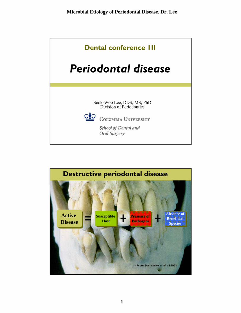
Active DiseaseActive Disease
Susceptible Host
Susceptible Host
Presence of Pathogens
Presence of Pathogens
Absence ofBeneficial
Species
Absence ofBeneficial
Species
-- From Socransky et al. (1992)
Destructive periodontal disease
Microbial Etiology of Periodontal Disease, Dr. Lee
2
Dental plaque biofilm infection
●Ecological point of viewEcological community evolved for survival as a wholeComplex community of more than 400 bacterial species
●Dynamic equilibrium between bacteria and a host defense
Adopted survival strategies favoring growth in plaque“Selection” of “pathogenic” bacteria among microbial community
Selection pressure coupled to environmental changesDisturbed equilibrium leading to pathologyOpportunistic infection
Difficulties in defining Periodontal Pathogens
●Classical Koch’s Postulatedesigned for monoinfections
●Technical difficulties●Conceptual problems●Data analysis
From Socransky et al. J. Clin Periodontol, 14:588-593, 1987

Microbial Etiology of Periodontal Disease, Dr. Lee
3
100 Years of Periodontal Microbiology
Specific
Non-specific
Specific
1890
1930
1970
Fusoformis fusiformis (1890)Streptococci (1906)Spirochetes (1912)Amoeba (1915)
Mixed Infection - Fusospirochetal (1930)Mixed Infection - with Black pigmented
Bacteroides (1955)
Spirochete - ANUG (1965)A. viscosus (1969)
A. actinomycetemcomitans (1976)P. gingivalis (1980)P. intermedia (1980)T. denticolaT. forsythia (B. forsythus)1990
Health vs. disease microflora in dental plaque
Potential pathogens

Microbial Etiology of Periodontal Disease, Dr. Lee
4
Microbiota Associated with Periodontal health, Gingivitis, and Advanced periodontal disease
0%
20%
40%
60%
80%
100%
Healthy -supragingival
Gingivitiscrevicluar
Gram-negative rods
Gram-positive rods
Gram-negativecocciGram-positive cocci
Development of dental plaque biofilm

Microbial Etiology of Periodontal Disease, Dr. Lee
5
Microbial complexes in biofilms
● Not randomly exist, rather as specific associations among bacterial species
● Socransky et al. (1998) examined over 13,000 subgingival plaque samples from 185 adults, and identified six specific microbial groups of bacterial species

Microbial Etiology of Periodontal Disease, Dr. Lee
6
S. mitusS. oralis
S. sanguisStreptococcus sp.
S. gordoniiS. intermedius
S. noxiaA. antino. b
Subgingival microbial complex
P. intermediaP. nigrescensP. microsF. nuc. nucleatumF. nuc. vincentil F. nuc. polymorphum F. periodonticum
P. gingivalisB. forsythusT. denticola
V. parvulaA. odontolyticus
E. corrodensC. gingivalisC. sputigenaC. ochraceaC. concisusA. actino. a
Actinomyces species
S. constellatus
C. gracilis
C. rectus
E. nodatum
C. showae
Criteria for defining putative periodontal pathogens
●Association with disease
●Elimination should result in clinical improvement
●Host response to pathogens
●Virulence factors
●Animal studies demonstrating tissue destruction

Microbial Etiology of Periodontal Disease, Dr. Lee
7
Possible etiologic agents of periodontal disease
●Actinobacillus actinomycetemcomitans●Porphyromonas gingivalis●Tannerella forsythia (Bacteroides forsythus)●Treponema denticola●Prevotella intermedia●Fusobacterium nucleatum●Eikenella corrodens●Campylobacter rectus (Wolinella recta)●Peptostreptococcus micros●Streptococcus intermedius
Actinobacillus actinomycetemcomitans
● First recognized as a possible periodontal pathogen in LJP (Newman et al., 1976)
● Majority of LJP patients have high Ab titers against Aa● Successful therapy lead to elimination or significant decrease of the species ● Potential virulence factors; leukotoxin, cytolethal distending toxin, invasion,
apoptosis● Induce disease in experimental animals● Eleveated in “active lesions”, compared with non-progressing sites● Virulent clonal type of Aa
LJP patients exhibit specific RFLP pattern, while healthy pts exhibit other patternsIncreased leukotoxin production by Aa strains isolated from families of African origin, a 530 bp deletion in the promoter of the leukotoxin gene operon
22.5 X more likely to convert to LJP than who had Aa strains with the full length leukotoxin promoter region
● Associated with refractory periodontitis in adult patients

Microbial Etiology of Periodontal Disease, Dr. Lee
8
Phenotypes – gram stain
A. actinomycetemcomitans F. nucleatum
Porphyromonas gingivalis
● Gram (-), anaerobic, asaccharolytic, black-pigmented bacterium
● Suspected periodontopathic microorganismAssociation
Elevated in periodontal lesions, rare in healthElimination or suppression resulted in successful therapy
Immunological correlation Elevated systemic and local antibody in periodontitis
Animal pathogenicityMonkey, dog, and rodent models
Putative virulent factors

Microbial Etiology of Periodontal Disease, Dr. Lee
9
Spirochetes
● G (-), anaerobic, spiral, highly motile● ANUG● Increased numbers in deep periodontal pockets● Difficulty in distinguishing individual species
15 subgingival spirochetes describedObscure classification - Small, medium, or large
● T. denticolaMore common in diseased, subgingival site
● Uncultivated “pathogen-related oral spirochetesDetected by Ab cross-reactivity to T. pallidum antibody
Prevotella intermedia/Prevotella nigrescens
● Strains of “P. intermedia” separated into two species, P. intermedia and P. nigrescins
● Hemagglutination activity
● Adherence activity
● Induce alveolar bone loss
● In certain forms of periodontitis
● Successful therapy leads to decrease in P. intermedia

Microbial Etiology of Periodontal Disease, Dr. Lee
10
● G(-), anaerobic, spindle-shaped rod● Has been recognized as part of the subgingival microbiota
for over 100 years● The most common isolate found in cultural studies of
subgingival plaque samples:7-10% of total isolates● Prevalent in subjects with periodontitis and periodontal
abscess● Invasion of epithelial cell● Apoptosis activity
Fusobacterium nucleatum
Other species● Campylobacter rectus
Produce leukotoxinContains the S-layerStimulate gingival fibroblast to produce IL-6 and IL-8
● Eikenella corrodens
● Peptostreptococcus microsG(+), anaerobic, small asaccharolyticLong been associated with mixed anaerobic infections
● Selemonas speciesCurved shape, tumbling motilityS. noxia found in deep pockets, conversion from healthy to disease site
● Eubacterium specues
● The “milleri” streptococciS. anginosus, S. constellatus, S. intermedius

Microbial Etiology of Periodontal Disease, Dr. Lee
11
Virus and periodontal disease
● Involvement of herpesvirus (human cytomegalovirus, HCMV and Epstein-Barr virus, EBV)
Genomes of HCMV and EBV occur at high frequency in aggressive, HIV-associated, ANUG, and advanced type periodontitis associated with medical disorders
● HCMV infects periodontal monocytes/macrophages and lymphocytes, and EBV infects periodontal B-lymphocytes
● Herpesvirus-infected inflammatory cells may Elicit tissue-destroying cytokinesExert diminished ability to defend against bacterial challenge
Herpesvirus-like virions
Gingival epithelial cells of HIV-associated necrotizing ulcerative periodontitis.

Microbial Etiology of Periodontal Disease, Dr. Lee
12
Microbial pathogenicity
● PathogenicityThe likelihood of causing disease
●VirulenceA quantitative measure of pathogenicityVirulent, avirulent strain
●Virulence factorsGene products that enhance a microorganism’s potential to cause diseaseVirulence genes
● “the pathogenic personality” of a specific pathogen
Virulence factors
●Gene products that enhance a microorganism’s potential to cause disease
● Involved in all steps of pathogenicityAttach to or enter host tissueEvade host responsesProliferateDamage the hostTransmit itself to new hosts
●Virulence genes

Microbial Etiology of Periodontal Disease, Dr. Lee
13
Expression of virulence factors
● Constitutive● Under specific environmental signals
Can be identified by mimicking environmental signals in the laboratoryMany virulence-associated genes are coordinately regulated by environmental signals
● Only in vivoCannot be identified in the laboratory Anthrax toxin, cholera toxin
Identifying virulence factors
●Microbiological and biochemical studiesIn vitro isolation and characterizationIn vivo systems
● Genetic studiesStudy of genes involved in virulenceGenetic transmission systemRecombinant DNA technology
Isogenic mutantsMolecular form of Koch’s postulates (Falkow)

Microbial Etiology of Periodontal Disease, Dr. Lee
14
Virulence factors of A. actinomycemtemcomitans
● Leukotoxin (RTX)Induce apoptosis
● Cytolethal distending toxin (CDT)● Chaperonin 60● LPS
Apoptosis, bone resorption, etc● OMP, vesicles● Fimbriae ● Actinobacillin● Collagenase● Immunosuppressive factor
Virulence factors of P. gingivalis
● Involved in colonization and attachmentFimbriae, hemagglutinins, OMPs, and vesicles
● Involved in evading (modulating) host responsesIg and complement proteases, LPS, capsule, other antiphagocytic products
● Involved in multiplyingProteinases, hemolysins
● Involved in damaging host tissues and spreadingProteinases (Arg-, Lys-gingipains), Collagenase, trypsin-like activity, fibrinolytic , keratinolytic, and other hydrolytic activities

Microbial Etiology of Periodontal Disease, Dr. Lee
15
An Example of Studying Microbial Pathogenesis
Hypothesis
S-layer of T. forsythia is a virulence factor
Tannerella forsythia (formerly B. forsythus)
● T. forsythia is a gram-negative, filament-shaped, non-motile, non-pigmented oral bacterium
● T. forsythia has been associated with advanced and recurrent periodontitis
● Implicated as one of three strong candidates for etiologic agents of periodontal disease
Actinobacillus actinomycemtemcomitansPorphyromonas gingivalisTannerella forsythia
● One of “red complex” pathogenic bacteria

Microbial Etiology of Periodontal Disease, Dr. Lee
16
Morphology of T. forsythia
Gram stain EM Negative stainingColony
● Pathogenicity is virtually unknownLittle information on virulence factorsFastidious nature of microorganisms
● Putative Virulence factorsProteolytic enzymes, trypsin-like enzymesSialidase (Neuraminidase)Leucin-rich surface protein (BspA)
BspA isogenic mutantAdhesin, inducing alveolar bone loss (mice)
Surface (S-) layer ?
Virulence factors of T. forsythia
Microbial Etiology of Periodontal Disease, Dr. Lee
17
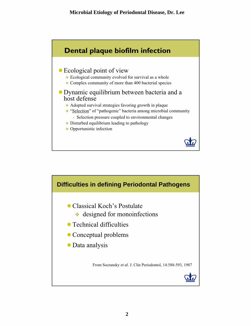
Surface layer of T. forsythia
Thin section of T. forsythia. S: S-layer; Om: outer membrane; Pm: plasma membrane
S
PmOm
Identification of the genes responsible for causing disease
●A Molecular form of Koch’s postulatesThe phenotype should be associated with pathogenic species (strains)Specific inactivation of genes associated with virulence should lead to a decrease in virulenceComplementing inactivated genes with the wild-type genes should restore full virulence
Falkow, 1988

Microbial Etiology of Periodontal Disease, Dr. Lee
18
Isolation of S-layer from T. forsythia
Negative staining
Most abundant cellular proteins
Hemagglutination activity of T. forsythia
Isolated S-layerWhole cell
Microbial Etiology of Periodontal Disease, Dr. Lee
19
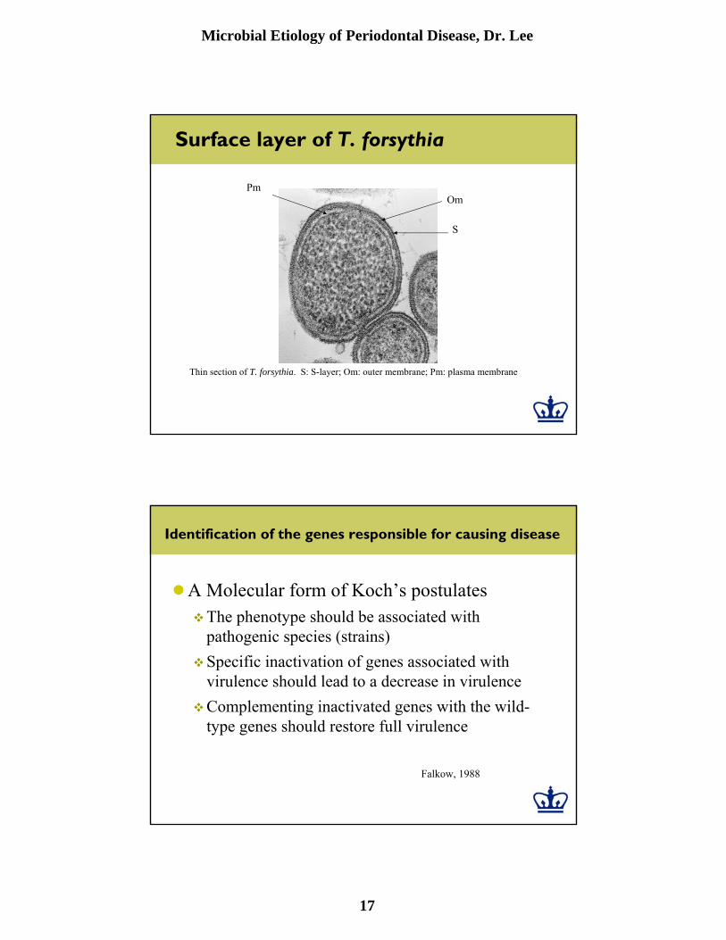
T. forsythia adheres to KB cells
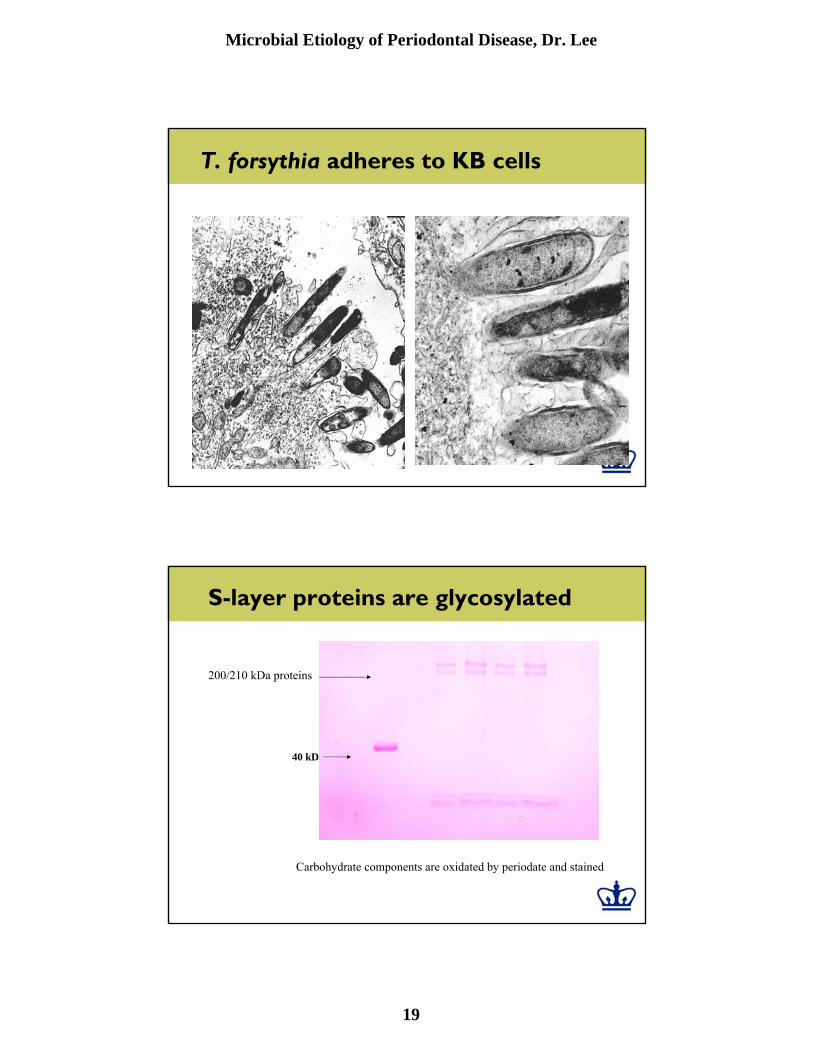
S-layer proteins are glycosylated
Carbohydrate components are oxidated by periodate and stained
200/210 kDa proteins
40 kD

Microbial Etiology of Periodontal Disease, Dr. Lee
20
tfsA
tfsB
Operon structure of tfsAB
AA BBC CMW MW
PCR RT-PCR
MWMW
A B C
tfsA tfsB5’ 3’
mRNA
Pr ATG ATG
A B C
7.8 kb
7.8 kb
Northern blot

Microbial Etiology of Periodontal Disease, Dr. Lee
21
Confirming S-layer as a virulence factor
●Construction of isogenic mutants lacking S-layer
●Use of relevant animal model for periodontal disease in testing virulence/pathogenicity