desmoid tumor of the rectus abdominis muscle in a postpartum … · cytic nevus that became...
TRANSCRIPT

Vol. 39 / No. 4 / July 2012
439
to the upper eyelid wrinkle; from the upper eyelid wrinkle to the upper eyelid borderline; the eyelash area; the lower eyelid boundary to the lower eyelid wrinkle; the isthmus area; the medial area of the eye-lid; the forehead area close to the eyebrow; and the nose and canthus. Most people with congenital melanocytic nevus in the eyelid or the periorbital area have considerable functional and aesthetic stress. In addition, for a pal-pebra with serious deformation, the suitable timing of surgery is preschool age, as the patient may be teased [4]. The most appropriate type of reconstructive pro-cedure should be selected according to the anatomi-cal units and size. A lesion that is smaller than 1/4 of the upper and lower eyelids can be reconstructed by advancing a nearby skin flap. A lesion that is large can be reconstructed with a full-thickness graft using a retroauricular flap, a lesion in an area that is close to the eyebrow can be reconstructed using an island flap and full-thickness flap surgery, and a large lesion on the forehead and the isthmus area can be recon-structed via local flap surgery using a tissue expander. As the lesions in the patient in this case belonged to zone 2 and caused no malformation of the eyelid, the functional and aesthetic problems could be solved us-ing a relatively simple procedure. Although no case of a congenital divided melano-cytic nevus that became malignant has been reported, it is considered desirable that lesions be excised as completely as possible. Although laser irradiation can be performed at certain intervals, it has the disadvan-tage of possibly leaving a pigmented lesion after the treatment. A skin graft after complete excision is commonly performed [2]. However, it may be difficult to have satisfactory results with a skin graft due to the differ-ence in the color and texture of the skin. In this case, the upper eyelid scar was successfully hidden and a satisfactory outcome was achieved via complete ana-tomical excision of the nevi and via reconstruction using blepharoplasty. No particular complications were observed in the 6 months follow-up, and the patient’s psychological stress due to the nevus was cleared and he was very satisfied (Fig. 3).
References
1. McDonnell PJ, Mayou BJ. Congenital divided naevus of the eyelids. Br J Ophthalmol 1988;72:198-201.
2. Margulis A, Adler N, Bauer BS. Congenital melanocytic
nevi of the eyelids and periorbital region. Plast Reconstr Surg 2009;124:1273-83.
3. Sevel D. A reappraisal of the development of the eyelids. Eye (Lond) 1988;2:123-9.
4. Bellier-Waast F, Perrot P, Duteille F, et al. Surgical treatment for giant congenital nevi: what are the psychosocial consequences for the child and family? Ann Chir Plast Esthet 2008;53:408-14.
Desmoid Tumor of the Rectus Abdominis Muscle in a Postpartum PatientSeung Ho Choi, Jung Ho Lee, Bommie F. Seo, Sang Wha Kim, Jong Won Rhie, Sang Tae AhnDepartment of Plastic and Reconstructive Surgery, The Catholic University of Korea College of Medicine, Seoul, Korea
Correspondence: Jung Ho LeeDepartment of Plastic and Reconstructive Surgery, The Catholic University of Korea College of Medicine, 222 Banpo-daero, Seocho-gu, Seoul 137-040, KoreaTel: +82-2-2258-6144, Fax: +82-2-594-7230E-mail: [email protected]
No potential conflict of interest relevant to this article was reported.
Received: 4 May 2012 • Revised: 12 Jun 2012 • Accepted: 13 Jun 2012pISSN: 2234-6163 • eISSN: 2234-6171http://dx.doi.org/10.5999/aps.2012.39.4.439 • Arch Plast Surg 2012;39:439-441
Copyright 2012 The Korean Society of Plastic and Reconstructive SurgeonsThis is an Open Access article distributed under the terms of the Creative Commons Attribution Non-Commercial License (http://creativecommons.org/licenses/by-nc/3.0/) which permits unrestricted non-commercial use, distribution, and reproduction in any medium, provided the original work is properly cited.
Desmoid tumors, also known as aggressive fibroma-tosis, are uncommon mesenchymal neoplasms. They account for only 0.03% of all tumors and 3% of all soft-tissue neoplasms [1]. A pregnancy-associated desmoid tumor is an even less common disease very rarely reported in published articles. Although it grows by locally infiltrating its neighboring tissues and structures, it is devoid of metastatic evolution. It usually occurs between the ages of 25 and 40 years, with a strong prevalence among women in the fertile age group [1]. A 36-year-old woman presented with a painless mass in the right upper quadrant of her abdomen. She had discovered the mass six months earlier and stated that the size of the mass had not increased. She had no relevant family history of related ailments such as of the familial adenomatous polyposis syndrome
Images
440
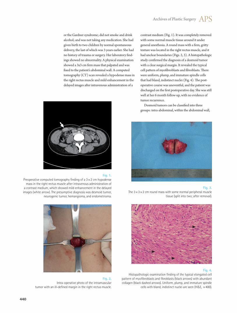
Fig. 4. Histopathologic examination finding of the typical elongated cell
pattern of myofibroblasts and fibroblasts (black arrows) with abundant collagen (black dashed arrows). Uniform, plump, and immature spindle
cells with bland, indistinct nuclei are seen (H&E, ×400).
or the Gardner syndrome; did not smoke and drink alcohol; and was not taking any medication. She had given birth to two children by normal spontaneous delivery, the last of which was 3 years earlier. She had no history of trauma or surgery. Her laboratory find-ings showed no abnormality. A physical examination showed a 3x3 cm firm mass that palpated and was fixed to the patient’s abdominal wall. A computed tomography (CT) scan revealed a hypodense mass in the right rectus muscle and mild enhancement in the delayed images after intravenous administration of a
Fig. 1. Preoperative computed tomography finding of a 3×2 cm hypodense
mass in the right rectus muscle after intravenous administration of a contrast medium, which showed mild enhancement in the delayed
images (white arrow). The presumptive diagnosis was desmoid tumor, neurogenic tumor, hemangioma, and endometrioma.
contrast medium (Fig. 1). It was completely removed with some normal muscle tissue around it under general anesthesia. A round mass with a firm, gritty texture was located in the right rectus muscle, and it had unclear boundaries (Figs. 2, 3). A histopathologic study confirmed the diagnosis of a desmoid tumor with a clear surgical margin. It revealed the typical cell pattern of myofibroblasts and fibroblasts. There were uniform, plump, and immature spindle cells that had bland, indistinct nuclei (Fig. 4). The post-operative course was uneventful, and the patient was discharged on the first postoperative day. She was still well at her 6 month follow-up, with no evidence of tumor recurrence. Desmoid tumors can be classified into three groups: intra-abdominal, within the abdominal wall,
Fig. 3. The 3×3×2 cm round mass with some normal peripheral muscle
tissue (split into two; after removal).
Fig. 2. Intra-operative photo of the intramuscular
tumor with an ill-defined margin in the right rectus muscle.

Vol. 39 / No. 4 / July 2012
441
[3]. Stable asymptomatic desmoids can be observed and followed up with imaging at a 3- to 6-month interval. Treatment is necessary for symptomatic patients, especially those with a mass effect on criti-cal structures. Surgery is the mainstay treatment in patients with extra-abdominal desmoid tumors, including abdominal wall desmoid tumors, as in this case. Though a few authors report that recurrence is independent of the negative margin, most investiga-tors recommend resection with a wide margin of surrounding normal tissue to minimize the risk of local recurrence. In addition, a retrospective analysis of 189 desmoid tumor patients revealed that the pres-ence of a tumor at the microscopic surgical margin was the single most significant determinant of the recurrence of desmoid tumor in patients treated with surgery. Thus, in some cases of large desmoid tumors, reconstruction procedures such as transposition of aponeurotic-muscular flaps or use of a mesh product should be considered preoperatively [4]. In conclusion, the diagnosis of desmoid tumor should be strongly considered in female patients who have experienced childbirth presenting with an abdominal wall mass even though it is a rare entity. Our case of desmoid tumor in a postpartum patient, a very rare occurrence, was completely treated with surgery and is under follow-up without any relapse.
References
1. Economou A, Pitta X, Andreadis E, et al. Desmoid tumor of the abdominal wall: a case report. J Med Case Rep 2011;5:326.
2. De Cian F, Delay E, Rudigoz RC, et al. Desmoid tumor arising in a cesarean section scar during pregnancy: monitoring and management. Gynecol Oncol 1999;75: 145-8.
3. Carneiro C, Hurtubis C, Singh M, et al. Desmoid tumors of the right rectus abdominus muscle in postpartum women. Arch Gynecol Obstet 2009;279:869-73.
4. Johner A, Tiwari P, Zetler P, et al. Abdominal wall desmoid tumors associated with pregnancy: current concepts. Expert Rev Anticancer Ther 2009;9:1675-82.
5. Shinagare AB, Ramaiya NH, Jagannathan JP, et al. A to Z of desmoid tumors. AJR Am J Roentgenol 2011;197:W1008-14.
and extra-abdominal. The histologic findings on these lesions are identical. Sixty percent of desmoids are extra-abdominally located, and 15% appear in the abdominal cavity. Another 25% of desmoids are in the abdominal wall [2]. Extra-abdominal desmoid tu-mors are widely distributed but are commonly found in the shoulder girdle, trunk, and lower extremities. Most intra-abdominal desmoid tumors occur in patients with familial adenomatosis polyposis, and when these two conditions occur together, they are often called “Gardner syndrome.” A clear link between the development of abdomi-nal wall desmoid tumors and the pregnancy and post-partum state has appeared in many case reports in the literature [3]. Interestingly, in most of these cases, des-moid tumors almost always occur in the abdominal wall of postpartum women, and there is a predilection for the right rectus abdominis muscle, as in this case. The definite etiology of desmoid tumors, whether pregnancy-associated or otherwise, is currently un-known. It has been postulated that the hormonal and immunologic changes in the pregnant or postpartum state appear to be important factors. The local effect due to the mechanical stress from a gravid uterus may also contribute to the occurrence of a desmoid tumor [4]. Histologically, desmoid tumors consist of elon-gated fibroblasts and myofibroblasts characterized by elongated, tapered cytoplasm; elongated, vesicular, and typical appearing nuclei; and multiple small nu-cleoli. The cells are linearly arranged and surrounded and separated from each other by collagen [1]. Ultrasonography has shown that desmoid tumors appear as well-defined lesions with variable echo-genicity. Their lateral borders may occasionally ap-pear ill-defined or irregular [1]. The CT appearance of desmoid tumors depends on their composition. They may appear homogeneous or heterogeneous and hypo-, iso-, or hyperintense compared with the attenuation of the muscles. The degree of their en-hancement after the intravenous administration of a contrast medium is variable. The magnetic resonance imaging (MRI) features of desmoid tumors also show wide variability depending on the stage in which they are imaged. Characteristic MRI findings include poor margination, low signal intensity on T1-weighted images and heterogeneity on T2-weighted images, and variable contrast enhancement. MRI may have a prognostic value because a higher T2 signal is report-edly associated with faster growth [5]. The treatment options for desmoid tumors can vary according to the individual patient’s condition