design of thiol–ene photoclick hydrogels using facile
TRANSCRIPT

Design of Thiol–ene Photoclick Hydrogels Using Facile Techniques for Cell Culture Applications
Journal: Biomaterials Science
Manuscript ID: BM-ART-05-2014-000187.R1
Article Type: Paper
Date Submitted by the Author: 06-Aug-2014
Complete List of Authors: Sawicki, Lisa; University of Delaware, Chemical Engineering Kloxin, April; University of Delaware, Chemical & Biomolecular Engineering and Materials Science & Engineering
Biomaterials Science

1
Design of Thiol–ene Photoclick Hydrogels Using
Facile Techniques for Cell Culture Applications
Lisa A. Sawicki,a April M. Kloxin
a,b
aDepartment of Chemical and Biomolecular Engineering, University of Delaware, Newark, DE
19716, USA. Email: [email protected]
bDepartment of Materials Science and Engineering, University of Delaware, Newark, DE 19716,
USA.
ABSTRACT
Thiol–ene ‘click’ chemistries have been widely used in biomaterials applications, including
drug delivery, tissue engineering, and controlled cell culture, owing to their rapid,
cytocompatible, and often orthogonal reactivity. In particular, hydrogel-based biomaterials
formed by photoinitiated thiol–ene reactions afford spatiotemporal control over the biochemical
and biomechanical properties of the network for creating synthetic materials that mimic the
extracellular matrix or enable controlled drug release. However, the use of charged peptides
Page 1 of 45 Biomaterials Science
2
functionalized with cysteines, which can form disulfides prior to reaction, and vinyl monomers
that require multistep syntheses and contain ester bonds, may lead to undesired inhomogeneity or
degradation under cell culture conditions. Here, we designed a thiol–ene hydrogel formed by the
reaction of alloxycarbonyl-functionalized peptides and thiol-functionalized poly(ethylene
glycol). Hydrogels were polymerized by free radical initiation under cytocompatible doses of
long wavelength ultraviolet light in the presence of water-soluble photoinitiators (lithium
acylphosphinate, LAP, and 2-hydroxy-1-[4-(2-hydroxyethoxy) phenyl]-2-methyl-1-propanone,
Irgacure 2959). Mechanical properties of these hydrogels were controlled by varying the
monomer concentration to mimic a range of soft tissue environments, and hydrogel stability in
cell culture medium was observed over weeks. Patterns of biochemical cues were created within
the hydrogels post-formation and confirmed through the incorporation of fluorescently-labeled
peptides and Ellman’s assay to detect free thiols. Human mesenchymal stem cells remained
viable after encapsulation and subsequent photopatterning, demonstrating the utility of the
monomers and hydrogels for three-dimensional cell culture. This facile approach enables the
formation and characterization of hydrogels with well-defined, spatially-specific properties and
expands the suite of monomers available for three-dimensional cell culture and other biological
applications.
INTRODUCTION
Click chemistries for the formation and modification of biomaterials have garnered significant
and growing interest for numerous applications, including drug delivery, tissue engineering, and
controlled cell culture.1, 2
A number of functional groups undergo efficient and highly selective
Page 2 of 45Biomaterials Science

3
click reactions under a variety of cytocompatible conditions, making them well suited for the
manipulation of biomaterial properties in the presence of cells.3, 4
These reactions include
radically initiated thiol–ene and thiol–yne,5, 6
thiol-Michael addition,7, 8
spontaneous reaction of
azides with strained alkynes,9, 10
and spontaneous reaction of tetrazine with norbornene and
transcyclooctene,11, 12
which have been used to examine the effects of matrix properties on cell
behavior,6, 7, 9, 11
to label cells and biomolecules,10, 12
and to form carriers for drug delivery.13
Amongst these, thiol–ene click chemistries have been examined broadly for the formation and
modification of hydrogel-based biomaterials owing to their ease of use and the availability of
thiols in many biomolecules.14
Hydrogels formed by thiol–ene click reactions have been constructed with a range of
cytocompatible polymers and copolymers, such as poly(ethylene glycol) (PEG),15
hyaluronic
acid,16
and poly(ethylene glycol)-poly(lactic acid),17
and modified with peptides and proteins,
such as GPQG↓IWGQ,18
IPVS↓LRSG,18
and RGDS,19
to impart specific biological activity.16, 20
Various vinyl functional groups have been investigated for this purpose, including norbornene,19
vinyl sulfone,8 and allyl ether.
21 For example, the Michael-type addition of thiols on peptides
with vinyl groups (‘ene’s) on vinyl sulfone-modified PEG has been widely employed to design
hydrogels with controlled, cell-responsive properties for use in drug delivery or tissue
engineering.8, 22
These reactions proceed via a step growth mechanism,5, 14
resulting in a
homogeneous network structure with robust mechanical properties for applications in cell culture
and delivery.23
Photoinitiated thiol–ene systems are particularly attractive for hydrogel formation and
modification because they allow user-directed control over the presentation of biophysical or
biochemical cues in space and in time to promote specific cellular functions and toward
Page 3 of 45 Biomaterials Science
4
mimicking the dynamic structure or composition of the native extracellular matrix (ECM) in
vitro.24, 25
Peptides modified with cysteines and polymers modified with acrylates (mixed step
and chain growth mechanism) or norbornenes (step growth mechanism) have been extensively
used owing to their rapid reaction under cytocompatible photopolymerization conditions.19, 26, 27
For example, Fairbanks et al. first demonstrated that norbornene-modified PEG reacts within
minutes with cysteine-modified, enzymatically degradable crosslinking peptides in the presence
of a radical initiator to form hydrogels by step growth free radical polymerization.19
This strategy
(vinyl-modified PEG) has been used to encapsulate a number of cell types including, but not
limited, to osteoblasts, chondrocytes, mesenchymal stem cells (MSCs), and smooth muscle
cells.28
These chemistries also have been used to create new biomaterial systems, such as a
hydrogel formed by the reaction of norbornene-modified hyaluronic acid with a dithiol
crosslinker and modified with patterns of biochemical cues at select time points.16
Despite their great utility, there are a few potential concerns when using these existing thiol–
ene photoclick systems. Recently, Shih and Lin observed that ester bonds present in polymers
modified with various vinyl groups (e.g., acrylic acid or norbornene carboxylic acid) degrade
over relatively short times in water or cell culture conditions (i.e., days to weeks), where the
hydrolysis rate is affected by the incorporation of different charged peptide sequences.29
Preprogrammed degradation afforded by hydrolysis allows cell spreading within the matrix;
however, it is often desirable for the rate of degradation to respond dynamically to cell secreted
enzymes or an externally-applied stimulus (e.g., light). Toward designing alternate systems with
controlled degradation (e.g., cell-secreted enzymes or light), polymer precursors modified with
amine functional groups instead of hydroxyls been utilized, introducing more water-stable amide
bonds upon reaction with carboxylic acid-containing functional groups.30, 31
Despite this
Page 4 of 45Biomaterials Science

5
increased stability, there is typically increased cost or synthetic processing associated with using
these materials. Additionally, the formation of disulfide bonds between cysteine-modified
charged peptides32
before reaction may deplete the concentration of thiols present in the reaction
solution, resulting in an off-stoichiometry mixture, defects in the network structure, and slower
polymerization times.33, 34
Herein, an approach to rapid thiol–ene photoclick polymerization between a vinyl-modified
peptide and thiol-modified PEG is presented (Figure 1). A multiarm PEG thiol is used as the
‘backbone’ of the hydrogel structure with thiols on each arm connected by ether bonds. The
PEG backbone is not charged, limiting potential disulfide formation,35
and ether bonds
neighboring thiol functional groups provide a water-stable base for the introduction of
enzymatically degradable peptide sequences for cell-dictated degradation. The alloxycarbonyl
(alloc) group, which is used to protect the amines of amino acids (e.g., lysine) during peptide
synthesis, is incorporated within pendant (single)15, 36, 37
and crosslink (double) peptide
sequences to provide vinyls for reaction with the PEG thiol backbone. The use of lithium
acylphosphinate (LAP) as a photoinitiator, which has increased rates of initiation and
polymerization relative to other water-soluble photoinitiators,38
allows the rapid reaction of the
alloc-modified peptides with the multiarm PEG thiol to form hydrogels under cytocompatible
doses of long wavelength ultraviolet (UV) light (10 mW/cm2, 365 nm).
39 Further, these
monomers may be purchased commercially or synthesized with relatively simple techniques
presented here, making the system accessible to researchers in a variety of fields. In this article,
the polymerization, mechanical properties, stability, cytocompatibility, and spatial patterning of
these robust thiol–ene photoclick hydrogels are characterized to define and demonstrate their
potential for use as three-dimensional (3D) mimics of the ECM, particularly for the evaluation of
Page 5 of 45 Biomaterials Science

6
cell-matrix interactions. In addition to the application of these materials in controlled cell culture
models, we believe this approach may be useful for the in situ modification of assembling
peptides (e.g., adding functionalities to supramolecular structures to allow electrical conduction,
enhance imaging, or promote specific biological interactions)40, 41
and even in membrane
applications (e.g., forming stable, charged PEG-based membranes for batteries).42
MATERIALS AND METHODS
Synthesis of PEG-thiol macromer. Poly(ethylene glycol)-tetrathiol (PEG4SH) is
commercially available (JenKem Technology USA, Creative PEGWorks) or can been
synthesized as was done here using a modified version of published protocols.43
Briefly, four-
arm PEG (Mn ~ 20,000 g/mol, 10 g) (JenKem USA) was dissolved in anhydrous tetrahydrofuran
(THF, 70 mL) (Fisher Scientific) and purged with argon, and argon-purged sodium hydroxide
(NaH, 4x molar excess with respect to –OH groups) (Sigma Aldrich) suspended in THF was
transferred via cannula under argon to the dissolved PEG. Allyl bromide (3x molar excess with
respect to –OH groups) (Acros Organics) dissolved in 30 mL of THF subsequently was added.
The PEG-allyl solution was refluxed overnight at 40 °C under argon and precipitated in ice cold
ethyl ether to generate allyl ether-modified PEG (PEG4AE). The PEG4AE was dissolved in
dichloromethane (40 mL) (Fisher Scientific) with a photoinitiator (2,2-dimethoxy-1,2-
diphenylethan-1-one, I651, 0.5% w/w) (Acros Organics) and trace trifluoroacetic acid (TFA, ~
100 µL) (Acros Organics) and purged with argon. Thioacetic acid (2x molar excess with respect
to allyl) (Acros Organics) was added, and the solution was purged with argon and subsequently
exposed to UV light (365 nm at 10-15 mW/cm2
for 45 minutes) to yield PEG-thioacetate
Page 6 of 45Biomaterials Science
7
(PEG4TA) after precipitation in ice cold diethyl ether. Last, PEG4TA was dissolved in 60-70
mL of water and purged with argon. An equal volume of 1 M sodium hydroxide (Fisher
Scientific) purged with argon was added to the PEG4TA (0.5 M final concentration) to generate
the thiol end groups on the final PEG4SH product. The reaction immediately was neutralized
with hydrochloric acid (final pH 1-2) (Fisher Scientific) and PEG4SH extracted with chloroform
and trace TFA (to prevent disulfide formation) and precipitated in ice cold diethyl ether. To
wash and collect all intermediates and the final product after precipitation, samples were
centrifuged at 0 °C for 20 minutes at 4400 rpm for a total of 3 washes and dessicated under
vacuum at room temperature overnight. All intermediates and the final product were
characterized with proton nuclear magnetic resonance (1H NMR) in DMSO: PEG4AE 5.1-5.2
(m, 1H) 5.2-5.3 (m, 1H) 5.8-5.9 (m, 1H); PEG4TA 2.3 (s, 3H); PEG4SH 2.3 (m, 1H) for a single
arm of the tetrafunctional monomer (Supplemental Figure S1).
Synthesis of alloc-functionalized peptides. The pendant cell adhesion sequence
K(alloc)GWGRGDS (RGDS), a ubiquitous sequence found in many ECM proteins including
fibronectin and vitronectin,44
was synthesized to promote cell adhesion (amino acid(s) with
reactive functional groups in bold). Non-degradable, water-soluble crosslinking sequences were
synthesized: K(alloc)RGKGRKGK(alloc)G37
(RGKGRK2alloc) (primary sequence used in
hydrogel development) and K(alloc)GKGWGKGK(alloc)G (GKGWGKG2alloc) and
CGKGWGKGCG (GKGWGKG2SH) (sequences with reduced charge and including tryptophan
for easily assessing their concentration). Additionally, an enzymatically degradable, water-
soluble crosslinking sequence KK(alloc)GGPQG↓IWGQGK(alloc)K (GPQGIWGQ2alloc)
(broadly degradable by matrix metalloproteinases (MMP)-1, 2, 3, 8, and 9)18
was synthesized to
Page 7 of 45 Biomaterials Science
8
promote cell viability and allow spreading in longer cell culture and photopatterning
experiments. Each was synthesized by standard solid phase peptide synthesis (SPPS) techniques
using Fmoc chemistry on MBHA rink amide resin (0.59 mmol/g; 0.25 mmol scale)
(Novabiochem) with a peptide synthesizer (Protein Technologies PS3). Fmoc-protected amino
acids, including the commercially-available alloc-protected lysine, and o-(benzotriazol-1-yl)-
n,n,n’,n’-tetramethyluronium hexafluorophosphate (HBTU) (4x excess) (Chem-Impex
International) were loaded into cartridges and coupled on resin. Fmoc deprotection was carried
out using 20% piperidine (Sigma Aldrich) in n,n-dimethylformamide (DMF) (Fisher Scientific)
prior to each amino acid coupling in 0.4 M methylmorpholine in DMF. Peptide products were
cleaved in 95% v/v trifluoroacetic acid (TFA), 2.5% v/v triisopropylsilane (TIPS) (Acros
Organics), and 2.5% v/v water with 5% w/v dithiothreitol (DTT) (Research Products
International Corporation) to prevent disulfide formation and 2.5% w/v phenol (Sigma Aldrich)
to protect tryptophan (W). After cleavage from the resin, peptides were precipitated in ice cold
diethyl ether, centrifuged at 3000 rpm and 4 °C for 5 minutes for a total of three washes and
dessicated under vacuum overnight at room temperature. Dry raw peptide product was purified
by high-performance liquid chromatography (HPLC) and analyzed by matrix-assisted laser
desorption/ionization (MALDI, crystallized with α-cyano-4-hydroxycinnamic acid, Acros
Organics) or electrospray ionization (ESI) mass spectrometry to confirm synthesis of each
desired peptide (Supplemental Figure S2).
A fluorescently-labeled pendant peptide, Alexa Fluor 488-AhxWGRGDSK(alloc)G
(AF488RGDS), also was designed for photopatterning experiments using published protocols.37
After Fmoc deprotection of Ahx on the N’-terminus of the peptide, 1 mg Alexa Fluor® 488
Carboxylic Acid, 2,3,5,6-Tetrafluorophenyl Ester, 5-isomer (Invitrogen) was stirred with 0.25
Page 8 of 45Biomaterials Science

9
mmol peptide on resin in 4 mL DMF and 50 µL n,n’-diisopropylethylamine (DIPEA) (Chem-
Impex International) overnight. The peptide was cleaved from resin, precipitated, and analyzed
by HPLC and ESI mass spectrometry (Supplemental Figure S2).
Synthesis of LAP initiator. The LAP initiator was synthesized using previously-described
methods.38
Briefly, 2,4,8-trimethylbenzoyl chloride (1.6 g, 0.009 mol) (Sigma Aldrich) was
added to dimethyl phenylphosphonite (1.5 g, 0.009 mol) (Acros Organics) and reacted overnight
at room temperature under argon. Lithium bromide (4x molar excess) (Sigma Aldrich) in 2-
butanone (Sigma Aldrich) was added to the reaction solution and heated to 50 °C for 10 minutes.
The white precipitate was filtered and rinsed 3 times with 2-butanone, and the final powder
product dried and analyzed by 1H NMR, matching literature (Supplemental Figure S3).
38
Hydrogel formation. All monomers and initiators were prepared in Dulbecco’s phosphate
buffered saline (PBS) (Life Technologies) immediately before polymerization. For the various
experiments described below, solutions of PEG4SH, RGKGRK2alloc (unless noted otherwise),
and RGDS (7.5, 10, 12.5 wt% with respect to PEG, 2 mM RGDS) were prepared at
stoichiometric ratios of thiol functional groups to alloc functional groups (1:1 SH:alloc) and
containing a photoinitiator, either LAP (1.1 and 2.2 mM) or Irgacure 2959 (I2959) (2.2 mM).
Hydrogels were formed upon irradiation of the monomer-initiator solution with cytocompatible
doses of long wavelength UV light (365 nm at 10 mW/cm2, International Light IL1400A
Radiometer/Photometer) in the specific geometries described below.
Page 9 of 45 Biomaterials Science
10
Rheometry. Hydrogels were formed in situ on a photorheometer (TA AR-G2 with UV light
attachment, Exfo Omnicure Series 2000 light source, 365 nm filter, SilverLine UV Radiometer
M007-153) to estimate the polymerization times for different initiator types and monomer
concentrations. I2959 (2.2 mM) or LAP (1.1 or 2.2 mM) photoinitiators were added to 10 wt%
PEG monomer solutions containing stoichiometrically balanced amounts (1:1 SH:alloc) of
RGKGRK2alloc to compare the effects of initiator type on polymerization time (n=3). PEG
monomer solutions (7.5, 10, and 12.5 wt%) containing stoichiometrically balanced amounts of
RGKGRK2alloc and RGDS (2 mM) were mixed with 2.2 mM LAP to compare the effects of
monomer concentration on polymerization time (n=6). Finally, PEG4SH or PEG4AE monomer
solutions (10 wt%) containing stoichiometrically balanced amounts of alloc (RGKGRK2alloc,
GKGWGKG2alloc, GPQGIWGQ2alloc) or thiol-modified crosslinkers (PEG2SH,
GKGWGKG2SH) were mixed with 2.2 mM LAP to compare the effects of crosslinker and
functional group chemistry on polymerization time (n=3). These solutions were placed between
parallel plates (8 mm diameter, 200 µm gap) and UV light (365 nm at 10 mW/cm2) applied 1
minute after starting rheometric measurements. Storage (G') and loss moduli (G") were recorded
over time at 2% applied strain and 6 rad/s frequency. From the data, an approximate time for
complete gelation was defined to be when the percent change in modulus between consecutive
data points was less than 0.1%.
For swollen modulus experiments, 7.5, 10, and 12.5 wt% hydrogels were polymerized within a
1-mm thick mold (2 microscope slides treated with Rain-X separated by a 1-mm rubber gasket).
After polymerization, discs (8 mm diameter) were punched from the gel slab and swollen
overnight in PBS. Strain sweeps (1 rad/s frequency, 1-100% strain) and frequency sweeps (1-
100 rad/s frequency, 5% strain) were conducted on swollen gels to determine the linear
Page 10 of 45Biomaterials Science
11
viscoelastic regime for the material. The swollen gels were then placed between parallel plates
on the rheometer and G' and G" were measured at 5% strain and 5 rad/s frequency (within the
linear viscoelastic regime) (n=6).
Hydrogel swelling. Experiments to determine volumetric swelling ratios (Q) were performed
on 7.5, 10, and 12.5 wt% hydrogels. Discs (8 mm diameter) were punched from gels
polymerized between glass slides separated by a 1-mm thick gasket, ensuring sufficient mass for
measuring dry weight, and swollen overnight in PBS. After recording swollen mass (Ms), the
gels were lyophilized and the dry masses were measured (Md) (n=6). Volumetric swelling ratio
was calculated by the relationships:
� = ��
��, � = 1 +
�� ����
�� ������ − 1�
where q is the swelling ratio, ρpolymer = 1.07 g/mL45
for PEG, and ρsolvent = 1.00 g/mL for PBS.
Experiments to determine gel stability after polymerization were performed on gels incubated
in PBS and cell culture medium at 37 °C over a 3 week time course. Gels (10 wt%) were
polymerized for 5 minutes in 5-mm diameter molds (1-mL syringes with tips cut off) under
sterile conditions and placed in sterile PBS and cell culture medium. Ms and Md were recorded
for the gels after 1, 7, 14, and 21 days (n=6). Values for the volumetric swelling ratio (Q) were
calculated as described above.
Detection of unreacted thiols. To initially quantify the photoaddition of biochemical cues,
hydrogels (10 wt% with respect to PEG) were polymerized (1 or 5 minutes) between glass slides
Page 11 of 45 Biomaterials Science

12
separated by a 0.254-mm thick gasket (McMaster-Carr) and off-stoichiometry such that
approximately 2 mM free thiol remained in the unswollen gel after polymerization. Discs (5 mm
diameter) were punched from these gels for further treatment and analysis. Half of the gel discs
were swollen in PBS containing LAP initiator (2.2 mM) and excess pendant peptide (20 mg/mL,
K(alloc)GWGRGDS) and incubated at room temperature for 1 hour. After 1 hour, these gels
were exposed to UV light for 1 or 5 minutes to initiate the photoaddition of the RGDS. The
other half of the gels remained in PBS as a control. Free thiol concentrations in the gels were
quantitatively detected by Ellman’s assay as described below.
Briefly, the swollen volume of the gels was predicted using the measured Q value (estimated at
19.3 µL). Ellman’s reaction buffer (20.7 µL) containing 0.1 M sodium phosphate (Sigma
Aldrich) and 1 mM ethylenediaminetetraacetic acid (Sigma Aldrich) at pH 7.5-8 was added to
the gels for a total volume of 40 µL. Ellman’s reagent (7.2 µL, 4 mg in 1 mL reaction buffer)
(Fisher Scientific) was diluted in 360 µL of reaction buffer and added to each well containing a
gel. Gels were incubated in the reagent for 1 hour and 30 minutes, the estimated time for the
diffusion of the yellow NTB2-
dianion out of the gel so that the supernatant and gel colors match
(by visual inspection). Finally, a calibration curve of L-cysteine hydrochloride monohydrate
(Sigma Aldrich) (0-2 mM) was made to calculate the concentration of thiols detected in each gel.
Absorbance of each condition was measured at 405 nM (Biotek Synergy H4 automated plate
reader).
To determine the free thiol concentration in conditions for photopatterning in the presence of
encapsulated cells, 10 wt% gels were polymerized in syringe tips (20 µL) such that
approximately 2 mM free thiol remained in the unswollen gel after polymerization. Gels
polymerized for 1 and 5 minutes were placed immediately in PBS as a control (n=3). Additional
Page 12 of 45Biomaterials Science

13
gels polymerized for 1 minute were immediately placed in solutions of PBS containing 3 mg/mL
RGDS and 2.2 mM LAP and incubated at 37 °C for 30 minutes (n=3) or 1 hour 30 minutes
(n=3). After incubation, these gels were exposed to a second dose of UV light for 1 minute to
attach the biochemical cue (RGDS) to remaining free thiols. Free thiol concentrations in the gels
were quantitatively detected by Ellman’s assay as described above, accounting for larger gel size
(swollen volume = 84.8 µL; add 15.2 µL of PBS to gel in well plate for 100 µL total volume; add
18 µL Ellman’s reagent in 900 µL Ellman’s buffer to each well).
Spatially-specific photopatterning of biochemical cues. Hydrogels (10 wt%) were
polymerized between glass slides spaced by a 0.254-mm gasket and off-stoichiometry to have a
final free thiol concentration of 2 mM within the as prepared gel (prior to equilibrium swelling).
The hydrogel was left on one of the glass slides for subsequent treatments and rinsed with PBS
for 1 hour. Rinsed gels were placed in solution containing pendant peptides (AF488RGDS or
RGDS) mixed with 2.2 mM LAP initiator for 1 hour and 30 minutes to allow diffusion of the
peptides and initiator into the gel network prior to subsequent patterning. Photomasks with lines
of increasing thickness (0.2-1 mm width) or square patterns (0.4 mm edge) purchased from
Advanced Reproductions Corporation were placed ink-side down on top of the samples and
exposed to collimated UV light (Inpro Technologies collimating adaptor, Exfo Omnicure Series
2000 light source) for 1 minute (365nm at 10 mW/cm2). Gels were rinsed 3x for 40 minutes
each with PBS to remove excess pendant peptide after photoaddition. Samples containing the
patterned AF488RGDS were imaged with a confocal microscope (Zeiss 510 NLO). Ellman’s
reagent was applied to the gels containing RGDS and imaged immediately on a
stereomicroscope (Zeiss Stemi 2000-C).
Page 13 of 45 Biomaterials Science
14
Culture and encapsulation of human mesenchymal stem cells. Human mesenchymal stem
cells (hMSCs) isolated from human bone marrow (Lonza)46
were cultured on tissue-culture
treated polystyrene in cell culture medium46
and harvested at ~70-80% confluency (Passage 2, 3)
for experiments. For evaluating the effects of light, cells were trypsinized from culture plates,
counted (hemacytometer), centrifuged (5 minutes, 1000 rpm), and plated at a density of 20000
cells/cm2 in 96-well plates. For cell encapsulation and photopatterning experiments, cells were
trypsinized from culture plates, counted (hemocytometer), centrifuged (5 minutes, 1000 rpm),
and resuspended at desired densities in monomer solution (10 wt%) with and without RGDS.
The mixtures of cells in monomer solution were polymerized in syringe molds at cytocompatible
wavelengths and doses of UV light (365 nm at 10 mW/cm2), encapsulating cells within the
hydrogel matrix.
Metabolic activity of hMSCs in photopatterned and non-patterned hydrogels. Cells were
suspended in monomer solution (10 wt%, 3000 cells/µL) containing 2 mM RGDS and
polymerized in syringe tip molds (20 µL) for 1 and 5 minutes (n=6, non-patterned). Immediately
after polymerization, gels were placed in cell culture medium to rinse out unreacted monomer
and photoinitiator (30 minutes). After rinsing, the medium was replaced with fresh medium and
gels were incubated at 37 °C for subsequent analysis. For photopatterned gels, cells were
suspended in monomer solution (10 wt%, 3000 cells/µL) without RGDS and polymerized for 1
minute such that 2 mM free thiols remained in the unswollen gel for subsequent modification.
After polymerization, the gels were incubated in PBS containing 3 mg/mL RGDS and 2.2 mM
LAP for 30 minutes or 1 hour 30 minutes at 37 °C before exposure to a second dose of UV light
(1 minute) to covalently link RGDS within the network (n=6). Patterned gels were immediately
Page 14 of 45Biomaterials Science
15
placed in cell culture medium (30 minutes) to rinse out excess monomer and photoinitiator. At 1
and 3 days post-encapsulation (D1 and D3), metabolic activity was assessed by CellTiter 96
(Promega) (n=3 each condition, each time point).
To assess the effect of light alone on cell function, plated cells (20000 cells/cm2) were exposed
to UV light (1 min of 365 nm at 10 mW/cm2). Metabolic activity was assessed by CellTiter 96 at
D1 and D3 compared to control (no light) (n=3 each condition, each time point).
Viability of hMSCs in photopatterned and non-patterned hydrogels. To initially study the
viability of cells encapsulated in hydrogels, 3000 cells/µL were encapsulated in non-degradable
gels (10 wt%, 2 mM RGDS before swelling) polymerized for 1 and 5 minutes. Additional studies
were performed to determine the effect of cell density on viability post-encapsulation, with cells
encapsulated in in non-degradable gels (10 wt%, 2 mM RGDS before swelling) at 3000 and
30000 cells/µL. Viability was quantified at 3 days post-encapsulation with a LIVE/DEAD
Viability/Cytotoxicity Kit for mammalian cells (Invitrogen), and gels were imaged with a
confocal microscope (Zeiss 510 NLO).
To study viability of cells in photopatterned hydrogels over longer times in culture, cells were
encapsulated in gels (10 wt%, 20 µL, 3000 cells/µL) crosslinked with the degradable
(GPQGIWGQ2alloc) peptide sequence such that 2 mM free thiol remained in unswollen gels
post-polymerization (1 minute). Gels were placed in PBS containing 3 mg/mL RGDS and 2.2
mM LAP for 1 hour and exposed to a second dose of UV light (1 minute) to allow attachment of
RGDS to the network. Viability was assessed 6 days after encapsulation with the LIVE/DEAD
Viability/Cytotoxicity Kit for mammalian cells, providing time for hMSCs to partially degrade
and attach to the hydrogel matrix.
Page 15 of 45 Biomaterials Science
16
RESULTS AND DISCUSSION
Click chemistries for hydrogel formation are of interest in many biomaterials applications.
Their efficient reactions under mild conditions enable hydrogel formation and modification in
the presence of proteins and cells,3, 28
which is especially useful for designing materials that
mimic native tissue environments in vitro for cell culture. Light-mediated thiol–ene click
reactions in particular are of great utility for control over the presentation of biomechanical and
biochemical cues in space and time within these systems. Here, we describe a new approach to
utilizing thiol–ene chemistry for hydrogel formation and spatially-specific patterning in cell
culture applications with alloc-functionalized peptides and thiol-terminated PEG. This strategy
enables rapid and consistent polymerization of hydrogels controlled by the application light, the
formation of a stable bioinert base matrix, and the spatial presentation of biochemical cues
within the hydrogel network.
Initiator selection allows rapid polymerization under cytocompatible conditions
Thiol–ene reactions for biomaterial applications can occur spontaneously in aqueous solutions
in the presence of a base catalyst or upon the introduction of free radicals, depending on vinyl
group selection.14, 47
For example, base-catalyzed polymerization of hydrogels in the presence of
cells by Michael-type addition reactions between thiols and vinyl sulfones or maleimides has
been used to understand cell behavior, invasion, and differentiation in synthetic mimics of the
ECM.7, 22
Additionally, for control over when and where the reaction takes place, polymerization
of hydrogels by a photoinitiated, free radical step growth reactions between thiols and vinyls
Page 16 of 45Biomaterials Science

17
(e.g., norbornene) have been used with cytocompatible doses of UV or visible light depending on
initiator selection (e.g., Irgacure 2959,39
lithium acylphosphinate,38
or Eosin Y30
). While the
spatiotemporal control afforded by photopolymerization is quite useful, minimizing exposure to
light, particularly wavelengths in the UV, is crucial for polymerizations done in the presence of
cells.39, 48
Light-mediated reaction conditions that are cytocompatible and rapid for the
polymerization of monomers in aqueous solutions often are limited and are needed to reduce the
exposure time of cells and proteins to light and reactive components (particularly free radicals).
Toward addressing this, we aimed to establish conditions for the photopolymerization of
monomers functionalized with thiols and allocs to expand the suite of reactions for cell
encapsulation.
Previously, the general reaction of allyl- and thiol-functionalized monomers for hydrogel
formation was considered too slow for gel formation in the presence of cells, which may be due
to a rate-limiting chain transfer step,49
and has been described with limited use in cell culture
applications for the modification of synthetic hydrogel matrices with pendant alloc-modified
peptide tethers.15, 37
Here, we examined water-soluble initiator and monomer compositions to
identify cytocompatible conditions for alloc-based hydrogel formation. Hydrogels were
polymerized in situ on a rheometer to monitor polymerization times of gels formed with different
water-soluble photoinitiators (LAP and I2959) and initial monomer concentrations (7.5, 10, 12.5
wt% with respect to PEG). Two initiator concentrations were selected (1.1 and 2.2 mM) to
match concentrations that have been used to polymerize other types of hydrogels in the presence
of cells,39
as cell viability previously has been observed to be sensitive to the concentration of
LAP owing to robust free radical generation with irradiation at 365 nm.38
Page 17 of 45 Biomaterials Science

18
The rheological data collected by in situ polymerization of hydrogels demonstrates the
efficiency of the LAP initiator for the radical reaction of thiol with vinyl functional groups. The
slope of the moduli over time for the 1.1 and 2.2 mM LAP conditions becomes approximately 0
after complete gelation, whereas the I2959 continues to slowly increase (slope = 1.5 to 5 Pa/s)
(Figure 2a) indicating a less rapid reaction. While presentation of hydrogel moduli (y-axis) on a
log scale is typical, we have chosen to present moduli on an absolute (normalized) scale to
demonstrate the efficiency of the LAP initiator in achieving complete gelation when compared
directly to I2959. Further, the polymerization times of the gels formed using 1.1 and 2.2 mM
LAP were determined to be approximately 5 and 15 times faster than those using I2959 as the
initiator (2.60 ± 0.03 and 0.96 ± 0.05 min, respectively, vs. 13.59 ± 1.15 min) (Figure 2b). This
order of magnitude difference in polymerization time is comparable to differences observed
between LAP and I2959 in the polymerization of other functional groups, such as the chain
growth polymerization of PEG-diacrylate with LAP (10 times faster than with I2959),38
and
arises from the increased absorbance of and radical generation by LAP relative to I2959 at long
wavelengths of UV light (365 nm). Moving forward, we focused on the 2.2 mM LAP
polymerization condition, which provided the most rapid gel formation. However, the 1.1 mM
LAP condition may be attractive for investigations in the future for specific cell culture
applications as higher initiator concentrations can result in lower cell viability.39
In addition to comparing the effect of different initiating conditions on polymerization rates,
the concentration of monomers initially present also must be considered. The availability of
terminal functional groups for reaction influences the time to complete gelation, especially at low
concentrations where the distance between functional groups is greater and, after reaction of one
end group, can decrease the probability of reaction with a functional group on a different
Page 18 of 45Biomaterials Science

19
monomer.50
We observe that the lowest initial monomer concentration (7.5 wt%) corresponds to
the longest polymerization time (3.00 ± 0.22 min) while the higher concentrations (10, 12.5
wt%) polymerize in shorter time periods (0.79 ± 0.03, 0.88 ± 0.11 min) (Figure 2b). The
polymerization time for the 7.5 wt% gels is statistically different from the 10 and 12.5 wt% gels
(p < 0.05); however, the 10 and 12.5 wt% gels are not (p > 0.05). The more rapid
polymerization times of the higher concentration conditions may be attributed to the increased
concentration of functional groups.
Finally, we investigated the polymerization of several different alloc-modified peptides
(RGKGRK2alloc, GKGWGKG2alloc, GPQGIWGQ2alloc) with PEG4SH to understand if there
may be any effects of peptide sequence on polymerization time (Figure 2c). We observed the
most rapid polymerization with the highly charged RGKGRK2alloc crosslinking peptide
followed by the less charged GPQGIWGQ2alloc and GKGWGKG2alloc peptides (40 seconds
slower), indicating that charge may play a role in the polymerization of the system and should be
considered when designing and utilizing different peptide sequences. All peptides led to
complete gelation within 2.5 minutes after UV light was applied and, consequently, are
promising and appropriate for cell encapsulation, as discussed further below. We briefly
compared to the polymerization of PEG4AE with different thiol-containing crosslinkers
(PEG2SH, GKGWGKG2SH) to examine the effect of monomer chemistry on the polymerization
rate (Supplemental Figure S4). The polymerization of this ‘inverse’ system was consistently
slower than the alloc system, which may be related to the reactivity of the allyl and thiol groups
being affected by neighboring substituents (i.e., oxycarbonyl [alloc] vs. ether [AE]51
or
neighboring amino acids52
). Further, in our hands, we observe variability in the final moduli and
polymerization times for PEG4AE and peptide2SH gels, which we speculate is partially due to
Page 19 of 45 Biomaterials Science

20
the propensity for disulfide formation between thiols on these charged peptides.32
Presentation
of thiols from PEG, as demonstrated with the peptide2alloc system, allows consistent formation
of hydrogels under cytocompatible conditions.
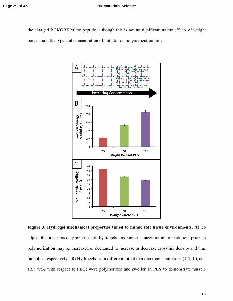
Hydrogel mechanical properties tuned to mimic soft tissue environments.
One common approach used to control or tune the initial mechanical properties of hydrogels is
varying the monomer concentration.53
Controlling the hydrogel mechanical properties, as
measured by modulus, can be critical in cell culture and regenerative medicine applications,
where the elasticity, or “stiffness”, of the microenvironment that surrounds a cell has been shown
to affect cell function and fate.54, 55
These properties also must be consistent from gel-to-gel for
a well-defined, controlled material system. Here, we aimed to establish hydrogel compositions
with a range of equilibrium-swollen moduli that mimic different soft tissues. Toward this, we
measured the swollen storage moduli (G') and volumetric swelling ratios (Q) of hydrogels
formed from different initial monomer concentrations (7.5, 10, and 12.5 wt% with respect to
PEG) (Figure 3).
We demonstrate for our material system that, by increasing the concentration of monomer in
the gel-forming solution, we can increase the modulus (Figure 3b; 7.5 wt%, G' = 553 ± 81 Pa;
10 wt%, G' = 1343 ± 49 Pa; 12.5 wt%, G' = 2147 ± 87 Pa), creating gels with a range of
elasticity comparable to native soft tissues (around the range of neural tissues to muscle, E ~ 1 to
10 kPa, where E ≈ 3G).55
Assuming that the theory of rubber elasticity holds for these swollen
gels, the behavior can be attributed to an increase in crosslink density (ρx) by56
� = ������� �⁄ .
Page 20 of 45Biomaterials Science

21
Similarly, we observed decreasing swelling ratios for increasing monomer concentrations
(Figure 3c; 7.5 wt%, Q = 41.4 ± 1.3; 10 wt%, Q = 33.3 ± 0.6; 12.5 wt%, Q = 29.2 ± 0.4).
Increased crosslink density inhibits how much a gel is able to swell, thus the inverse relationship
between ρx and Q is expected and observed. The results for the moduli and swelling ratios were
also found to be statistically significant (p < 0.05), indicating that the material system may be
easily tuned to have specific mechanical properties by varying the concentration of monomer
present within a gel.
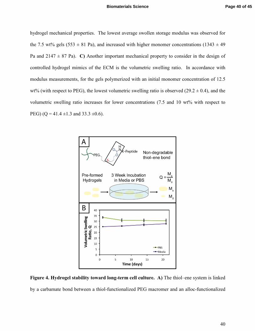
Hydrogel stability demonstrated for long-term culture
Hydrogel degradation over time often is desirable for cell culture applications to allow cellular
processes, such as growth, proliferation, and migration, which can be constrained or hindered by
a tightly crosslinked material.57
However, nonspecific degradation in aqueous solutions (e.g.,
hydrolytic cleavage of bonds within functional groups) can limit the degree of user control over
materials properties afforded by the addition of enzymatically degradable peptide crosslinks18
or
photodegradable chemistries,58
resulting in unintended or premature hydrogel degradation such
that the gel does not remain intact for appropriate time periods during cell culture. For example,
Shih and Lin have shown that step growth PEG-tetranorbornene-based thiol–ene hydrogels
completely degrade in 2-3 weeks at physiological pH (pH ~ 7.4), where the norbornene is linked
to PEG by an ester bond leading to hydrolytic degradation. Specifically, the degradation rate of
these hydrogels was influenced by the peptide crosslinker sequence, where peptides containing
hydrophobic or aromatic residues exhibited slower degradation (e.g., CGGGC sequence khyd =
0.049 ± 0.001 day-1
, CGGLC sequence khyd = 0.036 ± 0.002 day-1
).29
Page 21 of 45 Biomaterials Science
22
In the hydrogel system presented here, we aimed to create monomers free of ester bonds to
allow the creation of hydrogels that are stable under cell culture conditions. To assess the
stability of the resulting hydrogels, we monitored the volumetric swelling ratio (Q) of 10 wt%
gels incubated in PBS and cell culture medium at 37 oC over a period of three weeks (Figure 4),
a typical length for many two and three-dimensional cell culture experiments.3 For both
conditions, the Q values qualitatively are constant during the time course, and there is no
substantial degradation during the incubation period. Quantitatively, the p-values for the gels
incubated in PBS for different times are all greater than 0.05, indicating no statistical
significance between the gels for each time points and thus that degradation does not occur. For
the gels incubated in culture medium, when comparing days 1-14, the p-values are all greater
than 0.05. However, the day 21 time point is statistically different from the day 1 and 7 time
points (p < 0.05), indicating a slight change in swelling by 3 weeks. We hypothesize that
nonspecific degradation of the peptide crosslinker could be occurring over time in growth
medium, which is more complex than PBS and contains serum laden with enzymes, resulting in
this small but statistically significant increase in swelling. Despite this small swelling change,
the hydrogels remain robust and intact over multiple weeks in culture. The swelling ratios of
hydrogels in PBS versus media also are statistically significant for the entire incubation period,
which we speculate results from differences in the composition of PBS and growth media. With
this base system, various degradable peptide crosslinks derived from ECM proteins (e.g.,
GPQG↓IWGQ or IPVS↓LRSG derived from collagen I) can be incorporated within the gels to
allow cell-controlled matrix degradation, where the degradation rate of the matrix can be tuned
by peptide selection for different applications.18
Page 22 of 45Biomaterials Science
23
Biochemical cues spatially patterned within hydrogels
One benefit that photoclick chemistry provides in the design of hydrogels is the ability to
control the presentation of biochemical cues in space or time.59
Native tissues are dynamic
environments with gradients and defined regions of biological cues occurring at different times,
and the ability to capture this complexity in synthetic systems is important in understanding and
directing cellular processes.25
Here, we studied the photoaddition of a model biochemical cue to
our material i) to establish if excess free thiols could be modified after hydrogel formation and ii)
to demonstrate control over the spatial presentation of these cues. Specifically, an alloc-
modified integrin binding peptide (RGDS or AF488RGDS) was coupled homogenously or in
specific regions to hydrogels containing free thiols using photopatterning.
While one of our goals was to develop a hydrogel from accessible materials, we also aimed to
use simple techniques to characterize this system. Ellman’s assay, which can identify free thiols
in solution, is one such technique that has been used to quantify free thiols in materials post-
polymerization.60, 61
We have utilized this assay in a non-destructive method to quantify free
thiols in our hydrogels such that, if desired, gels may be rinsed of reagent, treated with tris(2-
carboxyethyl)phophine (TCEP), rinsed of TCEP, and re-used in additional studies. In addition to
quantifying free thiols, we wanted to demonstrate that Ellman’s reagent also could be used to
observe biochemical patterns created in gels with a reasonable degree of resolution as an
inexpensive and rapid alternative or complementary approach to using a fluorescently-tagged cue
(Figure 5a).
Toward achieving this, 10 wt% gels (0.254 mm thick between glass slides) were initially
polymerized off stoichiometry so that free thiols (2 mM at preparation prior to equilibrium
swelling) remained for later modification with the pendant RGDS peptide. Adjusting for
Page 23 of 45 Biomaterials Science
24
swelling, the free thiol concentration at equilibrium was estimated to be roughly ~ 0.61 ± 0.05
mM, so the free thiol concentration in hydrogels as measured by Ellman’s assay will be lower
than 2 mM. The free thiol concentrations of off-stoichiometry gels polymerized for 1 and 5
minutes subsequently was determined by Ellman’s assay to be 1.13 ± 0.09 mM and 0.97 ± 0.10
mM, respectively (Figure 5b, -RGDS condition). The gels polymerized for 1 and 5 minutes do
not have statistically different thiol concentrations (p > 0.05), supporting the results in Figure 2b
that the 10 wt% gels are completely formed in under one minute.
To initially determine if a model biochemical cue could be added to these gels, pre-formed
gels incubated in RGDS monomer (20 mg/mL ~20x excess to SH) with LAP (2.2 mM) were
exposed to UV light for 1 and 5 minutes. The thiol concentration after modification was
determined for each condition by Ellman’s (1 min = 0.01 ± 0.01 mM, 5 min = 0.003 ± 0.003
mM) (Figure 5b, +RGDS condition). These concentrations correspond to 93.1 and 94.8 %
modification of the remaining free thiols and 98.5 and 99.0 % total thiol modification, indicating
high coupling efficiency of the pendant peptide. There are slightly fewer free thiols in the gels
polymerized for 5 minutes indicating that a longer polymerization time results in higher
conversion of functional groups; however, there is no statistical significance between the two
conditions indicating that the effects of longer polymerization are ultimately negligible.
Hydrogels polymerized off-stoichiometry (2 mM free thiol at preparation) were then incubated in
growth medium at 37 °C for 3 days to determine if cues could be added at different times during
culture. Only trace free thiols were observed with Ellman’s assay after this 3-day incubation
(0.008 ± 0.002 mM), indicating the formation of disulfides either with components in the culture
medium or between free thiol end groups on PEG. To test this hypothesis, TCEP (10 mM in
PBS) was added to the gels for 1 hour to break potential disulfide bonds. Gels subsequently
Page 24 of 45Biomaterials Science

25
were rinsed, and the presence of free thiols was detected with Ellman’s (1.54 ± 0.09 mM)
(Supplemental Figure S5). This recovery of thiols confirms that a large portion of free thiols
post-polymerization were lost to disulfide formation upon incubation in culture medium. While
the application TCEP could be investigated as an approach to allow temporal photopatterning,
reducing agents such as it will negatively affect cell viability62
and may not be a practical option
for in situ photopatterning. However, different orthogonal chemistries2 could be utilized within
this base hydrogel system to allow the temporal addition of cues throughout long-term cell
culture in future investigations.
With the ability to add cues to the matrix after initial formation, spatially defined regions
of various cues of interest can be created toward directing the organization and function of cells
in three dimensions.10, 22, 63
Fluorescently-labeled cues are typically used to observe biochemical
patterns in hydrogel-based matrices with a high degree of resolution; however, this approach
requires additional expense and time for peptide labeling and fluorescence imaging. For a rapid
and inexpensive assessment of patterning, we examined using Ellman’s reagent to observe
spatially-defined patterns as a simple alternative or complementary approach for preliminary
evaluations. Hydrogels photopatterned with the AF488RGDS peptide demonstrate spatial
resolution of cue addition (Figure 5c) in the x, y, and z-directions for patterns of arbitrary shapes
(wide and narrow lines, squares). Next, to test Ellman’s as an alternative to fluorescently-labeled
evaluation, non-labeled RGDS was patterned into gels, and the gels were imaged under a light
microscope immediately after the application of Ellman’s reagent (Figure 5d). At short time
periods (< 5 minutes), we observed resolution of the patterns; however, as the products from
reaction with Ellman’s reagent diffused throughout the gel, the pattern began to disappear
(Supplemental Figure S6). While Ellman’s reagent is limited by the fast diffusion of the reaction
Page 25 of 45 Biomaterials Science
26
products, resulting in the short-term observation of patterns in the x-y plane only, we envision
using this test in initial or follow-up studies of photopatterning in thiol–ene hydrogels because it
is easy to use and provides almost instant results. Initially, one could test the ability to pattern a
hydrogel before building or purchasing a more expensive fluorescently-tagged peptide. In later
experiments, one could quickly confirm that a different peptide or peptide sequence is patterned
into the same system without having to build another labeled peptide and use an epi-fluorescent
or confocal microscope.
Encapsulated stem cells remain viable and metabolically active within patterned and non-
patterned hydrogels
Hydrogel systems for cell culture or delivery must not only be cytocompatible, but cells also
must be able to withstand their polymerization conditions for encapsulation within the matrix.
PEG, the primary component of the materials presented here, has been used in a variety of
hydrogel systems owing to its bioinert nature, providing a blank slate for the presentation of
peptide sequences or whole proteins to elicit specific cellular responses.25
Furthermore, cells
must be able to withstand multiple doses of UV light and radical initiator for the creation of
biochemical patterns within gels to direct cell behavior in three dimensions.
To evaluate the cytocompatibility of the initial polymerization conditions, we encapsulated
adult human stem cells, hMSCs, within non-degradable gels (10 wt%, 2.2 mM LAP, 2 mM
RGDS, 3000 cells/µL) polymerized for different lengths of time. Specifically, based on our
rheometric measurements, hydrogels were polymerized for the minimum amount of time
required to completely polymerize 10 and 12.5 wt% samples (1 minute) and in excess of the
minimum amount of time to polymerize 7.5 wt% samples (5 minutes). In addition, cell density
Page 26 of 45Biomaterials Science
27
was kept low to promote primarily cell-matrix interactions and fully understand the limits of cell
viability in the system when encapsulating a dilute, single-cell suspension. Cell viability and
metabolic activity subsequently were evaluated 1 and 3 days after polymerization to determine
polymerization conditions appropriate for the initial encapsulation and culture of cells,
respectively.
A membrane integrity assay (LIVE/DEAD Viability/Cytotoxicity Kit) of cells encapsulated
in gels (Figure 6a) showed a higher percentage of living cells in gels polymerized for 1 minute
(87 ± 2 %) in comparison to 5 minutes (81 ± 4 %) at day 3 in culture. While decreased cell
viability is observed for the 5 minute polymerization condition, which could limit the use of gels
with lower modulus in cell culture (e.g., 7.5 wt%), viability can be rescued by adjustment of
experimental parameters, including increased cell-cell contact (i.e., controlling the density of
encapsulated cells),64
incorporating biomimetic peptides that promote additional cell-matrix
interactions,20, 65
and lower initiator concentration (i.e., reducing concentration of radicals during
polymerization but at some cost to polymerization time).38
We increased the encapsulation
density of cells in non-degradable gels polymerized for 5 minutes (3000 to 30000 cells/µL) and
demonstrated a corresponding increase in viability (83 ± 2 % to 92 ± 1 %) (Supplemental Figure
S7). Accordingly, cell encapsulation density can be adjusted as appropriate to support viability
and function depending on the experimental variables to be studied and should be considered in
experimental design when using this system.
The metabolic activity of cells, an indicator of cell viability and function, also was monitored 1
and 3 days after encapsulation using CellTiter 96. Constant metabolic activity over time was
observed in the gels polymerized for 1 and 5 minutes over three days (p > 0.05) (Figure 6b).
Initially (D1) the metabolic activity of the gels polymerized for 5 minutes is statistically different
Page 27 of 45 Biomaterials Science
28
(p < 0.05) from gels polymerized for only one minute. However, by day 3, the metabolic activity
of the gels polymerized for 1 and 5 minutes is statistically similar (p > 0.05), indicating that the
initial effects of the polymerization are most apparent for longer irradiation time periods but do
not impact cell metabolic activity past the initial treatment. Here, the short-term effects of
encapsulation on cell survival appear minimal and similar to that observed in other hydrogels
formed by free radical initiation,38, 48
indicating that this new hydrogel system could support cell
culture or delivery in various experimental applications.
Note that all conditions in the metabolic activity experiments presented above were normalized
to cells encapsulated in hydrogels with 1 minute of light exposure. While normalization to
encapsulated cells without UV exposure is desirable, the hydrogel system presented cannot be
easily formed without light. To assess any effect of UV light alone on cell function, hMSCs
were seeded in 96-well plates and metabolic activity monitored 1 and 3 days after exposure to
UV. Light exposure did not significantly affect hMSC metabolic activity at either D1 or D3
post-irradiation (p > 0.05, compared to no UV control) (Supplemental Figure S8). This result is
consistent with the reports of others for single doses of UV light at 10 mW/cm2.66
Toward utilizing this system for patterning gels with biochemical cues during cell culture, we
sought to establish relatively mild photopatterning conditions to enable the application of
multiple doses of light and radicals within 24 hours of encapsulation. We first incubated gels
with 2 mM free thiols prior to swelling in serum-free and serum-containing, phenol red-free
growth medium for 2 hours at 37 °C. Only 0.26 ± 0.02 and 0.24 ± 0.04 mM free thiols remained
after incubation indicating free thiol consumption at a rate much faster than 24 hours
(Supplemental Figure S5); consequently, gels need to be incubated in PBS, rather than culture
medium, for photopatterning in the presence of cells. A balance must be struck between
Page 28 of 45Biomaterials Science
29
allowing time for diffusion of the peptide and initiator into the gels while minimizing the time
that cells are incubated in PBS during this process. To address this, we polymerized gels in
geometries in which cell encapsulation experiments were conducted (10 wt%, 20 µL gels in
syringe tips) for 1 minute and placed them immediately in the patterning solution (PBS
containing 3 mg/mL RGDS ~3x excess to SH and 2.2 mM LAP). Gels were incubated at 37 °C
for 30 minutes or 1 hour and 30 minutes, times longer and shorter than the time estimated for
diffusion of the monomer to the center of the gel assuming Fickian diffusion (td ~ 65 minutes):
!" =#$
%
where L is half the thickness of the unswollen gel (~ 0.625 mm) and % the diffusion coefficient
(~ 10-6
cm2/s based on proteins of similar molecular weight as the RGDS peptide).
67 A second
dose of UV light (1 minute) was applied to covalently link RGDS within the hydrogel. As
previously observed, free thiol concentration in gels polymerized for 1 and 5 minutes (without
patterning) was not statistically different (p > 0.05) and the patterned gels exhibit significantly
lower concentrations of free thiols post-patterning (p < 0.05 compared to that after 1 and 5
minute gel formation) at 0.30 ± 0.01 and 0.21 ± 0.02 mM, respectively (Figure 6c). These two
photopatterning conditions have statistically different thiol concentrations after polymerization
(p < 0.05), suggesting that the peptide and initiator may not have fully penetrated the gel during
this incubation time. To test this hypothesis, gels (10 wt%, 20 uL in syringe tips, 1 minute
polymerization) were incubated with AF488RGDS (3 mg/mL) and LAP (2.2 mM) in PBS for 30
minutes, 1 hour, and 1 hour 30 minutes, and exposed to UV light for 1 minute to allow covalent
attachment of the fluorescent peptide. Z-stack images through the entire gel depth (confocal)
Page 29 of 45 Biomaterials Science
30
indicate consistent patterning of the peptide through the gel depth for all conditions (Figure 6d,
Supplemental Figure S9). We speculate that the slight differences seen between the thiol
concentrations after patterning by Ellman’s assay (Figures 5b and 6c) are the result of small
variations between batches of PEG-4SH monomer and hydrogels or the relative excesses at
which the cues were tagged (20x for proof-of-concept and 3x for patterning in the presence of
cells).
To compare the effects of these photopatterning conditions on cell activity and viability, cells
encapsulated in non-degradable gels (3000 cells/µL, 1 minute UV exposure) were incubated for
30 minutes or 1 hour 30 minutes in PBS containing RGDS and LAP and a second dose of UV
light subsequently was applied for 1 minute. Cell metabolic activity for these photopatterning
conditions is statistically similar to the 1 minute hydrogel formation condition at days 1 and 3 (p
> 0.05), indicating that exposure to multiple polymerizations (formation + patterning) has a
minimal effect on cell function (Figure 6b). There appears to be a slight, but not statistically
significant, decrease in metabolic activity for each condition between days 1 and 3. We
hypothesize that this negligible decrease results from minor damage to cells in all cases by the
radically-mediated polymerizations, which shows up in reduced metabolic activity at day 3. No
statistical difference is observed between any condition at day 3. Taken together, no specific
effect of the photopatterning process is observed, and the photopatterning conditions assessed
here are appropriate for use in cell culture.
Finally, toward long-term culture of cells in patterned gels, hMSCs were encapsulated in cell-
degradable gels crosslinked with a MMP-cleavable peptide sequence18
(GPQGIWGQ2alloc) and
treated with 3 mg/mL RGDS and 2.2 mM LAP in PBS for 1 hour (between the minimum and
maximum incubation times tested for photopatterning) before a second dose of UV light was
Page 30 of 45Biomaterials Science

31
applied to photopattern RGDS within the network. After 6 days of culture, cells were stained
with the LIVE/DEAD Viability/Cytotoxicity Kit and imaged on a confocal microscope to
observe cell viability and any spreading within the network. Viability greater than 90% was
observed and a few cells exhibited protrusions (Figure 6e), indicative of adhesion to and
degradation of the matrix. Based on these results, this approach for cell encapsulation and
matrix photopatterning is promising for future studies to probe stem cell-material interactions
and direct cell function and fate in vitro.
CONCLUSIONS
In summary, we presented a novel hydrogel system formed by thiol–ene photoclick chemistry
through reaction of thiol-modified PEG and alloc-modified peptides. Use of the LAP
photoinitiator allowed rapid polymerization with cytocompatible doses of UV light and the
formation of hydrogels with appropriate mechanical properties to mimic soft tissues. These
hydrogels remain stable in cell culture conditions and encapsulated cells are viable within the
network. Biochemical cues were selectively patterned within the gels to demonstrate spatial
control over matrix properties, and cells remained viable. Further, the monomers used in the
design of this system may be synthesized using established protocols or commercially purchased,
making the material accessible for the facile and consistent formation of robust hydrogels to
mimic the ECM. In the future, this base material may be used with orthogonal click chemistries
to allow control over biochemical and biomechanical properties over days to weeks to study cell
response to changes in the surrounding environment and provides a useful platform to adapt for a
variety of biomaterials applications, including cell culture, tissue engineering, and drug delivery.
Page 31 of 45 Biomaterials Science

32
Specifically, toward application in culture and directing hMSC fate, gels could be patterned with
individual or multiple biochemical cues in spatially defined regions to drive cellular processes,
including adhesion, migration, proliferation, or differentiation.68, 69
ACKNOWLEDGEMENTS
This work was supported by the Institutional Development Award from the National Institutes of
Health (P20GM103541), the Pew Charitable Trusts (00026178), a National Science Foundation
Career Award (DMR-1253906), and the National Science Foundation IGERT SBE2 program at
the University of Delaware (fellowship to LAS). The authors thank the Delaware Biotechnology
Institute at the University of Delaware for training and access to confocal microscopy at the
BioImaging Center, Mr. Matthew Rehmann for generously providing hMSCs isolated from bone
marrow and the cysteine-modified peptide, Prof. Christopher J. Kloxin and Mr. Stephen Ma for
generously providing photomasks, Prof. Wilfred Chen for use of the automated plate reader, and
Mr. Eric Macedo for training in polymer and peptide synthesis techniques.
REFERENCES
1. H. C. Kolb, M. Finn and K. B. Sharpless, Angewandte Chemie International Edition,
2001, 40, 2004-2021.
2. W. Xi, T. F. Scott, C. J. Kloxin and C. N. Bowman, Advanced Functional Materials,
2014.
3. P. M. Kharkar, K. L. Kiick and A. M. Kloxin, Chemical Society Reviews, 2013, 42, 7335-
7372.
4. R. K. Iha, K. L. Wooley, A. M. Nyström, D. J. Burke, M. J. Kade and C. J. Hawker,
Chemical reviews, 2009, 109, 5620-5686.
5. M. J. Kade, D. J. Burke and C. J. Hawker, Journal of Polymer Science Part A: Polymer
Chemistry, 2010, 48, 743-750.
6. M. Lomba, L. Oriol, R. Alcalá, C. Sánchez, M. Moros, V. Grazú, J. L. Serrano and J. M.
De la Fuente, Macromolecular bioscience, 2011, 11, 1505-1514.
7. K. G. Robinson, T. Nie, A. D. Baldwin, E. C. Yang, K. L. Kiick and R. E. Akins, Journal
of Biomedical Materials Research Part A, 2012, 100, 1356-1367.
Page 32 of 45Biomaterials Science
33
8. M. Lutolf and J. Hubbell, Biomacromolecules, 2003, 4, 713-722.
9. J. Zheng, L. A. Smith Callahan, J. Hao, K. Guo, C. Wesdemiotis, R. Weiss and M. L.
Becker, ACS macro letters, 2012, 1, 1071-1073.
10. C. A. DeForest, B. D. Polizzotti and K. S. Anseth, Nature materials, 2009, 8, 659-664.
11. D. L. Alge, M. A. Azagarsamy, D. F. Donohue and K. S. Anseth, Biomacromolecules,
2013, 14, 949-953.
12. R. Selvaraj and J. M. Fox, Current opinion in chemical biology, 2013, 17, 753-760.
13. A. D. Baldwin and K. L. Kiick, Polymer chemistry, 2013, 4, 133-143.
14. C. E. Hoyle and C. N. Bowman, Angewandte Chemie International Edition, 2010, 49,
1540-1573.
15. B. D. Polizzotti, B. D. Fairbanks and K. S. Anseth, Biomacromolecules, 2008, 9, 1084-
1087.
16. W. M. Gramlich, I. L. Kim and J. A. Burdick, Biomaterials, 2013, 34, 9803-9811.
17. C. R. Nuttelman, M. A. Rice, A. E. Rydholm, C. N. Salinas, D. N. Shah and K. S.
Anseth, Progress in polymer science, 2008, 33, 167-179.
18. J. Patterson and J. A. Hubbell, Biomaterials, 2010, 31, 7836-7845.
19. B. D. Fairbanks, M. P. Schwartz, A. E. Halevi, C. R. Nuttelman, C. N. Bowman and K.
S. Anseth, Advanced Materials, 2009, 21, 5005-5010.
20. M. Lutolf and J. Hubbell, Nature biotechnology, 2005, 23, 47-55.
21. A. E. Rydholm, S. K. Reddy, K. S. Anseth and C. N. Bowman, Polymer, 2007, 48, 4589-
4600.
22. K. A. Mosiewicz, L. Kolb, A. J. van der Vlies, M. M. Martino, P. S. Lienemann, J. A.
Hubbell, M. Ehrbar and M. P. Lutolf, Nature materials, 2013, 12, 1072-1078.
23. T. Yang, H. Long, M. Malkoch, E. Kristofer Gamstedt, L. Berglund and A. Hult, Journal
of Polymer Science Part A: Polymer Chemistry, 2011, 49, 4044-4054.
24. J. A. Burdick and W. L. Murphy, Nature communications, 2012, 3, 1269.
25. M. S. Rehmann and A. M. Kloxin, Soft Matter, 2013, 9, 6737-6746.
26. C.-C. Lin, A. Raza and H. Shih, Biomaterials, 2011, 32, 9685-9695.
27. C. N. Salinas, B. B. Cole, A. M. Kasko and K. S. Anseth, Tissue engineering, 2007, 13,
1025-1034.
28. J. L. Ifkovits and J. A. Burdick, Tissue engineering, 2007, 13, 2369-2385.
29. H. Shih and C.-C. Lin, Biomacromolecules, 2012, 13, 2003-2012.
30. H. Shih and C. C. Lin, Macromolecular rapid communications, 2013, 34, 269-273.
31. J. J. Roberts and S. J. Bryant, Biomaterials, 2013, 34, 9969-9979.
32. R. Sanchez, M. Riddle, J. Woo and J. Momand, Protein Science, 2008, 17, 473-481.
33. S. T. Gould, N. J. Darling and K. S. Anseth, Acta biomaterialia, 2012, 8, 3201-3209.
34. C. A. DeForest, E. A. Sims and K. S. Anseth, Chemistry of materials, 2010, 22, 4783-
4790.
35. M. M. Kreevoy, E. T. Harper, R. E. Duvall, H. S. Wilgus III and L. T. Ditsch, Journal of
the American Chemical Society, 1960, 82, 4899-4902.
36. A. A. Aimetti, R. K. Shoemaker, C.-C. Lin and K. S. Anseth, Chemical Communications,
2010, 46, 4061-4063.
37. C. A. DeForest and K. S. Anseth, Nature chemistry, 2011, 3, 925-931.
38. B. D. Fairbanks, M. P. Schwartz, C. N. Bowman and K. S. Anseth, Biomaterials, 2009,
30, 6702-6707.
Page 33 of 45 Biomaterials Science

34
39. S. J. Bryant, C. R. Nuttelman and K. S. Anseth, Journal of Biomaterials Science,
Polymer Edition, 2000, 11, 439-457.
40. Z. N. Mahmoud, S. B. Gunnoo, A. R. Thomson, J. M. Fletcher and D. N. Woolfson,
Biomaterials, 2011, 32, 3712-3720.
41. J. A. Johnson, M. Finn, J. T. Koberstein and N. J. Turro, Macromolecular rapid
communications, 2008, 29, 1052-1072.
42. R. Khurana, J. Schaefer, L. A. Archer and G. W. Coates, Journal of the American
Chemical Society, 2014.
43. B. D. Fairbanks, S. P. Singh, C. N. Bowman and K. S. Anseth, Macromolecules, 2011,
44, 2444-2450.
44. E. Ruoslahti and M. D. Pierschbacher, Science, 1987, 238, 491-497.
45. S. J. Bryant and K. S. Anseth, in Scaffolding in Tissue Engineering, eds. P. X. Ma and J.
Elisseeff, Marcel Dekker, Inc., Boca Raton, FL, 1 edn., 2005, pp. 71-90.
46. S. B. Anderson, C.-C. Lin, D. V. Kuntzler and K. S. Anseth, Biomaterials, 2011, 32,
3564-3574.
47. A. B. Lowe, Polymer chemistry, 2010, 1, 17-36.
48. S. Gerecht, J. A. Burdick, L. S. Ferreira, S. A. Townsend, R. Langer and G. Vunjak-
Novakovic, Proceedings of the National Academy of Sciences, 2007, 104, 11298-11303.
49. N. B. Cramer, S. K. Reddy, A. K. O'Brien and C. N. Bowman, Macromolecules, 2003,
36, 7964-7969.
50. J. E. Elliott, M. Macdonald, J. Nie and C. N. Bowman, Polymer, 2004, 45, 1503-1510.
51. V. Zubov, M. V. Kumar, M. Masterova and V. Kabanov, Journal of Macromolecular
Science—Chemistry, 1979, 13, 111-131.
52. C. N. Salinas and K. S. Anseth, Macromolecules, 2008, 41, 6019-6026.
53. M. W. Toepke, N. A. Impellitteri, J. M. Theisen and W. L. Murphy, Macromolecular
Materials and Engineering, 2013, 298, 699-703.
54. D. E. Discher, P. Janmey and Y.-l. Wang, Science, 2005, 310, 1139-1143.
55. I. Levental, P. C. Georges and P. A. Janmey, Soft Matter, 2007, 3, 299-306.
56. M. P. Schwartz, B. D. Fairbanks, R. E. Rogers, R. Rangarajan, M. H. Zaman and K. S.
Anseth, Integrative Biology, 2010, 2, 32-40.
57. G. Raeber, M. Lutolf and J. Hubbell, Biophysical journal, 2005, 89, 1374-1388.
58. A. M. Kloxin, A. M. Kasko, C. N. Salinas and K. S. Anseth, Science, 2009, 324, 59-63.
59. M. Guvendiren and J. A. Burdick, Current opinion in biotechnology, 2013, 24, 841-846.
60. B. Fejerskov, B. E. Jensen, N. B. Jensen, S.-F. Chong and A. N. Zelikin, ACS applied
materials & interfaces, 2012, 4, 4981-4990.
61. S. Kobel, M. Limacher, S. Gobaa, T. Laroche and M. P. Lutolf, Langmuir, 2009, 25,
8774-8779.
62. X. Ren, Y. J. Lee, H. J. Han and I. S. Kim, Chemosphere, 2008, 74, 84-88.
63. S.-H. Lee, J. J. Moon and J. L. West, Biomaterials, 2008, 29, 2962-2968.
64. C.-C. Lin and K. S. Anseth, Proceedings of the National Academy of Sciences, 2011, 108,
6380-6385.
65. C. R. Nuttelman, M. C. Tripodi and K. S. Anseth, Matrix biology, 2005, 24, 208-218.
66. M. Guvendiren and J. A. Burdick, Nature communications, 2012, 3, 792.
67. L. M. Weber, C. G. Lopez and K. S. Anseth, Journal of Biomedical Materials Research
Part A, 2009, 90, 720-729.
68. R. A. Marklein and J. A. Burdick, Advanced Materials, 2010, 22, 175-189.
Page 34 of 45Biomaterials Science
35
69. M. P. Lutolf, P. M. Gilbert and H. M. Blau, Nature, 2009, 462, 433-441.
Page 35 of 45 Biomaterials Science

36
FIGURES
Figure 1. Hydrogels formed by thiol–ene photoclick reactions for cell culture applications.
A) Monomers functionalized with thiols or with alloc groups were synthesized for hydrogel
formation using thiol–ene click chemistry: multi-armed PEG was modified with thiols (right) and
peptides containing alloc-protected lysines (1 or 2) (left). Upon the application of light, these
functional groups react by a step growth mechanism, where an initiating species generates a thiyl
radical that attacks the pendant ‘ene’ and forms a stable covalent bond between the monomers in
solution.19
B) This material system is promising for cell encapsulation and three-dimensional
cell culture, where the thiol-modified PEG is crosslinked with alloc-containing peptides in the
presence of cells allowing their encapsulation for in vitro studies. Capitalizing on the spatial
Page 36 of 45Biomaterials Science

37
control enabled by the thiol-ene photoclick reaction, pendant peptides (containing one alloc) can
be added within the network during or after gel formation to promote cell-matrix interactions.
Page 37 of 45 Biomaterials Science

38
Figure 2. In situ polymerization of PEG hydrogels with different photoinitiators. A)
Hydrogels (10 wt% with respect to PEG) were polymerized in situ on a rheometer to monitor gel
formation over time using various initiator conditions. After 1 minute on the rheometer, UV
light (10 mW/cm2, 365 nm) was applied to samples. The storage moduli of gels polymerized
using 1.1 and 2.2 mM LAP initiator begin to increase within 30 seconds after light application
and finish forming in approximately 1-3 minutes (modulus levels off). Gels polymerized using
2.2 mM I2959 begin to form 4 minutes after UV light application and reach complete formation
in approximately 13-14 minutes of exposure. The rapid polymerizations observed for the LAP
initiator are relevant for cell culture applications. Representative data for each condition is
shown here. B) Complete polymerization, defined here as the point where the change in
modulus between consecutive data points is less than 0.1 %, was determined for various initiator
and monomer concentrations. As shown in (A), the LAP initiator exhibits the most rapid
polymerization, with times of 0.96 ± 0.05 and 2.60 ± 0.03 minutes for 2.2 and 1.1 mM LAP,
respectively. Complete polymerization for I2959, at the highest concentration compared to LAP
(2.2 mM), occurs in 13.59 ± 1.15 minutes. Using 2.2 mM LAP, hydrogels from various initial
monomer concentrations (7.5, 10, and 12.5 wt% with respect to PEG) were polymerized. The
7.5 wt% condition exhibited the slowest polymerization rate of 3.00 ± 0.22 minutes due to fewer
functional groups that are available to react. The 10 and 12.5 wt% gels polymerized in 0.79 ±
0.03 and 0.88 ± 0.11 minutes as the number of functional groups in solution is higher at the start
of polymerization. C) Hydrogels were polymerized with different alloc-modified peptides to
evaluate any effect of peptide chemistry on polymerization rate. The less charged
GKGWGKG2alloc and GPQGIWGQ2alloc peptides take 40 seconds longer to polymerize than
Page 38 of 45Biomaterials Science
39
the charged RGKGRK2alloc peptide, although this is not as significant as the effects of weight
percent and the type and concentration of initiator on polymerization time.
Figure 3. Hydrogel mechanical properties tuned to mimic soft tissue environments. A) To
adjust the mechanical properties of hydrogels, monomer concentration in solution prior to
polymerization may be increased or decreased to increase or decrease crosslink density and thus
modulus, respectively. B) Hydrogels from different initial monomer concentrations (7.5, 10, and
12.5 wt% with respect to PEG) were polymerized and swollen in PBS to demonstrate tunable
Page 39 of 45 Biomaterials Science
40
hydrogel mechanical properties. The lowest average swollen storage modulus was observed for
the 7.5 wt% gels (553 ± 81 Pa), and increased with higher monomer concentrations (1343 ± 49
Pa and 2147 ± 87 Pa). C) Another important mechanical property to consider in the design of
controlled hydrogel mimics of the ECM is the volumetric swelling ratio. In accordance with
modulus measurements, for the gels polymerized with an initial monomer concentration of 12.5
wt% (with respect to PEG), the lowest volumetric swelling ratio is observed (29.2 ± 0.4), and the
volumetric swelling ratio increases for lower concentrations (7.5 and 10 wt% with respect to
PEG) (Q = 41.4 ±1.3 and 33.3 ±0.6).
Figure 4. Hydrogel stability toward long-term cell culture. A) The thiol–ene system is linked
by a carbamate bond between a thiol-functionalized PEG macromer and an alloc-functionalized
Page 40 of 45Biomaterials Science

41
peptide, which both lack hydrolytically cleavable bonds (e.g., esters). To evaluate gel stability
for controlled cell culture over several weeks, 10 wt% hydrogels (with respect to PEG) were
incubated in cell culture medium and PBS for 3 weeks and volumetric swelling ratios (Q)
measured. B) The swelling of gels incubated in PBS did not significantly change (p > 0.05 for
all samples) indicating stability over the time period (Q = 33.4 ± 0.8, 30.8 ± 1.3, 30.6 ± 1.3, 30.3
±1.5). The swelling of gels incubated in growth media slightly increased through the incubation
period such that the first two and last Q values are different (p < 0.05), although consecutive
points are not (p > 0.05), which may be attributed partly to non-specific degradation of the
hydrogel by enzymes present in the more complex growth medium (Q = 25.1 ± 0.2, 25.6 ± .4,
26.8 ± 0.8, 27.6 ± 0.4).
Page 41 of 45 Biomaterials Science
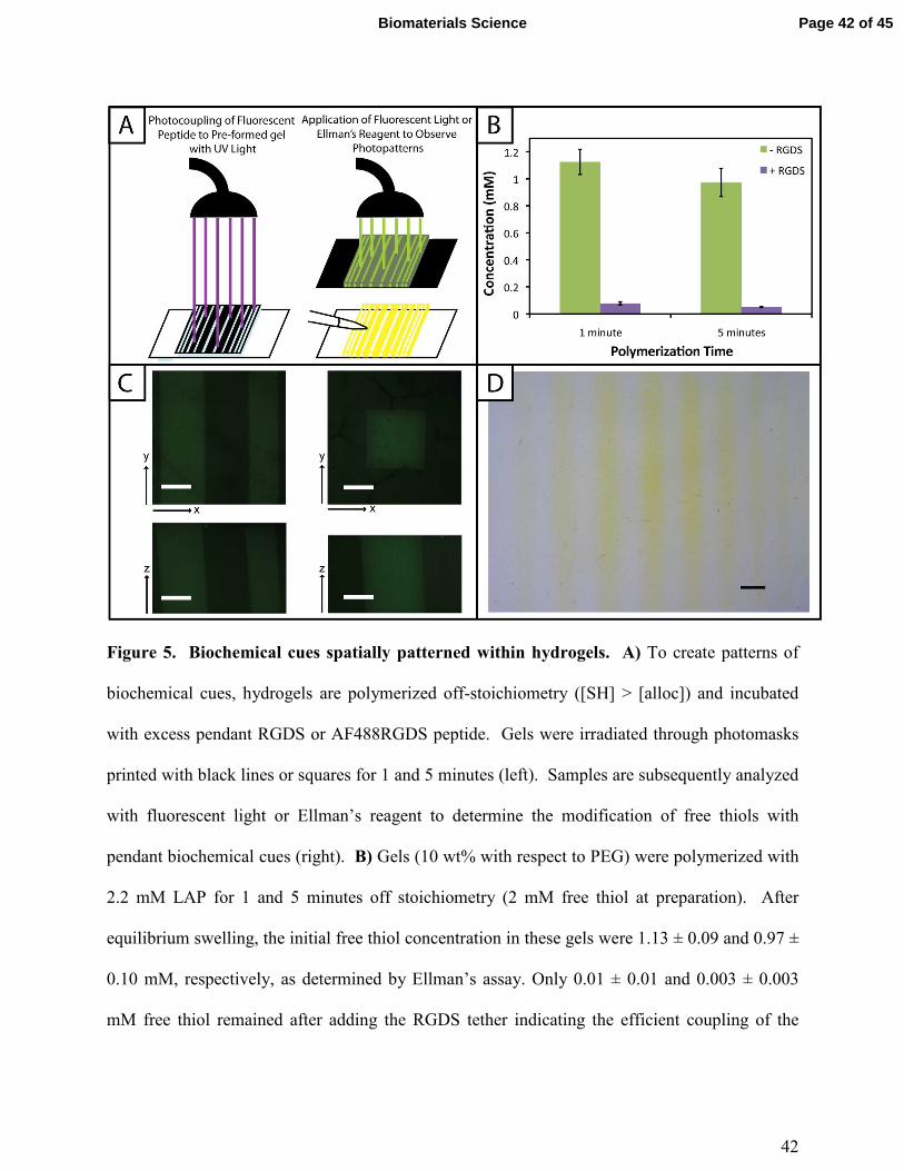
42
Figure 5. Biochemical cues spatially patterned within hydrogels. A) To create patterns of
biochemical cues, hydrogels are polymerized off-stoichiometry ([SH] > [alloc]) and incubated
with excess pendant RGDS or AF488RGDS peptide. Gels were irradiated through photomasks
printed with black lines or squares for 1 and 5 minutes (left). Samples are subsequently analyzed
with fluorescent light or Ellman’s reagent to determine the modification of free thiols with
pendant biochemical cues (right). B) Gels (10 wt% with respect to PEG) were polymerized with
2.2 mM LAP for 1 and 5 minutes off stoichiometry (2 mM free thiol at preparation). After
equilibrium swelling, the initial free thiol concentration in these gels were 1.13 ± 0.09 and 0.97 ±
0.10 mM, respectively, as determined by Ellman’s assay. Only 0.01 ± 0.01 and 0.003 ± 0.003
mM free thiol remained after adding the RGDS tether indicating the efficient coupling of the
Page 42 of 45Biomaterials Science
43
model biochemical cue to the hydrogel network. C) Following the setup shown in (A), arbitrary
patterns (squares, 1600 µm2; lines of different thickness, 200 - 1000 µm) of a fluorescent peptide
(AF488RGDS) were created within pre-formed hydrogels and imaged on a confocal microscope
for analysis. Resolution of the pattern is observed in the x-, y-, and z-planes indicating selective
coupling to only regions of the gel that were exposed to light. (Scale bar, 200 µm) D) As a
quick and inexpensive alternative to fluorescence, a non-fluorescent pendant peptide (RGDS)
was photopatterned (lines of different thickness) into pre-formed hydrogels. Ellman’s reagent
was directly applied to the top of these gels to identify regions lacking the pendant peptide
(yellow) with resolution in the x- and y-planes over short times (< 5 min). (Scale bar, 1 mm)
Page 43 of 45 Biomaterials Science
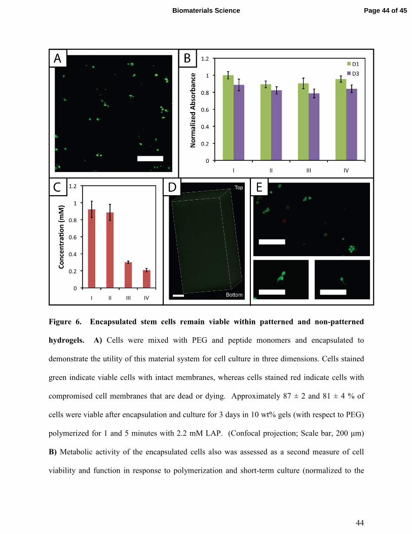
44
Figure 6. Encapsulated stem cells remain viable within patterned and non-patterned
hydrogels. A) Cells were mixed with PEG and peptide monomers and encapsulated to
demonstrate the utility of this material system for cell culture in three dimensions. Cells stained
green indicate viable cells with intact membranes, whereas cells stained red indicate cells with
compromised cell membranes that are dead or dying. Approximately 87 ± 2 and 81 ± 4 % of
cells were viable after encapsulation and culture for 3 days in 10 wt% gels (with respect to PEG)
polymerized for 1 and 5 minutes with 2.2 mM LAP. (Confocal projection; Scale bar, 200 µm)
B) Metabolic activity of the encapsulated cells also was assessed as a second measure of cell
viability and function in response to polymerization and short-term culture (normalized to the
Page 44 of 45Biomaterials Science

45
gels polymerized for 1 minute 1 day after encapsulation). Various encapsulation and
photopatterning conditions were tested (I) 1 minute encapsulation, (II) 5 minutes encapsulation,
(III) 1 minute encapsulation + 30 minutes PBS/RGDS/LAP + 1 minute photopatterning, (IV) 1
minute encapsulation + 1 hour 30 minutes PBS/RGDS/LAP + 1 minute photopatterning.
Condition II for day 1 is statistically different (p < 0.05) from I, indicating that longer exposure
times to UV and radicals can initially affect viability. However, at day 3, conditions I and II are
statistically similar, indicating ‘recovery’ of the cells post-encapsulation. The photopatterning
conditions III and IV are statistically similar to I at days 1 and 3; thus, incubation in PBS and a
second dose of UV light do not significantly impact cell function. C) Ellman’s assay was
conducted on hydrogels processed under conditions used in panel B (I, II, III, and IV). The
initial encapsulation conditions I and II have statistically similar free thiol concentrations,
consistent with prior gel formation results. The photopatterning conditions III and IV are
statistically different from I and II, indicating the ability to covalently attach peptides within the
network post-polymerization under mild conditions for cell culture. Further, III and IV are
statistically different from each other, suggesting that photopatterning may be diffusion-limited
in thicker gels used for cell encapsulation. To evaluate this, D) gels incubated with 3 mg/mL
AF488RGDS and 2.2 mM LAP for 30 minutes were patterned with a second dose of UV light
for 1 minute. Uniform attachment of the fluorescent cue is observed throughout the entire gel
depth (~1.6 mm). (Confocal z-stack; Scale Bar, 200 µm) E) Cells encapsulated in a MMP-
degradable hydrogel patterned with RGDS remain viable (> 90%) over 6 days in culture (top).
Several encapsulated cells began to form protrusions by day 6, characteristic of degradation and
cell adhesion to the matrix (bottom). (Scale bars, 200 µm)
Page 45 of 45 Biomaterials Science