design and synthesis of sigma-1 probe for positron
TRANSCRIPT
DESIGN AND SYNTHESIS OF SIGMA-1 PROBE FOR POSITRON EMISSION TOMOGRAPHY (PET) IMAGING
by May May Ly
A thesis submitted to the faculty of The University of Mississippi in partial fulfillment of the requirements of the Sally McDonnell Barksdale Honors College.
Oxford May 2015
Approved by
_____________________________________ Advisor: Dr. Christopher McCurdy
_____________________________________ Reader: Dr. Stephen Cutler
_____________________________________ Reader: Dr. Donna West-Strum

ii
© 2015
May May Ly ALL RIGHTS RESERVED

iii
ACKNOWLEDGEMENTS
I would like to acknowledge Dr. Christopher McCurdy. I could never express enough appreciation for his support of this project. I approached him nearly two years ago, and he welcomed me into his research laboratory with open arms. He welcomed me into my research the entire way and has provided encouragement and motivation when I needed it most. Without his overall supervision, this project would not have been possible.
I would like to thank my second and third readers, Dr. Stephen Cutler and Dr. Donna West-Strum respectively. I appreciate their time and valuable input toward this project.
I would also like to acknowledge Mr. Walid Alsharif for his guidance and patience in training me for the type of work involved in this project. Additionally, I would also like to acknowledge the remainder of Dr. McCurdy’s research laboratory group for addressing questions concerning this project.
I would like to acknowledge The University of Mississippi School of Pharmacy and Sally McDonnel Barksdale Honors College. The academic challenges and developmental opportunities throughout the past four years have made this project possible. The constant search for knowledge challenged and inspired me to work hard throughout my undergraduate career.
I am greatly appreciative for the endless support and encouragement from my friends and family.

iv
ABSTRACT MAY MAY LY: Design and Synthesis of Sigma-1 Probe for Positron Emission
Tomography (PET) Imaging (Under the direction of Dr. Christopher McCurdy)
Sigma-1 receptor radioligands have the potential to detect and monitor various
neurological diseases. Because sigma-1 receptors are believed to be associated with
numerous psychiatric conditions and neurodegenerative diseases, radioligands for sigma-
1 receptors have the potential to serve as novel diagnostic tools and may be useful in
assessing treatment effectiveness. A benzothiazolone 18F-labeled sigma-1 receptor ligand,
CM304, has demonstrated promise as a specific positron emission tomography
radiotracer for visualizing sigma-1 receptors in living subjects. Based on CM304, backup
molecules were created to improve the overall drug profile, including half- life and
bioavailability.

v
TABLE OF CONTENTS
ABSTRACT ..................................................................................................................... IV
LIST OF FIGURES ........................................................................................................ VI
INTRODUCTION............................................................................................................. 1
BACKGROUND ............................................................................................................... 5
CHEMISTRY .................................................................................................................. 15
CONCLUSIONS ............................................................................................................. 27
EXPERIMENTAL .......................................................................................................... 28
REFERENCES ................................................................................................................ 37
APPENDICES ................................................................................................................. 41

vi
LIST OF FIGURES
Figure 1 SN56
Figure 2 CM304
Figure 3 Cell uptake results for [18F]CM304 and [3H](+)-pentazocine
Figure 4 Time-activity curves from mouse positron emission tomography studies
Figure 5 Sagittal mouse PET images and ex vivo autoradiography of sagittal brain sections obtained 60 minutes after administration of [18F]CM304
Figure 6 Rat brain PET/CT and ex vivo autoradiography
Figure 7 Monkey brain PET/MR images from baseline dynamic imaging
Figure 8 Scheme 1
Figure 9 Friedel-Crafts Acylation
Figure 10 Scheme 2
Figure 11 Scheme 3
Figure 12 Electron Pushing Mechanism
Figure 13 Available PET Tracers for Sigma 1

1
INTRODUCTION
A potential biomarker associated with nerve injury and neuroinflammation is the
sigma-1 receptor. Sigma receptors were originally thought to belong to the opioid class of
receptors (Martin, 1976). Based on the psychotomimetic effects of (±)-SKF-10,047(N-
allylnormetazocine) and related benzomorpahns, which were not reversed by opioid
antagonists, Martin and co-workers named this distinct class of receptor as ‘sigma’,
derived from the first letter ‘S’ from SKF-10,047. It was then believed that sigma
receptor sites were identical to N-methyl-D-aspartate (NMDA) receptor/phencyclidine
(PCP) sites. This hypothesis was contradicted by studies using selective NMDA ligands
(Wong, 1988). Therefore, sigma receptors were then classified as a unique protein family.
Two known sigma receptor subtypes exist: sigma-1 and sigma-2. Of the two subtypes,
sigma-1 receptors are the most well-characterized because of the receptor sequence
information and availability of selective sigma-1 ligands. The sigma-1 receptor is a 25-29
kDa chaperone protein that is made up of 223 amino acids; it has been purified and
cloned from several species, including mouse, rat, guinea pig, and human (Hanner, 1996;
Mei, 2001; Pan, 1998; Seth, 1997; Seth, 1998). The protein shows no resemblance to
other known mammalian proteins; however, it has a 30% identity and 66% homology to
ergosterol-Δ8/Δ7-isomerase enzyme. Sigma-1 receptors do not possess sterol isomerase
activity though. It is believed that the receptor has two transmembrane-spanning regions
in the regions of amino acids 13 to 34 and 86 to 108, with the NH2 and COOH ends both
facing the interior of the cell.
2
Sigma-1 receptors are expressed throughout the body, including the heart, spleen,
brain, liver, gastrointestinal tract, kidney, adrenal glands, and reproductive organs.
Research has demonstrated that sigma-1 receptors are heavily concentrated within the
brain; they are predominately expressed in the specific areas associated with memory,
emotion, sensory functions, and motor functions (Cobos, 2008). The chief function of
sigma-1 receptors is the modulatory role on dopamine, acetylcholine, N-methyl-D-
aspartate, and opioid receptors (Bowen, 2000).
Mainly located at the endoplasmic reticulum of cells, sigma-1 receptors have been
associated with a host of biochemical processes and pathological conditions including
neurodegenerative diseases, psychiatric disorders, pain sensitization, drug addiction,
digestive function, regulation of smooth muscle contraction, and ischemia (James, 2012).
In most known cancers (e.g., breast, lung, colon, ovarian, prostate, brain), sigma-1
receptors are highly expressed. Sigma-1 receptors can regulate various ion channels,
including potassium channels, calcium channels, dopamine and gamma-amino butyric
acid (GABA) receptors thereby impacting neural excitability and transmission by
affecting the release of several neurotransmitters. These neurotransmitters include:
serotonin, dopamine, noradrenaline, glutamate, and GABA. Sigma-1 receptors appear to
play a significant role in pain modulation and are involved in “memorizing” pain by
synaptic plasticity and central sensitization. This mechanism is responsible for the
chronic and self-perpetuating nature of certain pain conditions.
Certain sigma-1 receptors agonists have also been shown to control endothelial
cell proliferation, improve cognition, provide neuroprotection, and act as antidepressant
agents, while antagonists inhibit cocaine-induced seizures and lessen neuropathic pain,
3
highlighting the potential of sigma-1 receptors as both a diagnostic and therapeutic target
(James, 2012). Over the past couple decades, several groups have reported the
development of high affinity sigma-1 receptor ligands, and of these, some have been
labeled with radioisotopes for use in positron emission tomography (PET) studies to
determine the origin of pain.
Current clinical imaging methods used to evaluate chronic pain, including
computed tomography (CT), ultrasound imaging (US), and magnetic resonance imaging
(MRI), are primarily focused on imaging anatomic alteration, which do not necessarily
reflect the origin of chronic pain (Jarvik, 2004). Various limitations in determining cause
and location of nerve injury are associated with these tests. These limitations include, but
are not restricted to: invasiveness, propensity to technical and operator-dependent errors,
and/or lack of sufficient sensitivity and specificity to diagnose nerve injury (Shen, 2015).
PET is an analytical imaging technology developed to use compounds labeled with
positron emitting radioisotopes as molecular probes to image and measure biochemical
processes of mammalian biology in vivo, which allows the development of instruments
with high-resolution along with high sensitivity (Phelps, 2000). Radioisotopes of oxygen
(14O, 15O), nitrogen (13N), carbon (11C), and most commonly fluorine (18F) can be
administered to a subject and detected externally are all positron emitters.
Molecular probes for PET are first developed by identifying a target process to be
studied and then making a positron labeled molecule through which an assay can be
performed. Because PET cannot provide direct chemical analysis of reaction products in
tissue, labeled molecules are used that trace a small number of steps of biochemical
process so that kinetic analysis can be used to estimate the concentration of reactants and

4
products over time and, from this, reaction rates (Phelps, 2000). Biochemists develop
molecular probes to isolate and accurately measure a limited number of steps in a
biochemical pathway. Drugs are designed to have limited interactions because the goal is
to modify the function of a key step in a biological process with minimal interaction with
other processes.
Before any PET procedure can become an important clinical tool, PET is a
nuclear medicine technique which requires the combination of a number of factors. These
factors include: (1) the necessary radioisotope has to be available on a daily basis, (2) an
imaging device must give good performance for the isotope of choice, and (3) the
combination of a radiopharmaceutical and a patient population for which the PET scan
provides significant diagnostic information in which is not readily available with other
techniques (Muehllehner, 2006). PET whole body imaging has the potential to identify
early disease, differentiate benign from malignant lesions, examine all organs for
metastases, and determine therapeutic effectiveness (Phelps, 2000). PET, biological, and
pharmaceutical sciences are merging together with common goals of building molecules
that can be used to image and measure biological functions within organ systems in living
subjects, and as drugs to modify the malfunction of disease. PET could potentially
provide more direct pathways from biology to the patient and link PET and
pharmaceutical sciences in molecular diagnostics and molecular therapeutics, especially
in various neurological diseases.

5
BACKGROUND
Examining sigma-1 receptors in living subjects with positron emission
tomography (PET) is an important step toward understanding the receptor’s functional
role and involvement in disease. PET radioligands specific for sigma-1 receptors could
possibly provide a noninvasive means of (1) investigating the distribution and function in
both normal and disease states in living subjects, (2) assessing receptor occupancy to
determine optimal doses of therapeutic drugs, (3) early detection and staging of sigma-1
receptor-related diseases, and (4) monitoring therapeutic response (James, 2012). In
2005, Yous and colleagues reported what has now been identified as a lead compound
from the benzothiazolone class of compounds, SN56 (shown in Figure 1), with the aim of
synthesizing a new, selective fluorinated PET radioligand for studying sigma-1 receptors
in living subjects. SN56 has demonstrated high affinity and apparent high selectivity for
the sigma-1 receptor.
N
S
O
N
Figure 1: SN56
6
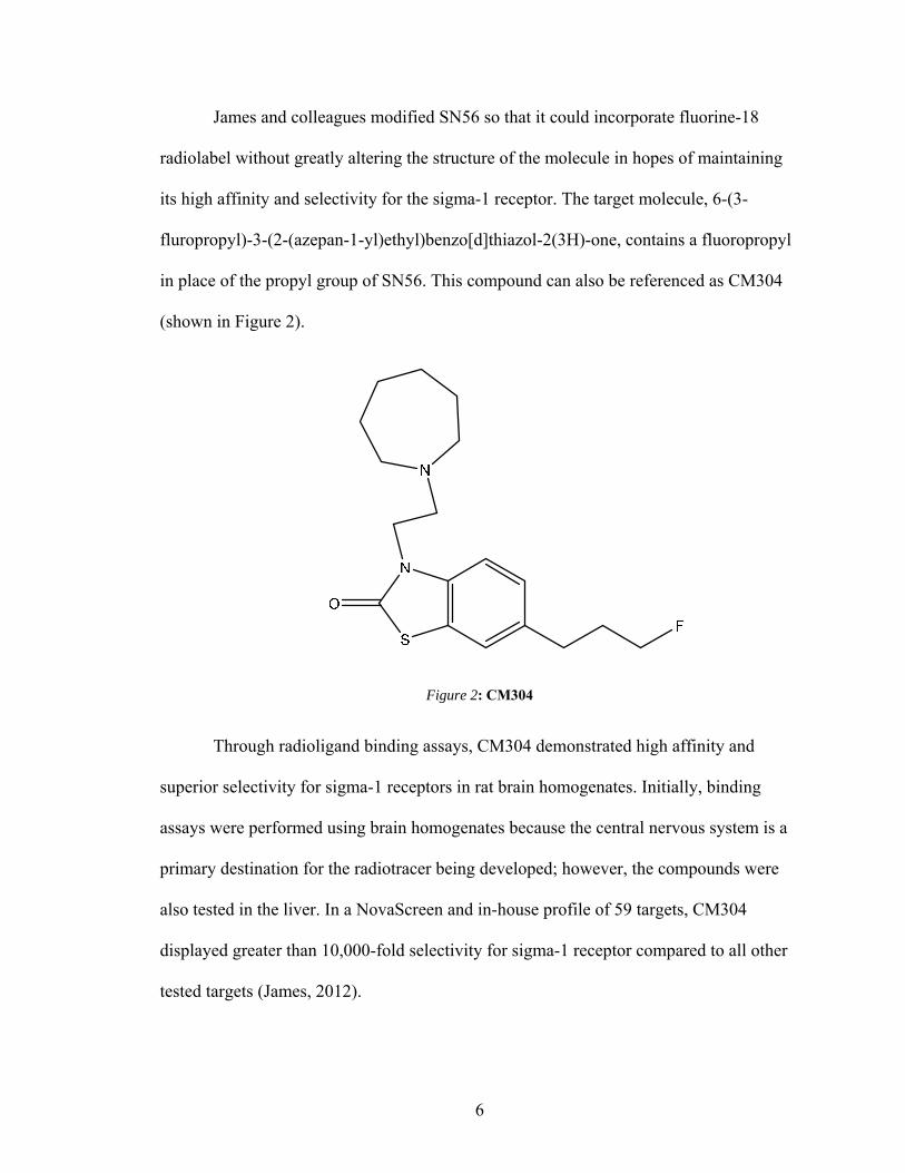
James and colleagues modified SN56 so that it could incorporate fluorine-18
radiolabel without greatly altering the structure of the molecule in hopes of maintaining
its high affinity and selectivity for the sigma-1 receptor. The target molecule, 6-(3-
fluropropyl)-3-(2-(azepan-1-yl)ethyl)benzo[d]thiazol-2(3H)-one, contains a fluoropropyl
in place of the propyl group of SN56. This compound can also be referenced as CM304
(shown in Figure 2).
Figure 2: CM304
Through radioligand binding assays, CM304 demonstrated high affinity and
superior selectivity for sigma-1 receptors in rat brain homogenates. Initially, binding
assays were performed using brain homogenates because the central nervous system is a
primary destination for the radiotracer being developed; however, the compounds were
also tested in the liver. In a NovaScreen and in-house profile of 59 targets, CM304
displayed greater than 10,000-fold selectivity for sigma-1 receptor compared to all other
tested targets (James, 2012).

7
CM304 was radiolabelled with fluorine-18 in order to trace the compound in
living subjects. The uptake of this radioactive compound in Chinese hamster ovary cells
was compared to the uptake of the known sigma-1 receptor ligand [3H](+)-pentazocine.
Control Chinese hamster ovary cells cells were transfected with a vector not containing
the sigma-1 receptor gene, while the experimental Chinese hamster ovary cells were
transfected with a vector containing sigma-1 receptor cDNA, which serves as a positive
control for sigma-1 receptor expression in cells. Cells were exposed to [18F]CM304 or
[3H](+)-pentazocine for 30 and 120 minutes. According to the data, there was a slight
increase in uptake for both radioligands between 30 minutes and 120 minutes in control
Chinese hamster ovary cells (James, 2012). In the Chinese hamster ovary cells
transfected with sigma-1 receptor cDNA, this increase was more profound and
numerically higher at both 30 minutes and 120 minutes than the control Chinese hamster
ovary cells. At 120 minutes, the uptake of [18F]CM304 in the experimental cells was 4-
fold higher than uptake in control Chinese hamster ovary cells as seen in Figure 3.
Figure 3: Cell uptake results for [18F]CM304 and [3H](+)-pentazocine. Results are expressed as counts per minute recorded in a sample from a particular cell per counts per minute recorded in medium per
amount of protein (μg) present in a sample from that well. Reprinted with permission from James, M. L.; Shen, B.; Zavaleta, C. L.; Nielsen, C. H.; Mesangeau, C.; Vuppala, P. K.; Chan, C.; Avery, B. A.; Fishback, J. A.; Matsumoto, R. R.; Gambhir, S. S.; McCurdy, C. R.; Chin, F. T. New positron emission tomography (PET) radioligand for imaging sigma-1 receptors in living subjects. J Med Chem. 2012, 55, 8272-8282. Copyright 2015 American Chemical Society.

8
Small animal PET was used to assess the in vivo kinetics of [18F]CM304 in
normal mice. Brain PET scanning began 1 minute before the administration of
[18F]CM304 and stopped 60 minutes later. The baseline time-activity curve, shown in
Figure 4, demonstrates that [18F]CM304 entered the brain quickly, spiked within the first
few minutes, and then gradually decreased over the remaining time of the scan.
[18F]CM304 reached its maximum uptake in mouse brain within the first few minutes of
imaging and gradually washed out to 65% of its maximum at 60 minutes after the
administration.
Figure 4: Time-activity curves (TACs) from mouse positron emission tomography studies. TACs represent accumulation of [18F]CM304 in whole mouse brain as a function of time for baseline, preblock
with haloperidol, preblock with FTC-146, and preblock with [18F]FTC-146. Reprinted with permission from James, M. L.; Shen, B.; Zavaleta, C. L.; Nielsen, C. H.; Mesangeau, C.; Vuppala, P. K.; Chan, C.; Avery, B. A.; Fishback, J. A.; Matsumoto, R. R.; Gambhir, S. S.; McCurdy, C. R.; Chin, F. T. New positron emission tomography (PET) radioligand for imaging sigma-1 receptors in living subjects. J Med Chem. 2012, 55, 8272-8282. Copyright 2015 American Chemical Society.
PET images provided visual evidence that [18F]CM304 rapidly crossed the blood-
brain barrier and appeared to accumulate in known sigma-1 receptor rich regions. An
accurate depiction of radioligand localization in specific brain regions could not be
determined solely with PET; therefore, ex vivo autoradiography was performed. Results
from the autoradiography demonstrated that [18F]CM304 accumulated in the midbrain,

9
facial nucleus, cortex, and hippocampus and to a lesser extent in the cerebellum and
thalamus (shown in Figure 5).
Figure 5: PET images and ex vivo autoradiography of sagittal brain sections (12 μm) obtained 60 minutes after administration of [18F]CM304. Sections used for autoradiography were stained with Nissl for anatomical correlation. PET images were acquired just before to perfusing mice and harvesting brain tissue for autoradiography. White dotted lines designate location of mouse brain in sagittal PET images: Cb = cerebellum, Ctx = cortex, FN = facial nucleus, H = hippocampus, Mb = midbrain, Ob = olfactory
bulb. Reprinted with permission from James, M. L.; Shen, B.; Zavaleta, C. L.; Nielsen, C. H.; Mesangeau, C.; Vuppala, P. K.; Chan, C.; Avery, B. A.; Fishback, J. A.; Matsumoto, R. R.; Gambhir, S. S.; McCurdy, C. R.; Chin, F. T. New positron emission tomography (PET) radioligand for imaging sigma-1 receptors in living subjects. J Med Chem. 2012, 55, 8272-8282. Copyright 2015 American Chemical Society.
[18F]CM304 was further assessed in rats and squirrel monkeys by James and
colleagues. Rats were administered [18F]CM304 intravenously, and static PET scans were
measured after 50 minutes. For blocking studies, rats were pretreated with BD1047 ten
minutes before tracer administration. A CT image was acquired after each PET scan to
provide an anatomic reference frame for the respective PET data. The rats were sacrificed
after PET imaging, and the brains were removed, which allowed for autoradiography to
be performed.
Baseline PET/CT images (shown in Figure 6) again demonstrated [18F]CM304’s
ability to cross the blood-brain barrier and accumulate in brain tissue. The PET/CT
images also revealed some accumulation of radioactivity in the rat skull. Accumulation of
[18F]CM304 was found in the brain stem, cerebellum, cortex, hypothalamus, nucleus
oculomotor, red nucleus, thalamus, caudate putamen, and hippocampus. There was little

10
uptake of [18F]CM304 in the corpus callosum and muscle. Both blocking studies showed
a reduction in the uptake of [18F]CM304.
Figure 6: Rat brain PET/CT and ex vivo autoradiography. (A) Baseline coronal PET/CT images 50-60 minutes after intravenous administration of [18F]CM304. (B) PET/CT images from blocking study. (C) Autoradiography and nissl/H&E staining of coronal brain and muscle sections from baseline study. (D) Autoradiography and nissl/H&E staining of coronal brain and muscle sections from blocking studies. Bst = brain stem; CC = corpus callosum; Cer = cerebellum; CP = caudate-putamen; Ctx = cortex; H = hippocampus; HTh = hypothalamus; NO = nucleus oculomotor; RN = red nucleus; Th = thalamus.
This research was originally published in JNM. Evaluation of σ-1 James, M. L.; Shen, B.; Nielsen, C. H.; Behera, D.; Buckmaster, C. L.; Mesangeau, C.; Zavaleta, C.; Vuppala, P. K.; Jamalapuram, S.; Avery, B.
A.; Lyons, D. M.; McCurdy, C. R.; Biswal, S.; Gambhir, S. S.; Chin, F. T. Evaluation of σ-1 receptor radioligand 18F-FTC-146 in rats and squirrel monkeys using PET. J Nucl Med. 2014, 55, 147-153. © by the
Society of Nuclear Medicine and Molecular Imaging, Inc.
Preliminary brain PET imaging of squirrel monkeys was performed to evaluate
the regional distribution and brain permeability of [18F]CM304 in a nonhuman primate. A
total of four brain PET images were obtained. The monkeys were injected intravenously
with [18F]CM304 in 500 microliters of sterile heparinized saline, followed by 500
microliters of sterile heparinized saline to flush the catheter. Seventy-five frames of
dynamic PET data was yielded over a 120-minute period. Blocking studies involved
pretreating monkeys with haloperidol 10 minutes before radioligand administration.
In Figure 7, baseline brain PET/MR images demonstrate [18F]CM304’s ability to
penetrate the monkey blood-brain barrier. The distribution of [18F]CM304 was shown to
accumulate in the cingulate, frontal cortex, occipital cortex, hippocampus, striatum,

11
cerebellum, and vermis. Also, PET images revealed some accumulation of radioactivity
in the monkey skull.
Figure 7: Monkey brain PET/MR images from baseline dynamic imaging. Summed axial images are shown for different time intervals after administration of [18F]CM304. Brain regions of interest are shown
in MR images and are labeled as (1) cingulate cortex, (2) frontal cortex, (3) striatum, (4) parietal cortex, (5) occipital cortex, (6) temporal cortex, (7) hippocampus, (8) cerebellum, and (9) vermis.
This research was originally published in JNM. Author(s). Evaluation of σ-1 James, M. L.; Shen, B.; Nielsen, C. H.; Behera, D.; Buckmaster, C. L.; Mesangeau, C.; Zavaleta, C.; Vuppala, P. K.; Jamalapuram, S.; Avery, B. A.; Lyons, D. M.; McCurdy, C. R.; Biswal, S.; Gambhir, S. S.; Chin, F. T. Evaluation of σ-1 receptor radioligand 18F-FTC-146 in rats and squirrel monkeys using PET. J Nucl Med. 2014, 55, 147-153.
© by the Society of Nuclear Medicine and Molecular Imaging, Inc.
Human serum/liver microsome studies were performed to attain information
about the potential of [18F]CM304 for eventual clinical translation. The metabolic
stability of CM304 was further investigated by determining its half-life in human liver
microsomes. CM304 was found to have a longer half-life in human liver microsomes
than in mouse and rat liver microsomes that was previously recorded by James and
colleagues. The human liver microsome had a half-life of 18.2 ± 3.6 minutes, while the

12
mouse and rat have half-lives of 4.2 ± 0.6 minutes and 12.6 ± 1.9 minutes, respectively.
Imaging sigma-1 receptors via PET could serve as a useful method for detecting sigma-1
receptor-related diseases and monitoring their treatment invasively. Sigma-1 PET
imaging could potentially help researchers investigate the in vivo role of sigma-1
receptors in different diseases and enhance the understanding of the
pathology/progression of such conditions. In order for PET radiotracers to be successful,
a wide range of criteria must be achieved. Ideally, PET radiotracers would possess the
following characteristics: high affinity, selectivity for target, ability to penetrate the
blood-brain barrier, non-substrate for efflux transporters, lack of troublesome
radiometabolites, low non-specific binding, suitable brain pharmacokinetics in relation to
radiolabel half-life, amenability to labeling with 11C or 18F at high specific radioactivity,
and safe for administration at low tracer dose (Pike, 2008). Several sigma-1 selective
PET radioligands have been synthesized; however, none have met all the criteria that
were listed above.
Based on the studies of [18F]CM304 in mice, rats, and squirrel monkeys, the
radioligand accumulated in regions known to contain high levels of sigma-1 receptors,
which suggests that the PET signal specifically binds to sigma-1 receptors in vivo.
Another common trend among these animal studies is the accumulation of radioactivity
in the skull. Metabolism leading to bone uptake is not desirable and can make
quantification of brain signal difficult because of the close proximity of skull to brain and
the partial-volume effect of PET. Accuracy of determining sigma-1 receptor levels may
be affected (James, 2014). Although the metabolism of [18F]CM304 is a limitation in
rodents and nonhuman primates, this does not completely reflect what would occur in

13
human studies. There have been cases of radiotracers in the literature, such as 18F-SP203,
that have defluorinated in nonhuman primates but demonstrated stability in human
studies (Brown, 2008). Furthermore, the fact that [18F]CM304 appeared to be more
stable in human liver microsomes, compared with mouse and rat liver microsomes, is a
good indicator that it might be more stable in humans.
As mentioned in the introduction, sigma-1 receptors may serve a potential role in
various neurological diseases, which include addiction, amnesia, chronic pain,
depression, Alzheimer’s disease, schizophrenia, stroke, retinal neural degeneration, HIV
infection, and cancer (Maurice, 2009). Sigma-1 receptors have long been recognized as
an endoplasmic reticulum protein. Its involvement in the various diseases has been
demonstrated by the molecular biological silencing of the receptor, excluding
schizophrenia and HIV infection (Maurice, 2009). The sigma-1 receptor has also been
identified as a receptor chaperone whose activity can be activated and deactivated by
specific ligands. The mechanism of action of sigma-1 receptors at the molecular level has
proven difficult to find by researchers because the sigma-1 receptor does not resemble
any of the mammalian proteins as previously mentioned. Several mechanisms of action
have been proposed, but the most prominent and the most explored molecular action of
sigma-1 receptors centers on the receptor’s interaction with voltage-regulated and ligand-
gated ion channels, including calcium channels, potassium channels, sodium channels,
chloride channels, small conductance calcium-activated potassium channels (SK
channels), NMDA receptors and inositol triphosphate (IP3) receptors. According to
Maurice and Su, the final output of the action of sigma-1 receptor agonists is to inhibit
the voltage-gated ion channels mentioned above, while they potentiate ligand-gated

14
channels (2009). Sigma-1 receptor antagonists block the inhibition or potentiation
induced by sigma-1 agonists.
Studies conducted by Tsai and colleagues have demonstrated that sigma-1
receptors and associated ligands modulate IP3 receptors at the ER membrane. Under
physiological conditions, the sigma-1 receptor chaperones the functional IP3 receptor at
the endoplasmic reticulum and mitochondrion interface to ensure proper Ca2+ signaling
from the endoplasmic reticulum to the mitochondria (Tsai, 2009). When cells lose Ca2+
homeostasis in the endoplasmic reticulum under pathological conditions, the sigma-1
receptor translocates and counteracts the arising apoptosis. Therefore, the sigma-1
receptor is a receptor chaperone essential for the metabotropic receptor signaling and for
the survival against cellular stress (Tsai, 2009).

15
CHEMISTRY
SN56 and CM304 have extremely similar structures, differing only in the
presence of the fluorine at the end of the propyl group. The same benzothiazolone core
was used because it has been previously observed by Yous and colleagues that the nature
of the surrounding of the tertiary amine nitrogen had an profound impact on sigma-1
affinity. In order to further investigate sigma receptor ligands, the fluoropropyl group was
extended to a fluorobutyl group. The insertion of an additional carbon to CM304 was
made in hopes of having similar or improved sigma-1 binding compared to CM304 or
SN56, without having sigma-2 affinity.
Both SN56 and CM304 were studied in an article written by James and colleagues
in 2012. SN56 had demonstrated high selectivity for sigma-1 receptors (>1,000 fold),
while CM304 showed even greater selectivity (>145,000 fold) for sigma-1 receptors.
Both compounds underwent binding assays, which were performed using brain
homogenates. The central nervous system was targeted primarily; however, the liver was
also utilized for sigma receptor binding assays. In the rat brain homogenates and the rat
liver homogenate binding assays for sigma-1 receptors, SN56 displayed a Ki of 1.7 ± 0.1
nanomolar and Ki = 1.61 ± 0.10 nanomolar, respectively. CM304 demonstrated a Ki of
0.0025 nanomolar in the rat brain homogenate binding assay for sigma-1 receptors and a
Ki of 0.96 ± 0.21 nanomolar in the rat liver homogenates. CM304 exhibited greater than
10,000-fold selectivity for sigma-1 receptors compared to all other tested targets. CM304
displayed less than 50% displacement of the radioligand at 10,000 nanomolar screening

16
concentration and less than 50% displacement for 10 targets at 100 nanomolar screening
concentration. These targets include, but are not limited to, α2-adrenoceptors, histamine
H2 receptors, muscarinic M2 receptors, peripheral muscarinic receptors, neuronal
nicotinic receptors, opioid receptors, norepinephrine transporters, calcium L type
channels, sodium channels site, and acetylcholine esterase.
To familiarize myself with the procedures for my proposed compound, I
performed Schemes 1 and 2 before starting Scheme 3. I got used to using the equipment
around the lab and familiarized myself with the reactions that were taking place. In
addition, I was synthesizing molecules that the lab needed to be sent off for further
investigation. By doing this, I was able to execute the steps confidently and
independently after several reactions were performed.
Reagents and starting materials were obtained from commercial suppliers and
were used without purification. Precoated silica gel GF Uniplates from Analtech were
used for thin-layer chromatography (TLC). Column chromatography was performed on
silica gel 60 (Sorbent Technologies).
3-(2-azepan-1-yl)ethyl)-6-(3-methoxypropyl)benzo[d]thiazol-2(3H)-one (5) was
prepared according to Scheme 1, which is depicted in Figure 8. Commercially available
2-hydroxybenzothiazole (1) was reacted with 3-chloroprionyl chloride. The ketone in M1
(2) was reduced by using triethylsilane and trifluoroacetic acid. The resulting compound,
M2 (3), was reacted with 2-(hexamethyleneimino)ethyl chloride hydrochloride to add an
azepane functional group to the existing nitrogen, which was an N-alkylation reaction. A

17
methoxy group replaced the chloride in M3 (4) after the compound was reacted with
sodium methoxide, giving the final compound.
Figure 8: Scheme 1 Reagents and conditions: (a) 3-chloroprionyl chloride, AlCl3, DMF, 45 to85 , 4h; (b) Et3SiH, TFA, rt, 4h; (c) 2-(hexamethyleneimino)ethyl chloride hydrochloride, NaHCO3, DMF, 120 , 30min (d) NaOCH3, MeOH, 85 , reflux, overnight (e) NaOCH3, MeOH, KI, 80 , 1h
The first step in the synthesis is a Friedel-Crafts acylation, which demonstrated in
Figure 9. In this specific reaction, 3-chloroprionyl chloride acts as the acyl halide. The
first step of an electrophilic aromatic substitution involves the formation of an acylium
ion. This occurs when the acyl halide reacts with the aluminum trichloride, AlCl3, a
Lewis acid catalyst. The AlCl3 enhances the electrophilicity of the acyl halide by
complexing with the chlorine. The chlorine is removed by the Lewis acid catalyst. The
acylium ion is then stabilized by resonance, which provides extra stability and prevents
the problems associated with the rearrangement of simple carbocation. The next step
involves the attack of the acylium ion on the 2-hydroxybenzothiazole as a new
electrophile to form one complex. In order for aromaticity to return to the benzene ring of

18
the 2-hydroxybenzothiazole, a proton leaves. While this is taking place, aluminum
tetrachloride, AlCl4, returns to remove a proton from the benzene ring, which allows for
the return of the ring’s aromaticity. In doing so, the original aluminum trichloride is
regenerated, along with hydrochloric acid. As a result, we have the first part of the final
product of the reaction, which is a ketone. The addition of water liberates the final
product as an acyl benzene.
Figure 9: Friedel-Crafts Acylation Mechanism

19
The synthesis of 3-(2-azepan-1-yl)ethyl)-6-(3-hydroxylpropyl)benzo[d]thiazol-
2(3H)-one (4) is illustrated in Figure 10. M2 (1) had been previously synthesized using
the first step from Scheme 1. The chlorine of the propyl chain in M2 (1) was replaced
with benzoic acid during a benzoate-ester reaction. The azepane group was linked to the
existing nitrogen of M5 (2). The resulting compound, M6 (3), was reduced using a
combination of sodium hydroxide and water.
Figure 10: Scheme 2 Reagents and conditions: (a) K2CO3, benzoic acid, DMF, 110 , 4h (b) K2CO3, 2-(hexamethyleneimino)ethyl chloride hydrochloride, DMF, 65 , 4h (c) MeOH, NaOH, H2O, 90 , 1h
The schematic for the synthesis of the modified 2(3H)-benzothiazolone is
depicted in Figure 11. The scheme followed a combination of Scheme 1 and Scheme 2
with a few exceptions. 4-chlorobutyryl chloride was used instead of 3-chloroprionyl
chloride, and additional steps were taken to reach the final product. After the hydrolysis
of 4-(3-(2-azepan-1-yl)ethyl)-2-oxo-2,3-dihydrobenzo[d]thiazol-6-yl)butyryl benzoate (5)
using sodium hydroxide and water, the resulting compound was reacted with p-

20
toluenesulfonyl chloride to give M13 (7). M13 was reacted with tetrabutylammonium
fluoride to replace the tosyl group with a fluorine to yield M14 (8).
Figure 11: Scheme 3 Reagents and conditions: (a) 4-chlorobutyryl chloride, AlCl3, DMF, 45 to85 , 6h; (b) Et3SiH, TFA, rt, 4h; (c) 2-(hexamethyleneimino)ethyl chloride hydrochloride, NaHCO3, DMF, 120 , 30min (d) K2CO3, benzoic acid, DMF, 110 , 2h (e) MeOH, NaOH, H2O, 90 , 1h (f) p-toluenesulfonyl chloride, DCM, triethylamine, rt, 8h (g) TBAF/THF, 60 , 4h
An electron pushing mechanism for Scheme 3 was created to further illustrate
how the reactions were taking place. This is shown in Figure 12.

21
Figure 12: Electron Pushing Mechanism
N
F
S
NO
N
F
S
HN
O
Cl
Cl
N
S
NO
N
Cl
H
BH
Cl
S
NO
N
Cl
B-H
Nucleophilic N attacks electrophilic carbon
B:=Base
S
NO
N
Cl
OHO
B:
OO
H S
NO
N
O
O
NaOHH2OOH
S
NO
N
HO
ClS
O
O
:Base= Pyridine or triethylamine
S
NO
N
OS
NO
N
OS
O
O
-HCl
+ B-H
S
HN
OCl
O
ClAlCl3 Cl
O
Cl-AlCl3 Cl
O
Cl
O
S
HN
O
O
ClH
Cl-AlCl3
S
HN
O
O
Cl
Si
H
S
HN O
Cl
O Si(C2H5)3
H
S
HN
O
Cl

22
DISCUSSION AND FUTURE DIRECTIONS
Findings from James and colleagues in 2012 demonstrated that the small
structural modification made to SN56 in order to create CM304 led to an improvement in
affinity and selectivity for sigma-1 receptors. Both the affinity and selectivity of CM304
are higher than values reported for most other known sigma-1 receptor ligands, such as
[11C]SA4503, [18F]FM-SA4503, [18F]FPS, and [18F]SFE (see Figure 13).
Figure 13: Available PET Tracers for Sigma-1
In the first scheme, a methoxy group was added to the end of the propyl chain in
SN56, resulting in 3-(2-azepan-1-yl)ethyl)-6-(3-methoxypropyl)benzo[d]thiazol-2(3H)-
one (M4). The purpose of this synthesis was to create a non-radioactive compound,
which is often referred to as a “cold” compound, that could be used as a PET ligand that

23
ultimately expresses similar or improved sigma-1 binding compared to CM304 or SN56
without having sigma-2 affinity. The methoxy group mimics the number of atoms in the
molecule’s tail. Therefore, the addition of the methoxy group should improve sigma-1
affinity; however, the effects of the oxygen are unknown. The methyl of the methoxy
group can be labeled with a 11C, which would provide an alternative to fluorine-based
imaging. The oxygen of the methoxy group should behave similarly to fluorine because it
should be able to accept hydrogen bonds due to the oxygen’s partial negative charge. The
oxygen also allows researchers to synthesize isotopes more easily. Because the structure
of M4 is similar to SN56, predictions can be made based off the previous studies on
SN56: M4 is expected to have relatively high affinity for sigma-1 receptors.
With a minor addition of a carbon on the propyl group of CM304, one could
suspect M14 to possess similar sigma-1 binding properties without sigma-2 affinity. The
goal of synthesizing M14 was to create a compound that have similar, possibly even
better, binding to sigma-1 compared to CM304 or SN56. Based on previous studies,
evidence suggests that there are several properties of M14 that would make it an ideal
PET radioligand (Yous, 2005). In an article by Yous and colleagues, compounds similar
to SN56, CM304, and M14 were synthesized, and the affinities for sigma-1 and sigma-2
receptors were evaluated. The same modifications were done to these similar compounds;
therefore, the same patterns are expected. Three specific modifications that were
displayed in this study to the molecules includes the addition of a methyl group to the
propyl chain, the inclusion of a seven-membered rings, and the addition of the fluorine.
All of these modifications were shown to improve sigma-1 receptor affinity; however, an
accurate assessment cannot be made.
24
Both compounds (M4 and M14) are expected to have relatively high affinity for
sigma-1 receptors because of the presence of the benzothiazole. The large increase in
affinity in CM304 to sigma-1 receptors may be due to the interaction with one or more
amino acid residues in the sigma-1 protein when engaged, which should be present with
these compounds.
Shen and colleagues established a rat-model of neuropathic pain and nerve injury
called the spared nerve injury (SNI) model. In this model, scientists carefully injury the
tibial and common peroneal branches but spared the sural branch of the left sciatic nerve.
After four weeks, pain behavioral response was tested using Von Frey filaments to
confirm development of pain in SNI rats and the lack of pain in sham-operated (surgical
wound but no nerve injury) and control rats (uninjured rats with no surgery or wound).
Von Frey filament tests indicated the development of allodynia (pain produced by an
otherwise non-painful stimulus) observed in the left hind paws of the SNI rats. The SNI
rats exhibited decreased paw withdrawal thresholds, indicating pain, in the injured left
hind limb compared to levels observed for the uninjured right side of SNI rats, sham-
operated rats, and control rats. [18F]CM304 accumulated in the injured nerve compared to
the uninjured right sciatic nerve of SNI rats as well as the nerves of the sham-operated
rats and control rates. The left injured nerves showed higher signals during
autoradiography. [18F]CM304 was able to detect the site of nerve injury in SNI rats via
PET/MR imaging and ex vivo autoradiography. The specific binding of [18F]CM304 to
sigma-1 receptor is further supported by the blocking studies with haloperidol. The
relationship between pain behavior (determined via Von Frey tests) and the normalized
PET signal in sciatic nerves, Shen found a strong negative correlation between the two.

25
That is, a lower Von Frey threshold denotes greater pain, which correlated with increased
PET signal.
Shen and colleagues’ study established four significant findings: (1) [18F]CM304-
PET/MRI enabled detection of peripheral nerve injury in a rat model of neuropathic pain;
(2) the normalized PET in injured sciatic nerves correlated with pain sensitivity and
severity; (3) the site of nerve injury was shown to contain increased expression of sigma-
1 receptors; and (4) there was significant correlation between the levels of sigma-1
receptors and the uptake of [18F]CM304 (2015).
As demonstrated by Shen and colleagues, specific binding of [18F]CM304 to
sigma-1 receptors in site of nerve injury suggest that increased sigma-1 receptor
expression is associated with pain generation and nerve injury. The strong correlation
between PET signal and pain levels indicates the possibility of providing accurate, non-
invasive estimates about pain severity. Furthermore, [18F]CM304 could provide
information about the precise location and magnitude of sigma-1 receptor expression as a
potential pain biomarker. The imaging approach of [18F]CM304 in combination of PET
with MRI satisfies the need for sensitivity and specificity to diagnose nerve injury
without being invasive. Sigma-1 receptor-PET imaging as a non-invasive biomarker of
nerve injury and inflammation could significantly impact pain therapeutics.
Future work with these compounds include tagging the precursor of M14, of 4-(3-
(2-(azepan-1-yl)ethyl)-2-oxo-2,3-dihydrobenzo[d]thiazol-6-yl)butyl 4-
methylbenzenesulfonate, with a radioactive fluorine. The synthesis of [18F]M14 could be
performed by the Molecule Imaging Program at Stanford Department of Radiology since

26
other compounds have previously been sent here. The strategy for generating [18F]M14
would involve the preparation of its tosylate precursor, M13, and its subsequent
radiolabeling with fluorine-18. Fluorine-18 radiolabelling could be accomplished by
reacting its tosylate precursor with cyclotron-produced 18F-fluorine as a Kryptofix-
222/K+/[18F]F- complex in DMSO at 150oC for 15 minutes (James 2012). This would
allow researchers to test the compound in animals with PET.
Further investigation involving sigma-1 receptors involves synthesizing selective
sigma-1 receptor PET radiotracers. CM304 has looked very promising as a potential PET
radioligand. Compared to other available sigma-1 receptor PET radiotracers, [18F]CM304
appears to have several key advantages, including its picomolar affinity for sigma-1
receptors; high brain uptake in mouse, rat, and monkey; favorable kinetic profile; and the
fact that it has the highest selectivity for sigma-1 receptors over sigma-2 receptors of any
sigma-1 receptor radiotracer (James, 2014). By using CM304 as the lead compound, we
would expect M14 to exert similar properties because the same modification was seen
from the reaction of SN56 to CM304.

27
CONCLUSIONS
Benzothiazole-based compounds were synthesized to serve as possible selective
sigma-1 receptor PET radioligands. Currently, no biological data have been collected on
these compounds. Therefore, the binding affinities and selectivity for sigma-1 receptors
are unknown. However, predictions about the general properties of the compounds
synthesized can be made. Each of the compounds is expected to have relatively high
selectivity and binding to sigma-1 receptors and lack of sigma-2 affinity due to the
compound’s similar structure to SN56 and CM304. Further studies will be conducted to
assess their selectivity and affinity toward sigma-1 receptors and their efficacy of
becoming a radioligand for PET imaging.

28
EXPERIMENTAL
Synthesis of 3-(2-azepan-1-yl)ethyl)-6-(3-methoxypropyl)benzo[d]thiazol-2(3H)-one.
6-(3-chloropropanoyl)benzo[d]thiazol-2(3H)-one (M1). Aluminum chloride (53.3
grams, 400 millimoles) was first added to an Erlenmeyer flask. Next, dimethylformamide
(8.6 milliliters, 115 millimoles) was slowly added under vigorous stirring. After 15
minutes of stirring, 2-hydroxybenzothiazole (6.04 grams, 40 millimoles) was added, and
the mixture was heated at 45C. After 15 minutes, 3-chloropropionyl chloride was added
and the reaction mixture was slowly raised to 85C and stirred for 3 hours. Once starting
material was no longer present in the reaction, the hot mixture was carefully poured onto
ice. 150 milliliters of 3N hydrochloric acid was added to neutralize the reaction, and the
mixture was stirred for 30 more minutes. The mixture was filtered by vacuum filtration,
and the crude product was collected and dissolved in a mixture of ethyl acetate and water.
The layers were then separated, and the organic layer was washed with brine and dried
with sodium sulfate. The organic layer was transferred to a round bottom flask, and the
solvent was removed in vacuo. The residue was recrystallized from toluene/dioxane to
give 5.50 grams (55.5%) of 6-(3-chloropropanoyl)benzo[d]thiazol-2(3H)-one as an
orange solid.
6-(3-chloropropyl)benzo[d]thiazol-2(3H)-one (M2). M1 (5.50 grams, 22.75 millimoles)
was added to a dry round bottom flask, and 30 milliliters of trifluoroacetic acid was
added. To the stirred solution, triethylsilane (8.36 mL, 52.32 millimoles) was added. The

29
reaction mixture was stirred for 4 hours at room temperature. The solvent was carefully
removed in vacuo, and the residue was purified by column chromatography using a
gradient of ethyl acetate and hexane to give 1.8776 grams (36.2%) of 6-(3-
chloropropyl)benzo[d]thiazol-2(3H)-one as a white crystalline solid.
3-(2-(azepan-1-yl)ethyl)-6-(3-chloropropyl)benzo[d]thiazol-2(3H)-one (M3). In a dry
round bottom flask, M2 (1.8776 grams, 8.246 millimoles) was dissolved in 25 milliliters
of dimethylformamide. 2-(Hexamethyleneimino)ethyl chloride hydrochloride (6.5351
grams, 32.98 millimoles) was added and the reaction mixture was heated to 120C. Then
sodium bicarbonate was slowly added and the reaction was left for 30 minutes. The
mixture was poured into water and extracted with ethyl acetate three times. The layers
were separated, and the organic layer was washed with brine and dried with sodium
sulfate. The organic layer was then transferred to a round bottom flask, and the solvent
was removed in vacuo. The residue was purified with column chromatography and
resulted in 1.10 grams (37.8%) of 3-(2-(azepan-1-yl)ethyl)-6-(3-
chloropropyl)benzo[d]thiazol-2(3H)-one as a white solid.
3-(2-azepan-1-yl)ethyl)-6-(3-methoxypropyl)benzo[d]thiazol-2(3H)-one (M4). M3
(1.0 grams, 2.83 millimoles) was dissolved in 10 milliliters of methanol in a dry round
bottom flask. Sodium methoxide (660 milligrams, 1.72 millimoles) was then added to the
reaction mixture. The reaction was heated under reflux at 85oC until starting material was
no longer present. The solvent was removed in vacuo, and the residue was subjected to
purification by silica gel column chromatography to afford approximately 150 milligrams
(15.2%) of 3-(2-azepan-1-yl)ethyl)-6-(3-methoxypropyl)benzo[d]thiazol-2(3H)-one as a
white solid.

30
Synthesis of 3-(2-azepan-1-yl)ethyl)-6-(3-hydroxylpropyl)benzo[d]thiazol-2(3H)-one.
3-(2-oxo-2,3-dihydrobenzo[d]thiazol-6-yl)propyl benzoate (M5). About 20 milliliters
of dimethylformamide was used to dissolve M2 (1.0 grams, 4.39 millimoles) in a round
bottom flask. Under mechanical stirring, potassium carbonate (1.517 grams, 10.979
millimoles) and benzoic acid (2.682 grams, 21.95 millimoles) were added to the reaction
mixture. The reaction was heated to 110oC. After 4 hours, the reaction was stopped and
left to cool down. The mixture was then poured into 200 milliliters of water and extracted
with ethyl acetate. The solvent was removed in vacuo, and the residue was purified
through a column chromatograph using ethyl acetate and hexane as the eluent to yield
1.221 grams (86.7%) of 3-(2-oxo-2,3-dihydrobenzo[d]thiazol-6-yl)propyl benzoate as a
white solid.
3-(3-(2-(azepan-1-yl)ethyl)-2-oxo-2,3-dihydrobenzo[d]thiazol-6-yl)propyl benzoate
(M6). In a dry round bottom flask, M5 (1.221 grams, 3.8986 millimoles) was dissolved in
about 20 milliliters of dimethylformamide. Potassium carbonate (1.6164 grams, 11.6958
millimoles) and 2-(hexamethyleneimino)ethyl chloride hydrochloride was added to the
solution under mechanical stirring. The reaction mixture was heated at 65oC for 4 hours.
Once the starting material was gone, the reaction was cooled. The mixture was then
poured into 200 milliliters of water and extracted with ethyl acetate. The organic layer
was washed with brine and dried with sodium sulfate. The solvent was removed in vacuo,
and the residue was chromatographed on a silica gel column, resulting in 1.3029 grams
(76.3%) of 3-(3-(2-(azepan-1-yl)ethyl)-2-oxo-2,3-dihydrobenzo[d]thiazol-6-yl)propyl
benzoate as a white solid.

31
3-(2-azepan-1-yl)ethyl)-6-(3-hydroxylpropyl)benzo[d]thiazol-2(3H)-one (M7). M6
(1.10 grams, 2.5117 millimoles) was dissolved in 15 milliliters of methanol. The solution
was then added to of a solution of sodium hydroxide (0.251 grams, 6.275 millimoles) in
15 milliliters of distilled water. The mixture was heated at 90oC for 1 hour, concentrated
in vacuo, poured into 1N hydrochloric acid, and extracted with ethyl acetate. The pH of
the aqueous layer was adjusted to 10 with potassium carbonate, and the mixture was
extracted with ethyl acetate. The organic layer was dried and transferred into a round
bottom flask to evaporate the solvent. The residue was purified by silica gel column
chromatography using methanol and dichloromethane as the eluent to give 330
milligrams (39.3%) of 3-(2-azepan-1-yl)ethyl)-6-(3-hydroxylpropyl)benzo[d]thiazol-
2(3H)-one.
Synthesis of 3-(2-azepan-1-yl)ethyl)-6-(4-fluorobutyl)benzo[d]thiazol-2(3H)-one.
6-(4-Chlorobutanoyl)benzo[d]thiazol-2(3H)-one (M8). Aluminum chloride (17.628
grams, 132.3 millimoles) was first added to an Erlenmeyer flask. Next,
dimethylformamide (3 milliliters, 40 millimoles) was slowly added under vigorous
stirring. After 15 minutes of stirring, 2-hydroxybenzothiazole (2.0 grams, 13.23
millimoles) was added, and the mixture was heated at 45C. After 15 minutes, 4-
chlorobutyryl chloride (2.224 milliliters, 19.845 millimoles) was added. After 1 hour, the
reaction mixture was slowly raised to 85C and stirred for 4 hours. Once starting material
was no longer present in the reaction, the hot mixture was carefully poured onto ice. 150
milliliters of 3N hydrochloric acid was added to neutralize the reaction, and the mixture
was stirred for 30 more minutes. Once the reaction mixture came into contact with the
ice, an exothermic reaction should have taken place; however, no heat was produced. The

32
product that was synthesized became a sticky residue. Filtration was supposed to take
place like in Scheme 1, but this step was skipped because the product remained in a
clump. Instead, the sticky product was then dissolved in ethyl acetate. Water was added,
and the layers were separated. The organic layer was washed with brine and dried with
sodium sulfate. The organic layer was transferred to a round bottom flask, and the
solvent was removed in vacuo. The residue was purified with a silica gel column
chromatography using a ethyl acetate and hexane eluent to afford 900 milligrams (26.7%)
of 6-(4- Chlorobutanoyl)benzo[d]thiazol-2(3H)-one. 1H NMR (400 MHz, Chloroform-d)
δ 10.67 (s, 1H), 8.05 (d, J = 8.1 Hz, 1H), 7.92 (dd, J = 8.4, 1.7 Hz, 1H), 7.28 – 7.23 (m,
1H), 3.68 (t, J = 6.1 Hz, 2H), 3.17 (t, J = 7.0 Hz, 2H), 2.23 (p, J = 6.7 Hz, 2H).13C NMR
(101 MHz, CDCl3) δ 197.28, 173.11, 139.45, 132.16, 127.13, 124.51, 122.84, 111.56,
44.61, 35.13, 26.77. MS (ESI) m/z (M+ + Na) 278
6-(4-Chlorobutyl)benzo-[d]thiazol-2(3H)-one (M9). M8 (0.900 grams, 3.529
millimoles) was transferred to a dry round bottom flask, and 5 milliliters of trifluoroacetic
acid was added. To the stirred solution, triethylsilane (1.368 milliliters, 8.1167
millimoles) was added. The reaction mixture was stirred for 3 hours at room temperature.
The solvent was carefully removed in vacuo, and the residue was purified by column
chromatography using a gradient of ethyl acetate and hexane to give 409 milligrams
(48.3%) of 6-(4-chlorobutyl)benzo[d]thiazol-2(3H)-one as a white crystalline solid. Used
without further purification.
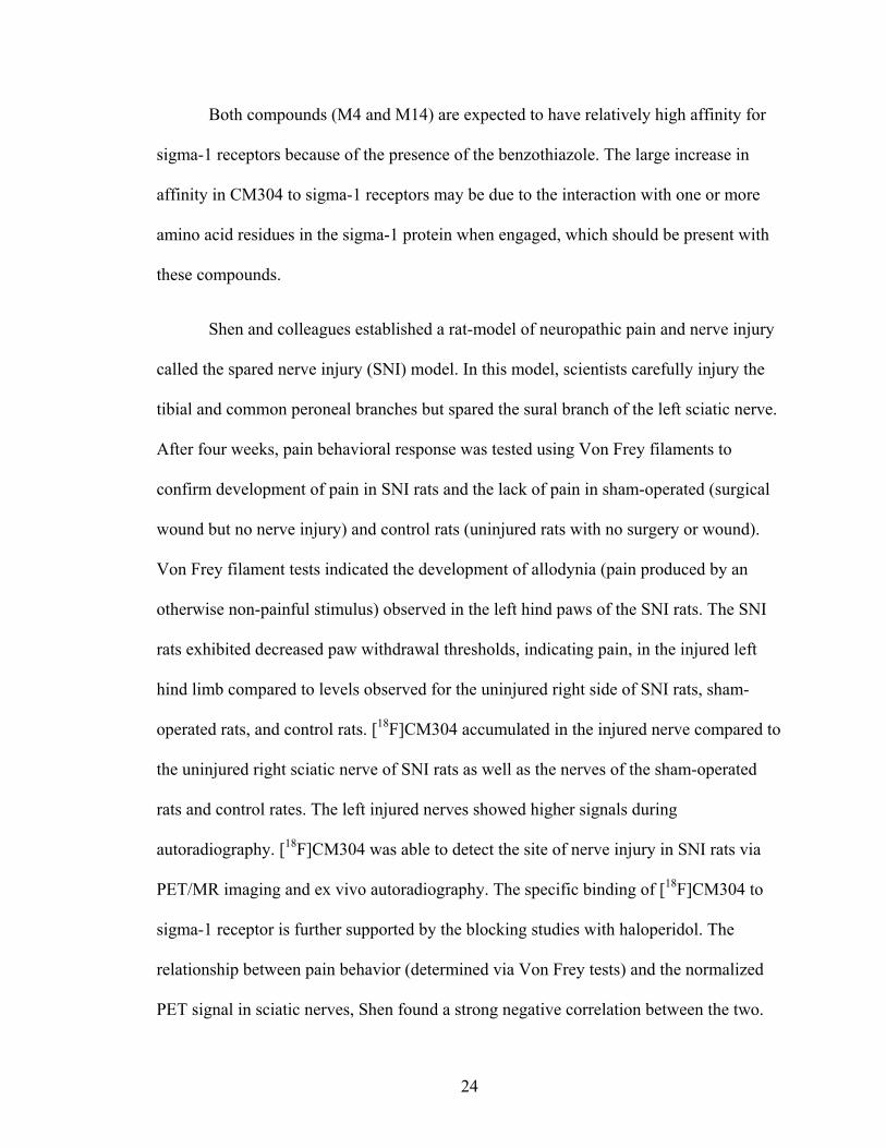
3-(2-azepan-1-yl)ethyl)-6-(4-chlorobutyl)benzo[d]thiazol-2(3H)-one (M10). In a dry
round bottom flask, M9 (0.6482 grams, 2.7 millimoles) was dissolved in 25 milliliters of
dimethylformamide. 2-(Hexamethyleneimino)ethyl chloride hydrochloride (2.141 grams,

33
10.8 millimoles) was added and the reaction mixture was heated to 120C. Then sodium
bicarbonate (2.271 grams, 27 millimoles) was slowly added and the reaction was left for
30 minutes. The mixture was poured into water and extracted with ethyl acetate. The
layers were separated, and the organic layer was washed with brine and dried with
sodium sulfate. The organic layer was then transferred to a round bottom flask, and the
solvent was removed in vacuo. The residue was purified with column chromatography
and resulted in 0.9425 grams (95.6%) of 3-(2-azepan-1-yl)ethyl)-6-(4-
chlorobutyl)benzo[d]thiazol-2(3H)-one as a white solid. 1H NMR (400 MHz,
Chloroform-d) δ 7.23 (s, 1H), 7.11 (dd, J = 8.3, 1.8 Hz, 1H), 7.02 (d, J = 8.2 Hz, 1H),
4.05 – 3.99 (m, 2H), 3.54 (t, J = 6.0 Hz, 2H), 2.84 – 2.78 (m, 2H), 2.74 (t, J = 5.5 Hz,
4H), 2.66 (q, J = 7.3, 6.2 Hz, 3H), 1.78 (dd, J = 7.9, 4.2 Hz, 4H), 1.63 (dd, J = 9.5, 4.9
Hz, 5H). 13C NMR (101 MHz, CDCl3) δ 169.77, 136.87, 135.37, 126.42, 122.78, 122.23,
110.54, 55.70, 54.68, 44.77, 41.17, 34.66, 31.90, 28.72, 28.17, 26.90. MS (ESI) m/z (M+
+ H) 367.5
4-(3-(2-azepan-1-yl)ethyl)-2-oxo-2,3-dihydrobenzo[d]thiazol-6-yl)butyl benzoate
(M11). Approximately 25 milliliters of dimethylformamide was added to M10 (0.8837
grams, 2.421 millimoles) in a round bottom flask. Potassium carbonate (0.8365 grams,
6.0525 millimoles) and benzoic acid (1.478 grams, 12.105 millimoles) were then added
to the solution. The reaction was heated to 110oC and stirred for 2 hours. The mixture
was poured into distilled water and extracted with ethyl acetate. The layers were
separated, and the organic layer was washed with brine and dried with sodium sulfate.
The solvent was evaporated in vacuo. The reaction resulted in 880 milligrams (80.8%) of
4-(3-(2-azepan-1-yl)ethyl)-2-oxo-2,3-dihydrobenzo[d]thiazol-6-yl)butyl benzoate as a

34
pale yellow viscous oil. 1H NMR (400 MHz, Chloroform-d) δ 8.08 – 8.01 (m, 2H), 7.56
(t, J = 7.4 Hz, 1H), 7.45 (q, J = 7.7, 6.9 Hz, 2H), 7.27 (s, 1H), 7.13 (q, J = 8.3 Hz, 2H),
4.35 (t, J = 5.6 Hz, 2H), 4.11 (t, J = 7.4 Hz, 2H), 2.86 (dt, J = 19.6, 6.1 Hz, 7H), 2.72 (d,
J = 7.2 Hz, 2H), 1.80 (dt, J = 7.7, 3.7 Hz, 3H), 1.68 (dd, J = 9.7, 4.9 Hz, 4H), 1.64 – 1.58
(m, 4H). 13C NMR (101 MHz, CDCl3) δ 169.85, 166.60, 137.16, 135.20, 132.88, 130.34,
129.51, 128.34, 126.61, 122.74, 122.27, 110.66, 64.67, 55.52, 54.35, 40.67, 35.04, 28.21,
27.98, 27.51, 26.91. MS (ESI) m/z (M+ + H) 453.56
3-(2-azepan-1-yl)ethyl)-6-(4-hydroxybutyl)benzo[d]thiazol-2(3H)-one (M12). M11
(0.850 grams, 1.890 millimoles) was dissolved in 15 milliliters of methanol. The solution
was added to sodium hydroxide (0.184 grams, 4.720 millimoles) that was in 15 milliliters
of water. The mixture was heated at 90oC for 1 hour and concentrated in vacuo. The
mixture was poured into 1N hydrochloric acid and extracted with ethyl acetate. The pH
of the aqueous layer was adjusted to 10 with potassium carbonate, and the mixture was
extracted with ethyl acetate. The organic layer was dried with sodium sulfate, and the
solvent was evaporated in vacuo. The reaction resulted in 430 milligrams (65%) of 3-(2-
azepan-1-yl)ethyl)-6-(4-hydroxybutyl)benzo[d]thiazol-2(3H)-one. 1H NMR (400 MHz,
Chloroform-d) δ 7.24 (d, J = 1.6 Hz, 1H), 7.15 – 7.07 (m, 2H), 4.14 – 4.05 (m, 2H), 3.66
(t, J = 6.4 Hz, 2H), 2.90 – 2.79 (m, 6H), 2.66 (t, J = 7.5 Hz, 2H), 1.72 – 1.55 (m, 13H).
13C NMR (101 MHz, CDCl3) δ 169.90, 137.57, 135.04, 126.63, 122.61, 122.28, 110.60,
77.36, 77.04, 76.72, 62.56, 55.61, 54.42, 40.65, 35.20, 32.13, 27.76, 27.55, 26.85. MS
(ESI) m/z (M+ + H) 349.54
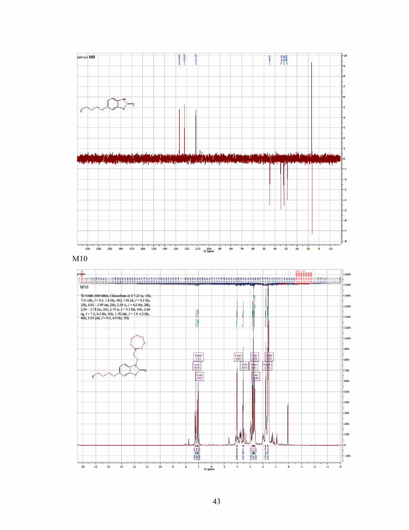
4-(3-(2-(azepan-1-yl)ethyl)-2-oxo-2,3-dihydrobenzo[d]thiazol-6-yl)butyl 4-
methylbenzenesulfonate (M13). A solution of M12 (0.200 grams, 0.572 millimoles) in

35
triethylamine (0.16 milliliters, 1.144 millimoles) and 5 milliliters of dichloromethane was
made. A solution of p-toluenesulfonyl chloride (0.120 grams, 0.6292 millimoles) in
dichloromethane (5 milliliters) was added to the existing mixture. The mixture was
stirred for 8 hours at room temperature. The solvent was evaporated in vacuo. The
residue was purified using silica gel column chromatography employing 2% methanol
and dichloromethane as the mobile phase to obtain 110 milligrams (38.1%) of 4-(3-(2-
(azepan-1-yl)ethyl)-2-oxo-2,3-dihydrobenzo[d]thiazol-6-yl)butyl 4-
methylbenzenesulfonate as a yellow viscous oil. 1H NMR (400 MHz, Chloroform-d) δ
7.81 – 7.74 (m, 2H), 7.34 (d, J = 7.9 Hz, 2H), 7.16 (d, J = 1.6 Hz, 1H), 7.09 – 7.01 (m,
2H), 4.04 (dd, J = 8.4, 6.4 Hz, 4H), 2.84 (dd, J = 8.5, 6.1 Hz, 2H), 2.79 – 2.75 (m, 4H),
2.59 (t, J = 6.9 Hz, 2H), 2.44 (s, 3H), 1.68 – 1.56 (m, 12H). 13C NMR (101 MHz, CDCl3)
δ 169.76, 144.75, 136.64, 135.34, 133.09, 129.83, 127.84, 126.46, 122.75, 122.21,
110.58, 77.36, 77.04, 76.73, 70.22, 55.68, 54.61, 41.04, 34.65, 28.23, 28.00, 27.31, 26.89,
21.63. MS (ESI) m/z (M+ + H) 503.56
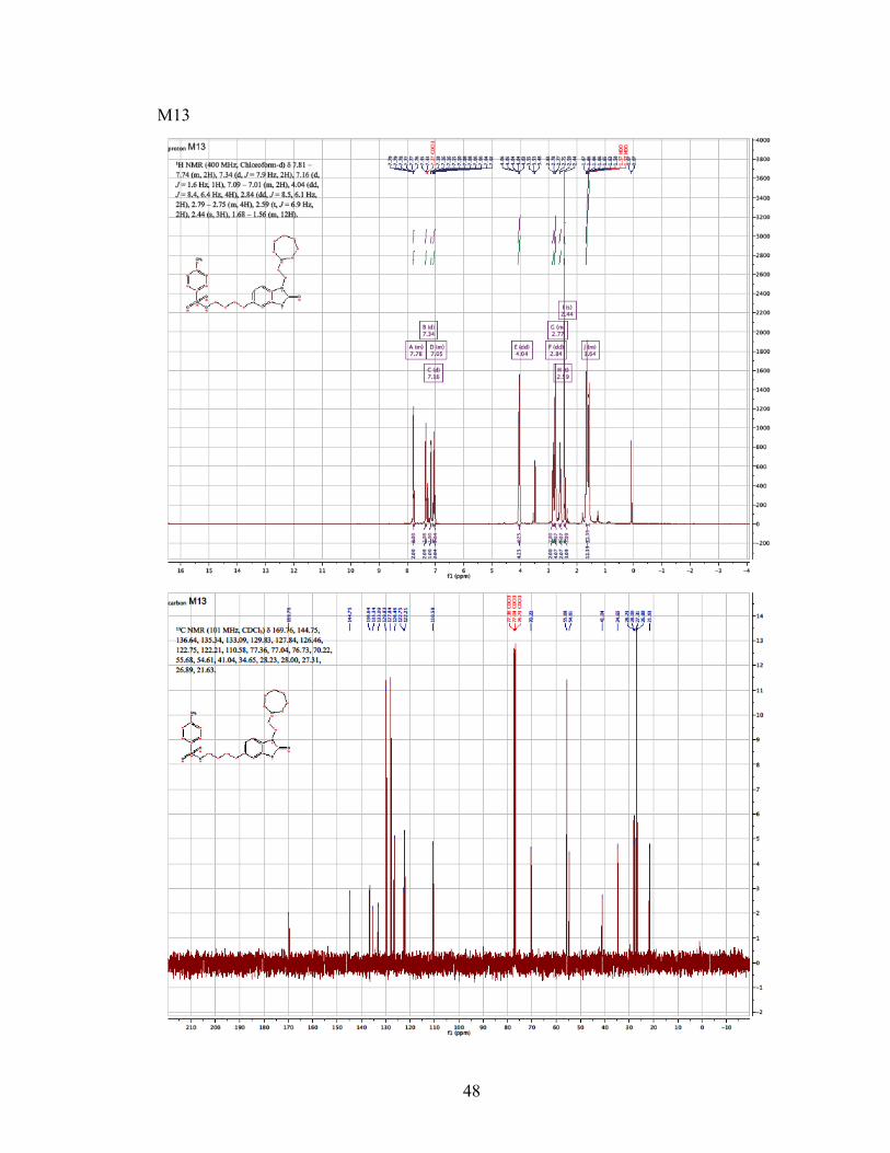
3-(2-azepan-1-yl)ethyl)-6-(4-fluorobutyl)benzo[d]thiazol-2(3H)-one (M14). A solution
of tetrabutylammonium fluoride (TBAF) in THF (1M/THF, 0.280 milliliters, 0.287
millimoles) was added to a solution of M13 (100 milligrams, 0.199 millimoles) in THF
(10 milliliters). The reaction mixture was heated to 60oC and stirred for 4 hours. The
solution was concentrated, and the residue was dissolved with 20 milliliters of ethyl
acetate. The solution was washed with 10% aqueous sodium bicarbonate solution, water,
and brine. The organic layers were dried with sodium sulfate and concentrated in vacuo.
The residue was purified by silica gel column chromatography using methanol and
chloroform as the eluent to obtain 30 milligrams (43.1%) of 3-(2-azepan-1-yl)ethyl)-6-(4-

36
fluorobutyl)benzo[d]thiazol-2(3H)-one as a yellow viscous oil. 1H NMR (400 MHz,
Methanol-d4) δ 7.33 (s, 1H), 7.17 (t, J = 6.5 Hz, 2H), 4.47 (d, J = 5.3 Hz, 1H), 4.36 (t, J
= 5.7 Hz, 1H), 4.05 (t, J = 7.1 Hz, 2H), 2.94 – 2.57 (m, 8H), 1.77 – 1.51 (m, 12H). 13C
NMR (101 MHz, MeOD) δ 170.31, 137.68, 135.06, 126.63, 122.20, 122.08, 110.74,
84.11, 82.48, 55.35, 54.33, 48.28, 48.07, 47.85, 47.64, 47.43, 47.21, 47.00, 40.17, 34.58,
29.74, 29.55, 29.41, 27.29, 27.12, 27.07, 26.50. MS (ESI) m/z (M+ + H) 351.68

37
REFERENCES
1. Bowen, W. D. σ receptors: recent advances and new clinical potentials. Pharm.
Acta. Helv. 2000, 74, 211-218.
2. Brown, A. K.; Kimura, Y.; Zoghbi, S. S.; Siméon, F. G.; Liow, J.; Kreisl, W. C.;
Taku, A.; Fujita, M.; Pike, V. W.; Innis, R. B. Metabotropic glutamate subtype 5
receptors are quantified in the human brain with novel radioligand for PET. J
Nucl Med. 2008, 49, 2042-2048.
3. Chapter 12: reactions of arenes, electrophilic aromatic substitution [Internet].
Calgary, Canada: University of Calgary [cited 2014 Dec 15]. Available from:
http://www.chem.ucalgary.ca/courses/350/Carey5th/Ch12/ch12-7.html.
4. Cobos, E. J.; Entrena, J. M.; Nieto, F. R.; Cendan, C. M.; Del Pozo, E.
Pharmacology and therapeutic potential of sigma-1 receptor ligands. Curr
Neuropharmcol. 2008, 6, 344-366.
5. de la Puente, B.; Nadal, X.; Portillo-Salido, E.; Sánchez-Arroyos, R.; Ovalle, S.;
Palcios, G.; Muro, A.; Romero, L.; Entrena, J. M.; Baeyens, J. M.; Lôpez-García,
J. A.; Maldonado, R.; Zamanillo, D.; Vela, J. M. Sigma-1 receptors regulate
activity-induced spinal sensitization and neuropathic pain after peripheral nerve
injury. Pain. 2009, 145, 294-303.
6. Drews, E.; Zimmer, A. Central sensitization needs sigma receptors. Pain. 2009,
145, 269-270.

38
7. Hanner M., Moebius, F. F., Flandorfer, A.; Knaus, H. G.; Striessnig, J.;
Kempner, E.; Glossmann, H. Purification, molecular cloning, and expression of
mammalian σ1-binding site. Proc. Natl. Acad. Sci. USA. 1996, 93, 8072-8077.
8. James, M. L.; Shen, B.; Nielsen, C. H.; Behera, D.; Buckmaster, C. L.;
Mesangeau, C.; Zavaleta, C.; Vuppala, P. K.; Jamalapuram, S.; Avery, B. A.;
Lyons, D. M.; McCurdy, C. R.; Biswal, S.; Gambhir, S. S.; Chin, F. T. Evaluation
of σ-1 receptor radioligand 18F-FTC-146 in rats and squirrel monkeys using PET.
J Nucl Med. 2014, 55, 147-153.
9. James, M. L.; Shen, B.; Zavaleta, C. L.; Nielsen, C. H.; Mesangeau, C.; Vuppala,
P. K.; Chan, C.; Avery, B. A.; Fishback, J. A.; Matsumoto, R. R.; Gambhir, S. S.;
McCurdy, C. R.; Chin, F. T. New positron emission tomography (PET)
radioligand for imaging sigma-1 receptors in living subjects. J Med Chem. 2012,
55, 8272-8282.
10. Jarvik, J. G.; Yuen, E.; Kliot, M. Diagnosis of carpal tunnel syndrome:
electrodiagnostic and MR imaging evaluation. Neuroimag Clin N Am. 2004, 14,
93-102.
11. Martin, W. R.; Eades, C. G.; Thompson, J. A.; Huppler, R. E.; Gilbert, P. E. The
effects of morphine- and nalorphine-like drugs in the nondependent and
morphine-dependent chronic spinal dog. J. Pharmacol. Exp. Thera. 1976, 197,
517-532.
12. Maurice, T.; Su, T. P. The pharmacology of sigma-1 receptors. Pharmacol. Ther.
2009, 124, 195-206.

39
13. Mei, J.; Pasternak, G. W. Molecular cloning and pharmacological characterization
of the rat σ1 receptor. Biochem. Pharmacol. 2001, 62, 349-355.
14. Muehllehner, G.; Karp, J. S. Positron emission tomography. Phys. Med. Biol.
2006, 51, 117-137.
15. Narayanan, S.; Bhat, R.; Mesangeau, C.; Poupaert, J. H.; McCurdy, C. R. Early
development of sigma-receptor ligands. Future Med. Chem. 2011, 3, 79-94.
16. Pan, Y. X.; Mei, J.; Xu, J.; Wan, B. L.; Zuckermann, A.; Pasternak, G. W.
Cloning and characterization of a mouse σ1 receptor. J. Neurochem. 1998, 70,
2279-2285.
17. Phelps, M. E. Positron emission tomography provides molecular imaging of
biological processes. Proc. Natl. Acad. Sci. U.S.A.. 2000, 97, 9226-9233.
18. Pike, V. W. PET radiotracers: crossing the blood-brain barrier and surviving
metabolism. Trends Pharmacol Sci. 2009, 30, 341-440.
19. Seth, P.; Fei, Y. J.; Li, H. W.; Huang, W.; Leibach, F. H.; Ganapathy, V. Cloning
and functional characterization of a σ receptor from rat brain. J. Neurochem.
1998, 70, 922-931.
20. Seth, P.; Leibach, F. H.; Ganapathy, V. Cloning and structural analysis of the
cDNA and the gene encoding the murine type 1 σ receptor. Biochem. Biophys.
Res. Commun. 1997, 241, 535-540.
21. Shen, B.; Behera, D.; James, M. L.; Borogohain, P.; Andrews, L.; Lutz, A. B.;
Mavlyutov, T.; Patankar, M.; Puoho, A. E.; McCurdy, C. R.; Gambhir, S. S.;
Biswal, S.; Chin, F. T. Identifying peripheral pain generators via PET/MR
imaging. Proc. Natl. Acad. Sci. U.S.A.. 2015.

40
22. Tsai, S. Y.; Hayashi, T.; Mori,T.; Su, T. P. Sigma-1 receptor chaperones and
diseases. Cent Nerv Syst Agents Med Chem. 2009, 9, 184-189.
23. Wong, E. H. F.; Knight, A. R.; Woodruff, G. N. [3H] MK-801 labels a site on the
N-methyl-D-aspartate receptor channel complex in rat brain membranes. J.
Neurochem. 1988, 50, 274-281.
24. Yous, S.; Wallez, V.; Belloir, M.; Caignard, D. H.; McCurdy, C. R.; Poupaert, J.
H. Novel 2(3H)-benzothiazolones as highly potent and selective sigma-1 receptor
ligands. Med. Chem. Res. 2005, 14, 158-168.

41
APPENDIX A: NUCLEAR MAGNETIC RESONANCE SPECTRA M8

42
M9
43
M10

44

45
M11

46
M12

47
48
M13

49
M14

50

51
APPENDIX B: MASS SPECTRUM DATA
M8
Mass Spectrum for M9 is not available.

52
M10
M11

53
M12
M13
54
M14