design and syntheses of potential drugs based on gaba ......design and syntheses of potential drugs...
TRANSCRIPT
Design and Syntheses of Potential Drugs Based on GABAA Receptor Pharmacophores
Ella Chow Clement
Dissertation submitted to the Faculty of the Virginia Polytechnic Institute and State University in partial fulfillment of the requirements
for the Degree of
Doctor of Philosopy in
Chemistry
Dr. Paul R. Carlier, Chairman Dr. Jeffrey R. Bloomquist Dr. Richard D. Gandour Dr. David G. I. Kingston
Dr. James M. Tanko
June 28, 2005 Blacksburg, Virginia
Keywords: GABAA receptor, Partial/full agonists, Superagonist, Antagonists, Non-zwitterionic GABA amide homodimers and heterodimers, 36Cl- Flux assay, [3H]Muscimol binding, ZAPA, PEG, Memory of Chirality
Copyright 2005, Ella Chow Clement
Design and Syntheses of Potential Drugs Based on GABAA Receptor Pharmocophores
Ella Chow Clement
ABSTRACT
Numerous previous studies of GABAAR ligands have suggested that GABAAR
agonists must be zwitterionic and feature an intercharge separation similar to that of
GABA (approx. 4.7-6.0 Å). We have demonstrated that monomeric, homodimeric and
heterodimeric non-zwitterionic GABA amides are partial, full, or superagonists at the
murine GABAA receptor (GABAAR). The agonism of these GABA amides is
comparable to that of THIP, as shown by in vitro assay results. The assay data indicate
that the agonism of GABA amides is tether length-dependent. Optimum agonism is
achieved with a tether length of four methylenes in GABA amide dimers and in GABA
amides bearing pendant amide or amino groups. We have further investigated the
structure-activity relationship for GABA amides on the GABAAR by performing
structural modifications to both the superagonist 2c and the agonist 6c. Synergism and
[3H]muscimol binding experiments show that 2c binds to the same sites as GABA.
Structural modification of 2c demonstrated that partial rigidification of the tether
eliminated agonism and caused ligands to behave as weak competitive antagonists. We
have also investigated the agonism of four ZAPA derivatives in 36Cl- uptake functional
assay. Two of them are found to be as potent as GABA.
In our studies of 1,4-benzodiazepines, our goal was to synthesize three different
subtypes of quaternary 1,4-benzodiazepines by use of the memory of chirality (MOC)
strategy. Disappointingly, most of the deprotonation/alkylations failed, due to various
reasons. The failure of the reactions of (S)-alanine-derived tetrahydro-1,4-
benzodiazepin-3-ones was probably due to either the unexpected side reactions or the
steric hindrance of enolate alkylation. In the case of tetrahydro-1,4-benzodiazepin-2-
ones, computational studies suggested that steric hindrance by both the benzo ring and
N4-allyl group might retard deprotonation at C3 by bulky bases like KHMDS or LDA.
Finally, (S)-serine-derived 1,4-benzodiazepin-2-ones and their elimination products (α-
methylene benzodiazepines) were prepared. These proved unreactive towards
deprotonation/alkylations and conjugate additions, respectively. The low reactivity of the
α-methylene benzodiazepines towards nucleophiles was attributed to highly delocalized
LUMOs that failed to direct nucleophiles to the β-carbons.
iii
Acknowledgements
First of all, I would like to thank my advisor, Dr. Paul Carlier, for giving me the
opportunity to come with him and study at Virginia Tech. I would also like to thank Dr.
Carlier for his guidance and financial support during my Ph.D. study. Secondly, I would
like to thank Dr. Jeffrey Bloomquist for his help and support in doing bioassay
experiments. Thirdly, I would like to thank my committee members for their assistance
in fulfilling departmental requirements. I would also like to thank Mr. Tom Glass and
Mr. Bill Bebout for their analytical services, and my group members for sharing lab
responsibility.
I would like to thank Mrs. Kay Castagnoli and Dr. Emre Isin for their friendship
and support. I also like to thank friends, especially Dr. Mary Dean Coleman, Miss Amy
Fletcher, Miss Jennifer Farris, and Ms. Mary Billings from the Graduate Christian
Fellowship for their prayers and support.
I would like to thank my family for their love and support during my study. I
would like to especially thank my father, who has gone to be with the Lord, for his great
influence in my life. Finally, I would like to thank my husband Jason for his love, help,
and support in every aspect of my studies and life.
iv

Table of Contents
List of Figures vii Chapter 1 Introduction and Background of GABAA Receptors ................................... 1
1.1 Background and Significance of the GABAA Receptor ......................................... 1 1.2 Classification of Three GABA Receptor Subtypes ................................................ 2 1.3 GABAA Receptor Physiology and Pharmacology.................................................. 6
1.3.1 Current Models for the Structure and Function of the GABAA Receptors. 6 1.3.2 Benzodiazepine Binding Sites of The GABAA Receptors ....................... 14 1.3.3 Barbiturates, Picrotoxinin and Steroid Anesthetic Binding Sites in the
GABAAR................................................................................................... 16 1.4 GABAA Receptor-Agonists and Partial Agonists................................................. 20
1.4.1 Structure and Conformation of Active GABA Analogs (Exogenous GABAA agonists)...................................................................................... 20
1.4.2 Endogenous Agonists................................................................................ 24 1.5 GABAA Agonist-Receptor Interactions ................................................................ 25
1.5.1 Electronic Factors ..................................................................................... 26 1.5.2 Structural and Conformational Factors..................................................... 26 1.5.3 Stereochemical Factors ............................................................................. 27 1.5.4 Current Agonist Binding model of the GABAA Receptor........................ 28
1.6 Traditional Assays for Agonists............................................................................ 32 1.7 Pharmacokinetic Aspects and Prodrugs................................................................ 34
References for chapter 1 ................................................................................................... 38 Chapter 2 Syntheses of GABAA receptor analogs...................................................... 47
2.1 Syntheses and Evaluation of homodimeric GABAA receptor analogs ................. 48 2.1.1 Bioassay Results of New GABA amide homodimers .............................. 49 2.1.2 Synthesis of Derivatives of ‘Superagonist’ Dimer 2c (4a-h).................... 53 2.1.3 Bioassay Results of the Derivatives (4a-h)............................................... 57 2.1.4 [3H]Muscimol Binding Experiments ........................................................ 59
2.2 Syntheses and Evaluation of heteromeric GABAA receptor analogs ................... 62 2.2.1 Bioassay Results of the Amide Heterodimers .......................................... 63
2.3 Syntheses and Evaluation of ZAPA and its derivatives........................................ 68 2.3.1 Synthesis of ZAPA and its derivatives ..................................................... 68 2.3.2 Bioassay Results of ZAPA and its derivatives ......................................... 72
2.4 Syntheses and Evaluation of PEG-linked GABAA receptor analogs.................... 76 2.4.1 Synthesis of PEG-linked GABA amide dimers ........................................ 76 2.4.2 Bioassay Results of PEG-linked GABA amide dimers ............................ 85 2.4.3 Synthesis of PEG-linked ZAPA analogs .................................................. 87
2.5 HPLC Analysis of GABA amides ........................................................................ 88 2.6 Conclusions........................................................................................................... 93
References and notes for chapter 2 ................................................................................... 95 Chapter 3 Experimental Section of GABA Project .................................................... 99
3.1 Chemistry.............................................................................................................. 99 3.1.1 General Methods....................................................................................... 99 3.1.2 Procedures............................................................................................... 100
3.2 Biological Assays................................................................................................ 128
v
References for chapter 3 ................................................................................................. 132 Chaper 4 Introduction and Background of 1,4-benzodiazopines ........................... 133
4.1 Importance of 1,4-benzodiazopine scaffolds in medical chemistry.................... 133 4.2 Importance of exploring enantiopure quaternary 1,4-benzodiazepines.............. 136 4.3 Overview of Pioneering Work in Carlier’s Research Group .............................. 139
Reference for chapter 4................................................................................................... 143 Chapter 5 Syntheses of 1,4-Benzodiazepine Analogs .............................................. 150
5.1 Investigation of Enantioselective Synthesis of Quaternary (S)-alanine Derived Tetrahydro-1,4-benzodiazepin-3-ones ................................................................ 150
5.1.1 Synthesis of (S)-alanine Derived Tetrahydro-1,4-benzodiazepin-3-ones150 5.1.2 Synthesis of Quaternary (S)-alanine Derived Tetrahydro-1,4-
benzodiazepin-3-ones Via “Memory of Chirality” (MOC) .................... 155 5.1.3 Computational Studies of Tetrahydro-1,4-benzodiazepin-3-ones .......... 182
5.2 Investigation of Enantioselective Synthesis of Quaternary (S)-phenylalanine Derived Tetrahydro-1,4-benzodiazepin-2-ones .................................................. 186
5.2.1 Synthesis of (S)-phenylalanine Derived Tetrahydro-1,4-benzodiazepin-2-ones ......................................................................................................... 186
5.2.2 Attempted Synthesis of Quaternary Tetrahydro-1,4-benzodiazepin-2-ones Via MOC................................................................................................. 189
5.2.3 Computational Studies of (S)-phenylalanine-derived Tetrahydro-1,4-benzodiazepin-2-ones ............................................................................. 191
5.3 Investigation of Enantioselective Synthesis of Quaternary (S)-serine -derived 1,4-benzodiazepin-2-ones through Michael Addition............................................... 194
5.3.1 Sythesis of (S)-serine -derived 1,4-benzodiazepin-2-ones...................... 197 5.3.2 Attempted Synthesis of 3,3-Substituted (S)-serine -derived 1,4-
benzodiazepin-2-ones ............................................................................. 199 5.3.3 Investigation of Enantioselective Synthesis of 1,4-benzodiazepin-2-ones
Via Michael addition............................................................................... 202 5.3.4 Computational Studies of 2-Methylenecycloalkanones and Heterocyclic 2-
methylenecycloalkanones ....................................................................... 206 5.4 Conclusions......................................................................................................... 211
References for chapter 5 ................................................................................................. 212 Chapter 6 Experimental Section of 1,4-Benzodiazepine Analogs............................ 218
6.1 General Methods................................................................................................. 218 6.2 Procedures........................................................................................................... 219
6.2.1 (S)-Alanine-derived tetrahydro-1,4-benzodiazepin-3-one project.......... 219 6.2.2 (S)-Phenylalanine-derived tetrahydro-1,4-benzodiazepine-2-one project
................................................................................................................. 236 6.2.3 (S)-Serine-derived 1,4-benzodiazepine-2-one project ............................ 239
References for chapter 6 ................................................................................................. 249
vi
List of Figures
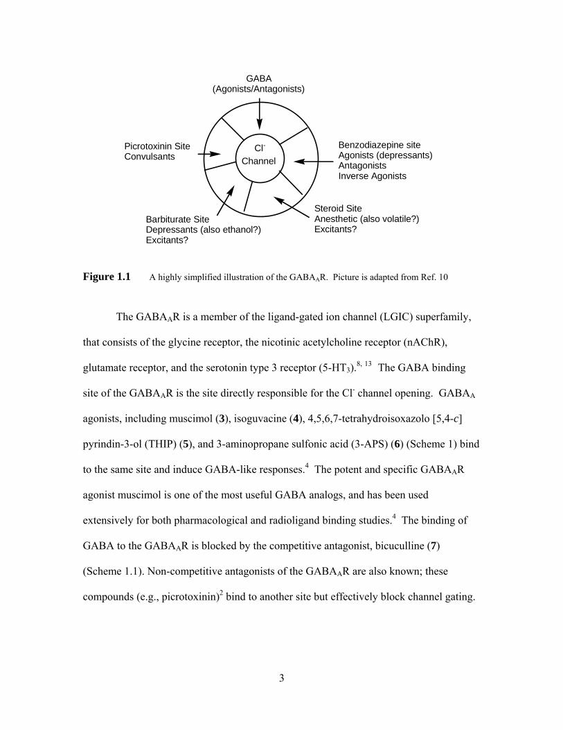
Figure 1.1 A highly simplified illustration of the GABAAR. Picture is adapted from Ref. 10......................................................................................................... 3
Figure 1.2 Proposed GABAAR structure showing the two functional agonist sites (G) and the benzodiazepine binding site (B). Letters A-F are designated as six discontinous polypeptide loops that are participated in the formation of agonist binding pocket. Picture is adapted from Ref 23 and 34 ................ 7
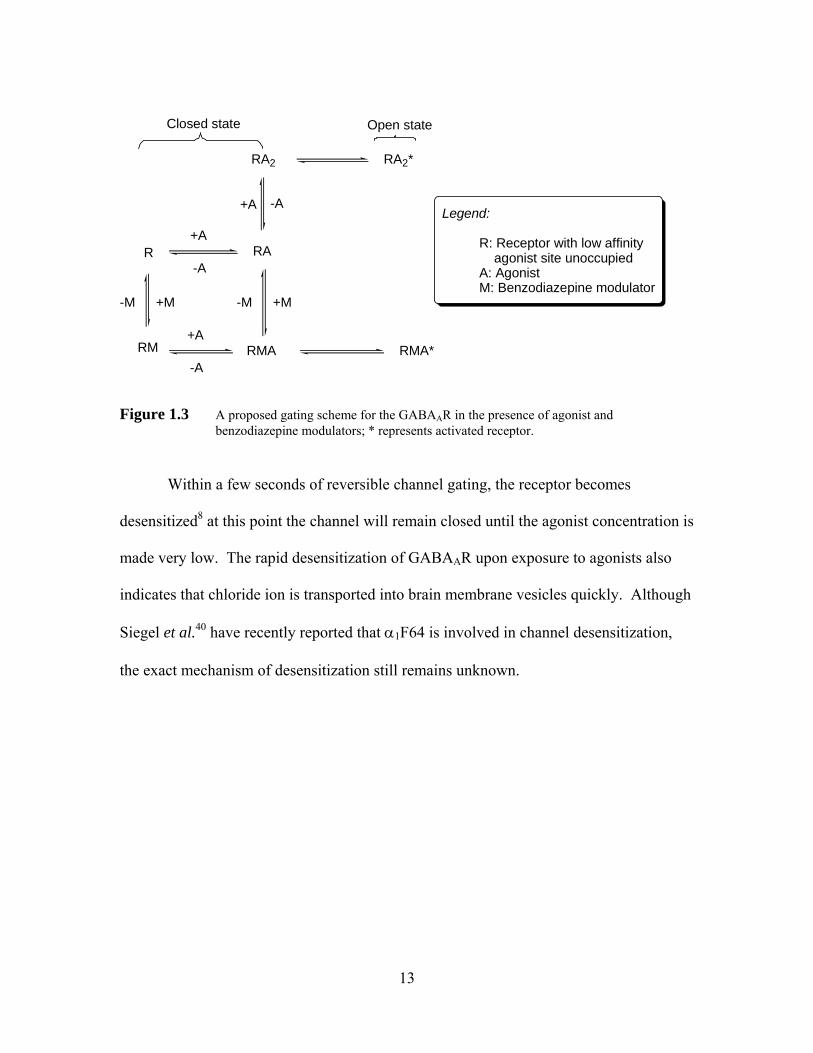
Figure 1.3 A proposed gating scheme for the GABAAR in the presence of agonist and benzodiazepine modulators; * represents activated receptor.................... 13
Figure 1.4 Enhancement of muscimol-induced 36Cl- uptake by diazepam. Picture is modified from Ref. 41............................................................................... 15
Figure 1.5 Potential conformational energy minima of GABA17 .............................. 21 Figure 1.6 Schematic description of receptor occupancy and gating of GABAAR.... 31 Figure 1.7 An illustration of the determination of the I/U ratio for Isoguvacine in
aqueous solution53..................................................................................... 35 Figure 1.8 GABAA agonists prodrugs........................................................................ 37 Figure 2.1 Effect of tether length of GABA amide dimers (n = number of
methylenes) on 36Cl- flux (at 1 mM drug) in mouse brain synaptoneurosomes, expressed as % uptake versus background (bkd, 100%, dotted line). An asterisk signifies that uptake is significantly different than background (p<0.05, t-test) ................................................ 50
Figure 2.2 Dose-response relationship of Cl- uptake and drug (GABA and 2a-e) concentrations in mouse brain synaptoneurosomes.................................. 50
Figure 2.3 Effect of nipecotic acid (100 μM) on GABA-induced Cl- uptake. Letters a and b signify that uptake is significantly different than background (p<0.05, t-test); and the uptake of experiment 1is not significantly different than that of experiment 2............................................................ 51
Figure 2.4 Effect of non-competitive (picrotoxinin, PTX) and competitive (bicuculline, BCC) inhibitors on 36Cl- ion flux elicited by 2c (1 mM) in mouse brain synaptoneurosomes; bkd represents background uptake in the absence of agonist. Asterisks indicate that the aonist-induced uptake in the presence of blocker significantly less than that observed with 1 mM 2c alone (p<0.05, t-test) ................................................................................. 52
Figure 2.5 Co-incubation experiment of (dose-response curve of GABA + 250 μM 2c) GABA and 2c...................................................................................... 53
Figure 2.6 Effects of GABA amide dimer derivatives 4a-h on ion flux elicited by GABA (0.1 mM); bkd represents background, uptake in the absence of agonist. Asterisks indicate that the GABA-induced uptake in the presence of the blocker at 1mM is significantly less than that observed with 0.1 mM GABA alone (p < 0.05, t-test)................................................................... 58
Figure 2.7 Inhibitory effect of 4b on concentration dependent chloride uptake by GABA in mouse brain synaptoneurosomes.............................................. 59
Figure 2.8 % Total binding of [3H]muscimol in the presence of unlabeled ligands and other unlabeled ligands in mouse brain synaptoneurosomes. An asterisk
vii
indicates that the binding is significantly different than from treatment 1 (p < 0.05, t-test) ............................................................................................. 61
Figure 2.9 Dose-response relationship of Cl- uptake and drug (GABA, 1a-b and THIP) concentrations in mouse brain synaptoneurosomes....................... 64
Figure 2.10 Effect of tether length of GABA amides (n = number of methylenes) on chloride flux (at 1 mM drug) in mouse brain synaptoneurosomes, expressed as % uptake versus background. An asterisk indicates that the uptake is significantly different than background (p < 0.05, t-test).......... 65
Figure 2.11 Dose-response relationship of Cl- uptake and drug (GABA, 5c and 6c) concentrations in mouse brain synaptoneurosomes.................................. 66
Figure 2.12 Dose-response relationship of Cl- uptake and drug (GABA, ZAPA, compound 11, 12 and 15) concentrations in mouse brain synaptoneurosomes ................................................................................... 73
Figure 2.13 Ion flux elicited by 97% pure E-isomer (with 3% Z-isomer ) of compound 12 in mouse brain synaptoneurosomes, expressed as % uptake versus background. An asterisk indicates that the uptake is significantly different than background (p < 0.05, t-test)............................................................. 74
Figure 2.14 Ion flux elicited by 4 different concentrations of compound 15 in mouse brain synaptoneurosomes, expressed as % uptake versus background..... 75
Figure 2.15 A schematic diagram of desolvation penalty effect in ligand-receptor interaction ................................................................................................. 76
Figure 2.16 Side view of X-ray crystal structure of the AChBP (pdb code 1I9B, graphic created with RasMol Molecular Graphics). HEPES ligands (red) represent the putative ACh binding sites .................................................. 78
Figure 2.17 A simplified schematic diagram of mouth-to-mouth distance between two agonist binding sites of on the GABAAR (top), and a bivalent ligand designed to span the sites (bottom, with an example of liagand (ZAPA in this case) and spacer) ................................................................................ 79
Figure 2.18 Ion flux elicited by 19a-d (500 μM) in mouse brain synaptoneurosomes; bkd represents background, uptake in the absence of agonist .................. 85
Figure 2.19 Effect of 19d on chloride uptake elicited by GABA (100 μM) in mouse brain synaptoneurosomes; backgroud uptake was at 100%. Dash line represented chloride uptake induced by GABA (100 μM). An asterisk signifies that uptake is significantly different than background (p<0.05, t-test)............................................................................................................ 87
Figure 2.20 Effect of NaTFA (1 mM) on chloride uptake elicited by GABA (1 mM), expressed as % uptake versus background (bkd)...................................... 92
Figure 5.1 HPLC chromatograms of (S)-56 (left) and its racemic mixture (S/R)-56 (right); X denotes an unknown impurity................................................. 182
Figure 5.2 B3LYP/6-31G(d) electronic energy difference between the (M)- and (P)- conformers of (S)-56; the sign of the dihedral angle C6-C7-N1-C2 defines the helical chirality descriptor (M (minus) or P (plus)) .......................... 184
Figure 5.3 B3LYP/6-31G(d) electronic energy difference between the (P)- and (M)- conformers of (S)-83; the sign of the dihedral angle C6-C7-N1-C2 defines the helical chirality descriptor (M (minus) or P (plus)); B denotes a base such as KHMDS or LDA........................................................................ 192
viii
Figure 5.4 Calculated dihedral angles (Cβ=Cα-C=O) of α,β-unsaturated carbonyl system of selected molecules at B3LYP/6-31G(d)................................. 207
Figure 5.5 Calculated dihedral angles(Cβ=Cα-C=O) of α,β-unsaturated carbonyl system of substrates that underwent successful conjugate addition at B3LYP/6-31G(d) .................................................................................... 208
Figure 5.6 LUMOs of selected compounds arranged to the right of or below the bold-line structural drawings. LUMOs were calculatid at B3LYP/6-31-G(d)210
ix
List of Schemes
Scheme 1.1 GABAAR, GABABR and GABACR Agonists and Antagonists ................. 4 Scheme 1.2 Important drugs and modulators for GABAAR ........................................ 17 Scheme 1.3 Conformationally restricted GABA analogs in their ionized and unionized
forms ......................................................................................................... 22 Scheme 1.4 Proposed mechanisms for the hydrolysis of progabide (40)95 .................. 38 Scheme 2.1 GABA amides examined previously by other workers ............................ 47 Scheme 2.2 Synthesis of GABA amide homodimers................................................... 48 Scheme 2.3 Design of derivatives of 2c (4a-h) ............................................................ 54 Scheme 2.4 Synthetic scheme of derivatives of 2c (4a-h) ........................................... 55 Scheme 2.5 Synthesis of dihydrochloride salts of diamine .......................................... 56 Scheme 2.6 Synthetic scheme for simple GABA amides ............................................ 62 Scheme 2.7 Synthesis of 1c under two different conditions......................................... 63 Scheme 2.8 Design of derivatives of 6c (7a-i) ............................................................. 67 Scheme 2.9 Synthesis of ZAPA derivatives................................................................. 71 Scheme 2.10 Proposed mechanism for the formation of compound 13 ......................... 72 Scheme 2.11 Selected successful examples of bivalent acetylcholinesterase inhibitors
(bis(7)-tacrine (both Carlier and Pang)40 and (S,S)-(-)-17 (Carlier),41 and its corresponding monomers (tacrine, (-)-Huperzine A and simplified fragment (±)-18)........................................................................................ 80
Scheme 2.12 Synthesis of PEG-linked GABA amide dimers ........................................ 83 Scheme 2.13 PEG-linked GABA amide heteromers (20a-b)......................................... 87 Scheme 2.14 Proposed mechanism for acid-catalyzed Boc deprotection of GABA
amides. Boc protected 5a was used as an example................................... 89 Scheme 4.1 Bioactive 1,4-benzodiazepin-2-ones 1-12 .............................................. 134 Scheme 4.2 Medicinally important tetrahydro-1,4-benzodiazepin-2 & 3-ones ......... 136 Scheme 4.3 The MOC alkylation protocol of Fuji & Kawabata30 ............................. 138 Scheme 4.4 Dynamic chirality of 1, 20a47.................................................................. 139 Scheme 4.5 Stereochemical cooperativity in 21a....................................................... 140 Scheme 4.6 Schematic of a successful MOC deprotonation/alkyaltion sequence of a
(S)-alanine-derived 1,4-benzodiazepin-2-one......................................... 141 Scheme 4.7 MOC enantioselective alkylation of (S)-alanine derived 1,4-
benzodiazepin-2-ones ............................................................................. 142 Scheme 5.1 Synthesis of tetrahydro-1,4-benzodiazepin-3-ones................................. 151 Scheme 5.2 Ring closure of heterocyclic ring (S)-9 to 11 at 200 ºC.......................... 153 Scheme 5.3 Synthetic scheme of carboxyl ester-accelerated substitution.................. 154 Scheme 5.4 Proposed pathways I and II for the deactivation of enolization for (S)-10
via single electron reduction ................................................................... 157 Scheme 5.5 A proposed mechanism involving addition reactions that deactivate
deprotonation of (S)-10 ........................................................................... 158 Scheme 5.6 1,2-Addition of pyrrolidine to 2-methylcyclohexanone proposed by
Collum et al.13......................................................................................... 159 Scheme 5.7 Conversion of nitro compounds 10 and 11 to other 1,4-benzodiazepin-3-
ones ......................................................................................................... 160
x
Scheme 5.8 A proposed mechanism for the formation and decomposition of O-alkylation product 31 .............................................................................. 163
Scheme 5.9 Deamination of (S)-27 and (S)-28........................................................... 164 Scheme 5.10 A proposed mechanism for reductive deamination of (S)-32 and 33 ..... 165
A possible rearrangement mechanism for the formation of compounds 40 and 46...................................................................................................... 170
Scheme 5.11 Proposed rearrangement mechanism for compounds 40 and 46 ............ 171 Scheme 5.12 Two ring contraction rearrangement mechanisms of diazepam proposed by
Fryer et al 20............................................................................................. 172 Scheme 5.13 Proposed reduction/oxidation free radical mechanism for the formation of
rearrangement products 40 and 46 .......................................................... 174 Scheme 5.14 A proposed mechanism for an example of Rh (I) or Ru (II) catalyzed
deallylations of tetrahydro-1,4-benzodiazepin-3-one (S)-33 .................. 177 Scheme 5.15 Palladium(0)-catalyzed deallylation of allylamines................................ 178 Scheme 5.16 Bundle’s oxidative deallyation protocol of (S)-10 and (S)-29 ................ 180 Scheme 5.17 A proposed mechanism of deallylation of (S)-10 ................................... 181 Scheme 5.18 Synthesis of (S)-75, (S)-76 and proposed mechanism for the
decomposition of (S)-75.......................................................................... 188 Scheme 5.19 Ring closure of heterocyclic ring............................................................ 189 Scheme 5.20 Design of 3,3-disubstituted 1,4-benzodiazepin-2-ones through conjugate
addition ................................................................................................... 195 Scheme 5.21 Synthesis of (3R)-90 by Evan et al.38...................................................... 195 Scheme 5.22 Synthetic scheme of 3,3-disubstituted 1,4-benzodiazepin-2-ones39 ....... 196 Scheme 5.23 Design of proposed MOC route to quaternary serine-derived 1,4-
benzodiazepin-2-ones. ............................................................................ 197 Scheme 5.24 Synthetic scheme of (S)-serine-derived 1,4-benzodiazepin-2-ones........ 198 Scheme 5.25 Ring closure of (S)-98 at 40 ºC. .............................................................. 199 Scheme 5.26 Elimination reaction of (3S)-101 and (3S)-102....................................... 200 Scheme 5.27 Examples of 1,4-conjugate addition to 2-methylenecycloalkanones...... 203 Scheme 5.28 Attempted conjugate additions to compound 106 .................................. 204
xi
List of Tables
Table 1.1 PRE regulation of GABAAR open properties*......................................... 19 Table 1.2 Residues lining the GABAAR agonist binding pocket ............................. 28 Table 1.3 Correlation of incubation time on GABA-mediated chloride flux and
EC5090 ........................................................................................................ 33
Table 1.4 pKa values, and I/U ratios of some GABAAR agonists and their ability to penetrate the BBB67 .................................................................................. 36
Table 2.1 GABAAR agonism (Emax), chloride uptake (EC50 values) and Hill slope of GABA amides and controls ...................................................................... 51
Table 2.2 GABAAR agonism by GABA amides and controls.................................. 65 Table 2.3 Two-step synthesis of n-alkylthioureas .................................................... 70 Table 2.4 GABAAR agonism, EC50 values and Hill slopes of GABA, ZAPA and
ZAPA derivatives...................................................................................... 74 Table 2.5 Selected commercially available PEGs and their molecular properties ... 81 Table 2.6 % Activations and yields of PNP activated-PEG dimers.......................... 83 Table 2.7 % Incorporations and yields of PEG-linked GABA amide dimers .......... 84 Table 2.8 MALDI-TOFMS data of PEG-linked GABA amide dimers.................... 85 Table 2.9 Attempted synthesis of PEG-linked ZAPA analogs ................................. 88 Table 2.10 HPLC* data of selected GABA amides.................................................... 91 Table 5.1 Ring closure reaction conditions............................................................. 152 Table 5.2 Attempted alkylations of (S)-10.............................................................. 156 Table 5.3 Attempted C-alkylations of (S)-29 and (S)-30 ........................................ 162 Table 5.4 Attempted alkylations of (S)-32 and (S)-33 ............................................ 166 Table 5.5 Attempted alkylations (S)-42 .................................................................. 168 Table 5.6 Summary of 1H NMR data of both compounds 40 and 46 ..................... 170 Table 5.7 Attempted deallylation of tetrahydro-1,4-benzodiazepin-3-ones ........... 176 Table 5.8 Calculated ratios of (P)- and (M)-conformers (cf Figure 5.1) and C2-CH3
proton signals of (S/R)-56. ...................................................................... 183 Table 5.9 Summary of sum of internal angles and energy differences (B3LYP/6-
31G(d)).................................................................................................... 186 Table 5.10 Attempted alkylations of (S)-83.............................................................. 190 Table 5.11 Selected structural parameters for (S)-alanine-derived tetrahydro-1,4-
benzodiazepin-2 & 3-ones equilibrium geometries ................................ 194 Table 5.12 Attempted alkylations of (3S)-99 and 100 .............................................. 202 Table 5.13 Attempted conjugate additions to 103 .................................................... 205 Table 6.1 HPLC data of chiral (S)-Alanine-derived tetrahydro-1,4-benzodiazepin-3-
ones ......................................................................................................... 219
xii
Chapter 1 Introduction and Background of GABAA Receptors
1.1 Background and Significance of the GABAA Receptor
Gamma-aminobutyric acid (GABA) is the major inhibitory neurotransmitter in
the mammalian central nervous system (CNS). It is found mainly in the brain,1 and is an
agonist at three receptor subtypes.2 Altered GABAergic function (GABA-activated
neurotransmission) in the brain is believed to responsible for the development of some
neurological and psychiatric disorders in humans such as Huntington’s chorea and
schizophrenia.1,3, 4
GABA, which was first discovered in the brain by Roberts and Awapara in 1950,
functions as an inhibitory neurotransmitter in the CNS, as suggested by
electrophysiological studies between 1950 and 1965.4 Since then, the complicated
mechanisms that contribute to GABA-mediated neurotransmission have been studied
extensively using electrophysiological, pharmacological, and molecular biological
techniques.5 Generally, GABA serves as an inhibitory neurotransmitter that decreases
neuronal excitability by increasing the conductance of post-synaptic membranes to Cl-
ion.6 -8 Its inhibitory effect plays an important role in all central neurons, and it has been
suggested that 30% or more of central neurons may use GABA as a neurotransmitter.1
GABA plays a part in the regulation of physiological mechanisms, including the
secretion of hormones such as prolactin and growth hormone. GABA is also involved in
the control of cardiovascular functions and pain processes. The synaptic mechanisms
associated with anxiety, and with feeding and aggressive behavior are also believed to be
under control of GABA.1 In addition, GABA also has physiological functions in the
1
peripheral nervous system (PNS). GABA receptors have been detected in the endocrine
glands, smooth muscles, and the female reproductive system.7
1.2 Classification of Three GABA Receptor Subtypes
GABA (1) is important for the overall balance between neuronal inhibition and
excitation, and is an agonist at three receptor subtypes: GABAA, GABAB, and GABAC
receptors.2, 9 The notion of GABAA and GABABRs were introduced by Hill and Bowery
in 1981, based on the discovery of difference between GABA-mediated activation of
bicuculline-sensitive chloride channels and GABA-mediated activation of cation
channels. These two receptors are pharmacologically, electrophysiologically and
biochemically different.2,4 The GABAAR is a ligand-gated chloride ion channel (Figure
1.1),10, 11 and has a number of binding sites for other ligands, including benzodiazepines,
barbiturates, convulsants such as picrotoxinin (2) (Scheme 1), some general anesthetics,
neurosteroids, and perhaps ethanol.2, , 10 12
2
Benzodiazepine siteAgonists (depressants)AntagonistsInverse Agonists
Steroid SiteAnesthetic (also volatile?)Excitants?
Barbiturate SiteDepressants (also ethanol?)Excitants?
Picrotoxinin SiteConvulsants
Cl-
Channel
GABA(Agonists/Antagonists)
Figure 1.1 A highly simplified illustration of the GABAAR. Picture is adapted from Ref. 10
The GABAAR is a member of the ligand-gated ion channel (LGIC) superfamily,
that consists of the glycine receptor, the nicotinic acetylcholine receptor (nAChR),
glutamate receptor, and the serotonin type 3 receptor (5-HT3).8, 13 The GABA binding
site of the GABAAR is the site directly responsible for the Cl- channel opening. GABAA
agonists, including muscimol (3), isoguvacine (4), 4,5,6,7-tetrahydroisoxazolo [5,4-c]
pyrindin-3-ol (THIP) (5), and 3-aminopropane sulfonic acid (3-APS) (6) (Scheme 1) bind
to the same site and induce GABA-like responses.4 The potent and specific GABAAR
agonist muscimol is one of the most useful GABA analogs, and has been used
extensively for both pharmacological and radioligand binding studies.4 The binding of
GABA to the GABAAR is blocked by the competitive antagonist, bicuculline (7)
(Scheme 1.1). Non-competitive antagonists of the GABAAR are also known; these
compounds (e.g., picrotoxinin)2 bind to another site but effectively block channel gating.
3
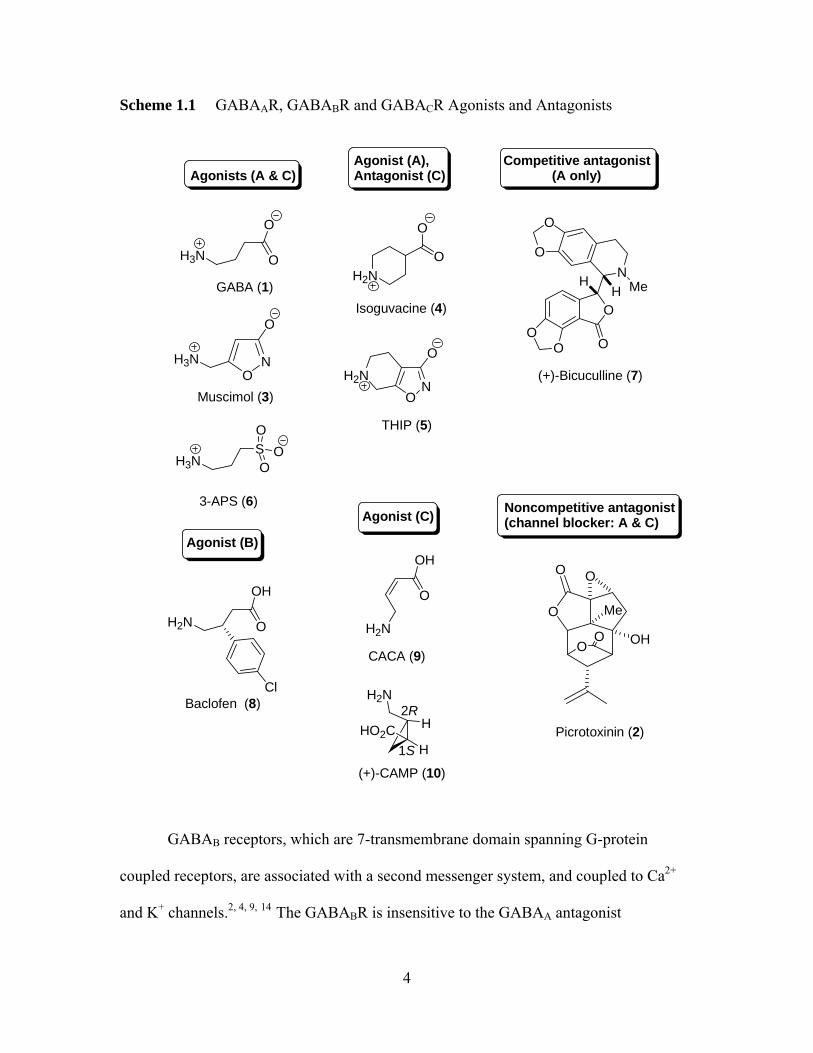
Scheme 1.1 GABAAR, GABABR and GABACR Agonists and Antagonists
ON
H2N
O
H2N
NO
O
H3N
O
O
THIP (5)
Isoguvacine (4)
3-APS (6)
Muscimol (3)
GABA (1)
H3N
O
O
H3NSO
OO
Agonists (A & C)Agonist (A),Antagonist (C)
Competitive antagonist (A only)
OO
O
O
N
O
O
MeHH
(+)-Bicuculline (7)
Agonist (B)
Agonist (C) Noncompetitive antagonist(channel blocker: A & C)
Baclofen (8)
H2N
OH
O
Cl
CACA (9)
OH
O
H2N
H
HHO2C
H2N
1S
2R
(+)-CAMP (10)
O
OH
Me
OO
OO
Picrotoxinin (2)
GABAB receptors, which are 7-transmembrane domain spanning G-protein
coupled receptors, are associated with a second messenger system, and coupled to Ca2+
and K+ channels.2, , , 4 9 14 The GABABR is insensitive to the GABAA antagonist
4
bicuculline, and to the GABAA agonists isoguvacine, THIP, and 3-APS.4 The GABABRs
are found on dopaminergic and glutamatergic neurons in the ventral tegmental area. The
systematic administration of GABABR agonist baclofen 8 has shown to decrease the
firing rate and bursting firing of ventral tegmental dopaminergic neurons as well as
reducing extracellular dopamine levels in the nucleus accumbens, suggesting the
inhibitory role of the GABABR in the mesolimbic circuitry.15 Clinical and preclinical
data also suggest the importance of the GABABR in therapeutic drug action. The
GABABR agonists such as baclofen have shown to demonstrate muscle relexant and
analgesic properties, reduce the craving for alcohol and nicotine consumption as well as
abuse of drugs such as cocaine, heroin, opiate and methamphetamine.15, ,16 17 In addition,
the data suggest that these agonists may be useful in treatment of gastrointestinal
disorders, asthma and overactive bladder. In the case of GABABR antagonists, animal
studies indicate that these antagonists display antiepileptic, neuroprotectant, cognition
enhancement and antidepressant properties.16, ,18 19
The GABAC receptor also gates a Cl- ion channel, which led to its initially being
classified as a subspecies of the GABAAR.2 However, unlike the GABAAR, the
GABACR is not widely distributed in the CNS; it appears principally in the vertebrate
retina.5 The GABACR is insensitive to the GABAB agonist-baclofen (8) (Scheme 1.1),
and the GABAA antagonist-bicuculline, as first identified by Johnston and colleagues in
1984. The GABAC receptor has been further identified by its response to agonist CACA
(cis-4-amino-crotonic acid) (9) and selective agonist (+)-CAMP (1S, 2R-2-
(aminomethyl)cyclopropanecarboxylic acid (10) (Scheme 1), to which the GABAAR is
insensitive.2, , 9 20 GABAC receptors are either homooligomers of ρ (ρ1-3) subunits or
5
pseudohomooligomers (ρ1ρ2),2 and cloning studies have shown that ρ1 shares some
homology with the α and β subunits of GABAAR (vide infra).5 The GABAAR and
GABACR share structural and functional features with other members of the LGIC
superfamily. However, many details of the physiology and pharmacology of GABACR
remain unknown.2, , 5 20
1.3 GABAA Receptor Physiology and Pharmacology 1.3.1 Current Models for the Structure and Function of the GABAA Receptors
GABAARs are chloride channel proteins, which are opened by the binding of
GABA agonists. GABAARs, like nicotinic acetylchlorine receptors (nAChRs), are 70 Å-
wide heteropentameric glycoproteins with a central water-filled pore,21 as suggested by
electron microscopic studies. Many GABAAR subunits have been identified by cDNA
cloning and sequencing (α1-6, β1-4, γ1-3, δ, ε, ρ1-2,θ and π).22, 23 The predominant native
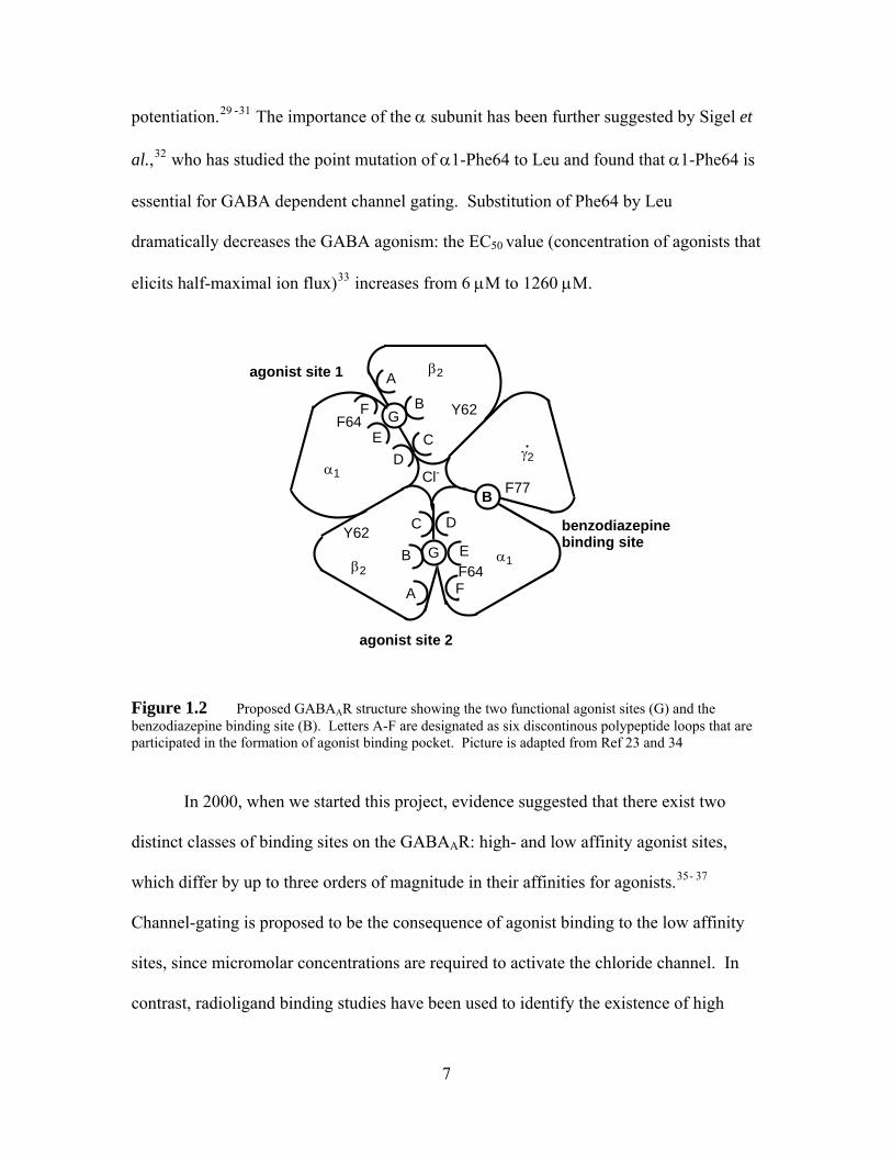
isoforms (60%) of GABAA receptors consist of 2α1, 2β2, and one γ2 subunit (Figure
1.2),14, , 24 25 as suggested by protein chemistry, antibody separations and site-directed
mutagenesis.26 The isoforms of each subunit class share 70 percent sequence identity,
and the subunits have approximately 20-30 percent sequence identity between classes.2, ,
,
4
12 27 Each individual subunit is about 50-60 kDa, which includes a large N-terminal
extracellular domain (ECD), a transmembrane domain (TMD) with four α-helical
segments (TM1-TM4), and a large intracellular domain.23, 28 The combination of α, β and
γ subunits is necessary to reconstitute the full pharmacological functions of the native
receptor, including sigmoidal dose-response to GABA and benzodiazepine
6
potentiation.29 -31 The importance of the α subunit has been further suggested by Sigel et
al.,32 who has studied the point mutation of α1-Phe64 to Leu and found that α1-Phe64 is
essential for GABA dependent channel gating. Substitution of Phe64 by Leu
dramatically decreases the GABA agonism: the EC50 value (concentration of agonists that
elicits half-maximal ion flux)33 increases from 6 μM to 1260 μM.
Cl-
agonist site 1 β2
γ2
B
α1
G
G
A
B
C
F
ED
A
B
C D
E
F
Y62
Y62
agonist site 2
benzodiazepine binding site
β2α1
F77
F64
F64
Figure 1.2 Proposed GABAAR structure showing the two functional agonist sites (G) and the benzodiazepine binding site (B). Letters A-F are designated as six discontinous polypeptide loops that are participated in the formation of agonist binding pocket. Picture is adapted from Ref 23 and 34
In 2000, when we started this project, evidence suggested that there exist two
distinct classes of binding sites on the GABAAR: high- and low affinity agonist sites,
which differ by up to three orders of magnitude in their affinities for agonists.35- 37
Channel-gating is proposed to be the consequence of agonist binding to the low affinity
sites, since micromolar concentrations are required to activate the chloride channel. In
contrast, radioligand binding studies have been used to identify the existence of high
7
affinity sites, since displacement of radiolabeled agonist from the high affinity sites
occurs at nanomolar concentrations.5,35 For instance, [3H]muscimol can be displaced from
membranes with nanomolar concentrations of muscimol or GABA. What possible
reasons are there for the observation of these two classes of sites? One possibility is that
the difference in apparent affinities is due to the fact that functional and binding assays
involve quite different measurements. That is, there is in fact only one type of agonist
binding site that exhibits high affinity under some conditions, and low affinity under
other conditions. The second possibility is that the high- and low- affinity sites are
structurally distinct. In 1989, Dunn36 reported characterization of both nanomolar and
micromolar binding of [3H]muscimol in the same membrane preparations. Dunn was the
first to use equilibrium binding studies to demonstrate a biphasic binding curve, and
concluded that the GABAAR contains structurally distinct low and high affinity sites.36
Her conclusions influenced our earlier thinking of the structure of the GABAAR, as will
be discussed in chapter 2. In line with this view, based on the photoaffinity
[3H]muscimol labeling studies in the bovine GABAAR α1- subunit, Olsen et al.29 suggest
that the high- and low-affinity sites may be differently located, but the data propose a
location for low affinity agonist binding that agrees with Sigel mutagenesis studies. The
amino acid residue which is labeled with [3H]muscimol was identified Olsen et al.29 as
Phe65. However, this Phe65 residue is the bovine homolog to Phe64 in the rat α1-
subunit, which was previously identified by the mutageneis studies of Sigel et al.32 as
possibly involving the functional GABA binding site.
Dunn's demonstration of two classes of binding sites is an important break-
through, but does not take into consideration the fact that a typical binding assay
8
membrane preparation is very different from a functional assay membrane preparation.
Functional assays (e.g., measurement of the uptake of 36Cl- into vesicles) are generally
carried out under approximately physiological conditions; typically with
synaptoneurosomes (which bear intact membranes) at 30 °C and pH 7.4. However,
binding assays (radioligand displacement studies of agonists from the high affinity sites)
are carried out under optimal ligand binding conditions.38 Typical binding assays are
done with frozen-thawed, well-washed, lysed membranes.36 Perhaps the preparation of
the membranes for a binding experiment actually creates high affinity sites that were not
there to begin with. In support of this idea, both Schwartz et al.38 and Brown et al.39
have done binding studies under conditions that more closely approximate functional
assays, and found only micromolar binding of [3H]muscimol. Their perhaps suggest that
high affinity sites are an artifact of the binding assay membranes preparation method.
In order to further test the theory of two distinct classes of binding site in the
GABAAR. Dunn et al. 35 have expressed single receptor isoforms in HEK (human
embryonic kidney)-293 cells and characterized both micromolar and nanomolar
concentration binding of [3H]muscimol in 2000. Interestingly, Dunn’s published study
does not address or offer any explanation to Schwartz’s results. On the other hand,
Dunn35 has also studied the effect of mutation of tyrosine residues at positions 62 and 74
to phenylalanine or serine on both [3H]muscimol binding in HEK-293 cells, and ion
channel activation in Xenopus oocytes, and found that Y62S mutation (mutation of
tyrosine to serine) completely eliminates high affinity binding in HEK-293 cells without
affecting low affinity binding, Emax, or benzodiazepine potentiation; however, both
mutations (Y62S and Y74F) increase the EC50 channel activation, 6- and 2-fold
9
respectively. Since disruption of the high affinity site does not have a significant effect
on receptor function, Dunn and coworkers suggest that the high affinity sites may play a
role in receptor desensitization (closed state of the receptor).35 In line with this idea,
Dunn et al.37 later on studied the effect of mutations of both Y62S and Y74F on the
desensitization of the GABAAR in Xenopus oocytes, and suggested that high affinity sites
stabilize the desensitized state of the receptor, based on the voltage clamp results. At low
concentration, the desensitization characteristics measured in Y74F mutant receptor is
similar to those of wild-type receptor; but the Y62S mutant receptor recovers from
desensitization even in the continued presence of GABA.
Based on Dunn's experiments, it is reasonable to believe that there are distinct
high and low affinity agonist sites on intact individual receptors, and that only the low
affinity sites are involved in channel gating. However, Sigel et al.40 in 2003 published a
study that challenged the idea of independent high- and low-affinity sites on a single
receptor. Sigel and co-worker investigated the effect of point mutation of α1F64L and
the similar mutation of β2Y62L on binding and receptor function. The data show that
α1F64L mutation strongly affects the channel activation (38-fold increase in EC50 value)
and desensitization by GABA; it also causes the maximum binding of [3H]muscimol to
drop dramatically. On the other hand, mutation of β2Y62L has little effect on binding
properties, channel activation (3.4-fold increase in EC50 value) and desensitization, as
compared with the wild-type receptor. The shift of 3.4-fold in EC50 value upon mutation
of β2Y62L is comparable to the shifts of 2- and 6-fold for β2Y62F and β2Y62S mutations
that are reported by Dunn et al.35 Apparently, mutation of α1F64L causes complete loss
of the high affinity sites and the channel activation to be affected strongly, but the
10
changes are not significant with the β2Y62L mutation. Therefore, Siegel et al. argue that
high- and low-affinity sites are probably structural identical.40
α1F64 has been previously shown to be important in channel activation, and is
photoaffinity labeled by [3H]muscimol.29, 32 This is consistent with Siegel’s recent
published data.40 However, the significant discrepancies between Siegel’s and Dunn’s
results require a good explanation. It is believed that the dissociation constant (KD) of
high affinity binding sites for GABAAR agonists is 10-30 nM, whereas that for low
affinity sites is 0.1-1.0 μM. Dunn has shown that β2Y62S mutation shifts KD of
[3H]muscimol to 300 nM in HEK293 cell, and this result leads Dunn and coworker to
believe the importance of β2Y62 in high affinity site binding.35 However, Siegel et al.
claim that KD values in the 100 nM range (150-300 nM) are difficult to measure due to
two reasons. Firstly, fast dissociation rates of [3H]muscimol lead to a significant loss of
observed binding. Secondly, non-specific binding becomes significant as compared with
specific binding at higher ligand concentrations. Thus Dunn’s data and conclusions are
subject to question.40 Siegel et al. also challenge the role of β2Y62 in the process of
desensitization. They believe that the voltage clamp measurements that were performed
by Dunn and co-worker37 have not been done in a classical way, which describes the
time-dependent current decay in the presence of the agonist. On the other hand, Siegel
and co-worker observe that β2Y62L mutation does not have significant effect on the time
course of desensitization after exposure to 1-10 mM GABA. In contrast, mutation of
α1F64L changes the current decay properties greatly.40 Based on these results, Siegel et
al. conclude that high-and low-affinity sites are structurally identical and these are the
two classes of site that are in equilibrium conformations during agonist binding. After
11
reviewing Sigel’s works and criticism of Dunn’s experiments, we also believe that high-
and low-affinity sites are interconvertible sites on the GABAAR. That is, these sites are
identically located and exhibit different affinities towards agonist binding.
Figure 1.2 shows a hypothetical model of the GABAA receptor with the agonist
and modulatory site at subunit interface. In the GABAAR, low affinity sites are proposed
to be located between α and β subunit interfaces, while both photoaffinity radiolabeling
experiments and site-directed mutagenesis studies suggest that the modulatory
benzodiazepine binding site is located between the α and γ subunits.28, 35 Based on
structural notation of the binding loops of X-ray crystal structure of AChBP and more
detailed analyses of the GABAAR binding sites, six discontinuous polypeptide loops
designated A-F at the α and β subunit interfaces are identified to take part in the
formation of the agonist binding pocket of the GABAAR (Figure 1.2).23 The charged
residues in the TM1-TM2 are believed to form the ion channel “gate” in the GABAAR.28
As we mentioned previously, GABAAR and nAChR belong to the same super family, and
share structural and functional features. AChBP (acetylcholine-binding protein) is a
structural and functional homologue of the extracellular portion of the α-subunit of
nAChR.41 Therefore, it is relevant to use structural notation of the binding loops of X-
ray crystal structure of AChBP in assigning the binding loops for the GABAAR.
Patch-clamp8 studies demonstrate that the dose-response curves of GABA
typically have a Hill slope of 2, implying that there are two low affinity binding sites
(agonist site 1 and 2, Figure 1.2) in the GABAAR and at least two agonist molecules (one
to each low affinity agonist site) are bound to these sites before the Cl- ion channel
opens.32, ,42 43 A proposed gating scheme is given in Figure 1.3.33
12
RAR
RM RMA
+M +M
+A
-M -M
-A
+A
-ARMA*
RA2 RA2*
+A -A
Closed state Open state
Legend: R: Receptor with low affinity agonist site unoccupied A: Agonist M: Benzodiazepine modulator
Figure 1.3 A proposed gating scheme for the GABAAR in the presence of agonist and
benzodiazepine modulators; * represents activated receptor.
Within a few seconds of reversible channel gating, the receptor becomes
desensitized8 at this point the channel will remain closed until the agonist concentration is
made very low. The rapid desensitization of GABAAR upon exposure to agonists also
indicates that chloride ion is transported into brain membrane vesicles quickly. Although
Siegel et al.40 have recently reported that α1F64 is involved in channel desensitization,
the exact mechanism of desensitization still remains unknown.
13
1.3.2 Benzodiazepine Binding Sites of The GABAA Receptors
The GABAAR is the target of a variety of clinically and pharmacologically
important drugs such as anxiolytic benzodiazepines (e.g., diazepam (11)). The
benzodiazepine binding site is believed to be located at the α1(+)/γ2 (-) subunit interface,
and the amino acid Phe77 residue has been identified to participate in the formation of
the benzodiazedine binding site (Figure 1.2).40 Benzodiazepines, which were first
introduced in the 1960s as sleeping pills, are agonistic modulators of the GABAAR.44
Agonistic modulators by definition, are ligands that bind to an allosteric site on the
GABAAR, and enhance the agonistic effect of GABA. Benzodiazepines such as
diazepam and flurazepam (12) have a sedative, anxiolytic, muscle relaxant, hypnotic and
anticonvulsant action.45 Therefore, benzodiazepine sites are significant therapeutic
targets of the GABAAR within the brain.12,46 Benzodiazepines such as chlordiazepoxide
and midazolam enhance the GABAAR function by increasing the apparent potency of
GABA.46,47 However, benzodiazepines cannot open the ion channel in the absence of
agonist. How do benzodiazepines exert this action on the GABAAR? Serfozo et al.
propose that benzodiazepines enhance chloride flux by introducing another gating
mechanism: only one agonist molecule is required to open the chloride channel with one
benzodiazepine bound.47 (As shown in Figure 1.3)
Many studies have demonstrated that benzodiazepine agonists such as Ro 11-
6896 [B10(+)] (13) (Scheme 2), diazepam, clonazepam and flurazepam, shift the GABA
agonist (GABA, muscimol) concentration-response curves shift to the left, indicating that
benzodiazepines potentiate GABAergic function by decreasing the agonist EC50 (Figure
1.4) with no effect on the maximal response induced by GABA.12, , , 48 49 50 Studies of
14
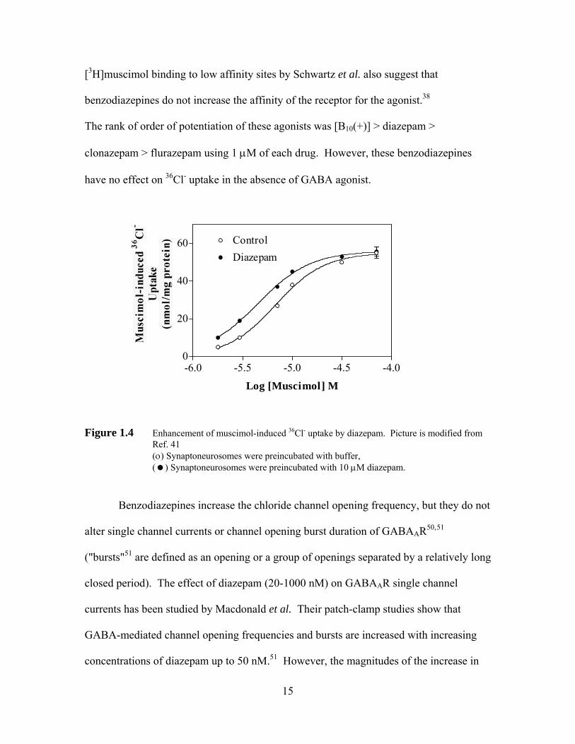
[3H]muscimol binding to low affinity sites by Schwartz et al. also suggest that
benzodiazepines do not increase the affinity of the receptor for the agonist.38
The rank of order of potentiation of these agonists was [B10(+)] > diazepam >
clonazepam > flurazepam using 1 μM of each drug. However, these benzodiazepines
have no effect on 36Cl- uptake in the absence of GABA agonist.
-6.0 -5.5 -5.0 -4.5 -4.00
20
40
60 ControlDiazepam
Log [Muscimol] M
Figure 1.4 Enhancement of muscimol-induced 36Cl- uptake by diazepam. Picture is modified from Ref. 41
(ο) Synaptoneurosomes were preincubated with buffer, ( ) Synaptoneurosomes were preincubated with 10 μM diazepam.
Benzodiazepines increase the chloride channel opening frequency, but they do not
alter single channel currents or channel opening burst duration of GABAAR50,51
("bursts"51 are defined as an opening or a group of openings separated by a relatively long
closed period). The effect of diazepam (20-1000 nM) on GABAAR single channel
currents has been studied by Macdonald et al. Their patch-clamp studies show that
GABA-mediated channel opening frequencies and bursts are increased with increasing
concentrations of diazepam up to 50 nM.51 However, the magnitudes of the increase in
15
channel opening and burst frequencies are decreased at higher concentration of diazepam
(250 nM). Macdonald et al.51 proposes that perhaps the rate of GABAAR channel
desensitization is increased with higher concentration of diazepam since the frequency of
bursts of channel openings is increased. In contrast, the mean GABAAR single-channel
current amplitudes are decreased by inverse agonists (ligands that exert a negative
allosteric effect on GABA agonism), such as methyl-6,7-dimethyoxyl-4-ethyl-β-
carboline-3-carboxylate (DMCM).51
1.3.3 Barbiturates, Picrotoxinin and Steroid Anesthetic Binding Sites in the GABAAR
The GABAAR is also the target of action of depressant and sedative-hypnotic
barbiturates (e.g., pentobarbital, PB, 14), and anesthetics such as etomidate (15),
propoflo, and alfaxalone (16) (Scheme 1.2).11 Electrophysiological studies have
demonstrated that at a 50-200 μM concentration range, barbituates such as pentobarbital
enhance GABAergic neurotransmission by increasing the mean channel open time to
chloride ion, but without affecting the channel opening frequency.52 The single-channel
bursting inward currents from spinal cord neurons of mice induced by both GABA (2
μM) and GABA plus PB (50 μM) have been studied by Macdonald et al., and they have
found that PB increases the mean open time of GABAAR from 3.5 ms (with GABA only)
to 8.3 ms (GABA plus PB).52
16
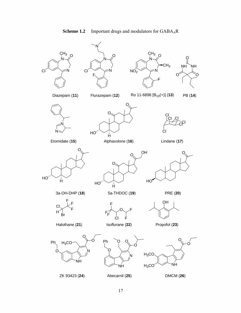
Scheme 1.2 Important drugs and modulators for GABAAR
N
N
Cl
CH3 O
N
N
Cl
O
N
N
NO2
CH3 O
N
FF
CH3 NH NH
O
O O
Diazepam (11) Flurazepam (12) Ro 11-6896 [B10(+)] (13) PB (14)
N
N
Etomidate (15)
O
HOH
O
Alphaxolone (16)
ClCl
Cl
ClClCl
Lindane (17)
O
HOH
3a-OH-DHP (18)
O
HOH
O
5a-THDOC (19)
OH O
HO
PRE (20)
FBr
ClH
FF
Halothane (21)
FO F
F
FCl F
Isoflurane (22)
OH
Propofol (23)
NH
NO
H3COO
Ph O
NH
N
OO
O
O
Ph O
NH
O
H3CO
H3CO
ZK 93423 (24) nil (25) DMCM (26)Abecar
17
Pentobarbital (50 μM) enhances the muscimol-mediated 36Cl- uptake by shifting
the muscimol concentration response curve to the left, decreasing the EC50 value from 7.0
μM to 2.5 μM.6 At higher concentrations (100-500 μM), barbituates are able to elicit Cl-
conductance in the absence of GABA, with a maximal response at 500 μM.14, 53
However, at concentrations higher than 500 μM, pentobarbital causes the functional
desensitization of the GABAAR. Higashi and Nishi54 propose that barbiturates at high
concentrations may induce a conformational change in the receptor complex, thereby
causing blockade of Cl- channels.6
Convulsant neurotoxicants such as picrotoxinin, and insecticides such as lindane,
(17)55 are channel blockers. The picrotoxinin site is located on or closely associated with
the chloride ion channel,56 and picrotoxinin inhibits the GABA-activated chloride influx
by causing a decrease in mean channel open time and stabilizing closed channel states.11,
57 The hypnotic neurosteroids such as 3α-OH-DHP (18) and 5α-THDOC (19)
(Scheme 1.2), are among the most potent known steroid modulators of GABAARs.14, 58
The synthetic anesthetic steroids such as pregnanolone (PRE, 20),59 ethanol, volatile gas
anesthetics halothane60 (21) and isoflurane (22),61 and propofol (23), also enhance
GABAAR chloride channel function and modulate binding in vitro. PRE is a "barbituate-
like modulator" of the GABAAR, which increases the average chloride channel open time
at low concentration (100 nM to 1 μM) and decreases the prolongation of the average
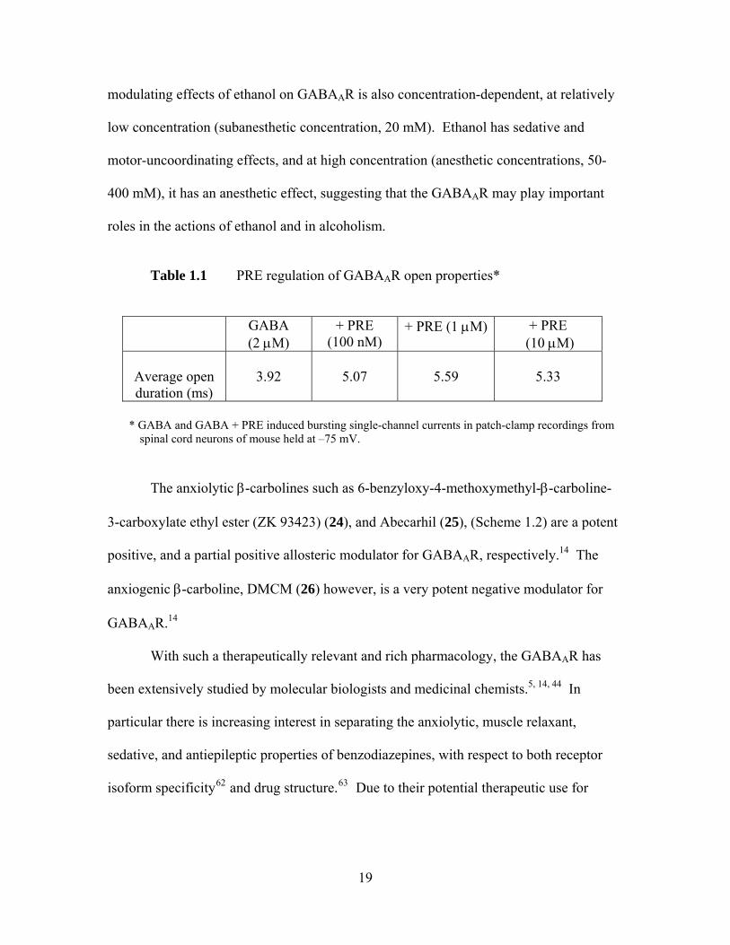
open duration at higher concentration (10 μM), (as shown in Table 1.1), indicating that
GABA-mediated chloride channel is blocked by PRE, the reason is that PRE at high
concentrations may also cause conformational change in the receptor complex. The
18
modulating effects of ethanol on GABAAR is also concentration-dependent, at relatively
low concentration (subanesthetic concentration, 20 mM). Ethanol has sedative and
motor-uncoordinating effects, and at high concentration (anesthetic concentrations, 50-
400 mM), it has an anesthetic effect, suggesting that the GABAAR may play important
roles in the actions of ethanol and in alcoholism.
Table 1.1 PRE regulation of GABAAR open properties*
GABA
(2 μM) + PRE
(100 nM) + PRE (1 μM) + PRE
(10 μM)
Average open duration (ms)
3.92
5.07
5.59
5.33
* GABA and GABA + PRE induced bursting single-channel currents in patch-clamp recordings from
spinal cord neurons of mouse held at –75 mV.
The anxiolytic β-carbolines such as 6-benzyloxy-4-methoxymethyl-β-carboline-
3-carboxylate ethyl ester (ZK 93423) (24), and Abecarhil (25), (Scheme 1.2) are a potent
positive, and a partial positive allosteric modulator for GABAAR, respectively.14 The
anxiogenic β-carboline, DMCM (26) however, is a very potent negative modulator for
GABAAR.14
With such a therapeutically relevant and rich pharmacology, the GABAAR has
been extensively studied by molecular biologists and medicinal chemists.5, , 14 44 In
particular there is increasing interest in separating the anxiolytic, muscle relaxant,
sedative, and antiepileptic properties of benzodiazepines, with respect to both receptor
isoform specificity62 and drug structure.63 Due to their potential therapeutic use for
19
treatment of anxiety disorders,7 epilepsy,3, 5 pain,5 and insomnias,64 design of novel
GABAAR agonists and partial agonists is also of considerable interest.
1.4 GABAA Receptor-Agonists and Partial Agonists
GABAAR agonists are compounds that induce GABA-gated chloride channel
opening. Channel opening is proposed to result from binding to the low affinity agonist
sites of GABAAR. The inhibitory nature of the GABA in the CNS encouraged the design
and development of various structural types of GABA agonists.5 In the past, based on the
conformational restriction of different parts of the GABA molecule and bioisosteric
replacements of the functional groups of this amino acid, a diverse group of specific
GABAA agonists have been developed. Some of these agonists have played an important
role in the development of the GABAAR pharmacology.5, 20
1.4.1 Structure and Conformation of Active GABA Analogs (Exogenous GABAA agonists)
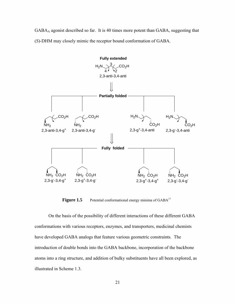
GABA is a flexible molecule, which can exist in several conformations, ranging
from “fully extended, partially folded, to fully folded,” as shown in
Figure 1.5.20, 65 There is great interest in determining which comformation GABA adopts
when bound to the receptor. If only staggered conformations (i.e. energy minima) are
allowed, 9 conformers must be considered. Of course, the receptor bound agonist may
exist in other conformations (e.g. eclipsed conformations) not described in Figure 1.5.
The different efficacies of enantiomers of (RS)-dihydromuscimol (DHM)66 suggest that
GABA may adopt a chiral conformation on binding. (S)-DHM (27) is the most potent
20
GABAA agonist described so far. It is 40 times more potent than GABA, suggesting that
(S)-DHM may closely mimic the receptor bound conformation of GABA.
CO2HH2N
Fully extended
2,3-anti-3,4-anti
Partially folded
CO2H
2,3-anti-3,4-g+NH2
CO2H
2,3-anti-3,4-g-NH2
H2N
2,3-g+-3,4-antiCO2H
H2N
2,3-g--3,4-antiCO2H
Fully folded
2,3-g--3,4-g+NH2 CO2H
2,3-g+-3,4-g-NH2 CO2H
2,3-g+-3,4-g+NH2 CO2H
2,3-g--3,4-g-NH2 CO2H
2
3
4
Figure 1.5 Potential conformational energy minima of GABA17
On the basis of the possibility of different interactions of these different GABA
conformations with various receptors, enzymes, and transporters, medicinal chemists
have developed GABA analogs that feature various geometric constraints. The
introduction of double bonds into the GABA backbone, incorporation of the backbone
atoms into a ring structure, and addition of bulky substituents have all been explored, as
illustrated in Scheme 1.3.
21
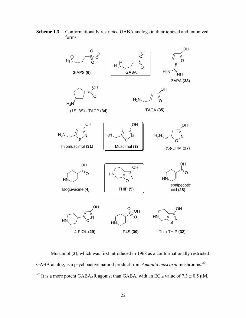
Scheme 1.3 Conformationally restricted GABA analogs in their ionized and unionized forms
ON
HN
OH
HN
NO
OH
H2N
SNHN
OH
NO
OH
H2NNS
OH
H2N
O
OH
HNO
OH
ON
OH
HN HN
SO
O OH
OH
O
H2N
O
OH
S
NHH2N
O
OH
H2N
THIP (5)Isoguvacine (4)
3-APS (6)
Muscimol (3)
Thio-THIP (32)
(S)-DHM (27)Thiomuscimol (31)
GABA
Isonipecotic acid (28)
4-PIOL (29) P4S (30)
(1S, 3S) - TACP (34)
ZAPA (33)
TACA (35)
H3N
O
O
H3NSO
OO
Muscimol (3), which was first introduced in 1968 as a conformationally restricted
GABA analog, is a psychoactive natural product from Amanita muscaria mushrooms.20,
67 It is a more potent GABAAR agonist than GABA, with an EC50 value of 7.3 ± 0.5 μM,
22

compared with 10 μM of GABA.6 However, muscimol is toxic and it will be
metabolized after peripheral administration, so it is not clinically useful.1, 3 The relatively
rigid muscimol analog, THIP (5), is a partial GABAAR agonist, and it has some
selectivity for particular β-subunits of the GABAA receptor.68 THIP is less potent than
musicmol, but it is comparatively non-toxic in rats and dogs, and also, it can penetrate the
blood brain barrier (BBB) very easily after systematic administration.3,65 THIP is a
proven antinociceptive agent-it can be as effective as morphine. Its analgesic action
therefore highlights the possible importance of GABA mechanisms in pain.65, 69
However its side effects, including sedation, dizziness, and blurred vision, prevent it from
being a useful therapeutic agent.14, 20 Based on the structure of THIP, several specific
monoheterocyclic GABAA agonists have been developed, including isoguvacine (4),
isonipecotic acid (28), 5-(4-piperidyl)isoxazol-3-ol (4-PIOL, 29) and piperidine-4-
sulfonic acid (P4S, 30). Isoguvacine (5) is approximately as potent as GABA in
competing for the binding of [3H]GABA, but it is slightly less potent than GABA in
mediating chloride flux.5, 70 4-PIOL is a partial agonist with an EC50 of 91 μM at
GABAA receptor, and it is approximately 200 times less potent than isoguvacine as an
agonist.15, 71
Both thiomuscimol (31) and DHM are derived from muscimol. The GABAA
agonism of muscimol is not lost upon certain minor structural modifications.
Thiomuscimol is approximately equipotent with muscimol as a GABAA agonist, whereas
"(S)-DHM (27) is the most potent GABAA agonist so far described", as demonstrated in
both functional and binding assays. However, replacement of oxygen in the iosoxazole
moiety in THIP by sulphur to give thio-THIP (32), is less potent than its parent
23
compound. Likewise, the sulfonic acid analog of GABA, 3-aminopropane sulfonic acid
(APS, 6) is less potent than GABA at inducing 36Cl- influx.42 However, 3-APS is twice
as potent as GABA in competing for the binding of [3H]GABA.72 This result highlights
the fact that agonist potency may not always track well with high-affinity binding.
(Z)-3-[(Aminoiminomethyl)thio]prop-2-enoic acid (ZAPA, 33), an
isothiouronium analog of GABA of restricted conformation, is more potent than GABA
on low-affinity GABA binding sites, with EC50 of 10.3 ± 0.7 μM, while the EC50 of
GABA is 12.8 ± 0.4 μM.14, 73 The Hill slopes of the concentration-dependent curve of
ZAPA are 2, indicating that 2 ZAPA molecules are needed to activate GABAAR.74 The
cyclopentane analogue of GABA, (1S, 3S)-(+)-trans-3-aminocyclopentane-1-carboxylic
acid ((+)-TACP, 34), is also a potent GABAA agonist.5, 14 The conformationally restricted
analog of isoguvacine and GABA, trans-4-aminocrotonic acid (TACA, 35), is an agonist
at GABAAR.
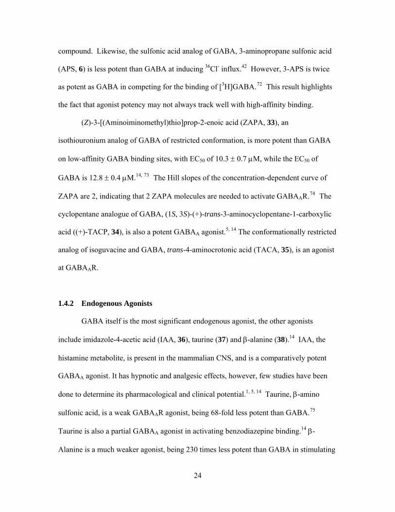
1.4.2 Endogenous Agonists
GABA itself is the most significant endogenous agonist, the other agonists
include imidazole-4-acetic acid (IAA, 36), taurine (37) and β-alanine (38).14 IAA, the
histamine metabolite, is present in the mammalian CNS, and is a comparatively potent
GABAA agonist. It has hypnotic and analgesic effects, however, few studies have been
done to determine its pharmacological and clinical potential.1, , 5 14 Taurine, β-amino
sulfonic acid, is a weak GABAAR agonist, being 68-fold less potent than GABA.75
Taurine is also a partial GABAA agonist in activating benzodiazepine binding.14 β-
Alanine is a much weaker agonist, being 230 times less potent than GABA in stimulating
24
GABAAR.14 Finally, the synthetic dipeptide, GABA-taurine (39) has no effect on
GABAAR.76
O
OH
IAA (36)
NNH
SO3H
Taurine (37)
H2NCOOH
β-Alanine (38)
H2N
SO3H
GABA-Taurine (39)
HN
H2NO
1.5 GABAA Agonist-Receptor Interactions
Based on structure-activity analyses, the GABAA receptor can tolerate some
bioisosteric alteration of the functional groups of GABA. For example, we have already
discussed the agonistic potency of muscimol, DHM, TACA and ZAPA. Surprisingly,
however minor structural modification of a particular agonist molecule usually results in
the loss of GABAA agonism.5, 74 Generally, the presence of negatively and positively
charged groups (zwitterionic) is essential, but not major for GABAA agonist activity.
The GABAAR agonism is maintained or enhanced if the GABA analogs resemble one of
the possible comformations of GABA. All of the potent GABAA agonists described so
far are structurally related to GABA.
At least three aspects of agonist structure affect GABAA agonism, including
electronic factors, structural and conformational factors, and stereochemical factors.77
We will also review the work on current agonist binding model of the GABAAR.
25
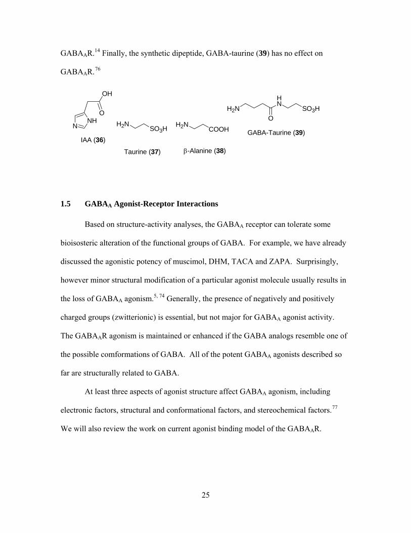
1.5.1 Electronic Factors
Prior to our work, it was believed that a zwitterionic structure of GABA analogs
is necessary for GABAAR agonism. In addition, on the basis of structure-activity
analysis, a high degree of delocalization of the positive charges reduces both GABAA
agonism and inhibition of GABAAR binding. IAA (36) is a good example. IAA is less
potent than GABA in both functional and binding assays. 77 As shown below, IAA
features more delocalization of positive charge than GABA. However, ZAPA is an
exceptional case, as we mentioned earlier, ZAPA is more potent than GABA as
GABAAR agonist.
O
O
SH
NH2H2N
ZAPA (28)
H3N
O
ONHHN
O
O
IAA (36) GABA (1)
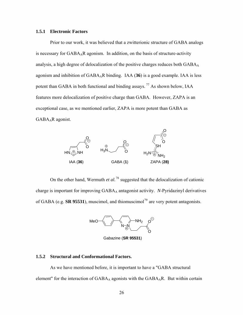
On the other hand, Wermuth et al.78 suggested that the delocalization of cationic
charge is important for improving GABAA antagonist activity. N-Pyridazinyl derivatives
of GABA (e.g. SR 95531), muscimol, and thiomuscimol79 are very potent antagonists.
MeON N
NH2 O
OGabazine (SR 95531)
1.5.2 Structural and Conformational Factors.
As we have mentioned before, it is important to have a "GABA structural
element" for the interaction of GABAA agonists with the GABAAR. But within certain
26
limits, the primary amino group of GABA can be substituted by other functional groups
without significant loss of GABAA agonism. ZAPA is a good example. However, an
incorporation of substituents onto the terminal amino groups of GABAA agonists
normally results in a significant or complete loss of GABAA agonism.5, , 77 80 N-Methyl-
GABA81 is almost inactive, N, N-dimethyl- and N, N,N-trimethyl-GABA are completely
inactive. The N-methyl derivatives of muscimol82 and thiomuscimol are only weak
GABAAR agonists.77, 83
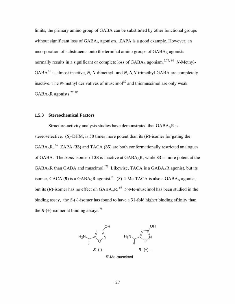
1.5.3 Stereochemical Factors
Structure-activity analysis studies have demonstrated that GABAAR is
stereoselective. (S)-DHM, is 50 times more potent than its (R)-isomer for gating the
GABAAR. 66 ZAPA (33) and TACA (35) are both conformationally restricted analogues
of GABA. The trans-isomer of 33 is inactive at GABAAR, while 33 is more potent at the
GABAAR than GABA and muscimol. 73 Likewise, TACA is a GABAAR agonist, but its
isomer, CACA (9) is a GABACR agonist.20 (S)-4-Me-TACA is also a GABAA agonist,
but its (R)-isomer has no effect on GABAAR. 66 5'-Me-muscimol has been studied in the
binding assay, the S-(-)-isomer has found to have a 31-fold higher binding affinity than
the R-(+)-isomer at binding assays.78
N
O
OH
H2N NO
OH
H2N
S- (-) - R- (+) -
5'-Me-muscimol
27

1.5.4 Current Agonist Binding model of the GABAA Receptor To date, direct structural determination of the GABAAR has been a challenge due
to its low abundance (pmol/mg of protein) and heterogeneous nature, in addition to the
difficulties of isolating and purifying whole membrane proteins. Many indirect but
useful methods such as site-directed mutagenesis, chemical modification and molecular
modeling were therefore used to provide insight into receptor structure and function.23
Photoaffinity labeling, site-directed mutagenesis and SCAM (substituted cysteine
accessibility method) have been used to identify many amino acid residues that
participate in agonist binding (Table 1.2).13, , -23 84 85 Many of these studies assayed in how
mutation affects GABA dose-response curves in both agonism and gating.
Table 1.2 Residues lining the GABAAR agonist binding pocket
Binding
loop notation*
A B C D E F
Residues of α1
subunits
-
-
-
Phe64, Phe65, Arg66, Arg67, Ser68, Ser69, Gln67, Trp69
Thr129, Arg131
Arg176, Val178, Val180, Asp183
Residues of β2
subunits
Tyr97, Leu99
Tyr198, Tyr200, Tyr157
Thr244, Tyr247, Thr202, Tyr205, Ser204, Arg207, Ser209
-
-
-
*Binding loop notations are depicted in Figure 1.2.
28

SCAM has been used to identify pore-lining residues in the binding pocket of
nicotinic and GABA receptors. This method is based on how the modification of the
substituted cysteine (selected amino acids were mutated to cysteine) by a reactive water-
soluble sulfhydryl compound (MTSEA-biotin: N-biotinylaminoethyl
methanethiosulfonate) could affect the receptor function. If the modification takes part in
a binding domain of the receptor, it is likely to cause a change in the functional properties
of the receptor. There are two factors that affect the rate of reaction of MTSEA-biotin
with introduced cysteine: ionization of the sulfhydryl side chain and steric hindrance.
Ionization of cysteine side chain is more likely to occur in aqueous environment. MTS
reacts 109-1010 times faster with ionized sulfhydryl side than they do with protonated
sulfhydryls. Steric hindrance reflects how easy or difficult it is for MTS to physically
access and interact with the sulfhydryl group. Therefore, whether the substituted cysteine
can be accessed by MTS and how quickly cysteine can react with MTS provide useful
information about the structure and the environment of the receptor.13 Based on the
accessibility pattern and the results of MTSEA-biotin reaction rates from G203C-S209C,
Czajkowski et al.13 have proposed that β2 loop C is a coil and the GABAAR binding
pocket is a narrow cleft that is located in both a hydrophobic environment and a sterically
hindered region. The results also suggest that the binding site constrict during gating due
to the movement of α1 and β2 domains of the GABA binding sites toward each other.
A flexible binding-site model has been used by Jones et al.86 to explain the
binding behavior of the GABAA agonists. They propose that the binding site behaves
like a pair of movable "arms" attached to rigid "anchor" sites by spring-like tethers,
which are separated by a distance. Binding induces movement of the arms to “hold” the
29
agonist molecule. Such process involves activation energy, which is consistent with the
idea that the gating of the ion channel is initiated by agonist-triggered movements within
the ligand binding site.87 On the other hand, Czajkowski et al.’s have proposed that the
binding site constricts during gating due to the movement of α1 and β2 domains of the
GABA binding site toward each other. Three arginine residues at the α1 and β2 domains
(β2-Arg207, α1-Arg66 and α1-Arg131) have been identified to directly stabilize GABA
during binding by electrostatic interaction between the positively charged guanido group
at the end of the arginine side chain and the negatively charged carboxylate group on the
GABA molecule. Czajkowski et al.87 propose that these three arginines form a triangle
of positive charge that collaborate to accommodate the negatively charged carboxylate
group of the GABA molecule. Since the GABA binding pocket is narrow, this “crown of
arginine” arrangement would have implied that GABA molecule is situated vertically
within the binding pocket, with the positively charged amino group pointing away from
the arginine crown.
Figure 1.6 is a simple description of receptor occupancy and gating of the
GABAAR. Basically, the agonists activate the Cl- channel of the GABAAR by interacting
with the agonist-binding sites, triggering a conformational change in the receptor
(structural transition from closed state, A2R to open state, A2R*) that results in the
opening of the ion channel. This process, which also known as gating happens quickly
and reversibly, but the exact mechanism involved in structural rearrangement, was not
well understood.28,88 Recently, several workers have studied the nature of the coupling
agonist binding to channel gating, and identified four amino acids in loop C α1 domain
30

and the charged residues located in flexible loops of either α1 or β2 subunits are involved
in coupling agonist binding to channel gating.13, , 28 89
A: Agonist
Closed Open R: Receptor
KD = Dissociation constant (agonist affinity) E = Agonist efficacy
Figure 1.6 Schematic description of receptor occupancy and gating of GABAAR
Based on SCAM, Wagner and Czajkowski13 have proposed that the residues Phe200,
Ser201, Tyr202 and Gly203 in loop C α1 subunit are involved in mediating GABA
binding. Three of the residues (Phe200 to Tyr202) appear to be outside the binding
pocket and buried in membrane lipid. Gly203 has been shown to be at the mouth of the
binding pocket. Harrison et al.89 show that electrostatic interactions between negatively
charged Asp57 and Asp149 in the flexible loops 2 and 7 in the extracellular domain, and
positively charged Lys279 in the short transmembrane linker (TM2-3L) region of the
GABAAR-α1 subunit are involved in the process of receptor activation. Apparently,
Asp149 and Lys279 move closer together during gating. In line with the results of this
view, Kash et al.28 also obtained the same observations using site-directed mutagenesis.
They identified the acidic residue Asp 146 in loop 7 of β2 subunit and basic residue
Lys215 in pre-transmembrane domain–1 are involved in coupling agonist binding to the
gating of channel through electrostatic interactions.
The studies described previously are based on how mutation changed the EC50
values of GABA. The question is how we can determine whether the amino residues
A + R A + AR A A2R 2R*D1 D2K K E
31

involved in mutation studies are related to coupling agonist binding to channel gating or
binding directly. The amino acid residues in Table 1 were identified lining the binding
pocket. Both site-directed mutagensis and SCAM have found that mutation of these
residues causes a large increase in EC50 values of GABA; there is no doubt that these
residues are directly involved in agonist binding and the subsequent opening of ion
channel. However, the residues like Phe200, Ser201, Tyr202, Gly203, Asp57, Asp 149,
Lys279, Asp 146 and Lys215 are identified either in extracellular domains or in
transmembrane domains. Mutation of these residues causes an increase in EC50 values of
the GABAAR agonists, especially partial agonist like P4S with a decrease in agonist
efficacy. Such decrease in agonist efficacy suggests that mutated amino residues are
involved in gating.
1.6 Traditional Assays for Agonists
Functional assays, such as patch-clamp and 36Cl- flux, provide functional
measurement of actions of GABA agonists and antagonists.90 Due to the unique
pharmacological and clinical importance of GABAAR, there is considerable interest in
studying the factors that would affect the functional assays of the GABAA agonists. As
regards 36Cl- flux, the first factor is the incubation time of agonists; generally, GABA-
mediated 36Cl- flux was complete within 15 seconds of incubation.42 Prolonged
incubation time results in a higher absolute chloride uptake, and therefore decreases the
EC50 of GABA, as listed in Table 1.3.90, 91 Longer incubation time also enhances the
modulatory effect of pentobarbital, indicating that the potentiation effect of pentobarbital
on GABA-activated 36Cl- flux takes place only after GABA has bound to the binding
32
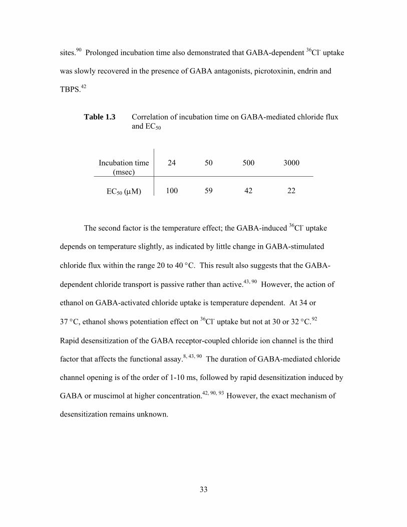
sites.90 Prolonged incubation time also demonstrated that GABA-dependent 36Cl- uptake
was slowly recovered in the presence of GABA antagonists, picrotoxinin, endrin and
TBPS.42
Table 1.3 Correlation of incubation time on GABA-mediated chloride flux and EC50
Incubation time (msec)
24
50
500
3000
EC50 (μM)
100
59
42
22
The second factor is the temperature effect; the GABA-induced 36Cl- uptake
depends on temperature slightly, as indicated by little change in GABA-stimulated
chloride flux within the range 20 to 40 °C. This result also suggests that the GABA-
dependent chloride transport is passive rather than active.43, 90 However, the action of
ethanol on GABA-activated chloride uptake is temperature dependent. At 34 or
37 °C, ethanol shows potentiation effect on 36Cl- uptake but not at 30 or 32 °C.92
Rapid desensitization of the GABA receptor-coupled chloride ion channel is the third
factor that affects the functional assay.8, , 43 90 The duration of GABA-mediated chloride
channel opening is of the order of 1-10 ms, followed by rapid desensitization induced by
GABA or muscimol at higher concentration.42, , 90 93 However, the exact mechanism of
desensitization remains unknown.
33

1.7 Pharmacokinetic Aspects and Prodrugs
All GABAAR agonists described so far have zwitterionic structures; however,
only non-ionized neutral molecules can penetrate the BBB. The inadequate ability of
GABAAR agonists to penetrate the BBB reflects the difficulties in developing such
agonists with satisfactory pharmacokinetics properties.5,77 The ratio between the
concentrations of ionized and unionized molecules (I/U ratio) indicates the ability of
GABAAR agonists to penetrate the BBB. This I/U ratio is a function of the difference
between the pKaI and pKa
II values.52,94 Figure 1.7 shows an illustration of the
determination of the I/U ratio for isoguvacine in aqueous solution. Basically, the I/U ratio
of neutral isoguvacine can be calculated in two ways, as the ratio between the intrinsic
constants Ka and Kb or between Kd and Kc. The approximate values of Ka and Kc are
measured by titration of isoguvacine. Species i will be the predominant form below pH
4, species ii will predominate at pH 5-9, and species iii will predominate at pH>10. The
values of Kb and Kd can not be determined by titration directly, since species iv will not
predominate at any pH, they are thus estimated indirectly by titration of appropriate
derivatives of isoguvacine, in which either the acid or the basic function, respectively, has
been blocked, as shown in Figure 1.7. From the values of Ka, Kc, KN, and KE, two
similar I/U ratios were calculated for isoguvacine. Values of pKa, and I/U ratios of some
GABAAR agonists and their ability to penetrate the BBB are shown in Table 1.4.67 In
general, the higher the I/U ratio, the smaller the fractions of un-ionized molecules will be
present in the solution, and the lower the ability of GABAAR agonists to penetrate the
BBB.1, , , 5 53 67
34
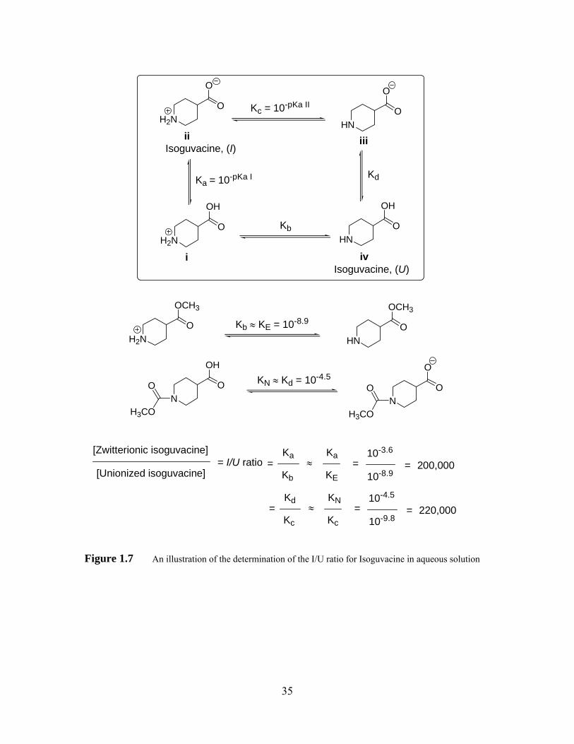
H2NO
O
H2NO
OH
HNO
O
HNO
OH
H2NO
OCH3
HNO
OCH3
Isoguvacine, (I)
Ka = 10-pKa I
Kc = 10-pKa II
Isoguvacine, (U)
Kb
Kd
Kb ≈ KE = 10-8.9
NO
OH
H3CO
ON
H3CO
O
[Zwitterionic isoguvacine]
[Unionized isoguvacine]=
Ka
Kb
Ka
KE=
10-8.9
10-3.6
= 200,000
i
ii iii
iv
O
OKN ≈ Kd = 10-4.5
≈= I/U ratio
=Kd
Kc Kc=
10-9.8
10-4.5
= 220,000 ≈KN
Figure 1.7 An illustration of the determination of the I/U ratio for Isoguvacine in aqueous solution
35
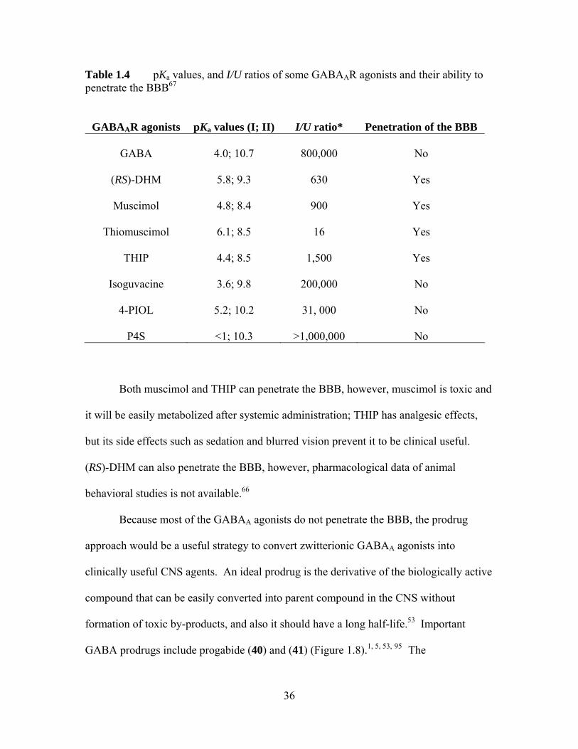
Table 1.4 pKa values, and I/U ratios of some GABAAR agonists and their ability to penetrate the BBB67
GABAAR agonists pKa values (I; II) I/U ratio* Penetration of the BBB
GABA
4.0; 10.7
800,000
No
(RS)-DHM
5.8; 9.3
630
Yes
Muscimol
4.8; 8.4
900
Yes
Thiomuscimol
6.1; 8.5
16
Yes
THIP
4.4; 8.5
1,500
Yes
Isoguvacine
3.6; 9.8
200,000
No
4-PIOL
5.2; 10.2
31, 000
No
P4S
<1; 10.3
>1,000,000
No
Both muscimol and THIP can penetrate the BBB, however, muscimol is toxic and
it will be easily metabolized after systemic administration; THIP has analgesic effects,
but its side effects such as sedation and blurred vision prevent it to be clinical useful.
(RS)-DHM can also penetrate the BBB, however, pharmacological data of animal
behavioral studies is not available.66
Because most of the GABAA agonists do not penetrate the BBB, the prodrug
approach would be a useful strategy to convert zwitterionic GABAA agonists into
clinically useful CNS agents. An ideal prodrug is the derivative of the biologically active
compound that can be easily converted into parent compound in the CNS without
formation of toxic by-products, and also it should have a long half-life.53 Important
GABA prodrugs include progabide (40) and (41) (Figure 1.8).1, , , 5 53 95 The
36
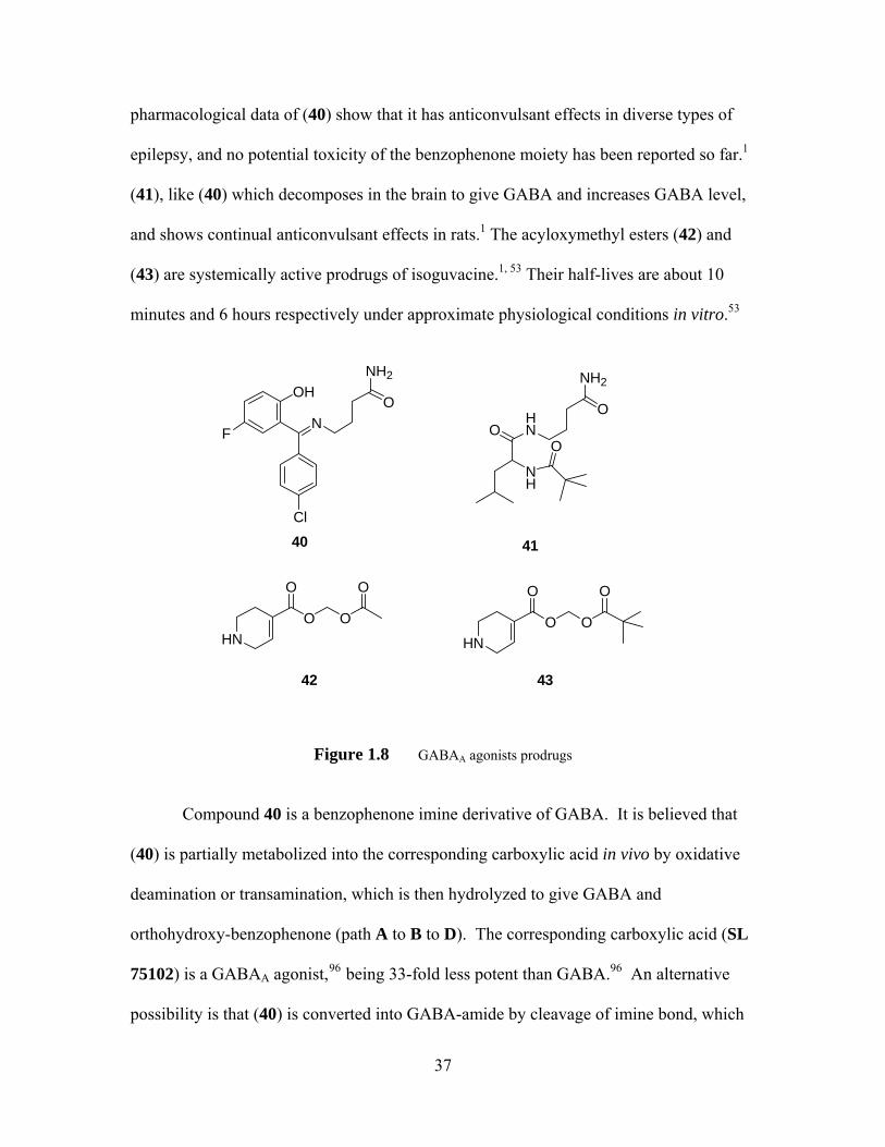
pharmacological data of (40) show that it has anticonvulsant effects in diverse types of
epilepsy, and no potential toxicity of the benzophenone moiety has been reported so far.1
(41), like (40) which decomposes in the brain to give GABA and increases GABA level,
and shows continual anticonvulsant effects in rats.1 The acyloxymethyl esters (42) and
(43) are systemically active prodrugs of isoguvacine.1, 53 Their half-lives are about 10
minutes and 6 hours respectively under approximate physiological conditions in vitro.53
Cl
N
OH
F
O
NH2
OHN
O
NH2
NH
O
HN
O
O O
O
40 41
HN
O
O O
O
42 43
Figure 1.8 GABAA agonists prodrugs
Compound 40 is a benzophenone imine derivative of GABA. It is believed that
(40) is partially metabolized into the corresponding carboxylic acid in vivo by oxidative
deamination or transamination, which is then hydrolyzed to give GABA and
orthohydroxy-benzophenone (path A to B to D). The corresponding carboxylic acid (SL
75102) is a GABAA agonist,96 being 33-fold less potent than GABA.96 An alternative
possibility is that (40) is converted into GABA-amide by cleavage of imine bond, which
37
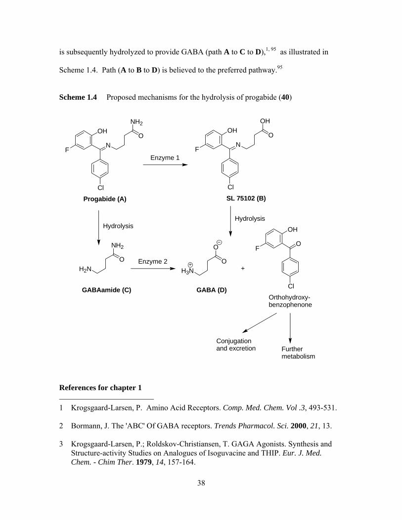
is subsequently hydrolyzed to provide GABA (path A to C to D),1, 95 as illustrated in
Scheme 1.4. Path (A to B to D) is believed to the preferred pathway.95
Scheme 1.4 Proposed mechanisms for the hydrolysis of progabide (40)
Cl
N
OH
F
O
NH2
Progabide (A)
Cl
N
OH
F
O
OH
SL 75102 (B)
Enzyme 1
H2NO
NH2
Hydrolysis
H3NO
O
Cl
O
OH
F
GABAamide (C) GABA (D)Orthohydroxy-benzophenone
Hydrolysis
+
Conjugation and excretion Further
metabolism
Enzyme 2
References for chapter 1
38
1 Krogsgaard-Larsen, P. Amino Acid Receptors. Comp. Med. Chem. Vol .3, 493-531. 2 Bormann, J. The 'ABC' Of GABA receptors. Trends Pharmacol. Sci. 2000, 21, 13. 3 Krogsgaard-Larsen, P.; Roldskov-Christiansen, T. GAGA Agonists. Synthesis and
Structure-activity Studies on Analogues of Isoguvacine and THIP. Eur. J. Med. Chem. - Chim Ther. 1979, 14, 157-164.
39
4 Sigel, G. J. In Basic Neurochemistry. Timothey, M. Delorey; Richard W. Olsen.
Raven Press, Ltd, New York. 1994, 389-399. 5 Krogsgaard-Larsen, P.; Fround, B.; Jrogensen, S. F.; Schousboe, A. GABAA Receptor
Agonists, Partial Agonists, and Antagonists. Design and Therapeutic Propects. J. Med. Chem. 1994, 37 (16), 2489-2505.
6 Schwartz, D. R.; Suzdak, P. D.; Paul, S. M. γ - Aminobutyric Acid (GABA)- and
Barbiturate-Mediated Cl- Uptake in Rate Brain Synaptoneurosomes: Evidence for Rapid Desensitization of the GABA Receptor-Coupled Chloride Ion Channel. Mol. Pharmacol. 1986, 30, 419-426.
7 Smart, T. G. In Amino Acid Neurotransmission; F. Anne Stephenson; A. J. Turner,
Eds.; Portland Press, Ltd, London. 1998, 37-63. 8 Bormann, J.; Clapham, D. E. γ–Aminobutyric acid receptor channels in adrenal
chromaffin cells: A patch-clamp study. Proc. Natl. Acad. Sci. USA. 1985, 82, 2168-2172.
9 Chebib, M.; Johnston, G. A. R. GABA-activated Ligand Gated Ion Channels:
Medicinal Chemistry and Molecular Biology. J. Med. Chem. 2000, 43, 1427-1447. 10 Macdonald R. L.; Olsen, R. W. GABAA Receptor Channels. Annu. Rev. Neurosci.
1994, 17, 569-602. 11 Sieghart, W. GABAA receptors: ligand-gated Cl- ion channels modulated by multiple
drug-binding sites. Trends Pharmacol. Sci. 1992, 13, 446-450. 12 Delorey, T. M.; Olsen, R. W. γ–Aminobutyric AcidA Receptor Structure and
Function. J. Biol. Chem. 1992, 267, 16747-16750. 13 Wagner, D. A.; Czajkowski, C. Structure and Dynamics of the GABA Binding
Pocket: A Narrowing Cleft that Constricts during Activation. J. Neuroscience 2001, 21, 67-74.
14 Johnston G. A. R. GABAA receptor pharmacology. Pharmacol. Ther. 1996, 69, 173-
198. 15 Paterson, N. E.; Froestl, W.; Markows, A. Repeated Administration of the GABAB
Receptor Agonist CGP44532 Decreased Nicotine Self-Administration, and Acute Administration Decreased Cue-Induced Reinstatement of Nicotine-Seeking in Rats. Neuropsychopharmacol. 2005, 30, 119-128.
40
16 Enna, S. J.; Bowery, N. G. GABAB receptor alterations as indicators of physiological
and pharmacological function. Biochemical Pharmacology. Biochem. Pharmacol. 2004, 68, 1541-1548.
17 Capasso, A. GABAB receptors are involved in the control of acute opiate withdrawal
in isolated tissue. Prog. Neuro-pshychopharmacol. Biol. Psychiat. 1999, 23, 289-299. 18 Xiong, Z.-Q.; Stringer, J. L. Effects of postsynaptic GABAB receptor activation on
epileptiform activity in hippocampal slices. Neuropharmacol. 2001, 40, 131-138. 19 Snead III, O. C. Antiabsence seizure activity of specific GABAB and γ-
hydroxybutyric acid receptor antagonist. Pharmacol. Biochem. Behavior. 1996, 53, 73-79.
20 Martin, D. L.; Olsen, R. W. In GABA in the Nervous System: The View at Fifty Years.
Lippincott Williams & Wilkins 2000, 65-80. 21 Nayeem, N.; Green, T. P.; Martin, I. L.; Barnard, E. A. Quaternary Structure of the
Native GABAA Receptor Determined by Electron Microscopic Image Analysis. J. Neurochem. 1994, 62, 815-818.
22 Boileau, A. J.; Evers, R. A.; Davis, A. F.; Czajkowski, C. Mapping the Agonist
Binding Site of the GABAA Receptor: Evidence for a β-Strand. J. Neurosci. 1999, 19, 4847-4854.
23 Kash, T. L.; Trudell, J. R.; Harrison, N. L. Structural elements involved in activation
of the γ-aminobutryic acid type A (GABAA) receptor. Biochem. Soc. Trans. 2004, 32, 540-546.
24 Smith, G. B.; Olsen, R. W. Functional domains of GABAA receptors. Trends
Pharmacol. Sci. 1995, 16, 162-168. 25 Mohler, H.; Rudolph, U. Selective GABAA circuits for novel CNS drugs. Drug
Discovery Today: Ther. Strategies. 2004, 1, 117-123. 26 Chang, Y.; Wang, R.; Barot, S.; Weiss D. S. Stoichemistry of a recombinant GABAA
receptor. J. Neurosci. 1996, 16, 5415-5424. 27 Smart T. G. In Amino Acid Neurotransmission; F. Anne Stephenson; A. J. Turner,
Eds.; Portland Press, Ltd, London. 1998, 65-92. 28 Kash, T. L.; Dizon, M.-J. F.; Trudell, J. R.; Harrison, N. L. Charged Residues in the
β2 Subunit Involved in GABAA Receptor Activation. J. Bio. Chem. 2004, 279, 4887-4893.
41
29 Smith, G. B.; R. Olsen W. Identification of a [3H] Musicmol Photoaffinity Substrate
in the Bovine γ-Aminobutyric AcidA Receptor α Subunit. J. Biol. Chem. 1994, 269, 20380-20387.
30 Luddens, H.; Korpi, E. R.; Seeburg, P. H. GABAA/Benzodiazepine Receptor
Heterogeneity Neurophysiological Implications. Neuropharmacol. 1995, 34, 245-254.
31 Pritchett, D. B.; Sontheimer, H.; Shivers, B. D.; Ymer, S.; Kettenmann, H.; Shofield,
P. R.; Seeburg, P. H. Importance of a novel GABAA receptor subunit for benzodiazepine pharmacology. Nature 1989, 338, 582-585.
32 Sigel, E.; Baur, R.; Kellenberger, S.; Malherbe, P. Point mutations affecting
antagonist affinity and agonist dependent gating of GABAA receptor channels. EMBO J. 1992, 11, 2017-2023.
33 Colquhoun, D. Binding, gating, affinity and efficacy: The Interpretation of structure-
activity relationships for agonists and of the effects of mutating receptors. Br. J. Pharmacol. 1998, 125, 924-947.
34 Baumann, S. W.; Baur, R.; Sigel, E. Individual properties of the two functional
agonist sites in GABAA receptors. J. Neurosci. 2003, 23, 11778. 35 Newell, J. G.; Davies, M.; Bateson, A. N.; Dunn, S. M. J. Tyrosine 62 of the γ-
Aminobutyric Acid Type A Receptor β2 Subunit Is an Important Determinant of High Affinity Agonist Binding. J. Biol. Chem. 2000, 275, 14198-14204.
36 Agey, M. W.; Dunn, S. M. J. Kinetics of [3H]Muscimol Binding to the GABAA
Receptor in Bovine Brain Membranes. Biochemistry, 1989, 28, 4200-4208. 37 Newell, J. G.; Dunn, S. M. J. Functional Consequences of the Loss of High Affinity
Agonist Binding to γ-Aminobutyric Acid Type A Receptors. J. Bio. Chem. 2002, 277, 21423-21430.
38 Edgar, P. P.; Schwartz, R. D. Functionally Relevant γ–Aminobutyric AcidA
Receptors: Equivalence between Receptor Affinity (Kd) and Potency (EC50)? Mol. Pharmacol. 1992, 41, 1124-1129.
39 DeLorey, T. M.; Brown, G. B. γ-Aminobutyric acidA Receptor Pharmacology in Rat
Cerebral Cortical Synaptoneurosomes. J. Neurochem. 1992, 58, 2162-2169. 40 Baur, R.; Sigel, E. On high- and low-affinity agonist sites in GABAA Receptor. J.
Neurosci. 2003, 87, 325-332.
42
41 Brejc, K.; Van Dijk, W. J.; Klaassen, R. V.; Schuurmans, M.; Van der Oost, J.; Smit,
A. B.; Sixma, T. K. Crystal structure of the an ACh-binding protein reveals the ligand-binding domain of nicotinic receptors. Nature, 2001, 411, 269-276.
42 Cupello, A.; Hyden, H. Binding experiment Kd values and physiologically active
GABA concentrations: an only apparent contradiction? Int. J. Neurochem. 1986, 30, 297-301.
43 Amin, J.; Welss, D. S. Binding experiment Kd values and physiologically active
GABA concentrations: an only apparent contradiction? Nature , 1993, 366, 565-569. 44 Lancel, M.; Steiger, A. Sleep and Its Modulation by Drugs That Affect GABAA
Receptor Function. Angew. Chem. Int. Ed. 1999, 111, 2852-2864. 45 Fryer, R. I. Ligand Interactions at the Benzodiazepine Receptor. Comp. Med. Chem.3,
540-543. 46 Olsen, R. W.; Venter, J. C. In the Benzodiazepine/GABA Receptors and Chloride
Channels: Structural and Functional Properties. Alan R. Liss, Inc. New York 1986, 97.
47 Serfozo, P.; Cash, D. Effect of benzodiazepine (chlordiazepoxide) on a GABAA
Receptor from rat brain: Requirement of only one bound GABA molecule for channel opening. FEBS. J. 1992, 310, 55-59.
48 Harris, R. A.; Allan, A. M. Functional Coupling of γ–Aminobutyric Acid Receptors
to Chloride Channels in Brain Membranes. Science 1985, 228, 1108-1110. 49 Morrow, A. L.; Paul, S. M. Benzodiazepine Enhancement of γ–Aminobutyric Acid-
Mediated Chloride Ion Flux in Rat Brain Synaptoneurosomes. J. Neurochem. 1988, 50, 302-306.
50 Vicini, S.; Mienville, J.-M.; Costa, E. Actions of Benzodiazepine and β-Carboline
Derivatives on γ–Aminobutyric Acid-Activated Cl- Channels Recorded from Membrane Patches of Neonatal Rat Cortical Neurons in Culture. J. Pharmaco. Exp. Ther. 1987, 243, 1195-1201.
51 Rogers, C. J.; Twyman, R. E.; Macdonald, R. L. Benzodiazepine and β-carboline
regulation of single GABAA receptor channels of mouse spinal neurons in culture. J Physiol. 1994, 475, 69-82.
52 Macdonald, R. L.; Rogers, C. J.; Twyman, R. E. Barbiturate regulation of kinetic
properties of the GABAA receptor channel of mouse spinal neurons in culture. J Physiol. 1989, 417, 486-500.
43
53 Krogsgaard-Larsen, P. In Amino Acid Receptors. Comp. Med. Chem. Vol 3, 388-409. 54 Higashi, H.; Nishi, S. Effect of barbiturates on the GABA receptor of rat primary
afferent neurons. J. Physiol.(Lond.) 1982, 332, 299-314. 55 Bloomquist, J. R. Ion Channels as Targets for Insecticides. Ann Rev. Entomol. 1996,
41,163-190. 56 Bloomquist, J. R. Intrinsic Lethality of Chloride Channel-Directed Insecticides and
Convulsants in Mammals. Toxicol. Lett. 1992, 60, 289-298. 57 Bloomquist, J. R.; Jackson, J. L.; Karr, L. L.; Ferguson, H. J.; Gajewski, R. P. A new
chemical class of insecticide acting upon the GABA receptor/chloride ionophore complex. Pesticide Sci. 1993, 39, 185-192.
58 Lan, N. C.; Gee, K. W.; Bolger, M. B; Chen, J. S. Differential Responses of
Expressed Recombinant Human γ–Aminobutyric AcidA Receptors to Neurosteroids. J. Neurochem. 1991, 57, 1818-1821.
59 Twyman, R. E.; Macdonald, R. L. Neurosteroid regulation of GABAA receptor
channel kinetic properties of mouse spinal neurons in culture. J Physiol. 1992, 456, 215-245.
60 Longoni, B.; Demontis, G. C.; Olsen, R. W. Enhancement of γ–Aminobutyric AcidA
Receptor Function and Binding By the Volatile Anesthetic Halothane. J. Pharmacol. Exp. Ther. 1993, 266, 153-159.
61 Quinlan, J. J.; Firestone, S.; Firestone, L. L. Isoflurane's enhancement of Chloride
Flux through Rat Brain gama-Aminobutyric Acid Type A Receptors Is Stereoselective. Anesthesiology, 1995, 83, 611-615.
62 McKernan, R. M.; Rosahl, T. W.; Reynolds, D. S.; Sur, C.; Wafford, K. A.; Atack, J.
R.; Farrar, S.; Myers, J.; Cook, G.; P. Ferris; Garrett; L. B.; Marshall, G.; Macauley, A.; Brown, N.; Howell, O.; Moore, W. K.; Carling, R. W.; Street, L. J.; Castro, J. L.; Ragan, C. I.; Dawson, G. R.; Whiting, P. J. Sedative but not anxiolytic properties of benzodiazepines are mediated by the GABAA receptor α1 subtype. Nature Neurosci. 2000, 3, 587-592.
63 Huang, Q.; He, X.; Ma, C.; Liu, R.; Yu, S.; Dayer, C. A.; Wenger, G. R.; Mckernan,
J. M. Pharmacophore GABAA/Receptor Models for /BzR Subtypes (α1β3γ2, α5β3γ2 and α6β3γ2) via a Comprehensive Ligand-Mapping Approach. J. Med. Chem. 2000, 43, 71-95.
64 Martin, D. L.; Olsen, R. W. In the GABA in the Nervous System: The View at Fifty
Years. Lippincott Williams & Wilkins 2000, 97.
44
65 Johnston, G. A. R.; Allan, R. D.; Kennedy, S. M. E.; Twitchin B. Systematic Study of
GABA Analogues of Restricted Conformation. In GABA- Neuro-Transmitters; Krogsgaard-Larsen, P.; Scheel-Kruger, J.; Kofod, H.; Eds.; Munkgaard: Copenhagen, 1979, 149-164.
66 Krogsgaard-Larsen, P.; Nielsen, L.; Falch, E.; Curtis, D. R. GABA Agonist.
Resolution, Absolute Stereochemistry, and Enantioselectivity of (S)-(+)- and (R)-(-)-Dihydromuscimol. J. Med. Chem. 1985, 28, 1612-1617.
67 Krogsgaard-Larsen, P.; Honoré, T.; Thysssen, K.. In GABA- Neuro-Transmitters; P.
Krogsgaard-Larsen; J. Scheel-Kruger; H. Kofod, Eds.; Munkgaard: Copenhagen, 1979, 201-216.
68 Bureau, M.; Olsen, R. W. Multiple distinct subunits of the γ–aminobutyric acidA
receptor protein show different ligand binding properties. Mol. Pharmacol. 1990, 37, 497-502.
69 Krogsgaard-Larsen, P.; Falch, E. The Binding of the GABA Agonist [3H] THIP to
Rat Brain Synaptic Membranes. J. Neurochem. 1982, 38, 1123. 70 Bloomquist, J. R.; Grubs, R. E.;.Soderlund, D. M.; Knipple, D. C. Prolonged
exposure to GABA activates GABA-gated chloride channels in the presence of channel-blocking convulsants. Comp. Biochem. Physiol. 1991, 99C, 397-402.
71 Kristiansen, U.; Lambert, J. D. C.; Falch, E.; Krogsgaard-Larsen, P.
Electrophysiological studies of the GABAA receptor ligand, 4-PIOL, on cultured hippocampal neurons. Br. J. Pharmacol. 1991, 104, 85-90.
72 Breckenridge, R. J.; Nicholson, S. H.; Nicol, A. J.; Suckling, C. J.; Leigh, B.; Iversen
L. Inhibition of [3H] GABA Binding to Postsynaptic Recetors in Human Cerebellar Synaptic Membranes by Carboxyl and Amino Derivatives of GABA. J Neurochem. 1981,37, 837-844.
73 Allan ,R. D.; Dickenson, H. W.; Hiern G. B. P.; Johnston, A. R.; Kazlauskas, R.
Isothiouronium compounds as γ–aminobutyric acid agonists. Br. J. Pharmacol. 1986, 88, 379-387.
74 Holden-Dye, L.; Walker, R. J. ZAPA, Z-3-[(aminoiminomethyl)thio]prop-2penoic
acid hydrochloride, a potent agonist at GABA-receptors on the Ascaris muscle cell. Br. J. Pharmacol. 1988, 95, 3-5.
75 Luu, M. D.; Morrow, A. L.; Paul, S. M.; Schwartz, R. D. Characterization of GABAA
Receptor-mediated 36Chloride Uptake in Rat Brain Synaptosomes. Life Sci. 1987, 41, 1277-1287.

45
76 Varga, V.; Kontro, P.; Oja, S. S. Modulation of GABAergic Neurotransmission in the
Brain by Dipetides. Neurochem. Res. 1988, 13, 1027-1034. 77 Olsen, R. W.; Venter, J. C. In the Benzodiazepine/GABA Receptors and Chloride
Channels: Structural and Functional Properties. Alan R. Liss, Inc. New York 1986, 73.
78 Wermuth, C. G.; Biziere, K. Pyridazyl-GABA derivatives: a new class of synthetic
GABAA antagonists. Trends Pharm. Sci. 1986, 7, 721-724. 79 Melikian, A.; Schlewer, G.; Chambon, J. P.; Wermuth, C. G. Condensation of
Muscimol or Thiomuscimol with Aminopyridazines Yields GABA-A Antagonists. J. Med. Chem. 1992, 35, 4092-4097.
80 Krogsgaard-Larsen, P.; Johnston, G. A. R.; Curtis, D. R.; Game, C. J. A.;
McCullonch, R. M. Structure and Biological Activity of A Series of Conformationally Restricted Analogues of GABA. J. Neurochem. 1975, 25, 803-809.
81 Falch, E.; Krogsgaard-Larsen, P.; Jacobsen, P.; Engesgaard, A.; Braestrup, C.; Crutis,
D. R. Synthesis, GABA agonist activity and effects on GABA and diazepam binding of some N-substituted analogues of GABA. Eur. J. Med. Chem. - Chim Ther. 1985, 20, 447-453.
82 Frey, M.; Jager, V. Synthesis of N-Substituted Muscimol Derivatives Including N-
Glycylmuscimol. Synthesis, 1985. 1100-1104. 83 Aprison, M. H.; Lipkowitz, K. B. Muscimol and N,N-Dimethylmuscimol: From a
GABA Agonist to a Glycine Antagonist. J. Neurosci. Res. 1992, 31, 166-174. 84 Bollan, K.; King, D.; Robertson, L. A.; Brown, K.; Taylor, P. M.; Moss, S. J.;
Connolly, C. N. GABAA Receptor Composition Is Determined by Distinct Assembly Signals within α and β Subunits. J. Bio. Chem. 2003, 278, 4747-4755.
85 Sedelnikova, A.; Smith, C. D.; Zakharkin, S. O.; Davis, D.; Weiss, D. S.; Chang, Y.
Mapping the ρ1 GABAC Receptor Agonist Binding Pocket. J. Bio. Chem. 2005, 280, 1535.
86 Jones, M. V.; Sahara, Y.; Dzubay, J. A.; Westbrook, G. L. Defining affinity with the
GABAA receptor. J. Neurosci. 1998, 18, 8590-8604. 87 Wagner, D. A.; Czajkowski, C.; Jone, M. V. An Arginine Involved in GABA
Binding and Unbinding But Not Gating of the GABAA Receptor. J. Neurosci. 2004, 24, 2733-2741..
46
88 Jones, M. V.; Jonas, P.; Sahara, Y.; Westbrook, G. L. Microscopic Kinetics and
Energetics Distinguish GABAA Receptor Agonists and Antagonists. Biophys. J. 2001, 81, 2660-2670.
89 Kash, T. L.; Jenkins, A.; Kelley, J. C.; Trudell, J. R.; Harrison, N. L. Coupling of
angonist binding to channel gating in the GABAA receptor. Nature, 2003, 421, 272-275.
90 Mihic, S. J.; Berckel V. B. N. M.; Wu, P. H.; Kalant, H. GABA and pentobarbital
potentiation of chloride influx into microsacs is influenced by incubation time. Brain Research, 1993, 619, 319-323.
91 Kardos, J.; Cash, D. J. Transmembrane 36Cl- Flux Measurements and Desensitization
of the γ-Aminobutyric AcidA Receptor. J. Neurochem. 1990, 55, 1095-1099. 92 McQuilkin S. J.; Harris R. A. Factors affecting actions of ethanol on GABA-activated
Chloride channels. Life Sci. 1990, 46, 527-541. 93 Mihic, S. J.; Wu, P. H.; Kalant, H. Potentiation of γ–Aminobutyric Acid-Mediated
Chloride Flux by Pentobarbital and Diazepam but Not Ethanol. J. Neurochem 1992, 58, 745-751.
94 Krogsgaard-Larsen, P.; Mikkelsen, H.; Jacobsen, P.; Falch E.; Curtis, D. R.; Peet, M.
J.; Leah, J. D. 4,5,6,7-Tetrahydroisothiazolo[5,4-c]pyridin-3-ol and Related Analogues of THIP. Sysnthesis and Biological Activity. J. Med. Chem. 1983, 26, 895-900.
95 Worms, P.; Depoortere, H.; Durand, A.; Morselli, P. L.; Lloyd, K. G.; Bartholini G.
γ–Aminobutyric Acid (GABA) Receptor Stimulation. I. Neuropharmacological Profiles of Progabide (SL 76002) and SL 75102, with Emphasis on Their Anticonvulsant Spectra. J Pharmacol. Exp. Ther. 1982, 220, 660.
96 Desarmenien, M.; Feltz, P.; Headley, P.M.; Santangelo, F. SL 75102 as a γ–
Aminobutyric Acid agonist: experiments on dorsal root ganglion neurones in vitro. Br. J. Pharmac. 1981, 72, 355-364.
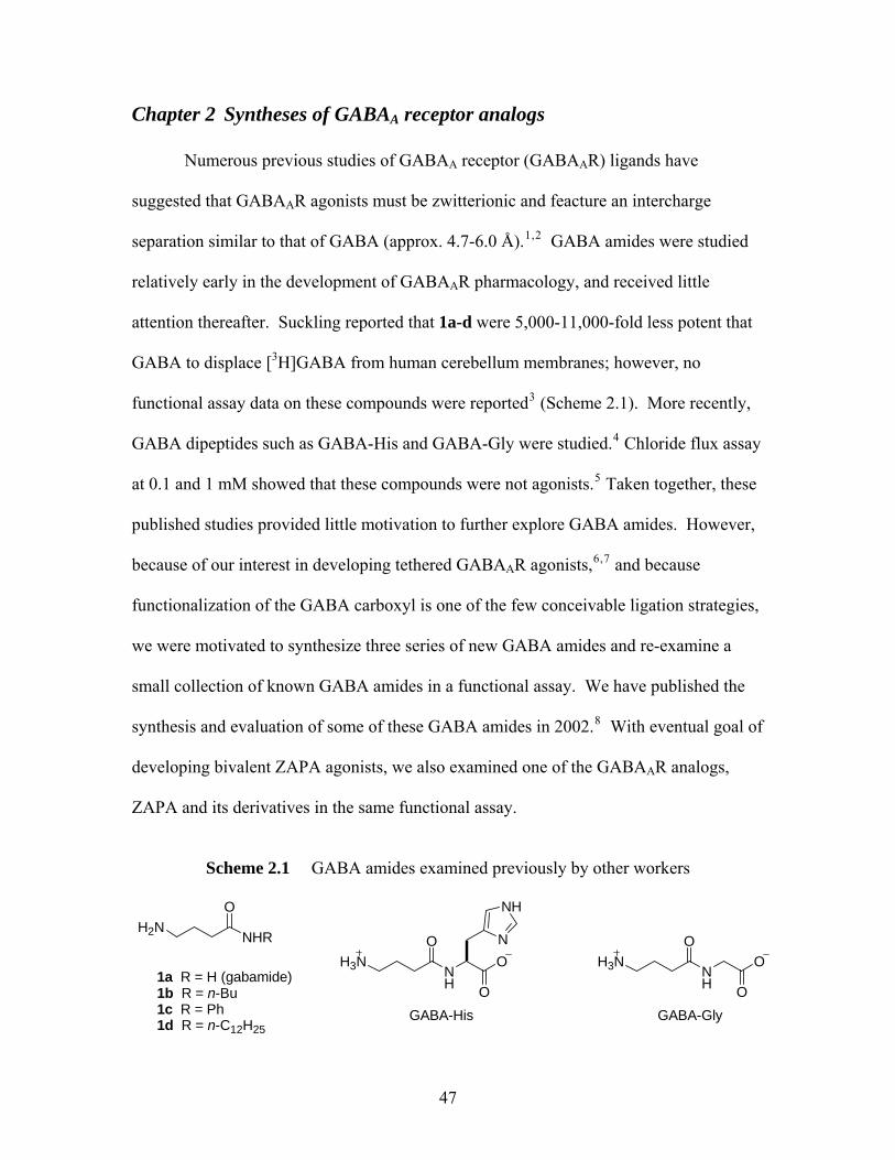
Chapter 2 Syntheses of GABAA receptor analogs
Numerous previous studies of GABAA receptor (GABAAR) ligands have
suggested that GABAAR agonists must be zwitterionic and feacture an intercharge
separation similar to that of GABA (approx. 4.7-6.0 Å).1,2 GABA amides were studied
relatively early in the development of GABAAR pharmacology, and received little
attention thereafter. Suckling reported that 1a-d were 5,000-11,000-fold less potent that
GABA to displace [3H]GABA from human cerebellum membranes; however, no
functional assay data on these compounds were reported3 (Scheme 2.1). More recently,
GABA dipeptides such as GABA-His and GABA-Gly were studied.4 Chloride flux assay
at 0.1 and 1 mM showed that these compounds were not agonists.5 Taken together, these
published studies provided little motivation to further explore GABA amides. However,
because of our interest in developing tethered GABAAR agonists,6,7 and because
functionalization of the GABA carboxyl is one of the few conceivable ligation strategies,
we were motivated to synthesize three series of new GABA amides and re-examine a
small collection of known GABA amides in a functional assay. We have published the
synthesis and evaluation of some of these GABA amides in 2002.8 With eventual goal of
developing bivalent ZAPA agonists, we also examined one of the GABAAR analogs,
ZAPA and its derivatives in the same functional assay.
Scheme 2.1 GABA amides examined previously by other workers
NHR
OH2N
1a R = H (gabamide)1b R = n-Bu 1c R = Ph1d R = n-C12H25
NH
OH3N O
O
N
NH
GABA-His
NH
OH3N O
OGABA-Gly
47
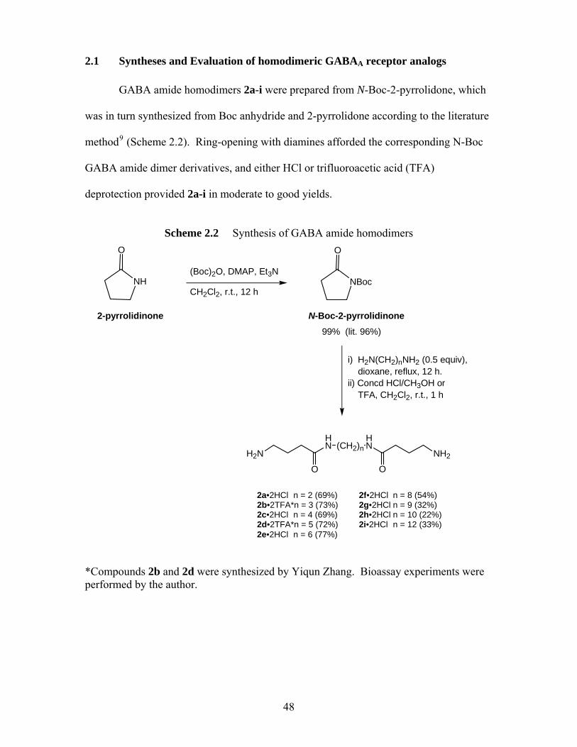
2.1 Syntheses and Evaluation of homodimeric GABAA receptor analogs
GABA amide homodimers 2a-i were prepared from N-Boc-2-pyrrolidone, which
was in turn synthesized from Boc anhydride and 2-pyrrolidone according to the literature
method9 (Scheme 2.2). Ring-opening with diamines afforded the corresponding N-Boc
GABA amide dimer derivatives, and either HCl or trifluoroacetic acid (TFA)
deprotection provided 2a-i in moderate to good yields.
Scheme 2.2 Synthesis of GABA amide homodimers
NBoc
O
N-Boc-2-pyrrolidinone
H2N
HN
O
(CH2)nHN
O
NH2
i) H2N(CH2)nNH2 (0.5 equiv), dioxane, reflux, 12 h.ii) Concd HCl/CH3OH or TFA, CH2Cl2, r.t., 1 h
NH
O
2-pyrrolidinone
(Boc)2O, DMAP, Et3N
CH2Cl2, r.t., 12 h
99% (lit. 96%)
2a•2HCl n = 2 (69%) 2f•2HCl n = 8 (54%) 2b•2TFA*n = 3 (73%) 2g•2HCl n = 9 (32%) 2c•2HCl n = 4 (69%) 2h•2HCl n = 10 (22%) 2d•2TFA*n = 5 (72%) 2i•2HCl n = 12 (33%) 2e•2HCl n = 6 (77%)
*Compounds 2b and 2d were synthesized by Yiqun Zhang. Bioassay experiments were performed by the author.
48
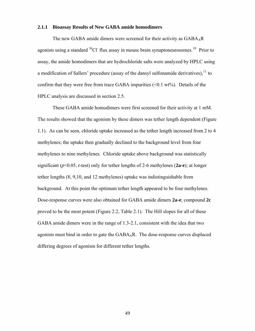
2.1.1 Bioassay Results of New GABA amide homodimers The new GABA amide dimers were screened for their activity as GABAAR
agonists using a standard 36Cl- flux assay in mouse brain synaptoneurosomes.10 Prior to
assay, the amide homodimers that are hydrochloride salts were analyzed by HPLC using
a modification of Sallers’ procedure (assay of the dansyl sulfonamide derivatives),11 to
confirm that they were free from trace GABA impurities (<0.1 wt%). Details of the
HPLC analysis are discussed in section 2.5.
These GABA amide homodimers were first screened for their activity at 1 mM.
The results showed that the agonism by these dimers was tether length dependent (Figure
1.1). As can be seen, chloride uptake increased as the tether length increased from 2 to 4
methylenes; the uptake then gradually declined to the background level from four
methylenes to nine methylenes. Chloride uptake above background was statistically
significant (p<0.05, t-test) only for tether lengths of 2-6 methylenes (2a-e); at longer
tether lengths (8, 9,10, and 12 methylenes) uptake was indistinguishable from
background. At this point the optimum tether length appeared to be four methylenes.
Dose-response curves were also obtained for GABA amide dimers 2a-e; compound 2c
proved to be the most potent (Figure 2.2, Table 2.1). The Hill slopes for all of these
GABA amide dimers were in the range of 1.3-2.1, consistent with the idea that two
agonists must bind in order to gate the GABAAR. The dose-response curves displaced
differing degrees of agonism for different tether lengths.
49
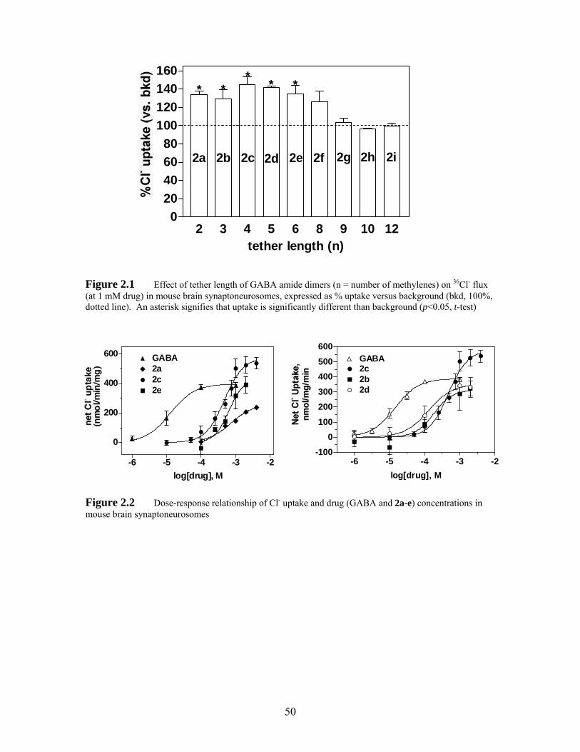
2 3 4 5 6 8 9 10 120
20406080
100120140160
** *
2a 2b 2c 2d 2e 2f 2g
* *
2h 2i
tether length (n)
Figure 2.1 Effect of tether length of GABA amide dimers (n = number of methylenes) on 36Cl- flux (at 1 mM drug) in mouse brain synaptoneurosomes, expressed as % uptake versus background (bkd, 100%, dotted line). An asterisk signifies that uptake is significantly different than background (p<0.05, t-test)
-6 -5 -4 -3 -2
0
200
400
600
2e
GABA
2c2a
log[drug], M
-6 -5 -4 -3 -2-100
0100200300400500600
2d2b
GABA2c
log[drug], M
Figure 2.2 Dose-response relationship of Cl- uptake and drug (GABA and 2a-e) concentrations in mouse brain synaptoneurosomes
50
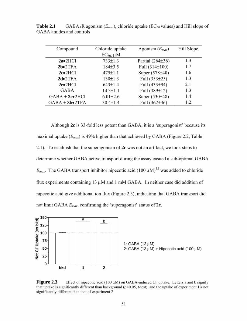
Table 2.1 GABAAR agonism (Emax), chloride uptake (EC50 values) and Hill slope of GABA amides and controls
Compound Chloride uptake EC50, μM
Agonism (Emax) Hill Slope
2a•2HCl 733±1.3 Partial (264±36) 1.3 2b•2TFA 184±3.5 Full (314±100) 1.7 2c•2HCl 475±1.1 Super (578±40) 1.6 2d•2TFA 130±1.3 Full (353±25) 1.3 2e•2HCl 643±1.4 Full (433±94) 2.1 GABA 14.3±1.1 Full (389±12) 1.3
GABA + 2c•2HCl 6.01±2.6 Super (530±48) 1.4 GABA + 3b•2TFA 30.4±1.4 Full (362±36) 1.2
Although 2c is 33-fold less potent than GABA, it is a ‘superagonist’ because its
maximal uptake (Emax) is 49% higher than that achieved by GABA (Figure 2.2, Table
2.1). To establish that the superagonism of 2c was not an artifact, we took steps to
determine whether GABA active transport during the assay casued a sub-optimal GABA
Emax. The GABA transport inhibitor nipecotic acid (100 μM)12 was added to chloride
flux experiments containing 13 μM and 1 mM GABA. In neither case did addition of
nipecotic acid give additional ion flux (Figure 2.3), indicating that GABA transport did
not limit GABA Emax, confirming the ‘superagonist’ status of 2c.
bkd 1 20
25
50
75
100
125
150
............................................................
ba
1: GABA (13 μM)
2: GABA (13 μM) + Nipecotic acid (100 μM)
Figure 2.3 Effect of nipecotic acid (100 μM) on GABA-induced Cl- uptake. Letters a and b signify that uptake is significantly different than background (p<0.05, t-test); and the uptake of experiment 1is not significantly different than that of experiment 2
51
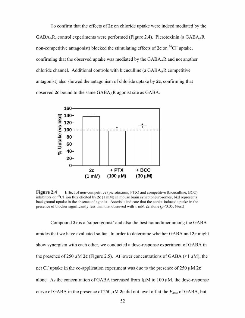
To confirm that the effects of 2c on chloride uptake were indeed mediated by the
GABAAR, control experiments were performed (Figure 2.4). Picrotoxinin (a GABAAR
non-competitive antagonist) blocked the stimulating effects of 2c on 36Cl- uptake,
confirming that the observed uptake was mediated by the GABAAR and not another
chloride channel. Additional controls with bicuculline (a GABAAR competitive
antagonist) also showed the antagonism of chloride uptake by 2c, confirming that
observed 2c bound to the same GABAAR agonist site as GABA.
020406080
100120140160
............................................................
2c(1 mM)
+ PTX(100 μM)
+ BCC(30 μM)
* *
Figure 2.4 Effect of non-competitive (picrotoxinin, PTX) and competitive (bicuculline, BCC) inhibitors on 36Cl- ion flux elicited by 2c (1 mM) in mouse brain synaptoneurosomes; bkd represents background uptake in the absence of agonist. Asterisks indicate that the aonist-induced uptake in the presence of blocker significantly less than that observed with 1 mM 2c alone (p<0.05, t-test)
Compound 2c is a ‘superagonist’ and also the best homodimer among the GABA
amides that we have evaluated so far. In order to determine whether GABA and 2c might
show synergism with each other, we conducted a dose-response experiment of GABA in
the presence of 250 μM 2c (Figure 2.5). At lower concentrations of GABA (<1 μM), the
net Cl- uptake in the co-application experiment was due to the presence of 250 μM 2c
alone. As the concentration of GABA increased from 1μM to 100 μM, the dose-response
curve of GABA in the presence of 250 μM 2c did not level off at the Emax of GABA, but
52
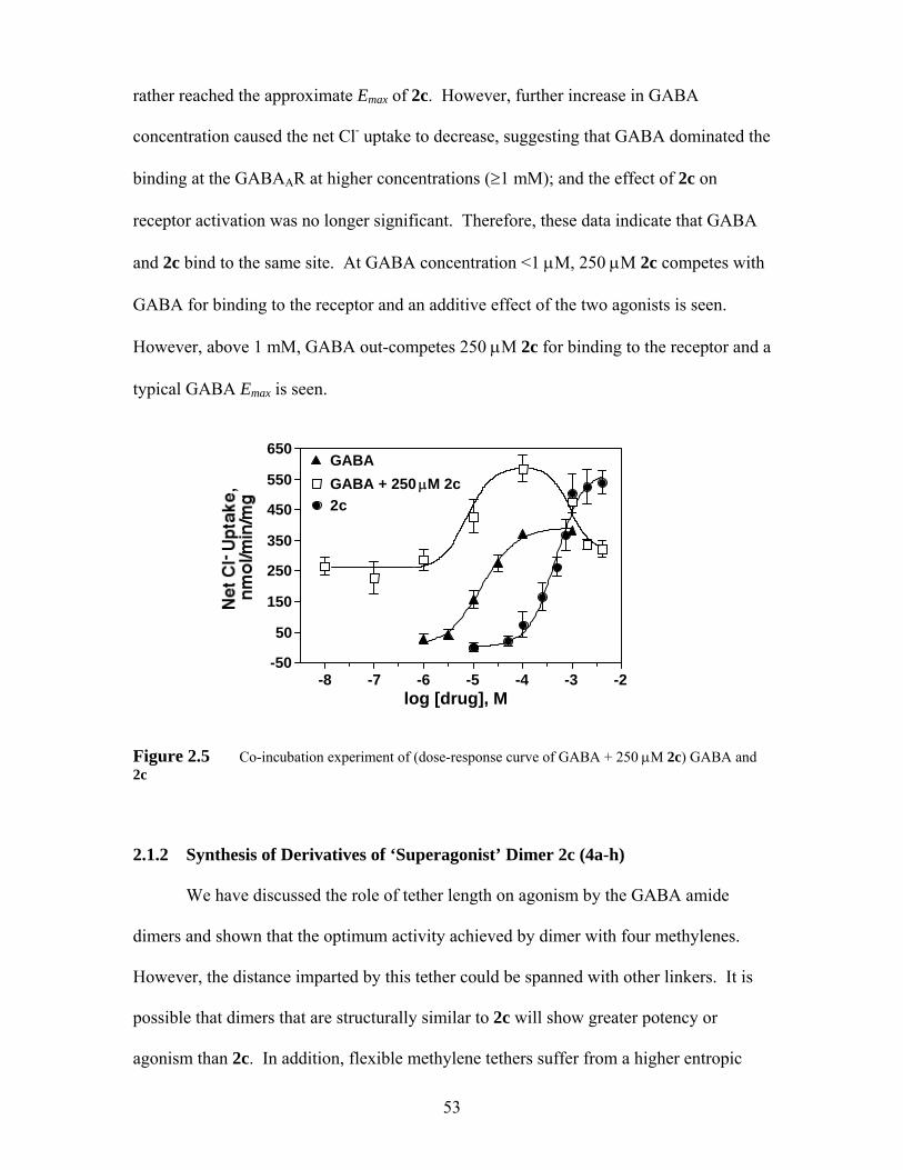
rather reached the approximate Emax of 2c. However, further increase in GABA
concentration caused the net Cl- uptake to decrease, suggesting that GABA dominated the
binding at the GABAAR at higher concentrations (≥1 mM); and the effect of 2c on
receptor activation was no longer significant. Therefore, these data indicate that GABA
and 2c bind to the same site. At GABA concentration <1 μM, 250 μM 2c competes with
GABA for binding to the receptor and an additive effect of the two agonists is seen.
However, above 1 mM, GABA out-competes 250 μM 2c for binding to the receptor and a
typical GABA Emax is seen.
-8 -7 -6 -5 -4 -3 -2-50
50
150
250
350
450
550
650GABAGABA + 250 μM 2c2c
log [drug], M
Figure 2.5 Co-incubation experiment of (dose-response curve of GABA + 250 μM 2c) GABA and 2c 2.1.2 Synthesis of Derivatives of ‘Superagonist’ Dimer 2c (4a-h)
We have discussed the role of tether length on agonism by the GABA amide
dimers and shown that the optimum activity achieved by dimer with four methylenes.
However, the distance imparted by this tether could be spanned with other linkers. It is
possible that dimers that are structurally similar to 2c will show greater potency or
agonism than 2c. In addition, flexible methylene tethers suffer from a higher entropic
53
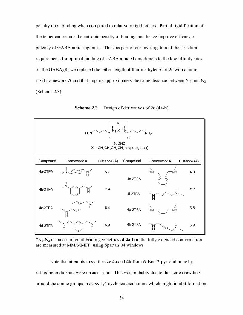
penalty upon binding when compared to relatively rigid tethers. Partial rigidification of
the tether can reduce the entropic penalty of binding, and hence improve efficacy or
potency of GABA amide agonists. Thus, as part of our investigation of the structural
requirements for optimal binding of GABA amide homodimers to the low-affinity sites
on the GABAAR, we replaced the tether length of four methylenes of 2c with a more
rigid framework A and that imparts approximately the same distance between N 1 and N2
(Scheme 2.3).
Scheme 2.3 Design of derivatives of 2c (4a-h)
NH
HN
NH
HN
NH
NH
NH
NH
NH
HN
HN NH
NH
HN
Compound Framework A
4a·2TFA
4b·2TFA
4c·2TFA
4d·2TFA
4e·2TFA
4f·2TFA
4g·2TFA
4h·2TFA
HN NH
H2NHN1
O
A
XHN2 NH2
O
Compound Framework A
2c·2HClX = CH2CH2CH2CH2 (superagonist)
Distance (Å) Distance (Å)
5.7
5.4
6.4
5.8
4.0
5.7
3.5
5.8
*N1-N2 distances of equilibrium geometries of 4a-h in the fully extended conformation are measured at MM/MMFF, using Spartan’04 windows
Note that attempts to synthesize 4a and 4b from N-Boc-2-pyrrolidinone by
refluxing in dioxane were unsuccessful. This was probably due to the steric crowding
around the amine groups in trans-1,4-cyclohexanediamine which might inhibit formation
54
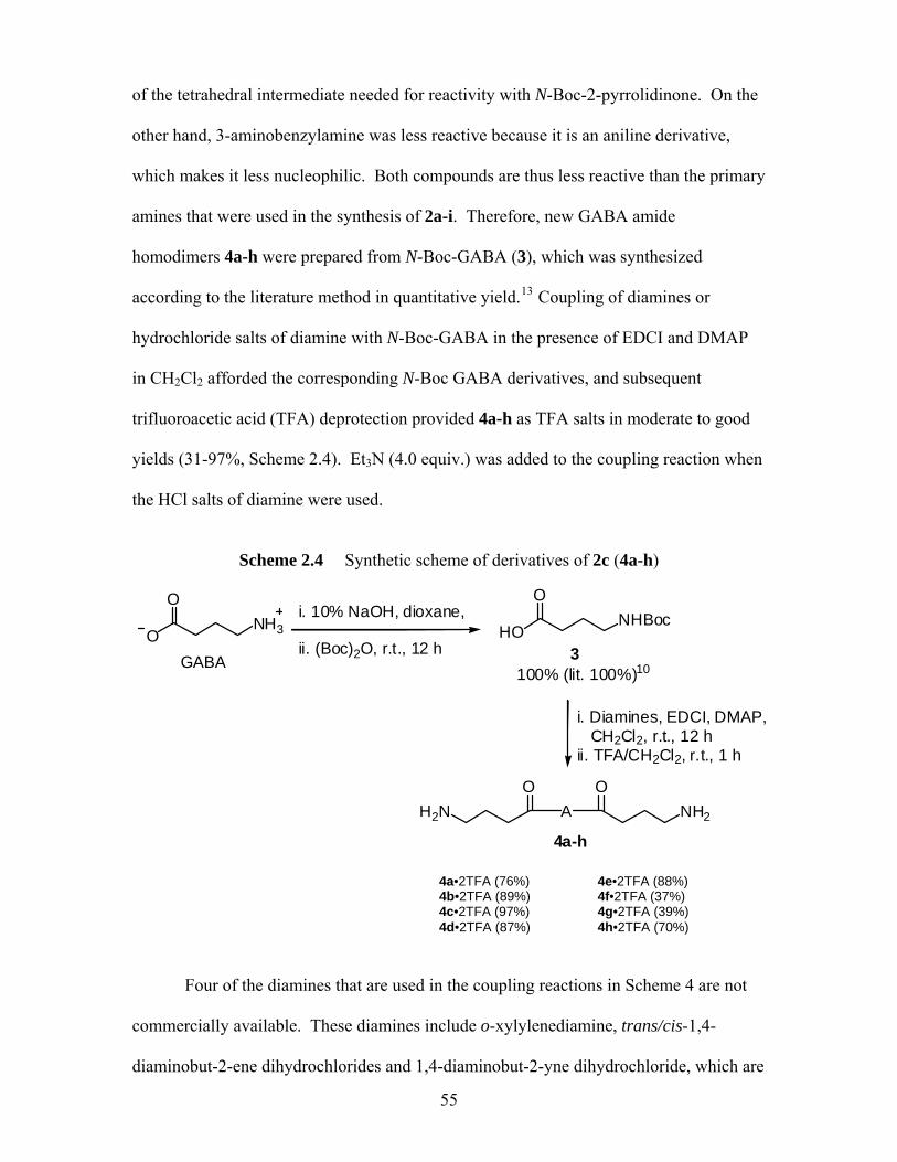
of the tetrahedral intermediate needed for reactivity with N-Boc-2-pyrrolidinone. On the
other hand, 3-aminobenzylamine was less reactive because it is an aniline derivative,
which makes it less nucleophilic. Both compounds are thus less reactive than the primary
amines that were used in the synthesis of 2a-i. Therefore, new GABA amide
homodimers 4a-h were prepared from N-Boc-GABA (3), which was synthesized
according to the literature method in quantitative yield.13 Coupling of diamines or
hydrochloride salts of diamine with N-Boc-GABA in the presence of EDCI and DMAP
in CH2Cl2 afforded the corresponding N-Boc GABA derivatives, and subsequent
trifluoroacetic acid (TFA) deprotection provided 4a-h as TFA salts in moderate to good
yields (31-97%, Scheme 2.4). Et3N (4.0 equiv.) was added to the coupling reaction when
the HCl salts of diamine were used.
Scheme 2.4 Synthetic scheme of derivatives of 2c (4a-h)
HONHBoc
O
100% (lit. 100%)10
ONH3
O
GABA
OH2N NH2
OA
i. Diamines, EDCI, DMAP, CH2Cl2, r.t., 12 hii. TFA/CH2Cl2, r.t., 1 h
4a-h
i. 10% NaOH, dioxane, ii. (Boc)2O, r.t., 12 h 3
4a•2TFA (76%) 4e•2TFA (88%)
4b•2TFA (89%) 4f•2TFA (37%) 4c•2TFA (97%) 4g•2TFA (39%) 4d•2TFA (87%) 4h•2TFA (70%)
Four of the diamines that are used in the coupling reactions in Scheme 4 are not
commercially available. These diamines include o-xylylenediamine, trans/cis-1,4-
diaminobut-2-ene dihydrochlorides and 1,4-diaminobut-2-yne dihydrochloride, which are
55
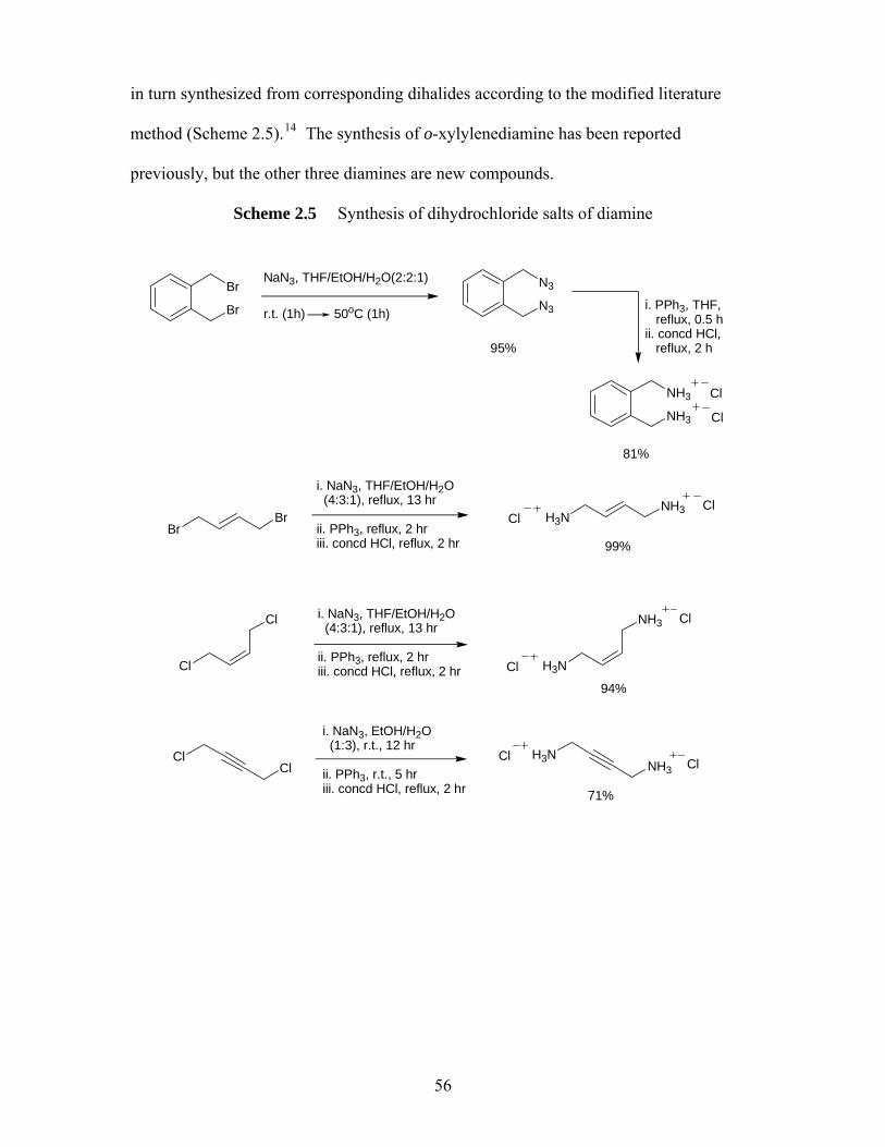
in turn synthesized from corresponding dihalides according to the modified literature
method (Scheme 2.5).14 The synthesis of o-xylylenediamine has been reported
previously, but the other three diamines are new compounds.
Scheme 2.5 Synthesis of dihydrochloride salts of diamine
Br
BrNaN3, THF/EtOH/H2O(2:2:1)
r.t. (1h) 50oC (1h) N3
N3
i. PPh3, THF, reflux, 0.5 hii. concd HCl, reflux, 2 h
NH3
NH3
Cl
Cl
95%
81%
Br
i. NaN3, THF/EtOH/H2O (4:3:1), reflux, 13 hr
ii. PPh3, reflux, 2 hriii. concd HCl, reflux, 2 hr
H3N
BrNH3
ClCl
99%
Cl
i. NaN3, THF/EtOH/H2O (4:3:1), reflux, 13 hr
ii. PPh3, reflux, 2 hriii. concd HCl, reflux, 2 hr H3N
Cl NH3 Cl
Cl
94%
Cl
i. NaN3, EtOH/H2O (1:3), r.t., 12 hr
ii. PPh3, r.t., 5 hriii. concd HCl, reflux, 2 hr
H3NCl NH3
Cl Cl
71%
56
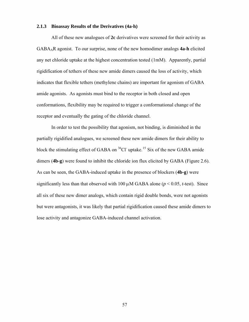
2.1.3 Bioassay Results of the Derivatives (4a-h)
All of these new analogues of 2c derivatives were screened for their activity as
GABAAR agonist. To our surprise, none of the new homodimer analogs 4a-h elicited
any net chloride uptake at the highest concentration tested (1mM). Apparently, partial
rigidification of tethers of these new amide dimers caused the loss of activity, which
indicates that flexible tethers (methylene chains) are important for agonism of GABA
amide agonists. As agonists must bind to the receptor in both closed and open
conformations, flexibility may be required to trigger a conformational change of the
receptor and eventually the gating of the chloride channel.
In order to test the possibility that agonism, not binding, is diminished in the
partially rigidified analogues, we screened these new amide dimers for their ability to
block the stimulating effect of GABA on 36Cl- uptake.15 Six of the new GABA amide
dimers (4b-g) were found to inhibit the chloride ion flux elicited by GABA (Figure 2.6).
As can be seen, the GABA-induced uptake in the presence of blockers (4b-g) were
significantly less than that observed with 100 μM GABA alone (p < 0.05, t-test). Since
all six of these new dimer analogs, which contain rigid double bonds, were not agonists
but were antagonists, it was likely that partial rigidification caused these amide dimers to
lose activity and antagonize GABA-induced channel activation.
57
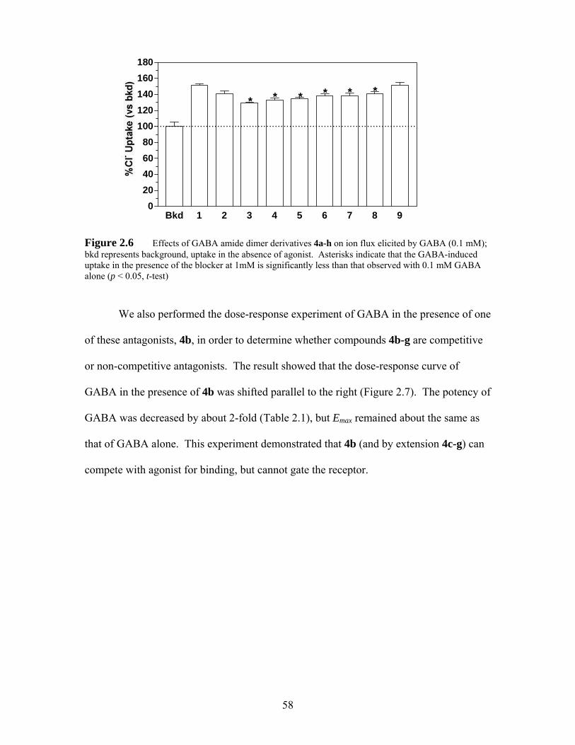
Bkd 1 2 3 4 5 6 7 8 90
20406080
100120140160180
...........................................................................................
* * * * * *
Figure 2.6 Effects of GABA amide dimer derivatives 4a-h on ion flux elicited by GABA (0.1 mM); bkd represents background, uptake in the absence of agonist. Asterisks indicate that the GABA-induced uptake in the presence of the blocker at 1mM is significantly less than that observed with 0.1 mM GABA alone (p < 0.05, t-test)
We also performed the dose-response experiment of GABA in the presence of one
of these antagonists, 4b, in order to determine whether compounds 4b-g are competitive
or non-competitive antagonists. The result showed that the dose-response curve of
GABA in the presence of 4b was shifted parallel to the right (Figure 2.7). The potency of
GABA was decreased by about 2-fold (Table 2.1), but Emax remained about the same as
that of GABA alone. This experiment demonstrated that 4b (and by extension 4c-g) can
compete with agonist for binding, but cannot gate the receptor.
58
-6.5 -5.5 -4.5 -3.5 -2.5
50
150
250
350 GABA + 4bGABA
log [drug], M
Figure 2.7 Inhibitory effect of 4b on concentration dependent chloride uptake by GABA in mouse brain synaptoneurosomes
2.1.4 [3H]Muscimol Binding Experiments
The inhibitory effect of these partial rigidified homodimers (4b-h) was further
examined in the binding experiments. The ability of GABA, 2c or 4b to displace
[3H]muscimol from binding sites were tested in two different assays (filtration and
centrifugation assays). Typical binding assays ([3H]muscimol or [3H]GABA) were done
with frozen-thawed, well-washed, lysed membranes to optimize the binding.16 However,
these studies generated little information regarding low affinity binding associated with
the channel gating in functional assay. In order to address this issue, both DeLorey and
Brown (filtration assay) and Schwartz et al.17 (centrifugation assay) have done binding
studies under conditions that more closely approximate functional assays, and found only
micromolar binding of [3H]muscimol (1.9 μM 16 and 2.0 μM17 respectively). Based on
Brown’s literature method, we carried out [3H]muscimol (70 nM) binding experiments in
the presence of three unlabeled ligands (1 mM GABA, 2c and 4b respectively). The
specific [3H]muscimol binding in the presence of 1 mM GABA or 2c was about 50% of
total binding. This value was comparable to the value that reported (40%) by DeLorey
59
and Brown.16 However, specific [3H]muscimol binding in the presence of the weak
antagonist 4b was indistinguishable from background (Figure 2.8). One possible reason
for the apparent lack of binding by 4b is the rapid washing and filtering used in the
filtration assay. Since 4b is a low potency antagonist, the wash and filtration may have
facilitated the dissociation of 4b from the binding sites; thus no binding was detected.
The centrifugation assay is better at detecting low affinity binding since no rapid
wash and filtration are required as in the filtration assay. Thus, dissociation of weakly
bound ligands to the binding sites should be minimal in the centrifugation assay. We
therefore conducted [3H]muscimol (70 nM) binding experiments in the presence of three
unlabeled ligands (1 mM GABA, 2c and 4b respectively) based on Schwartz’s method.17
Unlabeled mucimol (1 μM) was also added to each experiment to ensure the
measurement of low affinity-site binding. In the presence of 1 mM GABA or 2c, the
specific [3H]muscimol binding was about 40% (Figure 2.8). This result again indicated
that 2c bound to the same sites as GABA. The specific [3H]muscimol binding in the
presence of the weak antagonist 4b was only about 15%; however this result was
statistically significant. The result of 4b also shows that the centrifugation method is a
better technique for weak agonist or antagonist binding. In general, these results were in
good agreement with Schwartz’s published data (56 to 67% specific [3H]muscimol
binding in the presence of 1 mM GABA).17
60
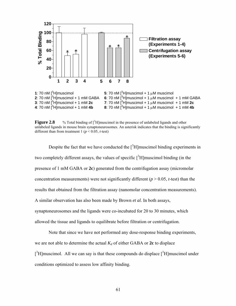
0
20
40
60
80
100
120
............................................................
*** *
* Filtration assay(Experiments 1-4)Centrifugation assay(Experiments 5-6)
1 2 3 4 5 6 7 8
1: 70 nM [3H]muscimol 5: 70 nM [3H]muscimol + 1 μM muscimol 2: 70 nM [3H]muscimol + 1 mM GABA 6: 70 nM [3H]muscimol + 1 μM muscimol + 1 mM GABA 3: 70 nM [3H]muscimol + 1 mM 2c 7: 70 nM [3H]muscimol + 1 μM muscimol + 1 mM 2c 4: 70 nM [3H]muscimol + 1 mM 4b 8: 70 nM [3H]muscimol + 1 μM muscimol + 1 mM 4b Figure 2.8 % Total binding of [3H]muscimol in the presence of unlabeled ligands and other unlabeled ligands in mouse brain synaptoneurosomes. An asterisk indicates that the binding is significantly different than from treatment 1 (p < 0.05, t-test)
Despite the fact that we have conducted the [3H]muscimol binding experiments in
two completely different assays, the values of specific [3H]muscimol binding (in the
presence of 1 mM GABA or 2c) generated from the centrifugation assay (micromolar
concentration measurements) were not significantly different (p > 0.05, t-test) than the
results that obtained from the filtration assay (nanomolar concentration measurements).
A similar observation has also been made by Brown et al. In both assays,
synaptoneurosomes and the ligands were co-incubated for 20 to 30 minutes, which
allowed the tissue and ligands to equilibrate before filtration or centrifugation.
Note that since we have not performed any dose-response binding experiments,
we are not able to determine the actual Kd of either GABA or 2c to displace
[3H]muscimol. All we can say is that these compounds do displace [3H]muscimol under
conditions optimized to assess low affinity binding.
61
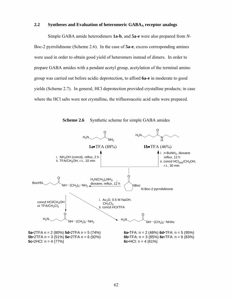
2.2 Syntheses and Evaluation of heteromeric GABAA receptor analogs Simple GABA amide heterodimers 1a-b, and 5a-e were also prepared from N-
Boc-2 pyrrolidinone (Scheme 2.6). In the case of 5a-e, excess corresponding amines
were used in order to obtain good yield of heteromers instead of dimers. In order to
prepare GABA amides with a pendant acetyl group, acetylation of the terminal amino
group was carried out before acidic deprotection, to afford 6a-e in moderate to good
yields (Scheme 2.7). In general, HCl deprotection provided crystalline products; in case
where the HCl salts were not crystalline, the trifluoroacetic acid salts were prepared.
Scheme 2.6 Synthetic scheme for simple GABA amides OO 1a•TFA (88%) 1b•TFA (46%)
NBoc
O
NH
OBocHN
(CH2)n NH2
H2N(CH2)nNH2, dioxane, reflux, 12 h
i. NH4OH (concd), reflux, 2 hii. TFA/CH3OH, r.t., 10 min
NH2H2N
i. n-BuNH2, dioxane reflux, 12 hii. concd HCl(aq)/CH3OH, r.t., 30 min
NH
H2N
i. Ac2O, 0.5 M NaOH, CH2Cl2, ii. concd HCl/TFA
N-Boc-2-pyrrolidinone
NH
OH2N
(CH2)n NHAc
concd HCl/CH3OHor TFA/CH2Cl2
NH
OH2N
(CH2)n NH2
5a•2TFA n = 2 (80%) 5d•2TFA n = 5 (74%) 5b•2TFA n = 3 (51%) 5e•2TFA n = 6 (92%) 5c•2HCl: n = 4 (77%)
6a•TFA: n = 2 (48%) 6d•TFA: n = 5 (95%) 6b•TFA: n = 3 (85%) 6e•TFA: n = 6 (83%) 6c•HCl: n = 4 (61%)
62
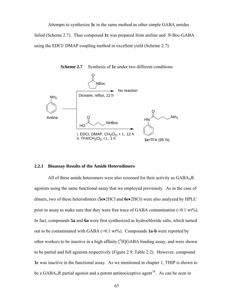
Attempts to synthesize 1c in the same method as other simple GABA amides
failed (Scheme 2.7). Thus compound 1c was prepared from aniline and N-Boc-GABA
using the EDCI/ DMAP coupling method in excellent yield (Scheme 2.7).
Scheme 2.7 Synthesis of 1c under two different conditions
NBoc
O
NH2
Aniline
Dioxane, reflux, 12 h
No reaction
HN
1c•TFA (95 %)
HO
ONHBoc
i. EDCI, DMAP, CH2Cl2, r. t., 12 hii. TFA/CH2Cl2, r.t., 1 h
NH2
O
2.2.1 Bioassay Results of the Amide Heterodimers
All of these amide heteromers were also screened for their activity as GABAAR
agonists using the same functional assay that we employed previously. As in the case of
dimers, two of these heterodimers (5c•2HCl and 6c•2HCl) were also analyzed by HPLC
prior to assay to make sure that they were free trace of GABA contamination (<0.1 wt%).
In fact, compounds 5a and 6a were first synthesized as hydrochloride salts, which turned
out to be contaminated with GABA (>0.1 wt%). Compounds 1a-b were reported by
other workers to be inactive in a high affinity [3H]GABA binding assay, and were shown
to be partial and full agonists respectively (Figure 2.9, Table 2.2). However, compound
1c was inactive in the functional assay. As we mentioned in chapter 1, THIP is shown to
be a GABAAR partial agonist and a potent antinociceptive agent18. As can be seen in
63
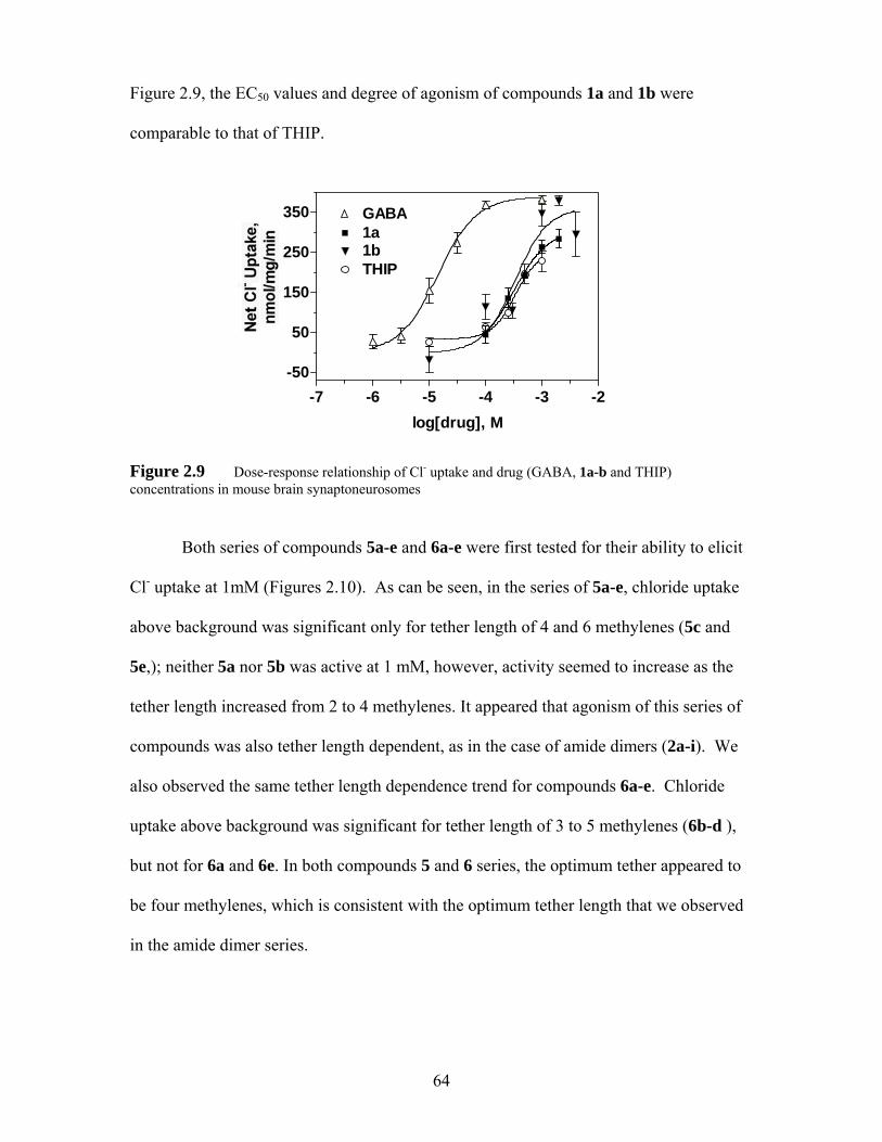
Figure 2.9, the EC50 values and degree of agonism of compounds 1a and 1b were
comparable to that of THIP.
-7 -6 -5 -4 -3 -2-50
50
150
250
3501aGABA
THIP1b
log[drug], M
Figure 2.9 Dose-response relationship of Cl- uptake and drug (GABA, 1a-b and THIP) concentrations in mouse brain synaptoneurosomes
Both series of compounds 5a-e and 6a-e were first tested for their ability to elicit
Cl- uptake at 1mM (Figures 2.10). As can be seen, in the series of 5a-e, chloride uptake
above background was significant only for tether length of 4 and 6 methylenes (5c and
5e,); neither 5a nor 5b was active at 1 mM, however, activity seemed to increase as the
tether length increased from 2 to 4 methylenes. It appeared that agonism of this series of
compounds was also tether length dependent, as in the case of amide dimers (2a-i). We
also observed the same tether length dependence trend for compounds 6a-e. Chloride
uptake above background was significant for tether length of 3 to 5 methylenes (6b-d ),
but not for 6a and 6e. In both compounds 5 and 6 series, the optimum tether appeared to
be four methylenes, which is consistent with the optimum tether length that we observed
in the amide dimer series.
64
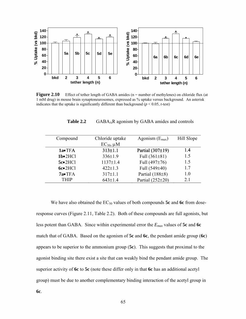
bkd 2 3 4 5 60
20406080
100120140
**
*
6b 6c 6d 6e
tether length (n)
6a
---------------------------------------------------------------------------
bkd 2 3 4 5 6
120100
020406080
140 *
5b 5c 5d 5e
tether length (n)
----------------------------------------------------------------------------------------------------
5a
***
Figure 2.10 Effect of tether length of GABA amides (n = number of methylenes) on chloride flux (at 1 mM drug) in mouse brain synaptoneurosomes, expressed as % uptake versus background. An asterisk indicates that the uptake is significantly different than background (p < 0.05, t-test)
Table 2.2 GABAAR agonism by GABA amides and controls
Compound Chloride uptake
EC50, μM Agonism (Emax) Hill Slope
1a•TFA A 313±1.1 313±1.1 Partial (307±19) Partial (307±19) 1.4 1.4 1b•2HCl 336±1.9 Full (361±81) 1.5 5c•2HCl 1137±1.4 Full (497±76) 1.5 6c•2HCl 422±1.3 Full (549±40) 1.7 7a•TFA 317±1.1 Partial (188±8) 1.0
THIP 643±1.4 Partial (252±20) 2.1
We have also obtained the EC50 values of both compounds 5c and 6c from dose-
response curves (Figure 2.11, Table 2.2). Both of these compounds are full agonists, but
less potent than GABA. Since within experimental error the Emax values of 5c and 6c
match that of GABA. Based on the agonism of 5c and 6c, the pendant amide group (6c)
appears to be superior to the ammonium group (5c). This suggests that proximal to the
agonist binding site there exist a site that can weakly bind the pendant amide group. The
superior activity of 6c to 5c (note these differ only in that 6c has an additional acetyl
group) must be due to another complementary binding interaction of the acetyl group in
6c.
65
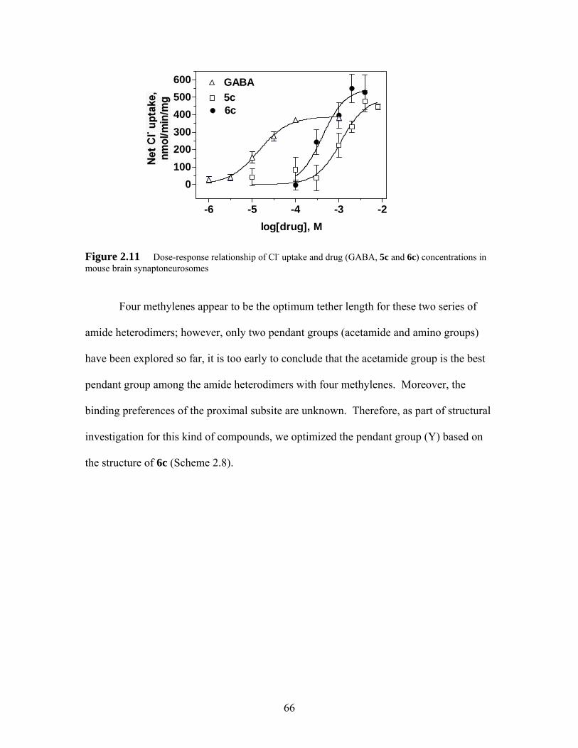
-6 -5 -4 -3 -2
0100200300400500600 GABA
5c6c
log[drug], M
Figure 2.11 Dose-response relationship of Cl- uptake and drug (GABA, 5c and 6c) concentrations in mouse brain synaptoneurosomes
Four methylenes appear to be the optimum tether length for these two series of
amide heterodimers; however, only two pendant groups (acetamide and amino groups)
have been explored so far, it is too early to conclude that the acetamide group is the best
pendant group among the amide heterodimers with four methylenes. Moreover, the
binding preferences of the proximal subsite are unknown. Therefore, as part of structural
investigation for this kind of compounds, we optimized the pendant group (Y) based on
the structure of 6c (Scheme 2.8).
66
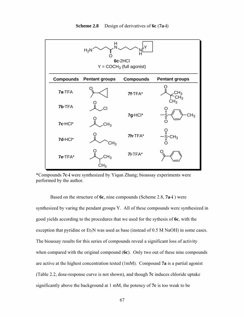
Scheme 2.8 Design of derivatives of 6c (7a-i)
Compounds CompoundsPentant groups Pentant groups
CH3SO
O
CH3SO
O
O
OCl
OCH3
O
CH3
OCH3
CH3
O
CH3
O
CH3CH3
7a·TFA
7h·TFA*
7g·HCl*
7f·TFA*
7e·TFA*
7d·HCl*
7c·HCl*
7b·TFA
7i·TFA*
H2NHN
ONH
Y
6c·2HClY = COCH3 (full agonist)
*Compounds 7c-i were synthesized by Yiqun Zhang; bioassay experiments were performed by the author.
Based on the structure of 6c, nine compounds (Scheme 2.8, 7a-i ) were
synthesized by varing the pendant groups Y. All of these compounds were synthesized in
good yields according to the procedures that we used for the sythesis of 6c, with the
exception that pyridine or Et3N was used as base (instead of 0.5 M NaOH) in some cases.
The bioassay results for this series of compounds reveal a significant loss of activity
when compared with the original compound (6c). Only two out of these nine compounds
are active at the highest concentration tested (1mM). Compound 7a is a partial agonist
(Table 2.2, dose-response curve is not shown), and though 7c induces chloride uptake
significantly above the background at 1 mM, the potency of 7c is too weak to be
67

determined. All of these nine compounds are structurally similar to 6c; however,
replacing the acetamide group with either the bulkier amide groups or sulfonamide
groups caused a loss of activity. These results suggest the important role of the actamide
group in investigated bioactivity. These results also suggested that the proximal binding
site for the pendant amide is small.
2.3 Syntheses and Evaluation of ZAPA and its derivatives
2.3.1 Synthesis of ZAPA and its derivatives
As mentioned in chapter 1, ZAPA (Z-3-[(aminoiminomethyl)thio]prop-2-enoic
acid), an isothiouronium analog of GABA (γ-aminobutyric acid), is a potent GABAAR
agonist, which is a potentially therapeutic agent for anxiety, epilepsy, and pain.19 ZAPA
has also shown good antidiabetic activity in animal models of non-insulin diabetes
mellitus (NIDDM).20 Interestingly, the E-isomer of ZAPA has been shown to be inactive
at the GABAAR.21 A structure-activity relationship study of ZAPA have been reported
by Allan et al.22 in 1997 in a guinea pig ileum contraction assay, which tested the ability
of ZAPA and its derivatives to produce GABA-like contractions in the guinea pig ileum.
Most of the ZAPA derivatives studied by these authors produced GABA-like agonism,
but all of them were less potent than either GABA or ZAPA.22 However, none of the
derivatives that studied by Allan and co-workers have been tested in a standard 36Cl- flux
or electrophysiological functional assay. Due to our interest in preparing ZAPA linked
bivalent agonists that can be tethered to other drug sites, we synthesized two new and two
known ZAPA derivatives and the tested these derivatives in the standard 36Cl- flux
functional assay for the first time.
68
ZAPA derivatives were prepared from the corresponding primary N-
alkylthioureas and propiolic acid. The traditional syntheses of primary or N, N’-
disubstituted thioureas usually involved a two-step process, which often required
vigorous conditions to cleave the benzoyl group from thiourea.22, 23 -27 We employed
several modified methods that were reported in the literature for the synthesis of n-
alkylthioureas. Most of the methods that we tried were unsuccessful. Prolonged heating
and use of strong bases led to the formation of side product n-alkylurea. Detailed
syntheses were shown in Table 2.3.
69
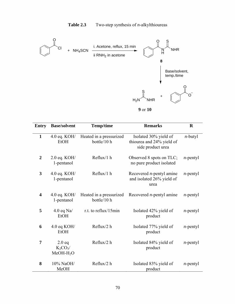
Table 2.3 Two-step synthesis of n-alkylthioureas
Cl
O
+ NH4SCNi. Acetone, reflux, 15 min
ii RNH2 in acetoneNH
O
NHR
S
Base/solvent,temp./time
H2N NHR
SO
O+
8
9 or 10
Entry
Base/solvent Temp/time Remarks R
1 4.0 eq. KOH/ EtOH
Heated in a pressurized bottle/10 h
Isolated 30% yield of thiourea and 24% yield of
side product urea
n-butyl
2 2.0 eq. KOH/ 1-pentanol
Reflux/1 h Observed 8 spots on TLC; no pure product isolated
n-pentyl
3 4.0 eq. KOH/ 1-pentanol
Reflux/1 h Recovered n-pentyl amine and isolated 26% yield of
urea
n-pentyl
4 4.0 eq. KOH/ 1-pentanol
Heated in a pressurized bottle/10 h
Recovered n-pentyl amine n-pentyl
5 4.0 eq Na/ EtOH
r.t. to reflux/15min
Isolated 42% yield of product
n-pentyl
6 4.0 eq KOH/ EtOH
Reflux/2 h Isolated 77% yield of product
n-pentyl
7 2.0 eq K2CO3/
MeOH-H2O
Reflux/2 h Isolated 84% yield of product
n-pentyl
8 10% NaOH/ MeOH
Reflux/2 h Isolated 83% yield of product
n-pentyl
70
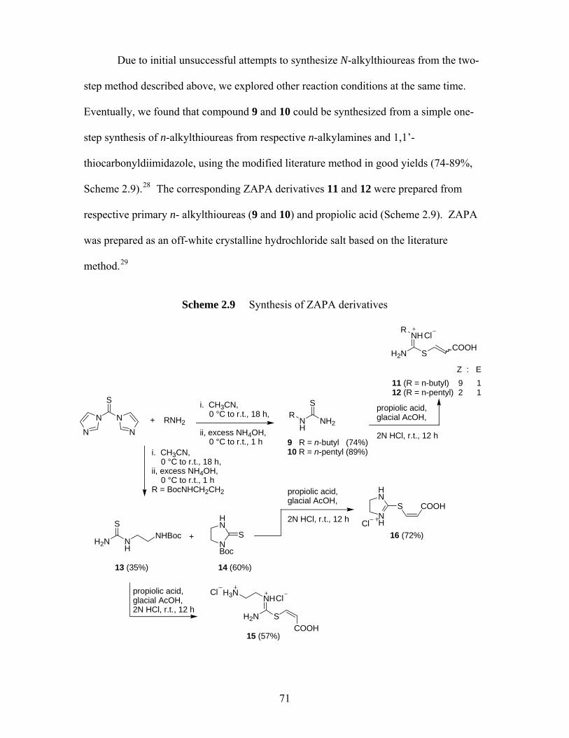
Due to initial unsuccessful attempts to synthesize N-alkylthioureas from the two-
step method described above, we explored other reaction conditions at the same time.
Eventually, we found that compound 9 and 10 could be synthesized from a simple one-
step synthesis of n-alkylthioureas from respective n-alkylamines and 1,1’-
thiocarbonyldiimidazole, using the modified literature method in good yields (74-89%,
Scheme 2.9).28 The corresponding ZAPA derivatives 11 and 12 were prepared from
respective primary n- alkylthioureas (9 and 10) and propiolic acid (Scheme 2.9). ZAPA
was prepared as an off-white crystalline hydrochloride salt based on the literature
method.29
Scheme 2.9 Synthesis of ZAPA derivatives
N N
S
N N+ RNH2 N
HNH2
RS
H2N S
NHCOOH
RCl
9 R = n-butyl (74%)10 R = n-pentyl (89%)
11 (R = n-butyl) 9 112 (R = n-pentyl) 2 1
Z : E
HN
NBoc
S
14 (60%)
HN
NH
S
Cl
COOH
16 (72%)
propiolic acid, glacial AcOH, 2N HCl, r.t., 12 h
i. CH3CN, 0 °C to r.t., 18 h, ii, excess NH4OH, 0 °C to r.t., 1 h
i. CH3CN, 0 °C to r.t., 18 h, ii, excess NH4OH, 0 °C to r.t., 1 hR = BocNHCH2CH2 propiolic acid,
glacial AcOH, 2N HCl, r.t., 12 h
H2N NH
NHBocS
+
13 (35%)
propiolic acid, glacial AcOH, 2N HCl, r.t., 12 h
H2N S
NH
COOH
H3NCl
Cl
15 (57%)
71
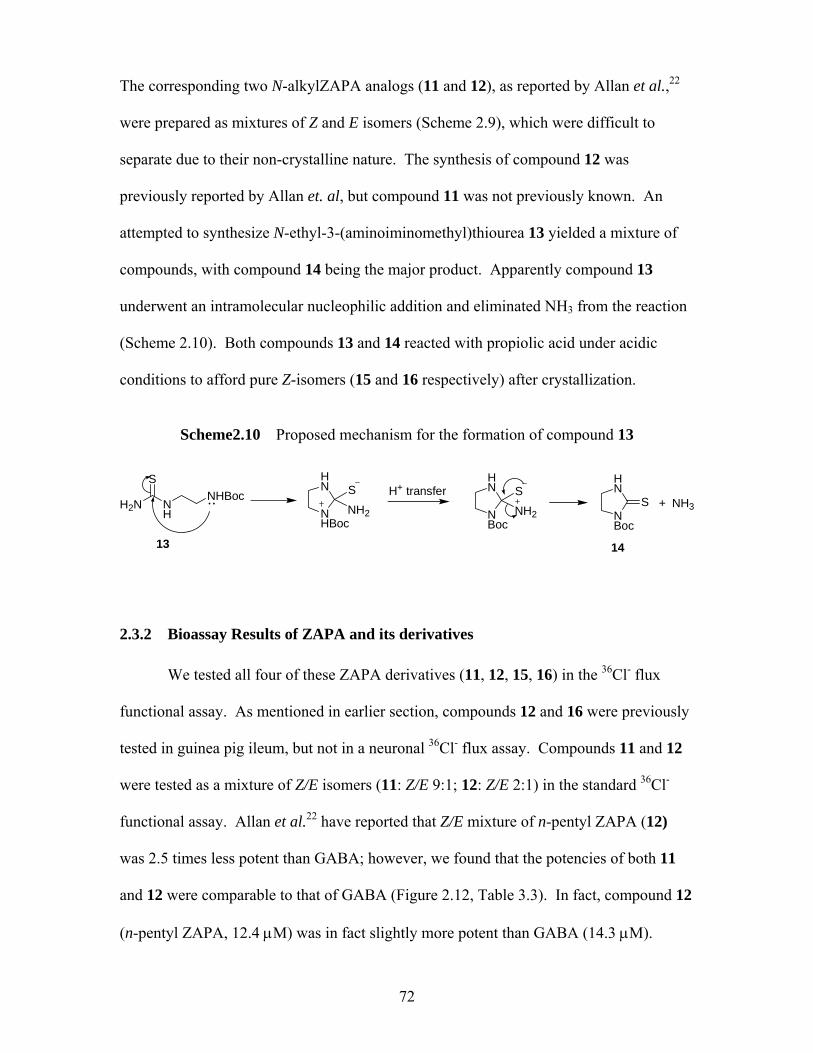
The corresponding two N-alkylZAPA analogs (11 and 12), as reported by Allan et al.,22
were prepared as mixtures of Z and E isomers (Scheme 2.9), which were difficult to
separate due to their non-crystalline nature. The synthesis of compound 12 was
previously reported by Allan et. al, but compound 11 was not previously known. An
attempted to synthesize N-ethyl-3-(aminoiminomethyl)thiourea 13 yielded a mixture of
compounds, with compound 14 being the major product. Apparently compound 13
underwent an intramolecular nucleophilic addition and eliminated NH3 from the reaction
(Scheme 2.10). Both compounds 13 and 14 reacted with propiolic acid under acidic
conditions to afford pure Z-isomers (15 and 16 respectively) after crystallization.
Scheme2.10 Proposed mechanism for the formation of compound 13
H2N NH
NHBocS
NHBoc
HN S
NH2
H+ transfer
NBoc
HN S
NH2 NBoc
HN
S + NH3
13 14
2.3.2 Bioassay Results of ZAPA and its derivatives
We tested all four of these ZAPA derivatives (11, 12, 15, 16) in the 36Cl- flux
functional assay. As mentioned in earlier section, compounds 12 and 16 were previously
tested in guinea pig ileum, but not in a neuronal 36Cl- flux assay. Compounds 11 and 12
were tested as a mixture of Z/E isomers (11: Z/E 9:1; 12: Z/E 2:1) in the standard 36Cl-
functional assay. Allan et al.22 have reported that Z/E mixture of n-pentyl ZAPA (12)
was 2.5 times less potent than GABA; however, we found that the potencies of both 11
and 12 were comparable to that of GABA (Figure 2.12, Table 3.3). In fact, compound 12
(n-pentyl ZAPA, 12.4 μM) was in fact slightly more potent than GABA (14.3 μM).
72
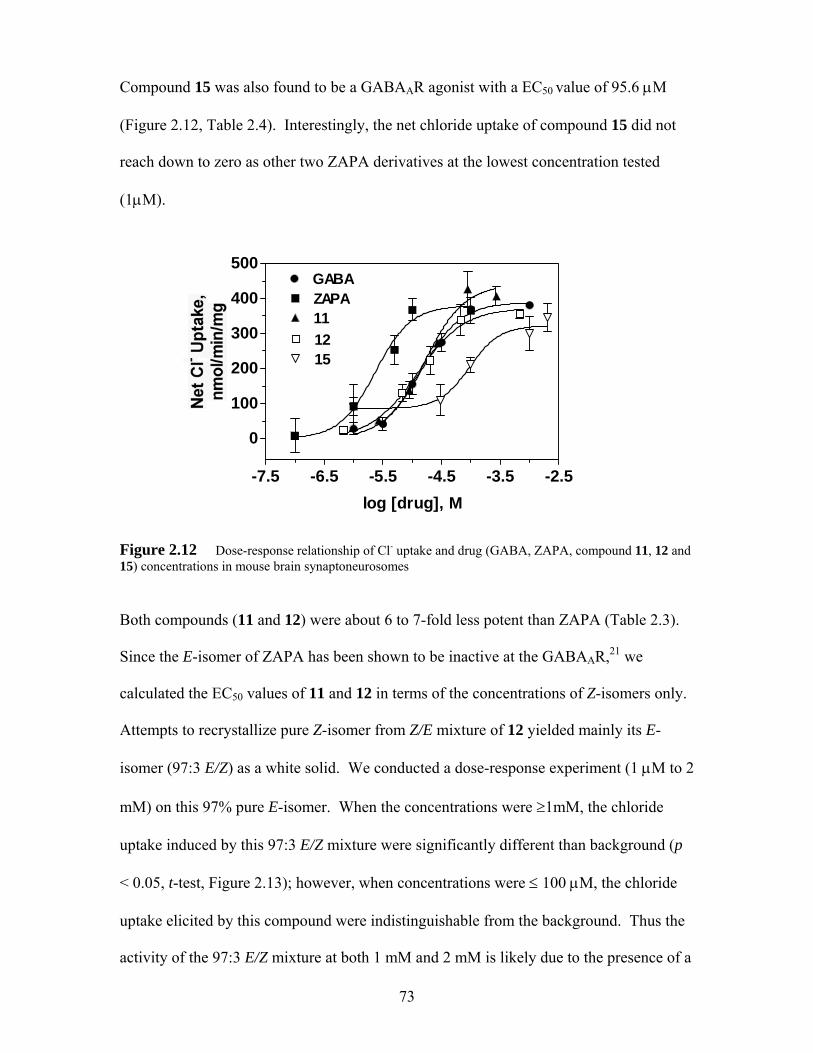
Compound 15 was also found to be a GABAAR agonist with a EC50 value of 95.6 μM
(Figure 2.12, Table 2.4). Interestingly, the net chloride uptake of compound 15 did not
reach down to zero as other two ZAPA derivatives at the lowest concentration tested
(1μM).
-7.5 -6.5 -5.5 -4.5 -3.5 -2.5
0
100
200
300
400
500
12
GABAZAPA11
15
log [drug], M
Figure 2.12 Dose-response relationship of Cl- uptake and drug (GABA, ZAPA, compound 11, 12 and 15) concentrations in mouse brain synaptoneurosomes
Both compounds (11 and 12) were about 6 to 7-fold less potent than ZAPA (Table 2.3).
Since the E-isomer of ZAPA has been shown to be inactive at the GABAAR,21 we
calculated the EC50 values of 11 and 12 in terms of the concentrations of Z-isomers only.
Attempts to recrystallize pure Z-isomer from Z/E mixture of 12 yielded mainly its E-
isomer (97:3 E/Z) as a white solid. We conducted a dose-response experiment (1 μM to 2
mM) on this 97% pure E-isomer. When the concentrations were ≥1mM, the chloride
uptake induced by this 97:3 E/Z mixture were significantly different than background (p
< 0.05, t-test, Figure 2.13); however, when concentrations were ≤ 100 μM, the chloride
uptake elicited by this compound were indistinguishable from the background. Thus the
activity of the 97:3 E/Z mixture at both 1 mM and 2 mM is likely due to the presence of a
73
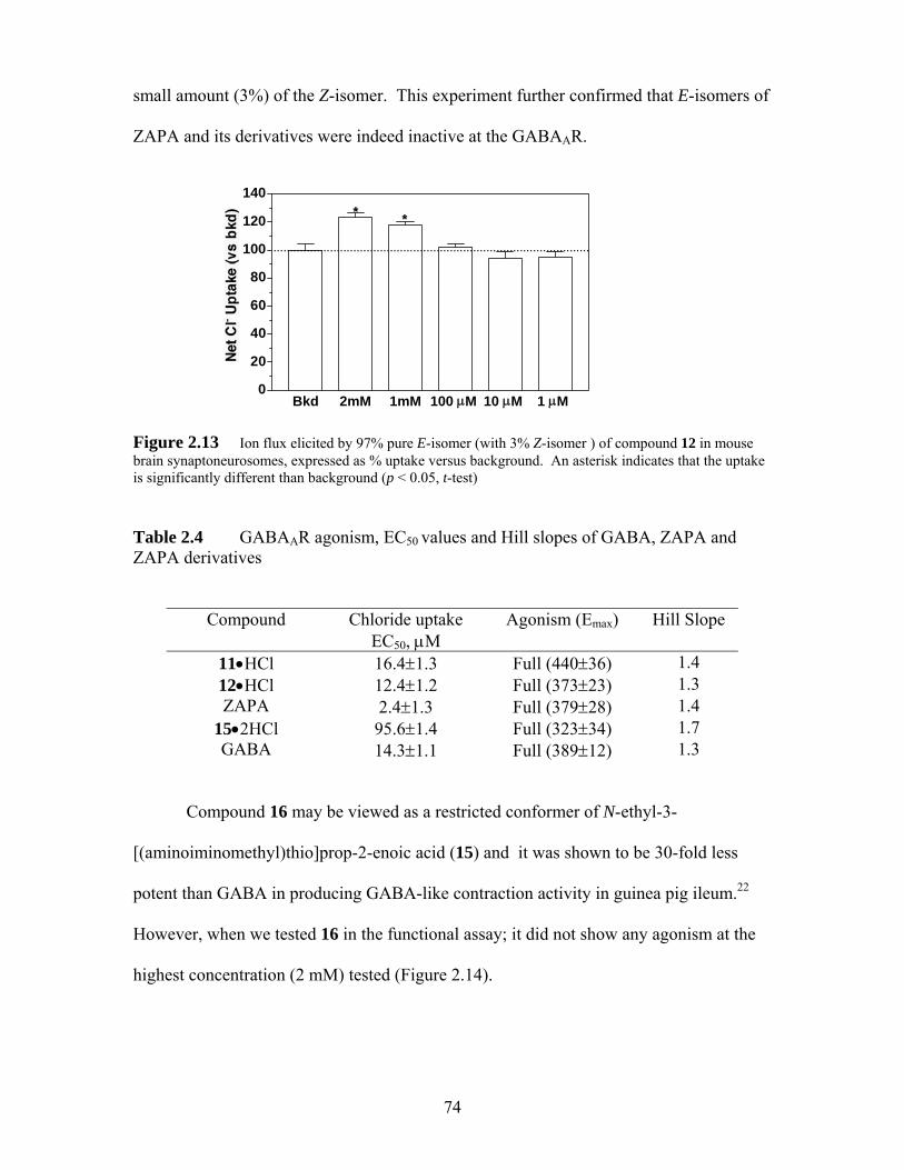
small amount (3%) of the Z-isomer. This experiment further confirmed that E-isomers of
ZAPA and its derivatives were indeed inactive at the GABAAR.
Bkd 2mM 1mM 100 μM 10 μM 1 μM0
20
40
60
80
100
120
140
...............................................................................................
* * Figure 2.13 Ion flux elicited by 97% pure E-isomer (with 3% Z-isomer ) of compound 12 in mouse brain synaptoneurosomes, expressed as % uptake versus background. An asterisk indicates that the uptake is significantly different than background (p < 0.05, t-test) Table 2.4 GABAAR agonism, EC50 values and Hill slopes of GABA, ZAPA and ZAPA derivatives
Compound Chloride uptake EC50, μM
Agonism (Emax) Hill Slope
11•HCl 16.4±1.3 Full (440±36) 1.4 12•HCl 12.4±1.2 Full (373±23) 1.3 ZAPA 2.4±1.3 Full (379±28) 1.4
15•2HCl 95.6±1.4 Full (323±34) 1.7 GABA 14.3±1.1 Full (389±12) 1.3
Compound 16 may be viewed as a restricted conformer of N-ethyl-3-
[(aminoiminomethyl)thio]prop-2-enoic acid (15) and it was shown to be 30-fold less
potent than GABA in producing GABA-like contraction activity in guinea pig ileum.22
However, when we tested 16 in the functional assay; it did not show any agonism at the
highest concentration (2 mM) tested (Figure 2.14).
74
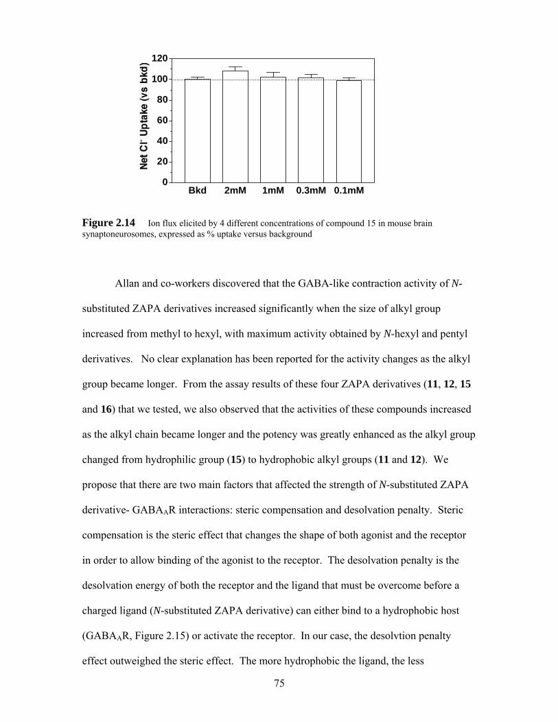
Bkd 2mM 1mM 0.3mM 0.1mM0
20
40
60
80
100
120
....................................................................................................................
Figure 2.14 Ion flux elicited by 4 different concentrations of compound 15 in mouse brain synaptoneurosomes, expressed as % uptake versus background
Allan and co-workers discovered that the GABA-like contraction activity of N-
substituted ZAPA derivatives increased significantly when the size of alkyl group
increased from methyl to hexyl, with maximum activity obtained by N-hexyl and pentyl
derivatives. No clear explanation has been reported for the activity changes as the alkyl
group became longer. From the assay results of these four ZAPA derivatives (11, 12, 15
and 16) that we tested, we also observed that the activities of these compounds increased
as the alkyl chain became longer and the potency was greatly enhanced as the alkyl group
changed from hydrophilic group (15) to hydrophobic alkyl groups (11 and 12). We
propose that there are two main factors that affected the strength of N-substituted ZAPA
derivative- GABAAR interactions: steric compensation and desolvation penalty. Steric
compensation is the steric effect that changes the shape of both agonist and the receptor
in order to allow binding of the agonist to the receptor. The desolvation penalty is the
desolvation energy of both the receptor and the ligand that must be overcome before a
charged ligand (N-substituted ZAPA derivative) can either bind to a hydrophobic host
(GABAAR, Figure 2.15) or activate the receptor. In our case, the desolvtion penalty
effect outweighed the steric effect. The more hydrophobic the ligand, the less
75
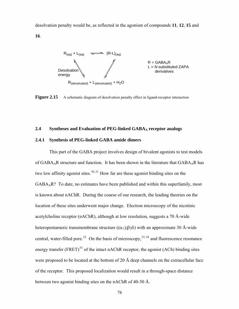
desolvation penalty would be, as reflected in the agonism of compounds 11, 12, 15 and
16.
R(aq) + L(aq) R = GABAAR
(R-L)(aq)
R(desolvated) + L(desolvated) + H2O
Desolvation energy
L = N-substituted ZAPA derivatives
Figure 2.15 A schematic diagram of desolvation penalty effect in ligand-receptor interaction
2.4 Syntheses and Evaluation of PEG-linked GABAA receptor analogs
2.4.1 Synthesis of PEG-linked GABA amide dimers This part of the GABA project involves design of bivalent agonists to test models
of GABAAR structure and function. It has been shown in the literature that GABAAR has
two low affinity agonist sites.30,31 How far are these agonist binding sites on the
GABAAR? To date, no estimates have been published and within this superfamily, most
is known about nAChR. During the course of our research, the leading theories on the
location of these sites underwent major change. Electron microscopy of the nicotinic
acetylcholine receptor (nAChR), although at low resolution, suggests a 70 Å-wide
heteropentameric transmembrane structure ((α1)2βγδ) with an approximate 30 Å-wide
central, water-filled pore.32 On the basis of microscopy,33,34 and fluorescence resonance
energy transfer (FRET)35 of the intact nAChR receptor, the agonist (ACh) binding sites
were proposed to be located at the bottom of 20 Å deep channels on the extracellular face
of the receptor. This proposed localization would result in a through-space distance
between two agonist binding sites on the nAChR of 40-50 Å.
76
However, Sixma et al.36 published an X-ray crystal structure of a related protein,
the acetylcholine binding protein (AChBP), that revealed the same general gross structure
features. AChBP is a soluble homopentameric protein found in the nail Lymnaea
stagnalis. AChBP is not a functional protein, and it does not possess the transmembrane
and intracellular portions of the nAChR. However, the primary sequence of the AChBP
and the extracellular portion of the α-subunit of the nAChR are highly similar. In
addition, the AChBP binds nAChR agonists and antagonists such as acetylcholine and
nicotine. Five HEPES (N-2-hydroxyethylpiperazine-N’-2-ethanesulphonic acid)
surfactants are also found to bind at residues corresponding to those proposed to line the
agonist binding sites in the nAChR. HEPES contains a positively charged quaternary
ammonium group, and thus it is similar to some known nicotinic receptor ligands. For
these reasons, the AChBP likely provides the clearest picture to date of the nAChR.
The ligand-binding sites in the AChBP structure are proposed to be at subunit
junctions on the external cylindrical portion of the receptor, approximately 22 Å below
the extracellular face (Figure 2.16). Figure 2.16 shows the homopentameric structure of
the AChBP and presence of 5 identical ligand binding sites at the subunit junctions.
77
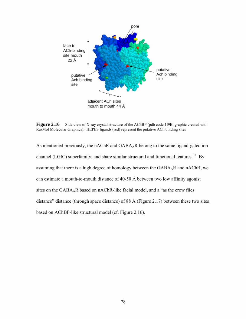
pore
face toACh-bindingsite mouth 22 Å
putativeAch binding site
putativeAch binding site
adjacent ACh sitesmouth to mouth 44 Å
Figure 2.16 Side view of X-ray crystal structure of the AChBP (pdb code 1I9B, graphic created with RasMol Molecular Graphics). HEPES ligands (red) represent the putative ACh binding sites
As mentioned previously, the nAChR and GABAAR belong to the same ligand-gated ion
channel (LGIC) superfamily, and share similar structural and functional features.37 By
assuming that there is a high degree of homology between the GABAAR and nAChR, we
can estimate a mouth-to-mouth distance of 40-50 Å between two low affinity agonist
sites on the GABAAR based on nAChR-like facial model, and a “as the crow flies
distance” distance (through space distance) of 88 Å (Figure 2.17) between these two sites
based on AChBP-like structural model (cf. Figure 2.16).
78
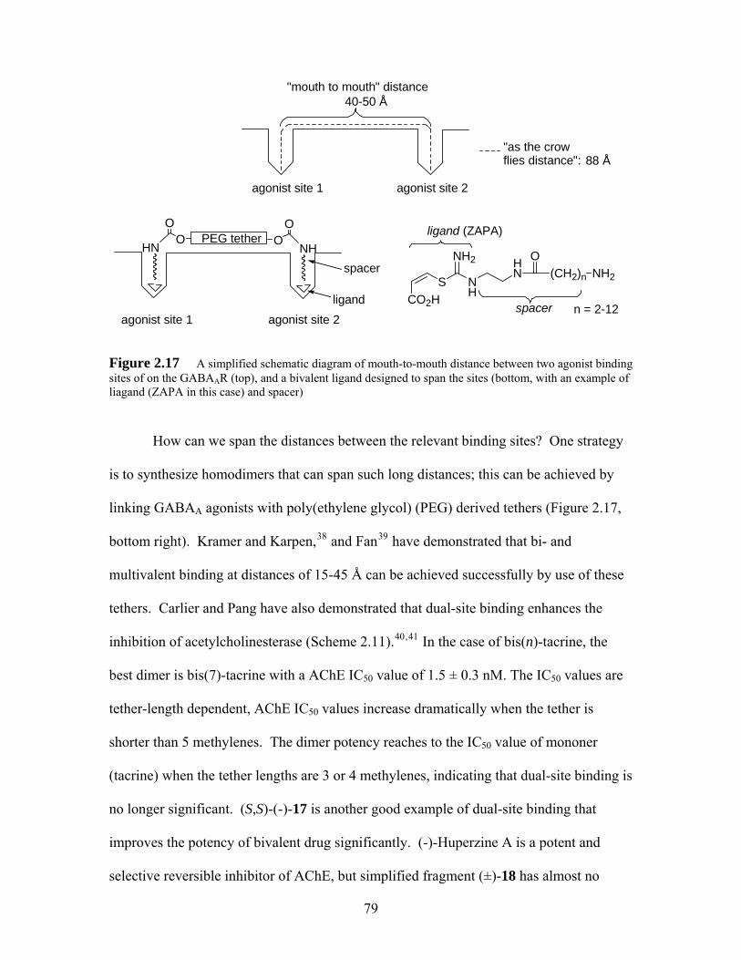
HNO
O
NHO
O
"mouth to mouth" distance
"as the crow flies distance":
agonist site 1 agonist site 2
agonist site 2
PEG tether
spacer
ligand
40-50 Å
agonist site 1
NH
S
NH2
CO2H
HN (CH2)n
ONH2
spacer
ligand (ZAPA)
n = 2-12
88 Å
Figure 2.17 A simplified schematic diagram of mouth-to-mouth distance between two agonist binding sites of on the GABAAR (top), and a bivalent ligand designed to span the sites (bottom, with an example of liagand (ZAPA in this case) and spacer)
How can we span the distances between the relevant binding sites? One strategy
is to synthesize homodimers that can span such long distances; this can be achieved by
linking GABAA agonists with poly(ethylene glycol) (PEG) derived tethers (Figure 2.17,
bottom right). Kramer and Karpen,38 and Fan39 have demonstrated that bi- and
multivalent binding at distances of 15-45 Å can be achieved successfully by use of these
tethers. Carlier and Pang have also demonstrated that dual-site binding enhances the
inhibition of acetylcholinesterase (Scheme 2.11).40,41 In the case of bis(n)-tacrine, the
best dimer is bis(7)-tacrine with a AChE IC50 value of 1.5 ± 0.3 nM. The IC50 values are
tether-length dependent, AChE IC50 values increase dramatically when the tether is
shorter than 5 methylenes. The dimer potency reaches to the IC50 value of mononer
(tacrine) when the tether lengths are 3 or 4 methylenes, indicating that dual-site binding is
no longer significant. (S,S)-(-)-17 is another good example of dual-site binding that
improves the potency of bivalent drug significantly. (-)-Huperzine A is a potent and
selective reversible inhibitor of AChE, but simplified fragment (±)-18 has almost no
79
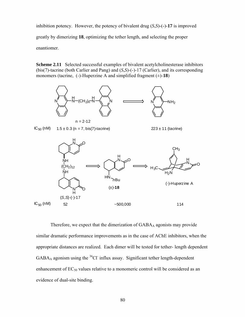
inhibition potency. However, the potency of bivalent drug (S,S)-(-)-17 is improved
greatly by dimerizing 18, optimizing the tether length, and selecting the proper
enantiomer.
Scheme 2.11 Selected successful examples of bivalent acetylcholinesterase inhibitors (bis(7)-tacrine (both Carlier and Pang) and (S,S)-(-)-17 (Carlier), and its corresponding monomers (tacrine, (-)-Huperzine A and simplified fragment (±)-18)
NHN (CH2)n
HN N N NH2
n = 2-12
I
C
50 (nM) 1.5 ± 0.3 (n = 7, bis(7)-tacrine) 223 ± 11 (tacrine)
HN O
CH3
80
NH(CH2)12NH
NH
O
HN O
HNnBu
H3C
HN O
I 50 (nM) 52 114
H2N
(-)-Huperzine A
~500,000
(±)-18
(S,S)-(-)-17
C
Therefore, we expect that the dimerization of GABAA agonists may provide
similar dramatic performance improvements as in the case of AChE inhibitors, when the
appropriate distances are realized. Each dimer will be tested for tether- length dependent
GABAA agonism using the 36Cl- influx assay. Significant tether length-dependent
enhancement of EC50 values relative to a monomeric control will be considered as an
evidence of dual-site binding.
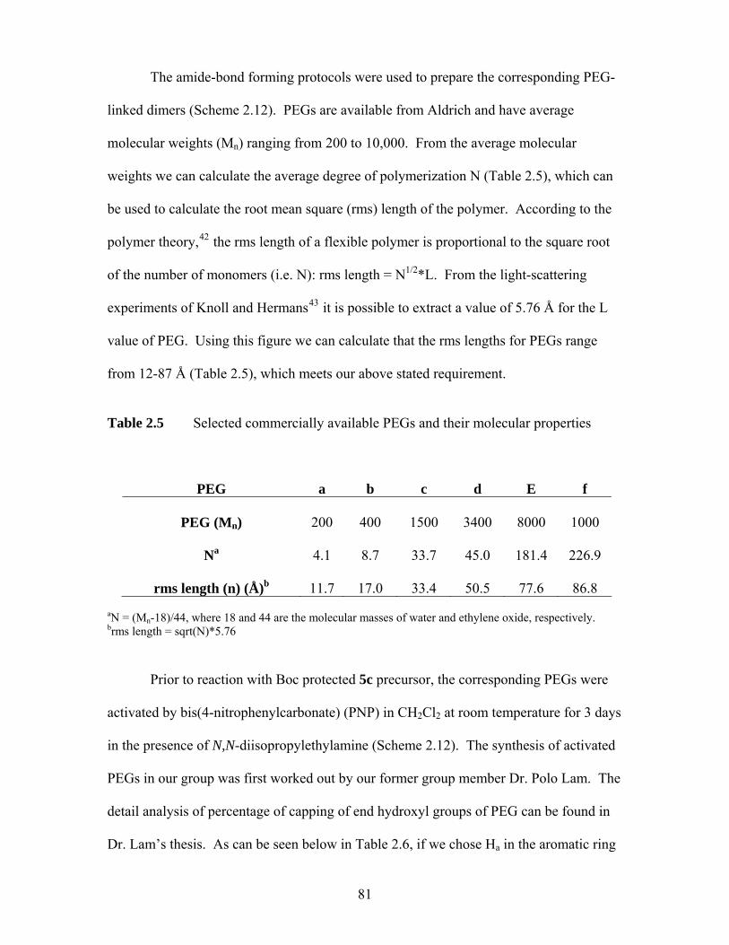
The amide-bond forming protocols were used to prepare the corresponding PEG-
linked dimers (Scheme 2.12). PEGs are available from Aldrich and have average
molecular weights (Mn) ranging from 200 to 10,000. From the average molecular
weights we can calculate the average degree of polymerization N (Table 2.5), which can
be used to calculate the root mean square (rms) length of the polymer. According to the
polymer theory,42 the rms length of a flexible polymer is proportional to the square root
of the number of monomers (i.e. N): rms length = N1/2*L. From the light-scattering
experiments of Knoll and Hermans43 it is possible to extract a value of 5.76 Å for the L
value of PEG. Using this figure we can calculate that the rms lengths for PEGs range
from 12-87 Å (Table 2.5), which meets our above stated requirement.
Table 2.5 Selected commercially available PEGs and their molecular properties
PEG
a
b
c
d
E
f
PEG (Mn)
200
400
1500
3400
8000
1000
Na
4.1
8.7
33.7
45.0
181.4
226.9
rms length (n) (Å)b
11.7
17.0
33.4
50.5
77.6
86.8
aN = (Mn-18)/44, where 18 and 44 are the molecular masses of water and ethylene oxide, respectively. brms length = sqrt(N)*5.76
Prior to reaction with Boc protected 5c precursor, the corresponding PEGs were
activated by bis(4-nitrophenylcarbonate) (PNP) in CH2Cl2 at room temperature for 3 days
in the presence of N,N-diisopropylethylamine (Scheme 2.12). The synthesis of activated
PEGs in our group was first worked out by our former group member Dr. Polo Lam. The
detail analysis of percentage of capping of end hydroxyl groups of PEG can be found in
Dr. Lam’s thesis. As can be seen below in Table 2.6, if we chose Ha in the aromatic ring
81
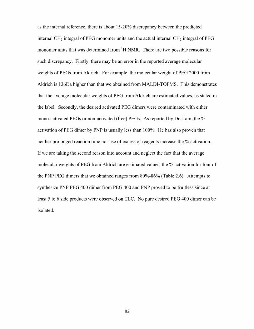
as the internal reference, there is about 15-20% discrepancy between the predicted
internal CH2 integral of PEG monomer units and the actual internal CH2 integral of PEG
monomer units that was determined from 1H NMR. There are two possible reasons for
such discrepancy. Firstly, there may be an error in the reported average molecular
weights of PEGs from Aldrich. For example, the molecular weight of PEG 2000 from
Aldrich is 136Da higher than that we obtained from MALDI-TOFMS. This demonstrates
that the average molecular weights of PEG from Aldrich are estimated values, as stated in
the label. Secondly, the desired activated PEG dimers were contaminated with either
mono-activated PEGs or non-activated (free) PEGs. As reported by Dr. Lam, the %
activation of PEG dimer by PNP is usually less than 100%. He has also proven that
neither prolonged reaction time nor use of excess of reagents increase the % activation.
If we are taking the second reason into account and neglect the fact that the average
molecular weights of PEG from Aldrich are estimated values, the % activation for four of
the PNP PEG dimers that we obtained ranges from 80%-86% (Table 2.6). Attempts to
synthesize PNP PEG 400 dimer from PEG 400 and PNP proved to be fruitless since at
least 5 to 6 side products were observed on TLC. No pure desired PEG 400 dimer can be
isolated.
82
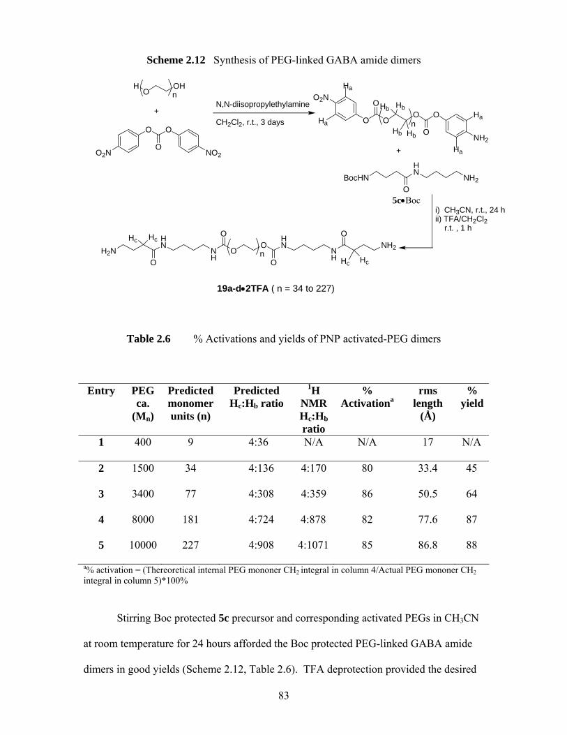
Scheme 2.12 Synthesis of PEG-linked GABA amide dimers
5c•Boc
H2NHN
NH
OO
HN
NHO
O
O
ONH2
n
BocHNHN
NH2O
+
OO
O
OnO
O
O2N
NH2
HO
OHn
+
Ha
N,N-diisopropylethylamine
CH2Cl2, r.t., 3 days
19a-d•2TFA ( n = 34 to 227)
Table 2.6 % Activations and yields of PNP activated-PEG dimers
Entry PEG ca.
(Mn)
Predicted monomer units (n)
Predicted Hc:Hb ratio
1H NMR Hc:Hb ratio
% Activationa
rms length
(Å)
% yield
1 400 9 4:36 N/A N/A 17 N/A
2 1500 34 4:136 4:170 80 33.4 45
3 3400 77 4:308 4:359 86 50.5 64
4 8000 181 4:724 4:878 82 77.6 87
5 10000 227 4:908 4:1071 85 86.8 88
a% activation = (Thereoretical internal PEG mononer CH2 integral in column 4/Actual PEG mononer CH2 integral in column 5)*100%
Stirring Boc protected 5c precursor and corresponding activated PEGs in CH3CN
at room temperature for 24 hours afforded the Boc protected PEG-linked GABA amide
dimers in good yields (Scheme 2.12, Table 2.6). TFA deprotection provided the desired
O O
OO2N NO2
i) CH3CN, r.t., 24 hii) TFA/CH2Cl2 r.t. , 1 h
Ha
Ha
Ha
Hb Hb
Hb Hb
HcHc
Hc Hc
83
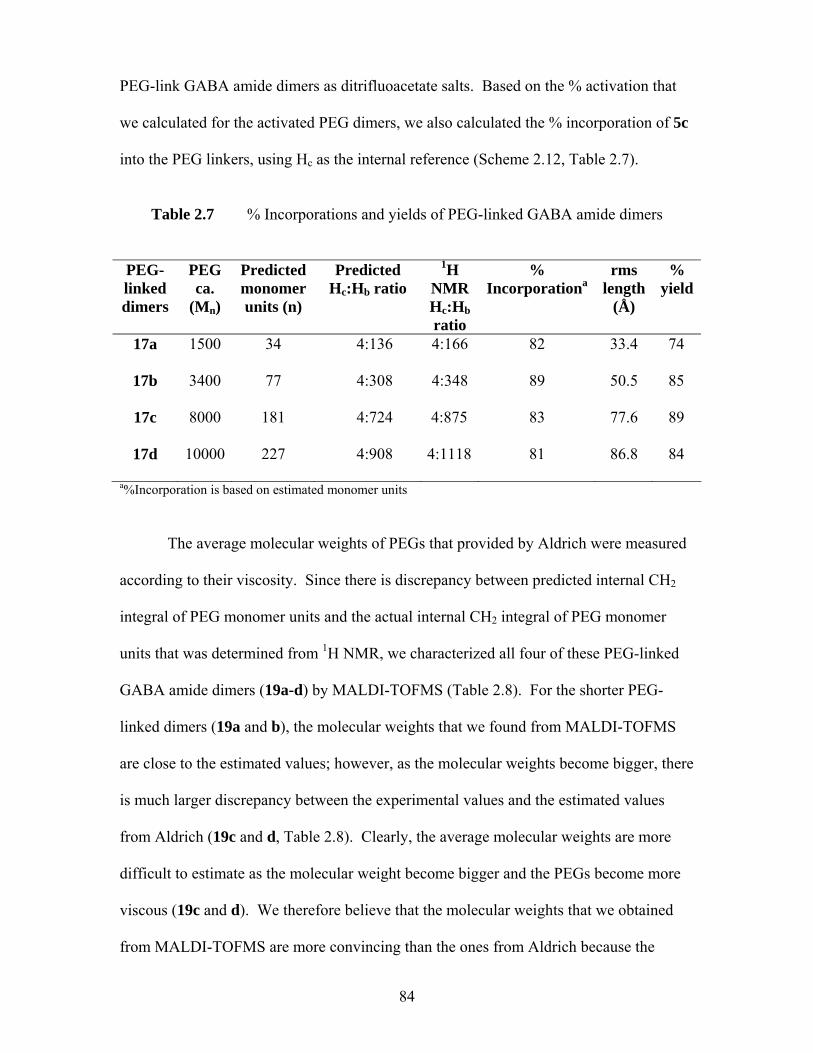
PEG-link GABA amide dimers as ditrifluoacetate salts. Based on the % activation that
we calculated for the activated PEG dimers, we also calculated the % incorporation of 5c
into the PEG linkers, using Hc as the internal reference (Scheme 2.12, Table 2.7).
Table 2.7 % Incorporations and yields of PEG-linked GABA amide dimers
PEG-linked dimers
PEG ca.
(Mn)
Predicted monomer units (n)
Predicted Hc:Hb ratio
1H NMR Hc:Hb ratio
% Incorporationa
rms length
(Å)
% yield
17a 1500 34 4:136 4:166 82 33.4 74
17b 3400 77 4:308 4:348 89 50.5 85
17c 8000 181 4:724 4:875 83 77.6 89
17d 10000 227 4:908 4:1118 81 86.8 84
a%Incorporation is based on estimated monomer units The average molecular weights of PEGs that provided by Aldrich were measured
according to their viscosity. Since there is discrepancy between predicted internal CH2
integral of PEG monomer units and the actual internal CH2 integral of PEG monomer
units that was determined from 1H NMR, we characterized all four of these PEG-linked
GABA amide dimers (19a-d) by MALDI-TOFMS (Table 2.8). For the shorter PEG-
linked dimers (19a and b), the molecular weights that we found from MALDI-TOFMS
are close to the estimated values; however, as the molecular weights become bigger, there
is much larger discrepancy between the experimental values and the estimated values
from Aldrich (19c and d, Table 2.8). Clearly, the average molecular weights are more
difficult to estimate as the molecular weight become bigger and the PEGs become more
viscous (19c and d). We therefore believe that the molecular weights that we obtained
from MALDI-TOFMS are more convincing than the ones from Aldrich because the
84
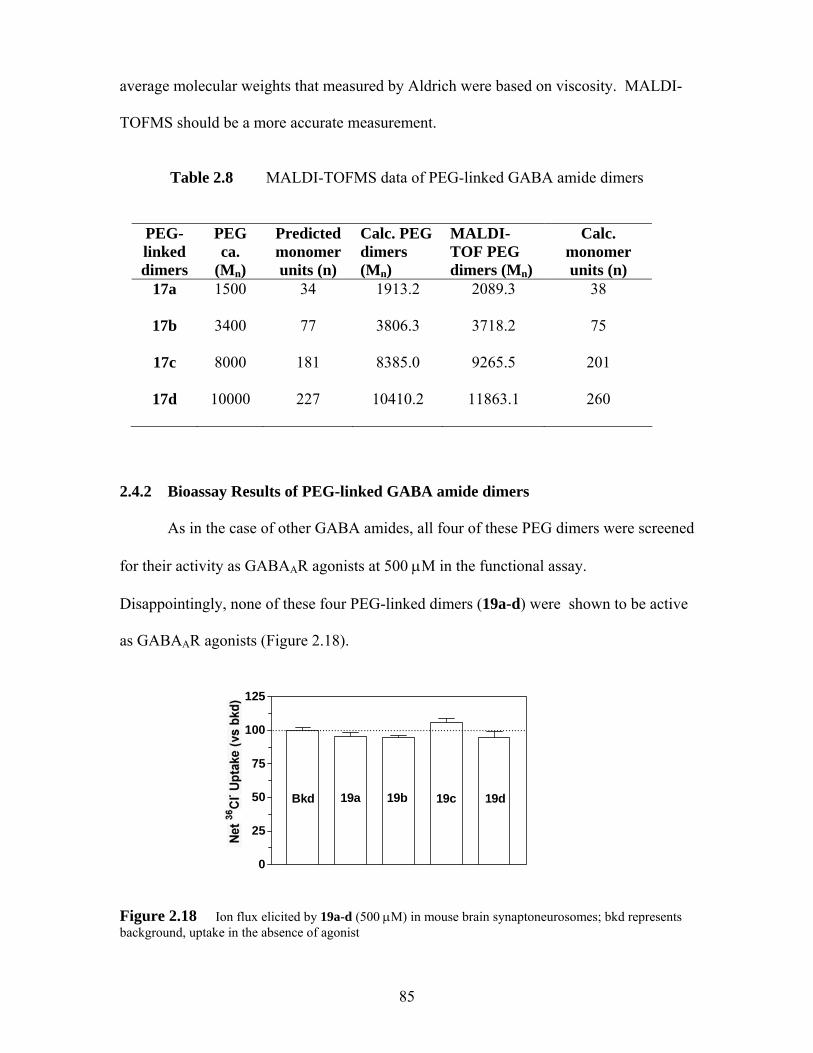
average molecular weights that measured by Aldrich were based on viscosity. MALDI-
TOFMS should be a more accurate measurement.
Table 2.8 MALDI-TOFMS data of PEG-linked GABA amide dimers
PEG-linked dimers
PEG ca.
(Mn)
Predicted monomer units (n)
Calc. PEG dimers (Mn)
MALDI-TOF PEG dimers (Mn)
Calc. monomer units (n)
17a 1500 34 1913.2 2089.3 38
17b 3400 77 3806.3 3718.2 75
17c 8000 181 8385.0 9265.5 201
17d 10000 227 10410.2 11863.1 260
2.4.2 Bioassay Results of PEG-linked GABA amide dimers
As in the case of other GABA amides, all four of these PEG dimers were screened
for their activity as GABAAR agonists at 500 μM in the functional assay.
Disappointingly, none of these four PEG-linked dimers (19a-d) were shown to be active
as GABAAR agonists (Figure 2.18).
0
25
50
75
100
125
Bkd 19a
......................................................................................
19b 19c 19d
Figure 2.18 Ion flux elicited by 19a-d (500 μM) in mouse brain synaptoneurosomes; bkd represents background, uptake in the absence of agonist
85
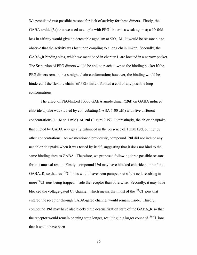
We postulated two possible reasons for lack of activity for these dimers. Firstly, the
GABA amide (5c) that we used to couple with PEG-linker is a weak agonist; a 10-fold
loss in affinity would give no detectable agonism at 500 μM. It would be reasonable to
observe that the activity was lost upon coupling to a long chain linker. Secondly, the
GABAAR binding sites, which we mentioned in chapter 1, are located in a narrow pocket.
The 5c portion of PEG dimers would be able to reach down to the binding pocket if the
PEG dimers remain in a straight chain conformation; however, the binding would be
hindered if the flexible chains of PEG linkers formed a coil or any possible loop
conformations.
The effect of PEG-linked 10000 GABA amide dimer (19d) on GABA induced
chloride uptake was studied by coincubating GABA (100 μM) with five different
concentrations (1 μM to 1 mM) of 19d (Figure 2.19). Interestingly, the chloride uptake
that elicted by GABA was greatly enhanced in the presence of 1 mM 19d, but not by
other concentrations. As we mentioned previously, compound 19d did not induce any
net chloride uptake when it was tested by itself, suggesting that it does not bind to the
same binding sites as GABA. Therefore, we proposed following three possible reasons
for this unusual result. Firstly, compound 19d may have blocked chloride pump of the
GABAAR, so that less 36Cl- ions would have been pumped out of the cell, resulting in
more 36Cl- ions being trapped inside the receptor than otherwise. Secondly, it may have
blocked the voltage-gated Cl- channel, which means that most of the 36Cl- ions that
entered the receptor through GABA-gated channel would remain inside. Thirdly,
compound 19d may have also blocked the desensitization state of the GABAAR so that
the receptor would remain opening state longer, resulting in a larger count of 36Cl- ions
that it would have been.
86
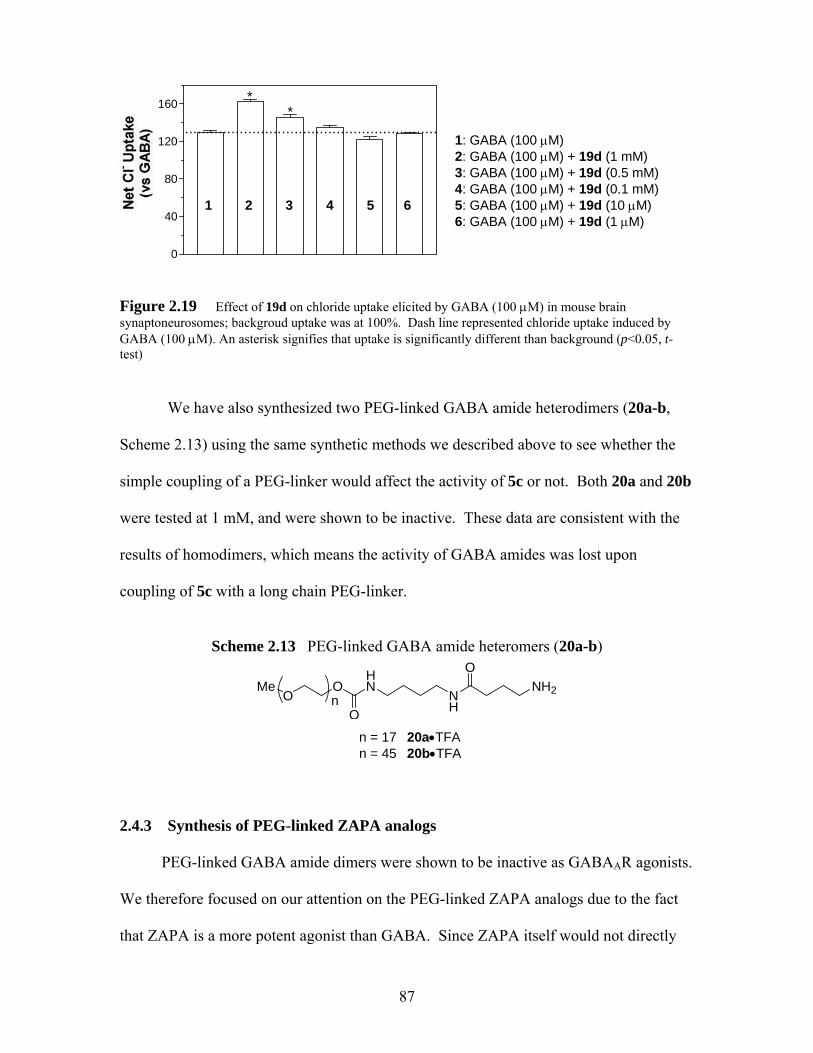
87
0
40
80
120
160
....... .. ........ ......... ......... .............. .... .. .. ...
**
1: GABA (100 μM)
2: GABA (100 μM) + 19d (1 mM) 3: GABA (100 μM) + 19d (0.5 mM) 4: GABA (100 μM) + 19d (0.1 mM) 1 2 3 4 5 6 5: GABA (100 μM) + 19d (10 μM) 6: GABA (100 μM) + 19d (1 μM) Figure 2.19 Effect of 19d on chloride uptake elicited by GABA (100 μM) in mouse brain synaptoneurosomes; backgroud uptake was at 100%. Dash line represented chloride uptake induced by GABA (100 μM). An asterisk signifies that uptake is significantly different than background (p<0.05, t-test)
We have also synthesized two PEG-linked GABA amide heterodimers (20a-b,
Scheme 2.13) using the same synthetic methods we described above to see whether the
simple coupling of a PEG-linker would affect the activity of 5c or not. Both 20a and 20b
were tested at 1 mM, and were shown to be inactive. These data are consistent with the
results of homodimers, which means the activity of GABA amides was lost upon
coupling of 5c with a long chain PEG-linker.
Scheme 2.13 PEG-linked GABA amide heteromers (20a-b)
Me
OO
HN
NHO
ONH2
n
n = 17 20a•TFA n = 45 20b•TFA
2.4.3 Synthesis of PEG-linked ZAPA analogs PEG-linked GABA amide dimers were shown to be inactive as GABAAR agonists.
We therefore focused on our attention on the PEG-linked ZAPA analogs due to the fact
that ZAPA is a more potent agonist than GABA. Since ZAPA itself would not directly
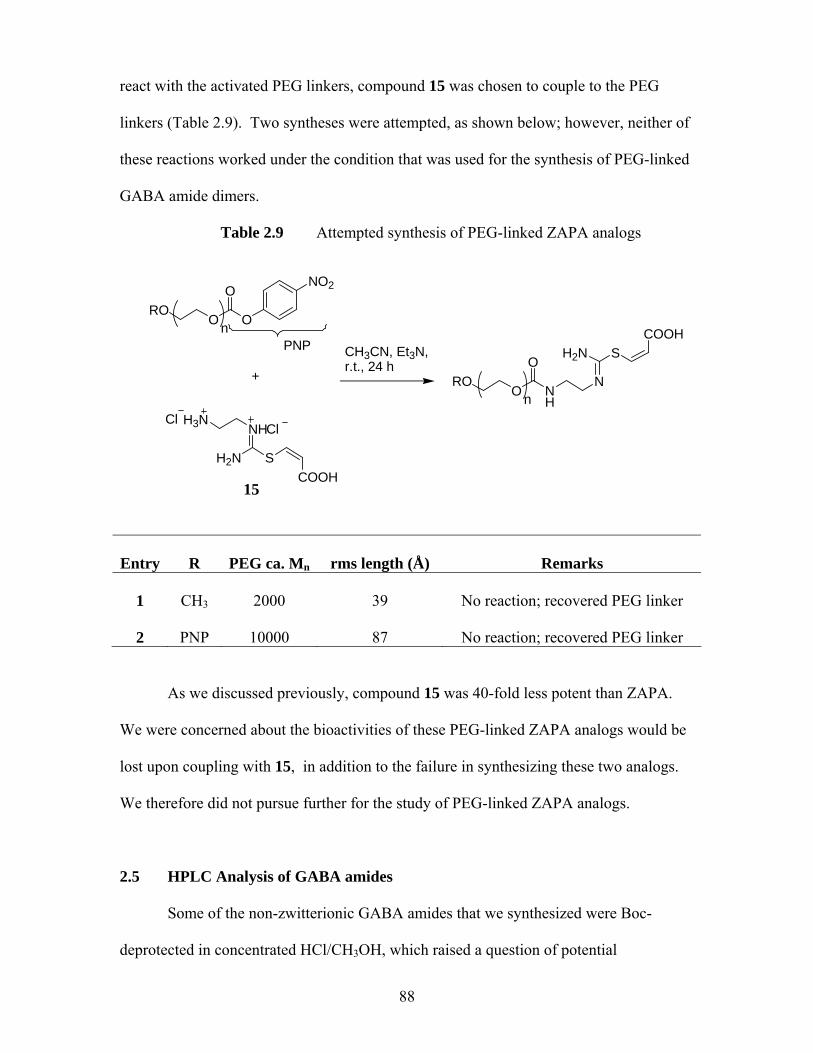
react with the activated PEG linkers, compound 15 was chosen to couple to the PEG
linkers (Table 2.9). Two syntheses were attempted, as shown below; however, neither of
these reactions worked under the condition that was used for the synthesis of PEG-linked
GABA amide dimers.
Table 2.9 Attempted synthesis of PEG-linked ZAPA analogs
15
+
ORO
O
n O
NO2
PNP
H2N S
NH
COOH
H3NCl
Cl
CH3CN, Et3N,r.t., 24 h
ORO
O
n NH
N
H2N SCOOH
Entry
R
PEG ca. Mn
rms length (Å)
Remarks
1
CH3
2000
39
No reaction; recovered PEG linker
2
PNP
10000
87
No reaction; recovered PEG linker
As we discussed previously, compound 15 was 40-fold less potent than ZAPA.
We were concerned about the bioactivities of these PEG-linked ZAPA analogs would be
lost upon coupling with 15, in addition to the failure in synthesizing these two analogs.
We therefore did not pursue further for the study of PEG-linked ZAPA analogs.
2.5 HPLC Analysis of GABA amides
Some of the non-zwitterionic GABA amides that we synthesized were Boc-
deprotected in concentrated HCl/CH3OH, which raised a question of potential
88

contamination of GABA amides with GABA. Scheme 2.14 showed how the
contamination would occur during deprotection of the Boc group, H2O would act as a
nucleophile and react with GABA amides under strong acid conditions.
Scheme 2.14 Proposed mechanism for acid-catalyzed Boc deprotection of GABA amides. Boc protected 5a was used as an example
5a•Boc
NH
HN
OO
O
H2Nconc. HCl,MeOH
NH
HN
OO
OH
H2N
H
H2O
NH
HN
OO
OH
H2N + HCl(aq)
NH
NH3
OH2N
carbamic acid
Cl
HCl(aq)
NH
NH3
OHH2N Cl
OH2
NH3
OCl
HO + H3NNH3Cl
Cl
+CO2
If this decomposition occurred, our GABA amide samples would be contaminated with
GABA. Thus the weak agonism displayed by GABA amides would be due to the
presence of trace of GABA. To rule out the possibility, we assayed our GABA amides
for free GABA using Saller’s modified procedure, we analyzed some of the GABA
amides by HPLC to ensure that these GABA amides were GABA free (<0.1 wt%).
Refluxing GABA or GABA amides with dansyl chloride in NaHCO3 and acetone/CH2Cl2
for 0.5 h to 12 h and followed by column chromatography (9:1 CH2Cl2-CH3OH)
afforded the desired dansylated products in good yields for GABA or mono-dansylated
GABA amides (Scheme 2.15).
89
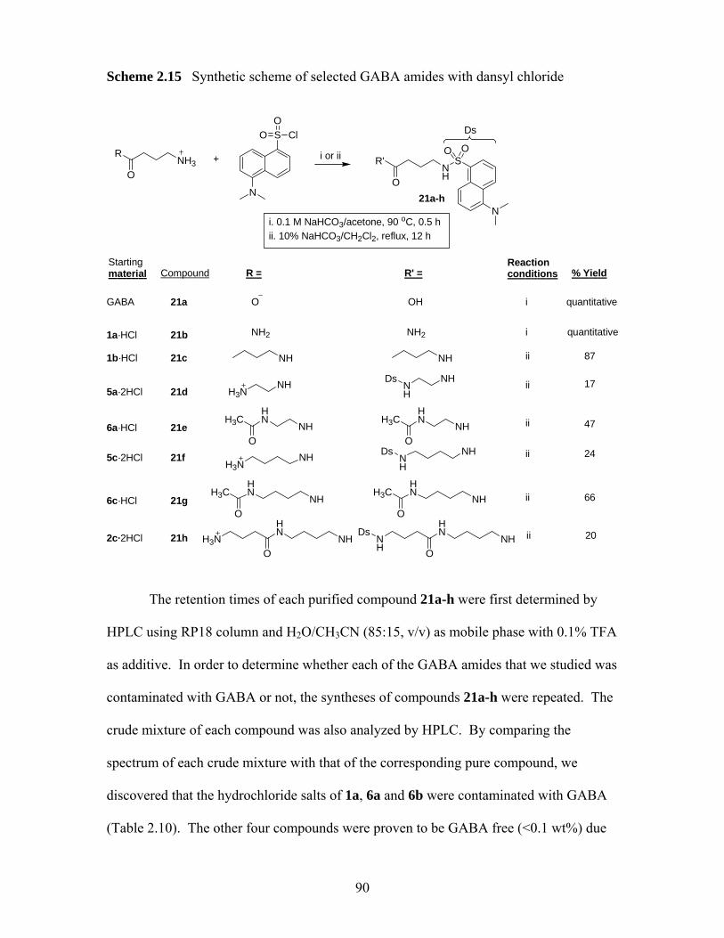
Scheme 2.15 Synthetic scheme of selected GABA amides with dansyl chloride
NH3R
O
N
S ClOO
+ i or iiNH
R'
O
SO O
N
Ds
i. 0.1 M NaHCO3/acetone, 90 oC, 0.5 hii. 10% NaHCO3/CH2Cl2, reflux, 12 h
90
Sm
tarting aterial R = R' =
Reactionconditions % Yield
GABA
·HCl
·HCl
·2HCl
·HCl
·2HCl
·HCl
·2HCl
O
1a
1b
5a
6a
5c
6c
2c
NH2
NH
H3NNH
HNH3C
O
H3NNH
HNH3C
O
NH
NH
H3NHN
NHO
OH
NH2
NH
NH
NHDs
HNH3C
ONH
NH
NH
HNH3C
ONH
NH
HN
NHO
Ds
Ds
i
i
ii
ii
ii
ii
ii
ii
quantitative
quantitative
87
17
47
24
66
20
Compound
21a-h
21a
21b
21c
21d
21e
21f
21g
21h
The retention times of each purified compound 21a-h were first determined by
HPLC using RP18 column and H2O/CH3CN (85:15, v/v) as mobile phase with 0.1% TFA
as additive. In order to determine whether each of the GABA amides that we studied was
contaminated with GABA or not, the syntheses of compounds 21a-h were repeated. The
crude mixture of each compound was also analyzed by HPLC. By comparing the
spectrum of each crude mixture with that of the corresponding pure compound, we
discovered that the hydrochloride salts of 1a, 6a and 6b were contaminated with GABA
(Table 2.10). The other four compounds were proven to be GABA free (<0.1 wt%) due
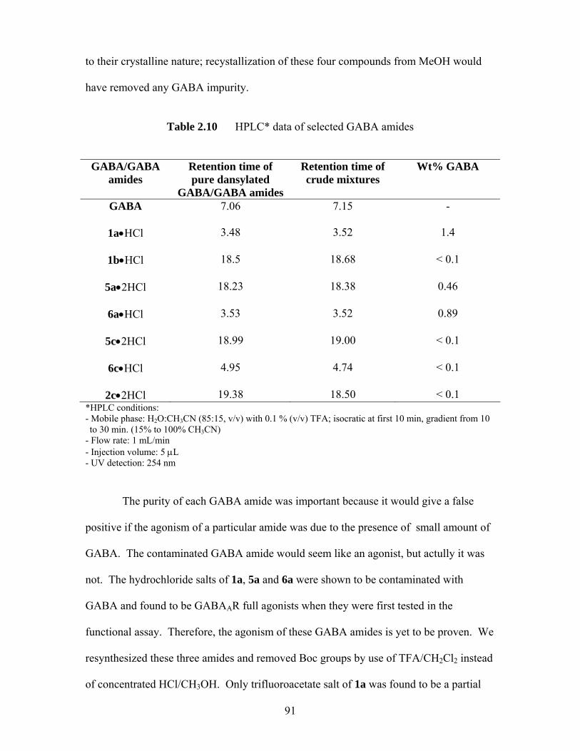
to their crystalline nature; recystallization of these four compounds from MeOH would
have removed any GABA impurity.
Table 2.10 HPLC* data of selected GABA amides
GABA/GABA
amides
Retention time of pure dansylated
GABA/GABA amides
Retention time of crude mixtures
Wt% GABA
GABA
7.06 7.15 -
1a•HCl
3.48 3.52 1.4
1b•HCl
18.5 18.68 < 0.1
5a•2HCl
18.23 18.38 0.46
6a•HCl
3.53 3.52 0.89
5c•2HCl
18.99 19.00 < 0.1
6c•HCl
4.95 4.74 < 0.1
2c•2HCl 19.38 18.50 < 0.1 *HPLC conditions: - Mobile phase: H2O:CH3CN (85:15, v/v) with 0.1 % (v/v) TFA; isocratic at first 10 min, gradient from 10 to 30 min. (15% to 100% CH3CN)
- Flow rate: 1 mL/min - Injection volume: 5 μL - UV detection: 254 nm
The purity of each GABA amide was important because it would give a false
positive if the agonism of a particular amide was due to the presence of small amount of
GABA. The contaminated GABA amide would seem like an agonist, but actully it was
not. The hydrochloride salts of 1a, 5a and 6a were shown to be contaminated with
GABA and found to be GABAAR full agonists when they were first tested in the
functional assay. Therefore, the agonism of these GABA amides is yet to be proven. We
resynthesized these three amides and removed Boc groups by use of TFA/CH2Cl2 instead
of concentrated HCl/CH3OH. Only trifluoroacetate salt of 1a was found to be a partial
91
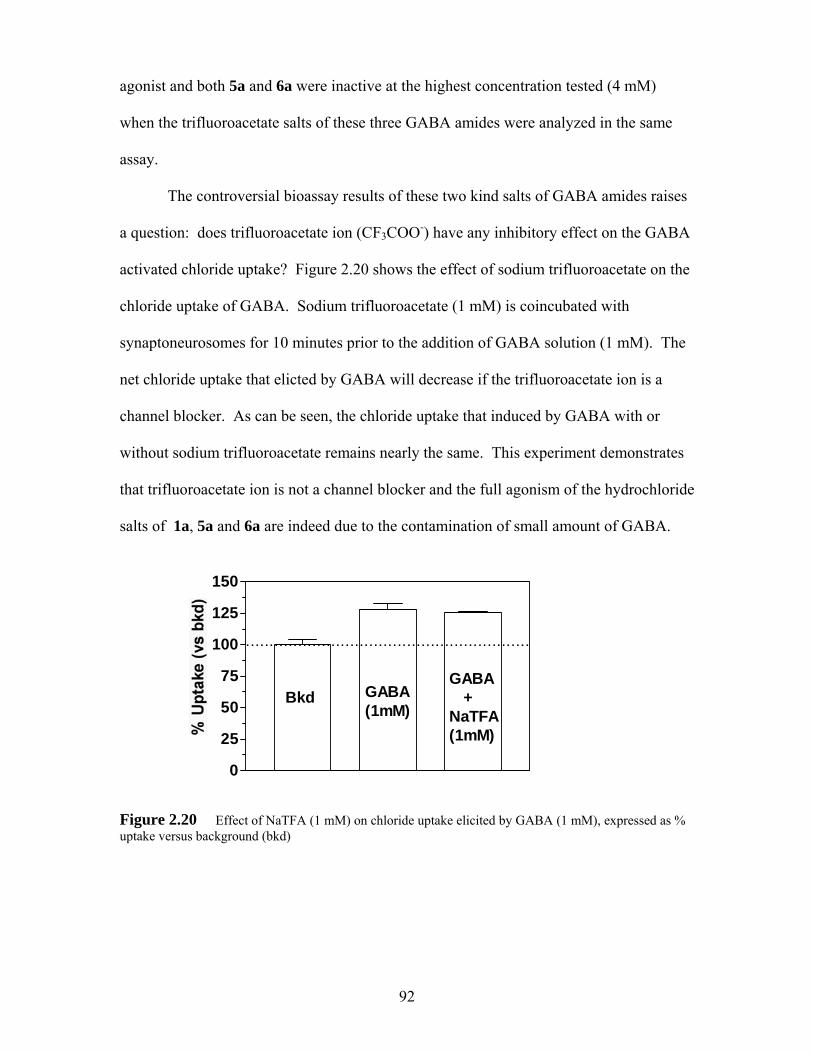
agonist and both 5a and 6a were inactive at the highest concentration tested (4 mM)
when the trifluoroacetate salts of these three GABA amides were analyzed in the same
assay.
The controversial bioassay results of these two kind salts of GABA amides raises
a question: does trifluoroacetate ion (CF3COO-) have any inhibitory effect on the GABA
activated chloride uptake? Figure 2.20 shows the effect of sodium trifluoroacetate on the
chloride uptake of GABA. Sodium trifluoroacetate (1 mM) is coincubated with
synaptoneurosomes for 10 minutes prior to the addition of GABA solution (1 mM). The
net chloride uptake that elicted by GABA will decrease if the trifluoroacetate ion is a
channel blocker. As can be seen, the chloride uptake that induced by GABA with or
without sodium trifluoroacetate remains nearly the same. This experiment demonstrates
that trifluoroacetate ion is not a channel blocker and the full agonism of the hydrochloride
salts of 1a, 5a and 6a are indeed due to the contamination of small amount of GABA.
0
25
50
75
100
125
150
Bkd GABA(1mM)
GABA +NaTFA(1mM)
............................................................
Figure 2.20 Effect of NaTFA (1 mM) on chloride uptake elicited by GABA (1 mM), expressed as % uptake versus background (bkd)
92

2.6 Conclusions
In chapter 2.1-2.2, we described the syntheses and bioassay results of non-
zwitterionic GABA amides. We first demonstrated that appropriately functionalized
GABA amides are partial, full and superagonists, despite their non-zwitterionic
structures. Note that none of these GABA amides has been tested in vivo assay, but the
agonism of these GABA amides is comparable to that of THIP, as shown by in vitro
assay results. The bioassay results of all three series of GABA amides showed that the
agonism of these amides are tether-length dependent, with a optimum tether length
appearing to be four methylenes. Based on the agonism of both homodimers and
heterodimers, we observed that the pendant amide group (2c and 6c) seemed to be
superior to the ammonium group (5c). We proposed that superior activity of 2c and 6c
over 5c the was due to the presence of a proximal subsite to the agonist binding site that
can weakly bind the pendant amide group.
In chapter 2.1, we focused on the syntheses and bioassay results of GABA amide
homodimers (2a-i). Compound 2c which has a flexible four methylene tether was found
to be the most potent dimer among this series of compounds. Compound 2c was a
‘superagonist’, and both co-application with GABA and [3H]musicimol binding
experiments demonstrated that 2c binds to the same site as GABA. Replacing the
flexible four methylenes tether of 2c by eight more rigid tethers (4a-h) revealed a
significant loss of activity compared with the original compound 2c. Six derivatives of
2c were found to be competitive antagonists. [3H]musicimol binding experiments further
confirmed the competitive antagonism of one of these antagonists (4b). Structural
modifications of 2c demonstrated the importance of flexible tether length for the agonism
of these GABA amide homodimers.
93

In chapter 2.2, the syntheses and bioassay results of two series of simple GABA
amide heterodimers were described. Compound 6, which has a pendant amide group was
found to be the best heterodimer among these two series of compounds. Optimization of
pendant amide group by other nine groups (7a-i) revealed a significant loss of activity
compared with the original compound 6c. Structural modifications of 6c demonstrated
that the amide group is important in the investigated bioactivity. It also showed that the
proximal binding site for the pendant amide is small. The superior activity of 6c over 7c
(note they differ only in that 6c has an additional amine group) must be due to another
complementary binding interaction of the amino group in 6c.
In chapter 2.3, we described the syntheses and first assays of four ZAPA
derivatives in the Cl- uptake functional assay. The agonism of these four ZAPA
derivatives was shown to be related to the size of the alkyl groups. The agonism of these
derivatives increased as the alkyl group became longer and the potency was greatly
improved as the alkyl group changed from hydrophilic to hydrophobic groups.
The syntheses and bioassay data of PEG-linked GABA amide homodimers were
described in chapter 2.4. PEG-linked dimers were designed to span the distances
between two low-affinity binding sites. However, little information is known about the
binding sites on the GABAAR and lack of crystal structure have made this approach
fruitless. GABA amide 5c lost activity upon coupling to PEG linkers. PEG-linked
GABA amide heterodimers were also shown to be inactive. Attempted to synthesize
PEG-linked ZAPA derivatives did not yield any desired products.
Finally, in chapter 2.5, the purity of some GABA amides were analyzed by HPLC
based on modification of Saller’s procedure (assay of the dansyl sulfonamide
derivatives). Three of the small GABA amides (1a, 5a and 6a) that we studied were
found to be contaminated with GABA impurity (>0.1 wt%). The contaminated GABA
94
amides gave false positive assay results, which demonstrated the importance of the purity
of GABA amides in bioassay experiments.
References and notes for chapter 2 1 Jones, M. V.; Shar, Y.; Dzubay, J. A.; Westbrook, G. L. Defining affinity with the
GABAA receptor. J. Neurosci. 1998, 18, 8590-8604. 2 Jones proposed that β-alanine, muscimol, and GABA bound to the GABAAR in their
fully extended conformations; the quoted 4.7-6.0 Å range spans the intercharge distances in these molecules.
3 Breckenridge, R. J.; Nicholson, S. H.; Nichol, A. J.; Suckling, C. J.; Leigh, B.;
Iversen, L. Inhibition of [3H] GABA Binding to Postsynaptic Recetors in Human Cerebellar Synaptic Membranes by Carboxyl and Amino Derivatives of GABA. J Neurochem. 1981,37, 837-844.
4 Varga, V.; Kontro, P.; Oja, S. S. Modulation of GABAergic Neurotransmission in the
Brain by Dipetides. Neurochem. Res. 1988, 13, 1027-1034. 5 Varga et al. showed that preincubation (5 min) of GABA-His and GABA-Gly with
mouse brain synaptosomal preparations enhanced 0.1 mM GABA-induced chloride flux. However, in the absence of GABA these compounds did not induce chloride flux.
6 Silman, I.; Karlin, A. Acetylcholine receptor covalent attachment of depolarizing
groups at the active site. Science (Washington, D. C.) 1969, 164, 1420-1421. 7 Li, L.; Zhong, W.; Zacharias, N.; Gibbs, C.; Lester, H. A.; Dougherty, D. A. The
tethered agonist approach to mapping ion channel proteins-toward a structural model for the agonist binding site of the nicotinic acetylcholine receptor. Chem. Biol. 2001, 8, 47-58.
8 Carlier, P. R.; Chow, E. S.-H.; Barlow, R. L.; Bloomquist, J. R. Discovery of Non-
Zwitterionic GABAA Receptor Full Agonists and a Superagonist. Bioorg. Med. Chem. Lett. 2002, 12, 1985-1988.
9 Flynn, D. L.; Zelle, R. E. Grieco, P. A. A Mild Two-Step Method for the Hydrolysis
/Methanolysis of Secondary Amides and Lactams. J. Org. Chem. 1983, 48, 2424-2427.
10 Bloomquist, J. R.; Soderlund, D. M. Neurotoxic Insecticides Inhibit GABA-
Dependent Chloride Uptake by Mouse Brain Vesicles. Biochem. Biophys. Res. Commun. 1985, 133, 37-43.
95
11 Saller, C. F.; Czupryna, M. J. γ-Aminobutyric acid, glutamate, glycine and taurine
analysis using revesed-phased high-performance liquid chromatography and ultraviolet detection of dansyl chloride derivatives. J. Chromatogr. 1989, 487, 167-172.
12 Luu, M. D.; Morrow, A. L.; Paul, S. M.; Schwartz, R. D. Characterization of GABAA
Receptor-mediated 36Chloride Uptake in Rat Brain Synaptosomes. Life Sci. 1987, 41, 1277-1287.
13 Schuda, P. F.; Greenlee, W. J.; Chakravarty, P. K. A Short and Efficient Synthesis of
(3S,4S)-4-[(tert-Butyloxycarbonyl)amino]-5-cyclohexyl-3-hydroxypentanoic Acid Ethyl Ester. J. Org. Chem. 1988, 53, 873-875.
14 Kawahara, S.-I.; Uchimaru, T. One-Pot Preparation of o-Xylylene Diamine and its
Related Amines. Zeit. fur Nat. B: Chem. Sci. 2000, 55, 985-987. 15 Compounds 3a-h were screened at 1 mM; they were pre-incubated with mouse brain
synaptoneurosomes (200 μL) for 10 minutes at room temperature. These compounds were then co-incubated with GABA (0.1 mM in 36Cl- flux buffer) for 15 seconds at 30°C.
16 DeLorey, T. M.; Brown, G. B. γ-Aminobutyric acidA Receptor Pharmacology in Rat
Cerebral Cortical Synaptoneurosomes. J. Neurochem. 1992, 58, 2162-2169. 17 Edgar, P. P.; Schwartz, R. D. Functionally Relevant γ–Aminobutyric AcidA
Receptors: Equivalence between Receptor Affinity (Kd) and Potency (EC50)? Mol. Pharmacol. 1992, 41, 1124-1129.
18 Johston, G. A. R.; Allan, R. D.; Kennedy, S. M. E.; Twitchin B. In GABA- Neuro-
Transmitters; Krogsgaard-Larsen, P.; Scheel-Kruger, J.; Kofod, H.; Eds.; Munkgaard: Copenhagen, 1979, 149-164.
19 Krogsgaard-Larsen, P.; Frolud, B.; Jorgensen, F.S.; Shousaboe, A. GABAA Receptor
Agonists, Partial Agonists, and Antagonists. Design and Therapeutic Propects. J. Med. Chem. 1994, 37 (16), 2489-2505.
20 Larsen, S. D.; Connell, M. A.; Cudahy, M. M.; Evans, B. R.; May, P. D.; Meglasson,
M. D.; O’Sullivan, T. J.; Schostarez, H. J.; Sih, J. C.; Stevens, F. C.; Tanis, S. P.; Tegley, C. M.; Tucker, J. A.; Vaillancourt, V. A.; Vidmar, T. J.; Watt, W.; Yu, J. H. Synthesis and biological activity of analogues of the antidiabetic/antiobesity agent 3-guanidinopropionic acid: discovery of a novel aminoguanidinoacetic acid antidiabetic agent. J. Med. Chem. 2001, 44, 1217-1230.
21 Allan, R. D.; Dickenson, H. W.; Hiern, B. P.; Johnson, G. A. R.; Kazlauskas, R.
Isothiouronium compounds as γ–aminobutyric acid agonists. Br. J. Pharmacol. 1986, 88, 379-387.
96
22 Allan, R. D.; Dickenson, H. W.; Johnston, G. A. R.; Kazlauskas, R.; Mewett, K. N.
Structural Analogues of ZAPA as GABAA Agonists. Neurochem. Int. 1997, 30, 583-591.
23 Takagi, S.; Tanaka, H.; Yano, M.; Machida, K. Studies on the Syntheses of
Polymethylene-bisthioureas and their Derivatives. I. Chem. Phar. Bull, 1959, 7, 206-208.
24 Poss, M. A.; Iwanowicz, E.; Reld, J. A.; Lin. J.; Gu Z. A Mild and Efficient Method
for the Preparation of Guanidines. Tetrahedron Lett. 1992, 33, 5933-5936. 25 Rasmussen, C. R.; Villani, F. J.; Weaner, L. E.; Reynolds, B. E.; Hood, A. R.;
Hecker, L. R.; Nortey, S. O.; Hanslin, N. A.; Costanzo, M .J.; Powell, E. T.; Molinari, A. J. Improved Procedures for the preparation of Cycloalkyl-, aryalkyl- and Arylthioureas. Synthesis, 1988, 456-459.
26 Tobe, Y.; Sasaki, S.-I.; Mizuno, M.; Hirose, K.; Naemura, K. Novel Self-Assembly of
m-Xylylene Type Dithioureas by Head -to-Tail Hydrogen Bonding. J. Org. Chem. 1998, 63, 7481-7489.
27 Buschauer, A. Synthesis and in vitro pharmacology of arpromidine and related
phenyl(pyridylalkyl)guanidines, a potential new class of positive inotropic drugs. J. Med. Chem. 1989, 32, 1963-1970.
28 Perkins, J. J.; ZarTman, A .E.; Meissner, R. S. Synthesis of 2-
(Alkylamino)benzimidazoles. Tetrahedron lett. 1999, 40, 1103-1106. 29 Nannini, G.; Perrone, E.; Severino, D.; Bedeschi, A.; Biasoli, G. Synthesis and
Structure-Activity Relationships of New 7-Vinylenethioacetamido and Thioacrylamindo Cephalosporins. J. Antibiot. 1981, 34, 412-426.
30 Kash, T. L.; Trudell, J. R.; Harrison, N. L. Structural elements involved in activation
of the γ-aminobutryic acid type A (GABAA) receptor. Biochem. Soc. Trans. 2004, 32, 540-546.
31 Baumann, S. W.; Baur, R.; Sigel, E. Individual properties of the two functional
agonist sites in GABAA receptors. J. Neurosci. 2003, 23, 11778. 32 Nayeem, N.; Green, T. P.; Martin, I. L.; Barnard, E. A. Quaternary Structure of the
Native GABAA Receptor Determined by Electron Microscopic Image Analysis. J. Neurochem. 1994, 62, 815-818.
33 Miyazawa, A.; Fuiyoshi, Y.; Stowell, M.; Unwin , N. Nicotinic Acetylcholine
Receptor at 4.6 Å Resolution: Transverse Tunnels in the Channel Wall. J. Mol. Biol. 1999, 288, 765-786.
97

34 Unwin, N. Acetylcholine receptor channel imaged in the open state. Nature, 1995,
373, 37-43. 35 Arias, H. R. Topology of ligand binding sites on the nicotinic acetylcholine receptor.
Brain, Res. Rev. 1997, 25, 133-191. 36 Brejc, K.; van Dijk, W. J.; Klaassen, R. V.; Schuurmans, M.; van der Oost, J.; Smit,
A. B.; Sixma, T. K. Crystal structure of an ACh-binding protein reveals the ligand-binding domain of nicotinic receptors. Nature, 2001, 411, 269-276.
37 Smith, G. B.; Olsen, R. W. Functional domains of GABAA receptors. Trends
Pharmacol. Sci. 1995, 16, 162-168. 38 Kramer, R. H.; Karpen, J. W. Spanning binding sites on allosteric proteins with
polymer-linked dimers. Nature 1998, 710-713. 39 Fan, E.; Zhang, Z.; Minke, W. E.; Hou, Z.; Verlinde, L. C.; Hol, W. G. J. High
affinity Pentavalent Ligands of Escherichia coli Heat-Labile Enterotoxin by Modulator Structure-Based Design. J. Am. Chem. Soc. 2000, 122, 2663-2664.
40 Carlier, P. R.; Han, Y. F.; Chow, E. S.-H.; C. Li, P.-L; Wang, H.; Lieu, T. X.;
Wong,H. S.; Pang, Y.-P. Evaluation of Short-tether Bis-THA AChE Inhibitors. A Further Test of the Dual Binding Site Hypothesis. Bioorg. Med. Chem. 1999, 7, 351-357.
41 Carlier, P. R.; Du, D.-M.; Han, Y.-F.; Liu, J.; Perola, E.; Williams, I. D.; Pang Y.-P.
Dimerization of an Inactive Fragment of Huperzine A Produces a Drug with twice Potency of the Natural Product. Angew. Chem. Int. Ed. in English 2000, 39, 1775-1777.
42 Hiemenz, P. C. Polymer Chemistry: The Basic Concepts; Marcel Dekker: New York,
1984, 48. 43 Knoll, D.; Hermans, J. J. Polymer-Protein Interactions. J. Biol Chem. 1983, 258.
5710-5715.
98
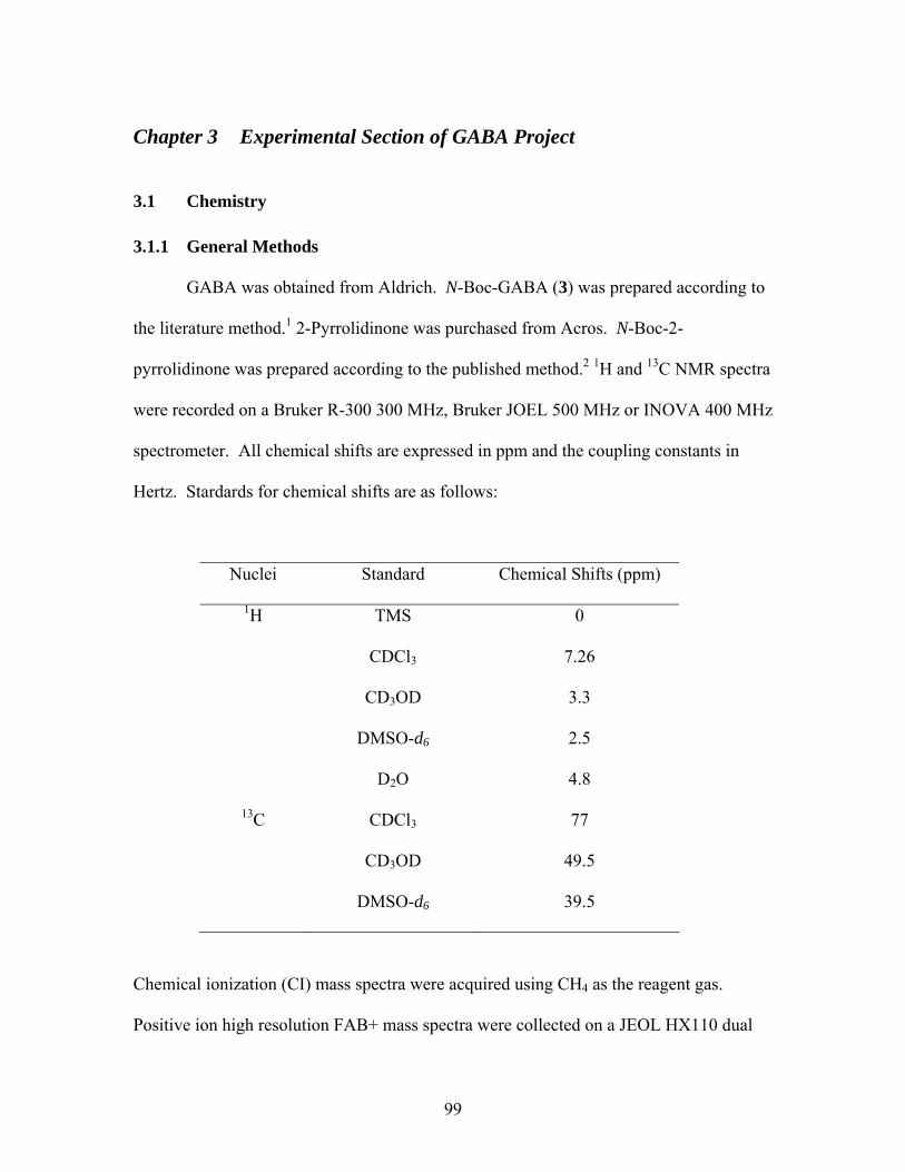
Chapter 3 Experimental Section of GABA Project
3.1 Chemistry 3.1.1 General Methods
GABA was obtained from Aldrich. N-Boc-GABA (3) was prepared according to
the literature method.1 2-Pyrrolidinone was purchased from Acros. N-Boc-2-
pyrrolidinone was prepared according to the published method.2 1H and 13C NMR spectra
were recorded on a Bruker R-300 300 MHz, Bruker JOEL 500 MHz or INOVA 400 MHz
spectrometer. All chemical shifts are expressed in ppm and the coupling constants in
Hertz. Stardards for chemical shifts are as follows:
Nuclei Standard Chemical Shifts (ppm)
1H TMS 0
CDCl3 7.26
CD3OD 3.3
DMSO-d6 2.5
D2O 4.8
13C CDCl3 77
CD3OD 49.5
DMSO-d6 39.5
Chemical ionization (CI) mass spectra were acquired using CH4 as the reagent gas.
Positive ion high resolution FAB+ mass spectra were collected on a JEOL HX110 dual
99
focusing mass spectrometer using a direct inlet with 3-nitrobenzyl alcohol as the sample
matrix. Elemental analysis was performed by Atlantic Microlab, Inc.
Analytical thin-layer chromatography (TLC) was performed with aluminum
sheets coated with RDH silica gel 60 F254. Flash column chromatography was
performed using VWR silica gel 60 (60-240 mesh). High-performance liquid
chromatography (HPLC) analysis was performed on a XterrraTM RP18 column (5.6 x 150
mm, 5 μM), detection at 254 nM, flow rate 1.0 mL/min with isocratic at first 10 minutes
and gradient in the next 20 minutes. Elution program: H2O:CH3CN (85/15, v/v) at 0-10
minutes with 0.1 % TFA as additive; 15-100% CH3CN at 10-30 minutes with 0.1 % TFA
as additive.
3.1.2 Procedures Synthesis of 4-amino-butyramide trifluoroacetic acid salt (gabamide, 1a)
N-Boc-2-pyrrolidinone (330.1 mg, 1.78 mmol) and concentrated NH3 (29.3 wt %,
10 mL) were combined and heated to reflux for 2 hours. After cooling to room
termperature. the mixture was diluted with CH2Cl2 (50 mL), which was then extracted
with H2O (40 mL). The CH2Cl2 layer was collected and the aqueous layer was extracted
with CH2Cl2 (3 x 50 mL). The combined CH2Cl2 layers were dried over Na2SO4, filtered
and evaporated under reduced pressure to afford the corresponding Boc-protected 1a as
white solid. The TFA salt was prepared by dissolving the white solid in CH3OH (1mL),
followed by addition of trifluoroacetic acid (2 mL). The resulting solution was stirred at
room temperature for 1 hour, and concentrated in vacuum to yield the title compound as a
brown oily liquid (317.9 mg, 88%). 1H NMR (CD3OD): δ 1.91 (5-let, J = 7.5 Hz, 2H),
100
2.35 (t, J = 7.2 Hz, 2 H), 2.96 (t, J = 7.6 Hz, 2 H); 13C NMR (CD3OD): δ 24.75, 33.58,
40.88, 177.79; MS (FAB+): calcd for C4H11N2O [M+1]+, 103.0866, found 103.1
Synthesis of N-4-aminobutyl butyramide hydrochloride salt (1b)
N-Boc-2-pyrrolidinone (524 mg, 2.83 mmol) and n-butylamine (1.1 mL, 8.49
mmol) were combined in dioxane (10 mL) and heated to reflux for overnight. After
cooling to room termperature, the mixture was diluted with CH2Cl2 (25 mL), which was
then extracted with H2O (2 x 25 mL). The CH2Cl2 layer was collected, dried over
MgSO4, filtered and evaporated under reduced pressure to give a crude brown residue.
Purified by flash column chromatography (10% MeOH-CH2Cl2 with 7 mL of
concentrated NH3 per liter) on silica gel afforded corresponding Boc-N protected 1b as
pale yellow solid. The HCl salt was prepared by redissolving the white solid in 5 mL of
CH3OH, followed by addition of HCl (37 % in H2O, 1 mL). The resulting solution was
stirred at room temperature for 2 h, and concentrated in vacuum to yield the title
compound as yellow solid (294 mg, 53 %). 1H NMR (CD3OD): δ 0.93 (t, J = 7.4 Hz,
3H), 1.32-1.39 (m, 2 H), 1.44-1.50 (m, 2 H), 1.91 (5-let, J = 7.3 Hz, 2H), 2.34 (t, J = 7.2
Hz, 2H), 2.96 (t, J = 7.6 Hz, 2H), 3.17 (t, J = 7.0 Hz, 2H); 13C NMR (CD3OD): δ 14.57,
21.58, 25.05, 32.97, 34.28, 40.70, 40.93, 174.88; MS (FAB+): calcd for C8H19N2O
[M+1]+ 159.1497, found 159.1493 (-2.7 ppm, -0.4 mmu).
Synthesis of N-(4’-aminobutanoyl)aniline trifluoroacetate (1c•TFA)
To a solution of Boc-GABA (624 mg, 3.07 mmol), EDCI (631 mg, 3.29 mmol)
and DMAP (295 mg, 2.41 mmol) in CH2Cl2 (10 mL) was added aniline (100 μL, 1.10
101
mmol). The yellow mixture was then stirred at room temperature for 12 hours. The
mixture was partitioned between CH2Cl2 (1 x 50 mL) and H2O (40 mL). The CH2Cl2
layers was collected, dried over Na2SO4, filtered and evaporated under reduced pressure.
Purified by flash column chromatography (5% MeOH-CH2Cl2 with 7 mL of concentrated
NH3 per liter) on silica gel afforded corresponding Boc-N protected 1c as a yellow solid.
The TFA salt was prepared by dissolving the yellow solid in CH2Cl2 (5mL), followed by
addition of trifluoroacetic acid (2 mL). The resulting solution was stirred at room
temperature for 1 hour, and concentrated in vacuo to yield the title compound 1c as a pale
yellow solid (256.5 mg, 95 %). 1H NMR (CD3OD): δ 2.00 (5-let, J = 7.1 Hz, 2H), 2.53
(t, J = 7.0 Hz, 2H), 3.01 (t, J = 7.6 Hz, 2H), 7.08 (t, J = 7.4 Hz, 1H), 7.29 (t, J = 7.8 Hz,
2H), 7.53 (apparent d, J = 7.6 Hz, 2H); 13C NMR (CD3OD): δ 24.75, 34.91, 40.87,
121.75, 125.77, 130.30, 140.19, 173.30; MS (FAB+): 179.1184, found 179.1191 (+3.7
ppm, +0.7 mmu).
General procedures for the preparation of GABA amide homodimers (2a-
i)
N-Boc-2-pyrrolidinone (3.90 mmol, 2.5 equiv) and 1,n-diaminoalkane (n = 2-12,
1.56 mmol, 1.0 equiv) were combined in 10 mL of dioxane, and refluxed overnight.
After cooling to room temperature, the volatiles were removed and ether was added to the
residue, which was then stirred for another hour. N-Boc-GABA amide dimer derivatives
were collected as white precipitates and washed with ether, followed by H2O. Either HCl
or trifluoroacetic acid deprotection provided the desired compounds (2a-i) in good yields.
102
N,N’-bis(4-amino-1-butanoyl)-1,2-ethanediamine dihydrochloride (2a) The general
procedures for the synthesis of homodimers were followed: N-Boc-2-pyrrolidinone 2
(1.79 mg, 9.67 mmol) and 1,2-diaminoethane (232 mg, 3.87 mmol) were combined in
dioxane (20 mL), and treated as above. The title compound 2a was obtained as a straw-
colored solid (836 mg, 71%). 1H NMR (CD3OD): δ 1.93 (5-let, J = 7.3 Hz, 4H), 2.39 (t,
J = 7.1 Hz, 4 H), 2.97 (t, J = 7.4 Hz, 4 H), 3.28 (s, 4 H); 13C NMR (CD3OD): δ 24.88,
34.27, 40.51, 40.91, 175.37; MS (CI+): calcd 231, found 231 [M+1]+. Anal.
(C10H26N4O2•2.1HCl) C, H, N.
N,N’-bis(4-amino-1- butanoyl)-1,4-butanediamine dihydrochloride (2c) The general
procedures for the synthesis of homodimers were followed: N-Boc-2-pyrrolidinone 2
(721 mg, 3.90 mmol) and 1,4-diaminobutane (138 mg, 1.56 mmol) were combined in
dioxane (10 mL), and treated as above. The title compound 2c was obtained as a pale
yellow solid (403 mg, 78%). 1H NMR (CD3OD): δ 1.53-1.56 (m, 4H), 1.95 (5-let, J =
7.3 Hz, 4 H), 2.40 (t, J = 7.2 Hz, 4 H), 2.95 (t, J = 7.4 Hz, 4 H); 3.21-3.23 (m, 4 H); 13C
NMR (CD3OD): δ 25.09, 28.01, 34.10, 40.79, 40.84, 175.34; MS (CI+): calcd 259, found
259 [M+1]+. Anal. (C12H26N4O2•2.2HCl•0.8H2O) C, H, N.
N,N’-bis(4-amino-1- butanoyl)-1,6-hexanediamine dihydrochloride (2e) The general
procedures for the synthesis of homodimers were followed: N-Boc-2-pyrrolidinone 2
(670 mg, 3.62 mmol) and 1,6-diaminohexane (168 mg, 1.45 mmol) were combined in
dioxane (10 mL), and treated as above. The title compound 2ewas obtained as a pale
yellow solid (403 mg, 77%). 1H NMR (CD3OD): δ 1.35-1.41 (m, 4H), 1.50-1.54 (m,
103
4H), 1.94 (5-let, J = 7.3 Hz, 4H), 2.39 (t, J = 7.2 Hz, 4 H), 2.97 (t, J = 7.4 Hz, 4 H), 3.18
(t, J = 6.9 Hz, 4 H); 13C NMR (CD3OD): δ 25.11, 28.00, 30.55, 34.10, 40.84, 41.06,
175.22; MS (CI+): calcd 287, found 287 [M+1]+. Anal. (C14H30N4O2•2.0HCl•0.2H2O)
C, H, N.
N,N’-bis(4-amino-1- butanoyl)-1,8-octanediamine dihydrochloride (2f) The general
procedures for the synthesis of homodimers were followed: N-Boc-2-pyrrolidinone 2
(526 mg, 2.84 mmol) and 1,8-diaminooctane (178 mg, 1.23 mmol) were combined in
dioxane (10 mL), and treated as above. The title compound 2f was obtained as a pale
yellow solid (290 mg, 59%). 1H NMR (CD3OD): δ 1.33 (s, br, 8H), 1.50-1.52 (m, 4H),
1.95 (5-let, J = 7.4 Hz, 4H), 2.42 (t, J = 7.3 Hz, 4 H), 2.98 (t, J = 7.4 Hz, 4 H), 3.20 (t, J =
7.0 Hz, 4 H); 13C NMR (CD3OD): δ 25.14, 28.34, 30.54, 30.68, 33.92, 40.77, 41.41,
175.46; MS (CI+): calcd 315, found 315 [M+1]+.
N,N’-bis(4-amino-1- butanoyl)-1,9-nonanediamine dihydrochloride (2g) The general
procedures for the synthesis of homodimers were followed: N-Boc-2-pyrrolidinone 2
(529 mg, 2.86 mmol) and 1,9-diaminononame (181 mg, 1.14 mmol) were combined in
dioxane (10 mL), and treated as above. The title compound 2g was obtained as a pale
yellow solid (195 mg, 43%). 1H NMR (CD3OD): δ 1.31 (s, br, 10H), 1.50 (s, br, 4H),
1.91 (5-let, J = 7.2 Hz, 4H), 2.34 (t, J = 7.1 Hz, 4 H), 2.96 (t, J = 7.4 Hz, 4 H), 3.15 (t, J =
7.0 Hz, 4 H); 13C NMR (CD3OD): δ 25.06, 28.49, 30.84, 30.86, 31.08, 34.29, 40.95,
40.98, 174.86; MS (CI+): calcd 329, found 329 [M+1]+.
104
N,N’-bis(4-amino-1- butanoyl)-1,10-decanediamine dihydrochloride (2h) The general
procedures for the synthesis of homodimers were followed: N-Boc-2-pyrrolidinone 2
(1.22 g, 6.61 mmol) and 1,10-diaminodecane (569 mg, 3.30 mmol) were combined in
dioxane (10 mL), and treated as above. The title compound 2h was obtained as a pale
yellow solid (443 mg, 32%). 1H NMR (CD3OD): δ 1.31 (s, br, 12H), 1.47-1.51 (m, 4H),
1.91 (5-let, J = 7.3 Hz, 4H), 2.34 (t, J = 7.1 Hz, 4 H), 2.96 (t, J = 7.5 Hz, 4 H), 3.15 (t, J =
7.1 Hz, 4 H); 13C NMR (CD3OD): δ 25.08, 28.53, 30.89, 30.91, 31.14, 34.30, 40.96,
41.01, 174.86; MS (CI+): calcd 343, found 343 [M+1]+.
N,N’-bis(4-amino-1- butanoyl)-1,12-dodecanediamine dihydrochloride (2i) The
general procedures for the synthesis of homodimers were followed: N-Boc-2-
pyrrolidinone 2 (532 mg, 2.87 mmol) and 1,12-diaminododecane (250 mg, 1.25 mmol)
were combined in dioxane (10 mL), and treated as above. The title compound 2i was
obtained as a pale yellow solid (266 mg, 48%). 1H NMR (CD3OD): δ 1.29 (s, br, 16H),
1.46-1.81 (s, br, 4H), 1.93 (5-let, J = 7.2 Hz, 4H), 2.34 (t, J = 7.1 Hz, 4 H), 2.96 (t, J =
7.4 Hz, 4 H), 3.15 (t, J = 7.1 Hz, 4 H); 13C NMR (CD3OD): δ 25.05, 28.53, 30.87, 30.92,
31.18, 31.19, 34.28, 40.93, 41.01, 174.86; MS (CI+): calcd 371, found 371 [M+1]+.
General procedures for the preparation of GABA amide dimer derivatives
(4a-h)
Diamines or diamine dihydrochloride salts (2.42 mmol, 1.0 equiv), Boc-GABA
(3)1 (6.04 mmol, 2.5 equiv), EDCI (6.04 mmol, 2.5 equiv), DMAP (6.04 mmol, 2.5
105
equiv) and Et3N (7.26 mmol, 3 equiv it was needed only when dihydrochlorides were
used) were combined in CH2Cl2 (or pyridine in case of hydrochloride salts were used) (10
mL), and stirred at room temperature for 12 hours. The mixture was then partitioned
between CH2Cl2 (30 mL) and KHSO4 (1N, 2 x 20 mL). The CH2Cl2 layer was collected,
dried over Na2SO4, filtered and evaporated in vacuo to dryness. Either recrystallization
or purification by column chromatography on silica gel yielded the corresponding N-Boc
GABA derivatives. Trifluoroacetic acid deprotection provided the desired compounds
(4a-h) in modest to good yields.
N,N’-bis(4-amino-1-butanoyl)-trans-1,4-cyclohexanediamine bis(trifluoroacetic acid)
salts (4a) The general procedures for the synthesis of 4a-h were followed: trans-1,4-
cyclohexanediamine (276 mg, 2.42 mmol), Boc-GABA (3)1 (1.23 g, 6.04 mmol), EDCI
(1.16 g, 6.04 mmol), DMAP (738 mg, 6.04 mmol) were combined in CH2Cl2 (10 mL)
and treated as above. The corresponding N-Boc GABA derivative was recrystallized
from EtOH/H2O (1:2). The title compound 4a was obtained as a brown solid (195 mg,
76%). 1H NMR (CD3OD): δ 1.29-1.34 (m, 4H), 1.86-1.93 (m, 8H), 2.28-2.32 (m, 4H),
2.94 (t, J = 7.6 Hz, 4H), 2.99 (t, J = 7.6 Hz, 4H), 3.60-3.63 (m, 2H); 13C NMR (CD3OD):
δ 24.99, 32.68 (two peaks overlapped), 34.29, 40.90, 174.19; HRMS (FAB+): calcd for
C14H31N4O2 [M+1]+ 285.2285, found 285.2293.
N,N’-bis(4-amino-1- butanoyl)-3-aminobenzylamine bis(trifluoroacetic acid) salts
(4b) The general procedures for the synthesis of 4a-h were followed: 3-
aminobenzylamine (287 mg, 0.94 mmol), Boc-GABA (3)1 (559 mg, 2.63 mmol), EDCI
106
(540 mg, 2.82 mmol), DMAP (253 mg, 2.07 mmol) were combined in CH2Cl2 (10 mL)
and treated as above. The corresponding N-Boc-GABA derivative was purified by flash
column chromatography on silica gel (15:85 MeOH-CH2Cl2 with 7 mL of concentrated
NH3 per liter). The title compound 4b was obtained as a yellow oil (437 mg, 89%). 1H
NMR (CD3OD): δ 1.92-2.05 (m, 4H), 2.41 (t, J = 7.0 Hz, 2H), 2.54 (t, J = 7.2 Hz, 2H),
3.00 (5-let J = 7.7 Hz, 4H), 4.35 (s, 2H), 7.03 (d, J = 7.6 Hz, 1H), 7.27 (t, J = 8.0 Hz,
1H), 7.40 (apparent d, J = 8.1 Hz, 1H), 7.56 (s, br, 1H); 13C NMR (CD3OD): δ 24.78,
24.90, 34.21, 34.93, 40.87, 40.92, 44.59, 120.50, 120.79, 124.80, 130.53, 140.48, 141.24,
173.33, 174.89; HRMS (FAB+): calcd for C15H25N4O2 [M+1]+ 293.1972, found
293.1989 (+4.0 ppm, +1.2 mmu).
N,N’-bis(4-amino-1- butanoyl)-4-aminobenzylamine bis(trifluoroacetic acid) salts
(4c) The general procedures for the synthesis of 4a-h were followed: 4-
aminobenzylamine (60 μL, 0.53 mmol), Boc-GABA (3)1 (448 g, 2.21 mmol), EDCI (423
mg, 2.21 mmol), DMAP (270 mg, 2.21 mmol) were combined in CH2Cl2 (10 mL) and
treated as above. The corresponding N-Boc-GABA derivative was purified by flash
column chromatography on silica gel (15:85 MeOH-CH2Cl2 with 7 mL of concentrated
NH3 per liter). The title compound 4c was obtained as yellow oil (251 mg, 97%). 1H
NMR (CD3OD): δ 1.93 (5-let, J = 7.5 Hz, 2H), 1.99 (5-let, J = 7.5 Hz, 2H), 2.38 (t, J =
7.0 Hz, 2H), 2.53 (t, J = 7.0 Hz, 2H), 4.32 (s, 2H), 7.23 (d, J = 8.4 Hz, 2H), 7.23
(apparent d, J = 8.4 Hz, 2H); 13C NMR (CD3OD): δ 24.74, 24.96, 34.21, 34.90, 40.94,
44.27, 96.33, 121.84, 129.65, 136.28, 139.38, 173.28, 174.80; HRMS (FAB+): calcd for
C15H25N4O2 [M+1]+ 293.1972, found 293.1975 (-0.8 ppm, -0.2 mmu).
107
N,N’-bis(4-amino-1- butanoyl)-m-xylylenediamine bis(trifluoroacetic acid) salts (4d)
The general procedures for the synthesis of 4a-h were followed: m-xylylendiamine (51.8
mg, 0.38 mmol), Boc-GABA (3)1 (238 g, 1.17 mmol), EDCI (224 mg, 1.17 mmol),
DMAP (143 mg, 1.17 mmol) were combined in CH2Cl2 (10 mL) and treated as above.
The corresponding N-Boc-GABA derivative was purified by flash column
chromatography on silica gel (10:90 MeOH-CH2Cl2 with 7 mL of concentrated NH3 per
liter). The title compound 4d was obtained as yellow oil (165 mg, 87%). 1H NMR
(CD3OD): δ 1.94 (5-let, J = 7.3 Hz, 4H), 2.39 (t, J = 7.2 Hz, 4H), 2.96 (t, J = 7.6 Hz, 4H),
4.35 (s, 4H), 7.17-7.20 (m, 3H), 7.26-7.30 (m, 1H); 13C NMR (CD3OD): δ 25.01, 34.21,
40.91, 44.58, 127.95, 128.38, 130.33, 140.82, 174.85; HRMS (FAB+): calcd for
C16H27N4O2 [M+1]+ 307.2129, found 307.2117 (-5.5 ppm, -1.7 mmu).
N,N’-bis(4-amino-1- butanoyl)-o-xylylenediamine bis(trifluoroacetic acid) salts (4e)
o-Xylylenediamine was prepared from α, α’-dibromo-o-xylene according to the literature
method in 81% yield.3 The general procedures for the synthesis of 4a-h were followed:
o-xylylenediamine (58 mg, 0.44 mmol), Boc-GABA (3)1 (265 g, 1.31 mmol), EDCI (251
mg, 1.31 mmol), DMAP (160 mg, 1.31 mmol) were combined in CH2Cl2 (10 mL) and
treated as above. The corresponding N-Boc GABA derivative was purified by flash
column chromatography on silica gel (10:90 MeOH-CH2Cl2 with 7 mL of concentrated
NH3 per liter). The title compound 4e was obtained as yellow oil (193 mg, 88%). 1H
NMR (CD3OD): δ 1.94 (5-let, J = 7.2 Hz, 4H), 2.39 (t, J = 7.0 Hz, 4H), 2.97 (t, J = 7.6
Hz, 4H), 4.43 (s, 4H), 7.24-7.30 (m, 4H); 13C NMR (CD3OD): δ 24.97, 34.13, 40.89,
108
42.12, 129.37, 130.33, 137.82, 174.77; HRMS (FAB+): calcd for C16H27N4O2 [M+1]+
307.2129, found 307.2133 (-0.4 ppm, -0.1 mmu).
N,N’-bis(4-amino-1-butanoyl)-trans-1,4-diaminobut-2-ene bis(trifluoroacetic acid)
salts (4f) trans-1,4-Diaminobut-2-ene dihydrochloride was prepared from trans-1,4-
dibromo-2-butene according to the literature method in 99% yield.3 The general
procedures for the synthesis of 4a-h were followed: trans-1,4-diaminobut-2-ene
dihydrochloride (147 mg, 0.92 mmol), Boc-GABA (3)1 (469 g, 2.31 mmol), EDCI (443
mg, 2.31 mmol), DMAP (282 mg, 2.31 mmol), Et3N (520 μL, 3.68 mmol) and were
combined in pyridine (10 mL) and treated as above. The corresponding N-Boc-GABA
derivative was recrystallized from Hexane/EtOAc/Dioxane (4:4:2). The title compound
4f was obtained as a brown oil (132 mg, 31%). 1H NMR (CD3OD): δ 1.91 (5-let, J = 7.3
Hz, 4H), 2.34 (t, J = 7.2 Hz, 4H), 2.95 (t, J = 7.6 Hz, 4H), 3.76-3.77 (m, 4H), 5.61-5.62
(m, 2H); 13C NMR (CD3OD): δ 24.94, 34.14, 40.89, 42.18, 129.58, 174.72; HRMS
(FAB+): calcd for for C12H25N4O2 257.1972 [M+1]+, found 257.1989 (+4.5 ppm, +1.2
mmu).
N,N’-bis(4-amino-1- butanoyl)-cis-1,4-diaminobut-2-ene bis(trifluoroacetic acid)
salts (4g) cis-1,4-Diaminobut-2-ene dihydrochloride was prepared from cis-1,4-dichloro-
2-butene according to the literature method in 94%.3 cis-1,4-diaminobut-2-ene
dihydrochloride (189 mg, 1.19 mmol), Boc-GABA (3)1 (603 g, 2.97 mmol), EDCI (569
mg, 2.97 mmol), DMAP (363 mg, 2.97 mmol), Et3N (498 μL, 3.57 mmol) and were
combined in pyridine (10 mL) and treated as above. The corresponding N-Boc-GABA
109
derivative was recrystallized from Hexane/EtOAc/Dioxane (4:4:2). The title compound
4g was obtained as brown oil (213 mg, 39%). 1H NMR (CD3OD): δ 1.91 (5-let, J = 7.3
Hz, 4H), 2.34 (t, J = 7.0 Hz, 4H), 2.96 (t, J = 7.4 Hz, 4H), 2.92-2.98 (m, 4H) 3.88
(apparent d, J = 5.2 Hz, 4H), 5.52 (t, J = 4.4 Hz, 2 H); 13C NMR (CD3OD): δ 24.94,
34.15, 37.85, 40.87, 129.85, 174.75; HRMS (FAB+): calcd for C12H25N4O2 [M+1]+
257.1972, found 257.1991 (+5.2 ppm, +1.3 mmu).
N,N’-bis(4-amino-1-butanoyl)-1,4-diaminobut-2-yne bis(trifluoroacetic acid) salts
(4h) 1,4-Diaminobut-2-yne dihydrochloride was prepared from 1,4-dichloro-2-butyne
according to the literature method in 71%.3 1,4-diaminobut-2-yne dihydrochloride (144
mg, 0.92 mmol), Boc-GABA (3)1 (465 g, 2.29 mmol), EDCI (439 mg, 2.29 mmol),
DMAP (280 mg, 2.29 mmol), Et3N (520 μL, 3.68 mmol) and were combined in pyridine
(10 mL) and treated as above. The corresponding N-Boc-GABA derivative was
recrystallized from Hexane/EtOAc/Dioxane (4:4:2). The title compound was obtained as
a brown oil (291 mg, 70%). 1H NMR (CD3OD): δ 1.87 (5-let, J = 7.3 Hz, 4H), 2.30 (t, J
= 7.0 Hz, 4H), 2.91 (t, J = 7.6 Hz, 4H), 3.91 (s, 4H); 13C NMR (CD3OD): δ 24.79, 30.21,
33.90, 40.84, 79.83, 174.56; HRMS (FAB+): calcd for C12H23N4O2 [M+1]+ 255.1816,
found 255.1819 (-0.8 ppm, -0.2mmu).
General procedures for the preparation of GABA amide heterodimers with
pendant ammonium group (5a-e)
N-Boc-2-pyrrolidone (3.95 mmol, 1 equiv) and 1,n-diaminoalkane (n = 3-6, 11.86
mmol, 3 equiv) were combined in dioxane (5 mL) and refluxed overnight. After cooling
110
to room temperature, the mixture was evaporated in vacuum to dryness. The
corresponding N-Boc-GABA amide derivatives were purified by flash column
chromatography (2:8 MeOH-CH2Cl2 with 7 mL of concentrated NH3 per liter) on silica
gel. Either HCl or trifluoroacetic acid deprotection provided the desired compounds (5a-
e) in good yields.
N-(4’-aminobutanoyl)-1,2-diaminoethane bis(trifluoroacetic acid) salts (5a) The
general procedures for the synthesis of heterodimers 5a-e were followed: N-Boc-2-
pyrrolidinone 2 (753 mg, 4.07 mmol) and 1,2-diaminoethane (816 μL, 12.20 mmol) were
combined in dioxane (10 mL), and treated as above. The title compound 5a was obtained
as brown oil (709 mg, 80%). 1H NMR (CD3OD): δ 1.94 (5-let, J = 7.1 Hz, 2H), 2.42 (t, J
= 7.0 Hz, 2H), 3.00 (t, J = 6.9 Hz, 2H), 3.07 (t, J = 6.2 Hz, 2H), 3.46 (t, J = 5.7 Hz, 2H);
13C NMR (CD3OD): δ 22.75, 32.20, 36.88, 39.10, 39.51, 174.45; MS (DICI+): calcd for
C6H16N3O [M+1]+ 146, found 146.
N-(4’-aminobutanoyl)-1,3-diaminopropane bis(trifluoroacetic acid) salts (5b) The
general procedures for the synthesis of heterodimers 5a-e were followed: N-Boc-2-
pyrrolidinone 2 (663 mg, 3.58 mmol) and 1,3-diaminopropane (1.51 mL, 17.9 mmol)
were combined in dioxane (20 mL), and treated as above. The title compound 5b was
obtained as brown oil (729 mg, 51%). 1H NMR (CD3OD): δ 1.92 (5-let, J = 7.3 Hz, 2H),
1.84 (5-let, J = 7.1 Hz, 2H), 2.36 (t, J = 7.2 Hz, 2H), 2.92-2.98 (m, 4H) 3.28 (t, J = 6.6
Hz, 2H); 13C NMR (CD3OD): δ 24.88, 29.21, 33.99, 37.53, 38.74, 40.86, 175.76; HRMS
(FAB+): calcd for C7H18N3O [M+1]+ 160.1450, found 160.1450 (+0.1ppm, 0.0 mmu).
111
N-(4’-aminobutanoyl)-1,4-diaminobutane bis(trifluoroacetic acid) salts (5c) The
general procedures for the synthesis of heterodimers 5a-e were followed: N-Boc-2-
pyrrolidinone 2 (731 mg, 3.95 mmol) and 1,4-diaminobutane (1.20 mL, 11.86 mmol)
were combined in dioxane (10 mL), and treated as above. The title compound 5c was
obtained as brown oil (733 mg, 75%). 1H NMR (CD3OD): δ 1.57-1.61 (m, 2H), 1.65-
1.70 (m, 2H), 1.92 (5-let, J = 7.3 Hz, 2H), 2.36 (t, J = 7.2 Hz, 2H), 2.93-2.98 (m, 4H)
3.22 (t, J = 6.9 Hz, 2H); 13C NMR (CD3OD): δ 23.21, 24.56, 25.98, 32.49, 38.31, 39.11,
173.34; HRMS (FAB+): calcd for C8H20N3O [M+1]+ 174.1606, found 174.1612 (+3.2
ppm, +0.6 mmu).
N-(4’-aminobutanoyl)-1,5-diaminopentane bis(trifluoroacetic acid) salts (5d) The
general procedures for the synthesis of heterodimers 5a-e were followed: N-Boc-2-
pyrrolidinone 2 (659 mg, 3.56 mmol) and 1,5-diaminopentane (1.25 mL, 10.68 mmol)
were combined in dioxane (20 mL), and treated as above. The title compound 5d was
obtained as brown oil (1.13 g, 74%). 1H NMR (CD3OD): δ 1.36-1.43 (m, 2H), 1.54 (5-
let, J = 7.4 Hz, 2H), 1.66 (5-let, J = 7.7 Hz, 2H), 1.91 (5-let, J = 7.2 Hz, 2H), 2.33 (t, J =
7.2 Hz, 2H), 2.89-2.97 (m, 4H) 3.18 (t, J = 7.2 Hz, 2H); 13C NMR (CD3OD): δ 25.01,
25.23, 28.67, 30.34, 34.20, 40.57, 40.89, 41.07, 174.98; HRMS (FAB+): calcd for
C9H22N3O2 [M+1]+ 188.1763, found 188.1757 (-3.1ppm, -0.6 mmu)
N-(4’-aminobutanoyl)-1,6-diaminohexane bis(trifluoroacetic acid) salts (5e) The
general procedures for the synthesis of heterodimers 5a-e were followed: N-Boc-2-
pyrrolidinone 2 (520 mg, 2.81 mmol) and 1,6-diaminohexane (1.63 g, 14.10 mmol) were
112

combined in dioxane (10 mL), and treated as above. The title compound 5e was obtained
as brown oil (1.15 g, 92%). 1H NMR (CD3OD): δ 1.34-1.45 (m, 4H), 1.52 (5-let, J = 7.0
Hz, 2H), 1.65 (5-let, J = 7.5 Hz, 2H), 1.90 (5-let, J = 7.4 Hz, 2H), 2.33 (t, J = 7.2 Hz,
2H), 2.89-2.97 (m, 4H) 3.18 (t, J = 7.0 Hz, 2H); 13C NMR (CD3OD): δ 25.02, 27.53,
27.89, 28.95, 30.63, 34.22, 40.75, 40.91, 41.13, 174.91; HRMS (FAB+): calcd for
C10H24N3O2 [M +1]+ 202.1914, found 202.1929 (+4.8 ppm, +1.0 mmu)
General procedures for the preparation of GABA amide heterodimers
with pendant acetamide group (6a-e)
To a solution of 5c (197.3 mg, 0.73 mmol) in CH2Cl2 (5 mL) was added acetic
anhydride (103 μL, 1.09 mmol) and 0.5 M NaOH (2.9 mL, 2.0 mmol). The reaction
mixture was stirred at room temperature for overnight. The mixture was then partitioned
between CH2Cl2 (20 mL) and H2O (2 x 20 mL). The CH2Cl2 layer was collected, dried
over Na2SO4, filtered and evaporated in vacuo to afford pure N-Boc-GABA amide
derivative of 6c as a pale yellow solid; and no further purification step was required.
Either HCl or trifluoroacetic acid deprotection provided the desired compounds (6a-e) in
good yields.
N-acetyl-N’-(4’-aminobutanoyl)-1,2-diaminoethane trifluoroacetic acid salt (6a) The
general procedures for the synthesis of 6c were followed: 5a (187.3 mg, 0.76 mmol) and
acetic anhydride (108 μL, 1.15 mmol) and 0.5 M NaOH (3.04 mL, 1.52 mmol) were
combined in CH2Cl2 (5 mL) and treated as above. The title compound 6a was obtained
113
as a brown oil (110 mg, 48%). 1H NMR (CD3OD): δ 1.91 (5-let, J = 7.0 Hz, 2H), 1.94 (s,
3H), 2.34 (t, J = 7.0 Hz, 2H), 2.96 (t, J = 7.4 Hz, 2H), 3.27 (s, br, 2H); MS (DICI-): calcd
for C8H16N3O2 [M-1] 186, found 186.
N-acetyl-N’-(4’-aminobutanoyl)-1,3-diaminopropane trifluoroacetic acid salt (6b)
The general procedures for the synthesis of 6c were followed: 5b (119 mg, 0.46 mmol)
and acetic anhydride (65 μL, 0.69 mmol) and 0.5 M NaOH (1.84 mL, 0.92 mmol) were
combined in CH2Cl2 (10 mL) and treated as above. The title compound 6b was obtained
as a brown oil (123 mg, 85%). 1H NMR (CD3OD): δ 1.67 (5-let, J = 6.8 Hz, 2H), 1.91
(5-let, J = 7.2 Hz, 2H), 1.93 (s, 3H), 2.34 (t, J = 7.0 Hz, 2H), 2.96 (t, J = 7.26Hz, 2H),
3.19 (q, J = 7.2 Hz, 4H); 13C NMR (CD3OD): δ 23.06, 24.96, 30.58, 34.25, 38.25, 38.28,
40.90, 173.90, 175.03; HRMS (FAB+): calcd for C9H20N3O2 [M+1]+ 202.1550, found
202.1559 (+1.7 ppm, +0.3 mmu).
N-acetyl-N’-(4’-aminobutanoyl)-1,4-diaminobutane hydrochloride (6c) The title
compound 6c was obtained as a yellow solid (112 mg, 61%). 1H NMR (CD3OD): δ 1.50-
1.53 (m, 4H), 1.92 (5-let, J = 7.3 Hz, 2H), 1.95 (s, 3H), 2.34 (t, J = 7.2 Hz, 2H), 2.96 (t, J
= 7.6 Hz, 2H), 3.17-3.19 (m, 4H); 13C NMR (CD3OD): δ 22.91, 25.02, 34.30, 40.54,
40.73, 40.97, 173.97, 174.95; HRMS (FAB+): calcd for C10H22N3O2 [M+1]+ 216.1712,
found 216.1716 (+2.3 ppm, +0.5 mmu)
N-acetyl-N’-(4’-aminobutanoyl)-1,5-diaminopetane trifluoroacetic acid salt (6d) The
general procedures for the synthesis of 6c were followed: 5d (108 mg, 0.37 mmol) and
114
acetic anhydride (53 μL, 0.56 mmol) and 0.5 M NaOH (1.5 mL, 0.74 mmol) were
combined in CH2Cl2 (10 mL) and treated as above. The title compound 6d was obtained
as a brown oil (121 mg, 95%). 1H NMR (CD3OD): δ 1.31-1.37 (m, 2H), 1.47-1.55 (m,
4H), 1.90 (5-let, J = 7.3 Hz, 2H), 1.92 (s, 3H), 2.33 (t, J = 7.2 Hz, 2H), 2.95 (t, J = 7.4
Hz, 2H), 3.12-3.18 (m, 4H); 13C NMR (CD3OD): δ 23.03, 25.00, 25.73, 30.47, 30.53,
34.26, 40.82, 40.95, 173.75, 174.89; HRMS (FAB+): calcd for C11H24N3O2 [M+1]+
230.1863, found 230.1877 (+3.7 ppm, +0.8 mmu).
N-acetyl-N’-(4’-aminobutanoyl)-1,6-diaminohexane trifluoroacetic acid salt (6e) The
general procedures for the synthesis of 6c were followed: 5e (157 mg, 0.52 mmol) and
acetic anhydride (75 μL, 0.78 mmol) and 0.5 M NaOH (2.0 mL, 1.04 mmol) were
combined in CH2Cl2 (12 mL) and treated as above. The title compound 6e was obtained
as a brown oil (155 mg, 83%). 1H NMR (CD3OD): δ 1.32-1.37 (m, 4H), 1.47-1.51 (m,
4H), 1.90 (5-let, J = 7.3 Hz, 2H), 1.91 (s, 3H), 2.32 (t, J = 7.0 Hz, 2H), 2.95 (t, J = 7.6
Hz, 2H), 3.12-3.18 (m, 4H); 13C NMR (CD3OD): δ 23.03, 25.03, 28.02, 30.71, 30.73,
34.26, 40.81, 40.84, 40.93, 173.70, 174.86; HRMS: calcd for C12H26N3O2 [M+1]+
244.2025, found 244.2017 (-3.3 ppm, -0.8 mmu).
General procedures for the preparation of GABA amide derivatives (7a-b)
To a solution of 5c (0.73 mmol) in CH2Cl2 (5 mL) was added a pendant group
synthon as described below (1.10 mmol) and 0.5 M NaOH or Et3N or pyridine. The
reaction mixture was stirred at room temperature for 12h. The mixture was then
115
partitioned between CH2Cl2 (20 mL) and H2O (2 x 20 mL). The CH2Cl2 layer was
collected, dried over Na2SO4, filtered and evaporated in vacuo to dryness. The
corresponding N-Boc GABA derivatives were purified by flash column chromatography
(2:8 MeOH-CH2Cl2 with 7 mL of concentrated NH3 per liter). Trifluoroacetic acid
deprotection provided the desired compounds (7a-b) in good yields.
N-cyclopropyl-N’-(4’-aminobutanoyl)-1,4-diaminobutane trifluoroacetic acid salt
(7a) The general procedures for the preparation of 7a-b were followed: 5c (105.3 mg,
0.39 mmol), cyclopropanecarbonyl chloride (45 mg, 0.59 mmol) and 0.5 M NaOH (1.6
mL, 0.78 mmol) were combined as above. The title compound 7a was obtained as a
yellow solid (111 mg, 80%). 1H NMR (CD3OD): δ 0.70-0.75 (m, 2H), 0.80-0.83 (m,
2H), 1.50-1.55 (m, 4H), 1.90 (5-let, J = 7.3 Hz, 2H), 2.33 (t, J = 7.0 Hz, 2H), 2.95 (t, J =
7.6 Hz, 2H), 3.16-3.19 (m, 4H); 13C NMR (CD3OD): δ 7.10, 14.82, 24.49, 27.68, 27.95,
33.74, 40.08, 40.17, 40.41, 174.42, 176.46. HRMS (FAB+): calcd C12H24N3O2 [M+1]+
242.1863, found 242.1869.
N-chloroacetyl-N’-(4’-aminobutanoyl)-1,4-diaminobutane trifluoroacetic acid salt
(7b) The general procedures for the preparation of 7a-b were followed: 5c (106.6 mg,
0.39 mmol), chloroacetic anhydride (100.8 mg, 0.59 mmol) and 0.5 M NaOH (1.6 mL,
0.78 mmol) were combined as above. The title compound 7b was obtained as a brown
oil (104 mg, 73%). 1H NMR (CD3OD): δ 1.50-1.55 (m, 4H), 1.90 (5-let, J = 7.3 Hz, 2H),
2.33 (t, J = 7.0 Hz, 2H), 2.95 (t, J = 7.4 Hz, 2H), 3.18-3.24 (m, 4H), 4.02 (s, 2H); 13C
116
NMR (CD3OD): δ 25.00, 28.08, 28.11, 34.24, 40.50, 40.89, 43.69, 96.32, 169.85, 174.93;
HRMS: calcd C10H21N3O2Cl, [M+1]+ 250.1317, found 250.1322.
Synthesis of N-benzoylamino-N-pentylthiourea (8)4
To a solution of NH4NCS (3.76 g, 49.4 mmol)) in dry acetone (37 mL) was added
benzyol chloride (5.8 mL, 49.4 mmol) dropwise and heated to reflux for 15 min. The
reaction mixture was allowed to cool to room temperature, followed by addition of n-
amylamine (7.0 mL) in acetone (10 mL). The reaction mixture was then stirred at room
temperature for additional 1 h. White precipitate was filtered off, and the filtrate was
concentrated under vacuum to afford the desired product 8 as yellow solid (7.90 g, 67%).
The desired product was pure by TLC and NMR. No further purification step was
required. 1H NMR (CDCl3): δ 0.92 (t, J = 7.0 Hz, 3H), 1.32-1.43 (m, 4H), 1.71 (5-let, J
= 7.6 Hz, 2H), 3.68 (q, J = 7.1 Hz, 2H), 7.48-7.53 (m, 2H), 7.56-7.62 (m, 1H), 7.79-7.84
(m, 1H), 9.07, (s, 1H), 10.73 (s, 1H); 13C NMR (CDCl3): δ 13.85, 22.24, 27.83, 29.01,
45.87, 127.30, 127.37, 128.52, 129.02, 131.81, 131.93, 133.41, 166.85, 179.62; HRMS:
calcd C10H21N3O2Cl, [M+1]+ 251.1218, found 251.1213 9 (-2.0 ppm, -0.5 mmu).
General procedures for the preparation of n-alkylthioureas (9, 10, 13 and
14)
To a stirring solution of 1,1’-thiocarbonyldiimidazole (4.83 mmol, 2.0 equiv) and
imidazole (7.23 mmol, 3.0 equiv) in CH3CN (15 mL) at 0 °C, was added n-alkylamine
(2.41 mmol, 1.0 equiv) in CH3CN (2 mL) dropwise. The yellow mixture was then stirred
117
at room temperature for 18 h. The reaction mixture was then cooled to 0 °C, NH4OH
(29.2 wt% in H2O, 1.5 mL) was added. The ice-bath was removed, and the reaction
mixture was stirred at room temperature for 1 h. Solvent was removed under reduced
pressure to afford brown liquid residue, which was then purified by column
chromatography on silica gel (1:9 CH2Cl2-CH3OH).
n-Butylthiourea (9) The general procedures for the preparation of n-alkylthioureas were
followed: 1,1’-thiocarbonyldiimidazole (1.18 g, 6.62 mmol), imidazole (676 mg, 9.92
mmol), and n-butylamine (327 μL, 3.31 mmol) were combined in CH3CN (15 mL) and
treated as above. The title compound 9 was obtained as a yellow solid (325 mg, 74%).
1H NMR (CDCl3): δ 0.9 (t, J = 7.4 Hz, 3H), 1.22-1.28 (m, 4H), 1.42-1.43 (m, 2H), 3.31
(s, br, 2H), 6.84 (s, br, 2H), 7.61 (s, br, 1H); 13C NMR (CDCl3): δ 13.61, 19.97, 30.27,
31.09, 43.88, 45.15, 180.53, 182.91; HRMS (FAB+): calcd for C5H13N2S [M+1]+
133.0799, found 133.0798 (-1.1 ppm, -0.1 mmu).
N-Pentylthiourea (10) The general procedures for the preparation of n-alkylthioureas
were followed: 1,1’-thiocarbonyldiimidazole (860 mg, 4.83 mmol), imidazole (492 mg,
7.23 mmol), and n-pentylamine (280 μL, 2.41 mmol) were combined in CH3CN (10 mL)
and treated as above. The title compound 10 was obtained as a yellow solid (314 mg,
89%). The title compound was obtained as yellow solid in 89% yield as described above.
1H NMR (DMSO): δ 0.85 (t, J = 7.1 Hz, 3H), 1.33-1.38 (m, 2H), 1.56 (s, br, 2H), 3.13 (s,
br, 1H), 3.49 (s, br, 1H), 6.40 (s, br, 2H), 7.17 (s, br, 1H); 13C NMR (CDCl3): δ 13.90,
118
21.85, 27.93, 28.53, 42.84, 43.85, 45.15, 180.21, 183.06; HRMS (FAB+): calcd for
C6H15N2S [M+1]+ 147.0956, found 147.0953 (-2.0 ppm, -0.3 mmu).
N-(2-diaminoethyl)thiourea (13) The general procedures for the preparation of n-
alkylthioureas were followed: 1,1’-thiocarbonyldiimidazole (884 mg, 4.96 mmol),
imidazole (507 mg, 7.44 mmol), and N-tert-butoxylcarbonyl-1,2-diaminoethane (397 mg,
2.48 mmol) were combined in CH3CN (9 mL) and treated as above. The title compound
13 was obtained as a yellow solid (191 mg, 35%). 1H NMR (CD3CN): δ 1.41 (s, 9H),
3.00-3.01 (m, 2H), 3.38 (s, br, 2H), 6.82 (s, br, 1H), 7.00 (s, br, 1H), 7.52 (s, br, 1H);
FTIR (neat film): 1511, 1683, 2979, 3184, 3295, 3350, 3374, 3403 cm-1. 13C NMR
(DMSO): 28.30, 43.56, 77.72, 155.70, 183.37.
N-ethylenethiourea (14) The title compound 14 was obtained as ayellow solid (303 mg,
60%) as described above. Compound 14 is a side product in the synthesis of 13. 1H
NMR (CDCl3): δ 1.54 (s, 9H), 3.57 (t, J = 8.7 Hz, 2H), 3.57 (apparent t, J = 8.8 Hz, 2H),
7.04 (s, 1H); FTIR (neat film): 1504, 1700, 1732, 2974, 3293, 3243 cm-1. MS (DIEI+):
calcd for C8H14N2O2S [M]+ 202.0776, found 202.
General procedures for the preparation of N-alkyl ZAPA (11, 12, 15 and
16)
To a solution of n-alkylthiourea (0.92 mmol, 1.0 equiv) in glacial acetic acid (3
mL) and 2 N HCl (10 mL) at room temperature was added propiolic acid (1.02 mmol, 1.1
119
equiv) in H2O (2 mL) dropwise. The reaction mixture was then stirred at room
temperature for 12 h. Solvent was removed under reduced pressure to provide gummy
residue, which would not recrystallize from any attempted solvent.
N-butyl -3-[(aminoiminomethyl)thio]prop-2-enoic acid (11) The general procedures
for the synthesis of N-alkyl ZAPA were followed: n-butylthiourea (218 mg, 0.92 mmol),
glacial acetic acid (3 mL), 2 N HCl (10 mL) and propiolic acid (115 μL, 1.02 mmol)
were combined and treated as above. The title compound 11 was obtained as yellow gum
of Z/E isomers (Z/E ratio: 9:1) in quantitative yield (395 mg, 100 %). 1H NMR
(CD3OD): δ 0.87 (t, J = 7.4 Hz, 3H), 1.3 (6-let, J = 7.5 Hz, 2H), 1.53 (5-let, J = 7.3 Hz,
2H), 3.33-3.39 (m, 2H), 6.23 (d, J = 9.6 Hz, 1H), 6.31 (d, J = 15.2 Hz, 0.12H), 7.65 (d, J
= 9.6 Hz, 1H), 7.75 (d, J = 15.6 Hz, 0.11H), 9.68 (s, 1H), 9.93 (s, 1H), 10.33 (s, 1H); 13C
NMR (CD3OD): δ 13.46, 19.30, 29.25, 43.92, 117.57, 138.36, 163.80, 167.20; HRMS
(FAB+): calcd for C8H15N2O2S [M+1]+ 203.0854, found 203.0865 (+5.3 ppm, +1.1
mmu).
N-pentyl -3-[(aminoiminomethyl)thio]prop-2-enoic acid (12) The general procedures
for the synthesis of N-alkyl ZAPA were followed: n-pentylthiourea (137 mg, 0.94
mmol), glacial acetic acid (3 mL), 2 N HCl (10 mL) and propiolic acid (64 μL, 1.03
mmol) were combined and treated as above. The title compound 12 was obtained as
yellow gum of Z/E isomers (Z/E ratio: 2:1) in quantitative yield (239 mg, 100 %). 1H
NMR (CD3OD): δ 0.96-0.99 (m, 3H), 1.39-1.43 (m, 4H), 1.71-1.74 (m, 2H), 3.40-3.45
(m, 2H), 6.28 (d, J = 9.2 Hz, 0.65H), 6.35 (d, J = 15.6 Hz, 0.31H), 7.52 (d, J = 9.2 Hz,
120
0.65H), 7.79 (d, J = 15.2 Hz, 0.31H); HRMS (FAB+): calcd for C9H17N2O2S [M+1]+
217.1011, found 217.1027 (+7.5 ppm, +1.6 mmu).
(Z)-N-aminoethyl-3-[(aminoiminomethyl)thio]prop-2-enoic acid (15) The general
procedures for the synthesis of N-alkyl ZAPA were followed: N-(2-
diaminoethyl)thiourea (228 mg, 1.04 mmol), glacial acetic acid (2 mL), 2 N HCl (5 mL)
and propiolic acid (78 μL, 1.25 mmol) were combined and treated as above. The title
compound 15 was obtained as an off-white solid after recrystallizing from iPr/CH2OH
(8:2) (154 mg, 57 %). 1H NMR (DMSO): δ 3.06-3.08(m, 2H), 3.71-3.72 (m, 2H), 6.22 (d,
J = 10 Hz, 1H), 7.77 (d, J = 10 Hz, 1H), 8.42 (s, 2H), 9.95 (s, 1H), 10.2 (s, 1H), 10.48 (s,
1H); 13C NMR (DMSO): δ 36.92, 41.81, 117.50, 138.69, 165.22, 167.26; HRMS
(FAB+): calcd for C6H12N3O2S [M+1]+ 190.0650, found 190.0643 (-3.8 ppm, -0.7 mmu).
(Z)-N-ethylene-3-[(aminoiminomethyl)thio]prop-2-enoic acid (16) The general
procedures for the synthesis of N-alkyl ZAPA were followed: N-ethylenethiourea (1.56
g, 7.12 mmol), glacial acetic acid (5 mL), 2 N HCl (15 mL) and propiolic acid (530 μL,
5.84 mmol) were combined and treated as above. The title compound 16 was obtained as
an off-white solid after recrystallizing from iPr/CH2OH (8:2) (1.07 g, 72 %). 1H NMR
(DMSO): δ 3.91 (s, 4H), 6.34 (d, J = 9 Hz, 1H), 7.76 (d, J = 9 Hz, 1H), 10.83 (s, 2H); 13C
NMR (DMSO): δ 45.28, 119.42, 135.39, 166.86, 167.26; HRMS (FAB+): calcd for
C6H11N2O2S [M+1]+ 173.0379, found 173.0362.
121
General procedures for the synthesis of PEG-linked GABA amide
homodimers (19a-d)
PNP (p-nitrophenyl carbonate) activated PEG 100009 (56 μmol, 1.0 equiv) and
5c•Boc (168 μmol, 3.0 equiv) were combined in CH3CN (500μL) and stirred at room
temperature for 24 h. The reaction mixture was then diluted to 50 mL CH2Cl2, washed
with 0.1 N HCl (1 x 40 mL) and 10% NaHCO3 (2 x 40 mL). The organic layer was
collected and dried over Na2SO4, filtered and concentrated under reduced pressure to
provide yellow oily residue. Column chromatography on silica gel, eluting with 95:5
CH2Cl2-CH3OH was used to remove the side product p-nitrophenol and with 80:20
CH2Cl2-CH3OH was used to yield the corresponding N-Boc PEG 10000 GABA amide.
Trifluoroacetic acid deprotection provided the desired compounds (19a-d) in good yields.
PEG 1500 bis[4-(4-aminobutanoyl)butyl] carbamate•2TFA (19a) PNP activated PEG
1500 (205 mg, 112 μmol) and 5c•Boc (91 mg, 336 μmol) were combined in CH3CN (620
μL) as described above. The title compound 19a was otained as off-white solid (197 mg,
74%). 1H NMR (CD3OD): δ 1.92 (5-let, J = 7.7 Hz, 4H), 2.33 (t, J = 7.5 Hz, 4H), 2.99 (t,
J = 7.6 Hz, 4H), 3.11-3.12 (m, 4H), 3.18-3.19 (m, 4H), 3.63-3.78 (m, 166H), 4.15
(apparent t, J = 5.1 Hz, 4H).
PEG 3400 bis[4-(4-aminobutanoyl)butyl] carbamate•2TFA (19b) PNP activated PEG
3400 (430 mg, 115 μmol) and 5c•Boc (93 mg, 346 μmol) were combined in CH3CN (640
μL) as described above. The title compound 19b was otained as off-white solid (384 mg,
122
85%). The title compound was otained as off-white solid in 85% yield as described
above. 1H NMR (CD3OD): δ 1.92 (5-let, J = 7.7 Hz, 4H), 2.33 (t, J = 7.0 Hz, 4H), 3.00
(t, J = 7.1 Hz, 4H), 3.11-3.12 (m, 4H), 3.19-3.20 (m, 4H), 3.61-3.70 (m, 348H), 4.14-4.16
(m, 4H).
PEG 8000 bis[4-(4-aminobutanoyl)butyl] carbamate•2TFA (19c) PNP activated PEG
8000 (408 mg, 49 μmol) and 5c•Boc (40 mg, 147 μmol) were combined in CH3CN (500
μL) as described above. The title compound 19c was otained as off-white solid (376 mg,
89%). 1H NMR (CD3OD): δ 1.93 (5-let, J = 7.5 Hz, 4H), 2.33 (t, J = 6.9 Hz, 4H), 3.01 (t,
J = 7.8 Hz, 4H), 3.11-3.12 (m, 4H), 3.20-3.21 (m, 4H), 3.50-3.78 (m, 875H), 4.14-4.16
(m, 4H).
PEG 10000 bis[4-(4-aminobutanoyl)butyl] carbamate•2TFA (19d) PNP activated
PEG 10000 (579 mg, 56 μmol) and 5c•Boc (45 mg, 168 μmol) were combined in
CH3CN (500 μL) as described above. The title compound 19d was otained as off-white
solid (529 mg, 89%). 1H NMR (CD3OD): δ 1.93 (5-let, J = 7.8 Hz, 4H), 2.33 (t, J = 7.0
Hz, 4H), 3.01 (t, J = 7.9 Hz, 4H), 3.11-3.12 (m, 4H), 3.20-3.21 (m, 4H), 3.51-3.3.78 (m,
1118H), 4.14-4.15 (m, 4H).
123
General procedures for the preparation of dansyl GABA and its derivatives
(21a-h)5
N,N’-bis[4’-(5’’-Dimethylnaphthyl-1’’-sulfonyl)amido butanoyl]-1,4-diaminobutane
(21h)
To a solution of 2c•2HCl (65.3 mg, 0.20 mmol) in 10% NaHCO3 (2 mL) was
added dansyl chloride (53.1 mg, 0.20 mmol) in 2 mL CH2Cl2. The two phase reaction
mixture was heated to reflux for 12 h. After cooling to room temperature, the reaction
mixture was partitioned between CH2Cl2 (2 x 25 mL) and H2O (1 x 25 mL). The
combined organic layer were dried over MgSO4, filtered and concentrated under reduced
pressure to provide a viscous yellow residue. Column chromatography on silica gel,
eluting with 90:10 CH2Cl2-CH3OH afforded the title compound 21h as a green solid
(27.3 mg, 20%). 1H NMR (CDCl3): δ 1.56 (s, br, 2H), 1.76-1.78 (m, 4H), 1.98 (s, 3H),
2.25 (t, J = 6.7 Hz, 4H), 2.86(s, 12H), 2.91 (apparent q, J = 5.8 Hz, 4H), 3.24-3.25 (m,
4H), 6.15 (t, J = 5.7 Hz, 2H), 6.47 (t, J = 5.6 Hz, 2H), 7.15 (d, J = 7.6 Hz, 2H), 7.50 (q, J
= 7.9 Hz, 4H), 8.20 (dd, J = 7.4 Hz, 2H), 8.31 (d, J = 8.7 Hz, 2H), 8.51 (d, J = 8.5 Hz,
1H); HRMS (FAB+): calcd for C36H49N6O6S2 [M+1]+ 725.315, found 725.455; HPLC:
18.38 min.
4’-(5’’-Dimethylnaphthyl-1’’-sulfonyl)amido butanoyl alcohol (21a) The general
procedures for the synthesis of 21h were followed: GABA (27.1 mg, 0.26 mmol in 1 mL
0.1 NaHCO3) and dansyl chloride (7.0 mg, 0.026 mmol) were combined acetone (2 mL)
and treated as above. The title compound 21a was obtained as a green solid (8.7 mg,
124
100%). 1H NMR (CDCl3): δ 1.73 (5-let, J = 6.8 Hz, 2H), 2.31 (apparent t, J = 6.8 Hz,
2H), 2.88 (s, 6H), 2.94-2.95 (m, 2H), 4.97 (s, br, 1H), 7.17 (d, J = 7.4 Hz, 1H), 7.50
(apparent d, J = 7.9 Hz, 1H), 7.55 (apparent, J = 8.3 Hz, 1H), 8.22 (dd, J = 7.3, 1.2 Hz,
1H), 8.25 (d, J = 8.5 Hz, 1H), 8.5(d, J = 8.5 Hz, 1H); HPLC: 7.06 min.
4’-(5’’-Dimethylnaphthyl-1’’-sulfonyl)amido butanoyl amine (21b) The general
procedures for the synthesis of 21h were followed: 1a•HCl (12.9 mg, 0.09 mmol in 1 mL
0.1 NaHCO3) and dansyl chloride (2.5 mg, 0.009 mmol) were combined acetone (2 mL)
and treated as above. The title compound 21b was obtained as a green solid (3.2 mg, 100
%). 1H NMR (CDCl3): δ 1.76 (5-let, J = 6.4 Hz, 2H), 2.34 (t, J = 6.9 Hz, 2H), 2.88 (s,
6H), 2.94 (q, J = 6.3 Hz, 2H), 5.36-5.38 (m, 2H), 5.54 (s, br, 1H), 7.18 (d, J = 7.4 Hz,
1H), 7.52 (apparent d, J = 8.5 Hz, 1H), 7.57 (apparent, J = 8.3 Hz, 1H), 8.21 (d, J = 8.5
Hz, 1H), 8.27 (d, J = 8.7 Hz, 1H), 8.53 (d, J = 8.5 Hz, 1H); HRMS (FAB+): calcd for
C16H21N2O3S [M+1]+ 336.1376, found 336.1380 (+1.2 ppm); HPLC: 3.48 min.
N-butyl-4’-(5’’-Dimethylnaphthyl-1’’-sulfonyl)amido butanoyl amine (21c) The
general procedures for the synthesis of 21h were followed: 1b•HCl (45.2 mg, 0.23 mmol
in 2 mL 10% NaHCO3) and dansyl chloride (75.1 mg, 0.28 mmol) were combined
CH2Cl2 (2 mL) and treated as above. The title compound 21c was obtained as a brown
solid (82.0 mg, 87%). 1H NMR (CDCl3): δ 0.90 (t, J = 7.4 Hz, 3H), 1.31 (6-let, J = 7.7
Hz, 2H), 1.43 (5-let, J = 7.3 Hz, 2H), 1.75 (5-let, J = 6.5 Hz, 2H), 2.16 (t, J = 6.8 Hz,
2H), 2.88 (s, 6H), 2.91 (q, J = 6.2 Hz, 2H), 3.17 (q, J = 7.1 Hz, 2H), 5.38 (t, J = 6.2 Hz,
125
2H), 5.52 (s, br, 1H), 7.17 (d, J = 7.1 Hz, 1H), 7.51 (apparent d, J = 8.7 Hz, 1H), 7.56
(apparent, J = 8.5 Hz, 1H), 8.21 (d, J = 8.5 Hz, 1H), 8.28 (d, J = 8.7 Hz, 1H), 8.52 (d, J =
8.5 Hz, 1H); HRMS (FAB+): calcd for C20H29N3O3S [M+1]+ 392.2008, found 392.2011
(+0.8 ppm/+0.3 mmu); HPLC: 18.50 min.
N,N’-bis[4’-(5’’-Dimethylnaphthyl-1’’-sulfonyl)amido] butanoyl-1,2-diaminoethane
(21d) The general procedures for the synthesis of 21h were followed: 5a•2HCl (30.8 mg,
0.14 mmol in 2 mL 10% NaHCO3) and dansyl chloride (25.3 mg, 0.09 mmol) were
combined CH2Cl2 (2 mL) and treated as above. The title compound 21d was obtained as
a green solid (9.6 mg, 17%). 1H NMR (CDCl3): δ 1.74 (5-let, J = 6.2 Hz, 2H), 2.18 (t, J
= 6.9 Hz, 2H), 2.84-2.91 (m, 16H), 3.06 (apparent q, J = 5.4 Hz, 2H), 3.28 (apparent q, J
= 5.4 Hz, 2H), 5.63 (t, J = 6.1 Hz, 1H), 6.06 (t, J = 6.1 Hz, 1H), 6.31 (t, J = 5.9 Hz, 1H),
7.13 (d, J = 7.5 Hz, 1H), 7.16 (d, J = 7.4 Hz, 1H), 7.47-7.57 (m, 4H), 8.17 (d, J = 8.5 Hz,
1H), 8.21 (d, J = 8.7 Hz, 1H), 8.32 (dd, J = 8.7, 2.6 Hz, 1H), 8.52 (d, J = 8.5 Hz, 1H);
HRMS (FAB+): calcd for C30H37N5O5S2 [M+1]+ 612.2314, found 612.2304 (-1.6 ppm);
HPLC: 18.29 min.
N-acetyl-N’-[4’-(5’’-Dimethylnaphthyl-1’’-sulfonyl)amidobutanoyl]-1,2-
diaminoethane (21e) The general procedures for the synthesis of 21h were followed:
6a•HCl (15.5 mg, 0.07 mmol in 2 mL 10% NaHCO3) and dansyl chloride (12.4 mg, 0.05
mmol) were combined CH2Cl2 (2 mL) and treated as above. The title compound 21e was
obtained as a brown solid (9.1 mg, 47%). 1H NMR (CDCl3): δ 1.79 (5-let, J = 6.4 Hz,
126
2H), 1.97 (s, 3H), 2.27 (t, J = 6.5 Hz, 2H), 2.88 (s, 6H), 2.91 (q, J = 6.1 Hz, 2H), 3.36-
3.39 (m, 4H), 5.71 (t, J = 6.2 Hz, 1H), 6.44-6.45 (m, 1H), 6.55 (s, br, 1H), 7.17 (d, J = 7.6
Hz, 1H), 7.52-7.55 (m, 2H), 8.18 (d, J = 8.4 Hz, 1H), 8.26 (d, J = 8.5 Hz, 1H), 8.53 (d, J
= 8.5 Hz, 1H); HRMS (FAB+): calcd for C20H28N4O4S [M+1]+ 421.1910, found
421.1915 (+1.1 ppm); HPLC: 4.88 min.
N,N’-bis[4’-(5’’-Dimethylnaphthyl-1’’-sulfonyl)amido] butanoyl-1,4-diaminobutane
(21f) The general procedures for the synthesis of 21h were followed: 5c•2HCl (23.4 mg,
0.10 mmol in 2 mL 10% NaHCO3) and dansyl chloride (14.9 mg, 0.06 mmol) were
combined CH2Cl2 (2 mL) and treated as above. The title compound 21f was obtained as a
green solid (8.4 mg, 24%). 1H NMR (CDCl3): δ 1.44-1.45 (m, 4H), 1.74 (5-let, J = 6.4
Hz, 2H), 2.18 (t, J = 6.8 Hz, 2H), 2.87-2.91 (m, 16H), 3.11 (apparent q, J = 6.1 Hz, 2H),
5.24 (t, J = 6.2 Hz, 1H), 5.44 (t, J = 6.1 Hz, 1H), 5.70 (t, J = 5.5 Hz, 1H), 7.16 (apparent
t, J = 8.5 Hz, 2H), 7.48-7.53 (m, 4H), 8.20 (d, J = 8.5 Hz, 1H), 8.23 (d, J = 8.7 Hz, 1H),
8.9 (t, J = 9.1 Hz, 2H), 8.52 (d, J = 8.5 Hz, 2H); HRMS (FAB+): calcd for C32H42N5O5S2
[M+1]+ 640.2622, found 640.25; HPLC: 18.99 min.
N-acetyl-N’-[4’-(5’’-Dimethylnaphthyl-1’’-sulfonyl)amidobutanoyl]-1,4-
diaminobutane (21g) The general procedures for the synthesis of 21h were followed:
6c•HCl (20 mg, 0.08 mmol in 2 mL 10% NaHCO3) and dansyl chloride (14.3 mg, 0.05
mmol) were combined CH2Cl2 (2 mL) and treated as above. The title compound 21g was
obtained as a green solid (15.6 mg, 66%). 1H NMR (CDCl3): δ 1.54-1.57 (m, 4H), 1.77
127
(5-let, J = 6.2 Hz, 2H), 1.98 (s, 3H), 2.23 (t, J = 6.2 Hz, 2H), 2.89-2.95 (m, 8H), 3.26-
3.27 (m, 4H), 5.78 (s, br, 1H), 6.17 (s, br, 2H), 7.22 (d, J = 7.4 Hz, 1H), 7.53 (apparent t,
J = 7.8 Hz, 2H), 7.56 (apparent t, J = 8.3 Hz, 2H), 8.20 (dd, J = 7.3, 0.9 Hz, 1H), 8.32 (d,
J = 8.7 Hz, 1H), 8.56 (d, J = 8.5 Hz, 1H); HRMS (FAB+): calcd for C22H32N4O4S
[M+1]+ 449.2223, found 449.2212 (-1.2 ppm/-0.5 mmu); HPLC: 4.95 min.
3.2 Biological Assays Agonism tests:6
The procedures for GABA-gated chloride flux into brain vesicles are similar to
those of Bloomquist and Soderlund.26 The brains of two male ICR mice (Harlan Sprague
Dawley, Dublin, VA) are removed following sacrifice by cervical dislocation. The
brains are minced in cold buffer containing 118 mM NaCl, 5 mM KCl, 1.2 mM MgSO4,
2.5 mM CaCl2, and 54 mM glucose, and 20 mM HEPES, pH = 7.4 (low-Cl buffer). The
tissue is homogenized gently by hand in 2.5 ml of cold buffer using 5 strokes of a Dounce
glass-glass homogenizer. The membranes are then diluted with 12 ml of additional cold
low-Cl buffer and filtered through 3 layers of 100 mesh nylon to remove any
unhomogenized tissue. The filtrate is then centrifuged for 15 min at 1000 g to pellet
synaptoneurosomes. Finally, the supernatant is decanted and the pellet gently
resuspended in 3 mL low-Cl buffer containing 1 mg/mL BSA. The influx of 36Cl- (> 3
mCi/g Cl, Amersham Corporation) is measured by vacuum filtration assay from
established methods.26 Membranes are aliquotted into triplicate tubes, flux is initiated by
the addition of agonist solution in high-Cl buffer (145 mM NaCl, 27 mM glucose)
128
containing 0.5 μCi/ml 36Cl- and agonist. After a 15 second incubation at 30 °C, uptake is
stopped by addition of 4 ml of ice cold high-Cl buffer w/o BSA and vacuum filtered
through Whatman GF/C glass fiber filters. The filter is then washed twice with 4 ml cold
buffer, and the radiation on the filter quantified by liquid scintillation counting.
Antagonism tests
Synaptoneurosomes are prepared as above. Membranes are aliquotted into
triplicate tubes, with compounds 4a-h added to the membranes in deionized water and
preincubated for 10 min at 25 °C. Flux is initiated by the addition of GABA solution
containing antagonist in high-Cl buffer (145 mM NaCl, 27 mM glucose) containing 0.5
μCi/ml 36Cl-. After a 15 second incubation at 30 °C, chloride uptakes are determined as
above.
Filtration binding assay:7
Synaptoneurosomes were prepared as in the chloride flux assay. A stock solution
of [3H]muscimol (70 nM) was prepared in HEPES buffer (same buffer that was used in
the chloride flux assay) immediately before assay. Stock solutions of unlabeled ligands
(0.5 M, GABA, 2c or 4b) were similarly prepared in HEPES buffer. Each assay tube was
charged with [3H]muscimol solution (50 μL) and unlabeled ligand (50 μL, GABA, 2c or
4b). Binding reactions were initiated by addition of 100 μL of synaptoneurosome
suspension for a total assay volume of 200 μL. Each triplicate set of sample was
incubated at 20 °C for 20 minutes. The binding reaction was terminated by diluting with
4 mL of ice-cold buffer. The tissue was immediately collected on Whatman GF/C filters
129
by vacuum fitration and washed once within 10 seconds with another 4 mL cold buffer.
Radioactivity was determined by scintillation counting.
Centrifugation binding assay:8
Synaptoneurosomes were prepared as in the chloride flux assay. A stock solution
of [3H]muscimol (70 nM) was prepared in HEPES buffer (same buffer that was used in
the chloride flux assay) immediately before assay. Stock solutions of unlabeled ligands
(0.5 M, GABA, 2c or 4b) were similarly prepared in HEPES buffer. Each assay tube was
charged with [3H]muscimol solution (50 μL) and unlabeled ligand (50 μL, GABA, 2c or
4b). Binding reactions were initiated by addition of 100 μL of synaptoneurosome
suspension for a total assay volume of 200 μL. Each triplicate set of sample was
incubated at 30 °C for 30 minutes. The binding reaction was terminated by
centrifugation (13,000 x g, 0-4 °C, 10 min), and the supernatant was removed. Pellets
were washed superficially, without disturbance, twice with 500 μL of cold buffer. The
washed pellet was mixed with 100 μL buffer and placed in a scintillation vial. The
centrifugation tube was subsequently rinsed with buffer twice (100 μL), and the washings
were also transferred to the scintillation vial. Radioactivity was determined by
scintillation counting.
Bradford Protein Assay-Dynex Plate Reader Assay
Stock solution of dye: the stock solution of dye is made of Coomassie Brilliant
Blue G250 (1g/500 mL EtOH). The dye working solution contains 50 mL of stock
130
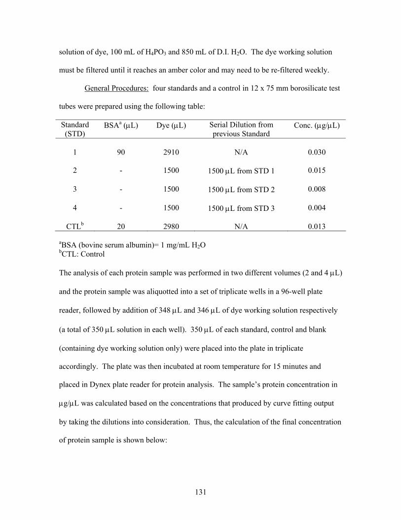
solution of dye, 100 mL of H4PO3 and 850 mL of D.I. H2O. The dye working solution
must be filtered until it reaches an amber color and may need to be re-filtered weekly.
General Procedures: four standards and a control in 12 x 75 mm borosilicate test
tubes were prepared using the following table:
Standard (STD)
BSAa (μL) Dye (μL) Serial Dilution from previous Standard
Conc. (μg/μL)
1
90
2910
N/A
0.030
2
-
1500
1500 μL from STD 1
0.015
3
-
1500
1500 μL from STD 2
0.008
4
-
1500
1500 μL from STD 3
0.004
CTLb
20
2980
N/A
0.013
aBSA (bovine serum albumin)= 1 mg/mL H2O bCTL: Control The analysis of each protein sample was performed in two different volumes (2 and 4 μL)
and the protein sample was aliquotted into a set of triplicate wells in a 96-well plate
reader, followed by addition of 348 μL and 346 μL of dye working solution respectively
(a total of 350 μL solution in each well). 350 μL of each standard, control and blank
(containing dye working solution only) were placed into the plate in triplicate
accordingly. The plate was then incubated at room temperature for 15 minutes and
placed in Dynex plate reader for protein analysis. The sample’s protein concentration in
μg/μL was calculated based on the concentrations that produced by curve fitting output
by taking the dilutions into consideration. Thus, the calculation of the final concentration
of protein sample is shown below:
131
Final concentration of each protein sample = (C1 x 175 + C2 x 87.5)/2 where C1 is
the concentration output (from curve fitting) of 2 μL protein sample and C2 is the
concentration output (from curve fitting) of 4 μL protein sample.
References for chapter 3 1 Schuda, P. F.; Greenlee, W. J.; Chakravarty, P. K. A Short and Efficient Synthesis of
(3S,4S)-4-[(tert-Butyloxycarbonyl)amino]-5-cyclohexyl-3-hydroxypentanoic Acid Ethyl Ester. J. Org. Chem. 1988, 53, 873-875.
2 Flynn, D. L.; Zelle, R. E.; Grieco, P. A. A Mild Two-Step Method for the Hydrolysis
/Methanolysis of Secondary Amides and Lactams. J. Org. Chem. 1983, 48, 2424-2427.
3 Kawahara, S.-I.; Uchimaru, T. One-Pot Preparation of o-Xylylene Diamine and its
Related Amines. Zeit. fur Nat. B: Chem. Sci. 2000, 55, 985-987. 4 Takagi, S.; Tanaka, H.; Yano, M.; Machida, K. Studies on the Syntheses of
Polymethylene-bisthioureas and their Derivatives. I. Chem. Phar. Bull, 1959, 7, 206-208.
5 Saller, C. F.; Czupryna, M. J. γ-Aminobutyric acid, glutamate, glycine and taurine
analysis using revesed-phased high-performance liquid chromatography and ultraviolet detection of dansyl chloride derivatives. J. Chromatogr. 1989, 487, 167-172.
6 Bloomquist, J. R.; Soderlund, D. M. Neurotoxic Insecticides Inhibit GABA-
Dependent Chloride Uptake by Mouse Brain Vesicles. Biochem. Biophys. Res. Commun. 1985, 133, 37-43.
7 DeLorey, T. M.; Brown, G. B. γ-Aminobutyric acidA Receptor Pharmacology in Rat
Cerebral Cortical Synaptoneurosomes. J. Neurochem. 1992, 58, 2162-2169.
8 Edgar, P. P.; Schwartz, R. D. Functionally Relevant γ–Aminobutyric AcidA Receptors: Equivalence between Receptor Affinity (Kd) and Potency (EC50)? Mol. Pharmacol. 1992, 41, 1124-1129.
9 The details of synthesis of PNP activated PEG: Lam, P. C.-H. PhD dissertation:
Experimental and Computational Studies in Bioorganic and Synthetic Organic Chemistry. 2004, 129-130.
132
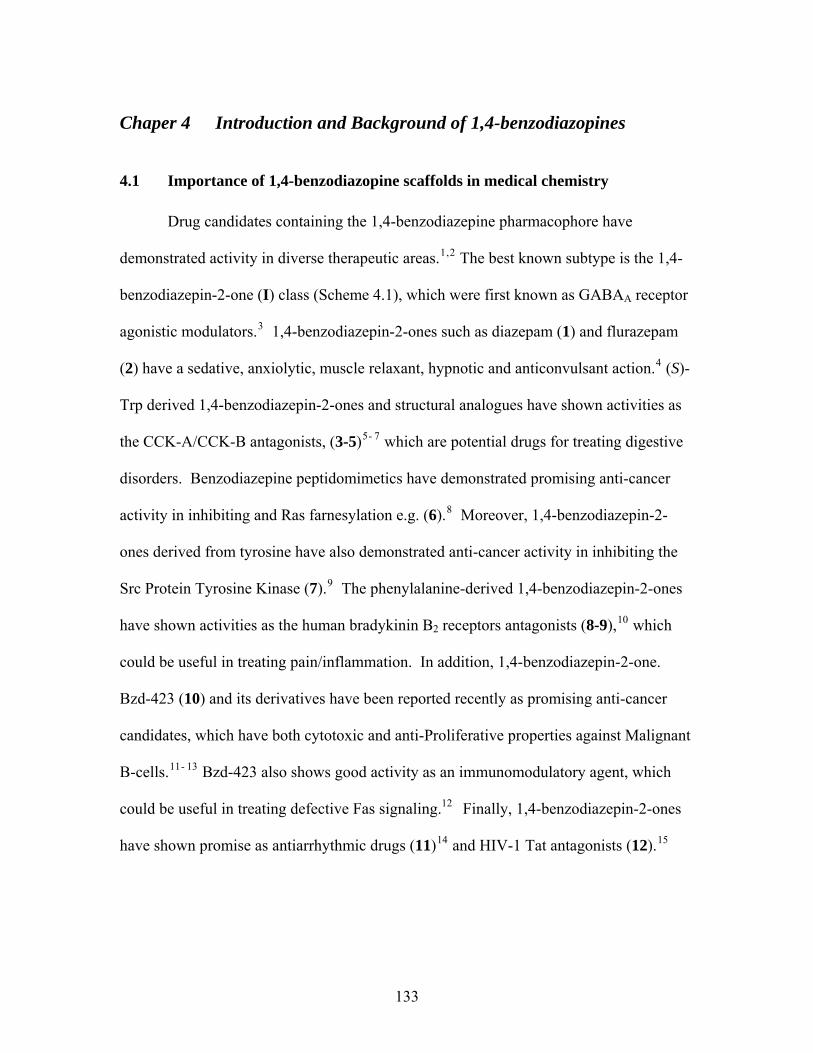
Chaper 4 Introduction and Background of 1,4-benzodiazopines
4.1 Importance of 1,4-benzodiazopine scaffolds in medical chemistry
Drug candidates containing the 1,4-benzodiazepine pharmacophore have
demonstrated activity in diverse therapeutic areas.1,2 The best known subtype is the 1,4-
benzodiazepin-2-one (I) class (Scheme 4.1), which were first known as GABAA receptor
agonistic modulators.3 1,4-benzodiazepin-2-ones such as diazepam (1) and flurazepam
(2) have a sedative, anxiolytic, muscle relaxant, hypnotic and anticonvulsant action.4 (S)-
Trp derived 1,4-benzodiazepin-2-ones and structural analogues have shown activities as
the CCK-A/CCK-B antagonists, (3-5)5- 7 which are potential drugs for treating digestive
disorders. Benzodiazepine peptidomimetics have demonstrated promising anti-cancer
activity in inhibiting and Ras farnesylation e.g. (6).8 Moreover, 1,4-benzodiazepin-2-
ones derived from tyrosine have also demonstrated anti-cancer activity in inhibiting the
Src Protein Tyrosine Kinase (7).9 The phenylalanine-derived 1,4-benzodiazepin-2-ones
have shown activities as the human bradykinin B2 receptors antagonists (8-9),10 which
could be useful in treating pain/inflammation. In addition, 1,4-benzodiazepin-2-one.
Bzd-423 (10) and its derivatives have been reported recently as promising anti-cancer
candidates, which have both cytotoxic and anti-Proliferative properties against Malignant
B-cells.11- 13 Bzd-423 also shows good activity as an immunomodulatory agent, which
could be useful in treating defective Fas signaling.12 Finally, 1,4-benzodiazepin-2-ones
have shown promise as antiarrhythmic drugs (11)14 and HIV-1 Tat antagonists (12).15
133
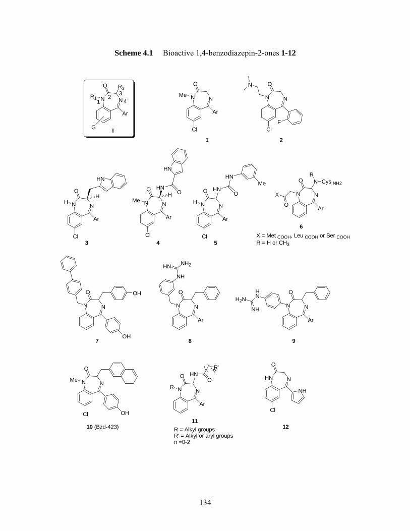
Scheme 4.1 Bioactive 1,4-benzodiazepin-2-ones 1-12
NN
O
Ar
R3
R1
G
12 3
4
I
NN
O
Ar
Me
1
Cl
NN
O
2
ClF
N
NN
O
Ar
H
3Cl
H
HN
NN
O
Ar
Me
4Cl
HNH O
HN
NN
O
Ar
H
5Cl
HN
HN
O
Me
NN
O
Ar
6
X
O
NR
Cys NH2
X = Met COOH, Leu COOH or Ser COOHR = H or CH3
NN
O
7OH
OH
NN
O
Ar
8
NH
NH2HN
NN
O
Ar
9
HNH2N
NH
NN
O
Me
10 (Bzd-423)
OHCl
NN
O
Ar
R
11
HN
R = Alkyl groupsR' = Alkyl or aryl groupsn =0-2
NHN
O
Cl
NH
12
O
R'n
134
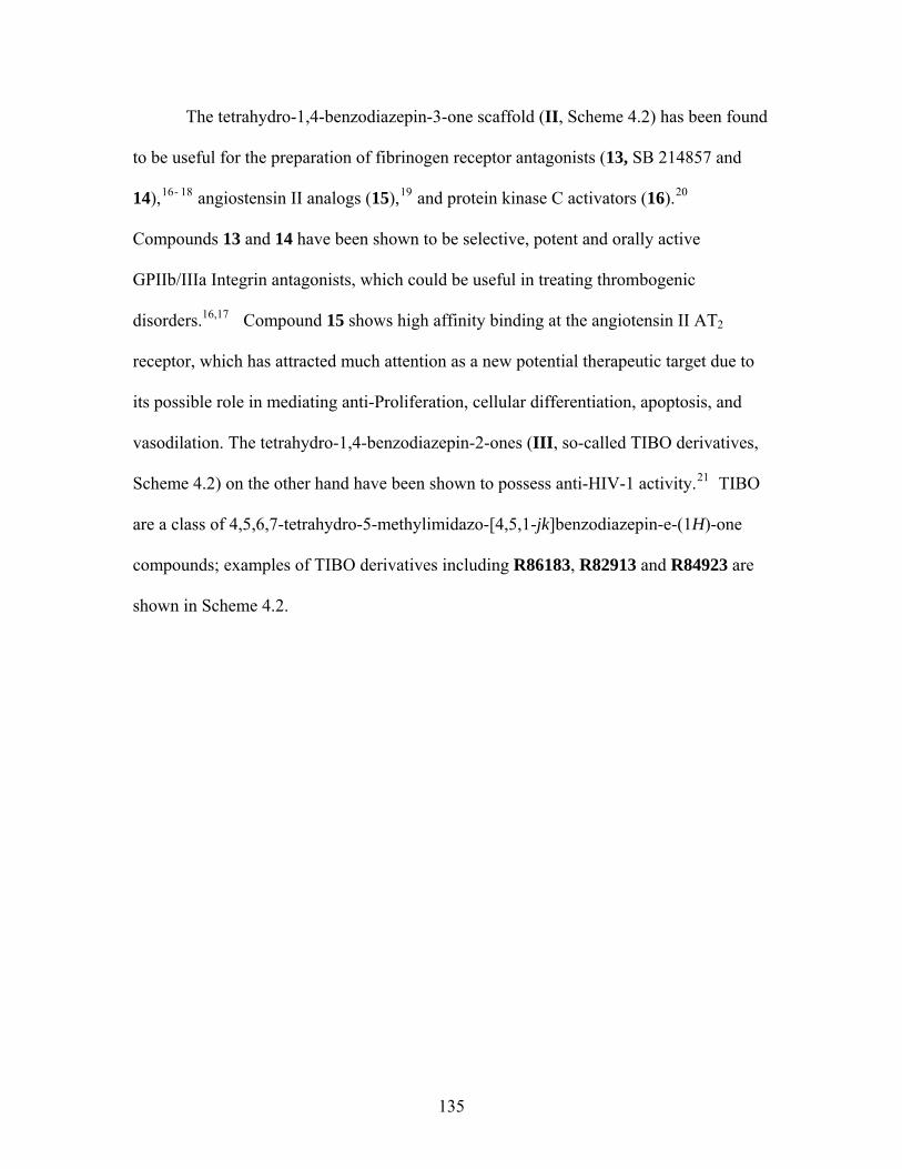
The tetrahydro-1,4-benzodiazepin-3-one scaffold (II, Scheme 4.2) has been found
to be useful for the preparation of fibrinogen receptor antagonists (13, SB 214857 and
14),16- 18 angiostensin II analogs (15),19 and protein kinase C activators (16).20
Compounds 13 and 14 have been shown to be selective, potent and orally active
GPIIb/IIIa Integrin antagonists, which could be useful in treating thrombogenic
disorders.16,17 Compound 15 shows high affinity binding at the angiotensin II AT2
receptor, which has attracted much attention as a new potential therapeutic target due to
its possible role in mediating anti-Proliferation, cellular differentiation, apoptosis, and
vasodilation. The tetrahydro-1,4-benzodiazepin-2-ones (III, so-called TIBO derivatives,
Scheme 4.2) on the other hand have been shown to possess anti-HIV-1 activity.21 TIBO
are a class of 4,5,6,7-tetrahydro-5-methylimidazo-[4,5,1-jk]benzodiazepin-e-(1H)-one
compounds; examples of TIBO derivatives including R86183, R82913 and R84923 are
shown in Scheme 4.2.
135
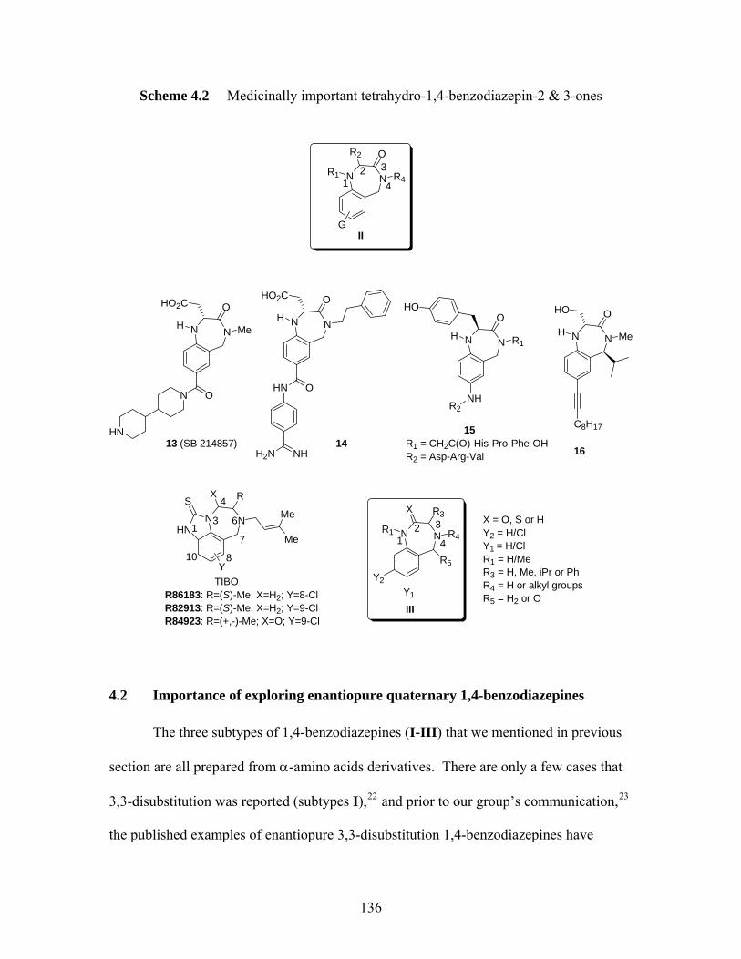
Scheme 4.2 Medicinally important tetrahydro-1,4-benzodiazepin-2 & 3-ones
NNR1
O
R4
R2
12 3
4
GII
NNR1 R412 3
4
III
X R3
NNH
O
Me
HO2C
N O
HN
NNH
OHO2C
HN O
H2N NH13 (SB 214857) 14
NNH
O
NH
R1 = CH2C(O)-His-Pro-Phe-OHR2 = Asp-Arg-Val
R2
HO
R1
15
NNH
OHO
Me
16
C8H17
Y2Y1
R5
X = O, S or HY2 = H/ClY1 = H/ClR1 = H/MeR3 = H, Me, iPr or PhR4 = H or alkyl groupsR5 = H2 or O
NNHN
S
Y
X R
Me
Me1
3
4
6
7
810
R86183: R=(S)-Me; X=H2; Y=8-ClR82913: R=(S)-Me; X=H2; Y=9-ClR84923: R=(+,-)-Me; X=O; Y=9-Cl
TIBO
4.2 Importance of exploring enantiopure quaternary 1,4-benzodiazepines
The three subtypes of 1,4-benzodiazepines (I-III) that we mentioned in previous
section are all prepared from α-amino acids derivatives. There are only a few cases that
3,3-disubstitution was reported (subtypes I),22 and prior to our group’s communication,23
the published examples of enantiopure 3,3-disubstitution 1,4-benzodiazepines have
136

merely relied upon lipase-catalyzed kinetic resolution.24,25 One possibility for the
development of quaternary 1,4-benzodiazepines is to extend the existing synthetic
methods for I-III to the use of enantiomerically pure quaternary amino acids. However,
despite the fact that several asymmetric methods have been developed for the synthesis of
quaternary amino acids,26,27 few enantiomerically pure quaternary amino acids are
available commercially. Moreover, the amide (I and III) and amination (II) bond-
forming reactions during the ring closure that led to these heterocycles may be
problematic with the quaternary amino acids. A direct synthesis of quaternary 1,4-
benzodiazepines from commercially available ‘tertiary’ amino acids might then be the
key to therapeutically discovery quaternary 1,4-benzodiazepines as promising agents.
Our research group has developed such a procedure for the preparation of
quaternary 1,4-benzodiazepin-2-ones. The route to quaternary 1,4-benzodiazepin-2-ones
depends on enantioselective functionalization of the already-formed heterocycles, which
were prepared using normal, enantiomerically pure α-amino acids. This method relies on
the “memory of chirality (MOC)” imparted by the conformation of the benzodiazepine
ring. The concept of MOC was introduced by Fuji and Kawabata as a useful strategy for
asymmetric synthesis.28 As defined by Carlier, MOC is “often used to describe formal
substitution reactions at the sole sp3 center of an enantiopure starting material, which
proceed by trigonalization of that center, and yet retain high enantiomeric purity in the
product.” Such process can occur when the static, central chirality of the reactants is
preserved in the form of dynamic conformational chiral reactive intermediate.29 One of
the best examples of this phenomenon is demonstrated by Fuji and Kawabata (Scheme
4.3).30
137
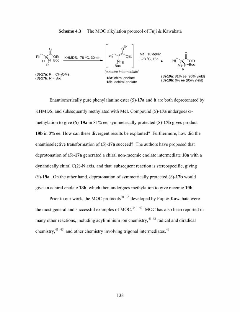
Scheme 4.3 The MOC alkylation protocol of Fuji & Kawabata
P
h OEtNH Boc
R
O
(S)-17a: R = CH2OMe(S)-17b: R = Boc
KHMDS, -78 oC, 30min Ph OEt
O
N RBoc
18a: chiral enolate18b: achiral enolate
MeI, 10 equiv. -78 oC, 16h Ph OEt
NMe BocR
O
(S)-19a: 81% ee (96% yield)(S)-19b: 0% ee (95% yield)
"putative intermediate"
Enantiomerically pure phenylalanine ester (S)-17a and b are both deprotonated by
KHMDS, and subsequently methylated with MeI. Compound (S)-17a undergoes α-
methylation to give (S)-19a in 81% ee, symmetrically protected (S)-17b gives product
19b in 0% ee. How can these divergent results be explanted? Furthermore, how did the
enantioselective transformation of (S)-17a succeed? The authors have proposed that
deprotonation of (S)-17a generated a chiral non-racemic enolate intermediate 18a with a
dynamically chiral C(2)-N axis, and that subsequent reaction is stereospecific, giving
(S)-19a. On the other hand, deprotonation of symmetrically protected (S)-17b would
give an achiral enolate 18b, which then undergoes methylation to give racemic 19b.
Prior to our work, the MOC protocols30 - 33 developed by Fuji & Kawabata were
the most general and successful examples of MOC.34- 40 MOC has also been reported in
many other reactions, including acyliminium ion chemistry,41,42 radical and diradical
chemistry,43- 45 and other chemistry involving trigonal intermediates.46
138
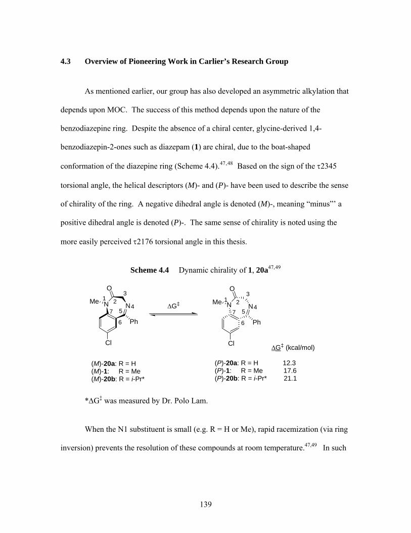
4.3 Overview of Pioneering Work in Carlier’s Research Group
As mentioned earlier, our group has also developed an asymmetric alkylation that
depends upon MOC. The success of this method depends upon the nature of the
benzodiazepine ring. Despite the absence of a chiral center, glycine-derived 1,4-
benzodiazepin-2-ones such as diazepam (1) are chiral, due to the boat-shaped
conformation of the diazepine ring (Scheme 4.4).47,48 Based on the sign of the τ2345
torsional angle, the helical descriptors (M)- and (P)- have been used to describe the sense
of chirality of the ring. A negative dihedral angle is denoted (M)-, meaning “minus”’ a
positive dihedral angle is denoted (P)-. The same sense of chirality is noted using the
more easily perceived τ2176 torsional angle in this thesis.
Scheme 4.4 Dynamic chirality of 1, 20a47,49
NN
Cl
Ph
O
MeΔG‡
1 23
45
6
7NN
Cl
Ph
O
Me 1 23
45
6
7
(M)-20a: R = H
*
(P)-20a: R = H 12.3(P)-1: R = Me 17.6 (P)-20b: R = i-Pr* 21.1
Δ
*ΔG‡ was measured by Dr. Polo Lam.
When the N1 substituent is small (e.g. R = H or Me), rapid racemization (via ring
inversion) prevents the resolution of these compounds at room temperature.47,49 In such
(M)-1: R = Me(M)-20b: R = i-Pr
G‡ (kcal/mol)
139
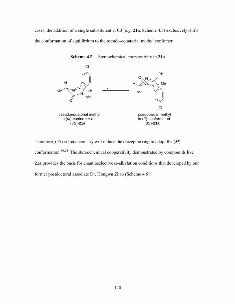
cases, the addition of a single substitutent at C3 (e.g. 21a, Scheme 4.5) exclusively shifts
the conformation of equilibrium to the pseudo-equatorial methyl confomer.
Scheme 4.5 Stereochemical cooperativity in 21a
H
MeN
O
N
Cl
Me
HN
N
Cl
OPh
Me
Ph
pseudoequatorial methyl in (M)-conformer of (3S)-21a
pseudoaxial methylin (P)-conformer of
(3S)-21a
Me
Therefore, (3S)-stereochemistry will induce the diazepine ring to adopt the (M)-
conformation.50, 51 The stereochemical cooperativity demonstrated by compounds like
21a provides the basis for enantioselective α-alkylation conditions that developed by our
former postdoctoral associate Dr. Hongwu Zhao (Scheme 4.6).
140
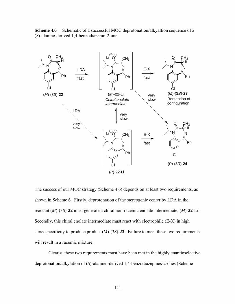
Scheme 4.6 Schematic of a successful MOC deprotonation/alkyaltion sequence of a (S)-alanine-derived 1,4-benzodiazepin-2-one
NN
Cl
Ph
O
LDA
fast
N
Cl
Ph
CH3O
E-X
fast
NN
Cl
Ph
O CH3E
N
Cl
Ph
CH3O
very slow
LDA
veryslow
Li
Li E-X
fast
NN
Cl
Ph
O CH3E
very slow
(M)-(3S)-22 (M)-22-Li
(P)-22-Li
(M)-(3S)-23
(P)-(3R)-24
Rentention of configuration
CH3H
Chiral enolate intermediate
The success of our MOC strategy (Scheme 4.6) depends on at least two requirements, as
shown in Scheme 6. Firstly, deprotonation of the stereogenic center by LDA in the
reactant (M)-(3S)-22 must generate a chiral non-racemic enolate intermediate, (M)-22-Li.
Secondly, this chiral enolate intermediate must react with electrophile (E-X) in high
stereospecificity to produce product (M)-(3S)-23. Failure to meet these two requirements
will result in a racemic mixture.
Clearly, these two requirements must have been met in the highly enantioselective
deprotonation/alkylation of (S)-alanine -derived 1,4-benzodiazepines-2-ones (Scheme
141
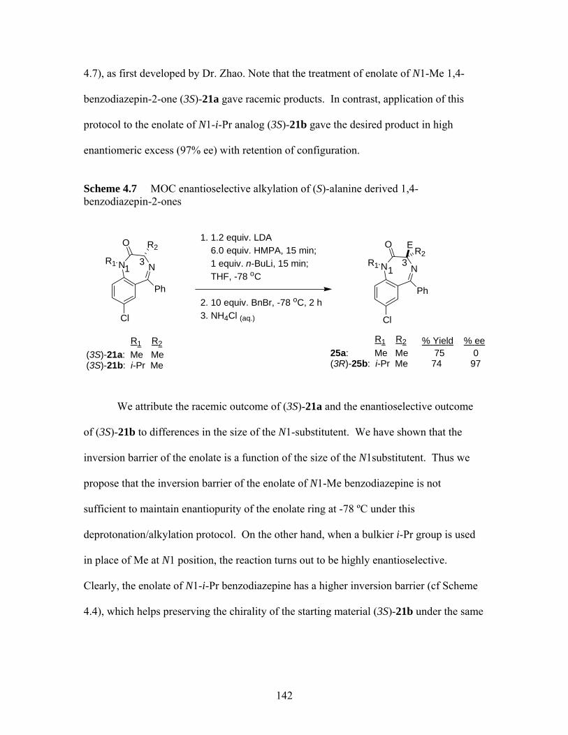
4.7), as first developed by Dr. Zhao. Note that the treatment of enolate of N1-Me 1,4-
benzodiazepin-2-one (3S)-21a gave racemic products. In contrast, application of this
protocol to the enolate of N1-i-Pr analog (3S)-21b gave the desired product in high
enantiomeric excess (97% ee) with retention of configuration.
Scheme 4.7 MOC enantioselective alkylation of (S)-alanine derived 1,4-benzodiazepin-2-ones
1. 1.2 equiv. LDA O OR2 E 6.0 equiv. HMPA, 15 min; 1 equiv. n-BuLi, 15 min; THF, -78 oC
2. 10 equiv. BnBr, -78 oC, 2 h3. NH4Cl (aq.)
R2
We attribute the racemic outcome of (3S)-21a and the enantioselective outcome
of (3S)-21b to differences in the size of the N1-substitutent. We have shown that the
inversion barrier of the enolate is a function of the size of the N1substitutent. Thus we
propose that the inversion barrier of the enolate of N1-Me benzodiazepine is not
sufficient to maintain enantiopurity of the enolate ring at -78 ºC under this
deprotonation/alkylation protocol. On the other hand, when a bulkier i-Pr group is used
in place of Me at N1 position, the reaction turns out to be highly enantioselective.
Clearly, the enolate of N1-i-Pr benzodiazepine has a higher inversion barrier (cf Scheme
4.4), which helps preserving the chirality of the starting material (3S)-21b under the same
NN
Cl
R1
Ph
NNR1
Cl
3 3
(3S)-21a: Me Me3S)-21b: i-P
1 1
Ph
25a: Me Me 75 0(3R)-25b: i-Pr Me 74 97
R R %
( r Me
R1 R2 1 2 Yield % ee
142

reaction conditions and thus yield the desired product in high enantiomeric excess with
retention of configuration.
Density functional theory (DFT) calculations of the inversion barriers of 1,4-
benzodiazepin-2-one enolates indicate that the equilibrium geometries of 21a and 21b are
chiral. The free energies of activation for ring inversion of 21a (N1-Me) and 21b (N1-i-
Pr) at 195 K are 12.4 and 17.5 kcal/mol, which correspond to racemization half-life
values of 0.11 min and 970 h, respectively. Dr. Polo Lam has performed enolate trapping
experiments that confirm this proposal.52
The optimized conditions involve use of LDA and n-BuLi as bases and HMPA as
additive and the reactions were conducted at -78 ºC. Dr. Zhao has discovered that HMPA
was essential for this type of reaction; only traces of the desired products were yielded in
the absence of HMPA. Use of DMPU in place of HMPA also proved to be fruitless. He
has also found that use of LiHMDS in place of LDA gave none of the desired products.
Using this basic MOC strategy, we will attempt to prepare three other classes of
1,4-benzodiazepine from enantiomerically pure α-amino acids. These include (S)-
alanine-derived tetrahydro-1,4-benzodiazepin-3-ones, (S)-phenylalanine-derived
tetrahydro-1,4-benzodiazepin-2-ones, and (S)-serine-derived 1,4-benzodiazepin-2-ones.
Reference for chapter 4 1 Ellman, J. A. Design, Sythesis, and Evaluation of Small-Molecule Libraries. Acc.
Chem. Res. 1996, 29, 132-143. 2 Boojamra, C. G.; Burow, K. M.; Thomson, L. A.; Ellman, J. A. Solid-Phase Sythesis
of 21,4-Benzodiazepin-2,5-diones. Library Preparation and Demonstration of Synthesis Generally. J. Org. Chem. 1997, 62, 1240.
143

3 Sakmann, B.; Hamill, O. P.; Bormann, J. Patch-clamp measurements of elementary
chloride currents activated by the putative inhibitory transmitters GABA and glycine in mammalian spinal neurons. J. Neural Transmission. 1983, 18, 83-95.
4 Lancel, M.; Steiger, A. Sleep and its modulation by drugs that affect GABAA
receptor function. Angew. Chem. Int. Ed. 1999, 111, 2852-2864. 5 Evans, B. E.; Rittle, K. E.; Bock, M. G.; DiPardo, R. M.; Freidinger, R. M.; Whitter,
W. L.; Could, N. P.; Lundell, G. F.; Homnick, C. F.; Veber, D. F.; Anderson, P. S.; Chang, R. S. L.; Lotti, V. J.; Cerino, D. J.; Chen, T. B.; King, P. J.; Kunkel, K. A.; Springer, J. P.; Hirschfield, J. Design of Nonpeptidal Ligands for a Peptide Receptor: Cholecystokinin Antagonists. J. Med. Chem. 1987, 30, 1229-1239.
6 Bock, M. G.; DiPardo, R. M.; Rittle, K. E.; Veber, D. F.; Anderson, P. S.; Freidinger,
R. M. Benzodiazpine Gastrin and Brain Cholecystkinin Receptor Ligands: L-365,260. J. Med. Chem. 1989, 32, 13-16.
7 Showell, G. A.; Bourrain, S.; Neduvelil, J. G.; Flethcher, S. R.; Baker, R.; Watt, A.
P.; Fletcher, A. E.; Freedman, S. B.; Kemp, J. A.; Marshall, G. R.; Patel, S.; Smith, A. J.; Matassa, V. G. High-affinity and Potent, Water-Soluble 5-Amino-1,4-Benzodiazepine CCKB/Gastrin Receptor Antagonists Containing a Cationic Solubilizing Group. J. Med. Chem. 1994, 37, 719-721.
8 James, G. L.; Goldstein, J. L.; Brown, M. S.; Rawson, T. E.; Somers, T. C.;
McDowell, R. S.; Crowley, C. W.; Lucas, B. K.; Levinson, A. D.; Masters, J. C. Benzodiazepine Peptidomimetics: Potent Inhibitors of Ras Farneslylation in Animal Cells. Science (Washington, D. C.) 1993, 260, 1937-1942.
9 Ramads, L.; Bunnin, B. A.; Plunkett, M, J.; Sun, G. Ellman, J. Gallick, G.; Budde, R.
J. A. Benzodiazepine Compounds as Inhibitors of the Src protein Tyrosine Kinase: Screening of a Combinatory Library of 1,4-Benzodiazepines. Arch. Biochem. Biophys. 1999, 368, 394-400.
10 Dziadulewicz, E. K.; Brown, M. C.; Dunstan, A. R.; Lee, W.; Said, N. B.; Garratt, P.
J. The design of non-peptide human bradykinin B2 receptor antagonists employing the benzodiazepine peptidomimetic scaffold. Bioorg. Med. Chem. Lett. 1999, 9, 463-468.
11 Boitano, A.; Ellman, J. A.; Glick, G. D.; Opipari, Jr., A. W. The Proapoptotic
Benzodiazepine Bz-423 Affects the Growth and Survival of Malignant B Cells. Cancer Res. 2003, 63, 6870-6876.
12 Bednarski, J. J.; Warner, R. E.; Rao, T.; Leonetti, F.; Yung, R.; Opipari, Jr., A. W.,
Glick, G. D. Attenuation of autoimmune disease in Fas-deficient mice by treatment with a cytotoxic benzodiazepine. Arthritic & Rheumatism, 2003, 48, 757-766.
144

13 Boitano, A.; Emal, C. D.; Leonetti, F,; Blatt, N. B.; Dineen, T. A;. Ellman, J. A.;
Roush, W. R.; Opipari, A. W. Structure activity studies of a novel cytotoxic benzodiazepine. Bioorg. Med. Chem. Lett. 2003, 63, 3327-3330.
14 Selnick, H. G.; Liverton, N. J.; Baldwin, J. J.; Butcher, J. W.; Claremon, D .A.;
Elliott, J. M.; Freidlinger, R. M.; King, S. A.; Libby, B. E.; Mclntyre, C. J.; Pribush, D. A.; Remy, D. C.; Smith, G. R.; Tebben, A. J.; Juriewicz, N. K.; Lynch, J. J.; Salata, J. J.; Snguinetti, M. C.; Siegl, P. K. S.; Slaughter, D. E.; Vyas, K. Class III Antiarrhythmic Activity in Vivo by Selective Blockade of the Slowly Activating Current Delayed Rectifier Potassium Current Iks by (R)-(2,4-Trifluoromethyl)-N-[2-oxo-5-phenyl-1-(2,2,2-trifluoroethyl)-2,3-dihydro-1H-benzo[e][1,4]diazepine-3-yl-acetaminde. J. Med. Chem. 1997, 40, 3865-3868.
15 Hsu, M.-C.; Schutt, A. D.; Holly, M.; Slice, L. W.; Sherman, M. I.; Richman, D. D.;
Potash, M .J.; Volsky, D. J. Inhibition of HIV Replication in Acute and Chronic Invections in Vitro by a Tat Antagonist. Science (Washington, D. C.) 1991, 254, 1799-1802.
16 Miller, W. H.; Ku, T. W.; Ali, F. E.; Bondinell, W. E.; Calvo, R. R.; Davis, L. D.;
Erhard, K. F.; Hall, L. B.; Huffman, W. F.; Keenan, R. M.; Kwon, C.; Newlander, K. A.; Ross, S. T.; Samanen, J. M.; Takata, D. T.; Yuan, C.-K. Enantiospecific Synthesis of SB 214857, a Potent, Orally Active Nonspecific Fibrinogen Receptor Antagonist. Tetrahedron Lett. 1995, 36, 9433-9436.
17 Samanen, J. M.; Ali, F. E.; Barton, L. S.; Bondinell, W. E.; Burgess, J. L.; Callahan,
J. F.; Calvo, R. R.; Chen, W.; Chen, L.; Erhard, K.; Feuerstein, G.; Heys, R.; Hwang, S.-M.; Jakas, D. R.; Keenan, R. M.; Ku, T. W.; Kwon, C.; Newlander, K. A.; Nichols, A.; Parker, M.; Peishoff, C. E.; Rhodes, G.; Ross. S.; Shu, A.; Simpson, R.; Takata, D.; Yellin, T. O.; Uzsinskas, I.; Venslavsky, J. W.; Yuan, C.-K,; Huffman, W. F. Potent, Selective Orally Active 3-Oxo-1,4-benzodiazepine GPIIb/IIIa Integrin Antagonists. J. Med. Chem. 1996, 39, 4867-4870.
18 Keenan, R. M.; Callahan, J. F.; Samanen, J. M.; Bondinell, W. E.; Calvo, R. R.;
DeBrosse, C.; Eggleston, D. S.; Haltiwanger, R. C.; Hwang, S.-M.; Jakas, D. R.; Ku, T. W.; Miller, W. H.; Newlander, K. A.; Nichols, A.; Parker, M. F.; Southhall, L. S.; Uzinskas, I.; Vasko-Moser, J. A.; Venslavsky, J. W.; Wong, A. S.; Huffman, W. F. Conformational Preferences in a Benzodiazepine Series of Potent Nonpeptide Fibrinogen Receptor Antagonists. J. Med. Chem. 1999, 42, 545-559.
19 Rosenström, U.; Sköld, C.; Lindeberg, G.; Botros, M.; Nyberg, F.; Karlén, A.;
Halberg, A Selective AT2 Receptor Ligand with a γ-Turn-Like Mimetic Replacing the Amino Acid Residues 4-5 of Angiotensin II. J. Med. Chem. 2004, 47, 859.
145
20 Ma, D.; Wang, G.; Wang, S.; Kozikowski, A. P.; Lewin, N. E.; Blumberg, P. M.
Synthesis and Protein Kinase Binding Activity of Benzolactam-V7. Bioorg. Med. Chem. Lett. 1999, 9, 1371-1374.
21 Breslin, H. J.; Kukla, M. J.; Kromis, T.; Cullis, H.; Knaep, F. D.; Pauwels, R.;
Andries, K.; Clercq, E. D.; Janssen, M. A. C.; Janssen, P. A. J. Synthesis and Anti-HIV Activity of 1,3,4,5-Tetrahydro-2H-1,4—benzodiazepine-2-one (TBO) Derivatives. Truncated 4,5,6,7-Tetrahydro-5-methylimidazolo[4,5,1-jk][1,4]benzodiazepin-2-(1H)-ones (TIBO) Analogues. Bioorg. Med. Chem. 1999, 7, 2427-2436.
22 Simonyi, M.; Kaksay, G.; Kovacs, I.; Tegyey, Z.; Parkanyi, L.; Kalman, A.; Otvos, L.
Conformational Recognition by Central Benzodiazepine Receptors. Bioorg. Chem. 1990, 18, 1-12.
23 Carlier, P. R.; Zhao, H.-W.; DeGuzman, J.; Lam, P. C.-H. Enantioselective Sythesis
of “Quaternary” 1,4-Benzodiazepin-2-one Scaffolds via Memory of Chirality. J. Am. Chem. Soc. 2003, 125, 11482-11483.
24 Avdagić, A.; Lesac, A.; Šunjić, V. First Example of the Solvent Effect on Absolute
Conformation of Chiral 3,3-Diustituted 1,4-benzodiazepin-2-ones. Tetrahedron 1999, 55, 1407.
25 Avdagić, A.; Lesac, A.; Majer, Z.; Hollòsi, M.; Šunjić, V. Lipase-Catalyzed
Acetylation of 3-Substituted 2,3-Dihydro-1H-1,4-benzodiazepin-2-ones: Effect of Temperature and Conformation on Enantioselectivity and Configuration. Helv. Chim, Acta 1998, 81, 1567-1582.
26 Seebach, D. Self-Regeneration of Stereocenters (SRS)-Applications, Limitations, and
Abandonment of a Synthetic Principle. Angew. Chem. Int. Ed. 1996, 35, 2708-2748. 27 Cativiela, C.; Díaz-de-Vallagas, M. D. Stereoselective synthesis of quaternary α-
amino acids. Tetrahedron: Asymmetry 1998, 40, 3517-3599. 28 Fuji, K.; Kawabata, T. Memory of Chirality-A New Principle in Enolate Chemistry.
Chem Eur. J. 1998, 4, 373-376. 29 Lam, P. C.-H.; Carlier, P. R. Experimental and Computational Studies of Ring
Inversion of 1,4-Benzodiazepin-2-ones: Implications for Memory of Chirality Transformation. J. Org. Chem. 2005, 70, 1530-1538.
30 Kawabata, T.; Suzuki, H.; Nagae, Y.; Fuji, K. Chiral Nonracemic Enolate with
Dynamic Axial Chirality: Direct Asymmetric α-Amino Acid Derivatives. Angew. Chem. Int. Ed. 2000, 39, 2155-2157.
146
31 Kawabata, T.; Wirth, T.; Yahiro, K.; Suzuki, H.; Fuji. K. Direct Asymmetric α-
Alkylation of Phenylalanine Derivatives Using No External Chiral Sources. J. Am. Chem. Soc. 1994, 116, 10809-10810.
32 Kawabata, T.; Chen, J.; Suzuki, H.; Nagae, Y.; Kinoshita, T.; Chnacharunee, S.; Fuju,
K. Memory of Chirality in Diastereoselective α-Alkylation of Isoleucine and allo-Isoleucine Derivatives.Org. Lett. 2000, 2, 3883-3885.
33 Kawabata, T.; Kawakami, S.-P.; Fuji, K. Enantioselective α-Allylation of a phenylalanine derivative under the control of aggregation of a chiral nonracemic enolate. Tetrahedron Lett. 2002, 43, 1465-1467.
34 Seebach, D.; Wasmuth, D. Alkylation of Amino Acids without Loss of Optical
Activity: α- and β-Alkylation of an Aspartic Acid Derivative. Angew. Chem Int. Ed. 1981, 20, 971.
. 35 Beagley, B.; Betts, M. J.; Pritchard, R. G. Schofield, A.; Stoodley, R. J.; Vohra,, S. A
Cyclisation Reaction of Methyl (4R)-3-(2-Diazo-3-oxoabutanoyl)thiazolidine-4-carboxylate which proceeds with Retention of Configuration, probably via a Planar Ester Enolate Intermediate possessing Axial Chirality. Chem. Commun. 1991, 924-925.
36 Kawabata, T.; Yahiro, K. Fuji, K. Memory of Chirality: Enantioselective Alkylation
Reactions at an Asymmetric Carbon Adjacent to a Carbonyl Group. J. Am. Chem. Soc. 1991, 113, 9694-9696.
37 Gees, T.; Schweizer, W. B.; Seebach, D. An Unusual Rearrangement of a Lithiated
N-Acyl-tetrahydroisoquinoline to an Amino-indan Skeleton and Structural Comparison of 3-Amino-2-methyllindane- and -tetrahydronaphthalene-2-carboxylic Acids as Possible Building Block for Peptide-Turn Mimics. Helv. Chim. Acta 1993, 76, 2640-2653.
38 Beagley, B.; Betts, M. J.; Pritchard, R. G. Schofield, A.; Stoodley, R. J.; Vohra,, S.
‘Hidden’ Axial Chirality as a Stereodirecting Element in Reactions Involving Enol(ate) Intermediate. Part1 1. Cyclisation Reactions of Methyl (4R)-3-(2-Diazo-3-oxoabutanoyl)thiazolidine-4-carboxylate and Related Compounds. J. Chem. Soc. Perkin Trans. I 1993, 1761-1770.
39 Gerona-Navarro, G.; Bonache, M. A.; Herranz, R.; García-López, M. T.; Gonzalez-
Muñiz, R. Entry to New Conformationally Constrained Amino Acids. First Synthesis of 3-Unsubstituted -4-Alkyl-4-carboxy-2-azedidinone Derivatives via an Intramolecular Nα-Cα-Cyclisation Strategy. J. Org. Chem. 2001, 66, 3538.
40 Brewster, A. G.; Jayatissa, J.; Mitchell, M. B.; Schofield, A.; Stoodley, R. J. Memory
of chirality effects in aldol cyclisations of 1-(3-oxobutyryl) derivatives of -L-4-oxaproline and L-proline isopropyl esters. Tetrhedron Lett. 2002, 43, 3919-3922.
147
41 Matsumura, Y.; Shirakawa, Y.; Satoh, Y.; Umino, M.; Tanaka, T.; Maki, T.;
Onomura, O. First Example of Memory of Chirality in Carbenium Ion Chemistry. Org. Lett. 2000, 2, 1689-1691.
42 Wanyoike, G. N.; Onomura, O.; Maki, T.; Matsumura, Y. Highly Enhanced
Enantioselectivtiy in the Memory of Chirality via Acyliminium Ions. Org. Lett. 2002, 4, 1875-1877.
43 Buckmelter, A. J.; Kim, A. I.; Rychnovsky, S. D. Conformational Memory in
Enantioselective Radical Reduction and a New Radical Clock Reaction. J. Am. Chem. Soc. 2000, 122, 9386-9390.
44 Giese, B.; Wettstein, P.; Stähelin, C.; Barbosa, F.; Neuberger, M.; Zehnder, M.;
Wessig, P. Memory of Chirality in Photochemistry. Angew. Chem. Int. Ed. 1999, 38, 2586-2587.
45 Griesbeck, A. G.; Kramer, W.; Lex, J. Diastereo- and Enantioselective Synthesis of
Pyrrolo[1,4]benzodiazepines through Decarboxylate Photocyclization. Angew. Chem. Int. Ed. 2001, 40, 577-579.
46 Schmalz, H.-G.; Koning, C. B. d.; Bernicke, D.; Siegel, S.; Pflethschinger, A.
Memory of Chirality in Electron Transfer Mediated Benzylic Umolung Reactions of Arene-Cr(CO3) complexes. Angew. Chem. Int. Ed. 1999, 38, 1620-1623.
47 Linscheid, P.; Lehn, J.-M. Étude cinétiques et conformationnelles par résonance
magnétique nucléaire. VII. Inversion de cycle dans des benzo-diazépinones. Bull. Chim. Soc. Fr. 1967, 992-997.
48 Konowal, A.; Snatzke, G.; Alebic-kolbah, T.; Kajfez, F.; Rendic, S.; Sunjic, V.
General Approach to Chiroptical Characterization of Binding to Prochiral and Chiral 1,4-Benzodiazepin-2-ones to Human Serum Albumin. Biochem. Pharmacol. 1979, 28, 3109-3113.
49 Gilman, N. W.; Rosen, P.; Earley, J. V.; Cook, C.; Todaro, L. J. Atropisomers of 1,4-
Benzodiazepines. Synthesis and Resolution of a Diazepam-Related 1,4-Benzodiazepine. J. Am. Chem. Soc. 1990, 112, 3969-3978.
50 Sunjic, V.; Lisini, A.; Sega. A.; Kovac, T.; Kajfez, F.; Ruscic, B. Conformation of 7-
Chloro-5-phenyl-d5-3(S)-mehtyl-dihydro-1,4-Benzodiazepin-2-one in Solution. J. Heterocylic Chem. 1979, 16, 757-761.
51 Paizs, B. Simonyi, M. Ring Inversion Barrier of Diazepam and Derivatives: An ab
Initio Study. Chirality 1999, 11, 651.
148

52 Lam, P. C.-H. PhD dissertation: Experimental and Computational Studies in
Bioorganic and Synthetic Organic Chemistry. 2004, 212-216.
149
Chapter 5 Syntheses of 1,4-Benzodiazepine Analogs
5.1 Investigation of Enantioselective Synthesis of Quaternary (S)-alanine Derived Tetrahydro-1,4-benzodiazepin-3-ones
As we mentioned in chapter 4, some members of the tetrahydro-1,4-
benzodiazepin-3-ones subtype have been found to be important therapeutic agents (13-
16, chapter 4). Note that all of these compounds (13-16) were prepared from
proteinogenic amino acid derivatives. The lack of 2,2-substituted analogs has motivated
us to extend our MOC strategy to the enantioselective synthesis of quaternary (S)-alanine
derived tetrahydro-1,4-benzodiazepin-3-ones.
5.1.1 Synthesis of (S)-alanine Derived Tetrahydro-1,4-benzodiazepin-3-ones
We have recently published the improved synthesis of (S)-alanine derived
tetrahydro-1,4-benzodiazepin-3-ones.1 The synthesis began with commercially available
2-fluoro-5-nitrotoluene 1 (Scheme 5.1) by adopting the basic strategy of Miller et al.:2
treatment with NBS in CCl4 afforded 2 as off-white, needle-like crystals in 66% yield.
Compound 2 was previously prepared by Sveinbjornsson et al.3 from o-fluorobenzyl
bromide and fuming nitric acid (>90%) in 81% yield; However, our attempts to repeat
this synthesis using regular nitric acid (70%) with concentrated sulfuric acid as catalyst,
gave a low yield (20%). Subsequent reaction of 2 with excess allyamine in THF gave 3
in 87%. The reaction of 2 with methylamine was however less selective, giving a
mixture of the desired product 4 and the undesired nucleophilic aromatic substitution
product 5, which were difficult to separate. Coupling of 3 with either (S)-Boc-Ala-OH,
150
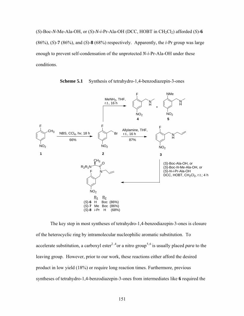
(S)-Boc-N-Me-Ala-OH, or (S)-N-i-Pr-Ala-OH (DCC, HOBT in CH2Cl2) afforded (S)-6
(86%), (S)-7 (86%), and (S)-8 (68%) respectively. Apparently, the i-Pr group was large
enough to prevent self-condensation of the unprotected N-i-Pr-Ala-OH under these
conditions.
Scheme 5.1 Synthesis of tetrahydro-1,4-benzodiazepin-3-ones
F
NO2
CH3 NBS, CCl4, hv, 18 h
F
NO2
BrAllylamine, THF, r.t., 16 h
F
NO2
NH
1 2 3
MeNH2, THF,r.t., 16 h
NMe
NO2
NH
5
F
NO2
NH
4
+
(S)-Boc-Ala-OH, or (S)-Boc-N-Me-Ala-OH, or(S)-N-i-Pr-Ala-OHDCC, HOBT, CH2Cl2, r.t.; 4 h
F
NO2
N
OR2R1N
CH3
66% 87%
(S)-6 H Boc (86%)(S)-7 Me Boc (86%)(S)-8 i-Pr H (68%)
R1 R2
The key step in most syntheses of tetrahydro-1,4-benzodiazepin-3-ones is closure
of the heterocyclic ring by intramolecular nucleophilic aromatic substitution. To
accelerate substitution, a carboxyl ester2 ,4or a nitro group5, 6 is usually placed para to the
leaving group. However, prior to our work, these reactions either afford the desired
product in low yield (18%) or require long reaction times. Furthermore, previous
syntheses of tetrahydro-1,4-benzodiazepin-3-ones from intermediates like 6 required the
151
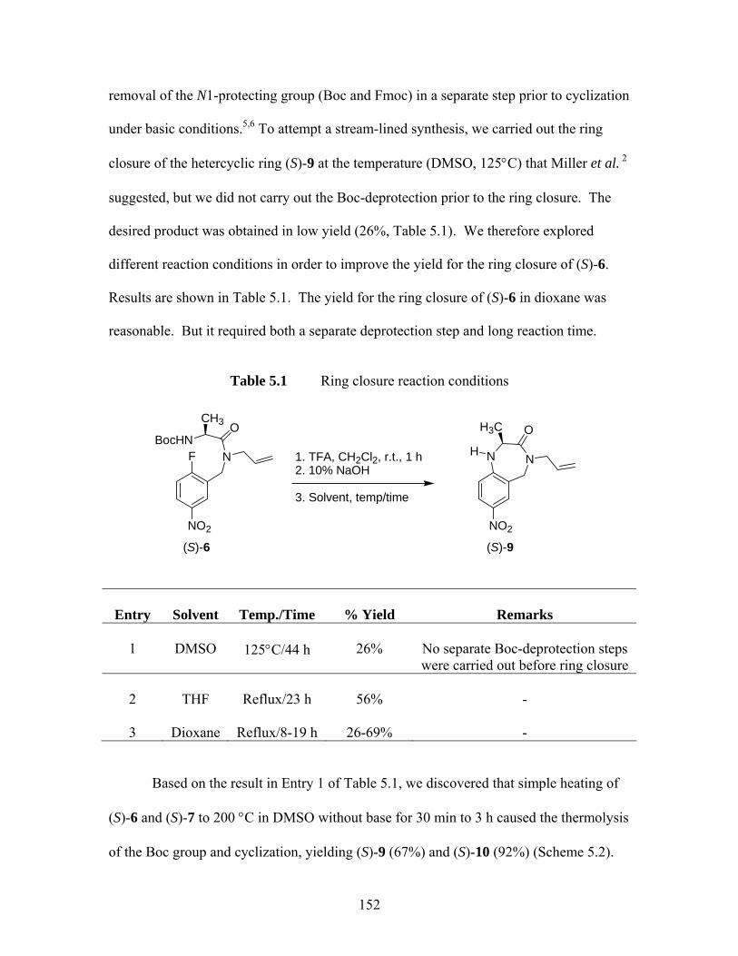
removal of the N1-protecting group (Boc and Fmoc) in a separate step prior to cyclization
under basic conditions.5,6 To attempt a stream-lined synthesis, we carried out the ring
closure of the hetercyclic ring (S)-9 at the temperature (DMSO, 125°C) that Miller et al. 2
suggested, but we did not carry out the Boc-deprotection prior to the ring closure. The
desired product was obtained in low yield (26%, Table 5.1). We therefore explored
different reaction conditions in order to improve the yield for the ring closure of (S)-6.
Results are shown in Table 5.1. The yield for the ring closure of (S)-6 in dioxane was
reasonable. But it required both a separate deprotection step and long reaction time.
Table 5.1 Ring closure reaction conditions
Entry
Solvent
Temp./Time
% Yield
Remarks 1
DMSO
125°C/44 h
26%
No separate Boc-deprotection steps were carried out before ring closure
2
THF
Reflux/23 h
56%
-
3
Dioxane
Reflux/8-19 h
26-69%
-
Based on the result in Entry 1 of Table 5.1, we discovered that simple heating of
(S)-6 and (S)-7 to 200 °C in DMSO without base for 30 min to 3 h caused the thermolysis
of the Boc group and cyclization, yielding (S)-9 (67%) and (S)-10 (92%) (Scheme 5.2).
F
NO2
N
OCH3 H3C O
BocHN1. TFA, CH2Cl2, r.t., 1 h2. 10% NaOH
3. Solvent, temp/time
NNH
NO2
(S)-6 (S)-9
152
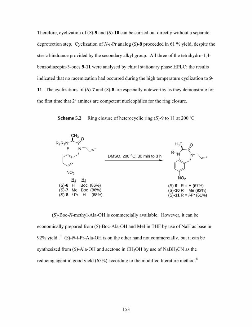
Therefore, cyclization of (S)-9 and (S)-10 can be carried out directly without a separate
deprotection step. Cyclization of N-i-Pr analog (S)-8 proceeded in 61 % yield, despite the
steric hindrance provided by the secondary alkyl group. All three of the tetrahydro-1,4-
benzodiazepin-3-ones 9-11 were analysed by chiral stationary phase HPLC; the results
indicated that no racemization had occurred during the high temperature cyclization to 9-
11. The cyclizations of (S)-7 and (S)-8 are especially noteworthy as they demonstrate for
the first time that 2º amines are competent nucleophiles for the ring closure.
Scheme 5.2 Ring closure of heterocyclic ring (S)-9 to 11 at 200 ºC
F
NO2
N
OR2R1N
CH3
(S)-6 H Boc (86%)(S)-7 Me Boc (86%)(S)-8 i-Pr H (68%)
DMSO, 200 oC, 30 min to 3 h NN
NO2
R
OH3C
(S)-9 R = H (67%)(S)-10 R = Me (92%)(S)-11 R = i-Pr (61%)
R1 R2
(S)-Boc-N-methyl-Ala-OH is commercially available. However, it can be
economically prepared from (S)-Boc-Ala-OH and MeI in THF by use of NaH as base in
92% yield .7 (S)-N-i-Pr-Ala-OH is on the other hand not commercially, but it can be
synthesized from (S)-Ala-OH and acetone in CH3OH by use of NaBH3CN as the
reducing agent in good yield (65%) according to the modified literature method.8
153
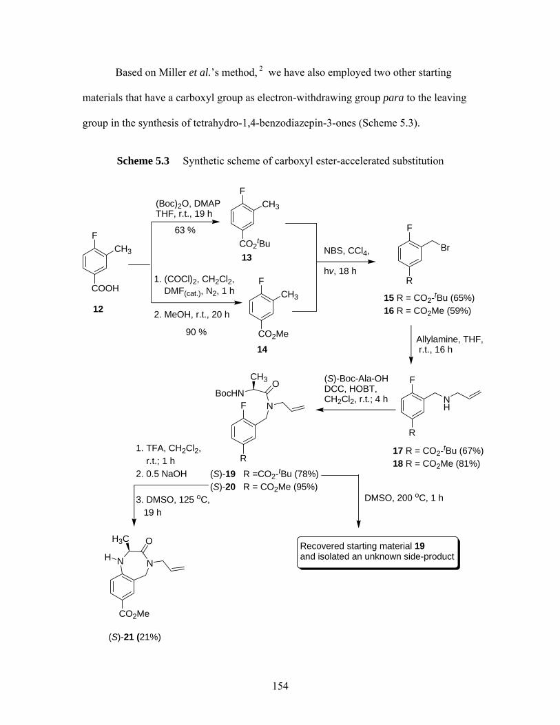
Based on Miller et al.’s method, 2 we have also employed two other starting
materials that have a carboxyl group as electron-withdrawing group para to the leaving
group in the synthesis of tetrahydro-1,4-benzodiazepin-3-ones (Scheme 5.3).
Scheme 5.3 Synthetic scheme of carboxyl ester-accelerated substitution
F
COOH
CH3 NBS, CCl4,
hv, 18 h
F
CO2tBu
CH3
Allylamine, THF, r.t., 16 h
F
R
NH
12
13
(S)-Boc-Ala-OHDCC, HOBT, CH2Cl2, r.t.; 4 h
F
R
N
OBocHN
CH3
1. TFA, CH2Cl2, r.t.; 1 h2. 0.5 NaOH
3. DMSO, 125 oC, 19 h
NN
CO2Me
H
OH3C
(S)-19 R =CO2-tBu (78%)(S)-20 R = CO2Me (95%)
F
R
Br
15 R = CO2-tBu (65%)16 R = CO2Me (59%)
(Boc)2O, DMAPTHF, r.t., 19 h
F
CO2Me
CH3
14
1. (COCl)2, CH2Cl2, DMF(cat.), N2, 1 h 2. MeOH, r.t., 20 h
63 %
90 %
17 R = CO2-tBu (67%)18 R = CO2Me (81%)
(S)-21 (21%)
DMSO, 200 oC, 1 h
Recovered starting material 19and isolated an unknown side-product
154
Reaction of 4-fluoro-3-methyl-benzoic acid with (Boc)2O9 and oxalyl chloride/MeOH10
afforded 13 (63%) and 14 (90%) accordingly; subsequent treatment with NBS in CCl4
afforded 15 and 16 in 65% and 59% yield correspondingly. Reactions of 15 and 16 with
allyamine and subsequent couplings with (S)-Boc-Ala-OH provided (S)-19 and (S)-20 in
good yields respectively. Attempts to ring close (S)-19 in DMSO at 200 °C were not
successful; a majority of the starting material (S)-19 was recovered, which demonstrated
that the successful cyclization of (S)-6 to 8 involved nucleophilic substitution of the
fluoride group by the corresponding amide, followed by loss of the Boc group. Small
amounts of an unidentified side product from attempted cyclization of (S)-19 were also
obtained. Therefore, due to the unsuccessful ring closure of (S)-19 at 200 °C, we carried
out the Boc deprotection step prior to performing the ring closure at 125 °C in DMSO.
The desired compound (S)-21 was however obtained in low yield (21%), which indicates
that stronger electron-withdrawing effect of NO2 group facilitates the ring closure of (S)-
9 to 11. Finally, attempts to methylate the N1of (S)-21 by use of NaH and MeOTf
yielded none of the desired product.
5.1.2 Synthesis of Quaternary (S)-alanine Derived Tetrahydro-1,4-benzodiazepin-3-ones Via “Memory of Chirality” (MOC)
We then attempted extension of our previously described11 synthesis of
quaternary tetrahydro-1,4-benzodiazepine-3-one (S)-10. Surprisingly, (S)-10 appears
unreactive toward alkylation under many different reaction conditions (Table 5.2).
Apparently, the electron-withdrawing NO2 group para to the N1-Me either prevents the
enolate formation or reduces the reactivity of the enolate towards the electrophile. We
155
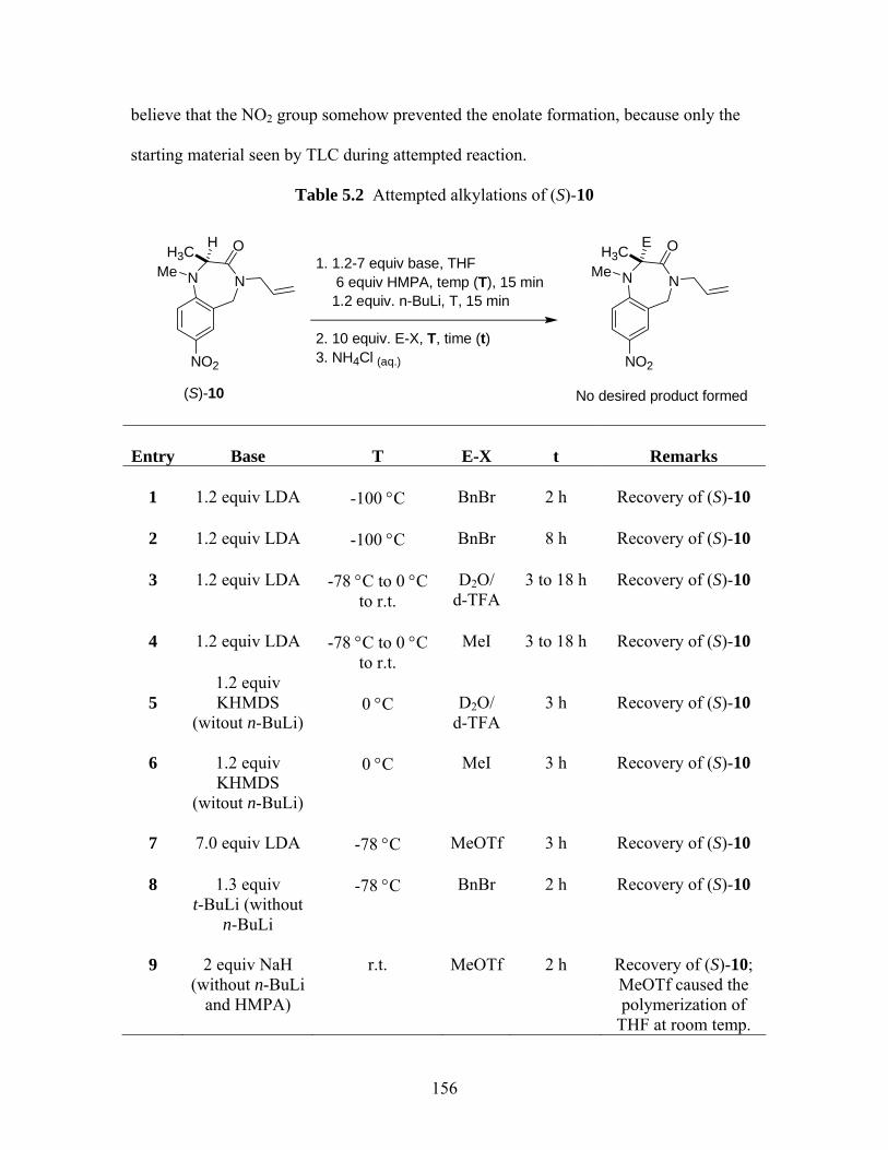
believe that the NO2 group somehow prevented the enolate formation, because only the
starting material seen by TLC during attempted reaction.
Table 5.2 Attempted alkylations of (S)-10
NNMe
NO2
O1. 1.2-7 equiv base, THF 6 equiv HMPA, temp (T), 15 min 1.2 equiv. n-BuLi, T, 15 min
2. 10 equiv. E-X, T, time (t)3. NH4Cl (aq.)
(S)-10
H3CH
NNMe
NO2
O
No desired product formed
H3CE
Entry
Base
T
E-X
t
Remarks
1
1.2 equiv LDA
-100 °C
BnBr
2 h
Recovery of (S)-10
2
1.2 equiv LDA
-100 °C
BnBr
8 h
Recovery of (S)-10
3
1.2 equiv LDA
-78 °C to 0 °C
to r.t.
D2O/
d-TFA
3 to 18 h
Recovery of (S)-10
4
1.2 equiv LDA
-78 °C to 0 °C
to r.t.
MeI
3 to 18 h
Recovery of (S)-10
5
1.2 equiv KHMDS
(witout n-BuLi)
0 °C
D2O/
d-TFA
3 h
Recovery of (S)-10
6
1.2 equiv KHMDS
(witout n-BuLi)
0 °C
MeI
3 h
Recovery of (S)-10
7
7.0 equiv LDA
-78 °C
MeOTf
3 h
Recovery of (S)-10
8
1.3 equiv
t-BuLi (without n-BuLi
-78 °C
BnBr
2 h
Recovery of (S)-10
9
2 equiv NaH
(without n-BuLi and HMPA)
r.t.
MeOTf
2 h
Recovery of (S)-10; MeOTf caused the polymerization of
THF at room temp.
156
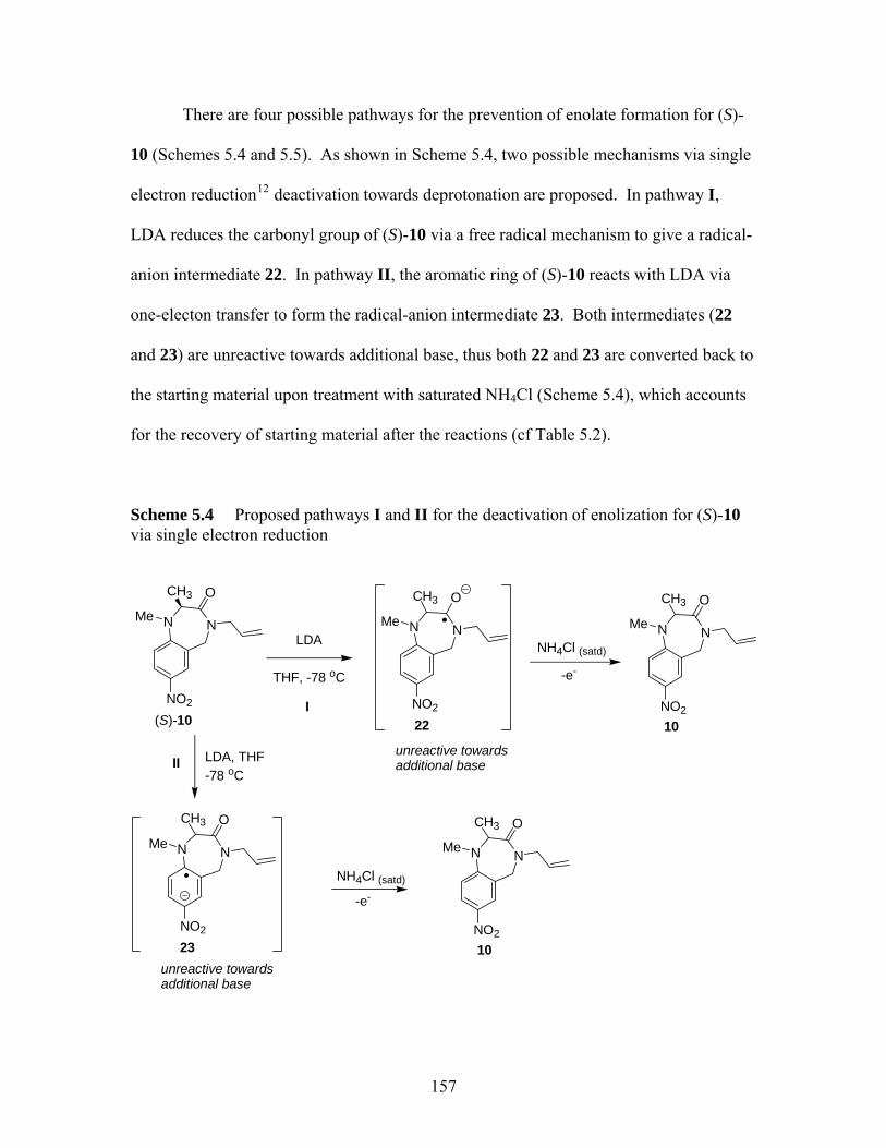
There are four possible pathways for the prevention of enolate formation for (S)-
10 (Schemes 5.4 and 5.5). As shown in Scheme 5.4, two possible mechanisms via single
electron reduction12 deactivation towards deprotonation are proposed. In pathway I,
LDA reduces the carbonyl group of (S)-10 via a free radical mechanism to give a radical-
anion intermediate 22. In pathway II, the aromatic ring of (S)-10 reacts with LDA via
one-electon transfer to form the radical-anion intermediate 23. Both intermediates (22
and 23) are unreactive towards additional base, thus both 22 and 23 are converted back to
the starting material upon treatment with saturated NH4Cl (Scheme 5.4), which accounts
for the recovery of starting material after the reactions (cf Table 5.2).
Scheme 5.4 Proposed pathways I and II for the deactivation of enolization for (S)-10 via single electron reduction
NNMe
NO2
OCH3
(S)-10
LDA
THF, -78 oC
NNMe
NO2
CH3
22I
LDA, THF-78 oC
NH4Cl (satd)
NNMe
NO2
OCH3
23
NH4Cl (satd)
II
O
-e-
-e-
NNMe
NO2
CH3 O
10
NNMe
NO2
CH3 O
10
unreactive towards additional base
unreactive towards additional base
157
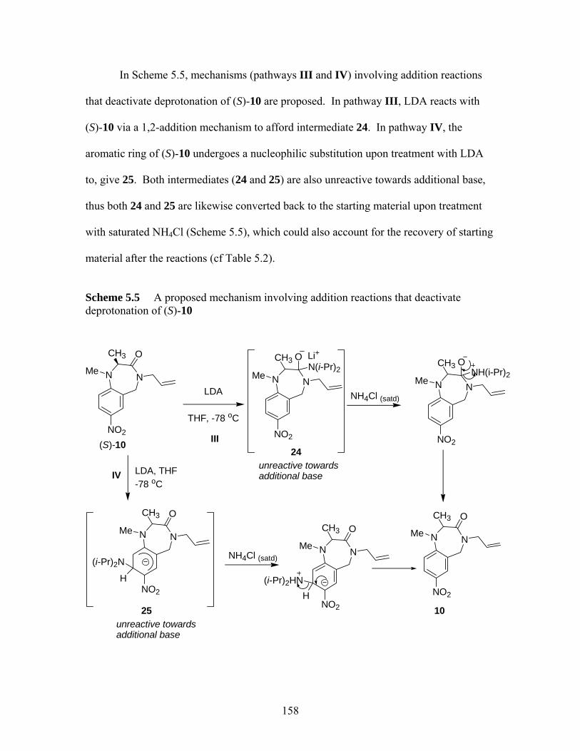
In Scheme 5.5, mechanisms (pathways III and IV) involving addition reactions
that deactivate deprotonation of (S)-10 are proposed. In pathway III, LDA reacts with
(S)-10 via a 1,2-addition mechanism to afford intermediate 24. In pathway IV, the
aromatic ring of (S)-10 undergoes a nucleophilic substitution upon treatment with LDA
to, give 25. Both intermediates (24 and 25) are also unreactive towards additional base,
thus both 24 and 25 are likewise converted back to the starting material upon treatment
with saturated NH4Cl (Scheme 5.5), which could also account for the recovery of starting
material after the reactions (cf Table 5.2).
Scheme 5.5 A proposed mechanism involving addition reactions that deactivate deprotonation of (S)-10
NNMe
NO2
OCH3
(S)-10
LDA
THF, -78 oC
NNMe
NO2
CH3
24
ON(i-Pr)2
III
LDA, THF-78 oC
NH4Cl (satd)NNMe
NO2
CH3 ONH(i-Pr)2
NNMe
NO2
OCH3
25
NNMe
NO2
OCH3
NH4Cl (satd)
IV
NNMe
NO2
OCH3
10
Li+
(i-Pr)2N
H (i-Pr)2HNH
unreactive towards additional base
unreactive towards additional base
158
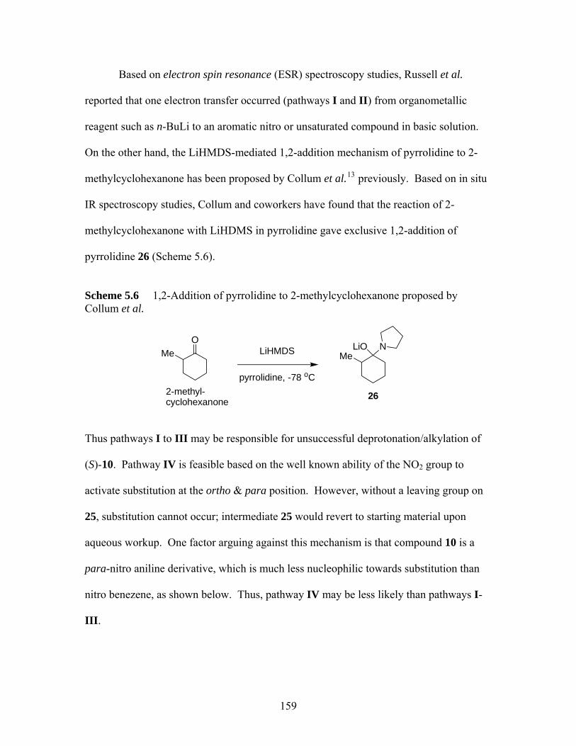
Based on electron spin resonance (ESR) spectroscopy studies, Russell et al.
reported that one electron transfer occurred (pathways I and II) from organometallic
reagent such as n-BuLi to an aromatic nitro or unsaturated compound in basic solution.
On the other hand, the LiHMDS-mediated 1,2-addition mechanism of pyrrolidine to 2-
methylcyclohexanone has been proposed by Collum et al.13 previously. Based on in situ
IR spectroscopy studies, Collum and coworkers have found that the reaction of 2-
methylcyclohexanone with LiHDMS in pyrrolidine gave exclusive 1,2-addition of
pyrrolidine 26 (Scheme 5.6).
Scheme 5.6 1,2-Addition of pyrrolidine to 2-methylcyclohexanone proposed by Collum et al.
O
Me LiHMDS
pyrrolidine, -78 oC
MeLiO N
262-methyl-cyclohexanone
Thus pathways I to III may be responsible for unsuccessful deprotonation/alkylation of
(S)-10. Pathway IV is feasible based on the well known ability of the NO2 group to
activate substitution at the ortho & para position. However, without a leaving group on
25, substitution cannot occur; intermediate 25 would revert to starting material upon
aqueous workup. One factor arguing against this mechanism is that compound 10 is a
para-nitro aniline derivative, which is much less nucleophilic towards substitution than
nitro benezene, as shown below. Thus, pathway IV may be less likely than pathways I-
III.
159
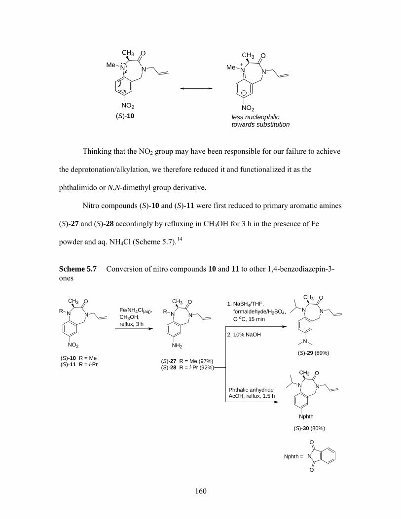
NNMe
NO2
OCH3
(S)-10
NNMe
NO2
OCH3
less nucleophilic towards substitution
Thinking that the NO2 group may have been responsible for our failure to achieve
the deprotonation/alkylation, we therefore reduced it and functionalized it as the
phthalimido or N,N-dimethyl group derivative.
Nitro compounds (S)-10 and (S)-11 were first reduced to primary aromatic amines
(S)-27 and (S)-28 accordingly by refluxing in CH3OH for 3 h in the presence of Fe
powder and aq. NH4Cl (Scheme 5.7).14
Scheme 5.7 Conversion of nitro compounds 10 and 11 to other 1,4-benzodiazepin-3-ones
CH3 OCH3 O
160
NNR
NO2
OCH3
(S)-10 R = Me(S)-11 R = i-Pr
Fe/NH4Cl(aq), CH3OH,reflux, 3 h
NNR
NH2
(S)-27 R = Me (97%)(S)-28 R = i-Pr (92%)
1. NaBH4/THF, formaldehyde/H2SO4, O oC, 15 min 2. 10% NaOH
NN
N
(S)-29 (89%)
Phthalic anhydrideAcOH, reflux, 1.5 h
NN
CH3 O
Nphth
(S)-30 (80%)
O
NNphth =
O
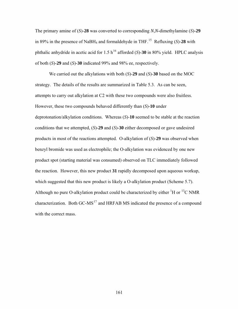
The primary amine of (S)-28 was converted to corresponding N,N-dimethylamine (S)-29
in 89% in the presence of NaBH4 and formaldehyde in THF.15 Refluxing (S)-28 with
phthalic anhydride in acetic acid for 1.5 h16 afforded (S)-30 in 80% yield. HPLC analysis
of both (S)-29 and (S)-30 indicated 99% and 98% ee, respectively.
We carried out the alkylations with both (S)-29 and (S)-30 based on the MOC
strategy. The details of the results are summarized in Table 5.3. As can be seen,
attempts to carry out alkylation at C2 with these two compounds were also fruitless.
However, these two compounds behaved differently than (S)-10 under
deprotonation/alkylation conditions. Whereas (S)-10 seemed to be stable at the reaction
conditions that we attempted, (S)-29 and (S)-30 either decomposed or gave undesired
products in most of the reactions attempted. O-alkylation of (S)-29 was observed when
benzyl bromide was used as electrophile; the O-alkylation was evidenced by one new
product spot (starting material was consumed) observed on TLC immediately followed
the reaction. However, this new product 31 rapidly decomposed upon aqueous workup,
which suggested that this new product is likely a O-alkylation product (Scheme 5.7).
Although no pure O-alkylation product could be characterized by either 1H or 13C NMR
characterization. Both GC-MS17 and HRFAB MS indicated the presence of a compound
with the correct mass.
161
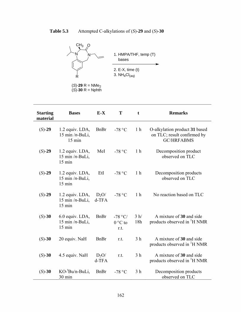
Table 5.3 Attempted C-alkylations of (S)-29 and (S)-30
NN
R
OCH3
(S)-29 R = NMe2(S)-30 R = Nphth
1. HMPA/THF, temp (T) bases 2. E-X, time (t)3. NH4Cl(aq)
Starting material
Bases
E-X
T
t
Remarks
(S)-29
1.2 equiv. LDA, 15 min /n-BuLi,
15 min
BnBr
-78 °C
1 h
O-alkylation product 31 based on TLC; result confirmed by
GC/HRFABMS
(S)-29
1.2 equiv. LDA, 15 min /n-BuLi, 15 min
MeI
-78 °C
1 h
Decomposition product
observed on TLC
(S)-29
1.2 equiv. LDA, 15 min /n-BuLi, 15 min
EtI
-78 °C
1 h
Decomposition products
observed on TLC
(S)-29
1.2 equiv. LDA, 15 min /n-BuLi, 15 min
D2O/
d-TFA
-78 °C
1 h
No reaction based on TLC
(S)-30
6.0 equiv. LDA, 15 min /n-BuLi, 15 min
BnBr
-78 °C/ 0 °C to
r.t.
3 h/ 18h
A mixture of 30 and side
products observed in 1H NMR
(S)-30
20 equiv. NaH
BnBr
r.t.
3 h
A mixture of 30 and side
products observed in 1H NMR
(S)-30
4.5 equiv. NaH
D2O/ d-TFA
r.t.
3 h
A mixture of 30 and side
products observed in 1H NMR
(S)-30
KO-tBu/n-BuLi, 30 min
BnBr
-78 °C
3 h
Decomposition products
observed on TLC
162
A proposed mechanism for the formation of O-alkylation product 31 and
subsequent decomposition of 31 upon aqueous workup is shown in Scheme 5.8. As seen
in Scheme 5.8, the enolate intermediate generated from deprotonation of 29 by bases
reacted with BnBr to give the O-alkylation product 31. Compound 31 was then
hydrolyzed by aq. NH4Cl to afford recovered starting material 29.
Scheme 5.8 A proposed mechanism for the formation and decomposition of O-alkylation product 31
NN
OH3C
HMPA/THF, LDA, -78 oC, 15 min n-BuLi, 15 min
(S)-29
N
NN
N
H3C OPh
NH4Cl(satd)
OH
HH
+
NN
N
H3C OPh
+ H2ONN
N
H3C OOH2
Ph
NN
N
H3C OOH
PhH
31
NN
N
H3C OH
-H+NN
N
H3C O
-PhCH2OH
29
, -78 oC, 1 h
NN
N
H3C O
enolate intermediate
Ph Br
Br
163
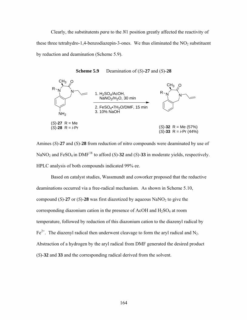
Clearly, the substitutents para to the N1 position greatly affected the reactivity of
these three tetrahydro-1,4-benzodiazepin-3-ones. We thus eliminated the NO2 substituent
by reduction and deamination (Scheme 5.9).
Scheme 5.9 Deamination of (S)-27 and (S)-28
NNR
NH2
OCH3
(S)-27 R = Me (S)-28 R = i-Pr
1. H2SO4/AcOH, NaNO2/H2O, 30 min 2. FeSO4•7H2O/DMF, 15 min3. 10% NaOH
NNR
OCH3
(S)-32 R = Me (57%)(S)-33 R = i-Pr (44%)
Amines (S)-27 and (S)-28 from reduction of nitro compounds were deaminated by use of
NaNO2 and FeSO4 in DMF18 to afford (S)-32 and (S)-33 in moderate yields, respectively.
HPLC analysis of both compounds indicated 99% ee.
Based on catalyst studies, Wassmundt and coworker proposed that the reductive
deaminations occurred via a free-radical mechanism. As shown in Scheme 5.10,
compound (S)-27 or (S)-28 was first diazotized by aqueous NaNO2 to give the
corresponding diazonium cation in the presence of AcOH and H2SO4 at room
temperature, followed by reduction of this diazonium cation to the diazenyl radical by
Fe2+. The diazenyl radical then underwent cleavage to form the aryl radical and N2.
Abstraction of a hydrogen by the aryl radical from DMF generated the desired product
(S)-32 and 33 and the corresponding radical derived from the solvent.
164
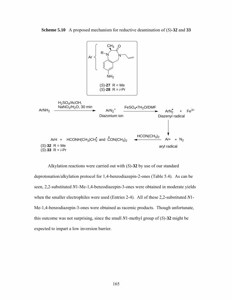
Scheme 5.10 A proposed mechanism for reductive deamination of (S)-32 and 33
NNR
NH2
OCH3
(S)-27 R = Me (S)-28 R = i-Pr
H2SO4/AcOH, NaNO2/H2O, 30 min FeSO4•7H2O/DMF
ArNH2
Ar
ArN2+ ArN2
ArHCON(CH3)2
ArH HCONH(CH3)CH2 and CON(CH3)2+
Diazonium ion
•
•• •
Diazenyl radical+ Fe3+
aryl radical
+ N2
(S)-32 R = Me (S)-33 R = i-Pr
Alkylation reactions were carried out with (S)-32 by use of our standard
deprotonation/alkylation protocol for 1,4-benzodiazepin-2-ones (Table 5.4). As can be
seen, 2,2-substituted N1-Me-1,4-benzodiazepin-3-ones were obtained in moderate yields
when the smaller electrophiles were used (Entries 2-4). All of these 2,2-substituted N1-
Me-1,4-benzodiazepin-3-ones were obtained as racemic products. Though unfortunate,
this outcome was not surprising, since the small N1-methyl group of (S)-32 might be
expected to impart a low inversion barrier.
165
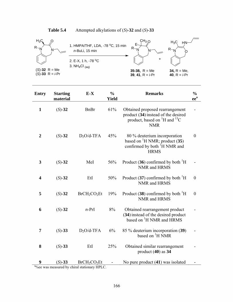
Table 5.4 Attempted alkylations of (S)-32 and (S)-33
Entry
Starting material
E-X
%
Yield
Remarks
% eea
1
(S)-32
BnBr
61%
Obtained proposed rearrangement product (34) instead of the desired
product, based on 1H and 13C NMR
-
2
(S)-32
D2O/d-TFA
45%
80 % deuterium incorporation
based on 1H NMR; product (35) confirmed by both 1H NMR and
HRMS
0
3
(S)-32
MeI
56%
Product (36) confirmed by both 1H
NMR and HRMS
-
4
(S)-32
EtI
50%
Product (37) confirmed by both 1H
NMR and HRMS
0
5
(S)-32
BrCH2CO2Et
19%
Product (38) confirmed by both 1H
NMR and HRMS
0
6
(S)-32
n-PrI
8%
Obtained rearrangement product
(34) instead of the desired product based on 1H NMR and HRMS
-
7
(S)-33
D2O/d-TFA
6%
85 % deuterium incorporation (39)
based on 1H NMR
-
8
(S)-33
EtI
25%
Obtained similar rearrangement
product (40) as 34
-
9
(S)-33
BrCH2CO2Et
-
No pure product (41) was isolated
-
a%ee was measured by chiral stationary HPLC.
NNR
OH3C1. HMPA/THF, LDA, -78 oC, 15 min n-BuLi, 15 min
2. E-X, 1 h, -78 oC3. NH4Cl (aq)
NNR
OCH3E
((S)-32 R = MeS)-33 R = i-Pr
35-38, R = Me 39, 41, R = i-Pr
34, R = Me, 40, R = i-Pr
ONR
+
H3C HN
166

On the other hand, when a larger electrophile like BnBr or n-PrI (Table 5.4, Entry 1 & 6)
was used, no desired product was obtained. Instead, these reactions yielded an isomer of
the starting material (S)-32, as confirmed by both GC-MS and HRMS. In any event, the
isolation of desired alkylation products 35-38, and the rearrangement product 34 indicates
that enolate formation from 32 is not problematic. Attempted deprotonation of N1-i-Pr
analogs (S)-33 was less successful. No alkylation products were obtained with EtI or
BrCH2CO2Et (Table 5.4, Entry 8 & 9). Instead the analogous N-iPr rearrangement
product 40 was however obtained. These results indicate that the reactivity of the enolate
is so low that the rearrangement reaction outcompetes alkylation.
To determine what role, if any, the allyl group played in this rearrangement. We
hydrogenated the allyl group to n-propyl group by 10% Pd/C in CH3OH to afford N4-n-
propyl benzodiazepine-3-one (S)-42 in quantitative yield.19 Results of alkylation of (S)-
42 are summarized in Table 5.5.
167
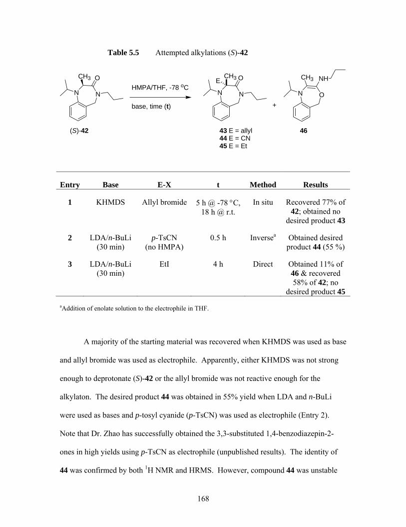
Table 5.5 Attempted alkylations (S)-42
NN
OCH3
(S)-42
HMPA/THF, -78 oC
base, time (t)
NN
O
43 E = allyl44 E = CN45 E = Et
ON
46
CH3E NHCH3
+
Entry Base E-X t Method Results
1 KHMDS Allyl bromide 5 h @ -78 °C, 18 h @ r.t.
In situ Recovered 77% of 42; obtained no
desired product 43
2 LDA/n-BuLi (30 min)
p-TsCN (no HMPA)
0.5 h Inversea Obtained desired product 44 (55 %)
3
LDA/n-BuLi (30 min)
EtI 4 h Direct Obtained 11% of 46 & recovered 58% of 42; no
desired product 45
aAddition of enolate solution to the electrophile in THF.
A majority of the starting material was recovered when KHMDS was used as base
and allyl bromide was used as electrophile. Apparently, either KHMDS was not strong
enough to deprotonate (S)-42 or the allyl bromide was not reactive enough for the
alkylaton. The desired product 44 was obtained in 55% yield when LDA and n-BuLi
were used as bases and p-tosyl cyanide (p-TsCN) was used as electrophile (Entry 2).
Note that Dr. Zhao has successfully obtained the 3,3-substituted 1,4-benzodiazepin-2-
ones in high yields using p-TsCN as electrophile (unpublished results). The identity of
44 was confirmed by both 1H NMR and HRMS. However, compound 44 was unstable
168
and decomposition was observed at room temperature. Finally, attempted reaction of the
enolate derived from (S)-42 with ethyl iodide gave an apparent rearrangement product 46.
The 1H NMR spectrum of 46 is highly similar to that of 40, except the allyl and n-propyl
regions (Table 5.6). The 1H NMR data of both compounds 40 and 46 are summarized in
Table 5.6. HRMS confirmed that 46 is a constitutional isomer of 42.
169
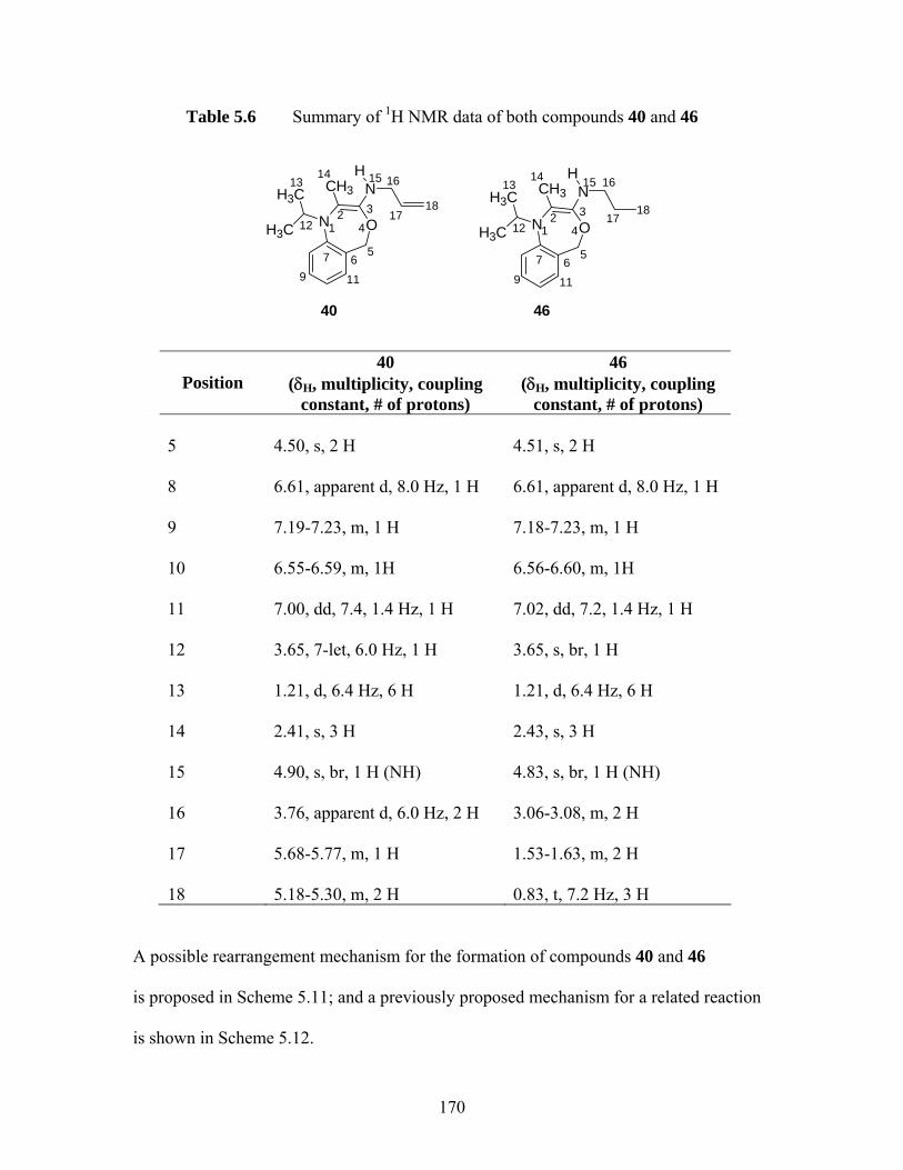
Table 5.6 Summary of 1H NMR data of both compounds 40 and 46
ONH3C
H3C NCH3
40
12 3
4
567
9
H
12
1314 15 16
17ONH3C
H3C NCH3
46
12 3
4
567
119
H
12
1314 15 16
1718 18
11
Position
40 (δH, multiplicity, coupling
constant, # of protons)
46 (δH, multiplicity, coupling
constant, # of protons) 5
4.50, s, 2 H
4.51, s, 2 H
8
6.61, apparent d, 8.0 Hz, 1 H
6.61, apparent d, 8.0 Hz, 1 H
9
7.19-7.23, m, 1 H
7.18-7.23, m, 1 H
10
6.55-6.59, m, 1H
6.56-6.60, m, 1H
11
7.00, dd, 7.4, 1.4 Hz, 1 H
7.02, dd, 7.2, 1.4 Hz, 1 H
12
3.65, 7-let, 6.0 Hz, 1 H
3.65, s, br, 1 H
13
1.21, d, 6.4 Hz, 6 H
1.21, d, 6.4 Hz, 6 H
14
2.41, s, 3 H
2.43, s, 3 H
15
4.90, s, br, 1 H (NH)
4.83, s, br, 1 H (NH)
16
3.76, apparent d, 6.0 Hz, 2 H
3.06-3.08, m, 2 H
17
5.68-5.77, m, 1 H
1.53-1.63, m, 2 H
18
5.18-5.30, m, 2 H
0.83, t, 7.2 Hz, 3 H
A possible rearrangement mechanism for the formation of compounds 40 and 46
is proposed in Scheme 5.11; and a previously proposed mechanism for a related reaction
is shown in Scheme 5.12.
170
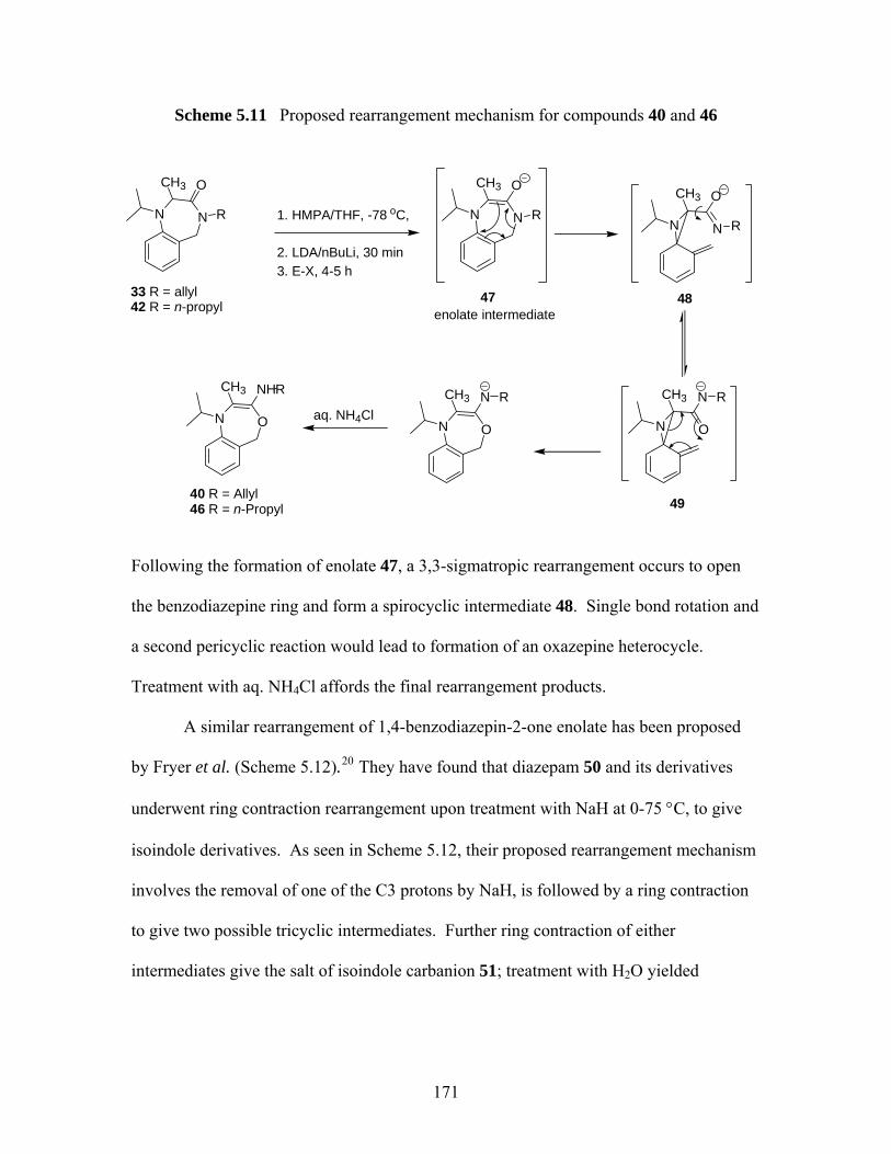
Scheme 5.11 Proposed rearrangement mechanism for compounds 40 and 46
NN
OCH3
R 1. HMPA/THF, -78 oC,
2. LDA/nBuLi, 30 min3. E-X, 4-5 h
NN
OCH3
RNN
OCH3
R
ON
NCH3 R
ON
NCH3 Raq. NH4ClON
NHCH3 R
40 R = Allyl46 R = n-Propyl
33 R = allyl 42 R = n-propyl enolate intermediate
47 48
49
Following the formation of enolate 47, a 3,3-sigmatropic rearrangement occurs to open
the benzodiazepine ring and form a spirocyclic intermediate 48. Single bond rotation and
a second pericyclic reaction would lead to formation of an oxazepine heterocycle.
Treatment with aq. NH4Cl affords the final rearrangement products.
A similar rearrangement of 1,4-benzodiazepin-2-one enolate has been proposed
by Fryer et al. (Scheme 5.12).20 They have found that diazepam 50 and its derivatives
underwent ring contraction rearrangement upon treatment with NaH at 0-75 °C, to give
isoindole derivatives. As seen in Scheme 5.12, their proposed rearrangement mechanism
involves the removal of one of the C3 protons by NaH, is followed by a ring contraction
to give two possible tricyclic intermediates. Further ring contraction of either
intermediates give the salt of isoindole carbanion 51; treatment with H2O yielded
171

isoindolenine 52. Though Fryer et al. did not draw a discrete enolate intermediate, the
basic mechanism remains unchanged.
Scheme 5.12 Two ring contraction rearrangement mechanisms of diazepam proposed by Fryer et al 20
The proposed mechanism in Scheme 5.11 like that in Scheme 5.12 involves the
disruption of aromaticity in the enolate intermediate 47 to form a strained 3-membered
ring intermediate 48. Such process would be expected to have a high activation barrier.
In order to assess the feasibility of our proposed mechanism (Scheme 5.11), we
conducted equilibrium geometry optimization calculations of both enolate 47 and
spirocylic intermediate 48 at B3LYP/6-31G(d) level. The energy difference between
these two intermediates (47 and 48) would give us an idea about the activation barrier of
this reaction. According to the energy that we obtained at B3LYP/6-31G(d), the enolate
intermediate 47 is lower in energy than the intermediate 48 by 27.0 kcal/mol, which
NN
Ph
H3C
O HH
HO
NNH3C
Ph O
NPh
CH3HN
tricyclic intermediate
NPh
OH3CHN H
H2ONN
Ph
H3C
O HH
H
NN
H3C
O
Ph
50
50
51
52
172

means that the activation barrier for this reaction would be higher than 27.0 kcal/mol.
Therefore, the proposed mechanism in Scheme 5.11 is not likely to be rapid at -78 °C.
Note that the rearrangement reported by Fryer et al. occurs at 0-75 °C (cf Scheme 5.12).
We therefore propose the following free radical mechanism (Scheme 5.13) to
account for the formation of the rearrangement products 40 and 46. As seen in Scheme
5.13, single electron reduction of either 33 or 42 by LDA gives a radical-anion
intermediate 53; fragmentation of the 7-membered ring forms a benzylic radical-anion
intermediate 54. Single bond rotation of 54 and cyclization lead to third radical-anion
intermediate 55. Finally, oxidation of 55 upon treatment of aq. NH4Cl affords the
rearrangement products. This proposed free radical mechanism is more feasible than that
proposed in Scheme 5.11 due to two reasons; firstly, this mechanism does not involve the
disruption of aromaticity of the aromatic ring nor involve the formation of a strained 3-
membered ring. Secondly, the benzylic radical intermediate 54 can be stabilized by the
aromatic ring by delocation.
173
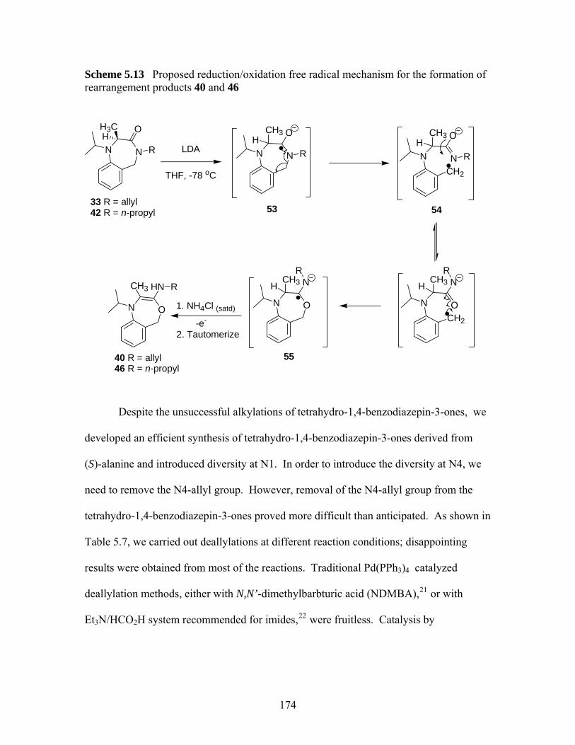
Scheme 5.13 Proposed reduction/oxidation free radical mechanism for the formation of rearrangement products 40 and 46
NN
O
R
33 R = allyl42 R = n-propyl
LDA
THF, -78 oC
NN R
53
1. NH4Cl (satd)ON
CH3
O
HN
-e-
CH2
NN R
54
O
CH2
ON
NR
ON
55
NR
R
40 R = allyl46 R = n-propyl
2. Tautomerize
HH3C
H HCH3 CH3
HCH3 H
CH3
Despite the unsuccessful alkylations of tetrahydro-1,4-benzodiazepin-3-ones, we
developed an efficient synthesis of tetrahydro-1,4-benzodiazepin-3-ones derived from
(S)-alanine and introduced diversity at N1. In order to introduce the diversity at N4, we
need to remove the N4-allyl group. However, removal of the N4-allyl group from the
tetrahydro-1,4-benzodiazepin-3-ones proved more difficult than anticipated. As shown in
Table 5.7, we carried out deallylations at different reaction conditions; disappointing
results were obtained from most of the reactions. Traditional Pd(PPh3)4 catalyzed
deallylation methods, either with N,N’-dimethylbarbturic acid (NDMBA),21 or with
Et3N/HCO2H system recommended for imides,22 were fruitless. Catalysis by
174
RhCl(PPh)323 also proved ineffectual, and RuCl2 3(PPh) 24 catalyzed deallylation
proceeded only in low to moderate yields.
175
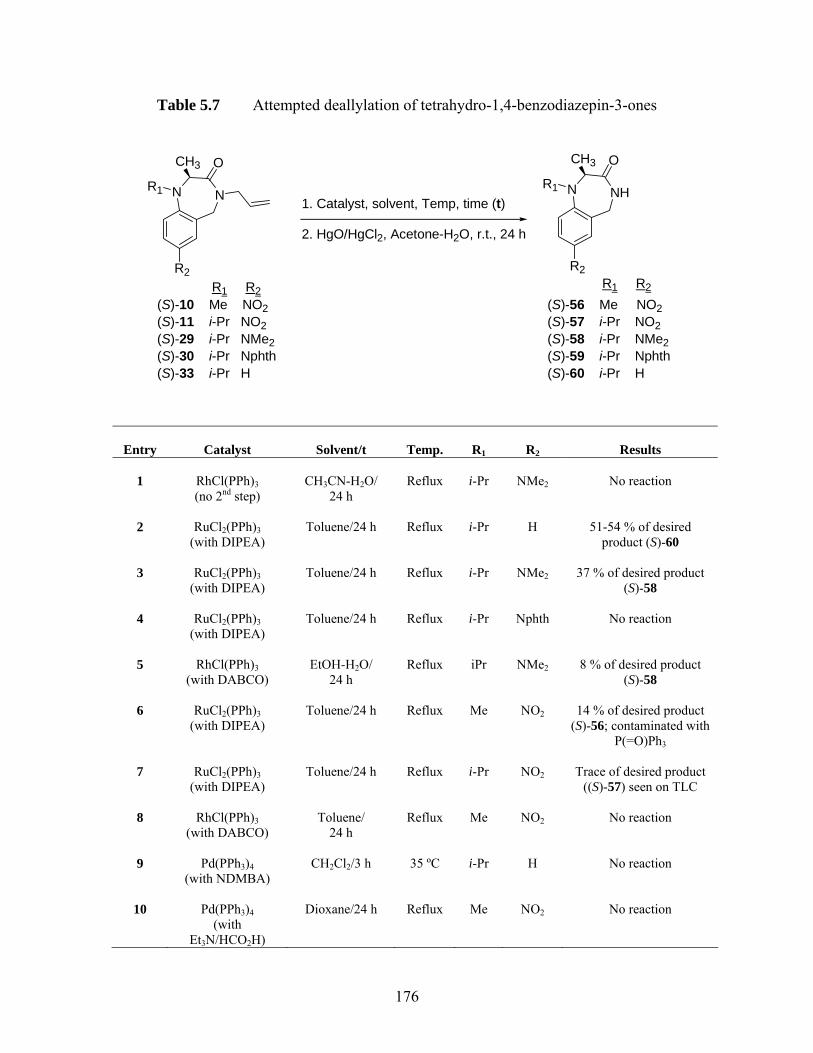
Table 5.7 Attempted deallylation of tetrahydro-1,4-benzodiazepin-3-ones
NNR1
R2
OCH3
1. Catalyst, solvent, Temp, time (t)
2. HgO/HgCl2, Acetone-H2O, r.t., 24 h
NHNR1
R2
OCH3
R1 R2(S)-10 Me NO2(S)-11 i-Pr NO2(S)-29 i-Pr NMe2(S)-30 i-Pr Nphth(S)-33 i-Pr H
R1 R2
(S)-56 Me NO2(S)-57 i-Pr NO2(S)-58 i-Pr NMe2(S)-59 i-Pr Nphth(S)-60 i-Pr H
Entry
Catalyst
Solvent/t
Temp.
R1
R2
Results
1
RhCl(PPh)3(no 2nd step)
CH3CN-H2O/
24 h
Reflux
i-Pr
NMe2
No reaction
2
RuCl2(PPh)3
(with DIPEA)
Toluene/24 h
Reflux
i-Pr
H
51-54 % of desired
product (S)-60
3
RuCl2(PPh)3 (with DIPEA)
Toluene/24 h
Reflux
i-Pr
NMe2
37 % of desired product
(S)-58
4
RuCl2(PPh)3 (with DIPEA)
Toluene/24 h
Reflux
i-Pr
Nphth
No reaction
5
RhCl(PPh)3
(with DABCO)
EtOH-H2O/
24 h
Reflux
iPr
NMe2
8 % of desired product
(S)-58
6
RuCl2(PPh)3 (with DIPEA)
Toluene/24 h
Reflux
Me
NO2
14 % of desired product
(S)-56; contaminated with P(=O)Ph3
7
RuCl2(PPh)3
(with DIPEA)
Toluene/24 h
Reflux
i-Pr
NO2
Trace of desired product
((S)-57) seen on TLC
8
RhCl(PPh)3(with DABCO)
Toluene/
24 h
Reflux
Me
NO2
No reaction
9
Pd(PPh3)4
(with NDMBA)
CH2Cl2/3 h
35 ºC
i-Pr
H
No reaction
10
Pd(PPh3)4
(with Et3N/HCO2H)
Dioxane/24 h
Reflux
Me
NO2
No reaction
176

Rh (I) or Ru (II) catalyzed deallylations involve isomerization of allylamine to
enamine 61 (Scheme 5.14), followed by acidic hydrolysis the amine. The isomerization
of 33 to 61 should not be problematic. However, for deprotection of N4-allyl of
tetrahydro-1,4-benzodiazepin-3-ones, the enamine intermediate 61 is actually an enamide
and is thus rather non basic. The unfavorable formation of acyliminium ion 62 is thus
likely the rate-determining step for Rh (I) or Ru (II) catalyzed deallylations of 33, which
may accounts for the low yielding in these reactions (cf Table 5.7, Entries 1-8).
Scheme 5.14 A proposed mechanism for an example of Rh (I) or Ru (II) catalyzed deallylations of tetrahydro-1,4-benzodiazepin-3-one (S)-33
NN
OH3C
Rh(I) or Ru(II) catalyst NN
OH3C
61
H+
NN
OH3C
62
H2O
NN
OH3COH2
proton transferNN
OH3CH
OH
NN
O
H
H3C
(S)-33
(S)-60
-CH3CH2CHO
In order to understand why the deallylations of tetrahydro-1,4-benzodiazepin-3-
ones by Pd(PPh3)4 in the presence of NMDBA were unsuccessful, we present in Scheme
5.15 a general catalytic cycle involved in the deallylation of allylamines by Pd(PPh3)4
/NMDBA as proposed by Guibé et al.
177
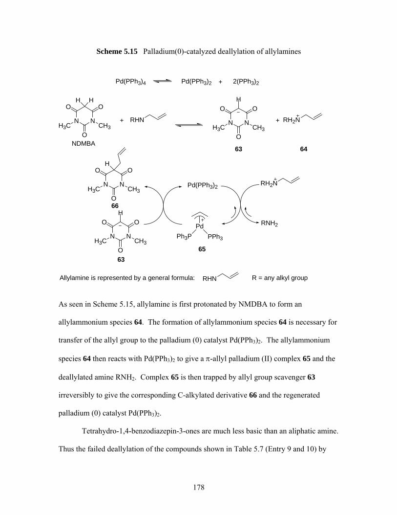
Scheme 5.15 Palladium(0)-catalyzed deallylation of allylamines
N N
O O
OH3C CH3
H H
RHN+ N N
O O
OH3C CH3
H
RH2N+
Pd(PPh3)4 Pd(PPh3)2 + 2(PPh3)2
RH2N
RNH2
Pd(PPh3)2
PdPPh3Ph3PN N
O O
OH3C CH3
H
N N
O O
OH3C CH3
H
NDMBA63
6365
66
RHNAllylamine is represented by a general formula: R = any alkyl group
64
As seen in Scheme 5.15, allylamine is first protonated by NMDBA to form an
allylammonium species 64. The formation of allylammonium species 64 is necessary for
transfer of the allyl group to the palladium (0) catalyst Pd(PPh3)2. The allylammonium
species 64 then reacts with Pd(PPh3)2 to give a π-allyl palladium (II) complex 65 and the
deallylated amine RNH2. Complex 65 is then trapped by allyl group scavenger 63
irreversibly to give the corresponding C-alkylated derivative 66 and the regenerated
palladium (0) catalyst Pd(PPh3)2.
Tetrahydro-1,4-benzodiazepin-3-ones are much less basic than an aliphatic amine.
Thus the failed deallylation of the compounds shown in Table 5.7 (Entry 9 and 10) by
178
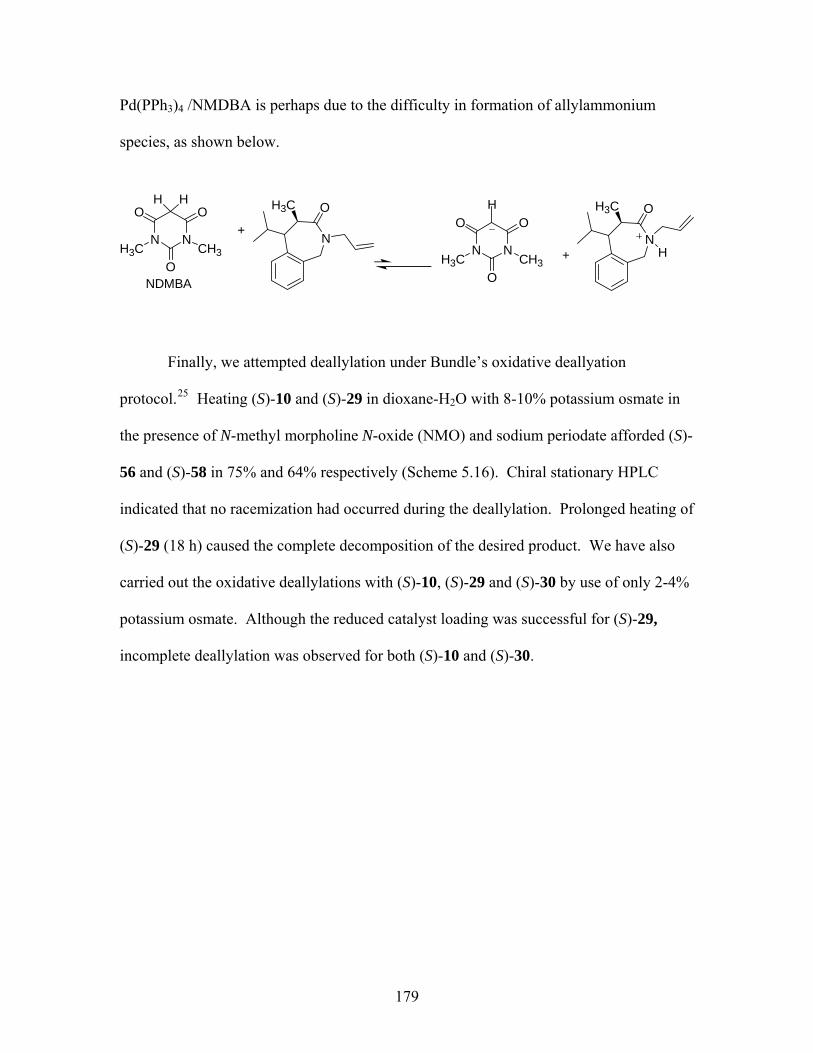
Pd(PPh3)4 /NMDBA is perhaps due to the difficulty in formation of allylammonium
species, as shown below.
N N
O O
OH3C CH3
H H
+N N
O O
OH3C CH3
H
+
NDMBA
N
OH3C
N
OH3C
H
Finally, we attempted deallylation under Bundle’s oxidative deallyation
protocol.25 Heating (S)-10 and (S)-29 in dioxane-H2O with 8-10% potassium osmate in
the presence of N-methyl morpholine N-oxide (NMO) and sodium periodate afforded (S)-
56 and (S)-58 in 75% and 64% respectively (Scheme 5.16). Chiral stationary HPLC
indicated that no racemization had occurred during the deallylation. Prolonged heating of
(S)-29 (18 h) caused the complete decomposition of the desired product. We have also
carried out the oxidative deallylations with (S)-10, (S)-29 and (S)-30 by use of only 2-4%
potassium osmate. Although the reduced catalyst loading was successful for (S)-29,
incomplete deallylation was observed for both (S)-10 and (S)-30.
179
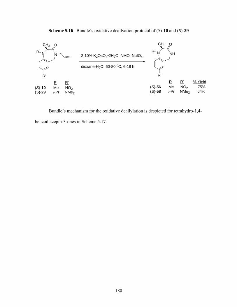
Scheme 5.16 Bundle’s oxidative deallyation protocol of (S)-10 and (S)-29
NNR
R'
OCH3
(S)-10 Me NO2 (S)-29 i-Pr NMe2
R R'
2-10% K2OsO4•2H2O, NMO, NaIO4,
dioxane-H2O, 60-80 oC, 6-18 h
NHNR
R'
OCH3
(S)-56 Me NO2 75%(S)-58 i-Pr NMe2 64%
R R' % Yield
Bundle’s mechanism for the oxidative deallylation is despicted for tetrahydro-1,4-
benzodiazepin-3-ones in Scheme 5.17.
180
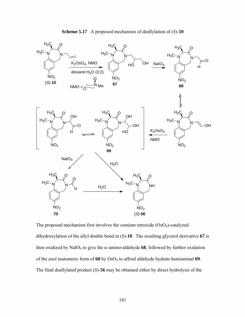
Scheme 5.17 A proposed mechanism of deallylation of (S)-10
OH
NNH3C
O
NO2
H3C
(S)-10
NNH3C
O
NO2
H3C
67
K2OsO4, NMO
dioxane:H2O (3:2)
HO OH NaIO4
NNH3CO
O
NO2
H3C
68
H
NNH3COH
O
NO2
H3C
K2OsO4
NMO
NNH3C
O
NO2
H3C
69
HO
OHNNH3C
O
O
NO2
H3C
H
OH
ON Me
O
NMO =
H2O
NHNH3C
O
NO2
H3C
(S)-56
NNH3C
O
NO2
H3C
70
H
O
H2O
NaIO4
The proposed mechanism first involves the osmium tetroxide (OsO4)-catalyzed
dihydroxylation of the allyl double bond in (S)-10. The resulting glycerol derivative 67 is
then oxidized by NaIO4 to give the α-amino-aldehyde 68, followed by further oxidation
of the enol tautomeric form of 68 by OsO4 to afford aldehyde hydrate hemiaminal 69.
The final deallylated product (S)-56 may be obtained either by direct hydrolysis of the
181
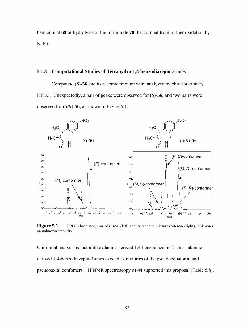
hemiaminal 69 or hydrolysis of the formimide 70 that formed from further oxidation by
NaIO4.
5.1.3 Computational Studies of Tetrahydro-1,4-benzodiazepin-3-ones
Compound (S)-56 and its racemic mixture were analyzed by chiral stationary
HPLC. Unexpectedly, a pair of peaks were observed for (S)-56, and two pairs were
observed for (S/R)-56, as shown in Figure 5.1.
NO2
(S)-56 (S/R)-56
N
NH
NO2
H3C
O
H3CN
H3C
(M, R)-conformer
Figure 5.1 HPLC chromatograms of (S)-56 (left) and its racemic mixture (S/R)-56 (right); X denotes an unknown impurity
Our initial analysis is that unlike alanine-derived 1,4-benzodiazepin-2-ones, alanine-
derived 1,4-benzodiazepin-3-ones existed as mixtures of the pseudoequatorial and
pseudoaxial confomers. 1H NMR spectroscopy of 44 supported this proposal (Table 5.8).
(P)-conformer
(M)-conformer
(P, S)-conformer
(M, R)-conformer
(M, S)-conformer(P, R)-conformer
xx
NH3C
HO
182
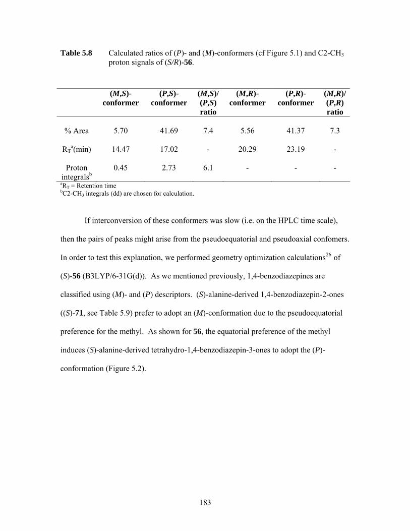
Table 5.8 Calculated ratios of (P)- and (M)-conformers (cf Figure 5.1) and C2-CH3 proton signals of (S/R)-56.
(M,S)-conformer
(P,S)-conformer
(M,S)/ (P,S) ratio
(M,R)-conformer
(P,R)-conformer
(M,R)/ (P,R) ratio
% Area
5.70
41.69
7.4
5.56
41.37
7.3
RT
a(min)
14.47
17.02 -
20.29
23.19
-
Proton
integralsb
0.45
2.73
6.1
-
-
-
aRT = Retention time bC2-CH3 integrals (dd) are chosen for calculation.
If interconversion of these conformers was slow (i.e. on the HPLC time scale),
then the pairs of peaks might arise from the pseudoequatorial and pseudoaxial confomers.
In order to test this explanation, we performed geometry optimization calculations26 of
(S)-56 (B3LYP/6-31G(d)). As we mentioned previously, 1,4-benzodiazepines are
classified using (M)- and (P) descriptors. (S)-alanine-derived 1,4-benzodiazepin-2-ones
((S)-71, see Table 5.9) prefer to adopt an (M)-conformation due to the pseudoequatorial
preference for the methyl. As shown for 56, the equatorial preference of the methyl
induces (S)-alanine-derived tetrahydro-1,4-benzodiazepin-3-ones to adopt the (P)-
conformation (Figure 5.2).
183

(P)-(S)-56 (M)-(S)-56 pseudoequatorial-Me pseudoaxial-Me 0 kcal/mol +2.0 kcal/mol
Figure 5.2 B3LYP/6-31G(d) electronic energy difference between the (M)- and (P)- conformers of (S)-56; the sign of the dihedral angle C6-C7-N1-C2 defines the helical chirality descriptor (M (minus) or P (plus))
As can be seen in Figure 5.2, two equilibrium geometries were found for the (S)-
enantiomer of 56. The (P)-conformer of 56, which features an equatorial C2-Me, is
lower in energy than the (M)-conformer by 2.0 kcal/mol. Thus the equatorial preference
for tetrahydro-1,4-benzodiazepin-3-ones is calculated to be considerably less than that of
tetrahydro-1,4-benzodiazepin-2-ones27 (Table 5.9). The calculated electronic energy
difference of (S)-56 and sum of internal angles of both (S)-56 and (S)-71 are summarized
in Table 5.9. The sum of the internal angles of the (P)- and (M)-conformers of (S)-56 are
829° and 844° respectively. These angles are between the value of 809° calculated for a
puckered ring from simple VSEPR considerations28 and the value of 900° calculated for
a flat heptagon.29 The calculated sum of internal angles for (S)-71 is 833°, which
corresponds to a calculated value of 829° based on the simple VSEPR considerations.
Since the calculated sum of internal angles for (S)-71 is close to the estimate based on
VSEPR considerations, while the sum of internal angles for (P)-S-56 and (M)-S-56 are
20o and 35o different than the VSEPR estimate, then N1-Me (S)-alanine-derived
184
tetrahydro-1,4-benzodiazepin-3-ones appear to be flatter than N1-Me (S)-alanine-derived
1,4-benzodiazepin-2-ones. This greater flatness may account for the smaller
pseudoequatorial preference for the methyl seen in the tetrahydro-1,4-benzodiazepin-3-
ones, because a flatter structure may make interconversion between the two
conformations easier.
To test our argument, we also performed equilibrium geometry calculations for
glycine-derived tetrahydro-1,4-benzodiazepine-3-one 72 by use of the same basis set.
The (P)- and (M)-conformers of 72 are enantiomers and therefore equal in energy. In the
equilibrium geometries, the sum of the internal angles of both (P) and (M)-conformers is
833° (Table 5.9), which is close to the sum of internal angles of (P)- and (M)-conformers
of (S)-56. So, since the sum of internal angles for (P)- and (M)-conformers of (S)-56 are
close to the sum of internal angles for (M)- or (P)-72, for which there is no
conformational preference, then these calculations support the idea that there is little
preference between (P)- and (M)-conformers of (S)-56 as well. The observed
pseudoequatorial:pseudoaxial ratios (cf Table 5.8) of 6:1 (1H NMR) and 7:1 (HPLC) for
(S)-56 correspond to an energy difference of 1.1 kcal/mol at 298 K, which is very close to
the calculated electronic energy difference of 2.0 kcal/mol.
185

Table 5.9 Summary of sum of internal angles and energy differences (B3LYP/6-31G(d))
NNH3C
O
H
NO2
NN
Cl
H3C
Ph
O CH3
(3S)-71 72
NNH3C
O
H
NO2
(2S)-56
H3C
Compound C3 methyl
ΔEa (kcal/mol)
Sum of internal angle (°) based on
computational studies
Sum of internal angle (°) based on simple
VSEPR considerations
(P)-(S)-56 eq 0 829 809
(M)-(S)-56 ax 2.0 844 809
(M)-(S)-71b eq 0 833 829
(P)-(S)-71b ax 4.7 848 829
(P)-72 na 0 833 809
(M)-72 na 0 833 809 aDifference in electronic energies between axial and equatorial conformers bΔE and sum of internal angles of compound 71 were calculated by Dr. Polo Lam.
5.2 Investigation of Enantioselective Synthesis of Quaternary (S)-phenylalanine Derived Tetrahydro-1,4-benzodiazepin-2-ones
5.2.1 Synthesis of (S)-phenylalanine Derived Tetrahydro-1,4-benzodiazepin-2-ones
As mentioned previously, various tetrahydro-1,4-benzodiazepin-2-ones have been
found to possess important biological activity.30 As in the case of tetrahydro-1,4-
186

benzodiazepin-3-ones, 3,3-substituted derivatives are unexplored territory. We are
therefore also interested in extending our MOC protocol to this subtype.
The synthesis of certain tetrahydro-1,4-benzodiazepin-2-ones has been previously
reported either by solid phase synthesis31 or via phenyliodine (III)bis(trifluoroacetate)-
mediated intramolecular cyclization.32 We carried out the synthesis of (S)-phenylalanine-
derived tetrahydro-1,4-benzodiazepin-2-ones based on modified literature method
(Scheme 5.18). The synthesis started with commercially available 2-nitrobenzylbromide
(73): treatment with (S)-phenylalanine methyl ester hydrochloride in the presence of
DIEA in DMF afforded (S)-74 in 95% yield. Heating of (S)-74 with either excess CH3I
or allyl bromide in DMF at the boiling point of the alkylating agent in the presence of
DIEA provided (S)-75 and (S)-76 respectively in good yields. However, attempts to
carry out reductive ring closure of (S)-75 by use of SnCl2•2H2O/NH4OAc were
completely unsuccessful. (S)-75 decomposed to (S)-N-methyl phenylalanine methyl ester
(77) and 2-amino benzylethylether (78) via elimination/addition (Scheme 5.18).
187
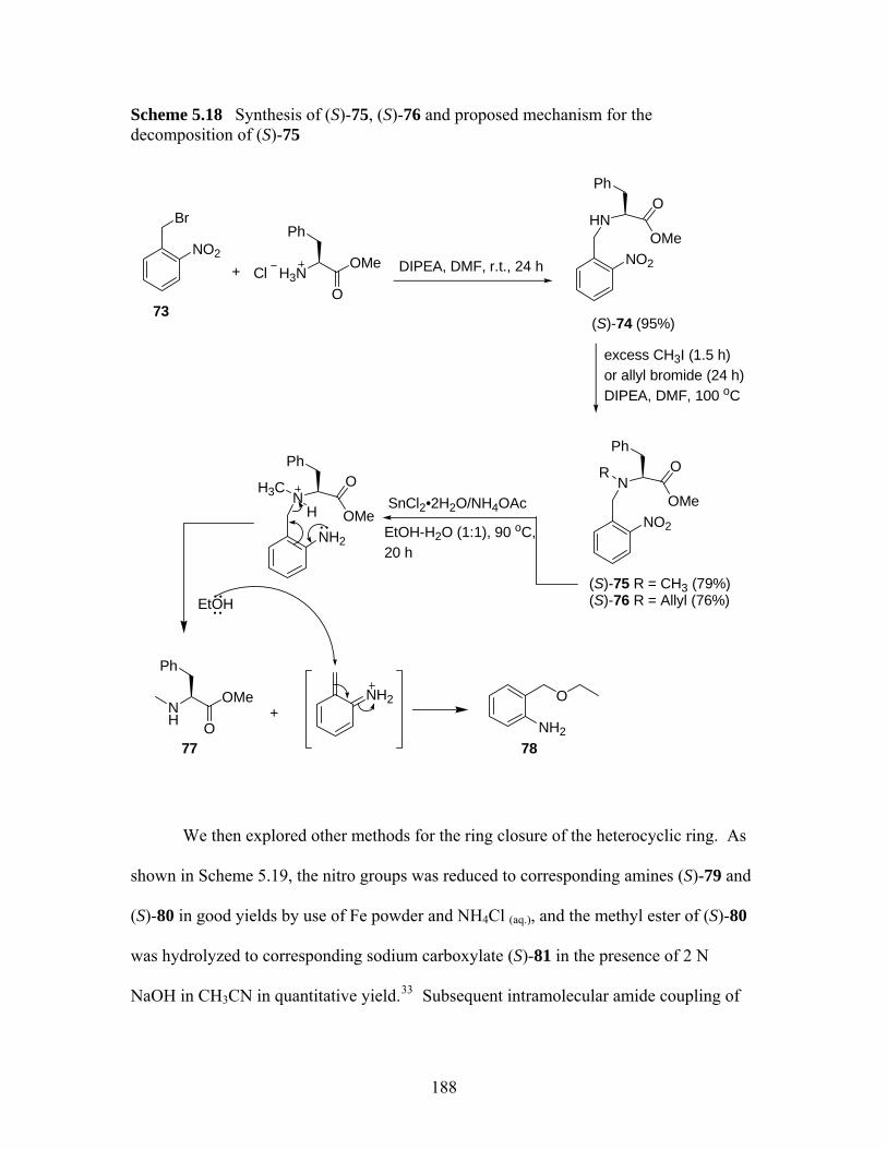
Scheme 5.18 Synthesis of (S)-75, (S)-76 and proposed mechanism for the decomposition of (S)-75
H3NOMe
O
Ph
Cl+ DIPEA, DMF, r.t., 24 hNO2
Br
NO2
HNOMe
OPh
excess CH3I (1.5 h)or allyl bromide (24 h)DIPEA, DMF, 100 oC
NO2
NOMe
OPh
R
73(S)-74 (95%)
(S)-75 R = CH3 (79%)(S)-76 R = Allyl (76%)
SnCl2•2H2O/NH4OAc
EtOH-H2O (1:1), 90 oC, 20 h
NH
OMe
O
Ph
+NH2
O
77 78
NH2
NOMe
OPh
H3C
H
NH2
EtOH
We then explored other methods for the ring closure of the heterocyclic ring. As
shown in Scheme 5.19, the nitro groups was reduced to corresponding amines (S)-79 and
(S)-80 in good yields by use of Fe powder and NH4Cl (aq.), and the methyl ester of (S)-80
was hydrolyzed to corresponding sodium carboxylate (S)-81 in the presence of 2 N
NaOH in CH3CN in quantitative yield.33 Subsequent intramolecular amide coupling of
188
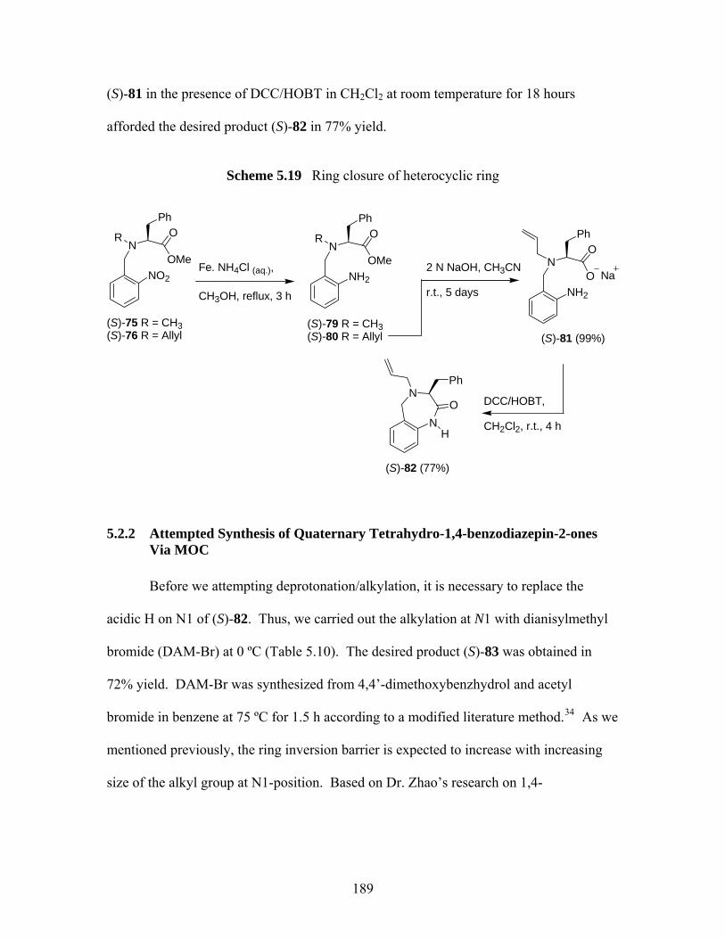
(S)-81 in the presence of DCC/HOBT in CH2Cl2 at room temperature for 18 hours
afforded the desired product (S)-82 in 77% yield.
Scheme 5.19 Ring closure of heterocyclic ring
NO2
NOMe
OR
(S)-75 R = CH3 (S)-76 R = Allyl
Fe. NH4Cl (aq.),
CH3OH, reflux, 3 h
NH2
NOMe
OR
(S)-79 R = CH3 (S)-80 R = Allyl
2 N NaOH, CH3CN
r.t., 5 days NH2
NO
O
(S)-81 (99%)
Na
DCC/HOBT,
CH2Cl2, r.t., 4 hN
N
H
O
Ph
PhPh Ph
(S)-82 (77%)
5.2.2 Attempted Synthesis of Quaternary Tetrahydro-1,4-benzodiazepin-2-ones Via MOC
Before we attempting deprotonation/alkylation, it is necessary to replace the
acidic H on N1 of (S)-82. Thus, we carried out the alkylation at N1 with dianisylmethyl
bromide (DAM-Br) at 0 ºC (Table 5.10). The desired product (S)-83 was obtained in
72% yield. DAM-Br was synthesized from 4,4’-dimethoxybenzhydrol and acetyl
bromide in benzene at 75 ºC for 1.5 h according to a modified literature method.34 As we
mentioned previously, the ring inversion barrier is expected to increase with increasing
size of the alkyl group at N1-position. Based on Dr. Zhao’s research on 1,4-
189
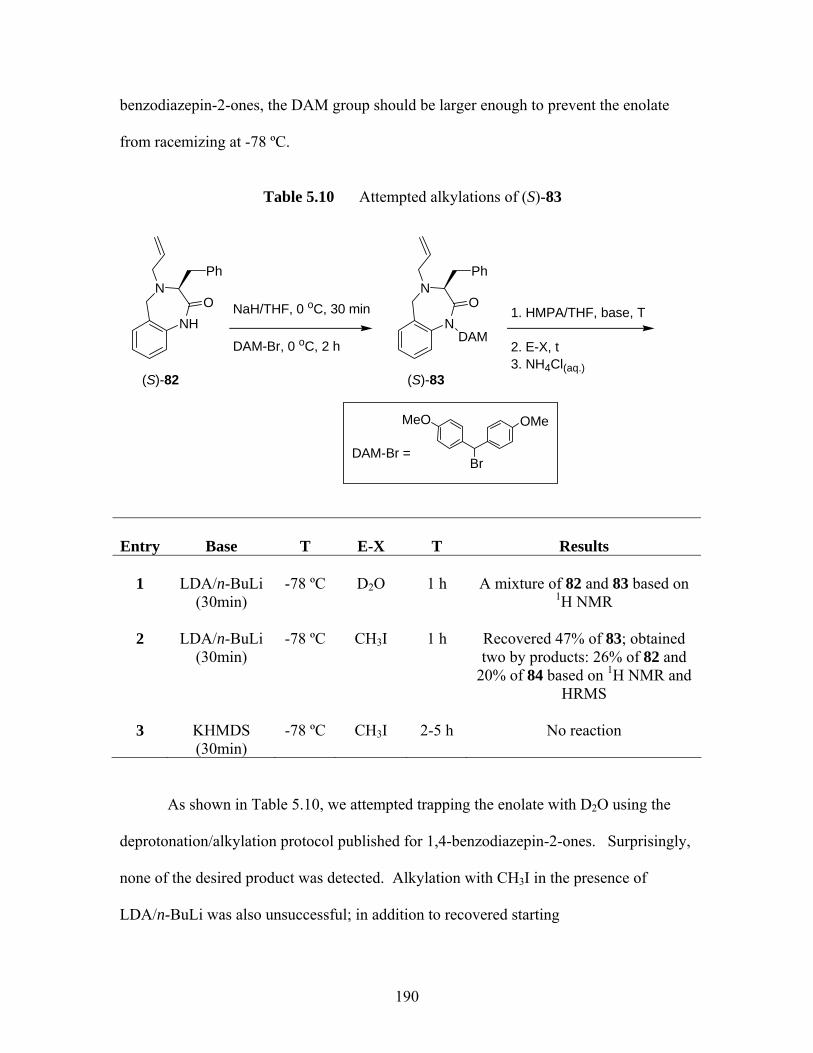
benzodiazepin-2-ones, the DAM group should be larger enough to prevent the enolate
from racemizing at -78 ºC.
Table 5.10 Attempted alkylations of (S)-83
NH
NO
Ph
(S)-82
NaH/THF, 0 oC, 30 min
DAM-Br, 0 oC, 2 h
N
NO
Ph
(S)-83
DAM
DAM-Br =Br
OMeMeO
1. HMPA/THF, base, T
2. E-X, t3. NH4Cl(aq.)
Entry
Base
T
E-X
T
Results
1
LDA/n-BuLi
(30min)
-78 ºC
D2O
1 h
A mixture of 82 and 83 based on
1H NMR 2
LDA/n-BuLi
(30min)
-78 ºC
CH3I
1 h
Recovered 47% of 83; obtained two by products: 26% of 82 and
20% of 84 based on 1H NMR and HRMS
3
KHMDS (30min)
-78 ºC
CH3I
2-5 h
No reaction
As shown in Table 5.10, we attempted trapping the enolate with D2O using the
deprotonation/alkylation protocol published for 1,4-benzodiazepin-2-ones. Surprisingly,
none of the desired product was detected. Alkylation with CH3I in the presence of
LDA/n-BuLi was also unsuccessful; in addition to recovered starting
190
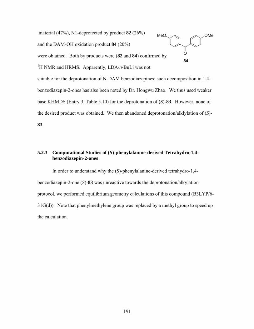
material (47%), N1-deprotected by product 82 (26%)
and the DAM-OH oxidation product 84 (20%)
were obtained. Both by products were (82 and 84) confirmed by
MeO
O
OMe
841H NMR and HRMS. Apparently, LDA/n-BuLi was not
suitable for the deprotonation of N-DAM benzodiazepines; such decomposition in 1,4-
benzodiazepin-2-ones has also been noted by Dr. Hongwu Zhao. We thus used weaker
base KHMDS (Entry 3, Table 5.10) for the deprotonation of (S)-83. However, none of
the desired product was obtained. We then abandoned deprotonation/alklylation of (S)-
83.
5.2.3 Computational Studies of (S)-phenylalanine-derived Tetrahydro-1,4-benzodiazepin-2-ones
In order to understand why the (S)-phenylalanine-derived tetrahydro-1,4-
benzodiazepin-2-one (S)-83 was unreactive towards the deprotonation/alkylation
protocol, we performed equilibrium geometry calculations of this compound (B3LYP/6-
31G(d)). Note that phenylmethylene group was replaced by a methyl group to speed up
the calculation.
191
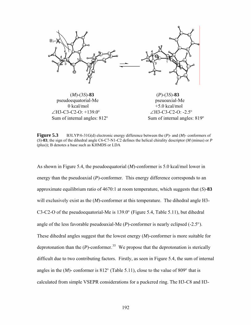
(M)-(3S)-83 (P)-(3S)-83
pseudoequatorial-Me pseuoaxial-Me 0 kcal/mol +5.0 kcal/mol ∠H3-C3-C2-O: +139.0° ∠H3-C3-C2-O: -2.5° Sum of internal angles: 812° Sum of internal angles: 819°
Figure 5.3 B3LYP/6-31G(d) electronic energy difference between the (P)- and (M)- conformers of (S)-83; the sign of the dihedral angle C6-C7-N1-C2 defines the helical chirality descriptor (M (minus) or P (plus)); B denotes a base such as KHMDS or LDA
As shown in Figure 5.4, the pseudoequatorial (M)-conformer is 5.0 kcal/mol lower in
energy than the pseudoaxial (P)-conformer. This energy difference corresponds to an
approximate equilibrium ratio of 4670:1 at room temperature, which suggests that (S)-83
will exclusively exist as the (M)-conformer at this temperature. The dihedral angle H3-
C3-C2-O of the pseudoequatorial-Me is 139.0° (Figure 5.4, Table 5.11), but dihedral
angle of the less favorable pseudoaxial-Me (P)-conformer is nearly eclipsed (-2.5°).
These dihedral angles suggest that the lowest energy (M)-conformer is more suitable for
deprotonation than the (P)-conformer.35 We propose that the deprotonation is sterically
difficult due to two contributing factors. Firstly, as seen in Figure 5.4, the sum of internal
angles in the (M)- conformer is 812° (Table 5.11), close to the value of 809° that is
calculated from simple VSEPR considerations for a puckered ring. The H3-C8 and H3-
192

C11 distances are 3.37 Å and 3.65 Å (Table 5.11) respectively. These relatively short
distances of suggest that the benzo ring might block the approach of bulky base to H3.
Secondly, N4-CH3 is pointing up and the distance of H3 to N4-CH3 is only 2.60 Å (Table
5.11), which also suggest that approach of bulky base to H3 may be sterically hindered
by N4-CH3 group. The steric hindrance from both benzo ring and N4-CH3 group may
account for the failure of deprotonation/alkylations of (S)-83. On the other hand, the
distances from H2 to C8/C11 and H2 to N1-CH3 (Table 5.11) of the (P)-conformer of the
(S)-alanine-derived tetrahydro-1,4-benzodiazepin-3-one such as (S)-32 are longer than
those of (S)-83. In these cases, H2 is free from steric hindrance from both the benzo ring
and N1-CH3 group, as both the benzo ring and the N1-CH3 group are not crowding
around H2. That is, H2 is more easily accessed by LDA due to its flatter ring geometry,
which alleviates the steric hindrance. The flatter ring geometry of (S)-32 is also reflected
in the sum of internal angles (Table 5.11). Furthermore, the dihedral angle H2-C2-C3-O
of pseudoequatorial-Me is 138° (Table 5.11), suggesting that the (P)-conformer of (S)-32
is suitable for deprotonation, which is consistent with the successful alkylations of (S)-32
(cf Table 5.4).
193
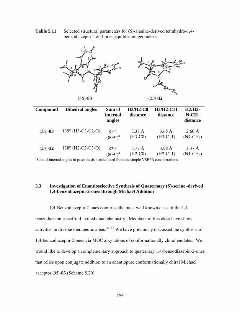
Table 5.11 Selected structural parameters for (S)-alanine-derived tetrahydro-1,4-benzodiazepin-2 & 3-ones equilibrium geometries
(3S)-83 (2S)-32
Compound Dihedral angles Sum of internal angles
H3/H2-C8 distance
H3/H2-C11 distance
H2/H3- N-CH3
distance
(3S)-83 139° (H3-C3-C2-O)
812° (809°)a
3.37 Å
(H3-C8)
3.65 Å
(H3-C11)
2.60 Å
(N4-CH3)
(2S)-32 138° (H2-C2-C3-O)
829° (809°)a
3.77 Å
(H2-C8)
3.98 Å
(H2-C11)
3.37 Å
(N1-CH3) aSum of internal angles in parenthesis is calculated from the simple VSEPR considerations
5.3 Investigation of Enantioselective Synthesis of Quaternary (S)-serine -derived 1,4-benzodiazepin-2-ones through Michael Addition
1,4-Benzodiazepin-2-ones comprise the most well known class of the 1,4-
benzodiazepine scaffold in medicinal chemistry. Members of this class have shown
activities in diverse therapeutic areas.36,37 We have previously discussed the synthesis of
1,4-benzodiazepin-2-ones via MOC alkylations of conformationally chiral enolates. We
would like to develop a complementary approach to quaternary 1,4-benzodiazepin-2-ones
that relies upon conjugate addition to an enantiopure conformationally chiral Michael
acceptor (M)-85 (Scheme 5.20).
194
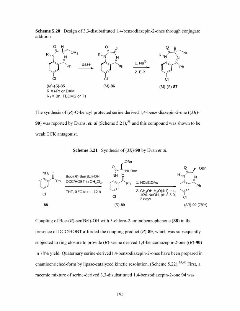
Scheme 5.20 Design of 3,3-disubstituted 1,4-benzodiazepin-2-ones through conjugate addition
NN
Ph
O HOR1
Cl
NN
Ph
O
Cl
(M)-(S)-85R = i-Pr or DAMR1 = Bn, TBDMS or Ts
(M)-86
Base 1. Nu
2. E-X
NN
Ph
O E
Cl
(M)-(S)-87
R R R Nu
The synthesis of (R)-O-benzyl protected serine derived 1,4-benzodiazepin-2-one ((3R)-
90) was reported by Evans, et. al (Scheme 5.21),38 and this compound was shown to be
weak CCK antagonist.
Scheme 5.21 Synthesis of (3R)-90 by Evan et al.
NH2
Ph
O
Cl
88
Boc-(R)-Ser(Bzl)-OH,DCC/HOBT in CH2Cl2,
THF, 0 oC to r.t., 12 h
NH
Ph
O
Cl
(R)-89
NHBocO
OBn
1. HCl/EtOAc
2. CH3OH-H2O(4:1), r.t., 10% NaOH, pH 8.5-9, 3 days
NN
Cl
H
O
Ph
OBn
(3R)-90 (78%)
Coupling of Boc-(R)-ser(Bzl)-OH with 5-chloro-2-aminobenzophenone (88) in the
presence of DCC/HOBT afforded the coupling product (R)-89, which was subsequently
subjected to ring closure to provide (R)-serine derived 1,4-benzodiazepin-2-one ((R)-90)
in 78% yield. Quaternary serine-derived1,4-benzodiazepin-2-ones have been prepared in
enantioenriched-form by lipase-catalyzed kinetic resolution. (Scheme 5.22).39, 40 First, a
racemic mixture of serine-derived 3,3-disubstituted 1,4-benzodiazepin-2-one 94 was
195
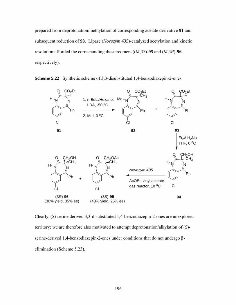
prepared from deprotonation/methylation of corresponding acetate derivative 91 and
subsequent reduction of 93. Lipase (Novozym 435)-catalyzed acetylation and kinetic
resolution afforded the corresponding diastereomers ((M,3S)-95 and (M,3R)-96
respectively).
Scheme 5.22 Synthetic scheme of 3,3-disubstituted 1,4-benzodiazepin-2-ones
NN
Cl
H
O
Ph
91
CO2EtH
1. n-BuLi/Hexane, LDA, -50 oC 2. MeI, 0 oC
NN
Cl
Me
O
Ph
92
CO2EtCH3
+
NN
Cl
H
O
Ph
93
CO2EtH
Et2AlH2NaTHF, 0 oC
NN
Cl
H
O
Ph
94
CH2OHCH3
NN
Cl
H
O
Ph
(3R)-96 (36% yield, 35% ee)
CH2OAcCH3
Novozym 435
AcOEt, vinyl acetategas reactor, 10 oC
NN
Cl
H
O
Ph
CH2OHCH3
+
(3S)-95 (48% yield, 25% ee)
Clearly, (S)-serine derived 3,3-disubstituted 1,4-benzodiazepin-2-ones are unexplored
territory; we are therefore also motivated to attempt deprotonation/alkylation of (S)-
serine-derived 1,4-benzodiazepin-2-ones under conditions that do not undergo β-
elimination (Scheme 5.23).
196
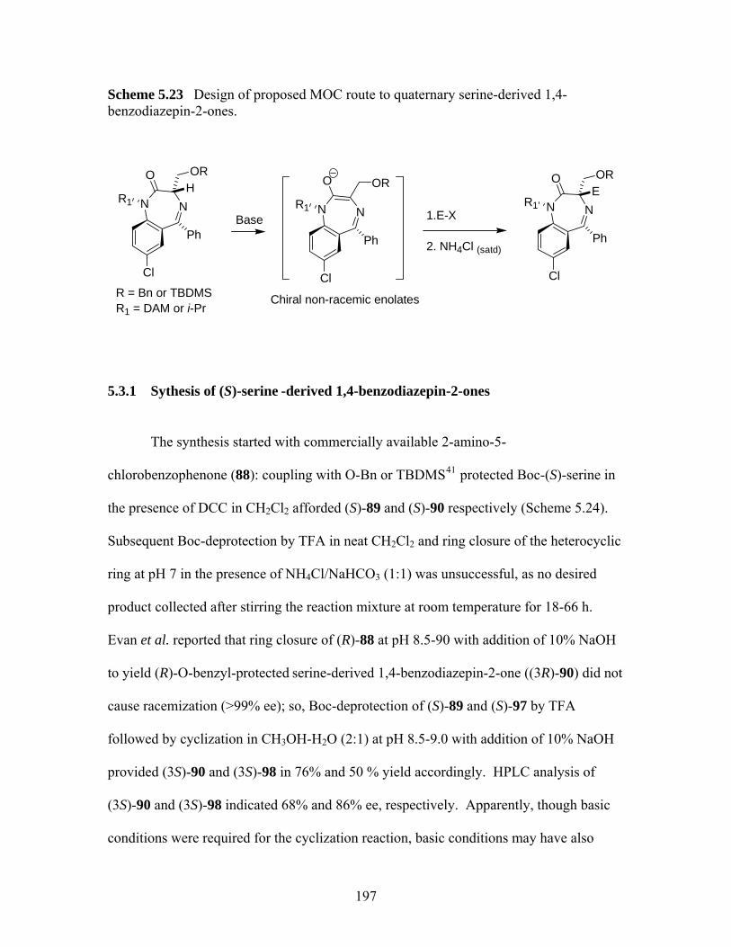
Scheme 5.23 Design of proposed MOC route to quaternary serine-derived 1,4-benzodiazepin-2-ones.
NN
Ph
Cl
R1
OE
NN
Ph
Cl
R1
OHOR
Base NN
Ph
Cl
R1
O OR
Chiral non-racemic enolates
1.E-X
2. NH4Cl (satd)
OR
R = Bn or TBDMSR1 = DAM or i-Pr
5.3.1 Sythesis of (S)-serine -derived 1,4-benzodiazepin-2-ones
The synthesis started with commercially available 2-amino-5-
chlorobenzophenone (88): coupling with O-Bn or TBDMS41 protected Boc-(S)-serine in
the presence of DCC in CH2Cl2 afforded (S)-89 and (S)-90 respectively (Scheme 5.24).
Subsequent Boc-deprotection by TFA in neat CH2Cl2 and ring closure of the heterocyclic
ring at pH 7 in the presence of NH4Cl/NaHCO3 (1:1) was unsuccessful, as no desired
product collected after stirring the reaction mixture at room temperature for 18-66 h.
Evan et al. reported that ring closure of (R)-88 at pH 8.5-90 with addition of 10% NaOH
to yield (R)-O-benzyl-protected serine-derived 1,4-benzodiazepin-2-one ((3R)-90) did not
cause racemization (>99% ee); so, Boc-deprotection of (S)-89 and (S)-97 by TFA
followed by cyclization in CH3OH-H2O (2:1) at pH 8.5-9.0 with addition of 10% NaOH
provided (3S)-90 and (3S)-98 in 76% and 50 % yield accordingly. HPLC analysis of
(3S)-90 and (3S)-98 indicated 68% and 86% ee, respectively. Apparently, though basic
conditions were required for the cyclization reaction, basic conditions may have also
197
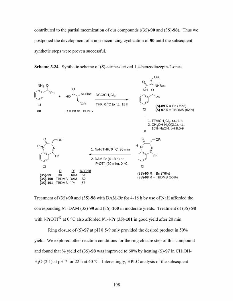
contributed to the partial racemization of our compounds ((3S)-90 and (3S)-98). Thus we
postponed the development of a non-racemizing cyclization of 90 until the subsequent
synthetic steps were proven successful.
Scheme 5.24 Synthetic scheme of (S)-serine-derived 1,4-benzodiazepin-2-ones
NH2
Ph
O
Cl
+
88
HONHBoc
O
OR
R = Bn or TBDMS
DCC/CH2Cl2,
THF, 0 oC to r.t., 18 h
NH
Ph
O
Cl(S)-89 R = Bn (79%)(S)-97 R = TBDMS (62%)
NHBocO
OR
1. TFA/CH2Cl2, r.t., 1 h2. CH3OH-H2O(2:1), r.t., 10% NaOH, pH 8.5-9
NN
Cl
H
O
Ph
OR
(3S)-90 R = Bn (76%)(3S)-98 R = TBDMS (50%)
1. NaH/THF, 0 oC, 30 min
2. DAM-Br (4-18 h) or iPrOTf (20 min), 0 oC,
NN
Cl
R'
O
Ph
OR
(3S)-99 Bn DAM 51 (3S)-100 TBDMS DAM 52(3S)-101 TBDMS i-Pr 67
R'R % Yield
Treatment of (3S)-90 and (3S)-98 with DAM-Br for 4-18 h by use of NaH afforded the
corresponding N1-DAM (3S)-99 and (3S)-100 in moderate yields. Treatment of (3S)-98
with i-PrOTf42 at 0 °C also afforded N1-i-Pr (3S)-101 in good yield after 20 min.
Ring closure of (S)-97 at pH 8.5-9 only provided the desired product in 50%
yield. We explored other reaction conditions for the ring closure step of this compound
and found that % yield of (3S)-98 was improved to 60% by heating (S)-97 in CH3OH-
H2O (2:1) at pH 7 for 22 h at 40 ºC. Interestingly, HPLC analysis of the subsequent
198

product (3S)-98 indicated >99% ee. The HPLC result suggested the importance of pH
during ring closure, but it is not clear why a similar result was not obtained with benzyl
protected analog 90. A side product (3S)-102 was observed due to the deprotection of
TBDMS group at higher temperature (Scheme 5.25). Treatment of (S)-102 with
TBDMSCl in DMF in the presence of imidazole recovered (S)-98 in 79% yield.
Scheme 5.25 Ring closure of (S)-98 at 40 ºC.
NH
Ph
O
Cl
(3S)-97
NHBocO
OTBDMS
1. TFA/CH2Cl2, r.t., 1 h
2. CH3OH-H2O(2:1), pH 7, 40 oC, 22 h
NN
Cl
H
O
Ph
OTBDMS
(3S)-98 (60%)
+
NN
Cl
H
O
Ph
OH
(3S)-102 (40%)
1. TBDMS-Cl/DMF imidazole, N2, r.t., 22 h
2. Satd. NH4Cl79%
5.3.2 Attempted Synthesis of 3,3-Substituted (S)-serine -derived 1,4-benzodiazepin-2-ones
From our first attempted deprotonation of (3S)-101, we were surprised to see that
(3S)-101 underwent elimination to give 103 (Scheme 5.26) in 55% yield when KHMDS
was used as base at -78 ºC. No such elimination was however observed in the
deprotonation of either (3S)-99 or (3S)-100, which contains N1-DAM group instead of
199
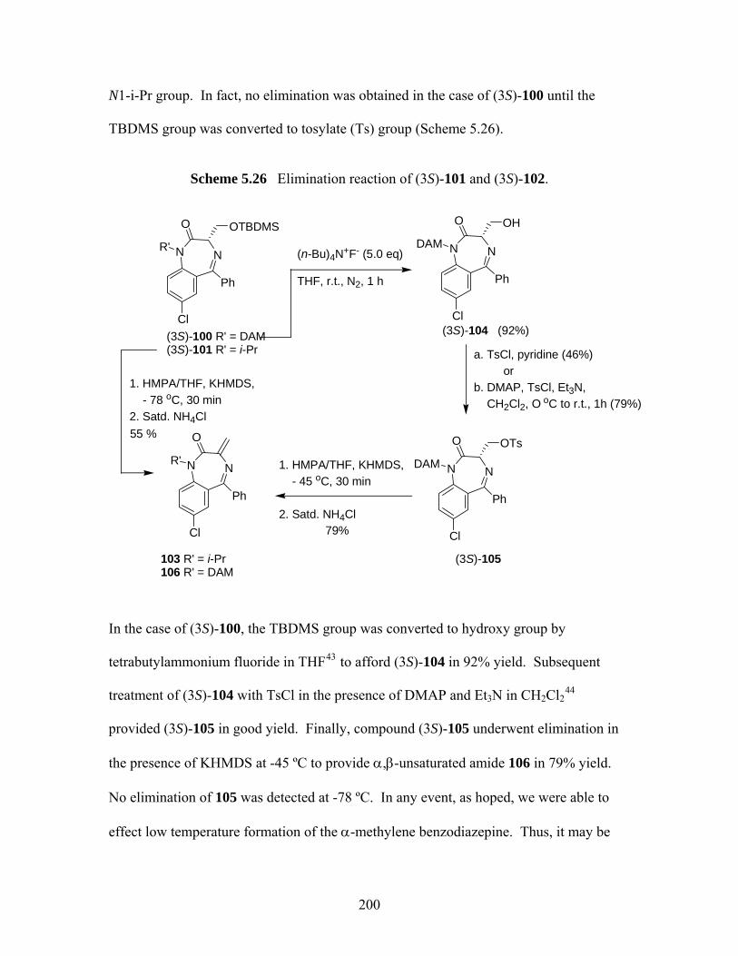
N1-i-Pr group. In fact, no elimination was obtained in the case of (3S)-100 until the
TBDMS group was converted to tosylate (Ts) group (Scheme 5.26).
Scheme 5.26 Elimination reaction of (3S)-101 and (3S)-102.
(n-Bu)4N+F- (5.0 eq)
THF, r.t., N2, 1 h
(3S)-100 R' = DAM(3S)-101 R' = i-Pr
NN
Cl
Ph
OTBDMSO
R'
(3S)-104 (92%)
NN
Cl
Ph
OHO
DAM
a. TsCl, pyridine (46%) orb. DMAP, TsCl, Et3N, CH2Cl2, O oC to r.t., 1h (79%)
(3S)-105
NN
Cl
Ph
OTsO
DAM
103 R' = i-Pr106 R' = DAM
NN
Cl
Ph
O
R'
79%
1. HMPA/THF, KHMDS, - 78 oC, 30 min2. Satd. NH4Cl55 %
1. HMPA/THF, KHMDS, - 45 oC, 30 min 2. Satd. NH4Cl
In the case of (3S)-100, the TBDMS group was converted to hydroxy group by
tetrabutylammonium fluoride in THF43 to afford (3S)-104 in 92% yield. Subsequent
treatment of (3S)-104 with TsCl in the presence of DMAP and Et3N in CH2Cl244
provided (3S)-105 in good yield. Finally, compound (3S)-105 underwent elimination in
the presence of KHMDS at -45 ºC to provide α,β-unsaturated amide 106 in 79% yield.
No elimination of 105 was detected at -78 ºC. In any event, as hoped, we were able to
effect low temperature formation of the α-methylene benzodiazepine. Thus, it may be
200
possible to generate 103 and 106 in enantiopure form as intermediates for conjugate
addition/alkylation (cf Scheme 5.20).
Before discussing our attempts at conjugate addition of 103 and 106, it is worthy
noting that the failure of 99 and 100 to undergo elimination with KHMDS at the
temperature as high as -45 °C, which means that these substrates might be useful for
deprotonation/alkylation (cf Scheme 5.23). Thus, we attempted several
deprotonation/alkylation experiments at both -78 °C and -45 °C. Results are summarized
in Table 5.12. To our surprise, these 1,4-benzodiazepin-2-ones were also unreactive
towards the deprotonation/alkylation protocol. Most of the starting material was
recovered at low reaction temperatures (-78 ºC); decomposition was observed when the
temperature was raised to -45 ºC and the reaction time was extended.
201

Table 5.12 Attempted alkylations of (3S)-99 and 100
NN
Cl
DAM
O
Ph
OR
(3S)-99 R = Bn(3S)-100 R = TBDMS
1. HMPA/solvent, T, base, 30 min
2. E-X, T, t3. Satd. NH4Cl
Entry
R
R’
Base/Solvent
T
E-X
t
Results
1
Bn
DAM
KHMDS/THF
-78 ºC
MeI
4 h
Recovered 50%
99 2
TBDMS
DAM
KHMDS/THF
-78 ºC
MeI
21 h
Recovered 70%
100 3
Bn
DAM
KHMDS/Et2O
-45 ºC
MeI
10
min.
Observed mainly
99 on TLC 4
Bn
DAM
KHMDS/Et2O
-45 ºC
BnBr
1.5 h
A mixture of 99
and decomposition
products on TLC 5
TBDMS
DAM
KHMDS/THF
-45 ºC
MeI
10
min.
Recovered 70%
100
5.3.3 Investigation of Enantioselective Synthesis of 1,4-benzodiazepin-2-ones Via
Michael addition
The successful use of organometallic (organolithium45- 48 or organocuprates49- 51)
reagents in enantio- or diasteroselective conjugate additions to acyclic α,β-unsaturated
amide/ketone/ester systems has been studied extensively in the literature. However, to
date, there are only a few published examples of conjugate additions of organometallic
202
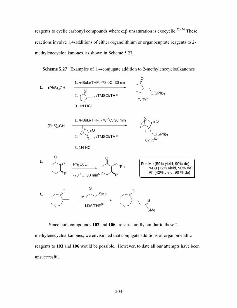
reagents to cyclic carbonyl compounds where α,β unsaturation is exocyclic.52- 54 These
reactions involve 1,4-additions of either organolithium or organocuprate reagents to 2-
methylenecycloalkanones, as shown in Scheme 5.27.
Scheme 5.27 Examples of 1,4-conjugate addition to 2-methylenecycloalkanones
O
O
(PhS)3CH1. n-BuLi/THF, -78 oC, 30 min
2. , /TMSCl/THF
3. 1N HCl
O
C(SPh)375 %52
(PhS)3CH1. n-BuLi/THF, -78 oC, 30 min
2. , /TMSCl/THF
3. 1N HCl
82 %52
O
C(SPh)3H
O
R
Ph2CuLi
-78 oC, 30 min53
O
R
PhR = Me (59% yield, 90% de) n-Bu (72% yield, 90% de) Ph (42% yield, 90 % de)
OMe
SSMe
LDA/THF54
O
S
SMe
1.
2.
3.
Since both compounds 103 and 106 are structurally similar to these 2-
methylenecycloalkanones, we envisioned that conjugate additions of organometallic
reagents to 103 and 106 would be possible. However, to date all our attempts have been
unsuccessful.
203

Our first attempts to effect conjugate addition employed n-BuLi in the reaction
with 106 to generate a lithium enolate at -78 °C, followed by trapping of this enolate
CH3I (Reaction 1, Scheme 5.28). To our surprise, mainly starting material 106 was
observed on TLC after 2.5 h. Attempts to carry out the 1,4-conjugate additions to this
α,β-unsaturated amide system in other reaction conditions49-51 were also unsuccessful, as
shown in Scheme 5.28.
Scheme 5.28 Attempted conjugate additions to compound 106
NN
Cl
DAM
O
Ph
1. HMPA/THF, -78 oC, n-BuLi, 15 min
2. CH3I, -78 oC, 2.5 h
106
1.No reaction
2.
1. 2 equiv MeLi Et2O, -45 oC, 30 min 2. 106 in Et2O, 30 min
3. satd. NH4Cl
3. CH3I, 2 h
4. satd. NH4Cl
No reaction;recovered 70 % of 106
No reaction
3.
1. CH3MgBr, Et2O, - 45 oC, 30 min
2. Add 106 in Et2O, 30 min3. MeI, 2 h3. Satd. NH4Cl
Observed a mixture of 106 and decomposition products on TLC
CuI
CuI
204

In the case of N-i-Pr analog 103, we attempted alkylations under two different
reaction conditions, as shown in Table 5.13. In Entry 1, organolithium/cuprate reagent
was used as the nucleophile, but disappointingly, starting material 103 was recovered
after the aqueous workup. At last, we attempted the conjugate reduction-alkylation of
103 (Entry 2 and 3) by use of hydrido(triphenylphosphine) copper (I) hexamer, which is
also known as Stryker’s reagent.55 The common application of Stryker’s reagent has
been in the conjugate reduction of α,β-unsaturated carbonyl compounds.55- 57 The
successful use of Stryker’s reagent in tandem 1,4-reduction/alkylation reactions of α,β-
unsaturated ketones has also been reported by Lipshutz et al.58 Other applications of
Stryker’s reagent includes tandem conjugate reduction-aldol cyclization.59, 60 Despite the
successful use of Stryker’s reagent in other reactions, no desired product was however
obtained in our case (Table 5.13, Entries 2 and 3).
Table 5.13 Attempted conjugate additions to 103
NN
Cl
O
Ph
103
1. Solvent/temp, organometallic reagent, t1
2. E-X, t23. Satd. NH4Cl
Entry
Solvent/temp
t1
Organometallic
reagent
E-X/t2
Results
1
Et2O/-78 °C
5 h
CuI/LiMe
CH3I/1
h
Recovered 103
2
Toluene/-45 °C
1 h
[(Ph3P)CuH]6
-
Recovered 69
% of 103 3
Toluene/reflux
1.5 h
[(Ph3P)CuH]6
-
Recovered 103
205
5.3.4 Computational Studies of 2-Methylenecycloalkanones and Heterocyclic 2-methylenecycloalkanones
In the hope of understanding the lack of reaction of 103 and 106 towards
nucleophiles, we studied the structures of many α-methylene cyclic carbonyl compounds.
In particular, we were interested to learn whether the reactivity towards conjugate
addition could be correlated with the dihedral angle made by the C=O and C=C. Since
the conjugate addition of organometallic reagents to α,β-unsaturated carbonyl system
would generate an enolate as the intermediate, which means the good conjugation of the
double bond with the carbonyl group should be required. We thus calculated dihedral
angles (Cβ=Cα-C=O) of the selected molecules at B3LYP/6-31G(d). Larger dihedral
angles would suggest the poor or no overlap of the double bond with the carbonyl group
during the enolate formation.Results are shown in Figure 5.5.
206
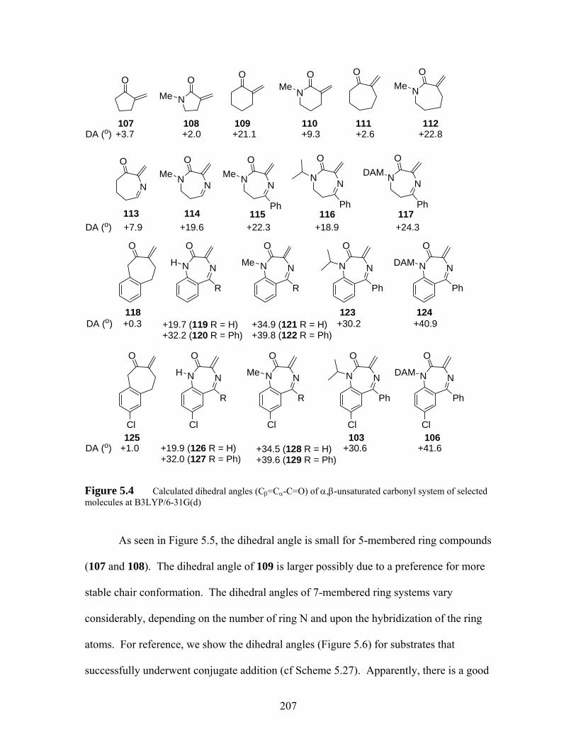
O O
207
O O OO
NMe NMe
DA (o) + +2.0 +21.1 +9.33.7
NMe
N
O
N N
OMe N N
OMe
Ph
N N
O
Ph
N N
ODAM
Ph
DA (o)
+2.6 +22.8
+7.9 +19.6 +22.3 +18.9 +24.3
10
NN
OO
R
H NN
O
R
Me NN
O
Ph
NN
O
Ph
DAM
DA (o) +0.3 +19.7 (119 R = H)+32.2 (120 R = Ph)
+34.9 (121 R = H)+39.8 (122 R = Ph)
+30.2 +40.9
NN
OO
R
H NN
O
R
Me NN
O
Ph
NN
O
Ph
DAM
DA (o) +1.0 +19.9 (126 R = H)+32.0 (127 R = Ph)
+34.5 (128 R = H)+39.6 (129 R = Ph)
+30.6 +41.6
Cl Cl Cl Cl Cl
7 108 109 110 111 112
113 114 115 116 117
118 123 124
125 103 106
Figure 5.4 Calculated dihedral angles (Cβ=Cα-C=O) of α,β-unsaturated carbonyl system of selected molecules at B3LYP/6-31G(d)
As seen in Figure 5.5, the dihedral angle is small for 5-membered ring compounds
(107 and 108). The dihedral angle of 109 is larger possibly due to a preference for more
stable chair conformation. The dihedral angles of 7-membered ring systems vary
considerably, depending on the number of ring N and upon the hybridization of the ring
atoms. For reference, we show the dihedral angles (Figure 5.6) for substrates that
successfully underwent conjugate addition (cf Scheme 5.27). Apparently, there is a good
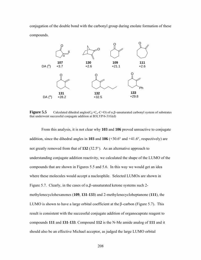
conjugation of the double bond with the carbonyl group during enolate formation of these
compounds.
O
O O O
Ph
DA (o) +21.1
+28.2 +32.5 +29.8
αβ
O
+3.7
O
+2.6107 130 109
131 132 133
O
+2.6111
o)DA (
Figure 5.5 Calculated dihedral angles(Cβ=Cα-C=O) of α,β-unsaturated carbonyl system of substrates that underwent successful conjugate addition at B3LYP/6-31G(d)
From this analysis, it is not clear why 103 and 106 proved unreactive to conjugate
addition, since the dihedral angles in 103 and 106 (+30.6° and +41.6°, respectively) are
not greatly removed from that of 132 (32.5°). As an alternative approach to
understanding conjugate addition reactivity, we calculated the shape of the LUMO of the
compounds that are shown in Figures 5.5 and 5.6. In this way we would get an idea
where these molecules would accept a nucleophile. Selected LUMOs are shown in
Figure 5.7. Clearly, in the cases of α,β-unsaturated ketone systems such 2-
methylenecyclohexanones (109, 131-133) and 2-methylenecycloheptanone (111), the
LUMO is shown to have a large orbital coefficient at the β-carbon (Figure 5.7). This
result is consistent with the successful conjugate addition of organocuprate reagent to
compounds 111 and 131-133. Compound 112 is the N-Me amide analog of 111 and it
should also be an effective Michael acceptor, as judged the large LUMO orbital
208
coefficient at the β-carbon (Figure 5.7). However, in the case of 113, the LUMO also
includes imine C=N. When both amide and imine are present on the ring, as in the case
of 116, LUMO is delocalized over the imine C=N and the 5-phenyl ring (Figure 5.7). In
the case of 103, which is the Michael acceptor we synthesized, the LUMO is very
delocalized over the imine and both benzene rings (Figure 5.7), but not over the carbonyl
group. As we mentioned previously, good conjugation of the double bond with the
carbonyl group should be required for the conjugate addition in order to form an enolate
intermediate. The highly delocalized LUMO of 103 over the imine and both benzene
rings instead of the carbonyl group might suggest that the enolate formation might not be
feasible for this reaction. Therefore, we suggest that this highly delocalized LUMO fails
to direct nucleophiles to the β-carbon, which accounts for the low reactivity of the α-
methylene benzodiazepines towards nucleophiles. One final caveat is that both the
dihedral angle and the LUMO structure approaches are ground state argument. A more
convincing explanation of the failed Miacheal addition reactions would require analysis
of possible reaction transition structures.
209
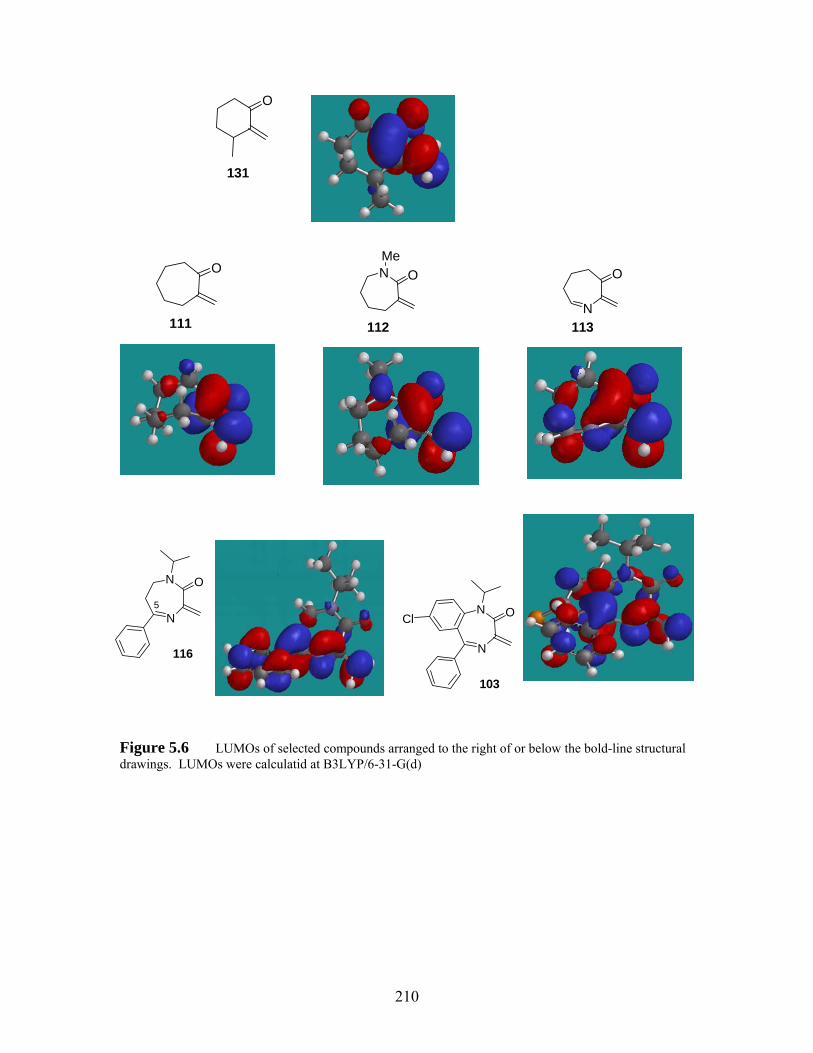
O
131
N OMe
112
O
N
O
113 111
N
N O
N
N OCl
103
5
116
Figure 5.6 LUMOs of selected compounds arranged to the right of or below the bold-line structural drawings. LUMOs were calculatid at B3LYP/6-31-G(d)
210
5.4 Conclusions
In chapter 5, we improved the synthesis of three different subtypes of 1,4-
benzodiazepines, including (S)-alanine derived tetrahydro-1,4-benzodiazepin-3-ones, (S)-
phenylalanine derived tetrahydro-1,4-benzodiazepin-2-ones and (S)-serine derived 1,4-
benzodiazepin-2-ones. Since the members of these subtypes possess important biological
activities, the synthetic methods described here may be useful for the corresponding
examples derived for other amino acids.
Our goal was to synthesize three different subtypes of quaternary 1,4-
benzodiazepines by use of the MOC strategy. Disappointingly, most of the
deprotonation/alkylations failed, due to various reasons. In the case (S)-alanine derived
tetrahydro-1,4-benzodiazepin-3-ones, 2,2-disubstituted analogs were obtained when
smaller electrophiles such as D2O, MeI or EtI were used in the alkylation of the N1-CH3
benzodiazepine. O-alkylation or rearrangement reaction was seen when larger
electrophiles such as BnBr or n-PrI were used in the reaction. The same rearrangement
reaction was seen in the case of N1-i-Pr tetrahydro-1,4-benzodiazepine-3-one. The
failure of these reactions was probably due to the steric hindrance of enolate alkylation.
The reason for our failure to achieve deprotonation/alkylation of tetrahydro-1,4-
benzodiazepin-2-ones are much less clear. Computational studies suggest that steric
hindrance by both benzo ring and N4-allyl group might retard deprotonation at C3 by
bulky bases like KHMDS or LDA.
Finally, (S)-serine derived 1,4-benzodiazepin-2-ones and their elimination
products were prepared. These proved unreactive towards deprotonation/alkylations and
conjugate additions respectively. The low reactivity of the α-methylene benzodiazepines
211

towards nucleophiles was attributed to highly delocalized LUMOs. In the ground state,
the LUMOs of these compounds were delocalized over the imine and both benzene rings,
but not the carbonyl group. These LUMOs would then fail to direct nucleophiles to the
β-carbons due to the lack of overlap between the carbonyl carbon and the α methylene
carbon in the LUMOs.
References for chapter 5
1 Clement, E. C.; Carlier, P. R. A Simple route to tetrahydro-1,4-benzodiazepin-3-ones
bearing diverse N1, N4, and C10 functionalization. Tetrahedron Lett. 2005, 46, 3633-3635.
2 Miller, W. H.; Ku, T. W.; Ali, F. E.; Bondinell, W. E.; Calvo, R. R.; Davis, L. D.;
Erhard, K. F.; Hall, L. B.; Huffman, W. F.; Keenan, R. M.; Kwon, C.; Newlander, K. A.; Ross, S. T.; Samanen, J. M.; Takata, D. T.; Yuan, C.-K. Enantiospecific Synthesis of SB 214857, a Potent, Orally Active Nonspecific Fibrinogen Receptor Antagonist. Tetrahedron Lett. 1995, 36, 9433.
3 Sveinbjornsson, A.; Vander Werf C. A. Synthesis of 2-Diethylaminoethyl-4-(7-
chloro-4-quinolyl)-aminofluorobenzene Dihydrobromide. J. Am. Chem. Soc. 1951, 73, 1378.
4 Samanen, J. M.; Ali, F. E.; Barton, L. S.; Bondinell, W. E.; Burgess, J. L.; Callahan,
J. F.; Calvo, R. R.; Chen, W.; Chen, L.; Erhard, K.; Feuerstein, G.; Heys, R.; Hwang, S.-M.; Jakas, D. R.; Keenan, R. M.; Ku, T. W.; Kwon, C.; Newlander, K. A.; Nichols, A.; Parker, M.; Peishoff, C. E.; Rhodes, G.; Ross. S.; Shu, A.; Simpson, R.; Takata, D.; Yellin, T. O.; Uzsinskas, I.; Venslavsky, J. W.; Yuan, C.-K,; Huffman, W. F. Potent, Selective Orally Active 3-Oxo-1,4-benzodiazepine GPIIb/IIIa Integrin Antagonists. J. Med. Chem. 1996, 39, 4867-4870.
5 Keenan, R. M.; Callahan, J. F.; Samanen, J. M.; Bondinell, W. E.; Calvo, R. R.;
DeBrosse, C.; Eggleston, D. S.; Haltiwanger, R. C.; Hwang, S.-M.; Jakas, D. R.; Ku, T. W.; Miller, W. H.; Newlander, K. A.; Nichols, A.; Parker, M. F.; Southhall, L. S.; Uzinskas, I.; Vasko-Moser, J. A.; Venslavsky, J. W.; Wong, A. S.; Huffman, W. F. Conformational Preferences in a Benzodiazepine Series of Potent Nonpeptide Fibrinogen Receptor Antagonists. J. Med. Chem. 1999, 42, 545-559.
212

6 Rosenström, U.; Sköld, C.; Lindeberg, G.; Botros, M.; Nyberg, F.; Karlén, A.;
Halberg, A Selective AT2 Receptor Ligand with a γ-Turn-Like Mimetic Replacing the Amino Acid Residues 4-5 of Angiotensin II. J. Med. Chem. 2004, 47, 859.
7 Schutkowski, M.; Mrestani-Klaus, C.; Neubert, K. Synthesis of dipeptide 4-
nitroanilides containing non-proteinogenic amino acids. Int. J. Pept. Protein Res. 1995, 45, 257-265.
8 Ohfune, Y.; Kurokawa, N.; Higuchi, N.; Saito. M.; Hashimoto, M.; Tanaka, T. An
efficient one-step reductive N-monoalkylation of α-amino acids. Chem. Lett. 1984, 441-444.
9 Takeda, K.; Akiyama, A.; Nakamura, H.; Takizama, S. -I.; Mizuno, Y.; Takayanagi,
H.; Harigaya, Y. Dicarbonates: Convenient 4-Dimethylaminopyridine Catalyzed Esterification Reagents. Synthesis, 1994, 1063.
10 Petersen, L.; Jensen, K. J. A New, Efficient Glycosylation Method for
Oligosaccharide Synthesis under Neutral Conditions: Preparation and Use of New DISAL Donors. J. Org. Chem. 2001, 66, 6268-6275.
11 Carlier, P. R.; Zhao, H.-W.; DeGuzman, J.; Lam, P. C.-H. Enantioselective Sythesis
of “Quaternary” 1,4-Benzodiazepin-2-one Scaffolds via Memory of Chirality. J. Am. Chem. Soc. 2003, 125, 11482-11483.
12 Russell, G.; Janzen, E. G.; Strom, E. T. Electron-Transfer Process. I. The Scope of the
Reaction between Carbanions or Nitrations and Unsaturated Electron Acceptors. J. Am. Chem. Soc. 1964, 86, 1807-1814.
13 Zhao, P.; Condo, A.; Keresztes, I.; Collum, D. B. Reactions of Ketones with Lithium
Hexamethyldisilazide: Competitive Enolizations and 1,2-Additions. J. Am. Chem. Soc. 2004, 126, 3113-3118.
14 Wissner, A.; Overbeek, E.; Reich, M. F.; Floyd, M. B.; Johnson, B. D.; Mamuya, N.;
Rosfjord, E. C.; Discafani, C.; Shi, X.-Q.; Rabindran, S. K.; Gruber, B. C.; Ye, F.; Hallett, W. A.; Nilakantan, R.; Shen, R.; Wang, Y.-F.; Greenberger, L. M.; Tsou, H.-R. Synthesis and Structure-Activity Relationship of 6,7-Disubstituted 4-Anilinoquinoline-3carbonitriles. The design of an Orally Active, Irreversible Inhibitor of the Tyrosine Kinase Activity of the Epidermal Growth Factor Receptor (EGFR) and the Human Epidermal Growth Factor Receptor-2 (HER-2) J. Med. Chem. 2003, 46, 49-63.
15 Giumanini, A. G.; Chiavari, G.; Gusiani, M. M.; Rossi, P. N-Permethylation of
Primary and Secondary Aromatic Amines. Synthesis, 1980, 743-746.
213
16 Perry, C. J.; Parveen, Z. The cyclisation of substituted phthalanilic acids in acetic acid
solution. A kinetic study of substituted N-phenylphthalimide formation. J. Chem. Soc., Perkin Trans. 2001, 2, 512-521.
17 GC-MS was performed by Dr. Emre Isin from Castagnoli’ group. 18 Wassmundt, F. M.; Kiesman, W. F. Efficient Catalytic of Hydrodediazoniations in
Dimethylformamide. J. Org. Chem. 1995, 60, 1713-1719. 19 Lubell, W.; Rapoport, H. Surrogates for Chiral Aminomalondialdehyde. Synthesis of
N-(9-Phenylfluoren-9-yl) and N-(9-Phenylfluoren-9-yl)vinylglycianl. J. Org. Chem. 1989, 54, 3824-3831.
20 Fryer, R. I.; Earley, J. V.; Sternbach, L. H.Quinazolines and 1,4-Benzodiazepines.
XLIV.1a The Formation of Isoindoles by the Ring Contraction of 1-Alkyl-1,4-benzodiazepines 1b. J. Org. Chem. 1969, 34, 649-654.
21 Garro-Helion, F.; Merzouk, A.; Guibe, F. Mild and Selective Palladium (0)-Catalyzed
Deallylation of Allylic Amine. Allylamine and Diallylamines as Very Convenient Ammonia Equivalents for the Synthesis of Primary Amines. J. Org. Chem. 1993, 58, 6109-6113.
22 Koch, T.; Hesse, M. Synthesis of Macrocyclic N-Unsubstituted Imides. Synthesis,
1992, 931. 23 Doi, H.; Sakai,T.; Yamada, K.-i.; Tomioka, K. N-Allyl- N-tert-
butyldimethylsilylamine for chiral ligand-controlled asymmetric conjugate addition to tert-butyl alkenoates. Chem. Commun. 2004, 1850.
24 Hu, Y.-J.; Dominique, R.; Das, S. K.; Roy, R. A facile new procedure for the
deprotection of allyl ethers under mild conditions. Can. J. Chem. 2000, 78, 838. 25 Kitov, P. I.; Bundle, D. R. Mild Oxidative One-Pot Allyl Group Cleavage. Org. Lett.
2001, 3, 2835-2838. 26 Geometry optimization calculations were conducted using PC Spartan 3.0. 27 Lam, P. C.-H.; Carlier, P. R. Experimental and Computational Studies of Ring
Inversion of 1,4-Benzodiazepin-2-ones: Implications for Memory of Chirality Transformation. J. Org. Chem. 2005, 70, 1530-1538.
28 The sum of internal angles in the benzodiazepine ring was calculated based on the standard bond angles for sp2 (120°) and sp3 (109.5°).
29 The sum of internal angles of a polygon is (180n-360°), where n is the number sides of the polygon.
214
30 Breslin, H. J.; Kukla, M. J.; Kromis, T.; Cullis, H.; Knaep, F. D.; Pauwels, R.;
Andries, K.; Clercq, E. D.; Janssen, M. A. C.; Janssen, P. A. J. Synthesis and Anti-HIV Activity of 1,3,4,5-Tetrahydro-2H-1,4—benzodiazepine-2-one (TBO) Derivatives. Truncated 4,5,6,7-Tetrahydro-5-methylimidazolo[4,5,1-jk][1,4]benzodiazepin-2-(1H)-ones (TIBO) Analogues. Bioorg. Med. Chem. 1999, 7, 2427-2436.
31 Wu. Z.; Ercole, F.; FitzGerald, M.; Perera, S.; Riley, P.; Campbell, R.; Pham, Y.;
Rea, P.; Sandanayake, S.; Mathieu, M. N.; Bray, A. M.; Ede, N. J. Synthesis of Tetrahydro-1,4-benzodiazepin-2-ones on Hydrophilic Polyamide SynPhase Lanterns J. Com. Chem. 2003, 5, 166-171.
32 Herrero, M. T.; Tellitu, I.; Dominguez, E.; Moreno, I.; SanMartin, R. A novel and
efficient iodine (III)-mediated access to 1,4-benzodiazepin-2ones Tetrahedron Lett. 2002, 43, 8273.
33 Cheguillaume, A.; Salaün, A.; Sinbandhit, S.; Potel, M.; Gall, P.; Gaudy-Floćh, M.;
Le Grel, P. Solution synthesis and characterization of aza-beta(3)-peptides (N(alpha)-substituted hydrazino acetic acid oligomers). J. Org. Chem. 2001, 66, 4923-4929.
34 Sekine, M.; Iwase, R.; Masuda, N.; Tsujiaki, H. Synthesis of Oligogibonucleotides by
Use of 4,4’,4’’-Tris(acyloxy)trityl Groups for Protection of the 6-Amino Group of Adenosine. Bull. Chem. Soc. Jpn, 1988, 61, 1669-1677.
35 Behnam, S. M.; Behnam, S. E.; Ando, K.; Green, N. S.; Houk, K. N. Stereoelectronic,
Torsional, and Steric Effect on Rates of Enolation of Ketones. J. Org. Chem. 2000, 65, 8970-8978.
36 Ellman, J. A. Design, Synthesis, and Evaluation of Small-Molecule Libraries. Acc.
Chem. Res. 1996, 29, 132-143. 37 Boojamra, C. G.; Burow, K. M.; Thomson, L. A.; Ellman, J. A. Solid-Phase Synthesis
of 1,4-benzodiazepine-2,5-diones. Library Preparation and Demonstration of Synthesis Generally. J. Org. Chem. 1997, 62, 1240-1256.
38 Evans, B. E.; Rittle, K. E.; Bock, M. G.; DiPardo, R. M.; Freidinger, R. M.; Whitter,
W. L.; Could, N. P.; Lundell, G. F.; Homnick, C. F.; Veber, D. F.; Anderson, P. S.; Chang, R. S. L.; Lotti, V. J.; Cerino, D. J.; Chen, T. B.; King, P. J.; Kunkel, K. A.; Springer, J. P.; Hirschfield, J. Design of Nonpeptidal Ligands for a Peptide Receptor: Cholecystokinin Antagonists. J. Med. Chem. 1987, 30, 1229-1239.
39 Avdagić, A.; Lesac, A.; Majer, Z.; Hollòsi, M.; Šunjić, V. Lipase-Catalyzed
Acetylation of 3-Substituted 2,3-Dihydro-1H-1,4-benzodiazepin-2-ones: Effect of Temperature and Conformation on Enantioselectivity and Configuration. Helv. Chim, Acta 1998, 81, 1567-1582.
215

40 Avdagić, A.; Šunjić, V. Biocatalytic Deracemization of 1,4-Benzodiazepines in the
synthesis of Enantiomerically Pure Serine. Helv. Chim, Acta 1998, 81, 85-93. 41 Boc-O-TBDMS-(S)-serine was synthesized from Boc-(S)-serine based on the
literature method (92% yield, lit. 85%). Yoo, D.-W.; Oh, J. S.; Lee, D. –W.; Kim, Y.-G. Efficient Synthesis of a Configurationally Stable L-Serinal Derivatives. J. Org. Chem. 2003, 68, 2979-2982.
42 Isopropyl triflate was prepared from isopropyl alcohol and triflate anhydride in
pyridine/CH2Cl2 at 0 ºC immediately prior to the alkylation. The synthesis of iPrOTf was developed by Dr. Hongwu Zhao.
43 Scheidt, K. A.; Chen, H.; Fellows, B. C.; Chemler, S. R.; Coffey, D. S.; Roush, W. R.
Tris(dimethylamino)sulfonium Difluorotrimethylsilicate (TAS-F), a Mild Reagent fro the Removal of Silicon Protecting Groups. J. Org. Chem. 1998, 63, 6436.
44 Langenhan, J. M.; Gellman, S. H. Preparation of Protected syn-α,β-Dialkyl-β-Amino
Acids That Contain Polar Side Chain Functionality. J. Org. Chem. 2003, 68, 6440-6443.
45 Etxebarria, J.; Vicario, J. L.; Badia, D.; Carrillo, L. Asymmetric Synthesis of β-
Amino Esters by Aza-Michael Reactions of α,β-Unsaturated Amides Using (S,S)-(+)-Pseudoephedrine as Chiral Auxiliary. J. Org. Chem. 2004, 69, 2588-2590.
46 Kanernasa, S.; Nomura, M.; Yoshinaga, S.; Yamamoto, H. High Enantiocontrol of
Michael Additions by Use of 2,2-Dimethyloxazolidine Chiral Auxiliaries. Exclusively ul, lk-1,4-Inductive Michael Additions of the Lithium (Z)-Enolates of (S)-4-Benzyl-2,2,5,5-tetramethyl-3-propanoyl oxazolidine to α,β-Unsaturated Ester. Tetrahedron 1995, 51, 10463-10476.
47 Oare, D. A.; Henderson, M. A.; Sanner, M. A.; Heathcock, C. H. Stereochemistry of
the Michael Additions of N,N-Disubstituted Amide and Thioamide Enolates to α,β-Unsaturated Ketones. J. Org. Chem. 1990, 55, 132-157.
48 Evans, D. A.; Ennis, M. D.; Le. T. J. Am .Chem. Soc. 1984, 106, 1154. 49 Dambacher, J.; Anness, R.; Pollock, P.; Bergdahl, M. Highly diastereoselective
conjugate additions of monoorganocopper reagents to a chiral imides. Tetrahedron 2004, 60, 2097-2110.
50 Arai, Y.; Ueda, K.; Xie, J.; Masaki, Y. 1,6-Asymmetric Induction During the
Conjugate Addition of Arylcopper Reagents to a Chiral Sulfinyl-Substituted Pyrrolyl-α,β-Unsaturated Amides. Chem. Pharm. Bull. 2001, 49, 1609-1614.
216

51 Kanemasa, S.; Suenaga, H.; Onimura, K. Effective Enantiocontrol in Conjugate
Additions of Organocuprates. Highly Selective 1,5-Chiral Induction in the Conjugate Additions of Cuprates to -α,β-Unsaturated Amide Derivatives of 2,2-Dimethyloxazolidine Chiral Auxilliaries. J. Org. Chem. 1994, 59, 6949-6954.
52 Liu, H.; Cohen, T. Yield and Selectivity Enhancement by Trimethylsilyl Chloride in
the Conjugate Addition of Stabilized Organolithiums. Tetrahedron Lett. 1995, 36, 8925-8928.
53 Tamura, R.; Watabe, K.-I.; Katayama, H.; Suzuki, H.; Yamamoto, Y. Asymmetric
Synthesis of 3-Substituted 2-exo-Methylenecyclohexanones via 1,5-Diastereoselection by Using a Chiral Amine. J. Org. Chem. 1990, 55, 408-410.
54 Berrada, S.; Metzner, P.; Rakotonirina, R. 1,4-Addition of lithium thioenolates of
dithio esters to ethylenic ketones. Bull de la Soc Chim de Fr. 1985, 5, 881-890. 55 Mahoney, W. S.; Brestensky, D. M.; Stryker, J. M. S. Selective hydride-mediated
conjugate reduction of α,β-unsaturated carbonyl compounds using [(Ph3P)CuH]6. J. Am.Chem. Soc. 1988, 110, 291-293.
56 Brestensky, D. M.; Stryker, J. M. S. Regioselective conjugate reduction and
reductive silylation of α,β-unsaturated aldehydes using copper(I) hydride complex [(Ph3P)CuH]6. Tetrahedron Lett. 1989, 5677-5780.
57 Koenig, T. M.; Daeuble, J. F.; Brestensky, D. M.; Stryker, J. M. S. Conjugate
reduction of polyfunctional α,β-unsaturated carbonyl compounds using [(Ph3P)CuH]6. Compatibility with halogen, sulfonate, and γ-oxygen and sulfur substituents. Tetrahedron Lett. 1990, 31, 3237-3240.
58 Lipshutz, B. H.; Chrisman, W.; Noson, K.; Papa, P.; Sclafani, J. A.; Vivian, R. W.,
Keith, J. M. Copper hydride-catalyzed tandem 1,4-reduction/alkylation reactions. Tetrahedron 2000, 56, 2779-2788.
59 Chiu, P.; Szeto, C.-P.; Geng, Z.; Cheng, K.-F.Tandem Conjugate Reduction-Aldol
Cyclization Using Stryker’s Reagent. Organic Lett. 2001, 3,1901-1903. 60 Chiu, P.; Szeto, C.-P.; Geng, Z.; Cheng, K.-F. Application of the tandem Stryker
reduction-aldol cyclization strategy to the asymmetric synthesis of lucinone. Tetrahedron Lett. 2001, 42, 4091-4093.
217
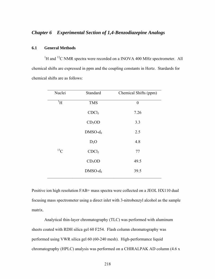
Chapter 6 Experimental Section of 1,4-Benzodiazepine Analogs
6.1 General Methods
1H and 13C NMR spectra were recorded on a INOVA 400 MHz spectrometer. All
chemical shifts are expressed in ppm and the coupling constants in Hertz. Stardards for
chemical shifts are as follows:
Nuclei Standard Chemical Shifts (ppm)
1H TMS 0
CDCl3 7.26
CD3OD 3.3
DMSO-d6 2.5
D2O 4.8
13C CDCl3 77
CD3OD 49.5
DMSO-d6 39.5
Positive ion high resolution FAB+ mass spectra were collected on a JEOL HX110 dual
focusing mass spectrometer using a direct inlet with 3-nitrobenzyl alcohol as the sample
matrix.
Analytical thin-layer chromatography (TLC) was performed with aluminum
sheets coated with RDH silica gel 60 F254. Flash column chromatography was
performed using VWR silica gel 60 (60-240 mesh). High-performance liquid
chromatography (HPLC) analysis was performed on a CHIRALPAK AD column (4.6 x
218

250 mm), detection at 254 nM, flow rate 1.0 mL/min unless specified with isocratic
solvent system; elution program: i-PrOH:Hexane (Table 6.1).
Table 6.1 HPLC data of chiral (S)-Alanine-derived tetrahydro-1,4-benzodiazepin-3-
ones
Racemic mixture Compounds
RT
a of (S)-enantiomer
RTa of (S)-
enantiomer RT
a of (R)-enantiomer
Elution
iPrOH:Hexane
Flow rate (min/mL)
9
15.52 min
12.08 min
15.63 min
20:80
1.0
10
13.02 min
13.10 min
15.06 min
15:85
1.0
11
26.16 min
26.01 min
29.61 min
1:99
2.0
29
9.56 min
9.53 min
11.06 min
5:95
1.0
30
13.31 min
13.36 min
16.62 min
20:80
1.0
32
12.16 min
12.16 min
13.29 min
3:97
1.0
33
11.24 min
11.14 min
12.92 min
2:98
1.0
56b
16.96 min
17.02 min
23.19 min
20:80
1.0
aRT = Retention time bCompound 56 exists as a mixture of pseudoequatorial (P)- and pseudoaxial (M)- conformers on the HPLC time scale and (P, S) is the major conformer.
6.2 Procedures
6.2.1 (S)-Alanine-derived tetrahydro-1,4-benzodiazepin-3-one project Both 2-fluoro-5-nitrotoluene and Boc-(S)-Ala-OH were purchased from ACROS,
and used without purification. Boc-(S)-N- Me-Ala-OH was prepared from Boc-(S)-Ala-
OH and MeI according to the literature method.1 (S)-N-i-Pr-Ala-OH was synthesized
from Boc-(S)-Ala-OH and acetone based on modified literature method.2
219

Synthesis of 2-Fluoro-5-nitrobenzyl bromide (2)
2-Fluoro-5-nitrotoluene (1) (2.74 g, 17.7 mmol) and NBS (3.14 g, 17.7 mmol)
were combined in CCl4 (35 mL). The yellow suspension was irradiated under a Hg lamp
for 19 hours. The white solid was filtered and washed with CCl4 (10 mL). The volatiles
were removed under reduced pressure to afford yellow liquid residue, which was then
mixed with EtOH/i-PrOH (20 mL, 95/5). The clear yellow solution was placed in the
freezer for overnight. Off-white, long needle-like crystals were obtained as desired
product (2.75 g, 66% yield) by filtration. 1H NMR (CDCl3): δ 4. 53 (s, 1H), 7.25 (t, J =
8.8 Hz, 1H), 8.21-8.25 (m, 1H), 8.36 (dd, J = 2.4, 8.4 Hz, 1H); 13C NMR (CDCl3): δ
23.55, 23.58, 116.79, 117.03, 126.07, 126.18, 127.06, 127.12, 162.65, 165.25; HRMS
(FAB-): calcd for C7H5BrFNO2 [M]- 232.9488, found 232.9560.
Synthesis of N-Allyl-2-fluoro-5-nitrobenzylamine (3)
To a stirring solution of allylamine (682 μL, 8.98 mmol) in THF (6 mL) at 0°C
was added 2 (1.05 g, 4.49 mmol) in THF (10 mL) dropwise. The ice-bath was removed,
and the reaction mixture was stirred at room temperature for 3 hours. The reaction
mixture was then partitioned between CH2Cl2 (2 x 40 mL) and 10% NaOH (40 mL). The
combined organic layers were dried over Na2SO4, filtered and evaporated to dryness. The
crude greenish yellow residue was purified by flash column chromatography on silica
gel, eluting with CH2Cl2-CH3OH (98/2) to afford the desired product as yellowish green
oil (822.3 mg, 87%). 1H NMR (CDCl3): δ 3.36 (ddd, J = 1.2, 3.2, 6 Hz, 2 H), 3.97 (s,
2H), 5.19-5.31 (m, 2H), 5.93-6.03 (m, 1H), 7.24, (t, J = 9 Hz, 1H), 8.20-8.24 (m, 1H),
8.42 (dd, J = 3.0, 6.2 Hz, 1H); 13C NMR (CDCl3): δ 46.66, 45.68, 51.69, 116.04, 116.28,
220

116.61, 124.44, 124.55, 126.02, 129.29, 129.47, 136.11, 144.32, 163.18, 165.73; HRMS
(FAB): calcd for C10H12FN2O2 [M+1]+ 211.0877, found 211.0896 (+6.2 ppm/+1.3
mmu).
General procedures for the coupling reactions: (S)-6 to (S)-83
To a stirring solution of 3 (1.0 equiv), DCC (1.2 equiv) and HOBT (1.2 equiv) in
CH2Cl2 (0.1 M) at 0°C was added (S)-alanine (1.2 equiv). The ice-bath was removed and
the greenish yellow suspension was stirred at room temperature for 12 hours. The white
precipitate was filtered off and washed with cold CH2Cl2 (10 mL). The filtrate was
evaporated under reduced pressure to provide greenish yellow oil residue, which was
purified by flash column chromatography (EtOAc-CH2Cl2 2/8) on silica gel.
Synthesis of (S)-Nα-Boc, N-allyl-N-(2-fluoro-5-nitrobenzyl)alaninamide (S-6)
Compound 3 (182 mg, 0.87 mmol) and Boc-(S)-Ala-OH (80.4 mg, 0.43 mmol)
were combined as describe above. The title compound was obtained as green oil (141.3
mg, 86%). 1H NMR (CDCl3): δ 1.36 (d, J = 6.4 Hz, 3 H), 1.40 (s, 1 H), 1.44 (s, 8 H),
2.78 (s, 3 H), 3.90-4.00 (m, 1 H), 4.09-4.18 (m, 1 H), 4.63-4.71 (m, 3 H), 5.09-5.33 (m, 3
H), 5.78-5.87 (m, 1 H), 7.19 (t, J = 9.2 Hz, 1 H), 8.11-8.21 (m, 2 H); 13C NMR (CDCl3):
δ 14.14, 19.22, 28.20, 28.26, 42.53, 42.57, 43.94, 46.18, 48.28, 50.30, 79.80, 116.18,
116.43, 118.23, 124.31, 124.88, 124.98, 125.55, 125.62, 126.12, 126.29, 131.78, 132.09,
144.52, 155.13, 162.89, 165.46, 174.09.
221
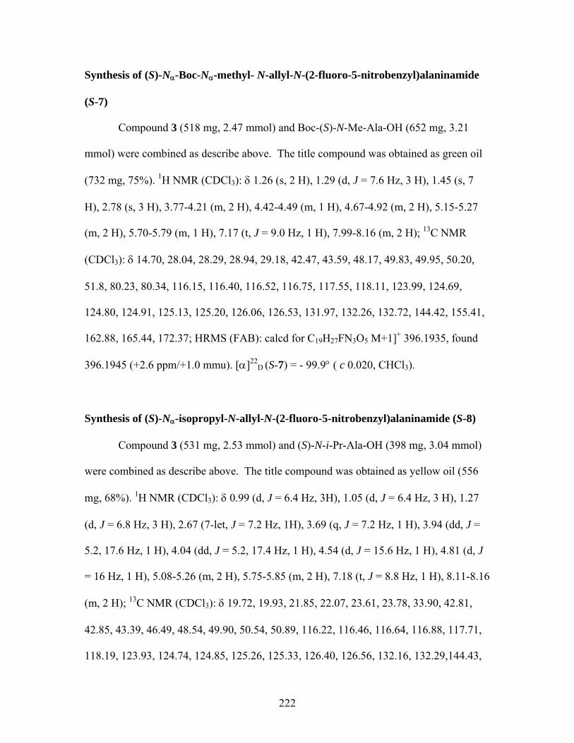
Synthesis of (S)-Nα-Boc-Nα-methyl- N-allyl-N-(2-fluoro-5-nitrobenzyl)alaninamide
(S-7)
Compound 3 (518 mg, 2.47 mmol) and Boc-(S)-N-Me-Ala-OH (652 mg, 3.21
mmol) were combined as describe above. The title compound was obtained as green oil
(732 mg, 75%). 1H NMR (CDCl3): δ 1.26 (s, 2 H), 1.29 (d, J = 7.6 Hz, 3 H), 1.45 (s, 7
H), 2.78 (s, 3 H), 3.77-4.21 (m, 2 H), 4.42-4.49 (m, 1 H), 4.67-4.92 (m, 2 H), 5.15-5.27
(m, 2 H), 5.70-5.79 (m, 1 H), 7.17 (t, J = 9.0 Hz, 1 H), 7.99-8.16 (m, 2 H); 13C NMR
(CDCl3): δ 14.70, 28.04, 28.29, 28.94, 29.18, 42.47, 43.59, 48.17, 49.83, 49.95, 50.20,
51.8, 80.23, 80.34, 116.15, 116.40, 116.52, 116.75, 117.55, 118.11, 123.99, 124.69,
124.80, 124.91, 125.13, 125.20, 126.06, 126.53, 131.97, 132.26, 132.72, 144.42, 155.41,
162.88, 165.44, 172.37; HRMS (FAB): calcd for C19H27FN3O5 M+1]+ 396.1935, found
396.1945 (+2.6 ppm/+1.0 mmu). [α]22D (S-7) = - 99.9° ( c 0.020, CHCl3).
Synthesis of (S)-Nα-isopropyl-N-allyl-N-(2-fluoro-5-nitrobenzyl)alaninamide (S-8)
Compound 3 (531 mg, 2.53 mmol) and (S)-N-i-Pr-Ala-OH (398 mg, 3.04 mmol)
were combined as describe above. The title compound was obtained as yellow oil (556
mg, 68%). 1H NMR (CDCl3): δ 0.99 (d, J = 6.4 Hz, 3H), 1.05 (d, J = 6.4 Hz, 3 H), 1.27
(d, J = 6.8 Hz, 3 H), 2.67 (7-let, J = 7.2 Hz, 1H), 3.69 (q, J = 7.2 Hz, 1 H), 3.94 (dd, J =
5.2, 17.6 Hz, 1 H), 4.04 (dd, J = 5.2, 17.4 Hz, 1 H), 4.54 (d, J = 15.6 Hz, 1 H), 4.81 (d, J
= 16 Hz, 1 H), 5.08-5.26 (m, 2 H), 5.75-5.85 (m, 2 H), 7.18 (t, J = 8.8 Hz, 1 H), 8.11-8.16
(m, 2 H); 13C NMR (CDCl3): δ 19.72, 19.93, 21.85, 22.07, 23.61, 23.78, 33.90, 42.81,
42.85, 43.39, 46.49, 48.54, 49.90, 50.54, 50.89, 116.22, 116.46, 116.64, 116.88, 117.71,
118.19, 123.93, 124.74, 124.85, 125.26, 125.33, 126.40, 126.56, 132.16, 132.29,144.43,
222
162.88, 165.44, 176.95; HRMS (FAB): calcd for C16H23FN3O5 [M+1]+ 324.1723, found
324.1703 (-6.3 ppm/-2.1 mmu). [α]22D (S-8) = - 53.4° ( c 0.016, CHCl3).
General procedures for the ring closure reactions 0.1-0.2 M of (S)-6, (S)-7, or (S)-8 in DMSO was heated to 200°C for 30 minutes to 3
hours. After cooling to room temperature, the reaction mixture was partitioned between
Et2O (2 x 30 mL) and H2O (30 mL). The ether layers were dried over Na2SO4, filtered,
and evaporated to dryness to provide brown oily residue, which was then purified by
flash column chromatography (CH2Cl2-EtOAc-Hexane: 7/2/1) on silica gel to afford the
desired product.
Synthesis of (S)-N4-allyl-2-methyl-10-nitro-tetrahydro-1,4-benzodiapin-3-one (S-9)
(S)-6 (71.5 mg, 0.19 mmol) was dissolved in DMSO (1.9 mL) and heated to
200°C for 30 minutes. The title compound was obtained as yellow solid (32.5 mg, 67%)
after the workup as described above. 1H NMR (DMSO): δ 1.24 (d, J = 6.4 Hz, 3 H),
3.92-4.10 (m, 3 H), 5.04-5.14 (m, 2 H), 5.12 (d, J = 17.2 Hz, 1 H), 5.37 (d, J = 16.4 Hz, 1
H), 5.63-5.73 (m, 1 H), 6.65 (d, J = 9.2 Hz, 1 H), 7.20 (apparent d, J = 2.8 Hz, 1 NH),
7.85 (dd, J = 2.6, 9.0 Hz, 1 H), 7.92 (d, J = 2.8 Hz, 1 H); 13C NMR (DMSO): δ 16.10,
48.51, 48.72, 49.22, 66.38, 114.77, 116.84, 118.48, 125.06, 126.30, 133.56, 135.40,
153.12, 169.44. HRMS (FAB): calcd for C13H16N3O3 [M+1]+ 262.1192, found 262.1176
(-5.9ppm/-1.6mmu); HPLC (2:8 i-PrOH:Hexane, CHIRALPAK AD, 4.6 x 250 mm: 1.0
223
mL/min; UV detection at 254 nM): 15.52 min., 100 % ee; [α]22D (S-9) = - 528.8° ( c
0.013, DMSO).
Synthesis of (S)-N4-allyl-N1-methyl-2-methyl-10-nitro-tetrahydro-1,4-benzodiapin-3-
one (S-10)
(S)-7 (1.28 mg, 3.23 mmol) was dissolved in DMSO (32 mL) and heated to 200°C
for 3 hours. The title compound was obtained as yellow solid (814 mg, 92%) after the
workup as described above. 1H NMR (CDCl3): δ 1.52 (d, J = 6.4 Hz, 3 H), 3.01 (s, 3 H),
3.04-4.19 (m, 2 H), 4.18 (d, J = 16.4 Hz, 1 H), 4.59 (q, J = 6.9 Hz, 1 H), 4.87 (d, J = 16.4
Hz, 1 H), 5.17-5.22 (m, 2 H), 5.67-5.76 (m, 1 H), 6.82 (d, J = 10 Hz, 1 H), 7.91 (d, J =
2.8 Hz, 1 NH), 8.06 (dd, J = 2.8, 9.2 Hz, 1 H); 13C NMR (CDCl3): δ 15.36, 36.80, 49.52,
50.85, 57.83, 117.02, 118.26, 123.23, 124.89, 125.44, 132.80, 138.54, 155.19, 169.60.
HRMS (FAB): calcd for C14H18N3O3 [M+1]+ 276.1348, found 276.1351 (+1.0 ppm/+0.3
mmu). HPLC (15:85 i-PrOH:Hexane, 4.6 x 250 mm: 1.0 mL/min; UV detection at 254
nM: 13.02 min., 100 % ee; [α]22D (S-10) = - 890.6° ( c 0.014, CHCl3).
Synthesis of (S)-N4-allyl-N1-isopropyl-2-methyl-10-nitro-tetrahydro-1,4-
benzodiapin-3-one (S-11)
(S)-8 (733 mg, 2.27 mmol) was dissolved in DMSO (15 mL) and heated to 200°C
for 3 hours. The title compound was obtained as yellow solid (417 mg, 61%) after the
workup as described above. 1H NMR (CDCl3): δ 1.19 (d, J = 6.8 Hz, 3 H), 1.37 (d, J =
6.8 Hz, 3 H), 1.41 (d, J = 7.2 Hz, 3 H), 3.69 (7-let, J = 6.8 Hz, 1 H), 4.00-4.06 (m, 2 H),
4.14-4.20 (m, 1 H), 4.42 (q, J = 7.2 Hz, 1 H), 4.43 (d, J = 15.6 Hz, 1 H), 4.54 (d, J = 16.4
224
Hz, 1 H), 5.18-5.23 (m, 2 H), 5.69-5.79 (m, 1 H), 6.99 (d, J = 9.2 Hz, 1 H), 7.94 (d, J =
2.8 Hz, 1 NH), 8.06 (dd, J = 2.6, 9.0 Hz, 1 H); 13C NMR (CDCl3): δ 17.29, 21.39, 22.30,
50.39, 51.06, 51.49, 56.33, 118.20, 119.55, 124.26, 125.08, 125.53, 132.69, 139.57,
153.48, 171.63. HRMS (FAB): calcd for C16H22N3O3 [M+1]+ 304.1661, found 304.1659
(-0.7 ppm/-0.2 mmu). HPLC (1:99 i-PrOH:Hexane, CHIRALPAK AD, 4.6 x 250 mm:
2.0 mL/min; UV detection at 254 nM): 26.16 min., 100 % ee; [α]22D (S-11) = + 245.0° (
c 0.015, CHCl3).
General procedures for nitro group reduction4 Synthesis of (S)-N4-allyl-10-amino-N1-methyl-2-methyl-tetrahydro-1,4-benzodiapin-
3-one (S-27)
To a mixture of (S)-10 (829 mg, 3.01) and Fe powder (1.20 g, 21.4 mmol) was
added CH3OH (20 mL) and NH4Cl (1.47g, 27.5 mmol) in 10.5 mL H2O. The yellow
suspension was heated to reflux for 3 hours under N2. The hot reaction mixture with
black precipitate was filtered off immediately and washed with hot CH3OH (10 mL). The
filtrate was evaporated to dryness and partitioned between CH2Cl2 (2 x 50 mL) and 10%
NaHCO3 (50 mL). The combined organic layers were dried over Na2SO4, filtered, and
concentrated under vacuum to afford (S)-27 as the light green solid (715 mg, 97% yield).
The amine was immediately used next step without further purification.
225
Synthesis of (S)-N4-allyl-10-amino-N1-isopropyl-2-methyl-tetrahydro-1,4-
benzodiapin-3-one (S-28)
(S)-11 (109 mg, 0.37 mmol), Fe powder (141 mg, 2.52 mmol) and NH4Cl (178
mg, 3.33 mmol) in 1.2 mL H2O were treated as above to afford the desired amine (S)-28
as green oil (93 mg, 92%). The amine was immediately used next step without further
purification.
Synthesis of (S)-N4-allyl-N1-isopropyl-10-(N,N-dimethylamino)-2-methyl-
tetrahydro-1,4-benzodiapin-3-one (S)-295
To a mixture of formaldehyde (683 μL, 8.4 mmol) and first 2 equiv. of 3 M
H2SO4 (0.7 mL, 2.1 mmol) at 0 °C was added a suspension of S-28 (381 mg, 1.4 mmol)
and NaBH4 (370 mg, 9.77 mmol) dropwise. After the addition was complete, second 2
equiv. of 3 M H2SO4 (0.7 mL, 2.1 mmol) was added. The reaction mixture was stirred at
0 °C for 5 minutes. 10% NaOH was added to the reaction mixture until pH = 9. The
reaction mixture was then partitioned between CH2Cl2 (2 x 30 mL) and H2O (25 mL).
The combined organic layers were dried over Na2SO4, filtered, and concentrated on
vacuum to provide brown crude oil, which was then purified by flash column
chromatography (CH2Cl2-CH3OH: 98/2) on silica gel to afford the desired product (S)-29
as yellow oil (394.7 mg, 94 %). 1H NMR (CDCl3): δ 1.04 (d, J = 6.4 Hz, 3 H), 1.10 (d, J
= 7.2 Hz, 3 H), 1.25 (d, J = 6.4 Hz, 3 H), 2.90 (s, 6 H), 3.57 (7-let, J = 6.4 Hz, 1 H), 3.75
(d, J = 13.6 Hz, 1 H), 3.98 (dd, J = 6.0, 15.6 Hz, 1 H), 4.25 (q, J = 6.9 Hz, 1 H), 4.29 (dd,
J = 5.4, 15.4 Hz, 1 H), 5.17-5.22 (m, 2 H), 5.75-5.85 (m, 1 H), 6.53 (d, J = 3.2 H, 1 H),
6.68 (d, J = 14.0 Hz, 1 H), 7.00 (d, J = 8.4 Hz, 1 H); 13C NMR (CDCl3): δ 16.45, 22.71,
226
22.76, 30.28, 41.06, 49.44, 51.44, 51.59, 58.37, 112.83, 112.88, 116.84, 125.06, 133.47,
133.59, 137.58, 146.80, 173.60. HRMS (FAB): calcd for C18H27N3O [M+1]+ 301.2154,
found 301.2145 (-3.0 ppm/-0.9 mmu); HPLC (5:95 i-PrOH:Hexane, CHIRALPAK AD,
4.6 x 250 mm: 1.0 mL/min; UV detection at 254 nM): 10.33 min., 99 % ee; [α]22D (S-29)
= + 168.5° ( c 0.015, CHCl3).
Synthesis of (S)-N4-allyl-N1-isopropyl-10-phthalimido-2-methyl-tetrahydro-1,4-
benzodiapin-3-one (S)-306
Compound 27 (92.6 mg, 0.34 mmol) and phthalic anhydride (51 mg, 0.34 mmol)
were combined in AcOH (3 mL), and heated to reflux for 1.5 hours. After cooling to
room temperature, the reaction mixture was partitioned between CH2Cl2 (2 x 30 mL) and
H2O (25 mL). The combined organic layers were dried over Na2SO4, filtered, and
evaporated to dryness to provide brown oily residue, which was then purified by flash
column chromatography (CH2Cl2-EtOAc-Hexane: 7/2/1) on silica gel to afford the
desired product (S)-30 as yellow foamy solid (109.2 mg, 80 %). 1H NMR (CDCl3): δ 1.11
(d, J = 6.4 Hz, 3 H), 1.22 (d, J = 7.2 Hz, 3 H), 1.34 (d, J = 6.4 Hz, 3 H), 3.73 (7-let, J =
6.6 Hz, 1 H), 3.96-4.01 (m, 1 H), 4.00 (d, J = 14.8 Hz, 1 H), 4.24-4.27 (m, 1 H), 4.31 (q,
J = 7.3 Hz, 1 H), 4.77 (d, J = 14.4 Hz, 1 H), 5.16-5.21 (m, 2 H), 5.73-5.82 (m, 1 H), 7.17-
7.19 (m, 2 H), 7.31 (dd, J = 2.6, 8.6 Hz, 1 H), 7.76-7.80 (m, 1 H), 7.91-7.96 (m, 1 H); 13C
NMR (CDCl3): δ 16.99, 22.40, 22.61, 50.27, 50.91, 50.98, 57.33, 117.48, 123.81, 123.64,
125.18, 126.36, 126.78, 130.91, 131.70, 133.10, 134.36, 147.90, 167.39, 172.73. HRMS
(FAB): calcd for C24H26N3O3 [M+1]+ 404.1974, found 404.1989 (+3.6 ppm/+1.4 mmu).
227
HPLC (2:8 i-PrOH:Hexane, CHIRALPAK AD, 4.6 x 250 mm: 1.0 mL/min; UV
detection at 254 nM): 13.31 min., 98 % ee; [α]22D (S-30) = + 138.8° ( c 0.020, CHCl3).
General procedures for aromatic deamination7
Synthesis of (S)-N4-allyl-N1-methyl-2-methyl-tetrahydro-1,4-benzodiapin-3-one (S)-
32
Compound 27 (192 mg, 0.78 mmol) was dissolved in a warm solution of H2SO4
(84 μL, 1.57 mmol) and AcOH (2 mL). After cooling to room temperature, NaNO2 (81
mg, 1.17 mmol) in H2O (1.2 mL) was added to in 15 minutes. After stirring at room
temperature for additional 15 minutes, the reaction mixture was added to a suspension of
Fe2SO4•7H2O (217 mg, 0.78 mmol) and DMF (11 mL). Gas evolution was observed and
the reaction mixture was stirred at room temperature for 30 more minutes or until the
cease of gas evolution. The reaction mixture was then partitioned between CH2Cl2 (3 x
30 mL) and NaOH (25 mL). The combined organic layers were dried over Na2SO4,
filtered, and evaporated to dryness to provide brown oily residue, which was then
purified by flash column chromatography (CH2Cl2-CH3OH: 96/4) on silica gel to afford
the desired product as brown oil (90 mg, 57%). 1H NMR (CDCl3): δ 1.30 (d, J = 7.2 Hz,
3 H), 2.85 (s, 3 H), 3.98-4.04 (m, 2 H), 4.01 (d, J = 15.2 Hz, 1 H), 4.20 (q, J = 6.9 Hz, 1
H), 4.79 (d, J = 15.2 Hz, 1 H), 5.16-5.22 (m, 2 H), 5.71-5.81 (m, 1 H), 6.89 (ddd, J = 0.9,
8.0, 7.6 Hz, 1 H), 6.99 (apparent dd, J = 1.0, 8.1 Hz, 1 H), 7.05 (apparent dd, J = 1.0, 7.4
Hz, 1 H), 7.25 (apparent ddd, J = 1.6, 7.6, 7.6 Hz, 1 H); 13C NMR (CDCl3): δ 14.82,
38.49, 50.33, 50.71, 60.67, 117.30, 120.77, 121.20, 128.42, 128.65, 128.72, 133.40,
228
150.10, 171.33. HRMS (FAB): calcd for C14H19N2O [M+1]+ 231.1497, found 231.1490 (-
3.2 ppm/-0.7 mmu); HPLC (3:97 i-PrOH:Hexane, CHIRALPAK AD, 4.6 x 250 mm: 1.0
mL/min; UV detection at 254 nM): 12.16 min., 100 % ee; [α]22D (S-32) = + 94.1° ( c
0.005, CHCl3).
Synthesis of (S)-N4-allyl-N1-isopropyl-2-methyl-tetrahydro-1,4-benzodiapin-3-one
(S)-33
Compound 27 (350 mg, 1.28 mmol), H2SO4 (205 μL, 3.85 mmol) and AcOH (4
mL), NaNO2 (132 mg, 1.92 mmol) and Fe2SO4•7H2O (354 mg, 1.28 mmol) in DMF (20
mL) were treated as above. The title compound was obtained as yellow solid (147 mg,
44%). 1H NMR (CDCl3): δ 1.08 (d, J = 6.4 Hz, 3 H), 1.14 (d, J = 7.2 Hz, 3 H), 1.32 (d, J
= 6.4 Hz, 3 H), 3.69 (7-let, J = 6.5 Hz, 1 H), 3.89 (d, J = 14.4 Hz, 1 H), 3.95 (dd, J = 6.0,
15.6 Hz, 1 H), 4.30 (q, J = 6.5 Hz, 1 H), 4.30-4.35 (m, 1 H), 4.80 (d, J = 14.4 Hz, 1 H),
5.16-5.29 (m, 2 H), 5.74-5.84 (m, 1 H), 6.96 (apparent ddd, J = 1.1, 7.4, 7.4 Hz, 1 H),
7.09-7.13 (m, 1 H), 7.25 (apparent ddd, J = 1.6, 7.6, 7.8 Hz, 1 H); 13C NMR (CDCl3): δ
16.83, 22.59, 22.61, 49.89, 51.09, 51.11, 57.59, 117.07, 122.33, 123.28, 128.29, 128.75,
131.50, 133.29, 148.14, 172.98. HRMS (FAB): calcd for C16H23N2O [M+1]+ 259.1810,
found 259.1813 (+1.0 ppm/+0.3 mmu); HPLC (2:98 i-PrOH:Hexane, CHIRALPAK AD,
4.6 x 250 mm: 1.0 mL/min; UV detection at 254 nM) : 11.24 min., 99 % ee; [α]22D (S-33)
= + 279.3° ( c 0.006, CHCl3).
229
Synthesis of (S)-N1-isopropyl-N4-propyl 2-methyl-tetrahydro-1,4-benzodiapin-3-one
(S)-428
Compound (S)-33 (87 mg, 0.32 mmol) and 10% Pd/C (34 mg, 0.032 mmol) were
combined in degassed CH3OH (3 mL) and stirred under H2 at room temperature for 2
hours. Pd/C was filtered off through celite. The filtrate was concentrated under reduced
pressure to afford off-white solid as desired product (S)-42 (87 mg, 100%). The desired
product was pure by both NMR and TLC, no further purification step was required. 1H
NMR (CDCl3): δ 0.90 (t, J = 7.2 Hz, 3 H), 1.06 (d, J = 6.8 Hz, 3 H), 1.13 (d, J = 6.8 Hz,
3 H), 1.31 (d, J = 6.4 Hz, 3 H), 1.60 (d, J = 7.1 Hz, 2 H), 3.33-3.40 (m, 1 H), 3.50-3.57
(m, 1 H), 3.68 (7-let, J = 6.5 Hz, 1 H), 3.90 (d, J = 14.4 Hz, 1 H), 4.26 (q, J = 6.9 Hz, 1
H), 4.86 (d, J = 14.0 Hz, 1 H), 6.9 (t, J = 7.4 Hz, 1 H), 7.08 (J = 7.4 Hz, 1 H), 7.25 (d, J =
7.2 Hz, 1 H), 7.22-7.26 (m, 1 H); 13C NMR (CDCl3): δ 11.27, 16.74, 21.06, 22.44, 22.76,
49.77, 50.71, 52.00, 57.40, 122.12, 123.21, 128.19, 128.53, 131.32, 148.05, 172.71.
HRMS (FAB): calcd for C16H25N2O [M+1]+ 261.1967, found 261.1979 (+4.7 ppm/+1.2
mmu); [α]22D (S-42) = + 207.9° ( c 0.013, CHCl3).
General procedures for Bundle’s9 oxidative deallylation Synthesis of (S)-N1-methyl-2-methyl-10-nitro-tetrahydro-1,4-benzodiapin-3-one (S-
56)
To a solution of 10 (19.3 mg, 0.07 mmol) in dioxane (1 mL) and water (100 μL)
was added a mixture of 4-methyl-morpholine N-oxide (24.6 mg, 0.21 mmol) and
K2OsO4•2H2O (2.06 mg, 0.0056 mmol) in t-BuOH (0.5 mL) followed by addition of a
230
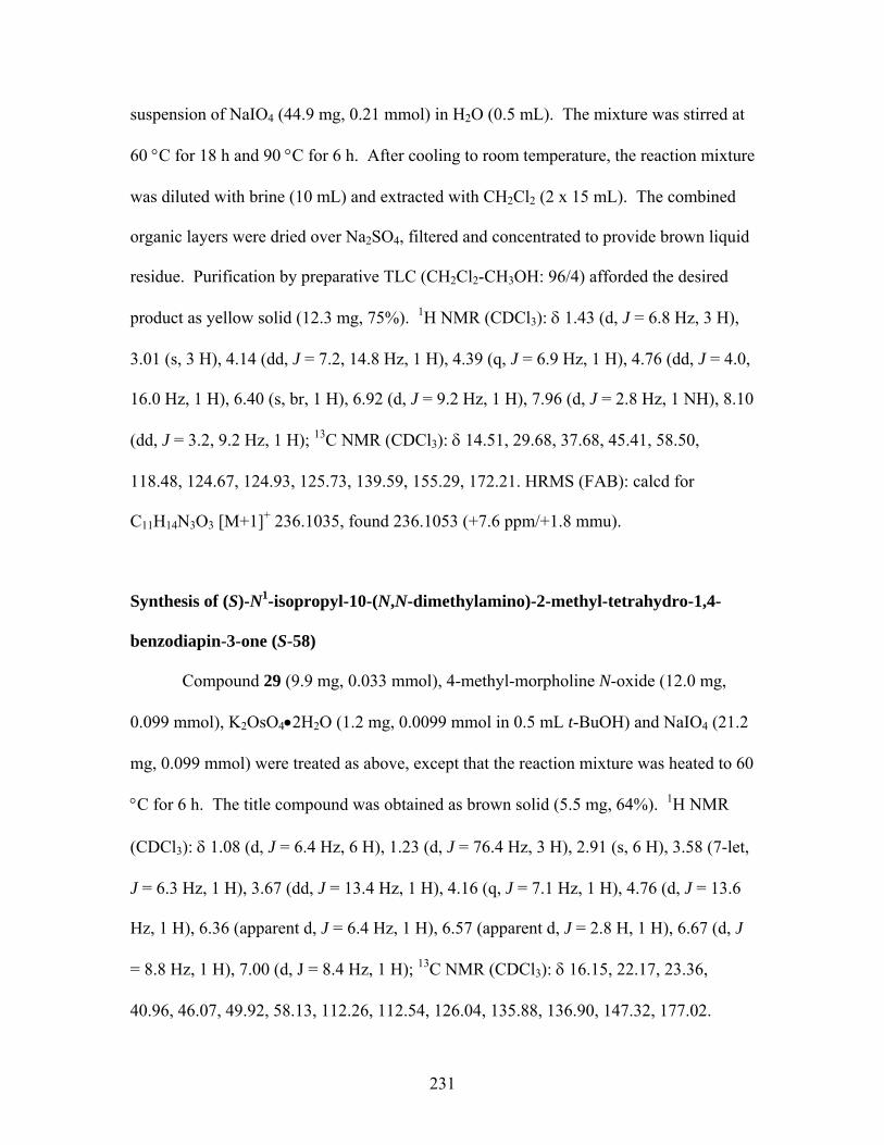
suspension of NaIO4 (44.9 mg, 0.21 mmol) in H2O (0.5 mL). The mixture was stirred at
60 °C for 18 h and 90 °C for 6 h. After cooling to room temperature, the reaction mixture
was diluted with brine (10 mL) and extracted with CH2Cl2 (2 x 15 mL). The combined
organic layers were dried over Na2SO4, filtered and concentrated to provide brown liquid
residue. Purification by preparative TLC (CH2Cl2-CH3OH: 96/4) afforded the desired
product as yellow solid (12.3 mg, 75%). 1H NMR (CDCl3): δ 1.43 (d, J = 6.8 Hz, 3 H),
3.01 (s, 3 H), 4.14 (dd, J = 7.2, 14.8 Hz, 1 H), 4.39 (q, J = 6.9 Hz, 1 H), 4.76 (dd, J = 4.0,
16.0 Hz, 1 H), 6.40 (s, br, 1 H), 6.92 (d, J = 9.2 Hz, 1 H), 7.96 (d, J = 2.8 Hz, 1 NH), 8.10
(dd, J = 3.2, 9.2 Hz, 1 H); 13C NMR (CDCl3): δ 14.51, 29.68, 37.68, 45.41, 58.50,
118.48, 124.67, 124.93, 125.73, 139.59, 155.29, 172.21. HRMS (FAB): calcd for
C11H14N3O3 [M+1]+ 236.1035, found 236.1053 (+7.6 ppm/+1.8 mmu).
Synthesis of (S)-N1-isopropyl-10-(N,N-dimethylamino)-2-methyl-tetrahydro-1,4-
benzodiapin-3-one (S-58)
Compound 29 (9.9 mg, 0.033 mmol), 4-methyl-morpholine N-oxide (12.0 mg,
0.099 mmol), K2OsO4•2H2O (1.2 mg, 0.0099 mmol in 0.5 mL t-BuOH) and NaIO4 (21.2
mg, 0.099 mmol) were treated as above, except that the reaction mixture was heated to 60
°C for 6 h. The title compound was obtained as brown solid (5.5 mg, 64%). 1H NMR
(CDCl3): δ 1.08 (d, J = 6.4 Hz, 6 H), 1.23 (d, J = 76.4 Hz, 3 H), 2.91 (s, 6 H), 3.58 (7-let,
J = 6.3 Hz, 1 H), 3.67 (dd, J = 13.4 Hz, 1 H), 4.16 (q, J = 7.1 Hz, 1 H), 4.76 (d, J = 13.6
Hz, 1 H), 6.36 (apparent d, J = 6.4 Hz, 1 H), 6.57 (apparent d, J = 2.8 H, 1 H), 6.67 (d, J
= 8.8 Hz, 1 H), 7.00 (d, J = 8.4 Hz, 1 H); 13C NMR (CDCl3): δ 16.15, 22.17, 23.36,
40.96, 46.07, 49.92, 58.13, 112.26, 112.54, 126.04, 135.88, 136.90, 147.32, 177.02.
231
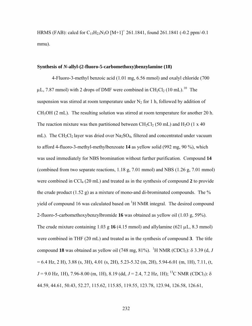
HRMS (FAB): calcd for C15H23N3O [M+1]+ 261.1841, found 261.1841 (-0.2 ppm/-0.1
mmu).
Synthesis of N-allyl-(2-fluoro-5-carbomethoxy)benzylamine (18)
4-Fluoro-3-methyl benzoic acid (1.01 mg, 6.56 mmol) and oxalyl chloride (700
μL, 7.87 mmol) with 2 drops of DMF were combined in CH2Cl2 (10 mL).10 The
suspension was stirred at room temperature under N2 for 1 h, followed by addition of
CH3OH (2 mL). The resulting solution was stirred at room temperature for another 20 h.
The reaction mixture was then partitioned between CH2Cl2 (50 mL) and H2O (1 x 40
mL). The CH2Cl2 layer was dried over Na2SO4, filtered and concentrated under vacuum
to afford 4-fluoro-3-methyl-methylbenzoate 14 as yellow solid (992 mg, 90 %), which
was used immediately for NBS bromination without further purification. Compound 14
(combined from two separate reactions, 1.18 g, 7.01 mmol) and NBS (1.26 g, 7.01 mmol)
were combined in CCl4 (20 mL) and treated as in the synthesis of compound 2 to provide
the crude product (1.52 g) as a mixture of mono-and di-brominated compounds. The %
yield of compound 16 was calculated based on 1H NMR integral. The desired compound
2-fluoro-5-carbomethoxybenzylbromide 16 was obtained as yellow oil (1.03 g, 59%).
The crude mixture containing 1.03 g 16 (4.15 mmol) and allylamine (621 μL, 8.3 mmol)
were combined in THF (20 mL) and treated as in the synthesis of compound 3. The title
compound 18 was obtained as yellow oil (748 mg, 81%). 1H NMR (CDCl3): δ 3.39 (d, J
= 6.4 Hz, 2 H), 3.88 (s, 3H), 4.01 (s, 2H), 5.23-5.32 (m, 2H), 5.94-6.01 (m, 1H), 7.11, (t,
J = 9.0 Hz, 1H), 7.96-8.00 (m, 1H), 8.19 (dd, J = 2.4, 7.2 Hz, 1H); 13C NMR (CDCl3): δ
44.59, 44.61, 50.43, 52.27, 115.62, 115.85, 119.55, 123.78, 123.94, 126.58, 126.61,
232
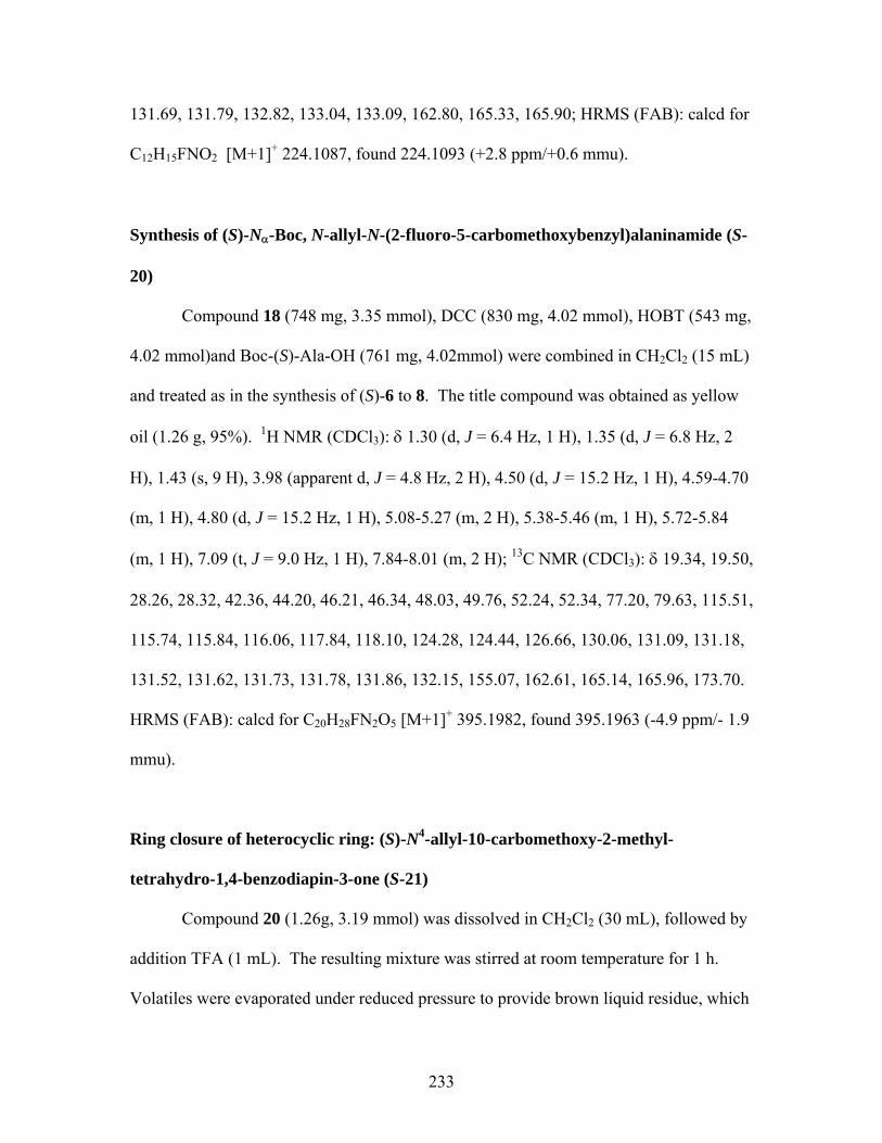
131.69, 131.79, 132.82, 133.04, 133.09, 162.80, 165.33, 165.90; HRMS (FAB): calcd for
C12H15FNO2 [M+1]+ 224.1087, found 224.1093 (+2.8 ppm/+0.6 mmu).
Synthesis of (S)-Nα-Boc, N-allyl-N-(2-fluoro-5-carbomethoxybenzyl)alaninamide (S-
20)
Compound 18 (748 mg, 3.35 mmol), DCC (830 mg, 4.02 mmol), HOBT (543 mg,
4.02 mmol)and Boc-(S)-Ala-OH (761 mg, 4.02mmol) were combined in CH2Cl2 (15 mL)
and treated as in the synthesis of (S)-6 to 8. The title compound was obtained as yellow
oil (1.26 g, 95%). 1H NMR (CDCl3): δ 1.30 (d, J = 6.4 Hz, 1 H), 1.35 (d, J = 6.8 Hz, 2
H), 1.43 (s, 9 H), 3.98 (apparent d, J = 4.8 Hz, 2 H), 4.50 (d, J = 15.2 Hz, 1 H), 4.59-4.70
(m, 1 H), 4.80 (d, J = 15.2 Hz, 1 H), 5.08-5.27 (m, 2 H), 5.38-5.46 (m, 1 H), 5.72-5.84
(m, 1 H), 7.09 (t, J = 9.0 Hz, 1 H), 7.84-8.01 (m, 2 H); 13C NMR (CDCl3): δ 19.34, 19.50,
28.26, 28.32, 42.36, 44.20, 46.21, 46.34, 48.03, 49.76, 52.24, 52.34, 77.20, 79.63, 115.51,
115.74, 115.84, 116.06, 117.84, 118.10, 124.28, 124.44, 126.66, 130.06, 131.09, 131.18,
131.52, 131.62, 131.73, 131.78, 131.86, 132.15, 155.07, 162.61, 165.14, 165.96, 173.70.
HRMS (FAB): calcd for C20H28FN2O5 [M+1]+ 395.1982, found 395.1963 (-4.9 ppm/- 1.9
mmu).
Ring closure of heterocyclic ring: (S)-N4-allyl-10-carbomethoxy-2-methyl-
tetrahydro-1,4-benzodiapin-3-one (S-21)
Compound 20 (1.26g, 3.19 mmol) was dissolved in CH2Cl2 (30 mL), followed by
addition TFA (1 mL). The resulting mixture was stirred at room temperature for 1 h.
Volatiles were evaporated under reduced pressure to provide brown liquid residue, which
233
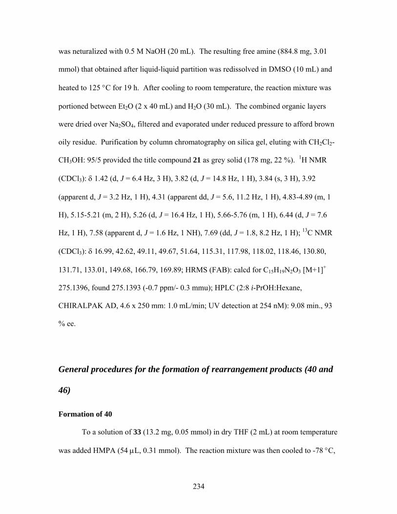
was neturalized with 0.5 M NaOH (20 mL). The resulting free amine (884.8 mg, 3.01
mmol) that obtained after liquid-liquid partition was redissolved in DMSO (10 mL) and
heated to 125 °C for 19 h. After cooling to room temperature, the reaction mixture was
portioned between Et2O (2 x 40 mL) and H2O (30 mL). The combined organic layers
were dried over Na2SO4, filtered and evaporated under reduced pressure to afford brown
oily residue. Purification by column chromatography on silica gel, eluting with CH2Cl2-
CH3OH: 95/5 provided the title compound 21 as grey solid (178 mg, 22 %). 1H NMR
(CDCl3): δ 1.42 (d, J = 6.4 Hz, 3 H), 3.82 (d, J = 14.8 Hz, 1 H), 3.84 (s, 3 H), 3.92
(apparent d, J = 3.2 Hz, 1 H), 4.31 (apparent dd, J = 5.6, 11.2 Hz, 1 H), 4.83-4.89 (m, 1
H), 5.15-5.21 (m, 2 H), 5.26 (d, J = 16.4 Hz, 1 H), 5.66-5.76 (m, 1 H), 6.44 (d, J = 7.6
Hz, 1 H), 7.58 (apparent d, J = 1.6 Hz, 1 NH), 7.69 (dd, J = 1.8, 8.2 Hz, 1 H); 13C NMR
(CDCl3): δ 16.99, 42.62, 49.11, 49.67, 51.64, 115.31, 117.98, 118.02, 118.46, 130.80,
131.71, 133.01, 149.68, 166.79, 169.89; HRMS (FAB): calcd for C15H19N2O3 [M+1]+
275.1396, found 275.1393 (-0.7 ppm/- 0.3 mmu); HPLC (2:8 i-PrOH:Hexane,
CHIRALPAK AD, 4.6 x 250 mm: 1.0 mL/min; UV detection at 254 nM): 9.08 min., 93
% ee.
General procedures for the formation of rearrangement products (40 and
46)
Formation of 40 To a solution of 33 (13.2 mg, 0.05 mmol) in dry THF (2 mL) at room temperature
was added HMPA (54 μL, 0.31 mmol). The reaction mixture was then cooled to -78 °C,
234
followed by addition of LDA (1.5 M in cyclohexane, 68 μL, 0.102 mmol). The resulting
orange mixture was stirred at -78 °C for 15 min. n-BuLi (2.5 M in hexanes, 41 μL,
0.102 mmol) was then added to it, and the reaction mixture was stirred at -78 °C for
another 15 min. before the addition of EtI (42 μL, 0.51 mmol). The corresponding
reaction mixture was stirred at -78 °C for further 1 h, followed by addition of NH4Cl(satd.)
(10 mL). The mixture was allowed to warm up to room temperature and diluted with
CH2Cl2 (15 mL). The organic layer was dried over Na2SO4, filtered and evaporated
under reduced pressure to afford brown oily residue. Purification on preparative TLC
(EtOAc-CH2Cl2: 20/80) afforded the rearrangement product as yellow solid 40 (3.0 mg,
23 %). 1H NMR (CDCl3): δ 1.21 (d, J = 6.4 Hz, 6 H), 2.41 (s, 3 H), 3.63-3.68 (m, 1 H),
3.76 (apparent d, J = 6.0 Hz, 2 H), 4.50 (s, 2 H), 4.90 (s, br, 1 NH), 5.15-5.30 (m, 2 H),
5.68-5.77 (m, 1 H), 6.55-6.65 (m, 2 H), 7.00 (dd, J = 1.4, 7.4 Hz, 1 H), 7.19-7.23 (m, 1
H).
Formation of 46 Compound 42 (18.7 mg, 0.068 mmol), HMPA (71 μL, 0.41 mmol), LDA (227,
μL, 0.34 mmol), n-BuLi (136 μL, 0.34 mmol) and EtI (55 μL, 0.68 mmol) were
combined in THF (1 mL) and treated as above. The title compound 46 was obtained as
yellow solid (3.1 mg, 17%). 1H NMR (CDCl3): δ 0.83 (t, J = 7.2 Hz, 3 H), 1.21 (d, J =
6.4 Hz, 6 H), 1.53-1.63 (m, 2 H), 2.43 (s, 3 H), 3.04-3.08 (m, 2 H), 3.65 (s, br, 1 H), 4.51
(s, 2 H), 4.83 (s, br, 1 NH), 6.56-6.65 (m, 2 H), 7.02 (dd, J = 1.4, 7.2 Hz, 1 H), 7.18-7.23
(m, 1 H); HRMS (FAB): calcd for C16H23N2O [M]+ 259.1810, found 259.1823 (+6.0
ppm/+1.5 mmu);
235

6.2.2 (S)-Phenylalanine-derived tetrahydro-1,4-benzodiazepine-2-one project
(S)-Phenylalanine methyl ester hydrochloride was purchased from Aldrich. 2-
Nitrobenzylbromide was obtained from ACROS. Both reagents were used without
purification.
Synthesis of (S)-N-(2-nitrobenzylphenylalanine)methyl ester (S)-7411
To a solution of (S)-phenylalanine methyl ester hydrochloride (1.06 g, 4.90 mmol)
and 2-nitrobenzylbromide 73 (1.06 g, 4.90 mmol) in DMF (10 mL) was added DIEA
(1.75 mL, 9.80 mmol). The resulting reaction mixture was stirred at room temperature
for 24 h. The volatiles were removed under reduced pressure to provide a yellow liquid
residue, which was then partitioned between Et2O (2 x 50 mL) and 10 % NaHCO3 (1 x 50
mL). The combined Et2O layers were dried over Na2SO4, filtered and evaporated under
reduced pressure to afford yellow oily residue. Purification by column chromatography
on silica gel, eluting with CH2Cl2-EtOAc-Hexane: 38/2/60 provided the title compound
74 as greenish yellow oil (1.31 g, 95 %). 1H NMR (CDCl3): δ 2.07 (s, br, 1 H), 2.91 (dd,
J = 7.6, 13.6 Hz, 2 H), 2.99 (dd, J = 6.0, 13.6 Hz, 1 H), 3.48-3.51(m, 1 H), 3.66 (s, 3 H),
3.93 (d, J = 15.2 Hz, 1 H), 4.10 (d, J = 15.2 Hz, 1 H), 7.14-7.16 (m, 2 H), 7.20-7.30 (m, 3
H), 7.35-7.39 (m, 1 H), 7.44-7.51 (m, 2 H), 7.90 (dd, J = 1.2, 8.0 Hz, 1 H); 13C NMR
(CDCl3): δ 39.73, 48.97, 51.76, 62.42, 124.67, 126.72, 127.88, 128.39, 129.19, 130.78,
133.01, 135.17, 137.13, 148.96, 174.65; HRMS (FAB): calcd for C17H19N2O4 [M+1]+
315.1345, found 315.1344 (-0.3 ppm/- 0.1 mmu).
236

General procedures for the preparation of (S)-75 and (S)-76
Synthesis of (S)-N-methyl-N’-(2-nitrobenzylphenylalanine)methyl ester (S)-75
To a solution of 74 (107 mg, 0.39 mmol) in DMF (5 mL) was added CH3I (188
μl, 3.03 mmol) and DIEA (69 μl, 0.39 mmol). The resulting green reaction mixture was
heated to 100 °C for 1.5 h. The volatiles were removed under reduced pressure to
provide a brown liquid residue, which was then partitioned between Et2O (2 x 40 mL)
and 10 % NaHCO3 (1 x 30 mL). The combined Et2O layers were dried over Na2SO4,
filtered and evaporated under reduced pressure to afford yellow oily residue. Purification
by column chromatography on silica gel, eluting with CH2Cl2-EtOAc: 97/3 provided the
title compound (S)-75 as green oil (88 mg, 76 %). 1H NMR (CDCl3): δ 2.43 (s, 3 H),
2.89 (dd, J = 7.2, 14.0 Hz, 2 H), 3.11 (dd, J = 8.0, 13.6 Hz, 1 H), 3.55 (apparent t, J = 8.0
Hz, 1 H), 3.69 (s, 3 H), 4.03 (apparent d, J = 2.0 Hz, 1 H), 7.15-7.17 (m, 2 H), 7.19-7.29
(m, 3 H), 7.29-7.37 (m, 2 H), 7.40-7.45 (m, 1 H), 7.80-7.82 (m, 1 H); 13C NMR (CDCl3):
δ 35.84, 37.54, 51.15, 55.96, 67.65, 124.36, 126.31, 127.75, 128.31, 129.14, 130.79,
132.46, 134.51, 138.24, 149.57, 171.98; HRMS (FAB): calcd for C18H21N2O4 [M+1]+
329.1501, found 329.1491 (-3.1 ppm/- 1.0 mmu).
Synthesis of (S)-N-allyl-N’-(2-nitrobenzylphenylalanine)methyl ester (S)-76
Compound 74 (132 mg, 0.49 mmol), allylbromide (342 μl, 3.92 mmol) and DIEA
(86μl, 0.49 mmol) were combined in DMF (5 mL) and treated as above. The title
compound (S)-76 was obtained as green oil (118 mg, 75 %). 1H NMR (CDCl3): δ 2.94
(dd, J = 8.0, 14.0 Hz, 2 H), 3.10-3.18 (m, 2 H), 3.37-3.42 (m, 1H), 3.63 (t, J = 7.6 Hz, 1
H), 3.70 (s, 3 H), 4.03 (d, J = 16.4 Hz, 1 H), 4.22 (d, J = 16.4 Hz, 1 H), 5.09-5.19 (m, 2
237
H), 5.66-5.76 (m, 1 H), 7.12-7.14 (m, 2 H), 7.22-7.30 (m, 3 H), 7.32-7.41 (m, 3 H), 7.82-
7.85 (m, 1 H); 13C NMR (CDCl3): δ 35.77, 51.27, 51.43, 54.04, 64.38, 117.80, 124.17,
126.35, 127.46, 128.25, 129.32, 130.67, 132.54, 134.99, 135.57, 138.19, 149.53, 172.45.
Synthesis of (S)-N4-allyl-3-benzyl-tetrahydro-1,4-benzodiapin-2-one (S)-82
Compound (S)-76 was reduced to corresponding primary amine by following the
general procedures in the synthesis of (S)-27. Compound 76 (435 mg, 1.35 mmol), Fe
powder (530 mg, 9.46 mmol), and NH4Cl (650 mg, 12.16 mmol in 6.0 mL H2O) were
combined in CH3OH (16 mL) and treated as described previously. The corresponding
primary amine (S)-80 was obtained as brown oil (385 mg, 98%). Compound (S)-80 was
used immediately in next step. To a solution of (S)-80 (216 mg, 0.74 mmol) in CH3CN
(2.2 mL) was added 2 N NaOH (0.5 mL).12 The resulting mixture was stirred at room
temperature for 5 days. Volatiles were removed under reduced pressure to provide (S)-81
as sodium salt (246 mg, 100%). No further purification was performed. Compound (S)-
81 (220 mg, 0.66 mmol), DCC (227 mg, 1.10 mmol) and HOBT (149 mg, 1.10 mmol)
were combined in CH2Cl2 (10 mL) and treated as in the synthesis of (S)-6 to 8. The title
compound (S)-82 was obtained as yellow solid (148 mg, 77%). 1H NMR (CDCl3): δ 2.81
(dd, J = 7.6, 14.0 Hz, 2 H), 3.14-3.22 (m, 2 H), 3.31-3.36 (m, 1H), 3.70 (t, J = 7.6 Hz, 1
H), 3.87 (s, 3 H), 5.15-5.22 (m, 2 H), 5.72-5.82 (m, 1 H), 6.93 (d, J = 8.0 Hz, 1 H), 7.10-
7.30 (m, 8 H), 8.42 (s, br, 1 H); 13C NMR (CDCl3): δ 36.65, 53.88, 55.93, 66.48, 118.07,
120.35, 124.71, 126.18, 128.13, 128.55, 129.10, 129.25, 130.26, 135.36, 137.12, 138.63,
173.33; HRMS (FAB): calcd for C19H21N2O [M+1]+ 293.1654, found 293.1645 (-3.1
ppm/- 0.9 mmu).
238
Synthesis of (S)-N4-allyl-3-benzyl-N1-DAM-tetrahydro-1,4-benzodiapin-2-one (S)-83
Compound (S)-82 (143 mg, 0.48 mmol) and NaH (60 wt% in oil, 23.2 mg, 0.58
mmol) were combined in THF (5 mL) and stirred at 0 °C for 30 min., followed by
addition of DAM-Br (178 mg, 0.58 mmol) in THF (2 mL) dropwise. The resulting green
suspension was stirred at 0 °C for 2 h. The excess NaH was quenched by H2O (10 mL) at
0 °C. After the gas evolution was completed, the reaction mixture was partitioned
between CH2Cl2 (2 x 30 mL) and H2O (1 x 20 mL). The combined CH2Cl2 layers were
dried over Na2SO4, filtered and evaporated under reduced pressure to afford yellow oily
residue. Purification by column chromatography on silica gel, eluting with CH2Cl2-
EtOAc: 9/1 provided the title compound (S)-83 as yellow solid (181 mg, 72 %). 1H NMR
(CDCl3): δ 2.75 (apparent d, J = 10.0 Hz, 1 H), 3.19-3.35 (m, 3 H), 3.46-3.48 (m, 2H),
3.65 –3.81 (m, 1 H), 3.71 (s, br, 3 H), 3.80 (s, br, 3 H), 6.62 (apparent d, J = 7.6 Hz, 2 H),
6.73 (apparent d, J = 7.6 Hz, 2 H), 6.83-6.93 (m, 5 H), 7.07-7.29 (m, 9 H); 13C NMR
(CDCl3): δ 34.83, 54.22, 54.73, 55.08, 55.23, 63.04, 64.82, 113.23, 113.45, 113.77,
118.04, 123.67, 126.09, 127.71, 127.97, 128.21, 129.04, 129.54, 129.88, 130.76, 131.26,
135.85, 138.97, 141.11, 158.45, 158.55, 169.37; HRMS (FAB): calcd for C34H35N2O3
[M+1]+ 519.2648, found 519.2661 (+2.6 ppm/+1.3 mmu).
6.2.3 (S)-Serine-derived 1,4-benzodiazepine-2-one project Both Boc-(S)-serine and Boc-O-benzyl-(S)-serine were purchased from Advanced
ChemTech, and used without purification. Boc-O-TBDMS-(S)-serine was synthesized
from tert-butyldimethylsilyl chloride (TBDMSCl) and Boc-(S)-serine based on the
239
literature method.13 2-Amino-5-chloro-benzophenone was obtained from ACORS, and
also used without purification.
Synthesis of Nα-Boc-O-benzyl-N-(2-benzoyl-4-chloro)phenyl serinamide (S-89)
To a solution of 2-amino-5-chlorobenzophenone 88 (1.74 g, 7.53 mmol) and Boc-
O-benzyl-(S)-serine (2.02 g, 6.85 mmol) in THF (15 mL) was added DCC (1.55 g, 7.53
mmol) in 7.5 mL CH2Cl2 at 0 °C. The yellow mixture was allowed at warm up to room
temperature at its own accord and stirred at room temperature for 18 h. White precipitate
was filtered off and washed with cold CH2Cl2 (10 mL), and the filtrate was evaporated
under reduced pressure to provide crude yellow oil. Purification by column
chromatography on silica gel, eluting with CH2Cl2-EtOAc: 9/1 afforded the desired
product as off-white solid (2.75 g, 79%). 1H NMR (CDCl3): δ 3.69 (dd, J = 4.4, 9.6 Hz, 1
H), 4.06 (apparent d, J = 7.2 Hz, 1 H), 4.43 (s, br, 1 H), 4.53 (apparent d, J = 4.2 Hz, 2
H), 5.56 (apparent d, J = 6.4 Hz, 1 H), 6.73 (apparent d, J = 7.6 Hz, 2 H), 7.24 (s, 5 H),
7.48-7.55 (m, 4 H), 7.63 (apparent tt J = 1.3, 7.4 Hz, 1 H), 7.68-7.70 (m, 2 H), 8.66 (d, J
= 8.4 Hz, 1 H), 11.34 (s, 1 NH); 13C NMR (CDCl3): δ 28.24, 55.76, 69.52, 73.35, 80.56,
122.93, 125.17, 127.55, 127.64, 127.76, 128.35, 128.44, 128.57, 129.92, 132.42, 132.84,
133.67, 137.35, 137.77, 138.25, 169.83, 197.48; HRMS (FAB): calcd for C28H30N2O5Cl
[M+1]+ 509.1843, found 5109.1828 (-3.0 ppm/-1.5 mmu).
240
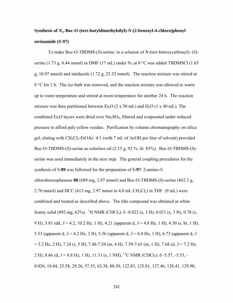
Synthesis of Nα-Boc-O-(tert-butyldimethylsilyl)-N-(2-benzoyl-4-chloro)phenyl
serinamide (S-97)
To make Boc-O-TBDMS-(S)-serine: to a solution of N-(tert-butoxycarbonyl)- (S)-
serine (1.73 g, 8.44 mmol) in DMF (17 mL) under N2 at 0 °C was added TBDMSCl (1.65
g, 10.97 mmol) and imidazole (1.72 g, 25.32 mmol). The reaction mixture was stirred at
0 °C for 1 h. The ice-bath was removed, and the reaction mixture was allowed to warm
up to room temperature and stirred at room temperature for another 24 h. The reaction
mixture was then partitioned between Et2O (2 x 50 mL) and H2O (1 x 40 mL). The
combined Et2O layers were dried over Na2SO4, filtered and evaporated under reduced
pressure to afford pale yellow residue. Purification by column chromatography on silica
gel, eluting with CH2Cl2-EtOAc: 4/1 (with 7 mL of AcOH per liter of solvent) provided
Boc-O-TBDMS-(S)-serine as colorless oil (2.15 g, 92 %, lit. 85%). Boc-O-TBDMS-(S)-
serine was used immediately in the next step. The general coupling procedures for the
synthesis of S-89 was followed for the preparation of S-97: 2-amino-5-
chlorobenzophenone 88 (689 mg, 2.97 mmol) and Boc-O-TBDMS-(S)-serine (862.3 g,
2.70 mmol) and DCC (613 mg, 2.97 mmol in 4.0 mL CH2Cl2) in THF (9 mL) were
combined and treated as described above. The title compound was obtained as white
foamy solid (892 mg, 62%). 1H NMR (CDCl3): δ -0.022 (s, 3 H), 0.021 (s, 3 H), 0.78 (s,
9 H), 3.81 (dd, J = 4.2, 10.2 Hz, 1 H), 4.21 (apparent d, J = 4.8 Hz, 1 H), 4.30 (s, br, 1 H),
5.53 (apparent d, J = 4.2 Hz, 2 H), 5.56 (apparent d, J = 6.4 Hz, 1 H), 6.73 (apparent d, J
= 5.2 Hz, 2 H), 7.24 (s, 5 H), 7.48-7.54 (m, 4 H), 7.59-7.63 (m, 1 H), 7.68 (d, J = 7.2 Hz,
2 H), 8.66 (d, J = 8.8 Hz, 1 H), 11.31 (s, 1 NH); 13C NMR (CDCl3): δ -5.57, -5.53, -
0.026, 18.44, 25.58, 28.26, 57.55, 63.38, 80.50, 122.83, 125.01, 127.46, 128.41, 129.90,
241

132.47, 132.81, 133.73, 137.83, 138.28, 155.54, 170.19, 197.58; LRMS (FAB): calcd for
C27H38N2O5ClSi [M+1]+ 533, found 533.
Synthesis of (S)-3-benzyloxymethyl)-10-chloro-5-phenyl-1,4-benzodiazepin-2-one (S-
90)
To a solution of S-89 (557 mg, 1.10 mmol) in CH2Cl2 (10 mL) was added TFA (1
mL). The resulting solution was stirred at room temperature for 1 h. Volatiles were
removed under reduced pressure to provide yellow residue, which was immediately
redissolved in CH3OH-H2O (10 mL: 5 mL), and the pH was adjusted to pH 8.5-9.0 with
10 % NaOH. The yellow mixture was stirred at room temperature for 65 h. Solvent was
removed under reduced pressure and the yellow residue was partitioned between CH2Cl2
(2 x 50 mL) and H2O (1 x 40 mL). The combined CH2Cl2 layers were dried over
Na2SO4, filtered and evaporated under reduced pressure to afford yellow oil. Purification
by column chromatography on silica gel, eluting with CH2Cl2-EtOAc: 9/1 provided the
title compound as white foamy solid (328 mg, 76 %). 1H NMR (CDCl3): δ 3.86 (t, J = 6.4
Hz, 1 H), 4.21 (dd, J = 6.2, 9.4 Hz, 1 H), 4.45 (dd, J = 6.6, 9.4 Hz, 1 H), 4.71 (s, 2 H),
7.08 (d, J = 8.4 Hz, 1 H), 7.28-7.49 (m, 9 H), 7.53-7.55 (m, 2 H), 8.97 (s, 1 NH); 13C
NMR (CDCl3): δ 63.41, 70.29, 73.69, 122.90, 126.94, 127.54, 127.63, 127.80, 128.33,
128.35, 128.47, 128.75, 128.82, 129.71, 130.39, 130.60, 131.75, 136.88, 138.41, 140.86,
168.62, 170.81; HRMS (FAB): calcd for C23H20N2O2Cl [M+1]+ 391.1213, found
391.1188 (-6.5 ppm/-2.5 mmu).
242
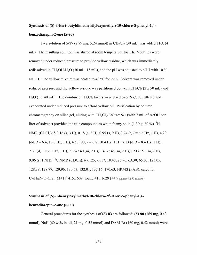
Synthesis of (S)-3-(tert-butyldimethylsilyloxymethyl)-10-chloro-5-phenyl-1,4-
benzodiazepin-2-one (S-98)
To a solution of S-97 (2.79 mg, 5.24 mmol) in CH2Cl2 (30 mL) was added TFA (4
mL). The resulting solution was stirred at room temperature for 1 h. Volatiles were
removed under reduced pressure to provide yellow residue, which was immediately
redissolved in CH3OH-H2O (30 mL: 15 mL), and the pH was adjusted to pH 7 with 10 %
NaOH. The yellow mixture was heated to 40 °C for 22 h. Solvent was removed under
reduced pressure and the yellow residue was partitioned between CH2Cl2 (2 x 50 mL) and
H2O (1 x 40 mL). The combined CH2Cl2 layers were dried over Na2SO4, filtered and
evaporated under reduced pressure to afford yellow oil. Purification by column
chromatography on silica gel, eluting with CH2Cl2-EtOAc: 9/1 (with 7 mL of AcOH per
liter of solvent) provided the title compound as white foamy solid (1.30 g, 60 %). 1H
NMR (CDCl3): δ 0.16 (s, 3 H), 0.18 (s, 3 H), 0.95 (s, 9 H), 3.74 (t, J = 6.6 Hz, 1 H), 4.29
(dd, J = 6.4, 10.0 Hz, 1 H), 4.58 (dd, J = 6.8, 10.4 Hz, 1 H), 7.13 (d, J = 8.4 Hz, 1 H),
7.31 (d, J = 2.0 Hz, 1 H), 7.36-7.40 (m, 2 H), 7.43-7.48 (m, 2 H), 7.51-7.53 (m, 2 H),
9.06 (s, 1 NH); 13C NMR (CDCl3): δ -5.25, -5.17, 18.48, 25.96, 63.30, 65.08, 123.05,
128.38, 128.77, 129.96, 130.63, 132.01, 137.16, 170.63; HRMS (FAB): calcd for
C22H28N2O2ClSi [M+1]+ 415.1609, found 415.1629 (+4.9 ppm/+2.0 mmu).
Synthesis of (S)-3-benzyloxylmethyl-10-chloro-N1-DAM-5-phenyl-1,4-
benzodiazepin-2-one (S-99)
General procedures for the synthesis of (S)-83 are followed: (S)-90 (169 mg, 0.43
mmol), NaH (60 wt% in oil, 21 mg, 0.52 mmol) and DAM-Br (160 mg, 0.52 mmol) were
243
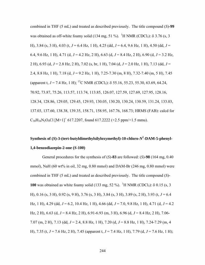
combined in THF (5 mL) and treated as described previously. The title compound (S)-99
was obtained as off-white foamy solid (134 mg, 51 %). 1H NMR (CDCl3): δ 3.76 (s, 3
H), 3.84 (s, 3 H), 4.03 (t, J = 6.4 Hz, 1 H), 4.25 (dd, J = 6.4, 9.6 Hz, 1 H), 4.50 (dd, J =
6.4, 9.6 Hz, 1 H), 4.71 (d, J = 4.2 Hz, 2 H), 6.63 (d, J = 8.4 Hz, 2 H), 6.90 (d, J = 3.2 Hz,
2 H), 6.93 (d, J = 2.8 Hz, 2 H), 7.02 (s, br, 1 H), 7.04 (d, J = 2.0 Hz, 1 H), 7.13 (dd, J =
2.4, 8.8 Hz, 1 H), 7.18 (d, J = 9.2 Hz, 1 H), 7.25-7.30 (m, 8 H), 7.32-7.40 (m, 5 H), 7.45
(apparent t, J = 7.4 Hz, 1 H); 13C NMR (CDCl3): δ 55.16, 55.23, 55.30, 63.69, 64.24,
70.92, 73.87, 75.26, 113.57, 113.74, 113.85, 126.07, 127.59, 127.69, 127.95, 128.16,
128.34, 128.86, 129.05, 129.45, 129.93, 130.05, 130.20, 130.24, 130.59, 131.24, 133.03,
137.03, 137.60, 138.38, 139.35, 158.71, 158.95, 167.76, 168.73; HRMS (FAB): calcd for
C38H34N2O4Cl [M+1]+ 617.2207, found 617.2222 (+2.5 ppm/+1.5 mmu).
Synthesis of (S)-3-(tert-butyldimethylsilyloxymethyl)-10-chloro-N1-DAM-5-phenyl-
1,4-benzodiazepin-2-one (S-100)
General procedures for the synthesis of (S)-83 are followed: (S)-98 (164 mg, 0.40
mmol), NaH (60 wt% in oil, 32 mg, 0.80 mmol) and DAM-Br (246 mg, 0.80 mmol) were
combined in THF (5 mL) and treated as described previously. The title compound (S)-
100 was obtained as white foamy solid (133 mg, 52 %). 1H NMR (CDCl3): δ 0.15 (s, 3
H), 0.16 (s, 3 H), 0.92 (s, 9 H), 3.76 (s, 3 H), 3.84 (s, 3 H), 3.89 (s, 2 H), 3.93 (t, J = 6.4
Hz, 1 H), 4.29 (dd, J = 6.2, 10.4 Hz, 1 H), 4.66 (dd, J = 7.0, 9.8 Hz, 1 H), 4.71 (d, J = 4.2
Hz, 2 H), 6.63 (d, J = 8.4 Hz, 2 H), 6.91-6.93 (m, 3 H), 6.96 (d, J = 8.4 Hz, 2 H), 7.06-
7.07 (m, 2 H), 7.13 (dd, J = 2.4, 8.8 Hz, 1 H), 7.20 (d, J = 8.8 Hz, 1 H), 7.24-7.29 (m, 4
H), 7.35 (t, J = 7.6 Hz, 2 H), 7.45 (apparent t, J = 7.4 Hz, 1 H), 7.79 (d, J = 7.6 Hz, 1 H);
244
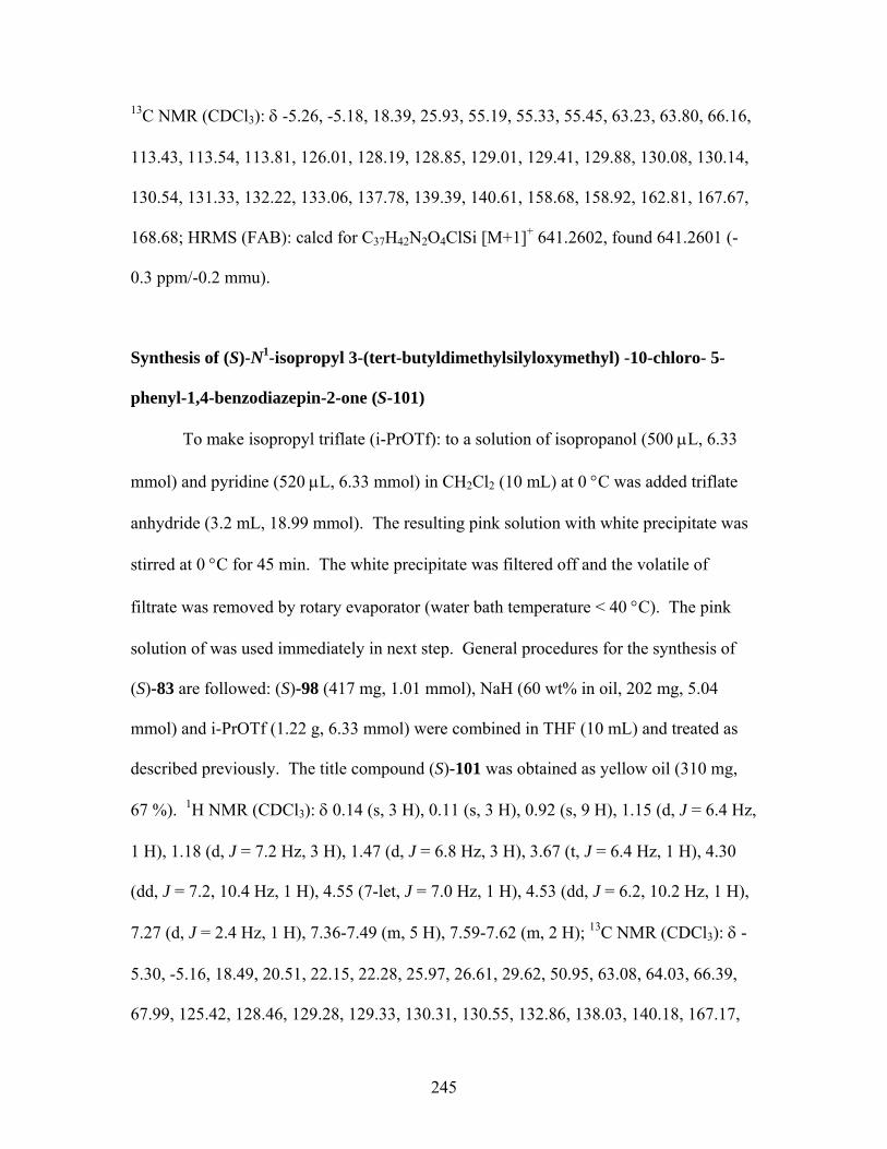
13C NMR (CDCl3): δ -5.26, -5.18, 18.39, 25.93, 55.19, 55.33, 55.45, 63.23, 63.80, 66.16,
113.43, 113.54, 113.81, 126.01, 128.19, 128.85, 129.01, 129.41, 129.88, 130.08, 130.14,
130.54, 131.33, 132.22, 133.06, 137.78, 139.39, 140.61, 158.68, 158.92, 162.81, 167.67,
168.68; HRMS (FAB): calcd for C37H42N2O4ClSi [M+1]+ 641.2602, found 641.2601 (-
0.3 ppm/-0.2 mmu).
Synthesis of (S)-N1-isopropyl 3-(tert-butyldimethylsilyloxymethyl) -10-chloro- 5-
phenyl-1,4-benzodiazepin-2-one (S-101)
To make isopropyl triflate (i-PrOTf): to a solution of isopropanol (500 μL, 6.33
mmol) and pyridine (520 μL, 6.33 mmol) in CH2Cl2 (10 mL) at 0 °C was added triflate
anhydride (3.2 mL, 18.99 mmol). The resulting pink solution with white precipitate was
stirred at 0 °C for 45 min. The white precipitate was filtered off and the volatile of
filtrate was removed by rotary evaporator (water bath temperature < 40 °C). The pink
solution of was used immediately in next step. General procedures for the synthesis of
(S)-83 are followed: (S)-98 (417 mg, 1.01 mmol), NaH (60 wt% in oil, 202 mg, 5.04
mmol) and i-PrOTf (1.22 g, 6.33 mmol) were combined in THF (10 mL) and treated as
described previously. The title compound (S)-101 was obtained as yellow oil (310 mg,
67 %). 1H NMR (CDCl3): δ 0.14 (s, 3 H), 0.11 (s, 3 H), 0.92 (s, 9 H), 1.15 (d, J = 6.4 Hz,
1 H), 1.18 (d, J = 7.2 Hz, 3 H), 1.47 (d, J = 6.8 Hz, 3 H), 3.67 (t, J = 6.4 Hz, 1 H), 4.30
(dd, J = 7.2, 10.4 Hz, 1 H), 4.55 (7-let, J = 7.0 Hz, 1 H), 4.53 (dd, J = 6.2, 10.2 Hz, 1 H),
7.27 (d, J = 2.4 Hz, 1 H), 7.36-7.49 (m, 5 H), 7.59-7.62 (m, 2 H); 13C NMR (CDCl3): δ -
5.30, -5.16, 18.49, 20.51, 22.15, 22.28, 25.97, 26.61, 29.62, 50.95, 63.08, 64.03, 66.39,
67.99, 125.42, 128.46, 129.28, 129.33, 130.31, 130.55, 132.86, 138.03, 140.18, 167.17,
245
168.80; HRMS (FAB): calcd for C25H34N2O2ClSi [M+1]+ 457.2078, found 457.2062 (-
3.5 ppm/-1.6 mmu).
Synthesis of (S)-10-chloro-N1-DAM-3-(hydroxymethyl)-5-phenyl-1,4-benzodiazepin-
2-one (S-104)
To a solution of (S)-100 (677 mg, 1.06 mmol) in THF (10.6 mL) under N2 at
room temperature was added tetrabutylammonium fluoride14 (5.3 mL, 5.29 mmol)
dropwise. The resulting orange mixture was stirred at room temperature under N2 for 1
h. The volatile was removed under reduced pressure and the brown residue was
partitioned between CH2Cl2 (2 x 40 mL) and H2O (1 x 30 mL). The combined CH2Cl2
layers were dried over Na2SO4, filtered and evaporated under reduced pressure to afford
brown oil. Purification by column chromatography on silica gel, eluting with CH2Cl2-
EtOAc: 9/1 (with 7 mL of AcOH per liter of solvent) provided the title compound as
yellow foamy solid (511mg, 92 %). 1H NMR (CDCl3): δ 2.87 (t, J = 7.2 Hz, 1 H), 4.30
(dd, J = 7.2, 10.4 Hz, 1 H), 3.76 (s, 3 H), 3.84 (s, 3 H), 3.96 (dd, J = 4.2, 7.6 Hz, 1 H),
4.19-4.25 (m, 1 H), 4.48-4.54 (m, 1 H), 6.63-6.67 (m, 1 H), 6.64 (d, J = 8.8 Hz, 1 H),
6.90-6.96 (m, 2 H), 6.94 (d, J = 8.8 Hz, 1 H), 6.98 (s, br, 1 H), 7.15 (dd, J = 2.6, 9.0 Hz, 1
H), 7.20 (s, 1 H), 7.22-7.30 (m, 4 H), 7.36 (apparent t, J = 7.6 Hz, 2 H), 7.47 (dd, J = 1.5,
7.4 Hz, 1 H); 13C NMR (CDCl3): δ 55.20, 55.33, 63.03, 63.99, 64.59, 113.63, 113.95,
126.09, 128.25, 128.78, 129.05, 129.42, 129.88, 129.91, 130.09, 130.45, 130.77, 131.25,
133.06, 137.53, 139.04, 158.80, 159.03, 168.31, 170.41; HRMS (FAB): calcd for
C31H28N2O4Cl [M+1]+ 527.1738, found 527.1735 (-0.5 ppm/-0.2 mmu).
246
Synthesis of (S)-10-chloro-N1-DAM-5-phenyl-3-(tosylatemethyl)-1,4-benzodiazepin-
2-one (S-105)
To a solution of (S)-104 (491 mg, 0.93 mmol), DMAP (137 mg, 1.12 mmol), and
TsCl (1.06 g, 5.58 mmol) in CH2Cl2 (10 mL) at 0 °C was added Et3N (790 μL, 5.58
mmol).15 The ice-bath was removed and the resulting mixture was then stirred at room
temperature for 1 h. The volatile was removed under reduced pressure and the brown
residue was partitioned between CH2Cl2 (2 x 40 mL) and H2O (1 x 30 mL). The
combined CH2Cl2 layers were dried over Na2SO4, filtered and evaporated under reduced
pressure to afford brown oil. Purification by column chromatography on silica gel,
eluting with CH2Cl2-EtOAc: 97/3 provided the title compound (S)-105 as yellow foamy
solid (555mg, 88 %). 1H NMR (CDCl3): δ 2.41 (s, 3 H), 3.75 (s, 3 H), 3.85 (s, 3 H), 4.11
(dd, J = 5.6, 11.2 Hz, 1 H), 4.77 (dd, J = 7.2, 10.0 Hz, 1 H), 4.89 (dd, J = 5.6, 10.4 Hz, 1
H), 6.60-6.63 (m, 1 H), 6.62 (d, J = 8.8 Hz, 1 H), 6.86 (d, J = 8.8 Hz, 2 H), 6.92-6.95 (m,
1 H), 6.93 (d, J = 8.8 Hz, 2 H), 7.04 (apparent d, J = 1.2 Hz, 1 H), 7.18-7.23 (m, 5 H),
7.32-7.36 (m, 4 H), 7.46 (apparent t, J = 7.2 Hz, 1 H), 7.86 (d, J = 8.8 Hz, 2 H); 13C NMR
(CDCl3): δ 21.66, 29.68, 55.18, 55.33, 62.75, 63.96, 70.46, 113.64, 113.92, 126.20,
128.12, 128.21, 128.82, 129.17, 129.48, 129.69, 129.83, 129.89, 130.33, 130.74, 130.91,
131.21, 132.76, 137.16, 138.94, 144.79, 158.82, 159.07, 167.62, 168.27; HRMS (FAB):
calcd for C38H34N2O6ClS [M+1]+ 681.1826, found 681.1807 (-2.8 ppm/-1.9 mmu).
247

Synthesis of N1-isopropyl-10-chloro-3-methylene-5-phenyl-1,4-benzodiazepin-2-one
(103)
To a solution of (S)-101 (19.7 mg, 0.043 mmol) and HMPA (46 μL, 0.26 mmol)
in THF (1 mL) at -78 °C was added KHMDS (350 μL, 0.17 mmol). The resulting orange
mixture was then stirred at -78 °C for 30 min. The reaction was then quenched by
saturated NH4Cl (10 mL) and partitioned between CH2Cl2 (2 x 10 mL) and H2O (1 x 10
mL). The combined CH2Cl2 layers were dried over Na2SO4, filtered and evaporated
under reduced pressure to afford brown oil. Purification by preparative TLC (CH2Cl2-
EtOAc-Hexane: 50/49/1 provided the title compound 103 as yellow solid (7.7mg, 55 %).
1H NMR (CDCl3): δ 1.28 (d, J = 6.8 Hz, 3 H), 1.57 (d, J = 6.8 Hz, 3 H), 4.49 (7-let, J =
7.0 Hz, 1 H), 4.82 (s, 1 H), 5.09 (s, 1 H), 7.26 (apparent t, J = 2.0 Hz, 1 H), 7.37 (d, J =
8.4 Hz, 1 H), 7.42-7.47 (m, 3 H), 7.52 (apparent tt, J = 1.6, 7.2 Hz, 1 H), 7.71-7.73 (m, 2
H); 13C NMR (CDCl3): δ 20.60, 22.35, 51.72, 104.46, 125.44, 128.52, 129.29, 129.45,
130.16, 130.62, 131.18, 133.08, 137.04, 139.51, 149.70, 165.43, 169.60; HRMS (FAB):
calcd for C19H18N2OCl [M+1]+ 325.1108, found 325.1093 (-4.5 ppm/-1.5 mmu).
Synthesis of 10-chloro-N1-DAM-3-methylene-5-phenyl-1,4-benzodiazepin-2-one
(106)
To a solution of (S)-105 (22.1 mg, 0.033 mmol) and HMPA (34 μL, 0.20 mmol)
in THF (2 mL) at -45 °C was added KHMDS (163 μL, 0.081 mmol). The resulting
orange mixture was then stirred at -45 °C for 2 h. The reaction was then quenched by
saturated NH4Cl (10 mL) and partitioned between CH2Cl2 (2 x 10 mL) and H2O (1 x 10
mL). The combined CH2Cl2 layers were dried over Na2SO4, filtered and evaporated
248
under reduced pressure to afford brown oil. Purification by column chromatography on
silica gel, eluting with CH2Cl2-EtOAc: 9/1 provided the title compound 106 as yellow
solid (13.1mg, 79 %). 1H NMR (CDCl3): δ 3.77 (s, 3 H), 3.85 (s, 3 H), 4.91 (s, 1 H), 5.22
(s, 1 H), 6.67 (d, J = 8.4 Hz, 2 H), 6.87 (s, br, 1 H), 6.94 (d, J = 8.4 Hz, 2 H), 7.01 (d, J =
8.8 Hz, 2 H), 7.04 (d, J = 2.4 Hz, 1 H), 7.10 (dd, J = 2.8, 8.8 Hz, 1 H), 7.19 (d, J = 8.8
Hz, 1 H), 7.26 (d, J = 8.4 Hz, 2H), 7.40-7.43 (m, 4 H), 7.49-7.61 (m, 1 H); 13C NMR
(CDCl3): δ 55.21, 55.33, 64.91, 104.91, 113.61, 113.83, 126.44, 128.33, 128.95, 129.05,
129.55, 129.93, 130.01, 130.11, 131.12, 131.17, 133.44, 137.03, 138.71, 149.19, 158.73,
159.00, 165.96, 169.41; HRMS (FAB): calcd for C31H26N2O3Cl [M+1]+ 509.1632, found
509.1602 (-5.9 ppm/-3.0 mmu).
References for chapter 6 1 Schutkowski, M.; Mrestani-Klaus, C.; Neubert, K. Synthesis of dipeptide 4-
nitroanilides containing non-proteinogenic amino acids. Int. J. Pept. Protein Res. 1995, 45, 257-265.
2 Ohfune, Y.; Kurokawa, N.; Higuchi, N.; Saito. M.; Hashimoto, M.; Tanaka, T. An
efficient one-step reductive N-monoalkylation of α-amino acids. Chem. Lett. 1984, 441-444.
3 Hart, B. R.; Rush, D. J.; Shea, K. J. Discrimination between Enantiomers of
Structurally Related Molecules: Separation of Benzodiazepines by Molecularly Imprinted Polymers. J. Am. Chem. Soc. 2000, 122, 460-465.
4 Wissner, A.; Overbeek, E.; Reich, M. F.; Floyd, M. B.; Johnson, B. D.; Mamuya, N.;
Rosfjord, E. C.; Discafani, C.; Shi, X.-Q.; Rabindran, S. K.; Gruber, B. C.; Ye, F.; Hallett, W. A.; Nilakantan, R.; Shen, R.; Wang, Y.-F.; Greenberger, L. M.; Tsou, H.-R. Synthesis and Structure-Activity Relationship of 6,7-Disubstituted 4-Anilinoquinoline-3carbonitriles. The design of an Orally Active, Irreversible Inhibitor of the Tyrosine Kinase Activity of the Epidermal Growth Factor Receptor (EGFR) and the Human Epidermal Growth Factor Receptor-2 (HER-2) J. Med. Chem. 2003, 46, 49-63.
249
5 Giumanini, A. G.; Chiavari, G.; Gusiani, M. M.; Rossi, P. N-Permethylation of
Primary and Secondary Aromatic Amines. Synthesis, 1980, 743-746. 6 Perry, C. J.; Parveen, Z. The cyclisation of substituted phthalanilic acids in acetic acid
solution. A kinetic study of substituted N-phenylphthalimide formation. J. Chem. Soc., Perkin Trans. 2001, 2, 512-521.
7 Wassmundt, F. M.; Kiesman, W. F. Efficient Catalytic of Hydrodediazoniations in
Dimethylformamide. J. Org. Chem. 1995, 60, 1713-1719. 8 Lubell, W.; Rapoport, H. Surrogates for Chiral Aminomalondialdehyde. Synthesis of
N-(9-Phenylfluoren-9-yl) and N-(9-Phenylfluoren-9-yl)vinylglycianl. J. Org. Chem. 1989, 54, 3824-3831.
9 Kitov, P. I.; Bundle, D. R. Mild Oxidative One-Pot Allyl Group Cleavage. Org. Lett.
2001, 3, 2835-2838. 10 Petersen, L.; Jensen, K. J. A New, Efficient Glycosylation Method for
Oligosaccharide Synthesis under Neutral Conditions: Preparation and Use of New DISAL Donors.J. Org. Chem. 2001, 66, 6268-6275.
11 Wu. Z.; Ercole, F.; FitzGerald, M.; Perera, S.; Riley, P.; Campbell, R.; Pham, Y.;
Rea, P.; Sandanayake, S.; Mathieu, M. N.; Bray, A. M.; Ede, N. J. Synthesis of Tetrahydro-1,4-benzodiazepine-2-ones on Hydrophilic Polyamide SynPhase Lanterns J. Com. Chem. 2003, 5, 166-171.
12 Cheguillaume, A.; Salaün, A.; Sinbandhit, S.; Potel, M.; Gall, P.; Gaudy-Floćh, M.;
Le Grel, P. Solution synthesis and characterization of aza-beta(3)-peptides (N(alpha)-substituted hydrazino acetic acid oligomers). J. Org. Chem. 2001, 66, 4923-4929.
13 Yoo, D.-W.; Oh, J. S.; Lee, D. -W.; Kim, Y.-G. Efficient Synthesis of a
Configurationally Stable L-Serinal Derivatives. J. Org. Chem. 2003, 68, 2979-2982. 14 Scheidt, K. A.; Chen, H.; Fellows, B. C.; Chemler, S. R.; Coffey, D. S.; Roush, W. R.
Tris(dimethylamino)sulfonium Difluorotrimethylsilicate (TAS-F), a Mild Reagent for the Removal of Silicon Protecting Groups. J. Org. Chem. 1998, 63, 6436.
15 Langenhan, J. M.; Gellman, S. H. Preparation of Protected syn-α,β-Dialkyl-β-Amino
Acids That Contain Polar Side Chain Functionality. J. Org. Chem. 2003, 68, 6440-6443.
250