design and structure-activity relationship of small
TRANSCRIPT

Virginia Commonwealth University Virginia Commonwealth University
VCU Scholars Compass VCU Scholars Compass
Theses and Dissertations Graduate School
2016
Design and Structure-Activity Relationship of Small Molecule C-Design and Structure-Activity Relationship of Small Molecule C-
terminal Binding Protein (CtBP) Inhibitors and Investigation of the terminal Binding Protein (CtBP) Inhibitors and Investigation of the
Scope of Palladium Multi-Walled Carbon Nanotubes (Pd-MWCNT) Scope of Palladium Multi-Walled Carbon Nanotubes (Pd-MWCNT)
Catalyst in C–H Activation Reactions Catalyst in C–H Activation Reactions
Sudha Korwar Virginia Commonwealth University
Follow this and additional works at: https://scholarscompass.vcu.edu/etd
Part of the Medicinal and Pharmaceutical Chemistry Commons
© The Author
Downloaded from Downloaded from https://scholarscompass.vcu.edu/etd/4146
This Dissertation is brought to you for free and open access by the Graduate School at VCU Scholars Compass. It has been accepted for inclusion in Theses and Dissertations by an authorized administrator of VCU Scholars Compass. For more information, please contact [email protected].

© Sudha Korwar 2016 All Rights Reserved

Design and Structure-Activity Relationship of Small Molecule C-terminal Binding
Protein (CtBP) Inhibitors and Investigation of the Scope of Palladium Multi-Walled
Carbon Nanotubes (Pd-MWCNT) Catalyst in C–H Activation Reactions
A dissertation submitted in partial fulfilment of the requirements for the degree of Doctor of Philosophy at Virginia Commonwealth University
by
Sudha Korwar
Master of Science, Virginia Commonwealth University, August 2012
Director: Keith C. Ellis Assistant Professor, Department of Medicinal Chemistry
Virginia Commonwealth University
Richmond, Virginia May 2016

ii
Acknowledgement
I would like to thank my advisor Dr. Keith Ellis. Thank you for your continuous support
and guidance throughout my graduate school life. It has been a great experience
working in your lab over the past six years, where I have developed my skills of organic
chemistry, both practical and theoretical. Thank you for being so patient with me at all
times.
I would like to thank my committee members Dr. Montserrat Samso, Dr. Martin Safo,
Dr. Glen Kellogg and Dr. Frank Gupton. Thank you all for your valuable research
advice and guidance. It is an honor to have you all on my committee.
I would like to acknowledge my current lab mate Ms. Nicole Luzi, her cheerful attitude
makes the lab environment very uplifting. Thanks to my previous lab mates Dr. Thuy
Nguyen, Dr. Jenson Verghese, Dr. Robert Coover and Ms. Lauren Gaskell for the
support and for all the fun times in the lab.
Special thanks to my amazing family for their constant support and encouragement.
Finally, I would like to thank my friends Samuel Sukumar, Rachel Wiltshire, Elisa
Palmer, Vasudha Surampudi, Leena Joseph, Guoyan Xu, Piyusha Pagare and
Gurpreet Bhandal for all your support. You have all made my graduate school life more
enjoyable.

iii
Table of Contents
Acknowledgement.……………………………………………………...............................ii List of Figures….……………………………………………………................................vii List of Schemes……………………………………………………………........................ix List of Tables……………………………………………………………...........................xii List of Abbreviations.…………………………………………………….........................xiii Abstract…………….……….………………………………………………………...........xiv Chapter: 1. Introduction .......................................................................................................... 1
1.1. Gene Regulation .............................................................................................. 1
1.2. CtBP ................................................................................................................. 2
1.2.1 Discovery .................................................................................................... 2
1.2.2. Isoforms and genes ................................................................................... 2
1.2.3. Localization ................................................................................................ 3
1.2.4. Oligomerization .......................................................................................... 4
1.2.5. CtBP Domain Arrangement ....................................................................... 5
1.3. Cytosolic and Nuclear functions of CtBP .......................................................... 6
1.3.1. Nuclear functions ....................................................................................... 6
1.3.2. Cytosolic functions ..................................................................................... 7
1.4. CtBP as a Transcriptional Corepressor ............................................................ 7
1.4.1. Mechanism of Transcriptional Repression by CtBP ................................... 8
1.5. Regulation of CtBP Activity ............................................................................ 10
iv
1.6. Role of CtBP .................................................................................................. 10
1.6.1. Role in Development ............................................................................... 10
1.6.2. Role of CtBP in Oncogenesis .................................................................. 11
1.7. Dehydrogenase Activity of CtBP .................................................................... 16
1.8. Targeting CtBP to Treat Cancer ..................................................................... 17
1.8.1. Peptide inhibitor of CtBP .......................................................................... 18
1.8.2. NSC95397 – A Small-molecule inhibitor of CtBP ..................................... 20
1.9. Scope of this dissertation ............................................................................... 21
2. Structure-Guided Design of CtBP Inhibitors ...................................................... 22
2.1. MTOB as an Inhibitor of CtBP ........................................................................ 22
2.2. Design of CtBP Inhibitors Based on MTOB .................................................... 25
2.3. Structure-Activity Relationship (SAR) Study .................................................. 27
2.3.1. Deconstruction analogues of phenylpyruvic acid ..................................... 27
2.3.2. Linker length analogues of phenylpyruvic acid ........................................ 28
2.3.3. Non-reducible Ketone Isosteres of phenylpyruvic acid ............................ 29
2.3.4. SAR of the Lead Compound HIPP ........................................................... 31
2.3.5. Second generation analogues ................................................................. 33
2.4. Syntheses and Biological Assays of CtBP Inhibitors ...................................... 37
2.4.1. Syntheses of the compounds ................................................................... 37
2.4.2. Biological Assays – Recombinant CtBP and Cellular assays .................. 41
2.4.3 LDH Assay to Determine Off-target Toxicity ............................................. 41
2.4.4. HPLC Study to Determine Stability of the Oxime Compounds ................. 42
2.5. Discussion ...................................................................................................... 42

v
2.5.1. Inhibitory activity against Recombinant CtBP .......................................... 42
2.5.2. Inhibitory activity against Cellular CtBP (HCT-116 Colon Cancer Cells) .. 43
2.5.3. SAR – Enzymatic vs Cellular Activity ....................................................... 43
2.5.4. Docking of Inhibitors in the Active Site of CtBP ....................................... 44
2.5.5. Off-target Toxicity .................................................................................... 44
2.5.6. Stability of Oximes ................................................................................... 45
2.5.7. Hypothesis and Model ............................................................................. 45
2.6. Computational Approach towards Design of Diverse CtBP Inhibitors ............ 47
2.7. Experimental Procedures ............................................................................... 51
2.7.1. General Chemical Methods ..................................................................... 51
2.7.2. Rationally Designed and Deconstruction Analogues ............................... 51
2.7.3. Synthesis of Hydroxyiminophenylpyruvic acid Analogues ....................... 54
2.7.4. Protein Production and Purification of CtBP2 .......................................... 67
2.7.5. Inhibition of Dehydrogenase Activity of Recombinant CtBP (NADH
inhibition assay) ................................................................................................. 67
2.7.6 Inhibition of Cell Growth (MTT Assay) ...................................................... 69
2.7.7. LDH Assay ............................................................................................... 70
2.7.8. Oxime Stability Studies ............................................................................ 71
2.7.9. HINT Scoring ........................................................................................... 75
3. C–H Activation ................................................................................................... 77
3.1. Introduction .................................................................................................... 77
3.1.1. Brief Examples of Use of C-H Activation in Synthesis ............................. 81
3.1.1.1. C–H activation in natural product synthesis .......................................... 81

vi
3.1.1.2. C–H activation in late-stage diversification ........................................... 83
3.2. C–H Activation by Palladium Catalysts .......................................................... 86
3.3. Mechanism of C–H activation ........................................................................ 88
3.3.1. N-Chelation-Directed C–H activation ....................................................... 88
3.4. Carbon–Oxygen, Carbon–Halogen, Carbon–Carbon Bond Forming Reactions
.............................................................................................................................. 90
3.4.1. Carbon–Oxygen Bond Forming Reactions .............................................. 90
3.4.2. Carbon–Halogen Bond Forming Reactions ............................................. 92
3.4.3. Carbon–Carbon Bond Forming Reactions ............................................... 97
3.5. Pd-MWCNT Catalyst .................................................................................... 103
3.6. Scope of this dissertation ............................................................................. 109
4. C-H Halogenation and Alkoxylation reactions catalyzed by Pd(II)-MWCNT
Catalyst .................................................................................................................. 110
4.1. Results and Discussion ................................................................................ 110
4.2. Experimental Procedures ............................................................................. 123
5. C-H Arylation Reactions Catalyzed by our Solid-Supported Pd(II)-MWCNT
Catalyst .................................................................................................................. 133
5.1. Results and Discussion ................................................................................ 133
5.2. Experimentals .............................................................................................. 142
6. References ...................................................................................................... 153

vii
List of Figures
Figure 1-1. Structural Features of CtBP15 .................................................................. 5
Figure 1-2. Multi-protein complexes with CtBP .......................................................... 7
Figure 1-3. Corepressor complex of CtBP (ref 27) .................................................... 9
Figure 1-4. Negative regulation of oncogenesis by CtBP through E1A (ref 27) ....... 14
Figure 1-5. Mechanism of hydride transfer .............................................................. 16
Figure 1-6. Bacteria reverse two hybrid system (ref 112) ........................................ 18
Figure 1-7. Structure of CP61 .................................................................................. 19
Figure 1-8. Structure of NSC95397 ......................................................................... 20
Figure 2-1. Crystal structure of CtBP1 in complex with MTOB ................................ 22
Figure 2-2. Interactions of MTOB with CtBP active site ........................................... 23
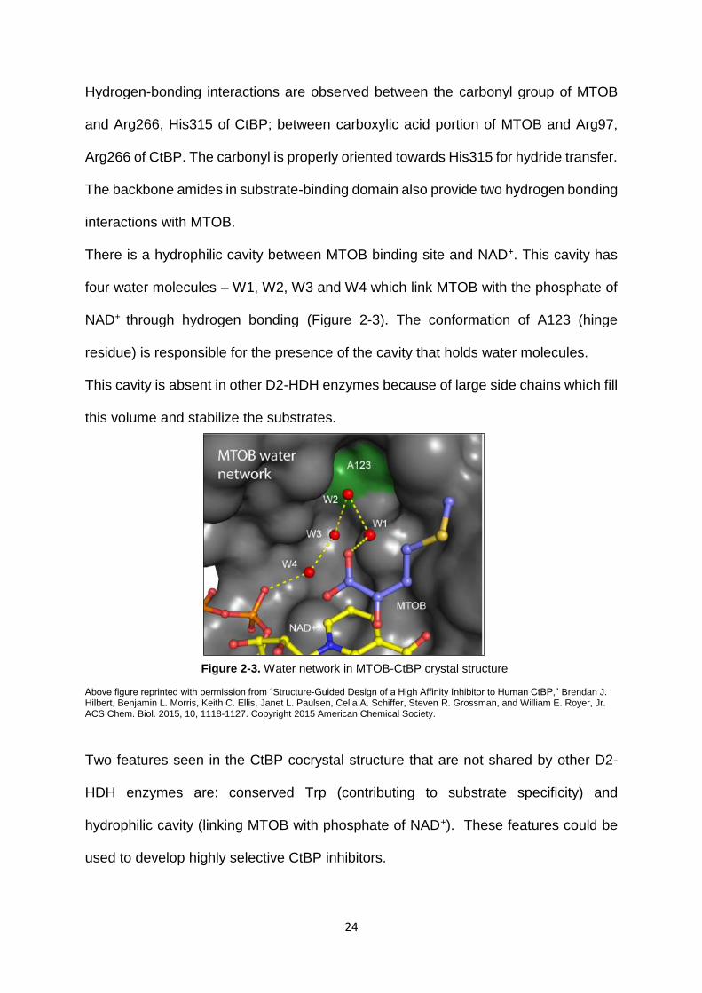
Figure 2-3. Water network in MTOB-CtBP crystal structure .................................... 24
Figure 2-4. Substrate conformation (yellow Coulombic) .......................................... 25
Figure 2-5. Non-canonical conformation .................................................................. 26
Figure 2-6. Water network of phenylpyruvic acid-CtBP crystal structure ................. 27
Figure 2-7. Oximes selected for synthesis ............................................................... 32
Figure 2-8. Second generation analogues ............................................................... 34
Figure 2-9. CtBP1-NADH-HIPP crystal structure ..................................................... 34
Figure 2-10. Interactions of HIPP at CtBP active site .............................................. 35
Figure 2-11. Water network of HIPP at the binding site ........................................... 36
Figure 2-12. Enzymatic vs cellular activity of inhibitors ............................................ 44
Figure 2-13. UNITY features based on HIPP .......................................................... 48

viii
Figure 2-14. Binding mode of compound ZINC02586210 ....................................... 49
Figure 2-15. Hits obtained through virtual screening to be tested ........................... 50
Figure 3-1. Ubiquitous nature of C-H bonds ............................................................ 77
Figure 3-2. C-H activation steps in Teleocidin B-4 core synthesis ........................... 81
Figure 3-3. C-H activation in late-stage diversification applications ......................... 84
Figure 3-4. Cost comparison of transition metals .................................................... 88
Figure 3-5. Single-walled and multi-walled carbon nanotubes .............................. 105
Figure 3-6. Pd decorated on MWCNT ................................................................... 106
Figure 4-1. Pd(II) and Pd(0) content in Pd(II)/MWCNT (a.) before and (b.) after a C-
H Activation Reaction as measured by XPS .......................................................... 117

ix
List of Schemes
Scheme 1-1. Reduction of MTOB by CtBP .............................................................. 17
Scheme 2-1. Deconstruction Analogues .................................................................. 28
Scheme 2-2. Non-reducible ketone isosteres of phenylpyruvic acid ........................ 30
Scheme 2-3. Synthesis of compound 2-5 ................................................................ 37
Scheme 2-4. Synthesis of compound 2-9 ................................................................ 37
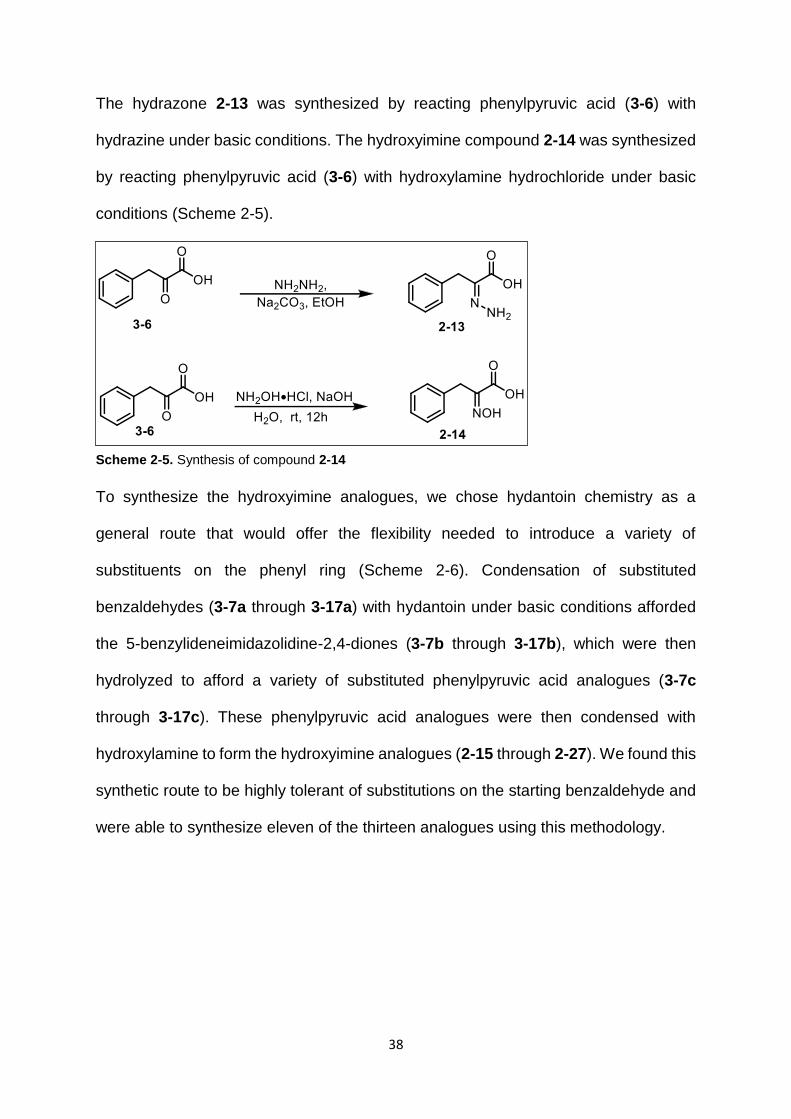
Scheme 2-5. Synthesis of compound 2-14 .............................................................. 38
Scheme 2-6. Synthesis of α-keto-acids and oximes from hydantoin ....................... 39
Scheme 2-7. Synthesis of α-keto-acids and oximes from 1,4-diacetylpiperazine-2,5-
dione ........................................................................................................................ 39
Scheme 2-8. Synthesis of second generation analogues ........................................ 40
Scheme 3-1. Traditional organic synthesis (A) vs C–H functionalization (B) ........... 78
Scheme 3-2. Comparison of traditional and C-H activation approaches .................. 78
Scheme 3-3. C-H activation ..................................................................................... 79
Scheme 3-4. Synthesis of intermediate 7 ................................................................ 82
Scheme 3-5. Synthesis of Teleocidin B-4 core ........................................................ 82
Scheme 3-6. Structural core diversification ............................................................. 83
Scheme 3-7. Late-stage diversification of celecoxib by C-H activation .................... 85
Scheme 3-8. Late-stage diversification of σ1 receptor ligands by C-H activation .... 85
Scheme 3-9. Organometallic mechanism ................................................................ 86
Scheme 3-10. Coordination mechanism of C–H activation ...................................... 87

x
Scheme 3-11. Pd(II)/Pd(IV) catalytic cycle for N-chelation-directed C-H activation
reactions ................................................................................................................... 89
Scheme 3-12. Palladium-catalyzed N-chelation-directed oxygenation reactions ..... 90
Scheme 3-13. Effect of solvent on oxygeantion reactions ....................................... 91
Scheme 3-14. Mechanism of C-H acetoxylation ...................................................... 91
Scheme 3-15. C-H acetoxylation by IOAc ............................................................... 92
Scheme 3-16. Pd-catalyzed chlorination of azobenzene ......................................... 92
Scheme 3-17. Halogenation of benzo[h]quinoline ................................................... 93
Scheme 3-18. Pd-catalyzed N-chelation-directed and non-catalyzed halogenation
reactions ................................................................................................................... 94
Scheme 3-19. Pd-catalyzed N-chelation-directed halogenation reactions ............... 94
Scheme 3-20. Mechanism of halogenation by Pd catalyst – Pd(II)/Pd(IV) cycle ..... 95
Scheme 3-21. Mechanism of halogenation by Pd catalyst – Pd(III)/Pd(III) cycle ..... 95
Scheme 3-22. Pd-catalyzed halogenation reaction by CuCl2 .................................. 95
Scheme 3-23. Pd-catalyzed halogenation by Suarez-type reagent ......................... 96
Scheme 3-24. Pd-catalyzed fluorination reactions ................................................... 96
Scheme 3-25. Pd-catalyzed C–H arylation reactions using [Ph2I]BF4 ..................... 98
Scheme 3-26. Pd-catalyzed C–H arylation reactions using asymmetrical arylating
reagents ................................................................................................................... 98
Scheme 3-27. Mechanism of Pd-catalyzed C–H arylation ....................................... 99
Scheme 3-28. Fluorenone synthesis by C–H arylation ............................................ 99
Scheme 3-29. Pd-catalyzed arylation reaction of aminoquinoline ......................... 100
Scheme 3-30. Mechanism of Pd-catalyzed arylation with AgOAc ......................... 100

xi
Scheme 3-31. Dimerization of arylpyridine ............................................................ 101
Scheme 3-32. Pd-catalyzed C–H arylation mechanism with oxone ....................... 101
Scheme 3-33. Oxidative cross coupling reaction on benzo[h]quinoline ................. 102
Scheme 3-34. Mechanism of oxidative cross coupling reaction on benzo[h]quinoline
............................................................................................................................... 102
Scheme 3-35. Arylation of isoxazole catalyzed by Pd on carbon .......................... 107
Scheme 3-36. Arylation of arenes using Pearlman's catalyst ................................ 107
Scheme 3-37. Arylation of aryl bromides catalyzed by Pd-CNT ............................ 108
Scheme 3-38. Suzuki cross-coupling catalyzed by Pd-MWCNT ........................... 108
Scheme 3-39. Arylation of thiophenes and benzothiophenes catalyzed by Pd on
carbon .................................................................................................................... 109
Scheme 3-40. Arylation reactions catalyzed by Pd on aluminium oxide ................ 109
Scheme 4-1. Methoxylation on Imatinib catalyzed by Pd-MWCNT ........................ 121
Scheme 4-2. Fluorination on 8-methylquinoline catalyzed by Pd-MWCNT ............ 122
Scheme 4-3. Trifluoromethylation on 2-phenylpyridine catalyzed by Pd-MWCNT . 122
Scheme 4-4. Trifluoromethylation on 8-methylquinoline catalyzed by Pd-MWCNT123
Scheme 5-1. C-H arylation on 8-methylquinoline .................................................. 139
Scheme 5-2. C-H arylation on 2-phenylpyridine .................................................... 139
Scheme 5-3. Challenges in C-H arylation reactions .............................................. 140
Scheme 5-4. Challenges in synthesis of arylating agents ...................................... 140
Scheme 5-5. Arylation of benzo[h]quinolone with 1,2-dichlorobenzene ................ 141
Scheme 5-6. Arylation of benzo[h]quinolone with 1,2-dimethoxybenzene ............. 141

xii
List of Tables
Table 1-1. Peptide inhibitors identified by SICLOPPS ............................................. 19
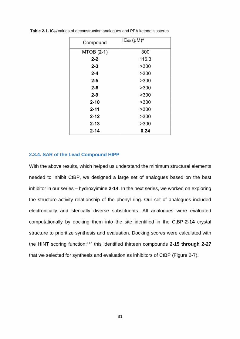
Table 2-1. IC50 values of deconstruction analogues and PPA ketone isosteres ...... 31
Table 2-2. IC50 values of oxime analogues .............................................................. 33
Table 4-1. C-H to C-O functionalizations catalyzed by Pd(II)-MWCNT .................. 112
Table 4-2. Steric Trend in C-H to C-O Alkyl Functionalizations Catalyzed by
Pd(II)/MWCNT ........................................................................................................ 113
Table 4-3. C-H to C-Cl/C-Br Functionalizations Catalyzed by Pd(II)/MWCNT ....... 115
Table 4-4. Recycling experiments with Pd(II)/MWCNT .......................................... 118
Table 4-5. Comparison of Turn Over Frequencies in C-H to C-OAc, C-OMe, C-Cl,
and C-Br Reactions Catalyzed by Solid-Supported Pd(II)/MWCNT and
Homogeneous Pd(OAc)2 ........................................................................................ 119
Table 5-1. C-H to C-C Functionalizations Catalyzed by Pd(II)/MWCNT ................ 134
Table 5-2. C-H to C-C Functionalizations Catalyzed by Pd(II)/MWCNT on 3-methyl-
2-phenylpyridine ..................................................................................................... 136
Table 5-3. C-H to C-C Functionalizations Catalyzed by Pd(II)/MWCNT on
benzo[h]quinoline ................................................................................................... 138

xiii
List of Abbreviations
APC – Adenomatous Polyposis Coli ACN – acetonitrile ARF – Alternate Reading Frame AcOH – acetic acid Bik – BCL2-interacting killer AgOAc – silver acetate BRCA1 – BReast CAncer gene 1 BDE – bond dissociation energy CBP – CREB Binding Protein BF4 – tetrafluoroborate CtBP – C-terminal Binding Protein CNT – carbon nanotube EMT – Epithelial Mesenchymal Transition DCE – 1,2-dichloroethane HDAC – Histone DeACetylase DCM – dichloromethane HIPP – 2-(hydroxyimino)-3-phenyl propanoic acid DMA – dimethylacetamide hTcf4 – human T-cell transcription factor-4 DMSO – dimethylsulfoxide LCoR – Ligand-dependent CoRepressor MeOH – methanol LDH – Lactate DeHydrogenase Mes – mesitylene LSD – Lysine-Specific histone Demethylase MWCNT – multi-walled carbon Nanotube MTOB – 4-(methylthio)-2-oxobutanoic acid NBS – N-bromosuccinimide PcG – Polycomb Group NCS – N-chlorosuccinimide pRb – Retinoblastoma protein Pd – palladium SUMO – Small Ubiquitin-related Modifier Pd(OAC)2 – palladium acetate PhI(OAc)2 – diacetoxyiodobenzene PhICl2 - dichloroiodo benzene SWCNT – single-walled carbon nanotube TBPB – tert-butyl peroxybenzoate

Abstract
DESIGN AND STRUCTURE-ACTIVITY RELATIONSHIP OF C-TERMINAL BINDING PROTEIN (CTBP) INHIBITORS AND INVESTIGATION OF THE SCOPE OF PALLADIUM MULTI-WALLED CARBON NANOTUBES (Pd-MWCNT) CATALYST IN C–H ACTIVATION REACTIONS
By Sudha Korwar, Ph.D.
A dissertation submitted in partial fulfilment of the requirements for the degree of
Doctor of Philosophy at Virginia Commonwealth University
Virginia Commonwealth University, 2016
Major Director: Keith C. Ellis, Assistant Professor, Department of Medicinal Chemistry
C-terminal binding proteins (CtBPs) are transcriptional co-repressors involved in
developmental processes, and also implicated in a number of breast, ovarian, colon
cancers, and resistance against cancer chemotherapy. CtBP is a validated novel
potential anti-cancer target. In this project we sought to develop potent and selective
small-molecule inhibitors of CtBP. Using a combination of classical medicinal
chemistry and modern computational approaches, we designed a potent inhibitor
HIPP (hydroxyimino-3-phenylpropanoic acid) that showed an IC50 of 0.24 µM against
recombinant CtBP. Further elucidation of the structure-activity relationship (SAR) of
HIPP led to the design of more potent inhibitors 3-Cl HIPP (CtBP IC50 = 0.17 µM) and
4-Cl HIPP (CtBP IC50 = 0.18 µM). These compounds also showed inhibition in HCT-
116 colon cancer cells with GI50 values ~ 1-4 mM. The compounds showed no off-

xv
target toxicity against a closely related protein. This is a starting point for the
development of CtBP inhibitors as anti-cancer therapeutics.
The second part of this dissertation focuses on C–H activation chemistry. C–H
activation is the most atom-economical method of introducing complexity into a
molecule, even at late stages of drug/product development. We have used solid-
supported palladium nanoparticle catalyst (Pd-MWCNT) to investigate the scope of
C–H activation reactions it can catalyse. Pd-MWCNT was found to efficiently catalyse
N-chelation directed C-H activation reactions – halogenations, oxygenations and
arylations. The turn-over numbers for these reactions were significantly higher than
that of the reported homogenous catalyst. The added advantages of reuse/recyclability
of catalyst, low contamination of metal in the final product make this catalyst very
attractive on an industrial scale. This work serves as a foundation for the further
development of Pd-MWCNT catalyst in late-stage synthesis of drugs and/or
diversification of products.
1
Design and Structure-Activity Relationship Study of Small Molecule C-terminal Binding Protein (CtBP) Inhibitors
1. Introduction
1.1. Gene Regulation
Gene expression is regulated by a combination of transcriptional activation and
transcriptional repression. DNA sequence-specific transcriptional repressors recruit
certain co-repressors to specific regions of DNA and inhibit gene expression of a single
gene or multiple genes by targeting the transcriptional machinery or altering the
chromatin structure. For example, several co-repressors recruit histone deacetylases
(HDACs) to remove acetyl group from the N-terminal tails of histones, resulting in
chromatin condensation, thereby preventing transcription factors access to DNA.
Examples of HDAC-dependent corepressors include Sin3, pRb, Groucho, Mi-
2/NuRD.1 Proper gene regulation through transcriptional regulation allows cells to
respond to stimuli, whereas improper gene regulation results in disease states.
Molecular control of the transcriptional machinery complex is a highly challenging
area, the investigation of which would help in the advancement of cancer therapy. This
dissertation focuses on CtBP (C-terminal Binding Protein) – transcriptional
corepressors, which have been implicated in development and oncogenesis.
Targeting CtBP has been shown to relieve the repression of apoptotic genes, tumor
suppression genes and genes involved in cell adhesion. This dissertation focuses on
the development of small molecule inhibitors that target CtBP and show potential as
novel anti-cancer therapeutics.

2
1.2. CtBP
The CtBP family of proteins function as transcriptional co-repressors and are highly
conserved among vertebrates and invertebrates. CtBP plays an important role during
early development, and has been implicated in various cancers. It modulates the
activity of oncogenes and tumor suppressor genes. CtBP represses transcription by
both chromatin modification-dependent and independent pathways, depending on the
promoter. CtBP proteins also act as a link between gene expression and metabolism,
as their transcriptional regulation function is dependent on the intracellular
NAD+/NADH ratio.
1.2.1 Discovery
CtBP is a 48 kDa cellular phosphoprotein (441 amino acids) that was first identified in
1993 by its binding to the C-terminal region (PLDLS motif) of E1A human adenovirus
oncoprotein,2, 3 during a search for proteins involved in negative modulation of
oncogenic transformation. This initial isoform was named CtBP1. CtBP2 (445 amino
acids, 48 kDa, 83% sequence homology to CtBP1) was identified by analysing
expressed sequence tag (EST) data bank sequences.4
1.2.2. Isoforms and genes
Invertebrates possess a single CtBP gene. They have different isoforms of CtBP as a
result of differential RNA processing. Vertebrates possess CtBP1 (Ctbp1 gene 4p16
chromosome) and CtBP2 (Ctbp2 gene 21q21.3 chromosome).4 Both vertebrate genes
code for different CtBP isoforms – CtBP-L (long) and CtBP-S (short).
CtBP1-S lacks the first 13 amino acids of the CtBP1-L N-terminal domain. CtBP1-S is
also known as CtBP3/BAR50, is N-terminally truncated CtBP1, which has
acyltransferase activity in Golgi.5

3
CtBP2-S does not possess the first 25 amino acids of the CtBP2-L N-terminal domain.
In the retina, a form of CtBP2 called RIBEYE protein (120 kDa) is expressed.6, 7 It has
an N-terminal domain fused to CtBP2 (amino acids 21-445).
1.2.3. Localization
CtBP isoforms are present in nucleus or cytoplasm based on the post-translational
modifications and the presence/absence of nuclear localization signal (NLS). The NLS
sequence is KRQR and corresponds to the residues 10-13 of hCtBP2. CtBP2-L can
heterodimerize with CtBP1 and translocate CtBP1 to the nucleus. CtBP1 isoforms do
not have NLS sequence, and are concentrated in the nucleus, significant quantities
are present in the cytosol. Neuronal nitric acid synthase (nNOS) binds to CtBP1 and
translocates it to the cytoplasm from the nucleus.8 CtBP2-S lacks the NLS sequence,
and hence is localised in the cytoplasm.9 Localization is not solely dependent on NLS,
as CtBP2-L dimer interface mutants localise in the cytoplasm.10 RIBEYE is localized
in the cytoplasm and is mainly present in the ribbon synapses.6, 11 , 12 The localization
of CtBP depends on the post-translational modifications, and is not consistent
throughout the literature. CtBP1-L and CtBP-1S have been observed both in
cytoplasm and nucleus.9 Phosphorylation of S158 by Pak1 kinase translocates CtBP1
to the cytoplasm, and inhibits the corepressor activity of CtBP1.13
SUMOylation of K428 by SUMO-1 (blocked by nNOS) localises it in the nucleus and
is required for corepression of E-cad by CtBP1.14 Dimerization, which is dependent
on NADH binding, also affects nuclear localization. Inhibition of CtBP dimerization
prevents nuclear localization.15

4
1.2.4. Oligomerization
CtBP forms dimers through the dehydrogenase domain similar to D-isomer specific
NAD-dependent 2-hydroxy acid dehydrogenase (D2-HDH) family members.
Oligomerization has been observed in bacterial D2-HDH enzymes – D-Lactate
Dehydrogenases16, 17 Hydroxyisocaproate Dehydrogenase,18 Formate
Dehydrogenase,19 D-Glycerate Dehydrogenase,20 and in human D2-HDH enzymes –
Glyoxylate Reductase/Hydroxypyruvate Reductase21 and D-3 Phosphoglycerate
Dehydrogenase.
CtBP is a redox sensor as its activity is dependent on the metabolic status of the cells
– NADH/NAD+ ratio. It binds to NADH with a 100-fold more affinity than NAD+.22 Once
NADH binds to CtBP, the protein undergoes dimerization,23 which is essential for the
activity of CtBP.10, 24-26 When NADH levels increase – conditions of hypoxia and high
extracellular glucose levels – NADH binds to CtBP, and the activity of CtBP increases.
Each monomer of CtBP contains a single PXDLS motif to which other proteins bind.
The presence of a single monomer results in competition between different factors
binding to CtBP, which disrupts transcriptional regulation.10 Dimerization of CtBP
increases the number of PXDLS sites, thereby providing a scaffold for other
transcriptional factors to bind. CtBP1 dimerization is required for its interaction with
E1A protein. NADH mediated dimerization enhances repression activity of CtBP.
CtBP1 mutants that cannot dimerize fail to effect transcriptional repression. NADH has
been found to be essential for dimerization and hence nuclear localisation, but not for
binding of other factors. Binding of other factors depends on the PXDLS motif.

5
1.2.5. CtBP Domain Arrangement
CtBP is highly homologous to D-isomer specific NAD-dependent 2-hydroxy acid
dehydrogenases (D2-HDH) – catalytic Histidine residue, Arg, Glu, and an NAD-binding
region (Figure 1-1).3 It has 3 domains – C-terminal domain, dehydrogenase domain
and N-terminal domain. The dehydrogenase domain has two domains – substrate
binding domain and coenzyme binding domain. Two features of CtBP that are involved
in recruiting cofactors/proteins are:
a. Hydrophobic cleft formed by N-terminal region in CtBP1-L (AA 27-121) that
recruits PLDLS motif containing factors (DNA binding proteins).
b. Surface groove on NADH-binding domain that recruits RRT motif containing
factors
C-terminal unstructured region has sites for SUMOylation and PDZ (structural domain
80-90 AA) binding.
Figure 1-1. Structural Features of CtBP15
CtBP1-L has the entire CtBP sequence, CtBP1-S has a truncation (blue region) in the
N-terminal region. In CtBP2-S, this truncation (25 AA) causes loss of NLS sequence,
thus localising it to the cytoplasm. D2-HDH domain has a substrate binding domain
(yellow, has PXDLS-binding motif), and coenzyme NADH binding domain (green, has
RRT-binding motif and catalytic triad REH), which are connected by hinges (Figure 1-
1).

6
1.3. Cytosolic and Nuclear functions of CtBP
1.3.1. Nuclear functions
CtBP forms large DNA-bound chromatin remodelling complexes and is involved in
transcriptional regulation (Figure 1-2). CtBP is recruited to DNA by several DNA
binding proteins through PXDLS-binding motif (substrate binding domain of N-terminal
region) of CtBP. DNA-binding proteins such as ZEB 1/2 (zinc finger protein) function
as bridges between CtBP PXDLS-binding motif and the promoters (for example E-
cadherin).10, 27-31 CtBP coenzyme binding domain has RRT binding motif
(RRTGXPPXL) that binds to cofactors that are involved in repression.32 Znf217 binds
to CtBP through both PXDLS motif and RRT motif,15, 33 thus it binds to CtBP dimers
(RRT-binding motif of one monomer, and PXDLS-binding motif of the second
monomer).
Numerous chromatin modifying proteins have been observed in CtBP transcriptional
complexes. It has been seen that class I HDACs – HDAC 1/2 and class II HDAC
proteins interact with CtBP.15, 34, 35 Other proteins such as HMT (Histone Methyl
Transferase), LSD-1 (Lysine Demethylase), G9A, GLP bind to the CtBP complex.15, 34
Histone-modifying proteins bind to CtBP either through other proteins or bind directly.
HDAC1/2, CoREST (corepressor of REST) bind directly through non-PXDLS
interactions.15, 34 CoREST recruits both HDAC1/2 and LSD-1 to CtBP.36, 37 However
LCoR corepressor interacts through PXDLS motif of CtBP to recruit HDAC1/2.38 Some
examples of chromatin modifying complexes include: Znf217 binding to CoREST that
recruits HDAC1/2 and LSD-1 to CtBP;33 Wiz protein that binds to G9A/GLP and CtBP
directly.39 Thus, depending on the context, various histone modifying proteins are
recruited through different DNA-binding proteins in different CtBP multi-subunit
regulatory complexes to regulate transcription.10

7
Figure 1-2. Multi-protein complexes with CtBP
1.3.2. Cytosolic functions
CtBP1 and CtBP2 play an important role in Golgi fission,40 vesicle formation41 and
synapse signalling.11, 42 CtBP1-S has functions in the Golgi – tubule constriction and
fissioning43 using acyl-coenzyme A (acyl-CoA) molecules. CtBP1-S also has acyl
transferase activity towards lysophosphatidic acid (LPA) and alters the membrane
properties in Golgi.5, 44 RIBEYE also has acyl transferase activity42 and is involved in
vesicle formation at ribbon synapses.6, 11, 42
1.4. CtBP as a Transcriptional Corepressor
The transcriptional repression role of CtBP was first suggested in a tethering
transcriptional assay45 involving E1A protein. Interaction of CtBP with the C-terminal
region of E1A inhibited the activity of conserved region 1 (CR1) of E1A. In-depth
studies on Drosophila CtBP (dCtBP) showed that dCtBP functions as a transcriptional
corepressor during development of embryo.30, 46 dCtBP has been found to interact with
Knirps, Snail (short range repressors) and Hairy (long range repressors). CtBPs have
8
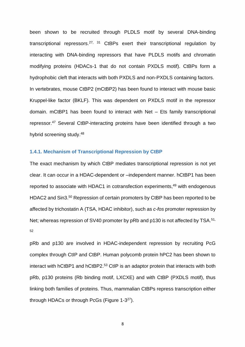
been shown to be recruited through PLDLS motif by several DNA-binding
transcriptional repressors.27, 31 CtBPs exert their transcriptional regulation by
interacting with DNA-binding repressors that have PLDLS motifs and chromatin
modifying proteins (HDACs-1 that do not contain PXDLS motif). CtBPs form a
hydrophobic cleft that interacts with both PXDLS and non-PXDLS containing factors.
In vertebrates, mouse CtBP2 (mCtBP2) has been found to interact with mouse basic
Kruppel-like factor (BKLF). This was dependent on PXDLS motif in the repressor
domain. mCtBP1 has been found to interact with Net – Ets family transcriptional
repressor.47 Several CtBP-interacting proteins have been identified through a two
hybrid screening study.48
1.4.1. Mechanism of Transcriptional Repression by CtBP
The exact mechanism by which CtBP mediates transcriptional repression is not yet
clear. It can occur in a HDAC-dependent or –independent manner. hCtBP1 has been
reported to associate with HDAC1 in cotransfection experiments,49 with endogenous
HDAC2 and Sin3.50 Repression of certain promoters by CtBP has been reported to be
affected by trichostatin A (TSA, HDAC inhibitor), such as c-fos promoter repression by
Net; whereas repression of SV40 promoter by pRb and p130 is not affected by TSA.51,
52
pRb and p130 are involved in HDAC-independent repression by recruiting PcG
complex through CtIP and CtBP. Human polycomb protein hPC2 has been shown to
interact with hCtBP1 and hCtBP2.53 CtIP is an adaptor protein that interacts with both
pRb, p130 proteins (Rb binding motif, LXCXE) and with CtBP (PXDLS motif), thus
linking both families of proteins. Thus, mammalian CtBPs repress transcription either
through HDACs or through PcGs (Figure 1-327).

9
In Drosophila, the involvement of deacetylases in dCtBP-mediated repression is
uncertain. The activity of dCtBP is not significantly affected in Rpd3 (HDAC
mammalian homolog) mutant embryos.54 The mutant embryos might be expressing
other deacetylases.
In contrast to the repressor functions, dCtBP has also been shown to possess context-
dependent weak transcriptional activational functions.55 This activation has been
observed in human HEK293 cells expressing E1A proteins. In the context of Hairy,
dCtBP might be antagonizing the activity of Gro.46 The regions of dCtBP that are
required for activation (His residue) and repressor functions are adjacent to each
other.
The corepressor complex of CtBP causes deacetylation and methylation of histone
H3-K9, demethylation of histone H3-K4.
Figure 1-3. Corepressor complex of CtBP (ref 27)

10
1.5. Regulation of CtBP Activity
CtBP is a phosphoprotein, its phosphorylation is cell-cycle dependent. It possess
consensus phosphorylation sites of DNA-PK (TQ or SQ) and has been found to
interact with DNA-PK.48, 56 Phosphorylation changes the localization of CtBP. The
activity of CtBP is also modulated by cellular energy levels/homeostasis as it binds to
NAD. CtBP binding to E1A C-terminal region is regulated by the nuclear acetylases
p300/CBP and P/CAF that acetylate the Lys residue (PLDLSCK) flanking CtBP binding
motif of E1A,57 resulting in decrease of CtBP binding.
1.6. Role of CtBP
CtBP plays an important role in development and oncogenesis
1.6.1. Role in Development
In Drosophila, dCtBP is involved in transcriptional regulation and plays an important
role during early embryo development and also during later developmental
processes.58, 59 The CtBP gene is involved in regulatory functions in the wing,60 eye,61,
62 sensory organs.63 Decrease in the levels of dCtBP leads to severe segmentation
defects, disruptions in anterior-posterior patterning.46 This is a result of loss of
repression of genes – eve, runt and hairy by short range repressors Knirps, Snail and
Kruppel. A transcriptional repressor Tramtrack69 (Ttk69) binds to dCtBP and acts as
a neural inhibitor during early eye development. This interaction determines the
number of photoreceptor cells produced.64
In Xenopus, the transcription factors xTcf-3, xFOG, xPc have been shown to interact
with xCtBP. xCtBP is involved in the development and is localized in the head, tail bud

11
and in central nervous system.65 xFOG represses RBC formation partially by
interacting with xCtBP.66
In mammals, mCtBP2 is expressed during embryogenesis, and mCtBP1 is expressed
throughout.67 CtBP1 is expressed in spinal chord, and CtBP2 is expressed in spinal
chord, limb buds, and root ganglia. Mutation in CtBP2 causes embryonic lethality.68
CtBP1-null mice are smaller in size than the wt mice, and have about 23% mortality
rate after birth within 20 days.68
In humans, both hCtBP1 and hCtBP2 are ubiquitously expressed in most human
tissues.53 TGIF is a transcriptional repressor of TGF-β activated genes. It binds to
CtBP protein. Mutations in TGIF leads to holoprosencephaly – brain malformations,
due to the loss of interaction with hCtBP1. The wt huntingtin protein mediates
transcriptional repression by interacting with CtBP.69 In Huntington’s disease (HD), the
protein contains polyglutamine expansion, which leads to reduced interaction with
CtBP, possibly leading to HD.
1.6.2. Role of CtBP in Oncogenesis
CtBP has been shown to bind to more than 1,800 promoters throughout the
mammalian genome.70 CtBP has been linked to the development of cancer in the
context of EMT (epithelial-to-mesenchymal) transition. EMT transition is an essential
process during development, in which epithelial cells lose cell adhesion property and
polarity, and acquire migratory, invasive properties.71 Though EMT is essential for
wound healing and during development, it increases the resistance to apoptosis and
metastasis of malignant tumors.72

12
CtBP has been shown to repress the transcription of pro-apoptotic factors,73 tumor
suppressors,74 cell adhesion molecules – E-cadherin,73 cell-cell junction proteins and
cytoskeletal proteins. Transcriptional repression of E-cadherin causes EMT. CtBP has
been shown to play a role in the progression of melanoma,75 pituitary tumors,76
prostate cancer,77 colon cancer78-83 and breast cancer.70, 84-87
Levels of CtBP are regulated by tumor suppressors, disruption of which leads to
cancers. In colorectal tumors, mutations in the tumor suppressor APC (adenomatous
polyposis coli) have been observed.88 APC degrades CtBP1.78 The levels of APC and
CtBP1 are inversely correlated during cancer initiation.78, 79
ARF is another tumor suppressor which targets CtBP for proteosomal degradation.
Binding of ARF to CtBP relieves the repression of a pro-apoptotic factor Bik.81 Bik
inhibits the function of anti-apoptotic factors, and sensitizes cells to apoptosis.89
Samples of colon cancer tissue showed high levels of CtBP and low levels of ARF,
whereas adjacent healthy tissues showed low levels of CtBP and normal levels of
ARF.83
In colon cancer cells, increase in NADH levels due to hypoxia increased cell
migration.24, 82 ARF antagonizes the function of CtBP, leading to Bik-mediated
apoptosis. Hence, loss of APC and ARF increases CtBP levels and activity leading to
tumorigenesis.
The role of CtBP in breast cancer has been highly studied. Depletion of CtBP stops
cell growth, whereas elevated CtBP levels lead to cell survival and metastasis.
Elevated CtBP1 levels have been observed in invasive ductal carcinoma tissues.85
Breast cancer cells show loss of E-cadherin, and low levels of the DNA repair protein
BRCA1 breast cancer gene. Knockdown of CtBP restores the levels of E-cadherin and

13
BRCA1.85 Decrease in the NADH/NAD+ ratio removes HDAC1 for the promoter,
leading to increase in BRCA1 levels.86 This might be the result of failure of CtBP
dimerization under low NADH levels, and failure to form a repressor complex. This is
the opposite effect of the “Warburg effect” in which high glycolysis increases NADH
levels leading to increased CtBP activity in cancers.90, 91 CtBP knockdown reduces cell
proliferation and sensitizes breast cancer cells to Cisplatin.84 Thus, breast cancer
involves downregulation of E-cadherin and BRCA1 by CtBP. CtBP-mediated
repression might be resulting in BRCA1 loss even in the absence of BRCA1
mutation.86
CtBP has been shown to be involved in oncogenesis through multiple
mechanisms/pathways explained below.
1.6.2.1. E1A Model – Tumor suppressor role
CtBP negatively regulates oncogenesis by interacting with E1A protein. E1A mutants
that lack the CtBP binding motif (PLDLS) in the C-terminal region cooperate with
activated Ras oncogene, and cause high-frequency in vitro transformation of primary
rat kidney cells.2, 3, 92 The resulting cells (E1A mutants) are very tumorigenic in mice
models, and the tumors are metastatic. There are three possible pathways by which
CtBP negatively regulates oncogenesis through E1A interaction (Figure 1-4):
a). Interaction of CtBP with the PLDLS motif in the C-terminal region might antagonise
the acetyl transferase activities of p300/CBP and P/CAF. This would result in the
inhibition of cell proliferation activity of the N-terminal region. It has also been shown
that pRb is acetylated by the E1A complex, which increases interaction between pRb
and Mdm2,93 resulting in the inactivation of pRb.94

14
b). CtBP interaction with E1A represses certain cellular genes. Acetylation of Lys
residues or deletion of the CtBP-interacting region (PLDLS motif) relieves repression.
c). wt E1A competitively interacts with CtBP and relieves the repression in
endogenous repression complexes resulting in the activation of certain genes that
modulates oncogenesis,49, 95, 96 whereas mutant E1A fails to relieve the repression.
1.6.2.2. Wnt Signaling Model – Tumor suppressor role
In mammals, CtBP might be playing a role in the Wnt signalling pathway during
development and oncogenesis. In the absence of Wnt signalling pathway, APC tumor
Figure 1-4. Negative regulation of oncogenesis by CtBP through E1A (ref 27)
15
suppressor protein degrades β-catenin; and hTcf-4 functions as a constitutive
repressor of Wnt target genes. hTcf-4 has two binding motifs for CtBP binding. When
Wnt signalling is activated, β-catenin levels increase. β-catenin binds to hTcf-4,
recruits the p300/CBP coactivator, which activates Wnt target genes cMyc, cyclin D1
resulting in oncogenesis. Mutations in APC and β-catenin have been shown to cause
tumorigenesis. Oncogenic mutations increase β-catenin levels. Mutations that affect
interaction of CtBP with hTcf-4 might contribute to colorectal carcinogenesis.97, 98 CtBP
might be contributing to the transcriptional repression activity of hTcf-4, and
antagonizing the β-catenin/CBP coactivator complex activity.
1.6.2.3. Evi-1 oncogene – Oncogenesis role
Evi-1 is a nuclear protein that functions as a sequence-specific transcriptional
repressor. It inhibits TGF-β signalling by causing repression of Smad-induced
transcription of TGF-β responsive genes,99 resulting in cell proliferation. Evi-1 is a
cellular oncogene that has been implicated in myeloid leukemogenesis in humans and
mice. It is highly expressed in human myeloid leukemias and in chronic ML. Evi-1
repressor domain contains two CtBP binding motifs. Mutants in these motifs do not
bind well with CtBP and fail to cause transcriptional repression.100, 101 CtBP might be
playing an important role in Evi-1 mediated leukemogenesis.
1.6.2.4. Ras signalling
CtBP modulates the activity of Net belonging to the family of Ets transcriptional
repressors.47 Net is regulated by Ras. Net interacts with serum response element
(SRE) and represses c-fos promoter. Ras signalling reverses this repression. Net
recruits CtBP and mediates repressor activity in the absence of Ras.

16
1.7. Dehydrogenase Activity of CtBP
CtBP is similar to the D2-HDH family of proteins. D2-HDH enzymes function by
transferring a hydride anion to substrate by simultaneous NADH oxidation, and in that
process convert the ketone carbonyl group of α-keto acid to hydroxyl group (Scheme
1-1, Figure 1-5).18, 102, 103 Keto-acid portion of the substrate is stabilized by arginine by
electrostatic interactions, glutamate makes hydrogen bonding interaction with histidine
residue, histidine proton polarizes carbonyl oxygen. NADH transfers hydride anion to
the carbon of substrate carbonyl group, carbonyl oxygen abstracts proton from
histidine giving the D-hydroxyacid product.
The dehydrogenase activity of CtBP was first discovered by its ability to convert
pyruvate to lactate by oxidation of NADH.23, 104 Search for potential substrates resulted
in the discovery of MTOB (4-methylthio-2-oxobutyric acid), as the putative
endogenous substrate for CtBP. MTOB is an intermediate in the methionine salvage
Figure 1-5. Mechanism of hydride transfer

17
pathway – following reduction, transamination of MTHB gives methionine.105 MTOB
was found to be 80-fold better substrate for CtBP1 than pyruvate.106 When the sulfur
in MTOB was replaced by a methylene group (2-oxohexanoic acid), the enzymatic
activity reduced by 8-fold. Thus, sulfur is essential for selectivity.106
The transcriptional regulation by CtBP does not require the dehydrogenase activity of
CtBP, which suggests that NADH binding is conserved for the purpose of
transcriptional regulation, and not for catalytic turnover.73
Though MTOB is a better substrate for CtBP than pyruvate, it is still a poor substrate
compared to the other D2-HDH family of proteins. For example, E. coli DGDH
catalyses substrate 2600 times more efficiently than MTOB catalysis by CtBP.106-108
1.8. Targeting CtBP to Treat Cancer
Reports show that CtBP can be inhibited by high levels (millimolar) of putative
substrate MTOB (shows bi-phasic kinetics), and that MTOB has anti-tumor effects in
breast and colon cancer cells.83 Although MTOB was shown to have an apoptosis-
inducing effect earlier, its targets were not known at that time.109 Straza et al reported
that MTOB at concentrations of around 4 mM displays cytotoxicity in HCT-116 cells.
MTOB acts by displacing CtBP from the Bik promoter, thereby relieving Bik-mediated
repression, finally resulting in apoptosis. MTOB has been shown to be effective in a
mouse xenograft model using p53-/- HCT-116 cells, but it showed no effect in normal
healthy mice, suggesting that it has no off-target toxicity issues. MTOB (10 mM) has
Scheme 1-1. Reduction of MTOB by CtBP

18
been shown to have anti-cancer effect in MCF-7 and MDA-MD231 cells through
inhibition of CtBP; it inhibited the repression of several genes associated with EMT
and genome stability.75 These preliminary data provide insights into targeting CTBP to
treat cancers. Other than the small molecule inhibitor MTOB, there are two other CtBP
inhibitors reported – peptide inhibitor, and a small molecule inhibitor NSC95397.
1.8.1. Peptide inhibitor of CtBP
Birts et al. reported a CtBP dimerization cyclic peptide inhibitor – cyclo-SGWTVVRMY
by high-throughput screening that assesses SICLOPPS (split-intein circular ligation of
peptides and proteins)110, 111 cyclic peptide libraries (cyclic
heptamer/octamer/nonamer) of about 64 million members (genetically encoded).112
This study proved that CtBP dimerization links cellular metabolism with mitotic fidelity.
The assay used a bacterial reverse two-hybrid system (RTHS) to analyse the link
between bacterial (E. coli) survival on a selective media and disruption of CtBP1
homodimer (NADH-dependent) fused to bacteriophage 434 repressor. The repressor
434 (bacteriophage DNA binding protein) is reconstituted when CtBP1 fusion protein
homodimerizes, leading to repressor binding to operator sites in E.coli chromosome,
this prevents transcription of three downstream reporter genes (HIS3, Kan and LacZ),
resulting in cell death in selective media. If the target proteins do not interact or when
the protein-protein interaction is inhibited by an inhibitor, the reporter genes are
expressed and cells survive in selective media (Figure 1-6).
Above figure reproduced from “A cyclic peptide inhibitor of C-terminal binding protein dimerization links metabolism with mitotic
fidelity in breast cancer cells,” C. N. Birts, S. K. Nijjar, C. A. Mardle, F. Hoakwie, P. J. Duriez, J. P. Blaydes and A. Tavassoli,
Chem. Sci. 2013, 4, 3046. DOI: 10.1039/C3SC50481F - Published by The Royal Society of Chemistry.
Figure 1-6. Bacteria reverse two hybrid system (ref 112)

19
The peptides contained these residues in common – serine (nucleophile for intein
processing), glycine (prevents racemization during synthesis), tryptophan
(chromophore for HPLC purification). Transformation was carried out on CtBP1 RTHS
with SICLOPPS plasmids, split-inteins were expressed that underwent processing to
give cyclic peptides. Only plasmids that produce cyclic peptides disrupting CtBP1
homodimerization allow for cell survival on selective media. After picking bacterial
colonies, plasmids were isolated and re-screened for non-specific inhibitors of the
RTHS (by using a different RTHS system). The SICLOPPS plasmids which gave rise
to three most potent peptides were sequenced for identity (Table1-1).
CP61 (Figure 1-7) has been shown to disrupt CtBP
dimerization both in vitro and in cells. It binds to CtBP1 (3
µM affinity), inhibits CtBP homo- and heterodimerization
with an in vitro IC50 of 19 ± 4 µM, but it requires fusion to
a cell penetrating molecule to enter the cell. CP61 does
not bind to the NADH-binding pocket of CtBP, so probably
it is an allosteric inhibitor. It did not show any inhibition of
LDH, thus it is very selective for CtBP. CP61 was used to establish that dimerization
of CtBP regulates mitotic fidelity in cancer cells. In breast cancer cells with high rate
Rank Name Target Peptide sequence
1 CP61 CtBP1/CtBP2 SGW TVVRMY
2 CP68 CtBP1/CtBP2 SGW PLSTWY
3 CP65 CtBP1/CtBP2 SGW RLIRLY
Table 1-1. Peptide inhibitors identified by SICLOPPS
Figure 1-7. Structure of CP61

20
of glycolysis, this compound reduced the mitotic fidelity, proliferation and colony
formation; but had no effect in cells with lower rate of glycolysis. This provides
evidence that the glycolytic state of cells is linked to mitotic cell cycle checkpoint
control through NADH regulation/detection by CtBPs. This inhibitor of dimerization is
important to study the roles of NADH-unbound CtBP.
1.8.2. NSC95397 – A Small-molecule inhibitor of CtBP
Blevins et al. reported a NSC95397, small molecule inhibitor of CtBP.113 They used a
high-throughput screening assay (AlphaScreen) to screen LOPAC library (Sigma-
Aldrich, 1280 bioactive compounds), and found NSC95397
(Figure 1-8) to be a good inhibitor of CtBP with an IC50 of 2.9
µM (inhibits CtBP-E1A interaction).
NSC95397 was found to be a weaker substrate of CtBP1
compared to MTOB; and was not found to inhibit LDH. MTOB
failed to inhibit CtBP-E1A interaction, in contrast NSC95397 inhibits CtBP-E1A
interaction. This rules out the suggestion that NSC95397 mode of action could be by
it acting as a CtBP1 substrate. NSC95397 reversed the repression of E-cadherin
promoter (in H1299 small cell lung carcinoma cells) by CtBP. It is a known inhibitor of
cdc25 phosphatase activity and spliceosomal activity. Thus, in order for it to be a useful
CtBP inhibitor, potency and specificity has to be improved.
The possible mechanism of action of this compound is that it might be locking CtBP1
in a conformation which prevents it from binding to transcriptional factors, or it could
be binding to the surface groove on CtBP at the conserved binding motif PXDLS,
thereby directly inhibiting interaction of CtBP with transcriptional factors.
Figure 1-8. Structure of NSC95397

21
1.9. Scope of this dissertation
This first project in my dissertation focuses on design and development of small
molecule inhibitors that target CtBP for use as anti-cancer therapeutics. We have used
a blend of classic traditional medicinal chemistry as well as modern computational
techniques to approach the lead compounds. We succeeded in obtaining two
compounds that are slightly more active in inhibiting CtBP and growth of cancer cells
compared to the reported lead compound HIPP. These two compounds were found to
be stable oximes, and did not have any off-target toxicity issues as determined by their
lack of activity against LDH, a closely related enzyme of CtBP.

22
2. Structure-Guided Design of CtBP Inhibitors
2.1. MTOB as an Inhibitor of CtBP
MTOB (2-1), a putative substrate of CtBP shows bi-phasic kinetics – it acts as a
substrate at lower concentrations, but acts as an inhibitor at higher concentrations. It
has been shown to interfere with the oncogenic activity of CtBP in cells and mice.114
MTOB displaced CtBP from the Bik promoter and thereby induced apoptosis in HCT-
116 colon cancer cells.83 In a mouse xenograft model, MTOB treated mice showed
prolonged survival and less tumor burden compared to non-treated mice. MTOB
shifted phenotypic indicators such as E-cadherin from mesenchymal to epithelial
phenotype.70
MTOB has provided a direction to develop small molecule inhibitors of CtBP (Figure
2-1). Hilbert et al have reported the crystal structures of human CtBP1 (28-253) and
CtBP2 (33-364) complexed with NAD(H) and ligand MTOB at 2.38 Å and 2.86 Å
resolution respectively.114
Above figure reprinted from “Crystal structures of human CtBP in complex with substrate MTOB reveal active site features useful for inhibitor design,” Brendan J. Hilbert, Steven R. Grossman, Celia A. Schiffer, and William E. Royer Jr. FEBS Lett. 2014, 588 (9), 1743-1748 Copyright 2014, with permission from Elsevier.
Figure 2-1. Crystal structure of CtBP1 in complex with MTOB

23
MTOB binds in the active site cleft between the coenzyme binding domain (125-319)
and the substrate binding domain (28-120, 327-353). MTOB does not cause any
tertiary or quarternary changes in the protein conformation upon binding. So, the
mechanism of CtBP inhibition by MTOB could be through the substrate turnover →
NAD+ generation and release → dimer dissociation → transcriptional regulation. Or,
high MTOB concentrations (via substrate inhibition) → inhibit NADH to NAD+
conversion → inhibit monomer-dimer cycling → transcriptional regulation.
Using the CtBP1(28-353)/MTOB/NAD+ crystal structure (Figure 2-2) as a starting
point, we wished to design, synthesize, and evaluate small molecules that would inhibit
the dehydrogenase activity of CtBP.
The interactions between MTOB and CtBP1 in the binding site show sulphur-pi
interactions between ‘S’ of MTOB and tryptophan 318 of CtBP (4 Å distance between
the two). This Trp has been shown to function as a dimerization switch in CtBP1.115
Arg97
MTOB
Arg266
NAD+
His315Trp318
Figure 2-2. Interactions of MTOB with CtBP active site
24
Hydrogen-bonding interactions are observed between the carbonyl group of MTOB
and Arg266, His315 of CtBP; between carboxylic acid portion of MTOB and Arg97,
Arg266 of CtBP. The carbonyl is properly oriented towards His315 for hydride transfer.
The backbone amides in substrate-binding domain also provide two hydrogen bonding
interactions with MTOB.
There is a hydrophilic cavity between MTOB binding site and NAD+. This cavity has
four water molecules – W1, W2, W3 and W4 which link MTOB with the phosphate of
NAD+ through hydrogen bonding (Figure 2-3). The conformation of A123 (hinge
residue) is responsible for the presence of the cavity that holds water molecules.
This cavity is absent in other D2-HDH enzymes because of large side chains which fill
this volume and stabilize the substrates.
Above figure reprinted with permission from “Structure-Guided Design of a High Affinity Inhibitor to Human CtBP,” Brendan J. Hilbert, Benjamin L. Morris, Keith C. Ellis, Janet L. Paulsen, Celia A. Schiffer, Steven R. Grossman, and William E. Royer, Jr. ACS Chem. Biol. 2015, 10, 1118-1127. Copyright 2015 American Chemical Society.
Two features seen in the CtBP cocrystal structure that are not shared by other D2-
HDH enzymes are: conserved Trp (contributing to substrate specificity) and
hydrophilic cavity (linking MTOB with phosphate of NAD+). These features could be
used to develop highly selective CtBP inhibitors.
Figure 2-3. Water network in MTOB-CtBP crystal structure

25
2.2. Design of CtBP Inhibitors Based on MTOB
Based on the interactions observed in the crystal structure of CtBP1(28-
353)/MTOB/NAD+, it was hypothesized that increasing the pi-interactions with CtBP
Trp318 might result in a potent inhibitor. Replacing the sulfur of MTOB with a
methylene group decreased the enzymatic activity by 8-fold.106 This showed that pi-
interaction is important for activity. Thus, the first step was to increase the pi-
interactions of MTOB. We replaced the sulfur in MTOB (2-1, IC50 = 300 μM), with a
phenyl ring to increase the π-interactions with Trp318 of CtBP (Scheme 2-1) giving
rise to phenylpyruvic acid 2-2 (PPA). Hilbert et al arrived at this compound
computationally through the Schrodinger Suite Glide program. Phenylpyruvic acid (2-
2) was tested in an NADH consumption assay, and was found to inhibit the reduction
reaction of MTOB to MTHB by CtBP. Phenylpyruvic acid (2-2) was found to have an
IC50 of 116 μM, which is about ~3-fold better inhibitor activity than MTOB (Table 2-1).
Hilbert et al reported the crystal structure of PPA complexed with CtBP1 and NAD+ at
2.1 Å resolution.116 The crystal structure of CtBP1 complexed with NAD+ and
phenylpyruvic acid shows similar interactions as that of MTOB, and does not induce
major conformational changes. The phenyl group makes pi-stacking interactions with
Trp318. One interesting feature is that PPA assumes two different and proportionally
Figure 2-4. Substrate conformation (yellow Coulombic)

26
equal conformations in the crystal structure (substrate and non-canonical). The
substrate conformation (Figure 2-4) is similar to that of MTOB, where the carbonyl of
PPA interacts through H-bonding with His315 and Arg266; and is oriented for hydride
transfer from His315. In contrast, in the non-canonical conformation (Figure 2-5), the
carboxylic acid group is oriented towards His315 and Arg266; and the carbonyl group
is oriented towards Ser100 and away from His315, so is not positioned for hydride
transfer. The binding affinities (energies) of both the conformations are not
substantially different.
Above figures 2-4 and 2-5 are reprinted with permission from “Structure-Guided Design of a High Affinity Inhibitor to Human CtBP,” Brendan J. Hilbert, Benjamin L. Morris, Keith C. Ellis, Janet L. Paulsen, Celia A. Schiffer, Steven R. Grossman, and William E. Royer, Jr. ACS Chem. Biol. 2015, 10, 1118-1127. Copyright 2015 American Chemical Society.
The water network is disrupted (different from that of MTOB, Figure 2-6). Because of
a different conformation of A123, the cavity is collapsed, as a result of which the waters
W2 and W3 are absent. In the substrate conformation, W1 (orange) is present;
whereas W1 is absent in the non-canonical conformation. W4 is present similar to the
position in the MTOB structure.
Figure 2-5. Non-canonical conformation

27
Above figure reprinted with permission from “Structure-Guided Design of a High Affinity Inhibitor to Human CtBP,” Brendan J. Hilbert, Benjamin L. Morris, Keith C. Ellis, Janet L. Paulsen, Celia A. Schiffer, Steven R. Grossman, and William E. Royer, Jr. ACS Chem. Biol. 2015, 10, 1118-1127. Copyright 2015 American Chemical Society.
2.3. Structure-Activity Relationship (SAR) Study
We further investigated the SAR of phenylpyruvic acid by synthesizing and testing
various analogues.
2.3.1. Deconstruction analogues of phenylpyruvic acid
Phenylpyruvic acid was deconstructed to investigate which structural features of the
molecule are important for activity. Three analogues were designed (Scheme 2-1): an
analogue in which phenyl ring was removed – pyruvate (2-3), an analogue in which
ketone was removed – hydrocinnamic acid (2-4), and an analogue in which carboxylic
acid group was removed – 1-phenylpropan-2-one (2-5). All of these compounds were
less active than phenylpyruvic acid.
Figure 2-6. Water network of phenylpyruvic acid-CtBP crystal structure

28
2.3.2. Linker length analogues of phenylpyruvic acid
In order to investigate what effect the linker length has on the activity, compounds 2-
6 (phenylglyoxylic acid – methylene spacer between phenyl ring and α-ketoacid
removed), 2-7 and 2-8 were designed. These compounds were also either
synthesized or purchased and tested for their ability to inhibit CtBP. 2-6 was less active
than phenylpyruvic acid (we were unable to synthesize 2-7 and 2-8).
Scheme 2-1. Deconstruction Analogues
29
2.3.3. Non-reducible Ketone Isosteres of phenylpyruvic acid
Though phenylpyruvic acid (2-2) inhibits the dehydrogenase activity of CtBP, it is a
substrate for the enzyme. To improve inhibition of CtBP, as well as to further establish
the SAR of phenylpyruvic acid, we hypothesized that stopping this chemical reduction
of phenylpyruvic acid (2-2) would result in better inhibitors. Thus, we designed
compounds based on the phenylpyruvic acid structure by replacing α–ketone with
isosteres that cannot be reduced by NADH.
The hypothesis was that, if CtBP is unable to reduce the compound, then the
compound would remain bound to CtBP and the turnover of the enzyme would be
greatly reduced, resulting in inhibition of the protein. The following analogues were
designed and tested (Scheme 2-2): ketone replaced by a sulfur – α-thioketone 2-9,
ketone replaced by a methylene – acrylic acid 2-10, ketone replaced by a carboxylic
acid – malonic acid 2-11, amide 2-12, ketone replaced by a hydrazine – hydrazone 2-
13, ketone replaced by an oxime – hydroxyimine (HIPP) 2-14.

30
Scheme 2-2. Non-reducible ketone isosteres of phenylpyruvic acid
All of these compounds were either synthesized or purchased and tested for their
ability to inhibit CtBP. Although most of these compounds were less active than
phenylpyruvic acid (Table 2-1), the compound hydroxyimine 2-14 inhibited CtBP with
an IC50 of 0.24 μM, a ~480-fold improvement over PPA (2-2). Hydroxyimine 2-14 was
therefore chosen for further structure-activity relationship studies.
31
Table 2-1. IC50 values of deconstruction analogues and PPA ketone isosteres
Compound IC50 (μM)a
MTOB (2-1) 300
2-2 116.3
2-3 >300
2-4 >300
2-5 >300
2-6 >300
2-9 >300
2-10 >300
2-11 >300
2-12 >300
2-13 >300
2-14 0.24
2.3.4. SAR of the Lead Compound HIPP
With the above results, which helped us understand the minimum structural elements
needed to inhibit CtBP, we designed a large set of analogues based on the best
inhibitor in our series – hydroxyimine 2-14. In the next series, we worked on exploring
the structure-activity relationship of the phenyl ring. Our set of analogues included
electronically and sterically diverse substituents. All analogues were evaluated
computationally by docking them into the site identified in the CtBP-2-14 crystal
structure to prioritize synthesis and evaluation. Docking scores were calculated with
the HINT scoring function;117 this identified thirteen compounds 2-15 through 2-27
that we selected for synthesis and evaluation as inhibitors of CtBP (Figure 2-7).

32
Figure 2-7. Oximes selected for synthesis
All these compounds were tested against recombinant CtBP and in HCT-116 cells.
Compounds 2-21 and 2-22 were found to be the most potent analogues in this series
based on the lead compound 2-14 (Table 2-2).

33
Table 2-2. IC50 values of oxime analogues
Compound Substituent CtBP IC50 (μM)a Cellular IC50 (mM)a
2-14 H
0.24 (0.21, 0.27) 4.12 (2.96, 5.73)
2-15 4-Me 0.32 (0.29, 0.37) 3.28 (2.51, 4.28)
2-16 3-Me 0.48 (0.43, 0.54) 3.26 (2.71, 3.93)
2-17 2-Me 8.73 (6.19, 12.29) 0.23 (0.16, 0.35)
2-18 4-OMe 2.16 (1.19, 3.90) 1.93 (1.65, 2.25)
2-19 3-OMe 0.88 (0.81, 0.97) 5.60 (3.56, 8.84)
2-20 2-OMe > 100 1.24 (1.02, 1.51)
2-21 4-Cl 0.18 (0.16, 0.20) 1.74 (1.47, 2.06)
2-22 3-Cl 0.17 (0.15, 0.19) 0.85 (0.76, 0.96)
2-23 2-Cl 7.65 (5.93, 9.86) 2.37 (1.83, 3.08)
2-24 4-OH 7.34 (5.26, 10.25) >10
2-25 3-OH 0.72 (0.67, 0.78) >10
2-26 4-F 0.30 (0.27, 0.33) 3.97 (3.52, 4.49)
2-27 4-CN 0.90 (0.82, 0.98) 1.10 (0.81, 1.49)
MTOB (2-1) --- n.d. 4.0
2.3.5. Second generation analogues
Based on the above results, we synthesized second generation analogues (Figure 2-
8). As the compounds 2-21 (4-Cl HIPP) and 2-22 (3-Cl HIPP) had very good activity,
the next obvious compound to synthesize was 3, 4-dichloro HIPP (2-28) to see if
incorporation of chloro groups at both the positions would give a better inhibitor. We
also synthesized 2,4-dichloro HIPP (2-29) to see if there would be any improvement

34
in the activity. The next compound we synthesized was 4-trifluromethyl HIPP (2-30) to
investigate if a more electron withdrawing group in the para position would improve
the activity relative to 4-Cl HIPP (2-21).
In parallel with this study to explore the structure-activity relationship of hydroxyimine
2-14, our collaborators co-crystallized 2-14 with CtBP1 and NADH at a resolution of
2.3 Å (Figure 2-9).118 It assumes the non-canonical conformation, i.e. the oxime group
is oriented away from His315, so hydride transfer cannot occur. If this compound were
to adopt a substrate conformation (i.e. oxime oriented towards His315), then a large
conformational change in the protein would be necessary to prevent oxime from
clashing with His315. The carboxylic acid group forms H-bonds with His315 and
Arg266, Coulombic interactions with Arg97. The oxime forms H-bond with Ser100. It
also forms a H-bond with an active site water molecule, which stabilizes it.
Figure 2-8. Second generation analogues
Figure 2-9. CtBP1-NADH-HIPP crystal structure

35
Above figure reprinted with permission from “Structure-Guided Design of a High Affinity Inhibitor to Human CtBP,” Brendan J. Hilbert, Benjamin L. Morris, Keith C. Ellis, Janet L. Paulsen, Celia A. Schiffer, Steven R. Grossman, and William E. Royer, Jr. ACS Chem. Biol. 2015, 10, 1118-1127. Copyright 2015 American Chemical Society.
The phenyl ring forms pi-stacking interactions with Trp318 (Figure 2-10, from PyMol).
This van der Waals interaction is 2-3 fold greater than the sulfur-pi interaction in
MTOB.
The water network is different from that of MTOB. W1 is displaced completely by the
hydroxyl group of the oxime. Both W2 and W3 have shifted. HIPP interacts directly
with W2. The positions of W2 and W3 clash (1.9 Å apart). There is no change in the
position of W4 (Figure 2-11).
Figure 2-10. Interactions of HIPP at CtBP active site

36
Above figure reprinted with permission from “Structure-Guided Design of a High Affinity Inhibitor to Human CtBP,” Brendan J. Hilbert, Benjamin L. Morris, Keith C. Ellis, Janet L. Paulsen, Celia A. Schiffer, Steven R. Grossman, and William E. Royer, Jr. ACS Chem. Biol. 2015, 10, 1118-1127. Copyright 2015 American Chemical Society.
HIPP (Kd = 0.37 µM) has been shown to bind to CtBP1 with 1000-fold more affinity
than that of MTOB (Kd = 1.26 mM) through Isothermal Titration Calorimetry (ITC)
experiment. Though HIPP is expected to show competitive inhibition (no change in
Vmax, but increase in Km) due to its binding in the substrate binding site, kinetic
experiments indicate non-competitive inhibition (decrease in Vmax, but no change in
Km). This could be because NADH binding causes a conformational change in the
protein leading to domain closure, which prevents release of NAD+ after product
(MTHB) release, and permits binding of a molecule of inhibitor (HIPP) or substrate
(MTOB). This forms an abortive ternary complex resulting in enzyme non-competitive
inhibition by HIPP. This model has not been completely investigated yet.
Figure 2-11. Water network of HIPP at the binding site

37
2.4. Syntheses and Biological Assays of CtBP Inhibitors
2.4.1. Syntheses of the compounds
The compounds MTOB (2-1), phenylpyruvic acid (2-2), pyruvic acid (2-3),
hydrocinnamic acid (2-4), phenylglyoxylic acid (2-6), 2-benzylacrylic acid (2-10),
benzylmalonic acid (2-11) and anilino(oxo)acetic acid (2-12) were purchased from
commercial sources.
The compound 2-5 was synthesized by reacting phenylacetic acid (3-1) with
dimethylhydroxylamine hydrochloride under EDC coupling conditions to give the
Weinreb amide 3-2. This underwent Grignard reaction with methylmagnesium bromide
under anhydrous conditions to furnish the product 2-5 (Scheme 2-3).
The compound 2-9 was synthesized by the reaction of benzaldehyde (3-3) and
rhodanine (3-4) to give the condensed intermediate benzalrhodanine (3-5), which
underwent hydrolysis under basic conditions to furnish the thioketone product 2-9
(Scheme 2-4).
Scheme 2-3. Synthesis of compound 2-5
Scheme 2-4. Synthesis of compound 2-9
38
The hydrazone 2-13 was synthesized by reacting phenylpyruvic acid (3-6) with
hydrazine under basic conditions. The hydroxyimine compound 2-14 was synthesized
by reacting phenylpyruvic acid (3-6) with hydroxylamine hydrochloride under basic
conditions (Scheme 2-5).
To synthesize the hydroxyimine analogues, we chose hydantoin chemistry as a
general route that would offer the flexibility needed to introduce a variety of
substituents on the phenyl ring (Scheme 2-6). Condensation of substituted
benzaldehydes (3-7a through 3-17a) with hydantoin under basic conditions afforded
the 5-benzylideneimidazolidine-2,4-diones (3-7b through 3-17b), which were then
hydrolyzed to afford a variety of substituted phenylpyruvic acid analogues (3-7c
through 3-17c). These phenylpyruvic acid analogues were then condensed with
hydroxylamine to form the hydroxyimine analogues (2-15 through 2-27). We found this
synthetic route to be highly tolerant of substitutions on the starting benzaldehyde and
were able to synthesize eleven of the thirteen analogues using this methodology.
Scheme 2-5. Synthesis of compound 2-14

39
For the two compounds (2-19 and 2-22) where condensation of the benzaldehyde with
the hydantoin failed to afford the 5-benzylideneimidazolidine-2,4-dione, we used the
alternative methodology shown in (Scheme 2-7).
Scheme 2-6. Synthesis of α-keto-acids and oximes from hydantoin
Scheme 2-7. Synthesis of α-keto-acids and oximes from 1,4-diacetylpiperazine-2,5-dione
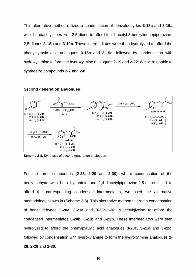
40
This alternative method utilized a condensation of benzaldehydes 3-18a and 3-19a
with 1,4-diacetylpiperazine-2,5-dione to afford the 1-acetyl-3-benzylidenepiperazine-
2,5-diones 3-18b and 3-19b. These intermediates were then hydrolyzed to afford the
phenylpyruvic acid analogues 3-18c and 3-19c, followed by condensation with
hydroxylamine to form the hydroxyimine analogues 2-19 and 2-22. We were unable to
synthesize compounds 2-7 and 2-8.
Second generation analogues
For the three compounds (2-28, 2-29 and 2-30), where condensation of the
benzaldehyde with both hydantoin and 1,4-diacetylpiperazine-2,5-dione failed to
afford the corresponding condensed intermediates, we used the alternative
methodology shown in (Scheme 2-8). This alternative method utilized a condensation
of benzaldehydes 3-20a, 3-21a and 3-22a with N-acetylglycine to afford the
condensed intermediates 3-20b, 3-21b and 3-22b. These intermediates were then
hydrolyzed to afford the phenylpyruvic acid analogues 3-20c, 3-21c and 3-22c,
followed by condensation with hydroxylamine to form the hydroxyimine analogues 2-
28, 2-29 and 2-30.
Scheme 2-8. Synthesis of second generation analogues

41
2.4.2. Biological Assays – Recombinant CtBP and Cellular assays
All the compounds synthesized above or purchased were tested against recombinant
CtBP enzyme and also in cellular growth/viability assays (HCT-116 p53-/- colon cancer
cells). While most of the compounds were inactive, three compounds 2-14, 2-21 and
2-22 showed good inhibitory activity against CtBP.
2.4.3 LDH Assay to Determine Off-target Toxicity
Lactate Dehydrogenase is closely related to CtBP in structure and function. We wished to test
if our compounds are specific against CtBP. Thus, three compounds (most potent in our
series) – HIPP, 3-Cl HIPP and 4-Cl HIPP were tested for their ability to inhibit L-Lactate
Dehydrogenase (LDH from rabbit muscle). Sodium oxamate (a known inhibitor of LDH) was
used as the standard. Stock solutions of the compounds in DMSO were added to a reaction
mixture containing phosphate buffer (pH 7.2), LDH, pyruvate and NADH. The enzyme
inhibition was measured by monitoring change in levels of NADH (absorbance) over a period
of 30 min. The minimal concentration of compounds leading to 50% inhibition of LDH activity
(IC50) was calculated using non-linear regression of the experimental data, using Prism
software (GraphPad).
The average IC50 (µM) values are reported below:
Compound IC50 (µM)
HIPP 42.53 ± 0.11
3-Cl-HIPP 94.86 ± 7.51
4-Cl-HIPP 181.9 ± 12.87
oxamate 45.80 ± 1.89
Detailed experimental procedure is reported in 6.4d.

42
2.4.4. HPLC Study to Determine Stability of the Oxime Compounds
As the in vitro activity and the in vivo activity of the oxime compounds did not show
proper correlation, we wanted to test if the oxime compounds are unstable under the
assay conditions, as instability of the compounds would explain the complex SAR
between in vitro and in vivo activities. We tested the stability of three most potent
(HIPP, 3-Cl HIPP and 4-Cl HIPP) compounds by HPLC. These compounds were found
to be stable.
2.5. Discussion
2.5.1. Inhibitory activity against Recombinant CtBP
Starting from MTOB (2-1, IC50 = 300 μM), phenylpyruvic acid (2-2) was found to be a
~3-fold better inhibitor than MTOB (Table 2-1) with an IC50 of 116 μM. Compounds 2-
3, 2-4, 2-5 and 2-6 were less active than phenylpyruvic acid (we were unable to
synthesize compounds 2-7 and 2-8). Compounds 2-9 through 2-13 were less active
than phenylpyruvic acid itself (Table 2-1), whereas hydroxyimine 2-14 inhibited CtBP
with an IC50 of 0.24 μM, a ~480-fold improvement over PPA.
Hydroxyimine analogues 2-15 through 2-27 were tested for their ability to inhibit the
functional dehydrogenase activity of CtBP in a NADH disappearance assay. Relative
to the parent hydroxylamine 2-14, we observed that ortho-substitutions on the ring
were detrimental to inhibitory activity [2-17 (2-Me), 2-20 (2-OMe), 2-23 (2-Cl), Table 2-
2]. Para- and meta-substitution by electron-donating groups [2-18 (4-OMe), 2-19 (3-
OMe), 2-24 (4-OH), 2-25 (3-OH)] resulted in a large decrease (4-OH) or a small
decrease (4-OMe, 3-OMe, 3-OH) in inhibitory activity. Para- and meta-substitution by
the electronically neutral methyl group [2-15 (4-Me), 2-16 (3-Me)] resulted in only a

43
small decrease in inhibitory activity. However, para- and meta-substitution by an
electron-withdrawing chlorine group [2-21 (4-Cl), 2-22 (3-Cl)] resulted in analogues
that were more active as inhibitors than parent hydroxyimine 2-9. Interestingly, this
effect seems specific to chlorine as para- substitutions with other electron-withdrawing
groups [2-26 (4-F), 2-27 (4-CN)] resulted in no increase in inhibition (2-26) or a
decrease in inhibitory activity (2-27).
We are yet to test the activity of our second generation analogues.
2.5.2. Inhibitory activity against Cellular CtBP (HCT-116 Colon Cancer Cells)
To determine whether inhibition of the dehydrogenase activity of CtBP by
hydroxyimine 2-14 and analogues 2-15 through 2-27 is effective at inhibiting cancer
cell growth, we carried out a short-term cell growth/viability assay (Table 2-2). HCT-
116 p53-/- colorectal cancer cells were treated with the compounds for 72 hours and
cell growth and viability was determined by MTT assay. Compounds 2-17, 2-20, 2-21,
2-22 and 2-27 were found to inhibit the growth of HCT-116 cells.
2.5.3. SAR – In vitro vs In vivo Activity
A complex structure/activity relationship was observed, between enzyme inhibition
and cell inhibitory activity, shown by the plot in Figure 2-12. A number of compounds
appeared to have cytotoxic effects out of proportion to their enzymatic activity,
consistent with off-target toxicity 2-18 (4-OMe), 2-27 (4-CN), while others had lower
than expected cellular inhibitory effects consistent with instability or poor intracellular
penetration 2-19 (3-OMe), 2-25 (3-OH). However, a series of compounds exhibited a
relatively consistent correlation of enzymatic and cellular inhibitory activity 2-15 (4-
Me), 2-16 (3-Me), 2-21 (4-Cl), 2-22 (3-Cl), 2-26 (4-F) with 2-21 (4-Cl) and 2-22 (3-Cl)

44
demonstrating the strongest enzymatic and cellular inhibitory activity of this subset of
the compounds.
Figure 2-12. Enzymatic vs cellular activity of inhibitors
2.5.4. Docking of Inhibitors in the Active Site of CtBP
More detailed computational docking with this crystal structure model were performed
to support the SAR studies. While no clear trend in docking scores vs. protein or
cellular inhibition IC50’s were found, the docked poses of designed analogues at CtBP
active site revealed factors important for binding and inhibition. Most importantly, the
phenyl rings of the ligands fit well in a hydrophobic region primarily formed by Try318,
Tyr76 and Met327. Compounds 2-15 and 2-21 form stronger hydrophobic and aryl-X
interactions in the pocket, compared to 2-24, where its polar (OH) substitution results
in unfavorable hydrophobic-polar interactions. These results will inform our design of
next-generation analogues.
2.5.5. Off-target Toxicity
The three most potent compounds in our series – HIPP, 3-Cl HIPP and 4-Cl HIPP
showed no inhibition against LDH, a closely related enzyme to CtBP. This shows that
45
our inhibitors are highly specific for CtBP. The aryl ring (complementary to Trp318 of
CtBP), and the α-keto acid portion of the molecule (in the hydrophilic cavity of CtBP)
might be conferring selectivity towards CtBP vs other dehydrogenases which lack
these two key features present in CtBP. So, the compounds 2-17, 2-18, 2-20, 2-23
might be targeting some other proteins resulting in their cytotoxic effects (poor in vitro
activity but good in vivo activity).
2.5.6. Stability of Oximes
As the in vitro activity and the in vivo activity of the oxime compounds did not show
proper correlation, we wanted to test if the oxime compounds are indeed stable under
the assay conditions, as instability of the compounds would explain the complex SAR
between in vitro and in vivo activities. We tested the stability of three most potent
(HIPP, 3-Cl HIPP and 4-Cl HIPP) compounds by HPLC (detailed procedure in section
2.7.8). These compounds were found to be stable. Thus, it is highly possible that
compounds 2-19 and 2-25 might be having cell permeability issues (good in vitro
activity but poor in vivo activity).
2.5.7. Hypothesis and Model
There is concrete evidence in the literature that targeting CtBP could be a highly tumor-
selective and effective strategy for treating cancer. CtBP is activated as a
transcriptional co-regulator only under conditions that are unique to tumor cells
(hypoxia and/or high NADH concentration – Warburg effect). Disrupting the
transcriptional co-regulatory function of CtBP in a tumorigenic environment has wide
ranging effects on multiple cellular mechanisms, as was seen in the genomic study by
Gardner and coworkers,70 and can restore the natural, endogenous function of tumor
suppressor genes.

46
While genetic inhibition of CtBP (with siRNA) and pharmacological inhibition with
peptides are effective tools for studying the role of CtBP in cancer biology, small
molecule inhibitors of CtBP are needed for further clinical drug development. Here we
report the first small molecule inhibitors for CtBP, and have optimized inhibition of the
dehydrogenase function of CtBP to a submicromolar level.
While these compounds are effective inhibitors of recombinant CtBP, high
concentrations are still required to inhibit growth in cells, with the in vivo cell growth
inhibitory activity of the best compounds disappointingly falling at just below 1 mM.
Earlier work with the parent compound MTOB, however, suggests that in long term
colony formation assays, GI50 values are usually 10-fold lower than those seen in short
term assays, and those studies are ongoing with the hydroxyimine derivatives.
Even taking the possible 10-fold lower GI50 of CtBP inhibitors in long-term assays into
account, the cellular inhibitory constants for the current series of CtBP inhibitors
remain 2-3 orders of magnitude higher than the in vitro IC50’s. A possible rationale for
this effect is that in cancer cells, CtBP has already formed transcriptional co-repression
complexes with NADH, transcription factors, and repressor enzymes, and is not
available for effective inhibitor binding. Binding of the inhibitor to CtBP either must
break up the transcriptional co-repression complex or wait until the complex
dissociates naturally in order to bind. This mechanistic model is consistent with our
data that the compounds effectively bind to and inhibit the dimeric form of CtBP
generated in the in vitro assay. We are currently working to investigate this
mechanistic model and develop analogues with improved activity in the cellular
assays. We are also using these new CtBP inhibitors as tools for studying the role of

47
CtBP in cell survival and migration, as well as the impact of restoring tumor suppressor
gene expression and inhibiting oncogenes in tumor cells.
2.6. Computational Approach towards Design of Diverse CtBP Inhibitors
Arriving at lead compounds quickly and efficiently (both time and cost-wise) is best
achieved by using a combination of different approaches. We used computational
approaches to design more CtBP inhibitors complementary to the traditional medicinal
chemistry SAR approach discussed above. Through computational approaches, a
large chemical space can be explored and diverse scaffolds of lead compounds could
be identified in less time, which is not always possible with the traditional medicinal
chemistry approach.
Virtual screening is a computational process in which a large set of compound
libraries can be screened to identify compounds that could potentially bind in a
particular proteins binding site. It is a process in which we can virtually filter
compounds that need to be synthesized/purchased and tested, thereby saving on the
time and resources. It can either be ligand-based or structure-based. In ligand-based
virtual screening, the features of a pharmacophore (from a known ligand of a protein)
are used to screen libraries of compounds. In structure-based virtual screening,
compounds are docked into the active site of the protein, and scored using scoring
functions to determine which compounds are likely to bind to the protein.
We carried out virtual screening of the ZINC database using UNITY in SYBYL using a
combination of both ligand-based and structure-based methods. We utilised the co-
crystal structure of CtBP-NADH-HIPP. The binding pocket with HIPP is shown in the
figure below (Figure 2-13).
UNITY query features: We defined the following features as queries in UNITY.

48
a. Aromatic feature – 1.5 Å radius – complementary to Trp318 (pi-stacking)
b. Aromatic feature – 1.0 Å radius – complementary to Tyr76 (pi-stacking)
c. Negative center – 1.0 Å radius – complementary to Arg266 (electrostatic)
d. Acceptor site – 1.0 Å radius – at His 315 (H-bond)
e. Exclusion spheres – 0.75 Å radius – receptor cavity (to avoid clashes)
f. Bond path constraint – 3-5 bonds – between negative center and aromatic
feature.
We performed a UNITY Flex search to screen the ZINC database of clean drug-like
subset (~10 million compounds). We obtained 289 hits.
Docking is a process in which the compounds are docked in the binding site of a
protein to see which conformations the compound can possibly bind in. We performed
docking of the 289 hits using GOLD. The binding cavity was defined as 10 Å radius
around Arg266, the number of GA runs was 50.
Scoring is a process in which the different docked poses are ranked based on the
favorable and unfavorable interactions. Higher score means more favourable
Trp318
NAD+
Tyr76
His315
Arg266
Arg97
*Aromatic
*Aromatic
*Negative center
*Acceptor site
Figure 2-13. UNITY features based on HIPP

49
interactions usually, although the poses have to be manually visualized to make sure
that the interactions are possible, and are not errors. We used HINT scoring function
to score all the solutions obtained from GOLD. Initial top HINT scores of best ligand
conformations 1290-1926. We then performed energy minimization of top scored 50
protein-ligand complexes (to mimic actual binding of compound to the protein) followed
by HINT rescoring of ligands. The final HINT scores were around 1362-2294.
We evaluated binding modes of these compounds, an example (ZINC 02586210, final
HINT score 2294) is shown below (Figure 2-14). There are two phenyl rings (yellow)
which show pi-stacking interactions with Tyr76 and Trp318. The carboxylate group
(red) shows electrostatic interactions with Arg266 and Arg97. The amine (blue) has a
H-bonding interaction with His315.
Based on the HINT scores, we ordered the following 10 hits, and are yet to test their
activity against CtBP (Figure 2-15).
Arg266
Arg97
Trp318
Tyr76
His315
Figure 2-14. Binding mode of compound ZINC02586210

50
Figure 2-15. Hits obtained through virtual screening to be tested

51
2.7. Experimental Procedures
2.7.1. General Chemical Methods
Reagents/chemicals, catalysts, solvents were purchased from Sigma-Aldrich, Fisher
and Alfa-Aesar. Analytical Thin Layer Chromatography (TLC) was performed using
silica gel GHLF plates (Analtech Inc.). Flash chromatography was performed on
TELEDYNE ISCO CombiFlash® Rf instrument using RediSep Rf Normal-phase Flash
Columns (4-gm, 12-gm, 24-gm or 40-gm). 1H NMR and 13C NMR experiments were
recorded on BRUKER 400MHz NMR instrument in deuterated solvents - chloroform
(CDCl3), acetone ((CD3)2CO), dimethyl sulfoxide ((CD3)2SO) or methanol (CD3OD). All
chemical shifts are reported in parts per million (ppm) with reference to chloroform,
acetone, DMSO and methanol residual peaks at 7.26, 2.05, 2.50 and 3.31 respectively
(1H NMR spectra); 77.16, 29.84, 39.52 and 49.00 respectively (13C NMR spectra). The
data is reported as: chemical shifts (ppm), multiplicity (s = singlet, d = doublet, t =
triplet, m = multiplet), coupling constant(s) (Hz) and integral values.
2.7.2. Rationally Designed and Deconstruction Analogues
The following compounds were purchased from commercial sources and tested for
CtBP inhibition without further purification: 4-methylthio-2-oxobutyric acid (MTOB, 2-
1, Sigma-Aldrich), phenyl pyruvic acid (2-2, Sigma-Aldrich), 2-benzylacrylic acid (2-10,
TCI America), benzylmalonic acid (2-11, Sigma Aldrich), anilino(oxo)acetic acid (2-12,
Sigma-Aldrich), pyruvate (2-3, Sigma-Aldrich), hydrocinnamic acid (2-4, Sigma
Aldrich), and phenylglyoxylic acid (2-6, Sigma-Aldrich).

52
3-Phenyl-2-thioxopropanoic acid (2-9). Rhodanine 3-4 (1.25 g, 9.4 mmol), anhydrous
sodium acetate (2.16 g, 26.4 mmol), and benzaldehyde 3-3 (1.0 g, 9.4 mmol) were
added to a vial followed by glacial acetic acid (17.8 mL, 0.53 M). The reaction was
heated to 115 °C for three hours, upon which time the reaction had turned orange and
developed a precipitate. The reaction mixture was cooled to room temperature and
poured into ice water, which caused additional precipitate to form. Upon warming to
room temperature, the precipitate was filtered and then dried in a oven for 36 hours to
afford a dry powder 3-5 (1.66 g, 80% yield). This intermediate was dissolved in water
(10 mL) and 6N sodium hydroxide (10 mL) and heated to 95 °C for 1 h. The reaction
mixture was cooled to room temperature, water (10 mL) was added, and 6N HCl was
added to crash out a white precipitate. This precipitate was filtered and purified by
flash chromatography (silica gel, 5% MeOH/DCM) to afford the product (0.86 g, 63%
yield). 1H NMR (400 MHz, (CD3)2SO) δ 7.74 (s, 1H), 7.68 (d, J = 7.4 Hz, 2H), 7.48 (t,
2H), 7.42-7.36 (m, 1H). HRMS C9H7O2S [M-H]- Expected: 179.0167, Found: 179.0164.
2-hydrazono-3-phenylpropanoic acid (2-13): Phenylpyruvic acid 3-6 (0.1 g, 0.61 mmol)
is dissolved in ethanol (0.4 ml). Hydrazine (0.02 mL, 0.61 mmol) is added and the
reaction mixture is stirred overnight at room temperature. Sodium carbonate (0.03 g,
0.31 mmol) is added and the reaction mixture is stirred for 2 h at room temperature.
The precipitated solid is filtered and dried to give the title compound in 62% yield (0.07

53
g, 0.38 mmol). 1H NMR (400 MHz, DMSO) δ 7.26 (d, J = 7.5 Hz, 2H), 7.19 (t, J = 7.7
Hz, 2H), 7.10 (t, J = 7.5 Hz, 1H), 6.26 (s, 2H), 3.78 (s, 2H); 13C NMR (125 MHz, DMSO)
δ 168.60, 146.52, 138.01, 128.68, 127.88, 125.37, 30.21. HRMS C9H10N2O2 [M+Na]-
Expected: 201.0634, Found: 201.0622.
2-(hydroxyimino)-3-phenylpropanoic acid (2-14): Phenyl pyruvic acid 3-6 (3.0 g, 18.28
mmol) was dissolved in a solution of NaOH (2.2 g, 54.83 mmol) in water (1 mL).
Hydroxylamine hydrochloride (1.9 g, 27.41 mmol) was added to the reaction and
stirred overnight at room temperature. 1N HCl was added to the reaction, the
precipitated product is filtered and dried. The crude product is purified using flash
chromatography (silica gel, 5% MeOH/DCM). Yield: 76% (2.50 g, 13.95 mmol). 1H
NMR (400 MHz, DMSO) δ 12.28 (s, 1H), 7.27 (m, 2H), 7.19 (m, 3H), 3.82 (s, 2H); 13C
NMR (125 MHz, DMSO) δ 165.14, 150.13, 136.68, 128.52, 128.32, 126.13, 29.85.
HRMS C9H9NO3 [M-H]- Expected: 178.0509, Found: 178.0494.
1-phenylpropan-2-one (2-5): Phenylacetic acid 3-1 (0.5 g, 3.7 mmol) was dissolved in
DCM (9.2 mL). To this, N-(3-dimethylaminopropyl)-N′-ethylcarbodiimide hydrochloride
(0.8 g, 4.4 mmol), 4-methylmorpholine (0.4 g, 3.7 mmol), N,O-Dimethylhydroxylamine
hydrochloride (0.4 g, 4.0 mmol) were added. The reaction mixture was stirred at room
temperature for 4 h. It was concentrated and diethyl ether was added. The organic
layer was washed with brine, dried over Na2SO4 and evaporated. Purification of the
54
residue on silica gel (5:1 hexanes/EtOAc) afforded the product N-methoxy-N-methyl-
2-phenylacetamide 3-2 in 87% yield. 0.6 g (3.2 mmol) of this product was dissolved in
ether (10.6 ml) under nitrogen at 0 oC. Methylmagnesium bromide solution (3.4 mL,
4.8 mmol, 1.4 M in THF:toluene 1:3) was added over 30 min. The reaction was stirred
at 0 oC for 1 h and at room temperature for 30 min. It was quenched at 0 oC with the
slow addition of 1 M HCl. The reaction was extracted with ether. The organic layer was
washed with brine, dried over Na2SO4 and evaporated. Purification of the residue on
silica gel (5:1 hexanes/EtOAc) afforded the product 1-phenylpropan-2-one in 89%
yield (0.38 g, 2.83 mmol). 1H NMR (400 MHz, MeOD) δ 7.21-7.33 (m, 5H), 3.75 (s,
2H), 2.15 (s, 3H); 13C NMR (125 MHz, MeOD) δ 209.24, 136.00, 130.58, 129.67,
127.97, 51.37, 29.27. HRMS C9H10O [M-H]- Expected: 133.0659, Found: 132.0813.
2.7.3. Synthesis of Hydroxyiminophenylpyruvic acid Analogues
2.7.3.1. Synthesis of keto-acid intermediates
General Procedure 1: Hydantoin (15.0 mmol) was dissolved in water (15.0 mL) at 70
oC. The pH of the solution was adjusted to 7 using saturated sodium bicarbonate
solution. Ethanolamine (1.4 mL) was added and the temperature was raised to 90 oC.
The corresponding aldehyde (15.0 mmol) in ethanol (15.0 mL) was added drop wise
to the reaction mixture. The reaction was refluxed at 120 oC for 5-10 h. After being
cooled to room temperature, the product was filtered, washed with alcohol/water (1:5)
and dried to give the corresponding benzalhydantoin intermediate. This corresponding
benzylhydantoin (8.98 mmol) was dissolved in 20% aqueous NaOH solution (28.4 mL)
and refluxed at 100 oC for 3 h. After being cooled to room temperature, 12N HCl (11.8
mL) was added. Sodium bicarbonate was added to bring the pH to 7. The reaction
mixture was extracted with ether until the ether layer was clear. This layer was
discarded. To the aqueous layer, 12N HCl (7.1 mL) was added. It was extracted with

55
ether until no more acid was obtained. The ether layer was dried to give the crude keto
acid. It was recrystallized in water to give pure product.
2-oxo-3-p-tolylpropanoic acid (3-7c): Procedure 1 was followed using 4-
methylbenzaldehyde. Yield 54% (1.0 g, 5.61 mmol). 1H NMR (400 MHz, MeOD) δ
7.64-7.66 (d, J = 8.1 Hz, 2H), 7.12-7.14 (d, J = 8.1 Hz, 2H), 6.46 (s, 1H), 2.32 (s, 3H);
13C NMR (125 MHz, MeOD) δ 168.46, 141.49, 138.44, 133.49, 130.72, 129.87,
111.79, 21.28. HRMS C10H10O3 [M-H]- Expected: 177.0557, Found: 177.0562.
2-oxo-3-m-tolylpropanoic acid (3-8c): Procedure 1 was followed using 3-
methylbenzaldehyde. Yield 57% (0.3 g, 1.68 mmol). 1H NMR (400 MHz, MeOD) δ 7.57
(d, J = 4.1 Hz, 1H), 7.55 (s, 1H), 7.18-7.22 (t, J = 7.5 Hz, 1H), 7.03-7.05 (d, J = 7.9 Hz,
1H), 6.45 (s, 1H), 2.32 (s, 3H); 13C NMR (125 MHz, MeOD) δ 170.0, 143.64, 140.38,
137.85, 132.89, 130.72, 129.55, 113.34, 23.07. HRMS C10H10O3 [M-H]- Expected:
177.0557, Found: 177.0565.
2-oxo-3-o-tolylpropanoic acid (3-9c): Procedure 1 was followed using 2-
methylbenzaldehyde. Yield 58% (0.08 g, 0.45 mmol). 1H NMR (400 MHz, (CD3)2CO)
δ 8.18 (d, J = 8.2 Hz, 1H), 7.85 (s, 1H), 7.11-7.23 (m, 4H), 6.74 (s, 1H), 2.38 (s, 3H);
13C NMR (125 MHz, (CD3)2CO) δ 167.10, 141.15, 137.11, 133.96, 130.84, 130.64,

56
128.34, 126.63, 107.60, 20.13. HRMS C10H10O3 [M-H]- Expected: 177.0557, Found:
177.0549.
3-(4-methoxyphenyl)-2-oxopropanoic acid (3-10c): Procedure 1 was followed using 4-
methoxybenzaldehyde. Yield 88% (1.8 g, 9.27 mmol). 1 H NMR (400 MHz, MeOD) δ
7.71-7.73 (d, J = 8.8 Hz, 2H), 6.88-6.90 (d, J = 8.9 Hz, 2H), 6.46 (s, 1H), 3.80 (s, 3H);
13C NMR (125 MHz, MeOD) δ 168.61, 160.57, 140.55, 132.24, 129.08, 114.76,
111.83, 55.69. HRMS C10H10O4 [M-H]- Expected: 193.0506, Found: 193.0511.
3-(2-methoxyphenyl)-2-oxopropanoic acid (3-11c): Procedure 1 was followed using 2-
methoxybenzaldehyde. Yield 56% (0.23 g, 1.29 mmol). 1H NMR (400 MHz, MeOD) δ
8.20-8.22 (m, 1H), 7.19-7.23 (m, 1H), 6.90-6.95 (m, 3H), 3.86 (s, 3H); 13C NMR (125
MHz, MeOD) δ 168.64, 158.20, 141.79, 131.72, 129.78, 124.98, 121.41, 111.46,
105.03, 56.11. HRMS C10H10O4 [M-H]- Expected: 193.0506, Found: 193.0496.
3-(4-chlorophenyl)-2-oxopropanoic acid (3-12c): Procedure 1 was followed using 4-
chlorobenzaldehyde. Yield 62% (1.1 g, 5.54 mmol). 1H NMR (400 MHz, MeOD) δ 7.74-
7.76 (d, J = 8.8 Hz, 2H), 7.30-7.32 (d, J = 8.5 Hz, 2H), 6.45 (s, 1H); 13C NMR (125

57
MHz, MeOD) δ 168.05, 142.95, 135.22, 133.81, 132.06, 129.35, 110.01. HRMS
C9H7O3Cl [M-H]- Expected: 197.0011, Found: 197.0016.
3-(2-chlorophenyl)-2-oxopropanoic acid (3-13c): Procedure 1 was followed using 2-
chlorobenzaldehyde. Yield 56% (0.3 g, 1.51 mmol). 1H NMR (400 MHz, MeOD) δ 8.32-
8.35 (d, J = 7.9 Hz, 1H), 7.38-7.40 (d, J = 8.1 Hz, 1H), 7.26-7.30 (t, J = 7.6 Hz, 1H),
7.19-7.21 (t, J = 7.8 Hz, 1H), 6.89 (s, 1H); 13C NMR (125 MHz, MeOD) δ 167.97,
143.77, 134.42, 134.04, 132.20, 130.31, 129.41, 127.73, 105.93. HRMS C9H7O3Cl [M-
H]- Expected: 197.0011, Found: 197.0010.
3-(4-hydroxyphenyl)-2-oxopropanoic acid (3-14c): Procedure 1 was followed using 4-
hydroxybenzaldehyde. Yield 74% (1.3 g, 7.22 mmol). 1H NMR (400 MHz, MeOD) δ
7.62-7.64 (d, J = 8.6 Hz, 2H), 6.75-6.77 (d, J = 8.8 Hz, 2H), 6.45 (s, 1H); 13C NMR
(125 MHz, MeOD) δ 168.74, 158.19, 139.97, 132.41, 127.94, 116.15, 112.35. HRMS
C9H8O4 [M-H]- Expected: 179.0350, Found: 179.0354.
3-(3-hydroxyphenyl)-2-oxopropanoic acid (3-15c): Procedure 1 was followed using 3-
hydroxybenzaldehyde. Yield 88% (0.7 g, 3.89 mmol). 1H NMR (400 MHz, (CD3)2SO)
δ 7.29 (s, 1H), 7.07-7.14 (m, 2H), 6.63-6.66 (m, 1H), 6.29 (s, 1H); 13C NMR (125 MHz,

58
(CD3)2SO) δ 166.28, 157.11, 141.56, 135.96, 129.02, 120.48, 115.78, 114.47, 109.76.
HRMS C9H8O4 [M-H]- Expected: 179.0350, Found: 179.0348.
3-(4-fluorophenyl)-2-oxopropanoic acid (3-16c): Procedure 1 was followed using 4-
fluorobenzaldehyde. Yield 71% (0.5 g, 2.74 mmol). 1H NMR (400 MHz, MeOD) δ 7.78-
7.81 (m, 2H), 7.02-7.07 (t, J = 8.9 Hz, 2H), 6.47 (s, 1H); 13C NMR (125 MHz, MeOD)
δ 168.26, 164.52, 162.07, 142.02, 132.57, 132.65, 115.88, 116.10, 110.42. HRMS
C9H7O3F [M-H]- Expected: 181.0306, Found: 181.0306.
3-(4-cyanophenyl)-2-oxopropanoic acid (3-17c): Procedure 1 was followed using 4-
cyanobenzaldehyde. Yield 70% (0.05 g, 0.26 mmol). 1H NMR (400 MHz, (CD3)2CO) δ
8.62 (d, J = 1.7 Hz, 1H), 8.01 (d, J = 8.6 Hz, 2H), 7.76 (d, J = 8.6 Hz, 2H), 6.60 (s, 1H);
13C NMR (125 MHz, (CD3)2CO) δ 167.49, 145.32, 141.54, 133.02, 132.69, 132.65,
131.03, 120.00, 110.87, 108.92. HRMS C10H7NO3 [M-H]- Expected: 188.0353, Found:
188.0346.
General Procedure 2: 1,4-diacetylpiperazine-2,5-dione119 (0.5 mmol) was dissolved
in DMF (1 mL). Triethylamine (0.5 mmol) was added followed by corresponding
aldehyde (0.5 mmol). The reaction mixture was stirred for 12 h at room temperature.
It was extracted with DCM, washed with NH4Cl solution. The organic layer was dried
over Na2SO4 and evaporated. Purification of the residue on silica gel afforded the
product (benzylidene)-piperazine-2,5-dione intermediate. This intermediate (0.1
mmol) was refluxed in 6 N HCl (4.0 ml) for 4 h. The reaction was cooled to room

59
temperature, extracted with ether, dried over Na2SO4 and evaporated to give the crude
keto acid. It was recrystallized in water to give pure product.
3-(3-methoxyphenyl)-2-oxopropanoic acid (3-18c): Procedure 2 was followed using 3-
methoxylbenzaldehyde. Yield 56% (0.5 g, 2.57 mmol). 1H NMR (400 MHz, MeOD) δ
7.44 (t, J = 2 Hz, 1H), 7.20-7.29 (m, 2H), 6.79-6.82 (m, 1H), 6.46 (s, 1H), 3.79 (s, 3H);
13C NMR (125 MHz, MeOD) δ 168.32, 161.04, 142.44, 137.66, 130.11, 123.52,
115.80, 114.39, 111.51, 55.62. HRMS C10H10O4 [M-H]- Expected: 193.0506, Found:
193.0495.
3-(3-chlorophenyl)-2-oxopropanoic acid (3-19c): Procedure 2 was followed using 3-
chlorobenzaldehyde. Yield 56% (0.12 g, 0.60 mmol). 1H NMR (400 MHz, (CD3)2CO) δ
7.96 (t, J = 1.96 Hz, 1H), 7.76 (dt, J = 7.8 Hz, 1H), 7.25-7.41 (m, 3H), 6.54 (s, 1H),
4.28 (s, 0.6H), 4.05 (d, J = 6.2 Hz, 0.5H); 13C NMR (125 MHz, (CD3)2CO)) δ 166.54,
137.85, 134.59, 130.74, 130.67, 129.80, 128.83, 127.99, 109.31. HRMS C9H7O3Cl [M-
H]- Expected: 197.0011, Found: 197.0005.
General Procedure 3120: Substituted benzaldehyde (2 mmol) was dissolved in acetic-
anhydride (0.25 M). N-acetyl glycine (2.4 mmol) was added followed by sodium
acetate (10 mmol). The reaction mixture was refluxed for 4 h at 140 oC. It was poured
into crushed ice, stirred for 30 min. Precipitate was separated, washed with cold water,
and dried under vacuum to afford the oxazolyl intermediate. This intermediate (0.4

60
mmol) was refluxed in 3 N HCl (4.0 ml) for 24 hours at 100 oC. The reaction was cooled
to room temperature, extracted with ether, washed with 6 N HCl, dried over Na2SO4
and evaporated to give the crude keto acid. It was recrystallized in water to give pure
product.
3-(3,4-dichlorophenyl)-2-oxopropanoic acid (3-20c): Procedure 3 was followed using
3,4-dichlorobenzaldehyde. Yield 71% (0.06 g, 0.28 mmol). 1H NMR (400 MHz,
(CD3)2CO) δ 8.1 (d, J = 1.89 Hz, 1H), 7.76 (dd, J = 8.49 Hz, 1H), 7.56 (d, J = 8.44 Hz,
1H), 6.53 (s, 1H); 13C NMR (125 MHz, (CD3)2CO)) δ 166.39, 143.09, 136.55, 132.63,
131.78, 131.29, 131.11, 130.27, 108.38. HRMS C9H6O3Cl2 [M-H]- Expected:
230.9627.
3-(2,4-dichlorophenyl)-2-oxopropanoic acid (3-20c): Procedure 3 was followed using
2,4-dichlorobenzaldehyde. Yield 77% (0.35 g, 1.5 mmol). 1H NMR (400 MHz, MeOD)
δ 8.34 (d, J = 8.79 Hz, 1H), 7.29-7.53 (m, 3H), 6.82 (s, 1H); 13C NMR (125 MHz,
MeOD) δ 167.62, 144.44, 134.76, 133.96, 132.96, 131.83, 130.41, 129.95, 129.78,
128.75, 128.35, 128.04, 127.61, 104.58. HRMS C9H6O3Cl2 [M-H]- Expected:
230.9627.

61
2-oxo-3-(4-(trifluoromethyl)phenyl)propanoic acid (3-20c): Procedure 3 was followed
using 3-trifluoromethylbenzaldehyde. Yield 90% (0.09 g, 0.41 mmol). 1H NMR (400
MHz, (MeOD) δ 7.93 (td J = 8.15 Hz, 2H), 7.59 (d, J = 8.15 Hz, 2H), 6.51 (s, 1H); 13C
NMR (125 MHz, MeOD) δ 167.71, 144.46, 140.44, 132.22, 130.82, 126.01, 125.97,
125.94, 125.64, 125.61, 109.28. HRMS C10H7O3F3 [M-H]- Expected: 231.0227.
2.7.3.2. Synthesis of Hydroxyimino (Oxime) Analogues
General Procedure 4: The corresponding keto-acid (0.50 mmol) is dissolved in a
solution of NaOH (1.51 mmol) in water (1 mL). Hydroxylamine hydrochloride (0.76
mmol) is added to the reaction and stirred overnight at room temperature. 1 N HCl is
added to the reaction, the precipitated product is filtered and dried. The crude product
is purified using flash chromatography (silica gel, 5% MeOH/DCM) as necessary.
2-(hydroxyimino)-3-p-tolylpropanoic acid (2-15): Title compound was prepared
following general procedure 4. Yield 55% (0.03 g, 0.16 mmol). 1H NMR (400 MHz,
MeOD) δ 7.05-7.14 (m, 4H), 3.86 (s, 2H), 2.27 (s, 3H); 13C NMR (125 MHz, MeOD) δ
166.90, 152.35, 136.96, 134.84, 129.96, 129.89, 30.51, 21.03. HRMS C10H11NO3 [M-
H]- Expected: 192.0666, Found:192.0673.

62
2-(hydroxyimino)-3-m-tolylpropanoic acid (2-16): Title compound was prepared
following general procedure 4. Yield 74% (0.04 g, 0.21 mmol). 1H NMR (400 MHz,
MeOD) δ 7.03-7.13 (m, 3H), 6.98 (d, J = 7.31 Hz, 1H), 3.87 (s, 2H), 2.28 (s, 3H); 13C
NMR (125 MHz, MeOD) δ 166.88, 152.22, 139.03, 137.84, 130.61, 129.26, 128.02,
127.02, 30.85, 21.42. HRMS C10H11NO3 [M-H]- Expected: 192.0666, Found:192.0656.
2-(hydroxyimino)-3-o-tolylpropanoic acid (2-17): Title compound was prepared
following general procedure 4. Yield 92% (0.12 g, 0.62 mmol). 1H NMR (400 MHz,
(CD3)2CO) δ 7.01-7.10 (m, 4H), 3.88 (s, 2H), 2.35 (s, 3H); 13C NMR (125 MHz,
(CD3)2CO) δ 167.77, 153.63, 137.10, 136.37, 130.67, 129.10, 126.82, 126.60, 28.37,
19.93. HRMS C10H11NO3 [M-H]- Expected: 192.0666, Found:192.0652.
2-(hydroxyimino)-3-(4-methoxyphenyl)propanoic acid (2-18): Title compound was
prepared following general procedure 4. Yield 71% (0.08 g, 0.36 mmol). 1H NMR (400
MHz, MeOD) δ 7.18 (d, J = 8.6 Hz, 2H), 6.79 (d, J = 8.8 Hz, 2H), 3.86 (s, 2H), 3.74 (s,
3H); 13C NMR (125 MHz, MeOD) δ 167.48, 159.71, 153.20, 131.07, 130.09, 114.79,
55.67, 30.16. HRMS C10H11NO4 [M-H]- Expected: 208.0615, Found:208.0601.

63
2-(hydroxyimino)-3-(3-methoxyphenyl)propanoic acid (2-19): Title compound was
prepared following general procedure 4. Yield 72% (0.08 g, 0.37 mmol). 1H NMR (400
MHz, MeOD) δ 7.13-7.17 (t, J = 8.1 Hz, 1H), 6.83 (m, 2H), 6.74 (dd, J = 8.3 Hz, 1H),
3.89 (s, 2H), 3.75 (s, 3H); 13C NMR (125 MHz, MeOD) δ 166.87, 161.21, 152.07,
139.41, 130.29, 122.35, 115.73, 112.86, 55.56, 30.95. HRMS C10H11NO4 [M-H]-
Expected: 208.0615, Found:208.0612.
2-(hydroxyimino)-3-(2-methoxyphenyl)propanoic acid (2-20): Title compound was
prepared following general procedure 4. Yield 56% (0.07 g, 0.33 mmol). 1H NMR (400
MHz, (CD3)2CO) δ 7.17 (t, J = 7.8 Hz, 1H), 7.00 (d, J = 7.6 Hz, 1H), 6.93 (d, J = 8.5
Hz, 1H), 6.83 (t, J = 7.6 Hz, 1H), 3.90 (s, 2H), 3.82 (s, 3H); 13C NMR (125 MHz,
(CD3)2CO) δ 165.17, 158.28, 151.76, 129.60, 28.34, 125.57, 121.06, 111.32, 55.72,
25.53. HRMS C10H11NO4 [M-H]- Expected: 208.0615, Found:208.0607.
3-(4-chlorophenyl)-2-(hydroxyimino)propanoic acid (2-21): Title compound was
prepared following general procedure 4. Yield 56% (0.06 g, 0.28 mmol). 1H NMR (400

64
MHz, DMSO) δ 12.34 (br s, 1H), 7.33 (d, J = 8.5 Hz, 2H), 7.20 (d, J = 8.5 Hz, 2H), 3.79
(s, 2 H); 13C NMR (125 MHz, MeOD) δ 166.71, 151.68, 136.88, 133.20, 131.63,
129.40, 30. 39. HRMS C9H8NO3Cl [M-H]- Expected: 212.0120, Found: 212.0102.
3-(3-chlorophenyl)-2-(hydroxyimino)propanoic acid (2-22): Title compound was
prepared following general procedure 4. Yield 66% (0.15 g, 0.70 mmol). 1H NMR (400
MHz, DMSO) δ 7.24-7.34 (m, J = 8.8 Hz, 3H), 7.17 (d, J = 7.3 Hz, 1H), 3.82 (s, 2H);
13C NMR (125 MHz, DMSO) δ 165.16, 139.26, 132.81, 130.14, 128.32, 127.32,
126.14, 29.64. HRMS C9H8O3NCl [M-H]- Expected: 212.0120, Found:212.0108.
3-(2-chlorophenyl)-2-(hydroxyimino)propanoic acid (2-23): Title compound was
prepared following general procedure 4. Yield 54% (0.02 g, 0.07 mmol). 1H NMR (400
MHz, DMSO) δ 7.41-7.44 (m, 1H), 7.21-7.28 (m, 2H), 7.03 (m, 1H), 3.88 (s, 2H); 13C
NMR (125 MHz, DMSO) δ 164.97, 149.01, 134.08, 132.83, 129.22, 129.11, 127.96,
127.11, 27.93. HRMS C9H8O3NCl [M-H]- Expected: 212.0120, Found: 212.0134.

65
2-(hydroxyimino)-3-(4-hydroxyphenyl)propanoic acid (2-24): Title compound was
prepared following general procedure 4. Yield 91% (0.10 g, 0.51 mmol). 1H NMR (400
MHz, MeOD) δ 7.08 (d, J = 8.5 Hz, 2H), 6.66 (d, J = 8.5 Hz, 2H), 3.80 (s, 2H); 13C
NMR (125 MHz, MeOD) δ 166.93, 156.93, 152.63, 131.06, 128.70, 116.13, 30.03.
HRMS C9H9NO4 [M-H]- Expected: 194.0459, Found:194.0459.
2-(hydroxyimino)-3-(3-hydroxyphenyl)propanoic acid (2-25): Title compound was
prepared following general procedure 4. Yield 65% (0.07 g, 0.36 mmol). 1H NMR (400
MHz, DMSO) δ 7.04 (t, J = 8.0 Hz, 1H), 6.55-6.62 (m, 3H), 3.72 (s, 2H); 13C NMR (125
MHz, DMSO) δ 165.18, 157.22, 137.89, 129.09, 119.24, 115.44, 113.07, 48.52, 29.74.
HRMS C9H9NO4 [M-H]- Expected: 194.0459, Found:194.0442.
3-(4-fluorophenyl)-2-(hydroxyimino)propanoic acid (2-26): Title compound was
prepared following general procedure 4. Yield 74% (0.08 g, 0.41 mmol). 1H NMR (400
MHz, DMSO) δ 7.20-7.24 (m, 2H), 7.10 (t, J = 8.8 Hz, 2H), 3.79 (s, 2H); 13C NMR (125
MHz, MeOD) δ 165.06, 161.94, 159.54, 150.03, 132.82, 132.79, 130.38, 130.30,
115.12, 114.91, 29.06. HRMS C9H8NO3F [M-H]- Expected: 196.0415, Found:
196.0398.

66
3-(4-cyanophenyl)-2-(hydroxyimino)propanoic acid (2-27): Title compound was
prepared following general procedure 4. Yield 64% (0.09 g, 0.44 mmol). 1H NMR (400
MHz, DMSO) δ 7.74 (d, J = 7.6 Hz, 2H), 7.37 (d, J = 8.1 Hz, 2H), 3.89 (s, 2H); 13C
NMR (125 MHz, DMSO) δ 164.93, 149.11, 142.74, 132.26, 129.52, 118.77, 109.06,
30.20. HRMS C10H8N2O3 [M-H]- Expected: 203.0462, Found: 203.0452.
3-(3,4-dichlorophenyl)-2-(hydroxyimino)propanoic acid (2-28): Title compound was
prepared following general procedure 4. Yield 66% (0.01 g, 0.04 mmol). 1H NMR (400
MHz, MeOD) δ 7.38-7.43 (m, 2H), 7.20 (dd, J = 8.34 Hz, 1H), 3.89 (s, 2H); 13C NMR
(125 MHz, MeOD) δ 166.80, 151.37, 138.94, 132.96, 132.01, 131.31, 131.14, 129.98,
30.22. HRMS C9H7Cl2NO3 [M-H]- Expected: 245.
3-(2,4-dichlorophenyl)-2-(hydroxyimino)propanoic acid (2-29): Title compound was
prepared following general procedure 4. Yield 66% (0.02 g, 0.08 mmol). 1H NMR (400
MHz, MeOD) δ 7.42 (d, J = 2.04 Hz, 1H), 7.21 (dd, J = 8.25 Hz, 1H), 7.09 (d, J = 8.34
Hz, 1H), 3.98 (s, 2H); 13C NMR (125 MHz, MeOD) δ 166.88, 150.90, 135.71, 134.68,
133.73, 131.82, 129.90, 128.07, 28.72. HRMS C9H7Cl2NO3 [M-H]- Expected: 245.97.
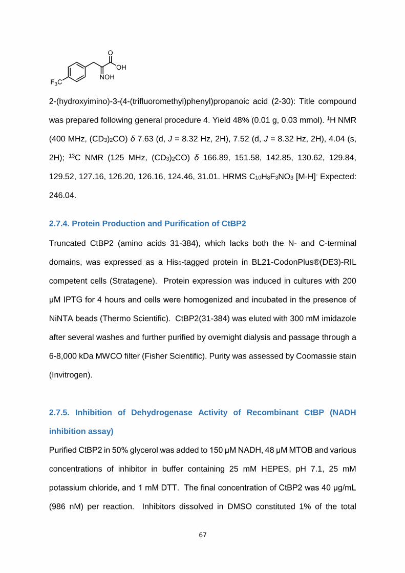
67
2-(hydroxyimino)-3-(4-(trifluoromethyl)phenyl)propanoic acid (2-30): Title compound
was prepared following general procedure 4. Yield 48% (0.01 g, 0.03 mmol). 1H NMR
(400 MHz, (CD3)2CO) δ 7.63 (d, J = 8.32 Hz, 2H), 7.52 (d, J = 8.32 Hz, 2H), 4.04 (s,
2H); 13C NMR (125 MHz, (CD3)2CO) δ 166.89, 151.58, 142.85, 130.62, 129.84,
129.52, 127.16, 126.20, 126.16, 124.46, 31.01. HRMS C10H8F3NO3 [M-H]- Expected:
246.04.
2.7.4. Protein Production and Purification of CtBP2
Truncated CtBP2 (amino acids 31-384), which lacks both the N- and C-terminal
domains, was expressed as a His6-tagged protein in BL21-CodonPlus®(DE3)-RIL
competent cells (Stratagene). Protein expression was induced in cultures with 200
μM IPTG for 4 hours and cells were homogenized and incubated in the presence of
NiNTA beads (Thermo Scientific). CtBP2(31-384) was eluted with 300 mM imidazole
after several washes and further purified by overnight dialysis and passage through a
6-8,000 kDa MWCO filter (Fisher Scientific). Purity was assessed by Coomassie stain
(Invitrogen).
2.7.5. Inhibition of Dehydrogenase Activity of Recombinant CtBP (NADH
inhibition assay)
Purified CtBP2 in 50% glycerol was added to 150 μM NADH, 48 μM MTOB and various
concentrations of inhibitor in buffer containing 25 mM HEPES, pH 7.1, 25 mM
potassium chloride, and 1 mM DTT. The final concentration of CtBP2 was 40 μg/mL
(986 nM) per reaction. Inhibitors dissolved in DMSO constituted 1% of the total

68
volume. For compounds 4-13, inhibitor concentrations ranging from 300 μM to 10 μM
and a no-drug (DMSO) control were tested. For 9 and analogues 14a-m, inhibitor
concentrations of 5 μM, 2.5 μM, 1 μM, 500 nM, 100 nM, 50 nM, 10 nM, and 0 nM
(DMSO control) were used. Three triplicate runs were performed for each inhibitor.
Reaction components were added to 96-well UV-Star Microplates (Greiner Bio-One)
and upon addition of CtBP, reactions were mixed vigorously and immediately read by
a Synergy H1 microplate reader (BioTek). Absorbance was recorded at A=340nm
every 30 sec for 15 min at 25 °C to measure CtBP2 dehydrogenase function (NADH,
but not NAD+ absorbs light at 340 nm). Change in absorbance was plotted after 15
minutes and IC50 concentrations were determined using Prism (Graphpad,Version
5.04). Representative IC50 curves for 2-14 (HIPP), 2-21 (4-Cl HIPP), and 2-22 (3-Cl
HIPP) are shown below.

69
2.7.6 Inhibition of Cell Growth (MTT Assay)
HCT-116 p53-/- colorectal cancer cell (~1,000 cells) in 100 μL media were plated in a
96 well plate. After a 24 hour incubation, inhibitors in DMSO were further diluted in
NaHCO3 (0.8% diluted DMSO final volume/well) and added to plates at 4, 2, 1, and
0.5 mM concentrations. 72 h after addition of inhibitor, 20 μL of MTT solution (Alfa

70
Aesar) was added to each well and cells were incubated a further 4 h. Media was
then aspirated and the MTT metabolic product formazan was resuspended in 200 μL
DMSO. Optical density was measured at 560 nm (subtracting background at 670 nm)
using a microplate reader and IC50 concentrations were determined using Prism
(Graphpad,Version 5.04).
2.7.7. LDH Assay
Three compounds – HIPP, 3-Cl HIPP and 4-Cl HIPP were tested for their ability to
inhibit LDH (which is closely related to CtBP in function). Sodium oxamate (a known
inhibitor of LDH) was used as the standard. Commercially available L-Lactic
Dehydrogenase (from rabbit muscle) was purchased from Sigma-Aldrich (USA). Stock
solutions of the compounds (15.36 mM in DMSO) were prepared. They were added
(in amounts of 0, 3, 6, 12, 24, 96 and 192 μM final concentrations) to a reaction mixture
containing 100 mM phosphate buffer (pH 7.2), LDH (0.5 U/mL – solution in 50%
glycerol), 200 μM pyruvate and 300 μM NADH at 0 oC. The final DMSO concentration
in all the wells was kept constant at 6.25%, and the final volume was 80 μL in each
well. The enzyme inhibition was measured by monitoring change in levels of NADH
over a period of 30 min. Absorbance was measured at 340 nm every 10 sec. The
assay was performed three times in 96-well-clear bottom plates (in triplicate each time)
using FlexStation plate reader (Molecular Devices) at 28 oC. The minimal
concentration of compounds leading to 50% inhibition of LDH activity (IC50) was
calculated using non-linear regression of the experimental data, using Prism software
(GraphPad).

71
2.7.8. Oxime Stability Studies
To study the stability of oximes under assay conditions, we ran HPLC samples (HIPP,
3-Cl HIPP and 4-Cl HIPP) to check if the compounds hydrolyze during assay runs. In
order to see if the compounds hydrolyze to parent phenylpyruvic acids, we ran controls
of PPA, 3-Cl PPA and 4-Cl PPA. All the samples were dissolved in DMSO. Final
concentration was 4 mM (15.36 mM DMSO stock was diluted in 25 mM phosphate
buffer pH 7.2 to give final conc. of 4 mM). The samples were incubated at room
temperature without shaking for 72 h. 20 μL aliquots were run on the HPLC at time
points 0 min, 24 h, 48 h and 72 h. The graphs of the runs are shown below.
Solvents used – Water (with 0.1% formic acid), ACN (with 0.1% formic acid)
Conditions: Initial 0 min 5% ACN/95% water,
0-10 min 5% → 95% ACN/5% water,
10-12 min 95% → 5% ACN/95% water
Column Flow: 1.000 mL/min
Stoptime: 12.00 min
Injection Volume: 20.0 µL
Wavelengths observed: 254 nm and 280 nm.
HIPP 0 min

72
HIPP 24 h
HIPP 48 h
HIPP 72 h
Phenylpyruvic acid
As seen in the graphs above, there is no significant change in the retention times
between 0 min vs 24 h, 48 h and 72 h of HIPP, which shows that the oxime is stable
up to at least 72 h. This retention time is different from that of phenylpyruvic acid,
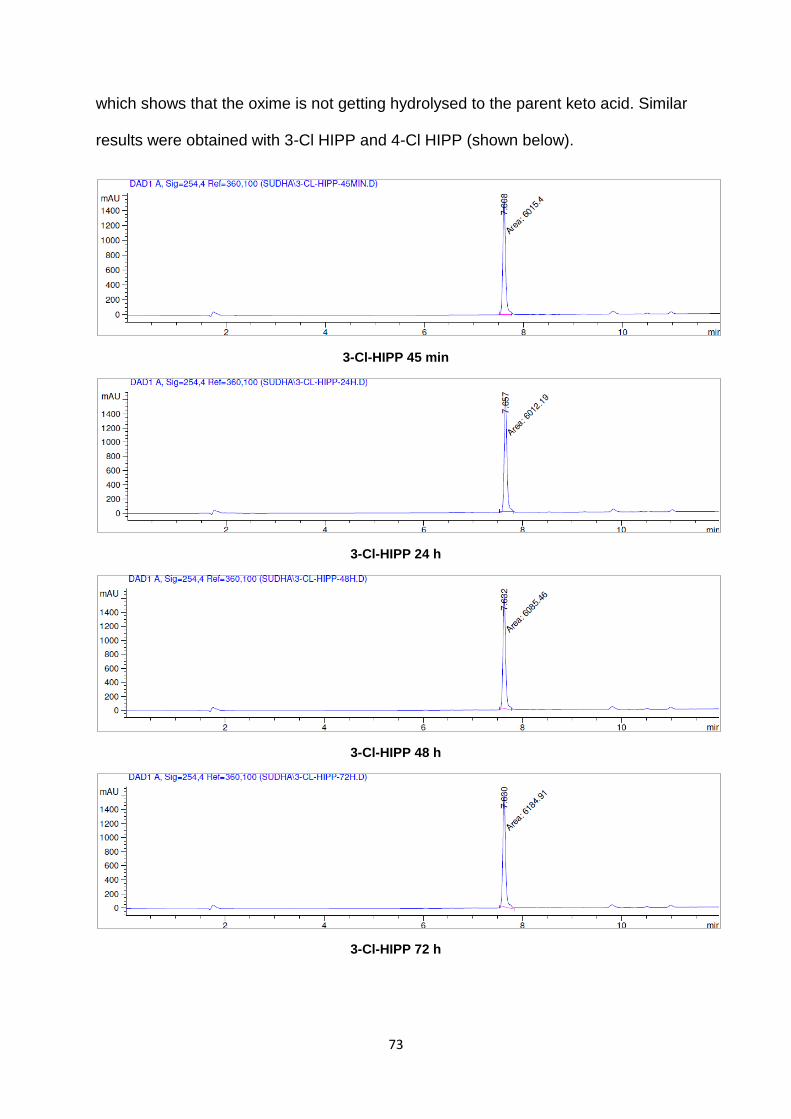
73
which shows that the oxime is not getting hydrolysed to the parent keto acid. Similar
results were obtained with 3-Cl HIPP and 4-Cl HIPP (shown below).
3-Cl-HIPP 45 min
3-Cl-HIPP 24 h
3-Cl-HIPP 48 h
3-Cl-HIPP 72 h

74
4-Cl-HIPP 0 min
4-Cl-HIPP 27 h
4-Cl-HIPP 48 h
4-Cl-HIPP 72 h

75
4-Cl-phenylpyruvic acid
2.7.9. HINT Scoring
Analogues of hydroxyimine 2-14 were modeled with SYBYL-X 2.1 (Tripos Inc.);
Gasteiger–Hückel charges were assigned and models were energy minimized with
the Tripos forcefield (10,000 iterations, termination gradient of 0.01 kcal/mol-Å). The
crystal structure of CtBP1/HIPP/NAD+ was used as the target for docking studies using
GOLD v5.2, with the binding defined as all the residues within 10 Å of the bound ligand
2-14. Since the ligands are analogues of 2-14, it is reasonable to expect that they will
adopt binding modes very similar to that of the parent compound. Thus, the binding
pose of 2-14 defined a scaffold match constraint, i.e., the common substructure forced
the corresponding atoms of ligands to lie in the exact, or very close, position within the
binding site. Default GOLD genetic algorithm parameters were used and a total of 50
solutions per compound were generated. The generated conformations were re-
ranked using the free-energy based HINT force-field, that quantifies all non-bonded
interactions in a biological environment, including hydrogen-bonding, electrostatic
interactions, hydrophobic interactions as well as desolvation energy. The ligand in its
most energetically favorable binding conformation and the CtBP1/NAD+ complex were
subjected to minimization (Powell, 2500 iterations, termination gradient 0.01 kcal/mol-

76
Å), to remove steric clashes and optimize the protein-ligand interactions within the
active site. HINT scores for the thus optimized complex were then recalculated.

77
Investigation of the Scope of Palladium Multi-Walled Carbon Nanotubes (Pd-
MWCNT) Catalyst in C–H Activation Reactions
3. C–H Activation
3.1. Introduction
Organic molecules comprise chains or rings of carbon atoms, capped with hydrogen
atom(s), and with or without heteroatoms (mostly nitrogen, oxygen, halogens, sulfur,
and phosphorous). These form simple hydrocarbons, natural products, proteins,
pharmaceuticals, plastics and agrochemicals. Carbon–hydrogen bonds are present
ubiquitously in nature (Figure 3-1).121
Figure 3-1. Ubiquitous nature of C-H bonds
Above figure from “C–H Bond Functionalization in Complex Organic Synthesis,” Kamil Godula and Dalibor Sames. Science
2006, 312, 67-75. Reprinted with permission from AAAS.

78
Scheme 3-1. Traditional organic synthesis (A) vs C–H functionalization (B)
Organic chemistry involves transformation of functional groups or highly reactive
structural features. C–H bonds are not considered as functional groups generally. In
traditional approaches, the presence of a heteroatom (halogen, oxygen) or
unsaturation is essential for the installation of a new bond, i.e. functional groups are
installed only on pre-functionalized substrates for the purpose of reactivity and to
achieve selectivity (Scheme 3-1).121 This forms the basis of all syntheses. Synthesis
of pre-functionalized substrates itself adds additional steps to the synthetic scheme of
the final product (Scheme 3-2).121 This is inefficient as it adds many steps to the overall
synthetic route.
Scheme 3-2. Comparison of traditional and C-H activation approaches

79
Although this is not an expensive approach on small scale reactions, it is very cost-
ineffective in the case of large scale reactions, especially in industries. Thus, solving
this issue will be very atom-economical and time-efficient.
One of the major challenges in organic chemistry has been the direct conversion of
carbon–hydrogen bonds into carbon–halogen, carbon–sulfur, carbon–nitrogen, and
carbon–carbon bonds using mild and selective methods. These transformations are
very useful as C–H bonds are ubiquitous in nature. Overcoming this challenge would
be of great use in the field of organic chemistry with wide applications in synthesis of
pharmaceutical products, natural products, polymers, and agrochemicals.
Direct transformation of a C–H bond to introduce a new functional group is the key to
overcome the above challenges. For example, the final product in scheme 3-2 can be
obtained by displacing just one hydrogen atom. This direct and selective
functionalization of a C–H bond by an organometallic catalyst is termed “C–H
Activation” (Scheme 3-3).122 This approach is atom- and step-economical, and can be
used to make synthetic connections that are not possible by traditional methods.
The four main challenges in direct C–H functionalization reactions are: reactivity,
chemoselectivity, regioselectivity and stereoselectivity.123
Scheme 3-3. C-H activation
80
Reactivity challenges arises because of the inert nature of most of the carbon-
hydrogen bonds (pKa >30-35, BDE 85-105 kcal/mol). Though many oxidation
reactions are thermodynamically favorable, the cleavage a of C–H bond has a high
kinetic barrier. This challenge has been overcome by many groups of researchers by
the use of transition metals. Transition metals (M) react with C–H bond to give C–M
bonds, which are much more reactive than the corresponding C–H bonds. These C–
M bonds can then be functionalized into various functional groups in a single step.124
Chemoselectivity challenges arises because of the difficulty in stopping reactions at
the required oxidation state, over-oxidized products are generally thermodynamically
favored. This challenge has been overcome by using excess substrate relative to
oxidant, low conversion of reactions.
Regioselectivity challenges arises because organic compounds invariably contain
several C–H bonds in different environments. This challenge has been overcome
through different approaches. The most common approach has been the use of
“directing groups” or “coordinating ligands” that are basic and coordinate to the metal
center, and deliver the catalyst to a proximal C–H bond. The intramolecular directing
groups are basic groups containing nitrogen such as pyridine or oxazole. This “ligand-
directed C–H activation” has been reported at several transition metal centers such as
Pd, Pt, Ru and Rh. Other approaches such as using substrates containing weaker C–
H bonds, having five- or six-membered cyclometalated species, and variation of
ligand-catalyst systems have been employed to achieve selectivity.
Stereoselectivity challenges are involved in the difficult process of generating new
stereocenters in a dia- or enantio- selective manner. This has not been explored as
much as other challenges. Approaches such as use of stereo center containing

81
substrates, or chiral catalyst-ligand systems have been used to overcome this
challenge.
3.1.1. Brief Examples of Use of C-H Activation in Synthesis
3.1.1.1. C–H activation in natural product synthesis
Figure 3-2. C-H activation steps in Teleocidin B-4 core synthesis
Above figure reprinted with permission from “C-C Bond Formation via C-H Bond Activation: Synthesis of the Core of Teleocidin B4.” Brian D. Dangel, Kamil Godula, So Won Youn, Bengü Sezen, and Dalibor Sames. JACS 2002, 124, 11856-11857. Copyright 2002, American Chemical Society.
C–H activation has been used in natural product synthesis. Dangel et al reported the
synthesis of a complex natural product fragment Teleocidin B-4 core by employing
four C–C bond forming steps (alkenylation of alkyl group, Friedel-Crafts reaction,
carbonylation and alkenylation of phenol, Figure 3-2).125
Starting from Schiff base 1, cyclometallation with PdCl2 gives the palladacycle 2, which
undergoes reaction with boronic acid to afford 4. Treatment with MeSO3H catalyzes a
Friedel-Crafts reaction to afford intermediate 5, which undergoes cyclopalladation to

82
afford 7 and 8 by carbonylation and hydrolysis of the Schiff base (Scheme 3-4).
Intermediate 7 is then alkylated to afford intermediate 10, which undergoes Pd-
catalyzed alkenylation to afford the final product Teleocidin B-4 core (Scheme 3-5).125
Scheme 3-4. Synthesis of intermediate 7
Above figure reprinted with permission from “C-C Bond Formation via C-H Bond Activation: Synthesis of the Core of Teleocidin B4.” Brian D. Dangel, Kamil Godula, So Won Youn, Bengü Sezen, and Dalibor Sames. JACS 2002, 124, 11856-11857. Copyright 2002, American Chemical Society.
Scheme 3-5. Synthesis of Teleocidin B-4 core
Conditions: a). t-BuOK, THF; b). BBr3, DCM; c). Pd(OAc)2, P(t-Bu)3, Cs2CO3, DMA.
Above figure reprinted with permission from “C-C Bond Formation via C-H Bond Activation: Synthesis of the Core of Teleocidin B4.” Brian D. Dangel, Kamil Godula, So Won Youn, Bengü Sezen, and Dalibor Sames. JACS 2002, 124, 11856-11857. Copyright 2002, American Chemical Society.

83
3.1.1.2. C–H activation in late-stage diversification
Development of new drugs requires the identification of lead compounds, and design
of analogs with improved activities. Thus, methods to quickly form analogs from a core
structure is highly desirable. In efforts towards this, C–H activation has also been
applied in “structural core diversification” in which various C–H bonds in a complex
molecule are selectively transformed to produce series of analogues from a common
substrate (Scheme 3-6).121 This is highly advantageous over traditional approaches
where multi-step and usually different synthetic routes are needed to arrive at series
of analogs.
Scheme 3-6. Structural core diversification
Late-stage diversification has been used in the synthesis of drug candidates, organic
light-emitting diodes (OLEDs), perylene tetracarboxylic acid bisimide (PBI), metal
organic frameworks (MOFs) and polymers (Figure 3-3).126

84
Figure 3-3. C-H activation in late-stage diversification applications
Above figure reprinted by permission from Macmillan Publishers Ltd: Nature Chemistry, “C-H bond activation enables the rapid
construction and late-stage diversification of functional molecules.” Joanna Wencel-Delord and Frank Glorius. 2013, 5, 369-
375. Copyright 2013.
Yu et al reported the late-stage diversification of celecoxib, an anti-inflammatory drug
(Scheme 3-7).127 Sulfonamide group is an essential part of the pharmacophore for
these compounds, converting it to perfluoroaryl sulfonamide allowed for its use as a
directing group. Using Pd catalysis, ortho-directed C–H activation led to the synthesis
of various ortho-substituted analogues in acceptable yields by olefination,
carboxylation, carbonylation, iodination, arylation and alkylation reactions. This is
advantageous over traditional approaches as it is more time-saving, and can install
groups that could be unfeasible by non C–H activation reactions.
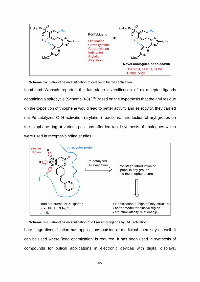
85
Scheme 3-7. Late-stage diversification of celecoxib by C-H activation
Itami and Wunsch reported the late-stage diversification of σ1 receptor ligands
containing a spirocycle (Scheme 3-8).128 Based on the hypothesis that the aryl residue
on the α-position of thiophene would lead to better activity and selectivity, they carried
out Pd-catalyzed C–H activation (arylation) reactions. Introduction of aryl groups on
the thiophene ring at various positions afforded rapid synthesis of analogues which
were used in receptor-binding studies.
Scheme 3-8. Late-stage diversification of σ1 receptor ligands by C-H activation
Late-stage diversification has applications outside of medicinal chemistry as well. It
can be used where ‘lead optimization’ is required. It has been used in synthesis of
compounds for optical applications in electronic devices with digital displays.

86
Triscyclometalllated Ir (III) complexes are useful as they have high emission-quantum
yields and longer lifetimes. Late-stage diversification has been used to synthesize
several analogues of such complexes.
3.2. C–H Activation by Palladium Catalysts
Homogenous transition metal catalysts have been extensively studied in C–H
activation reactions to produce aryl or alkyl halides, alcohols, amines and aryl
compounds. These transformations can occur through two general mechanisms –
“organometallic” and “coordination.”129
In the “organometallic” mechanism, there are two steps (Scheme 3-9). The first step
involves cleavage of a C–H bond to afford a C–M bond (M is a transition metal). The
second step involves functionalization of a C–M bond by an external reagent to afford
the product. The structural and electronic nature of the organometallic intermediate
dictates the regio-selectivity of the functionalization, which usually occurs at the less
hindered C–H bonds in the molecule.
Scheme 3-9. Organometallic mechanism
In the “coordination” mechanism, higher oxidation state metal complexes that contain
activated ligand (X, metal -oxo/imido/carbene) are formed (Scheme 3-10). This is
followed by reaction of X with the C–H bond, either through direct insertion or H-atom
abstraction, to finally afford the product. In this mechanism, the substrate undergoing
C–H activation does not interact directly with the transition metal, but only interacts
with the ligand (X).

87
Scheme 3-10. Coordination mechanism of C–H activation
This dissertation focuses on Pd-catalyzed C–H activation reactions. Palladium has
several advantages over other transition metal catalysts.124
Palladium is much cheaper than Pt and Rh (Figure 3-4). Pd centers can be used to
install a variety of bonds, such as C–O, C–halogen, C–N, C–S and C–C. Only few
other catalysts can catalyse such bond formations. This is because Pd is compatible
with a variety of oxidants. Pd can be utilized to achieve C–H activation not only at sp2
centers, but also at sp3 centers. Pd catalysed C–H activation reactions are not
sensitive to air and moisture; this makes it an attractive catalyst for industrial
applications.

88
Figure 3-4. Cost comparison of transition metals
3.3. Mechanism of C–H activation
3.3.1. N-Chelation-Directed C–H activation
Coordination-directed metalation is one of the approaches for C–H activation
reactions. It can be applied to both sp2 and sp3 carbons. This approach utilizes
heteroatoms such as oxygen or nitrogen (which are in proximity to the C–H to be
activated) to coordinate with the transition metal. This gives rise to a five- or six-
membered metallocycle, which is very reactive and subsequently forms the products.
In this dissertation, we will focus on N-chelation-directed C–H activation. Examples of
selective C−H to C−O,130-136 C−Halogen,130, 137, 138 C−C,139, 140 C−N,141, 142 C−F,143, 144
and C−CF145 transformations using N-chelation-directed palladium catalysis have
been reported in the literature.
Aug Sep Oct Nov Dec Jan Feb 2015 2015 2015 2015 2015 2016 2016
Price

89
The mechanism is explained in detail in the scheme below (Scheme 3-11):
In the first step, Pd(II) catalyst undergoes insertion into the proximal C–H bond to give
a five- or six-membered palladacycle. The selectivity in this “chelation-directed” step
is driven by directing groups such as nitrogen or oxygen that are basic in nature and
direct the catalyst to a specific C–H bond. In the second step, usually an oxidant
containing a functional group oxidizes Pd(II) to Pd(IV).146, 147 This is in contrast to the
traditional coupling reactions that undergo Pd(0)/Pd(II) cycle. In the last step, reductive
elimination causes Pd(II) to be regenerated, and the functional group is transferred to
the activated C–H bond.
Scheme 3-11. Pd(II)/Pd(IV) catalytic cycle for N-chelation-directed C-H activation reactions

90
3.4. Carbon–Oxygen, Carbon–Halogen, Carbon–Carbon Bond Forming
Reactions
In the past two decades, C–H activation has been reported by several groups to make
C–O, C–halogen, C–N, C–S and C–C bonds. This chapter focuses on the
oxygenation, halogenation and arylation reactions catalyzed by palladium catalysts.
This section focuses on homogenous palladium catalysts. Solid-supported and
heterogenous catalysts will be discussed in section 3.5.
3.4.1. Carbon–Oxygen Bond Forming Reactions
Pd-catalyzed ortho-oxygenation reactions use various oxidants. Reactions that utilize
Pd(OAc)2 catalyst and PhI(OAc)2 have been reported. In these reactions, nitrogen
containing groups such as pyridine,130, 148 pyrazole,130 isoxazoline,148 imine,130 oxime
ether149 function as directing groups to direct the acetoxylation to the ortho- position
(Scheme 3-12). Ketones and aldehydes are poor directing groups, and hence do not
undergo acetoxylation under these conditions.
Scheme 3-12. Palladium-catalyzed N-chelation-directed oxygenation reactions
The acetoxylation reactions are carried out under ambient air and moisture conditions.
Many functional groups in the molecules are well tolerated such as: aryl halides, nitro

91
groups, ketones, oximes. These features make Pd-catalyzed oxygenation reactions
very attractive.
When a substituent is present at the meta- position, the less hindered ortho- position
undergoes acetoxylation.
Scheme 3-13. Effect of solvent on oxygeantion reactions
Switching the solvent leads to the installation of various alkoxy groups in the
substrates. For example, MeOH solvent installs -OMe and EtOH solvent installs -OEt
(Scheme 3-13).130 PhI(OAc)2 might be forming PhI(OR)2 in situ.150 The following
mechanism has been proposed for C-H acetoxylation (Scheme 3-14).
Scheme 3-14. Mechanism of C-H acetoxylation

92
Other oxidants that have been used in these reactions are oxone, IOAc,151 and
dioxygen (Scheme 3-15).132
Scheme 3-15. C-H acetoxylation by IOAc
IOAc is generated in situ by reacting I2 with PhI(OAc)2 or AgOAc. The mechanism at
work when IOAc is utilised is: chelation-directed C–H activation, oxidation of PdII to
PdIV, reductive elimination forming C–I bond, displacement of I- by OAc-.
3.4.2. Carbon–Halogen Bond Forming Reactions
Pd catalyzed C–H activation is an efficient method to install halogens such as Cl, Br
and I. This was first reported in 1970, using Pd for the chlorination of azobenzene
(Scheme 3-16).152 Although this reaction used Cl2, and gave rise to a mixture of
isomers, there have been a lot of improvements since then in the use of more practical
oxidants and improvement of selectivity.
Scheme 3-16. Pd-catalyzed chlorination of azobenzene

93
Scheme 3-17. Halogenation of benzo[h]quinoline
Halogenation reactions catalyzed by Pd(OAc)2 using chlorinating agent N-
chlorosuccinimide (which also acts as an oxidant) gives ortho- chlorinated
benzo[h]quinoline product in excellent yields. In a similar reaction, using NBS gives
the brominated product (Scheme 3-17).130, 138
On the other hand, the more reactive PhICl2 gives the 5-chlorinated product. This
reaction occurs without N-chelation in the presence or absence of Pd catalyst
(Scheme 3-17). Thus, Pd catalysts can be used to afford products that are
complementary to the products obtained by electrophilic substitution reactions. Below
are some more examples. In the presence of Pd, N-chelation-directed ortho-
halogenation occurs. In the absence of Pd, electrophilic aromatic substitution occurs
(Scheme 3-18).138

94
Scheme 3-18. Pd-catalyzed N-chelation-directed and non-catalyzed halogenation reactions
The halogenation reactions using N-halosuccinimides and Pd catalysts has been
applied to a wide variety of substrates with basic directing groups such as pyridines,
oxime ethers, isoxazolines, isoquinolines (Scheme 3-19).137, 138 Using 2.5 equivalents
of N-halosuccinimide gives the di-ortho-halogenated product. In the presence of a
meta- substituent, the less hindered ortho- position is halogenated.
Scheme 3-19. Pd-catalyzed N-chelation-directed halogenation reactions

95
Mechanism of halogenation: There are two proposed models of halogenation.
Pd(II)/Pd(IV) cycle (Scheme 3-20). NCS is a strong oxidant that oxidizes Pd(II) to
Pd(IV).147
Scheme 3-20. Mechanism of halogenation by Pd catalyst – Pd(II)/Pd(IV) cycle
Or, Pd(III)-Pd(III) dimer cycle which occurs in the presence of PhICl2 (Scheme 3-21).153
Scheme 3-21. Mechanism of halogenation by Pd catalyst – Pd(III)/Pd(III) cycle
Other halogenating agents that have been used in C-H activation reactions are CuCl2,
CuBr2 (Scheme 3-22).137
Scheme 3-22. Pd-catalyzed halogenation reaction by CuCl2

96
Scheme 3-23. Pd-catalyzed halogenation by Suarez-type reagent
Yu et al have reported the use of Suarez-type reagents (XOAc) for halogenation of sp2
and sp3 C-H bonds (Scheme 3-23).154
Carbon-fluorine bond formation reactions have been carried out using Pd catalysts
and electrophilic fluorinating reagents (Scheme 3-24).143 This is a very important
transformation as it can be applied to PET imaging using radioactive fluorine. Instead
of re-developing synthetic routes to incorporate radioactive fluorine, it can be easily
installed through C-H activation reactions such as below in the final stages of the
synthetic routes. This would be a time- and cost-efficient process.
Scheme 3-24. Pd-catalyzed fluorination reactions

97
3.4.3. Carbon–Carbon Bond Forming Reactions
C–C bond formation is one of the most important reactions in organic chemistry.
Traditional carbon–carbon bond forming reactions include coupling reactions such as
Heck, Suzuki and Stille coupling reactions that utilize the Pd(0)/Pd(II) cycle, which
have led to an immense progress in the synthesis of organic molecules. But there is
still a need for improvement in the atom-economy, and cost-efficient processes. One
major disadvantage of these coupling reactions is that all of them require two pre-
functionalized substrates. This adds additional steps in the synthesis of the final
product. In efforts to overcome these disadvantages, C–H activation chemistry has
been developed as an alternative, in which carbon–carbon bond formation is achieved
using one or zero pre-functionalized substrates.
The general reaction conditions for coupling reactions include homogenous Pd
catalyst, base, ligand, and a solvent. Because the C–H bonds are inert (high
dissociation energy), usually high temperatures (>100 oC) and Pd catalyst loading (5-
20 mol%) are required for the reactions to take place. In heterocyclic structures where
the C–H bond is activated due to the presence of a heteroatom, the coupling reactions
can be run at lower temperatures155, 156 and catalyst loading.157, 158 Pd(OAc)2 has been
shown to catalyze arylation of substrates using diphenyliodinium salts as the arylating
agents. This reaction is driven by N-chelation of Pd. Aryl pyridines, quinolines,
oxazolidinones can undergo arylation reactions as they possess basic nitrogen
chelating group which directs arylation to the ortho- position (Scheme 3-25).139, 159-161
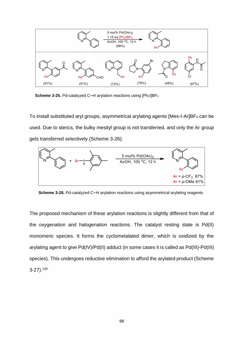
98
Scheme 3-25. Pd-catalyzed C–H arylation reactions using [Ph2I]BF4
To install substituted aryl groups, asymmetrical arylating agents [Mes-I-Ar]BF4 can be
used. Due to sterics, the bulky mesityl group is not transferred, and only the Ar group
gets transferred selectively (Scheme 3-26).
Scheme 3-26. Pd-catalyzed C–H arylation reactions using asymmetrical arylating reagents
The proposed mechanism of these arylation reactions is slightly different from that of
the oxygenation and halogenation reactions. The catalyst resting state is Pd(II)
monomeric species. It forms the cyclometalated dimer, which is oxidized by the
arylating agent to give Pd(IV)/Pd(II) adduct (in some cases it is called as Pd(III)-Pd(III)
species). This undergoes reductive elimination to afford the arylated product (Scheme
3-27).140

99
Scheme 3-27. Mechanism of Pd-catalyzed C–H arylation
Another example of sp2 C–H arylation that utilizes Pd(OAc)2 and AgOAc has been
reported. Amide162 and oxime ether163 containing substrates undergo arylation with
aryl iodides to afford the coupled product fluorenones (Scheme 3-28). These reactions
are tolerant to various functional groups.
Scheme 3-28. Fluorenone synthesis by C–H arylation

100
Scheme 3-29. Pd-catalyzed arylation reaction of aminoquinoline
Arylation of unactivated sp3 C–H bonds has also been achieved under similar
conditions. Pyridines,164 aminoquinolines165 function as the directing groups (Scheme
3-29).
The above reactions that utilize Ag have been proposed to go through Pd(II)/Pd(IV)
cycle. Ag salt is necessary for regenerating the Pd catalyst. Mechanistic studies have
been conducted on benzoic acids (Scheme 3-30).166
Scheme 3-30. Mechanism of Pd-catalyzed arylation with AgOAc
C–H Arylation without Prefunctionalized Arylating Reagents:
Reactions involving Pd-catalyzed ligand-directed arylations have been reported which
do not require the presence of prefunctionalized substrates. As an example, the
dimerization of arylpyridine in the presence of Pd(OAc)2 using oxone as oxidant
(Scheme 3-31).167 Meta substituents on the aryl ring resulted in low site selectivity of
arylation, in contrast to C–H arylation at Pd(II) centers.

101
Scheme 3-31. Dimerization of arylpyridine
So, the following pathway has been proposed for this type of arylation reactions.167
Cyclometalation at Pd(II) followed by oxidation of Pd(II) to Pd(IV) by oxone,
cyclometalation at Pd(IV), and reductive elimination to give the arylated product and
release of Pd(II) (Scheme 3-32).
Scheme 3-32. Pd-catalyzed C–H arylation mechanism with oxone
More recently, a general N-chelation directed Pd(OAc)2 catalyzed oxidative cross
coupling reaction on benzo[h]quinoline has been reported. This reaction uses Ag2CO3

102
as the oxidant, benzoquinone as the promoter, DMSO to protect the catalyst.168 This
reaction was shown to have a wide scope (Scheme 3-33).
Scheme 3-33. Oxidative cross coupling reaction on benzo[h]quinoline
These reactions are proposed to occur through the following mechanism:
cyclopalladation, Ar-H activation, reductive elimination to afford the arylated product,
oxidation of Pd(0) to Pd(II). Benzoquinone helps in the reductive elimination step
(Scheme 3-34).168-170
Scheme 3-34. Mechanism of oxidative cross coupling reaction on benzo[h]quinoline

103
3.5. Pd-MWCNT Catalyst
Homogenous catalysts such as palladium acetate show high conversion of substrates
to products because of easy access to all catalytic sites, but it is very difficult to remove
the catalyst from the reaction mixture due to its high solubility. Thus, commercialization
of homogenous catalyzed reactions has been rare.171 This has led to the development
of heterogeneous and solid-supported catalysts in which the catalyst is in a different
physical state from the solvent, and hence can be easily removed from the reaction
mixture.172 This makes the process of purification less tedious. Such catalysts can also
be reused/recycled, which makes the process very economical.
In heterogeneous catalysis, reactants are adsorbed on the solid-supported catalyst
surface, undergo reaction catalyzed by transition metal catalyst such as palladium,
products get desorbed from the catalyst surface, and are extracted from the reaction
mixture. Another mechanism of solid-supported catalysis is the “leaching-
redeposition” mechanism in which solid-supported Pd(0) nanocatalyst is oxidized to
Pd(II) and leaches from the support, catalyzes the conversion of substrate to product
in the reaction mixture, undergoes reduction to Pd(0) and is redeposited on the
support. This can be tested by two experiments – hot filtration and three-phase test.173
In the hot filtration test, a portion of the reaction mixture is filtered midway through the
reaction, and fresh reagents are introduced. Further conversion indicates presence of
soluble Pd species. Absence of conversion indicates absence of soluble Pd species,
but it could also mean that there is a fast leaching-redeposition of the Pd species,
which cannot be measured. Three-phase test174 is more reliable as a test for
heterogeneity – one substrate is immobilized on a solid-support, and the reaction is

104
carried out using a solid-supported catalyst and a soluble substrate. Product formation
indicates soluble Pd species catalyzing the reaction.
New supports and catalysts are being continuously developed. Several materials such
as polymers,175 dendrimers,176 metal oxides177 and carbonaceous materials178 have
been used as solid supports for Pd and other transition metal catalysts. Ideal
properties for the solid supports are – high surface area, chemical inertness, thermal
and mechanical stability.
Palladium metal has been shown to have a good catalytic activity in the form of
nanoparticles. Optimization of shape and size of these nanoparticles is essential for
improving catalytic activity.179 Metal-decorated carbon nanotubes (CNTs) – single-
walled and multi-walled (Figure 3-5)180 – are increasingly being used for many
applications such as optics, electronics and catalysis. They have high chemical
stability and surface area which makes them very efficient as catalysts. Graphene is
a 2D mono-layered structure of graphite packed into a honeycomb-like lattice. It has
excellent properties of thermal and mechanical stability. Reduction of graphene oxide
by hydrazine was an early method to synthesize graphene sheets.181 3D derivatives
of graphene are single walled carbon nanotubes (SWCNT) and multi-walled carbon
nanotubes (MWCNT). These have been shown to have excellent catalytic activity.
SWCNT consists of a single layer of graphene wrapped into a cylindrical column. The
synthesis of SWCNT is very difficult, and is achieved by chemical vapor deposition.
MWCNT have multiple sheets of graphene wrapped up into cylinders of varying
diameters. These can be prepared by chemical vapor deposition as well. MWCNT
have better stability compared to SWCNT owing to their multiple layers.

105
Figure 3-5. Single-walled and multi-walled carbon nanotubes
The above figure is from “Nanostructures: a platform for brain repair and augmentation,” Ruxandra Vidu, Masoud Rahman,
Morteza Mahmoudi, Marius Enachescu, Teodor D. Poteca and Ioan Opris. Front. Syst. Neurosci. 2014, 8, 91, 1-24.
Scheuermann et al have reported the synthesis of graphene supported palladium
nanoparticles.182 The synthesis of carbon nanotubes is complex and expensive, is
achieved through lasers, vapor deposition and other techniques.
Palladium is deposited on the nanotubes by the solvent-free methods of mortar-pestle
or ball-milling, or with a solvent-based method. A schematic representation of of Pd
decorated on MWCNT is shown in Figure 3-6.183

106
Figure 3-6. Pd decorated on MWCNT
The Gupton lab has provided us with Pd-MWCNT catalyst for carrying out C–H
activation reactions. Multi-walled nanotubes have an outer diameter of 6-9 mm, length
5 µm and 95% carbon. In the mortar-pestle method, the carbon nanotubes are mixed
with palladium acetate and ground using mortar and pestle until homogenous mixture
of Pd-MWCNT is obtained. In the ball-mill method, carbon nanotubes and palladium
acetate along with zirconia balls are placed in a ball mill and shaken mechanically
(back-and-forth 5.9 cm, side-to-side 2.5 cm, 115 volts, 1060 cycles/min) to afford Pd-
MWCNT.
The major advantages of solid-supported catalysts over that of homogenous catalysts
are: recyclability, less/no contamination of products by residual metal, and ease of
filtration and removal of catalyst from the reaction mixture.
The first arylation catalyzed by heterogenous Pd was reported in 1982 by Nakamura
et al.184 Arylation on muscimol was achieved using Pd/C catalyst on substituted
isoxazoles (Scheme 3-35).
Reactant
Product
Outer wall of
MWCNT
Pd

107
Scheme 3-35. Arylation of isoxazole catalyzed by Pd on carbon
Fagnou et al used Pd(OH)2 supported on activated charcoal (Pearlman’s catalyst) for
the arylations (intra- and inter-molecular) of arenes (Scheme 3-36).185
Scheme 3-36. Arylation of arenes using Pearlman's catalyst
There have been reports that have used Pd-CNT (carbon nanotubes) to achieve
Suzuki-Miyaura cross-coupling reaction. Suzuki reaction is a very powerful method to
synthesize biaryl compounds. Usually Suzuki reactions require the presence of a
ligand or an additive to prevent the aggregation of Pd0 during the course of the
reaction.
Zhang et al reported a ligandless and additive-free Suzuki reaction using Pd-CNT as
the catalyst (Scheme 3-37).186 Excellent yields were achieved with various aryl
bromides (both electron withdrawing and electron donating).

108
Scheme 3-37. Arylation of aryl bromides catalyzed by Pd-CNT
Siamaki et al reported the use of solid-supported Pd(0) on carbon nanotubes (Pd-
MWCNT 1-3 nm) as a highly efficient catalyst in the Suzuki cross-coupling reaction
(Scheme 3-38).187 Coupling reactions using these solid-supported nanoparticle
catalysts displayed remarkable catalytic activity with a high turnover number (TON
7250) and turnover frequency (TOF 217500 h-1). The reactions generally were
reported to be complete within 10 min, run at lower temperature (∼80 °C) using either
conventional or microwave heating, and run in either batch or flow format. They were
able to control the Pd(0)/Pd(II) ratio based on the method of preparation and the type
of carbon nanotube used as the solid support.
Scheme 3-38. Suzuki cross-coupling catalyzed by Pd-MWCNT
Tang et al reported the arylation of thiophenes and benzo[b]thiophenes using Pd/C as
the catalyst under ligand-free and additive-free reaction conditions (Scheme 3-39).188
This reaction was proved to be heterogenous in nature.

109
Scheme 3-39. Arylation of thiophenes and benzothiophenes catalyzed by Pd on carbon
Zhang et al reported ortho-directed C–C coupling reaction between 2-aryl pyridines
and aldehydes catalyzed by palladium nanoparticles via Pd0/II/IV cycle.189 Aluminum
oxide was used as support for palladium, and oxidant used was TBPB (Scheme 3-40).
Scheme 3-40. Arylation reactions catalyzed by Pd on aluminium oxide
3.6. Scope of this dissertation
The second project in my dissertation focuses on the C−H to C−OMe/OAc, C−Cl, C−Br
and C−C functionalizations using solid-supported Pd(II) multi-walled carbon nanotube
catalyst. The turn-over frequencies of solid-supported catalyst were higher than the
reported homogenous catalyst.

110
4. C-H Halogenation and Alkoxylation reactions catalyzed by Pd(II)-MWCNT
Catalyst
We wished to test the hypothesis that a solid-supported nanoparticle catalyst
containing predominantly Pd(II) can be used to catalyze oxidative N-chelation-directed
C−H activation reactions that undergo the Pd(II)/Pd(IV) catalytic cycle. As the catalyst,
we have prepared nanoparticles containing predominantly Pd(II) supported on multi-
walled carbon nanotubes (Pd(II)/MWCNT) by a modification of our previous
procedure. We have evaluated the ability of this catalyst to carry out oxidative C−H to
C−OMe/OAc, C−Cl, and C−Br functionalizations.
4.1. Results and Discussion
We initially chose substrates that have been previously reported to undergo the C−H
functionalization reactions so that we could compare the results of our solid-supported
Pd(II)/MWCNT catalyst to that of the known homogeneous Pd(II) system (Pd(OAc)2).
We first explored the C−H to C−O transformation, which utilizes PhI(OAc)2 as the
oxidant (Table 4-1).
Treatment of 8-methylquinoline with Pd(II)/ MWCNT and PhI(OAc)2 in acetic acid at
120 °C for 10 min (Table 4-1, entry 1) afforded the desired 8-(acetoxymethyl)-quinoline
in 90% yield, comparable to the previously reported yield of 88% with the
homogeneous catalyst. However, treatment of benzo[h]quinoline (entry 2) and 2-
phenylpyridine (entry 3) failed to give any of the desired acylation products, even at
extended reaction times and higher temperatures. Only starting material was
recovered in these reactions. Having confirmed that solid-supported Pd(II)/MWCNT
can catalyze a reaction that undergoes the Pd(II)/Pd(IV) catalytic cycle and

111
encouraged by the initial success of the acylation reaction with 8-methylquinoline, we
elected to evaluate whether other oxygen containing functional groups could be
installed using our catalyst. We repeated the reaction of 8-methylquinoline with
Pd(II)/MWCNT and PhI(OAc)2, except this time using methanol as a solvent (Table 4-
1, entry 4). We obtained a near-quantitative yield (99%) of the desired 8-
(methoxymethyl)quinoline in 10 min at 100 °C, which is a significant improvement over
the reported yield of 77% with the homogeneous catalyst system. Application of these
conditions to benzo[h]quinoline (entry 5) afforded a 90% yield of product, again
comparable to the reported yield with the homogeneous catalyst (95%). Finally,
treatment of 2-phenylpyridine with Pd(II)/MWCNT/PhI(OAc)2/MeOH (entry 6) did result
in formation of the product after an extended reaction time, albeit in much lower yield
(25%).

112
Table 4-1. C-H to C-O functionalizations catalyzed by Pd(II)-MWCNT
Solid-Supported Pd(II)/MWCNT
Yield and time with
Pd(OAc)2b
Entrya Product Temp Time Yield
1
120 °C 0.2 h 90% 88%,
22 h
2
120 °C 1 h 0% 86%,
12 h
3
120 °C 1 h 0% 52%,
12 h
4
100 °C 0.2 h 99% 77%,
18 h
5
100 °C 0.2 h 90% 95%,
22 h
6
100 °C 5 h 25% −c
a. Solvent for entries 1-3: AcOH; entries 4-6: MeOH. b. Previous reported results with homogeneous Pd(OAc)2; c. Yield with homogeneous Pd catalyst has not been reported.
We next sought to determine whether alcohols other than methanol could be installed
in substrates using the solid supported catalyst. Treatment of benzo[h]quinoline with
Pd(II)/MWCNT and PhI(OAc)2 in alcohol solvents (Table 4-2) demonstrated that
methoxy (from methanol, entry 1) and ethoxy (from ethanol, entry 2) functional groups
can be installed in moderate to high yields with our catalyst. However, as the size of
the alcohol increased, incorporation of the functional group into the product decreased

113
(with isopropyl, entry 3) and eventually failed (in the tert-butyl case, entry 4). Previously
reported data for the homogeneous catalyst system show only a moderate decrease
in yield as the size of the alcohol increases.
Table 4-2. Steric Trend in C-H to C-O Alkyl Functionalizations Catalyzed by Pd(II)/MWCNT
Entry ROH Product Yield
Pd-MWCNT Pd(OAc)2
1 MeOH
90% 95%
2 EtOH
73% 80%
3 iPrOH
33% 72%
4 tBuOH
0% −a
a. Yield with homogeneous Pd catalyst has not been reported.
Having demonstrated that solid-supported Pd(II)/MWCNT can catalyze the C−H to
C−O functionalization reaction, we next turned our attention to halogenation reactions
(Table 4-3). Treatment of benzo[h]quinoline with Pd(II)/MWCNT and N-
chlorosuccinimide (NCS) in acetonitrile at 100 °C for 5 h (Table 4-3, entry 1) afforded
the desired 10-chlorobenzo[h]quinoline in 92% yield, comparable to the previously

114
reported yield of 95% with the homogeneous catalyst. It should be noted that the
reaction with the Pd(II)/MWCNT is much faster than with homogeneous Pd(OAc)2 (5
h vs 3 days).
Chlorination of (E)-3,4-dihydronaphthalen-1-(2H)-one O-methyloxime (entry 2), 2-
phenylpyridine (entry 3), and 3-methyl-2-phenylpyridine (entry 4) in either acetic acid
(entries 2 and 4) or acetonitrile (entry 3) all afforded the desired chlorinated products
in moderate yields in reaction times from 10 min to 6 h. While the yields for these
transformations are lower than those for the comparable reaction with the
homogeneous catalyst, the reaction times are much shorter, pointing to faster reaction
kinetics (vide infra). The 2-phenylpyridine substrates (entries 3 and 4) required
additional catalyst loading to reach this level of conversion and product isolation. For
all of these substrates, further increasing the number of equivalents of NCS led to the
formation of other products.
Turning to the C−H to C−Br functionalizations, treatment of benzo[h]quinoline with
Pd(II)/MWCNT and N-bromosuccinimide (NBS) in acetonitrile at 100 °C for 1.5 h
(Table 4-3, entry 5) afforded the desired 10-bromobenzo[h]quinoline in 89% yield,
comparable to the previously reported yield of 93% with the homogeneous catalyst.
Again, it should be noted that the reaction with the Pd(II)/MWCNT is much faster than
that with homogeneous Pd(OAc)2 (1.5 h vs 1.5 days). Bromination of (E)-3,4-
dihydronaphthalen-1-(2H)-one O-methyloxime (entry 6), 2-phenylpyridine (entry 7),
and 3-methyl-2-phenylpyridine (entry 8) in either acetic acid (entries 6 and 8) or
acetonitrile (entry 7) all afforded the desired brominated products in moderate yields
in reaction times of 5−6 h. Again, the yields are lower than the comparable reaction
with homogeneous catalyst but the reaction times are much shorter, pointing to faster
reaction kinetics (vide infra).
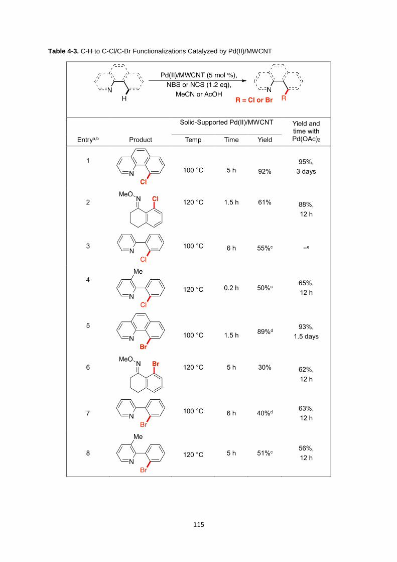
115
Table 4-3. C-H to C-Cl/C-Br Functionalizations Catalyzed by Pd(II)/MWCNT
Solid-Supported Pd(II)/MWCNT Yield and time with Pd(OAc)2 Entrya,b Product Temp Time Yield
1
100 °C
5 h
92%
95%,
3 days
2
120 °C
1.5 h
61% 88%,
12 h
3
100 °C 6 h 55%c −e
4
120 °C 0.2 h 50%c 65%,
12 h
5
100 °C
1.5 h 89%d
93%,
1.5 days
6
120 °C
5 h
30% 62%,
12 h
7
100 °C 6 h 40%d 63%,
12 h
8
120 °C 5 h 51%c 56%,
12 h

116
a. Oxidants for entries 1-4: NCS; entries 5-8: NBS. b. Solvents for entries 1, 3, 5, and 7: MeCN; entries 2, 4, 6, and 8: AcOH. c. 10 mol% Pd(II)/MWCNT was used. d. 1.5 eq of NBS was used. e. A directly analogous yield for the homogeneous catalyst has not been reported.
Both the benzo[h]quinoline and 2-phenylpyridine substrates (entries 5 and 7) required
additional NBS (1.5 equiv vs 1.2 equiv) to reach this level of conversion and product
isolation. Increasing the amount of NBS beyond 1.5 equiv led to the formation of other
products. For the 3-methyl-2-phenylpyridine (entry 8), additional catalyst loading was
required to reach this level of conversion and product isolation.
To characterize the solid-supported Pd(II)/MWCNT catalyst, we measured the
composition of the palladium both before and after C−H activation reaction by X-ray
photon spectroscopy (XPS, Figure 4-1). Prior to use in the C−H activation reactions,
the solid-supported catalyst was found to be a mixture of Pd(II) (76.54%) and Pd(0)
(23.46%), with Pd(II) being the predominant component. After the C−H to C−OMe
functionalization of benzo[h]quinoline (Table 4-1, entry 5), XPS showed that the solid-
supported catalyst contained only Pd(II), showing that all of the Pd(0) in the catalyst
prior to the reaction is converted to Pd(II), and supporting the mechanism that these
reactions use Pd(II) as the starting oxidation state.

117
Figure 4-1. Pd(II) and Pd(0) content in Pd(II)/MWCNT (a.) before and (b.) after a C-H Activation
Reaction as measured by XPS
To demonstrate the ability of the Pd(II)/MWCNT catalyst to be recycled, we ran the
C−H to C−OMe functionalization on 8-methylquinoline (Table 4-1, entry 4), recovered
the catalyst by centrifugation, and iteratively repeated the reaction with the same batch
of catalyst. We were able to recycle the catalyst a remarkable 16 times with minimal
reduction in yield and no catalyst deactivation (Table 4-4). We terminated the
experiment after 16 recycles and have yet to determine the limits of the recyclability of
the catalyst. These data demonstrate that there must be good retention or highly
efficient leaching/redeposition of Pd in these reactions. Otherwise, leaching would
result in loss of catalytic activity after multiple recycles.

118
Table 4-4. Recycling experiments with Pd(II)/MWCNT
Run Conversion (%)a
1 98 2 98 3 92 4 90 5 90 6 90 7 90 8 90 9 89 10 90 11 90 12 90 13 90 14 90 15 90 16 90 17 90
a. Conversion was determined by GC/MS based upon the consumption of the starting material
For all of the C−H activation reactions, we observed that the reactions catalyzed by
Pd(II)/MWCNT seemed to be faster than those with the homogeneous Pd(OAc)2. To
quantify this observation, we calculated turnover frequencies (TOFs) for one example
reaction of each functionalization with both the solid-supported Pd(II)/ MWCNT and
homogeneous palladium catalysts (Table 4-5). For the C−H to C−OAc and C−OMe
functionalization reactions, the turnover frequencies for the solid-supported
Pd(II)/MWCNT catalyst were ∼27-fold higher than that for the homogeneous catalyst.
For the C−H to C−Cl and C−Br reactions, the turnover frequencies for the solid-
supported Pd(II)/MWCNT catalyst were ∼4-fold higher. This represents a significant

119
improvement over previously reported results, particularly with regard to the C−H to
C−OMe functionalization reactions.
Table 4-5. Comparison of Turn Over Frequencies in C-H to C-OAc, C-OMe, C-Cl, and C-Br Reactions
Catalyzed by Solid-Supported Pd(II)/MWCNT and Homogeneous Pd(OAc)2
Turn Over Frequency (h-1)
Entry Product Pd(II)/MWCNT Pd(OAc)2 Fold-increase
1
106.44 4.02 26.48
2
106.94 3.79 28.22
3
3.60 0.73 4.90
4
9.96 2.63 3.75
To determine whether the palladium metal from the solid supported Pd(II)/MWCNT
catalyst leached into the C−H activation reactions and contaminated the products, we
removed the catalyst by filtration over Celite from a C−H to C−OMe functionalization
reaction on benzo[h]quinolone (Table 4-1, entry 5) and measured the palladium
content in solution by ICP-MS. The palladium content of the reaction mixture was
found to be <250 ppb, demonstrating that very little metal leached into the reaction
medium. Combined with the ease of removing the catalyst from the reaction mixtures,

120
this low level of palladium in the reaction mixture is an improvement on the existing
homogeneous catalyst for N-chelation-directed C−H activation reactions.
To demonstrate that the trace palladium in the reaction mixture is not the source of
catalytic activity, a hot filtration experiment was performed with benzo[h]quinoline
(Table 4-1, entry 5). After 10 min at 100 °C, the Pd(II)/MWCNT catalyst was removed
by hot filtration over Celite. Fresh substrate and oxidant were added to the filtrate,
which was reheated to 100°C. No further conversion to product or catalytic activity was
observed in the filtrate in the absence of Pd(II)/MWCNT, showing that the <250 ppb of
residual Pd that remains in solution is not adequate to catalyze the C−H activation
reaction.
In conclusion, we have demonstrated that solid-supported Pd(II)/MWCNT can
catalyze N-chelation-directed C−H to C−OAc, C−OMe, C−Cl, and C−Br activation
reactions. For all of the C−H activation reactions examined, the solid supported
catalyst demonstrated consistently higher turnover frequencies than the reported
homogeneous catalyst. The solid supported Pd(II)/MWCNT also offers the advantages
of ease of removal by filtration and low levels of residual palladium metal
contamination in the products. We are currently working to optimize the reactions
reported here as well as apply this catalyst to other N-chelation-directed C−H
activation reactions.

121
Challenges faced in methoxylation reaction:
In order to apply the methoxylation reaction in pharmaceutically relevant compounds,
we tried the methoxylation reaction on Imatinib, which is a tyrosine-kinase inhibitor
used in the treatment of cancers (chronic myeloid leukaemia). Unfortunately, this
reaction gave a mixture of products (seen as a single spot on TLC 5% MeOH-DCM)
which could not be separated (Scheme 4-1). The presence of multiple nitrogen atoms
(directing groups) might have resulted in the methoxylation at various positions. This
reaction needs to be further optimized to give a single product.
Scheme 4-1. Methoxylation on Imatinib catalyzed by Pd-MWCNT
Challenges faced in fluorination and trifluoromethylation reactions:
First, we tried the fluorination reaction on 8-methylquinoline using the electrophilic
fluorinating agent 1-Fluoro-2,4,6-trimethylpyridinium tetrafluoroborate (Scheme 4-2).
The reaction did not yield any product even after various optimization attempts. This
was not very surprising as fluorinating reactions are usually very difficult. The reported
fluorination reactions using homogenous Pd catalysts use microwave heating to get
the reactions to work. This shows that these reactions are thermodynamically difficult
to achieve.
The next step is to try using nucleophilic fluorinating agents.
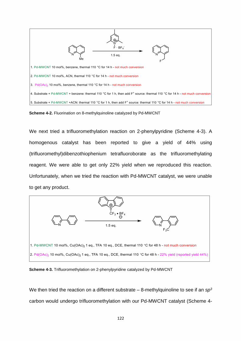
122
Scheme 4-2. Fluorination on 8-methylquinoline catalyzed by Pd-MWCNT
We next tried a trifluoromethylation reaction on 2-phenylpyridine (Scheme 4-3). A
homogenous catalyst has been reported to give a yield of 44% using
(trifluoromethyl)dibenzothiophenium tetrafluoroborate as the trifluoromethylating
reagent. We were able to get only 22% yield when we reproduced this reaction.
Unfortunately, when we tried the reaction with Pd-MWCNT catalyst, we were unable
to get any product.
Scheme 4-3. Trifluoromethylation on 2-phenylpyridine catalyzed by Pd-MWCNT
We then tried the reaction on a different substrate – 8-methylquinoline to see if an sp3
carbon would undergo trifluoromethylation with our Pd-MWCNT catalyst (Scheme 4-

123
4). This reaction did not afford any product. As this reaction utilizes Cu(OAc)2, we also
tried Pd-Cu nanocatalyst to see if the combined nanocatalyst would catalyze the
reaction. Unfortunately, we did not observe any formation of product.
Scheme 4-4. Trifluoromethylation on 8-methylquinoline catalyzed by Pd-MWCNT
These fluorination and trifluoromethylation reactions need to be further optimized in
order to make them work. At present, the Pd-MWCNT catalyst does not appear to be
a good catalyst for these reactions. Microwave heating of reactions and/or use of other
solid-supported catalyst systems would be the next step in this direction.
4.2. Experimental Procedures
General Chemical Methods
All the reactions were carried out in vials sealed with Teflon lined caps under ambient
atmosphere. Palladium (II) acetate (98% reagent grade) and multi-walled carbon
nanotubes (cat. #: 724769) were purchased from Sigma-Aldrich. Benzo[h]quinoline
and 3-methyl-2-phenylpyridine were purchased from TCI, 2-phenylpyridine was
purchased from Chem-Impex. All solvents were purchased from VWR and other
chemicals were purchased from Sigma-Aldrich and all were used without distillation or
purification. Analytical Thin Layer Chromatography (TLC) was performed using silica
gel GHLF plates (Analtech Inc., DE, USA). Flash chromatography was performed on
TELEDYNE ISCO CombiFlash® Rf instrument using RediSep Rf Normal-phase Flash

124
Columns (4-gm, 12-gm, 24-gm or 40-gm). NMR spectra were recorded on a Bruker
400 MHz instrument operating at 400 MHz for 1H and 125 MHz for 13C acquisitions.
Electrospray ionization (ESI) mass spectra were obtained from Perkin Elmer Flexar
UPLC/AxION2 TOF Mass Spectrometer. The X-ray photoelectron spectroscopy (XPS)
analysis was performed on a Thermo Fisher Scientific ESCALAB 250 using a
monochromatic Al KR X-ray. The Pd content in the Pd nanoparticles supported on
carbon nanotubes before and after reaction was determined using an Inductively
Coupled Plasma equipped with Mass Spectrometry (ICP-MS, Varian 820-MS).
Synthesis of Pd(II) nanoparticles on Multi-Walled Carbon Nanotubes Catalyst
[Pd(II)/MWCNT].
Palladium (II) acetate (0.103 g, 4.6 mmol) and multi-walled carbon nanotubes (0.500
g) were loaded in a 45 ml zirconia grinding vial. Two 12.77 mm diameter zirconia balls
are also placed in the vial before sealing. The container is then placed in an 8000 M
Spex Mixer/Mill. The contents in the mixer were shaken back and forth 5.9 cm and
side-to-side 2.5 cm for 10 minutes at room temperature at 115 volts (1060
cycles/minute). The resulting solid was collected and used directly in reactions.
NOTE: The shorter ball-milling time (10 minutes vs. 30 minutes used in the original
method) resulted in a higher Pd(II)/Pd(0) ratio (76.54% Pd(II), 23.46% Pd(0)).
Quinolin-8-ylmethyl acetate (1)
PhI(OAc)2 (89 mg, 0.28 mmol) and Pd-MWCNT (7 mg, 5 mol%) were added to a
solution of 8-methylquinoline (20 mg, 0.14 mmol) in AcOH (1.2 mL). The reaction was

125
heated at 120 oC for 10 min. After cooling to room temperature, it was diluted with
EtOAc and washed with brine. The organic layer was filtered through celite, Na2SO4
and dried. The crude product was purified by flash chromatography (silica gel, 20%
EtOAc/n-hexane) to give the product as a white solid (25 mg) in 90% yield. Analytical
data (1H NMR, 13C NMR, mass spec) matched the previously reported data.
8-(Methoxymethyl)quinoline (4)
PhI(OAc)2 (89 mg, 0.28 mmol) and Pd-MWCNT (7 mg, 5 mol%) were added to a
solution of 8-methylquinoline (20 mg, 0.14 mmol) in methanol (1.2 mL). The reaction
was heated at 100 oC for 10 min. After cooling to room temperature, it was diluted
with EtOAc, filtered through celite, Na2SO4 and dried. The crude product was purified
by flash chromatography (silica gel, 20% EtOAc/n-hexane) to give the product as
yellow oil (24 mg) in 99% yield. Analytical data (1H NMR, 13C NMR, mass spec)
matched the previously reported data.
10-Methoxybenzo[h]quinoline (5)
PhI(OAc)2 (89 mg, 0.28 mmol) and Pd-MWCNT (7 mg, 5 mol%) were added to a
solution of benzo[h]quinoline (25 mg, 0.14 mmol) in methanol (1.2 mL). The reaction
was heated at 100 oC for 10 min. After cooling to room temperature, it was diluted
with EtOAc, filtered through celite, Na2SO4 and dried. The crude product was purified

126
by flash chromatography (silica gel, 5% MeOH/DCM) to give the product as a pale
yellow solid (26 mg) in 90% yield. Analytical data (1H NMR, 13C NMR, mass spec)
matched the previously reported data.
2-(2-Methoxyphenyl)pyridine (6)
PhI(OAc)2 (87 mg, 0.27 mmol) and Pd-MWCNT (10 mg, 5 mol%) were added to a
solution of 2-phenylpyridine (30 mg, 0.19 mmol) in methanol (1.6 mL). The reaction
was heated at 100 oC for 5 h. After cooling to room temperature, it was diluted with
EtOAc, filtered through celite, Na2SO4 and dried. The crude product was purified by
flash chromatography (silica gel, 20% EtOAc/n-hexane) to give the product as a clear
liquid (9 mg) in 25% yield. Analytical data (1H NMR, 13C NMR, mass spec) matched
the previously reported data.
10-Ethoxybenzo[h]quinoline (7)
PhI(OAc)2 (89 mg, 0.28 mmol) and Pd-MWCNT (7 mg, 5 mol%) were added to a
solution of benzo[h]quinoline (25 mg, 0.14 mmol) in ethanol (1.2 mL). The reaction
was heated at 100 oC for 10 min. After cooling to room temperature, it was diluted
with EtOAc, filtered through celite, Na2SO4 and dried. The crude product was purified
by flash chromatography (silica gel, 5% MeOH/DCM) to give the product as thick oil

127
(22.6 mg) in 73% yield. Analytical data (1H NMR, 13C NMR, mass spec) matched the
previously reported data.
10-Isopropoxybenzo[h]quinoline (8)
PhI(OAc)2 (89 mg, 0.28 mmol) and Pd-MWCNT (7 mg, 5 mol%) were added to a
solution of benzo[h]quinoline (25 mg, 0.14 mmol) in isopropanol (1.2 mL). The reaction
was heated at 100 oC for 3 h. After cooling to room temperature, it was diluted with
EtOAc, filtered through celite, Na2SO4 and dried. The crude product was purified by
flash chromatography (silica gel, 5% MeOH/DCM) to give the product as brown oil (11
mg) in 33% yield. Analytical data (1H NMR, 13C NMR, mass spec) matched the
previously reported data.
10-Chlorobenzo[h]quinoline (10)
NCS (21 mg, 0.16 mmol) and Pd-MWCNT (7 mg, 5 mol%) were added to a solution
of benzo[h]quinoline (25 mg, 0.14 mmol) in ACN (1.2 mL). The reaction was heated at
100 oC for 5 h. After cooling to room temperature, it was diluted with EtOAc, filtered
through celite, Na2SO4 and dried. The crude product was purified by flash
chromatography (silica gel, 10% EtOAc/n-hexane) to give the product as a white solid
(27 mg) in 92% yield. Analytical data (1H NMR, 13C NMR, mass spec) matched the
previously reported data.

128
(E)-8-Chloro-3,4-dihydronaphthalen-1(2H)-one O-methyl oxime (11)
NCS (24 mg, 0.18 mmol) and Pd-MWCNT (9 mg, 5 mol%) were added to a solution
of (E)-3,4-dihydronaphthalen-1(2H)-one O-methyl oxime (30 mg, 0.17 mmol) in AcOH
(1.4 mL). The reaction was heated at 120 oC for 1.5 h. After cooling to room
temperature, it was diluted with EtOAc, washed with brine. The organic layer was
filtered through celite, Na2SO4 and dried. The crude product was purified by flash
chromatography (silica gel, 5% EtOAc/n-hexane) to give the product as clear oil (22
mg) in 61% yield. Analytical data (1H NMR, 13C NMR, mass spec) matched the
previously reported data.
2-(2-Chlorophenyl)pyridine (12)
NCS (26 mg, 0.19 mmol) and Pd-MWCNT (17 mg, 10 mol%) were added to a solution
of 2-phenylpyridine (25 mg, 0.16 mmol) in ACN (1.3 mL). The reaction was heated at
100 oC for 6 h. After cooling to room temperature, it was diluted with EtOAc, filtered
through celite, Na2SO4 and dried. The crude product was purified by flash
chromatography (silica gel, 10% EtOAc/n-hexane) to give the product as a clear liquid
(16.7 mg) in 55% yield. Analytical data (1H NMR, 13C NMR, mass spec) matched the
previously reported data.

129
2-(2-Chlorophenyl)-3-methylpyridine (13)
NCS (28 mg, 0.21 mmol) and Pd-MWCNT (19 mg, 10 mol%) were added to a solution
of 3-methyl-2-phenylpyridine (30 mg, 0.18 mmol) in AcOH (1.5 mL). The reaction was
heated at 120 oC for 10 min. After cooling to room temperature, it was diluted with
EtOAc, washed with brine. The organic layer was filtered through celite, Na2SO4 and
dried. The crude product was purified by flash chromatography (silica gel, 10%
EtOAc/n-hexane) to give the product as clear oil (18 mg) in 50% yield. Analytical data
(1H NMR, 13C NMR, mass spec) matched the previously reported data.
10-Bromobenzo[h]quinoline (14)
NBS (37 mg, 0.21 mmol) and Pd-MWCNT (7 mg, 5 mol%) were added to a solution of
benzo[h]quinoline (25 mg, 0.14 mmol) in ACN (1.2 mL). The reaction was heated at
100 oC for 1.5 h. After cooling to room temperature, it was diluted with EtOAc, filtered
through celite, Na2SO4 and dried. The crude product was purified by flash
chromatography (silica gel, 10% EtOAc/n-hexane) to give the product as a white solid
(27 mg) in 89% yield. Analytical data (1H NMR, 13C NMR, mass spec) matched the
previously reported data.
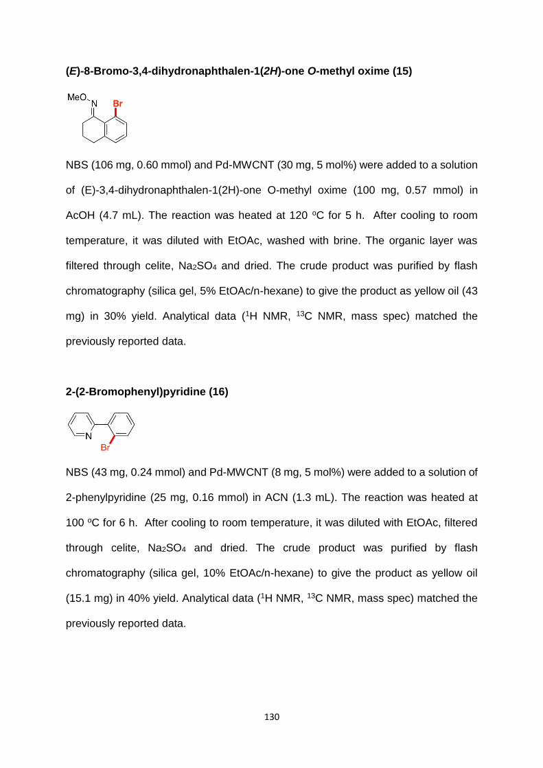
130
(E)-8-Bromo-3,4-dihydronaphthalen-1(2H)-one O-methyl oxime (15)
NBS (106 mg, 0.60 mmol) and Pd-MWCNT (30 mg, 5 mol%) were added to a solution
of (E)-3,4-dihydronaphthalen-1(2H)-one O-methyl oxime (100 mg, 0.57 mmol) in
AcOH (4.7 mL). The reaction was heated at 120 oC for 5 h. After cooling to room
temperature, it was diluted with EtOAc, washed with brine. The organic layer was
filtered through celite, Na2SO4 and dried. The crude product was purified by flash
chromatography (silica gel, 5% EtOAc/n-hexane) to give the product as yellow oil (43
mg) in 30% yield. Analytical data (1H NMR, 13C NMR, mass spec) matched the
previously reported data.
2-(2-Bromophenyl)pyridine (16)
NBS (43 mg, 0.24 mmol) and Pd-MWCNT (8 mg, 5 mol%) were added to a solution of
2-phenylpyridine (25 mg, 0.16 mmol) in ACN (1.3 mL). The reaction was heated at
100 oC for 6 h. After cooling to room temperature, it was diluted with EtOAc, filtered
through celite, Na2SO4 and dried. The crude product was purified by flash
chromatography (silica gel, 10% EtOAc/n-hexane) to give the product as yellow oil
(15.1 mg) in 40% yield. Analytical data (1H NMR, 13C NMR, mass spec) matched the
previously reported data.
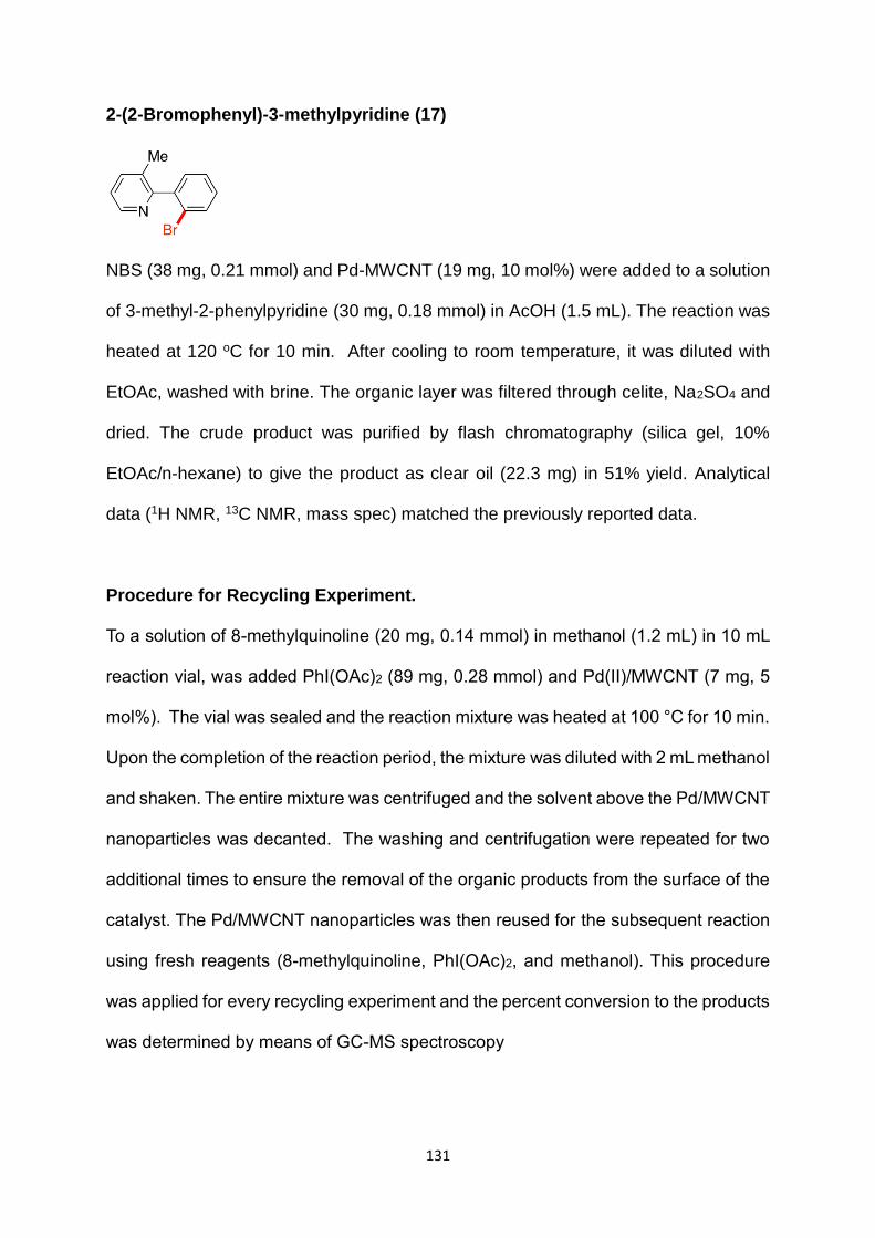
131
2-(2-Bromophenyl)-3-methylpyridine (17)
NBS (38 mg, 0.21 mmol) and Pd-MWCNT (19 mg, 10 mol%) were added to a solution
of 3-methyl-2-phenylpyridine (30 mg, 0.18 mmol) in AcOH (1.5 mL). The reaction was
heated at 120 oC for 10 min. After cooling to room temperature, it was diluted with
EtOAc, washed with brine. The organic layer was filtered through celite, Na2SO4 and
dried. The crude product was purified by flash chromatography (silica gel, 10%
EtOAc/n-hexane) to give the product as clear oil (22.3 mg) in 51% yield. Analytical
data (1H NMR, 13C NMR, mass spec) matched the previously reported data.
Procedure for Recycling Experiment.
To a solution of 8-methylquinoline (20 mg, 0.14 mmol) in methanol (1.2 mL) in 10 mL
reaction vial, was added PhI(OAc)2 (89 mg, 0.28 mmol) and Pd(II)/MWCNT (7 mg, 5
mol%). The vial was sealed and the reaction mixture was heated at 100 °C for 10 min.
Upon the completion of the reaction period, the mixture was diluted with 2 mL methanol
and shaken. The entire mixture was centrifuged and the solvent above the Pd/MWCNT
nanoparticles was decanted. The washing and centrifugation were repeated for two
additional times to ensure the removal of the organic products from the surface of the
catalyst. The Pd/MWCNT nanoparticles was then reused for the subsequent reaction
using fresh reagents (8-methylquinoline, PhI(OAc)2, and methanol). This procedure
was applied for every recycling experiment and the percent conversion to the products
was determined by means of GC-MS spectroscopy

132
Procedure for Hot filtration Experiment.
Benzo[h]quinoline (25 mg, 0.14 mmol) in methanol (1.2 mL) was heated in the
presence of Pd(II)/MWCNT (7 mg, 5 mol%) and PhI(OAc)2 (89 mg, 0.28 mmol) in a 10
mL reaction vial at 100 °C for 10 min, resulting in 95% conversion to the product
according to the GC-MS analysis. The reaction mixture was then hot filtered over celite
and the filtrate solution was subjected to ICP-MS in which the amount of Pd content
was determined to be <250 ppb. Fresh reagents (Benzo[h]quinoline (25 mg, 0.14
mmol), and PhI(OAc)2 (89 mg, 0.28 mmol)) were added to the filtrate solution, the
mixture was heated at 100 °C for additional 10 min. No further catalytic activity was
observed in this mixture by GC-MS spectroscopy.
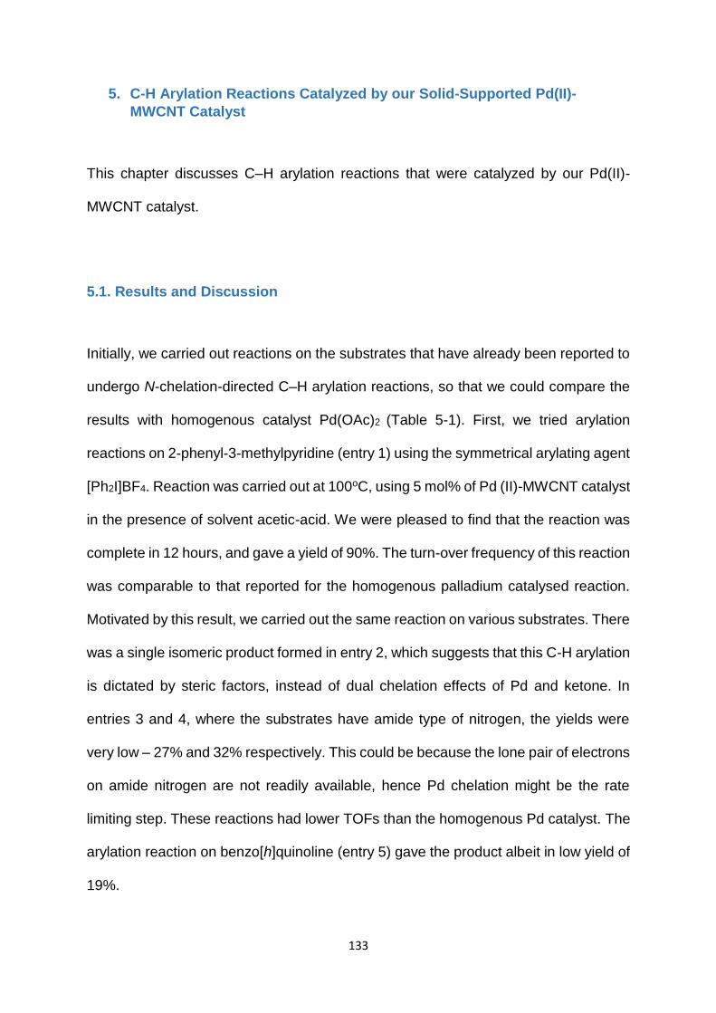
133
5. C-H Arylation Reactions Catalyzed by our Solid-Supported Pd(II)-
MWCNT Catalyst
This chapter discusses C–H arylation reactions that were catalyzed by our Pd(II)-
MWCNT catalyst.
5.1. Results and Discussion
Initially, we carried out reactions on the substrates that have already been reported to
undergo N-chelation-directed C–H arylation reactions, so that we could compare the
results with homogenous catalyst Pd(OAc)2 (Table 5-1). First, we tried arylation
reactions on 2-phenyl-3-methylpyridine (entry 1) using the symmetrical arylating agent
[Ph2I]BF4. Reaction was carried out at 100oC, using 5 mol% of Pd (II)-MWCNT catalyst
in the presence of solvent acetic-acid. We were pleased to find that the reaction was
complete in 12 hours, and gave a yield of 90%. The turn-over frequency of this reaction
was comparable to that reported for the homogenous palladium catalysed reaction.
Motivated by this result, we carried out the same reaction on various substrates. There
was a single isomeric product formed in entry 2, which suggests that this C-H arylation
is dictated by steric factors, instead of dual chelation effects of Pd and ketone. In
entries 3 and 4, where the substrates have amide type of nitrogen, the yields were
very low – 27% and 32% respectively. This could be because the lone pair of electrons
on amide nitrogen are not readily available, hence Pd chelation might be the rate
limiting step. These reactions had lower TOFs than the homogenous Pd catalyst. The
arylation reaction on benzo[h]quinoline (entry 5) gave the product albeit in low yield of
19%.

134
Table 5-1. C-H to C-C Functionalizations Catalyzed by Pd(II)/MWCNT
Entry
Product
Yield and time
with Pd(II)/MWCNT
Yield and
time with
Pd(OAc)2
1
90%, 12 h
88%, 12 h
2b
80%, 24 h
91%, 48 h
3c
27%, 12h
49%, 12h
4
32%, 24 h
75%, 24 h
5
19%, 12 h
NRd
a. Entries 1, 4, 5: [Ph2I]BF4 1.2 eq. b. [Ph2I]BF4 1.5 eq. c. [Ph2I]BF4 2 eq, solvent AcOH:Ac2O
(1:1). d. not reported under similar conditions
We next wished to expand the scope of our transformations to incorporate other aryl
rings (Table 5-2). In order to selectively transfer a specific aryl ring, we used
asymmetrical arylating agents [Mes-I-Ar] as reported by Sanford et al.139 The
asymmetrical arylating agents consist of a mesitylene group (which cannot be
transferred due to its high bulk) and a substituted aryl group (which would get
transferred selectively). Both electronically neutral aryl methyl group (entry 1) and
electronically poor aryl fluoro group (entry 2) were transferred with good yields (87%
and 79% respectively) with reaction rates almost equal to that of the homogenous
catalyst. In contrast, the electronically rich aryl methoxy group (entry 3) did not react
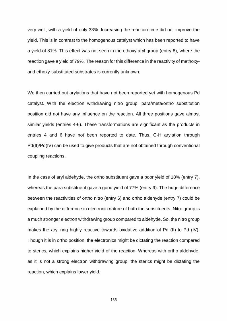
135
very well, with a yield of only 33%. Increasing the reaction time did not improve the
yield. This is in contrast to the homogenous catalyst which has been reported to have
a yield of 81%. This effect was not seen in the ethoxy aryl group (entry 8), where the
reaction gave a yield of 79%. The reason for this difference in the reactivity of methoxy-
and ethoxy-substituted substrates is currently unknown.
We then carried out arylations that have not been reported yet with homogenous Pd
catalyst. With the electron withdrawing nitro group, para/meta/ortho substitution
position did not have any influence on the reaction. All three positions gave almost
similar yields (entries 4-6). These transformations are significant as the products in
entries 4 and 6 have not been reported to date. Thus, C-H arylation through
Pd(II)/Pd(IV) can be used to give products that are not obtained through conventional
coupling reactions.
In the case of aryl aldehyde, the ortho substituent gave a poor yield of 18% (entry 7),
whereas the para substituent gave a good yield of 77% (entry 9). The huge difference
between the reactivities of ortho nitro (entry 6) and ortho aldehyde (entry 7) could be
explained by the difference in electronic nature of both the substituents. Nitro group is
a much stronger electron withdrawing group compared to aldehyde. So, the nitro group
makes the aryl ring highly reactive towards oxidative addition of Pd (II) to Pd (IV).
Though it is in ortho position, the electronics might be dictating the reaction compared
to sterics, which explains higher yield of the reaction. Whereas with ortho aldehyde,
as it is not a strong electron withdrawing group, the sterics might be dictating the
reaction, which explains lower yield.

136
Table 5-2. C-H to C-C Functionalizations Catalyzed by Pd(II)/MWCNT on 3-methyl-2-phenylpyridine
Entry
Product
Yield and time
with Pd(II)/MWCNT
Yield and time
with Pd(OAc)2
1
87%, 12 h
84%, 12 h
2
79%, 12 h
88%, 12 h
3
33%, 12h
81%, 12 h
4
61%, 12 h
NRa
5
69%, 12 h
NRa
6
58%, 12 h
NRa
7
18%, 12 h
NRa
8
79%, 12 h
NRa

137
9
77%, 12 h
NRa
a. not reported under similar conditions
We wished to expand C-H arylation catalysed by our Pd(II)-MWCNT catalyst to a more
rigid and bulky substrate benzo[h]quinoline (Table 5-3). We were pleased to obtain the
arylated product (entry 1) albeit in low yield of 19%. This transformation has been
achieved with very simple reaction conditions. This transformation has been reported
by Sanford et al, but using different conditions (89% yield using palladium acetate,
benzoquinone oxidant, DMSO and Ag2CO3). Though the yield with our solid-supported
catalyst is low, it is an attractive transformation due to its simplicity.
We then expanded this reaction to incorporate substituted aryl rings – electron
withdrawing aryl fluoro group (entry 2) and electronically neutral aryl methyl group
(entry 3). We were pleased to find that the fluoro substituent gave a yield of 35%,
which is higher than the unsubstituted phenyl ring (entry 1). This could be due to the
aryl ring being electron deficient due to the presence of fluoro group, thus making it
more reactive towards oxidative addition of Pd(II) to Pd(IV). Methyl substituted aryl
group gave a low yield of 15% (entry 3) comparable to that of unsubstituted phenyl
ring. This type of reaction is very attractive because the products in entries 2 and 3
have been reported but using more complicated reaction conditions and/or using
highly expensive catalysts such as Rh. We are in the process of optimizing our reaction
conditions to improve the yields further.

138
Table 5-3. C-H to C-C Functionalizations Catalyzed by Pd(II)/MWCNT on benzo[h]quinoline
Entry Product Yield and time
with Pd(II)/MWCNT
Yield and time with
Pd(OAc)2
1
19%, 12 h
NRa
2
35%, 12 h
NRa
3
15%, 12h
NRa
a. not reported under similar conditions
Arylation reaction challenges:
Arylation on 8-methylquinoline using the symmetrical arylating agent gave a mixture
of starting material and another isomer (which could not be identified, Scheme 5-1).
The reaction did not go to completion. This mixture could not be purified on normal
phase, so the yield of the product could not be determined. In order to optimize this
reaction, when AcOH was used as the solvent, diarylated product was formed and the
staring material was still not completely consumed.
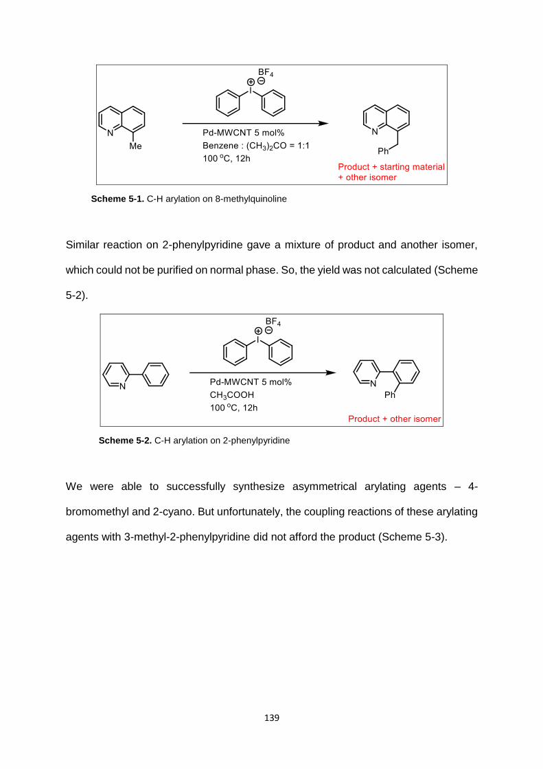
139
Scheme 5-1. C-H arylation on 8-methylquinoline
Similar reaction on 2-phenylpyridine gave a mixture of product and another isomer,
which could not be purified on normal phase. So, the yield was not calculated (Scheme
5-2).
Scheme 5-2. C-H arylation on 2-phenylpyridine
We were able to successfully synthesize asymmetrical arylating agents – 4-
bromomethyl and 2-cyano. But unfortunately, the coupling reactions of these arylating
agents with 3-methyl-2-phenylpyridine did not afford the product (Scheme 5-3).
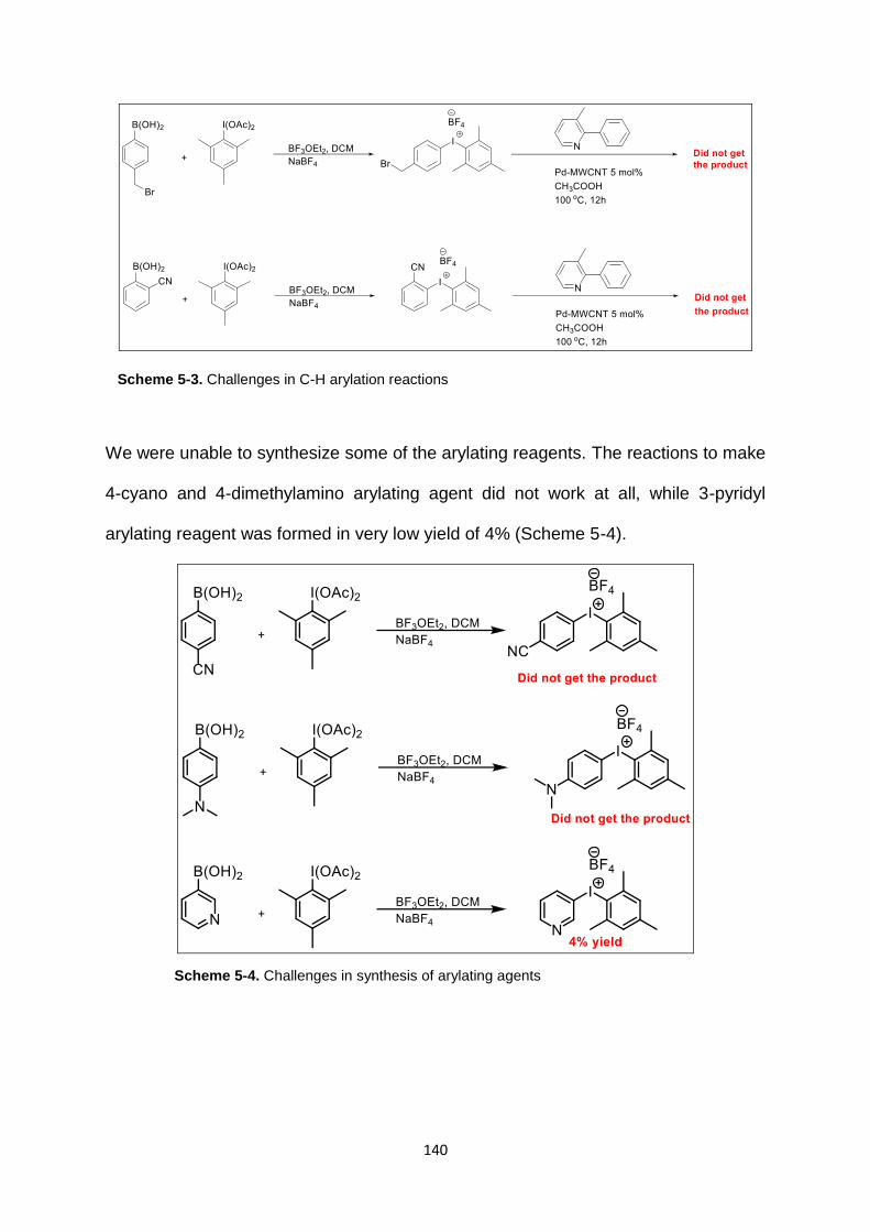
140
Scheme 5-3. Challenges in C-H arylation reactions
We were unable to synthesize some of the arylating reagents. The reactions to make
4-cyano and 4-dimethylamino arylating agent did not work at all, while 3-pyridyl
arylating reagent was formed in very low yield of 4% (Scheme 5-4).
Scheme 5-4. Challenges in synthesis of arylating agents

141
Arylation reaction between benzo[h]quinoline and 1,2-dichlorobenzene using
benzoquinone promoter, Ag2CO3 oxidant, Pd-MWCNT catalyst, and DMSO (to protect
catalyst) gave the product in good yield (68%, Scheme 5-5). In contrast, when the
reaction was tried on 1,2-dimethoxy benzene, the reaction did not work (Scheme 5-6).
This might be because methoxy is sterically bulkier than the chloro group. Sterics
might be dictating this reaction.
Scheme 5-5. Arylation of benzo[h]quinolone with 1,2-dichlorobenzene
Scheme 5-6. Arylation of benzo[h]quinolone with 1,2-dimethoxybenzene
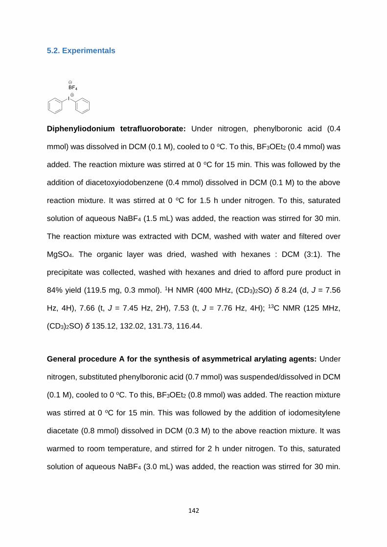
142
5.2. Experimentals
Diphenyliodonium tetrafluoroborate: Under nitrogen, phenylboronic acid (0.4
mmol) was dissolved in DCM (0.1 M), cooled to 0 oC. To this, BF3OEt2 (0.4 mmol) was
added. The reaction mixture was stirred at 0 oC for 15 min. This was followed by the
addition of diacetoxyiodobenzene (0.4 mmol) dissolved in DCM (0.1 M) to the above
reaction mixture. It was stirred at 0 oC for 1.5 h under nitrogen. To this, saturated
solution of aqueous NaBF4 (1.5 mL) was added, the reaction was stirred for 30 min.
The reaction mixture was extracted with DCM, washed with water and filtered over
MgSO4. The organic layer was dried, washed with hexanes : DCM (3:1). The
precipitate was collected, washed with hexanes and dried to afford pure product in
84% yield (119.5 mg, 0.3 mmol). 1H NMR (400 MHz, (CD3)2SO) δ 8.24 (d, J = 7.56
Hz, 4H), 7.66 (t, J = 7.45 Hz, 2H), 7.53 (t, J = 7.76 Hz, 4H); 13C NMR (125 MHz,
(CD3)2SO) δ 135.12, 132.02, 131.73, 116.44.
General procedure A for the synthesis of asymmetrical arylating agents: Under
nitrogen, substituted phenylboronic acid (0.7 mmol) was suspended/dissolved in DCM
(0.1 M), cooled to 0 oC. To this, BF3OEt2 (0.8 mmol) was added. The reaction mixture
was stirred at 0 oC for 15 min. This was followed by the addition of iodomesitylene
diacetate (0.8 mmol) dissolved in DCM (0.3 M) to the above reaction mixture. It was
warmed to room temperature, and stirred for 2 h under nitrogen. To this, saturated
solution of aqueous NaBF4 (3.0 mL) was added, the reaction was stirred for 30 min.

143
The reaction mixture was extracted with DCM, washed with water, filtered over
MgSO4. Organic layer was dried, washed with Et2O to afford pure product.
(4-fluorophenyl)(mesityl)iodonium tetrafluoroborate: General procedure A was
followed on substrate 4-fluorophenyl boronic acid to afford the product in 75% yield
(229.6 mg, 0.54 mmol). 1H NMR (400 MHz, (CD3)2SO) δ 8.02-8.04 (m, 2H), 7.36 (t, J
= 8.91 Hz, 2H), 7.21 (s, 2H), 2.60 (s, 6H), 2.29 (s, 3H); 13C NMR (125 MHz, (CD3)2SO)
δ 143.16, 141.47, 137.21, 137.12, 129.77, 122.94, 119.31, 119.08, 108.62, 30.62,
26.22, 20.47.
mesityl(p-tolyl)iodonium tetrafluoroborate: General procedure A was followed on
substrate 4-tolyl boronic acid to afford the product in 70% yield (217.2 mg, 0.51 mmol).
1H NMR (400 MHz, (CD3)2SO) δ 7.85 (d, J = 8.36 Hz, 2H), 7.30 (d, J = 8.44 Hz, 2H),
7.20 (s, 2H), 2.59 (s, 6H), 2.32 (s, 3H), 2.29 (s, 3H); 13C NMR (125 MHz, (CD3)2SO) δ
142.99, 142.23, 141.44, 134.43, 132.45, 129.71, 122.68, 110.83, 26.24, 20.73, 20.45.
(4-ethoxyphenyl)(mesityl)iodonium tetrafluoroborate: General procedure A was
followed on substrate 4-ethoxyphenyl boronic acid to afford the product in 95% yield
(142.1 mg, 0.31 mmol). 1H NMR (400 MHz, (CD3)2SO) δ 7.66 (d, J = 9.35 Hz, 2H),

144
7.26 (s, 1H), 7.09 (s, 2H), 6.91 (d, J = 9.39 Hz, 2H), 4.02 (q, J = 7.18 Hz, 6.93 Hz, 2H),
2.65 (s, 6H), 2.35 (s, 3H), 1.39 (t, J = 6.97 Hz, 3H); 13C NMR (125 MHz, (CD3)2SO) δ
160.99, 142.85, 141.29, 136.48, 129.64, 123.04, 117.86, 103.15, 63.75, 26.19, 20.43,
14.30.
(4-formylphenyl)(mesityl)iodonium tetrafluoroborate: General procedure A was
followed on substrate 4-formylphenyl boronic acid to afford the product in 69% yield
(101 mg, 0.23 mmol). 1H NMR (400 MHz, (CD3)2SO) δ 10.00 (s, 1H), 8.15 (d, J = 8.48
Hz, 2H), 7.95 (d, J = 8.48 Hz, 2H), 7.24 (s, 2H), 2.60 (s, 6H), 2.30 (s, 3H); 13C NMR
(125 MHz, (CD3)2SO) δ 192.32, 143.27, 141.58, 137.74, 134.85, 131.85, 129.83,
122.91, 120.71, 26.24, 20.46.
mesityl(4-methoxyphenyl)iodonium tetrafluoroborate: General procedure A was
followed on substrate 4-methoxyphenyl boronic acid to afford the product in 85% yield
(251.2 mg, 0.57 mmol). 1H NMR (400 MHz, (CD3)2SO) δ 7.91 (d, J = 9.1 Hz, 2H), 7.19
(s, 2H), 7.03 (d, J = 9.25 Hz, 2H), 3.78 (s, 3H), 2.60 (s, 6H), 2.29 (s, 3H); 13C NMR
(125 MHz, (CD3)2SO) δ 161.69, 142.83, 141.27, 136.46, 129.62, 123.01, 117.47,
103.32, 55.63, 26.17, 20.41.

145
mesityl(2-nitrophenyl)iodonium tetrafluoroborate: General procedure A was
followed on substrate 2-nitrophenyl boronic acid to afford the product in 79% yield (241
mg, 0.53 mmol). 1H NMR (400 MHz, (CD3)2SO) δ 8.59 (d, J = 8.09 Hz, 1H), 7.79-7.90
(m, 2H), 7.41 (s, 2H), 7.03 (d, J = 8.04 Hz, 1H), 2.56 (s, 6H), 2.42 (s, 3H); 13C NMR
(125 MHz, (CD3)2SO) δ 145.93, 144.72, 142.95, 138.08, 132.23, 130.41, 130.22,
127.87, 120.58, 108.39, 26.15, 20.72.
(2-formylphenyl)(mesityl)iodonium tetrafluoroborate: General procedure A was
followed on substrate 2-formylphenyl boronic acid to afford the product in 30% yield
(86 mg, 0.2 mmol. 1H NMR (400 MHz, (CD3)2SO) δ 10.32 (s, 1H), 8.43 (dd, J = 7.5
Hz, 1H), 7.91 (td, J = 7.28 Hz, 1H), 7.79 (td, J = 7.85 Hz, 1H), 7.39 (s, 2H), 6.90 (d, J
= 8.29 Hz, 1H), 3.32 (s, 9H), 2.42 (s, 3H); 13C NMR (125 MHz, (CD3)2SO) δ 195.04,
144.41, 143.05, 138.48, 138.41, 132.97, 131.57, 130.05, 128.92, 116.99, 110.14,
25.99, 20.68.
mesityl(2-nitrophenyl)iodonium tetrafluoroborate: General procedure A was
followed on substrate 3-nitrophenyl boronic acid. Product could not be isolated to get
a pure NMR spectrum.

146
mesityl(4-nitrophenyl)iodonium tetrafluoroborate: General procedure A was
followed on substrate 4-nitrophenyl boronic acid. Product could not be isolated to get
a pure NMR spectrum.
General procedure B for arylation using [Ph2I]BF4: Substrate (0.05 mmol) was
added to acetic-acid (0.12 M) followed by the addition of arylating agent [Ph2I]BF4
(0.08 mmol) and Pd(II)-MWCNT catalyst (5 mol%). The reaction mixture was heated
at 100 oC for 3 h. It was then cooled to room temperature, diluted with DCM, washed
with saturated aqueous NaHCO3, brine, and filtered over celite and MgSO4. The
organic layer was dried and purified over silica using 20% EtOAc-hexanes unless
otherwise noted.
1-(2-(pyridin-2-yl)-[1,1'-biphenyl]-4-yl)ethan-1-one: General procedure B was
followed on substrate 1-(3-(pyridin-2-yl)phenyl)ethan-1-one to afford the product in
80% yield (11.1 mg, 0.04 mmol). 1H NMR (400 MHz, (CD3)2CO) δ 8.60-8.62 (m, 1H),
8.27 (d, J = 1.8 Hz, 1H), 8.10 (dd, J = 8.1Hz, 1H), 7.58 (d, J = 8.0 Hz, 1H), 7.52 (td, J
= 7.7 Hz, 1H), 7.27-7.30 (m, 3H), 7.21-7.25 (m, 1H) 7.17-7.19 (m, 2H), 6.98 (dt, J =
7.9 Hz, 1H), 2.66 (s, 3H); 13C NMR (125 MHz, (CD3)2CO) δ 197.49, 159.32, 150.40,

147
145.86, 141.48, 140.98, 137.27, 136.30, 131.67, 131.50, 130.25, 129.07, 128.79,
128.22, 125.91, 122.73, 26.81.
1-(7-phenylindolin-1-yl)ethan-1-one: General procedure B was followed on
substrate 1-(indolin-1-yl)ethan-1-one, using a mixture of 1:1 CH3COOH: (CH3CO)2CO
as the solvent, and purified using 10% acetone-EtOAc to afford the product in 27%
yield (6 mg, 0.03 mmol). 1H NMR (400 MHz, (CD3)2CO) δ 7.38-7.48 (m, 4H), 7.16-7.31
(m, 4H), 4.24 (t, J = 7.6 Hz, 2H), 3.04 (t, J = 7.5 Hz, 2H), 2.76 (s, 3H); 13C NMR (125
MHz, (CD3)2CO) δ 142.25, 141.63, 137.65, 129.75, 129.64, 128.17, 127.75, 125.98,
124.63, 51.16, 22.82.
1-([1,1'-biphenyl]-2-yl)pyrrolidin-2-one: General procedure B was followed on
substrate 1-phenylpyrrolidin-2-one, and purified using 10% acetone-EtOAc to afford
the product in 32% yield (7 mg, 0.03 mmol). 1H NMR (400 MHz, (CDCl3) δ 7.31-7.42
(m, 9H), 3.21 (t, J = 6.7 Hz, 2H), 2.41 (t, J = 7.9 Hz, 2H), 1.83-1.90 (m, 2H); 13C NMR
(125 MHz, CDCl3) δ 175.72, 139.87, 139.36, 136.58, 131.01, 128.71, 128.56, 128.17,
127.73, 50.35, 31.35, 29.85, 19.15.
2-([1,1'-biphenyl]-2-yl)-3-methylpyridine: General procedure B was followed on
substrate 3-methyl-2-phenylpyridine, and purified using 5% EtOAc-DCM to afford the

148
product in 90% yield (19.5 mg, 0.08 mmol). 1H NMR (400 MHz, (CDCl3) δ 8.49 (dd, J
= 4.9 Hz, 1H), 7.42-7.50 (m, 3H), 7.38-7.41 (m, 1H), 7.28-7.30 (m, 1H), 7.08-7.17 (m,
6H), 1.76 (s, 3H); 13C NMR (125 MHz, CDCl3) δ 159.63, 146.65, 141.28, 140.87,
139.58, 137.64, 131.82, 130.04, 129.87, 129.43, 128.51, 127.93, 127.57, 126.78,
122.24, 18.95.
General procedure C for arylation using asymmetrical arylating agents:
Substrate (0.05 mmol) was added to acetic-acid (0.12 M) followed by the addition of
asymmetrical arylating agent [Mes-I-Ar]BF4 (0.08 mmol) and Pd(II)-MWCNT catalyst
(5 mol%). The reaction mixture was heated at 100 oC for 3 h. It was then cooled to
room temperature, diluted with DCM, washed with saturated aqueous NaHCO3, brine,
and filtered over celite and MgSO4. The organic layer was dried and purified over silica
using 20% EtOAc-hexanes unless otherwise noted.
3-methyl-2-(4'-methyl-[1,1'-biphenyl]-2-yl)pyridine: General procedure C was
followed on substrate 3-methyl-2-phenylpyridine, using [Mes-I-p-Me-C6H5]BF4 to
afford the product in 87% yield (20 mg, 0.08 mmol). 1H NMR (400 MHz, (CD3)2CO) δ
8.43 (dd, J = 4.9 Hz, 1H), 7.38-7.49 (m, 4H), 7.32-7.34 (m, 1H), 7.15 (q, 1H), 6.98-
7.03 (m, 4H), 2.66 (s, 3H), 1.74 (s, 3H); 13C NMR (125 MHz, (CD3)2CO) δ 160.66,
147.33, 141.47, 140.83, 139.27, 138.04, 137.09, 132.10, 130.86, 130.35, 129.92,
129.36, 129.02, 127.82, 123.02, 20.99, 19.00.
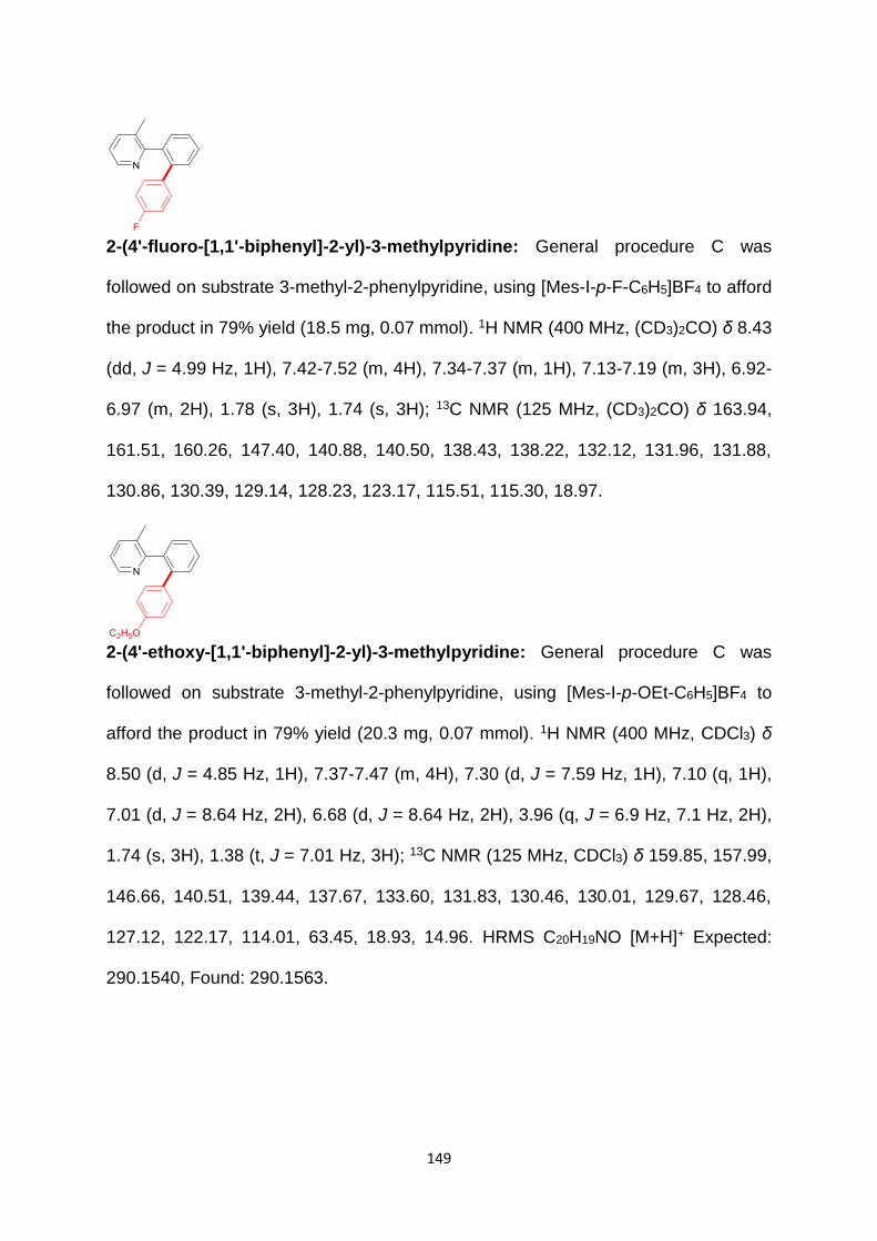
149
2-(4'-fluoro-[1,1'-biphenyl]-2-yl)-3-methylpyridine: General procedure C was
followed on substrate 3-methyl-2-phenylpyridine, using [Mes-I-p-F-C6H5]BF4 to afford
the product in 79% yield (18.5 mg, 0.07 mmol). 1H NMR (400 MHz, (CD3)2CO) δ 8.43
(dd, J = 4.99 Hz, 1H), 7.42-7.52 (m, 4H), 7.34-7.37 (m, 1H), 7.13-7.19 (m, 3H), 6.92-
6.97 (m, 2H), 1.78 (s, 3H), 1.74 (s, 3H); 13C NMR (125 MHz, (CD3)2CO) δ 163.94,
161.51, 160.26, 147.40, 140.88, 140.50, 138.43, 138.22, 132.12, 131.96, 131.88,
130.86, 130.39, 129.14, 128.23, 123.17, 115.51, 115.30, 18.97.
2-(4'-ethoxy-[1,1'-biphenyl]-2-yl)-3-methylpyridine: General procedure C was
followed on substrate 3-methyl-2-phenylpyridine, using [Mes-I-p-OEt-C6H5]BF4 to
afford the product in 79% yield (20.3 mg, 0.07 mmol). 1H NMR (400 MHz, CDCl3) δ
8.50 (d, J = 4.85 Hz, 1H), 7.37-7.47 (m, 4H), 7.30 (d, J = 7.59 Hz, 1H), 7.10 (q, 1H),
7.01 (d, J = 8.64 Hz, 2H), 6.68 (d, J = 8.64 Hz, 2H), 3.96 (q, J = 6.9 Hz, 7.1 Hz, 2H),
1.74 (s, 3H), 1.38 (t, J = 7.01 Hz, 3H); 13C NMR (125 MHz, CDCl3) δ 159.85, 157.99,
146.66, 140.51, 139.44, 137.67, 133.60, 131.83, 130.46, 130.01, 129.67, 128.46,
127.12, 122.17, 114.01, 63.45, 18.93, 14.96. HRMS C20H19NO [M+H]+ Expected:
290.1540, Found: 290.1563.

150
2'-(3-methylpyridin-2-yl)-[1,1'-biphenyl]-4-carbaldehyde: General procedure C
was followed on substrate 3-methyl-2-phenylpyridine, using [Mes-I-p-CHO-C6H5]BF4
to afford the product in 77% yield (18.5 mg, 0.07 mmol). 1H NMR (400 MHz, CDCl3) δ
9.94 (s, 1H), 8.47 (dd, J = 4.90 Hz, 1H), 7.68 (d, J = 8.53 Hz, 2H), 7.42-7.52 (m, 4H),
7.33 (d, J = 7.86 Hz, 1H), 7.28 (d, J = 8.13 Hz, 2H), 7.12 (q, 1H), 1.80 (s, 3H); 13C
NMR (125 MHz, CDCl3) δ 192.07, 158.90, 147.65, 146.78, 139.58, 137.95, 134.78,
131.74, 130.26, 130.03, 129.75, 129.40, 128.72, 128.55, 122.58, 18.97. HRMS
C19H15NO [M+H]+ Expected: 274.1227, Found: 274.1253.
3-methyl-2-(4'-nitro-[1,1'-biphenyl]-2-yl)pyridine: General procedure C was
followed on substrate 3-methyl-2-phenylpyridine, using [Mes-I-p-NO2-C6H5]BF4 to
afford the product in 61% yield (15.6 mg, 0.05 mmol). 1H NMR (400 MHz, CDCl3) δ
8.47 (dd, J = 4.96 Hz, 1H), 8.03 (dt, J = 8.95 Hz, 2H), 7.43-7.54 (m, 4H), 7.36 (d, J =
7.53 Hz, 1H), 7.26-7.29 (m, 2H), 7.14 (q, 1H), 1.83 (s, 3H); 13C NMR (125 MHz, CDCl3)
δ 158.49, 147.96, 146.81, 146.61, 139.58, 138.49, 137.85, 131.45, 130.16, 130.01,
129.56, 128.79, 128.64, 123.07, 122.56, 18.85. HRMS C18H14N2O2 [M+H]+ Expected:
291.1128, Found: 291.1110.

151
3-methyl-2-(3'-nitro-[1,1'-biphenyl]-2-yl)pyridine: General procedure C was
followed on substrate 3-methyl-2-phenylpyridine, using [Mes-I-m-NO2-C6H5]BF4 to
afford the product in 69% yield (17.7 mg, 0.06 mmol). 1H NMR (400 MHz, CDCl3) δ
8.48 (dd, J = 4.94 Hz, 1H), 8.01-8.05 (m, 2H), 7.41-7.55 (m, 5H), 7.37 (d, J = 7.7 Hz,
1H), 7.31 (td, J = 7.9 Hz, 1H), 7.13 (q, 1H), 1.85 (s, 3H); 13C NMR (125 MHz, CDCl3)
δ 158.58, 148.10, 146.93, 142.88, 139.63, 138.42, 138.08, 135.45, 131.66, 130.23,
129.78, 128.92, 128.81, 128.76, 1224.17, 122.73, 121.78, 19.03. HRMS C18H14N2O2
[M+H]+ Expected: 291.1128, Found: 291.1108.
3-methyl-2-(2'-nitro-[1,1'-biphenyl]-2-yl)pyridine: General procedure C was
followed on substrate 3-methyl-2-phenylpyridine, using [Mes-I-m-NO2-C6H5]BF4 to
afford the product in 58% yield (14.9 mg, 0.05 mmol). 1H NMR (400 MHz, CDCl3) δ
8.26 (dd, J = 4.7 Hz, 1H), 7.72 (dd, J = 8.1 Hz, 1H), 7.29-7.49 (m, 8H), 7.03 (q, 1H),
2.09 (s, 3H); 13C NMR (125 MHz, CDCl3) δ 157.59, 148.95, 146.21, 138.33, 137.16,
136.06, 133.64, 132.41, 132.03, 129.92, 129.67, 128.44, 128.10, 127.96, 124.09,
122.31, 19.10. HRMS C18H14N2O2 [M+H]+ Expected: 291.1128, Found: 291.1141.

152
2'-(3-methylpyridin-2-yl)-[1,1'-biphenyl]-2-carbaldehyde: General procedure C
was followed on substrate 3-methyl-2-phenylpyridine, using [Mes-I-m-NO2-C6H5]BF4
to afford the product in 18% yield (2.8 mg, 0.01 mmol). 1H NMR (400 MHz, CDCl3) δ
9.94 (s, 1H), 8.34 (s, 1H), 7.82 (d, J = 7.75 Hz, 1H), 7.30-7.54 (m, 7H), 7.18 (d, J =
7.58 Hz, 1H), 7.00-7.03 (m, 1H), 1.91 (s, 3H); 13C NMR (125 MHz, CDCl3) δ 192.02,
158.19, 146.63, 144.72, 140.62, 137.81, 137.04, 133.98, 132.82, 131.57, 131.41,
129.90, 128.50, 128.10, 127.69, 126.95, 122.43, 29.85, 19.14. HRMS C19H15NO
[M+H]+ Expected: 274.1227, Found: 274.1246.
2-(4'-methoxy-[1,1'-biphenyl]-2-yl)-3-methylpyridine: General procedure C was
followed on substrate 3-methyl-2-phenylpyridine, using [Mes-I-m-NO2-C6H5]BF4 to
afford the product in 33% yield (8.1 mg, 0.03 mmol). 1H NMR (400 MHz, CDCl3) δ 8.51
(dd, J = 5.09 Hz, 1H), 7.38-7.46 (m, 4H), 7.31 (dd, J = 8.1 Hz, 1H), 7.10 (q, 1H), 7.02-
7.04 (m, 2H), 6.69-6.71 (m, 2H), 3.75 (s, 3H), 1.75 (s, 3H); 13C NMR (125 MHz, CDCl3)
δ 159.81, 158.63, 146.65, 140.46, 139.41, 137.74, 133.75, 131.86, 130.49, 130.03,
129.71, 128.50.

153
6. References
1. Knoepfler, P. S.; Eisenman, R. N. Sin meets NuRD and other tails of repression. Cell 1999, 99, 447-50. 2. Boyd, J. M.; Subramanian, T.; Schaeper, U.; La Regina, M.; Bayley, S.; Chinnadurai, G. A region in the C-terminus of adenovirus 2/5 E1a protein is required for association with a cellular phosphoprotein and important for the negative modulation of T24-ras mediated transformation, tumorigenesis and metastasis. Embo j 1993, 12, 469-78. 3. Schaeper, U.; Boyd, J. M.; Verma, S.; Uhlmann, E.; Subramanian, T.; Chinnadurai, G. Molecular cloning and characterization of a cellular phosphoprotein that interacts with a conserved C-terminal domain of adenovirus E1A involved in negative modulation of oncogenic transformation. Proc Natl Acad Sci U S A 1995, 92, 10467-71. 4. Katsanis, N.; Fisher, E. M. A novel C-terminal binding protein (CTBP2) is closely related to CTBP1, an adenovirus E1A-binding protein, and maps to human chromosome 21q21.3. Genomics 1998, 47, 294-9. 5. Weigert, R.; Silletta, M. G.; Spano, S.; Turacchio, G.; Cericola, C.; Colanzi, A.; Senatore, S.; Mancini, R.; Polishchuk, E. V.; Salmona, M.; Facchiano, F.; Burger, K. N.; Mironov, A.; Luini, A.; Corda, D. CtBP/BARS induces fission of Golgi membranes by acylating lysophosphatidic acid. Nature 1999, 402, 429-33. 6. Schmitz, F.; Konigstorfer, A.; Sudhof, T. C. RIBEYE, a component of synaptic ribbons: a protein's journey through evolution provides insight into synaptic ribbon function. Neuron 2000, 28, 857-72. 7. Zenisek, D.; Horst, N. K.; Merrifield, C.; Sterling, P.; Matthews, G. Visualizing synaptic ribbons in the living cell. J Neurosci 2004, 24, 9752-9. 8. Riefler, G. M.; Firestein, B. L. Binding of neuronal nitric-oxide synthase (nNOS) to carboxyl-terminal-binding protein (CtBP) changes the localization of CtBP from the nucleus to the cytosol: a novel function for targeting by the PDZ domain of nNOS. J Biol Chem 2001, 276, 48262-8. 9. Verger, A.; Quinlan, K. G.; Crofts, L. A.; Spano, S.; Corda, D.; Kable, E. P.; Braet, F.; Crossley, M. Mechanisms directing the nuclear localization of the CtBP family proteins. Mol Cell Biol 2006, 26, 4882-94. 10. Zhao, L. J.; Kuppuswamy, M.; Vijayalingam, S.; Chinnadurai, G. Interaction of ZEB and histone deacetylase with the PLDLS-binding cleft region of monomeric C-terminal binding protein 2. BMC Mol Biol 2009, 10, 89. 11. Magupalli, V. G.; Schwarz, K.; Alpadi, K.; Natarajan, S.; Seigel, G. M.; Schmitz, F. Multiple RIBEYE-RIBEYE interactions create a dynamic scaffold for the formation of synaptic ribbons. J Neurosci 2008, 28, 7954-67. 12. tom Dieck, S.; Altrock, W. D.; Kessels, M. M.; Qualmann, B.; Regus, H.; Brauner, D.; Fejtova, A.; Bracko, O.; Gundelfinger, E. D.; Brandstatter, J. H. Molecular dissection of the photoreceptor ribbon synapse: physical interaction of Bassoon and RIBEYE is essential for the assembly of the ribbon complex. J Cell Biol 2005, 168, 825-36. 13. Barnes, C. J.; Vadlamudi, R. K.; Mishra, S. K.; Jacobson, R. H.; Li, F.; Kumar, R. Functional inactivation of a transcriptional corepressor by a signaling kinase. Nat Struct Biol 2003, 10, 622-8. 14. Lin, X.; Sun, B.; Liang, M.; Liang, Y. Y.; Gast, A.; Hildebrand, J.; Brunicardi, F. C.; Melchior, F.; Feng, X. H. Opposed regulation of corepressor CtBP by SUMOylation and PDZ binding. Mol Cell 2003, 11, 1389-96. 15. Kuppuswamy, M.; Vijayalingam, S.; Zhao, L. J.; Zhou, Y.; Subramanian, T.; Ryerse, J.; Chinnadurai, G. Role of the PLDLS-binding cleft region of CtBP1 in recruitment of core and auxiliary components of the corepressor complex. Mol Cell Biol 2008, 28, 269-81. 16. Razeto, A.; Kochhar, S.; Hottinger, H.; Dauter, M.; Wilson, K. S.; Lamzin, V. S. Domain closure, substrate specificity and catalysis of D-lactate dehydrogenase from Lactobacillus bulgaricus. J Mol Biol 2002, 318, 109-19.

154
17. Antonyuk, S. V.; Strange, R. W.; Ellis, M. J.; Bessho, Y.; Kuramitsu, S.; Inoue, Y.; Yokoyama, S.; Hasnain, S. S. Structure of D-lactate dehydrogenase from Aquifex aeolicus complexed with NAD(+) and lactic acid (or pyruvate). Acta Crystallogr Sect F Struct Biol Cryst Commun 2009, 65, 1209-13. 18. Dengler, U.; Niefind, K.; Kiess, M.; Schomburg, D. Crystal structure of a ternary complex of D-2-hydroxyisocaproate dehydrogenase from Lactobacillus casei, NAD+ and 2-oxoisocaproate at 1.9 A resolution. J Mol Biol 1997, 267, 640-60. 19. Lamzin, V. S.; Dauter, Z.; Popov, V. O.; Harutyunyan, E. H.; Wilson, K. S. High resolution structures of holo and apo formate dehydrogenase. J Mol Biol 1994, 236, 759-85. 20. Goldberg, J. D.; Yoshida, T.; Brick, P. Crystal structure of a NAD-dependent D-glycerate dehydrogenase at 2.4 A resolution. J Mol Biol 1994, 236, 1123-40. 21. Booth, M. P.; Conners, R.; Rumsby, G.; Brady, R. L. Structural basis of substrate specificity in human glyoxylate reductase/hydroxypyruvate reductase. J Mol Biol 2006, 360, 178-89. 22. Fjeld, C. C.; Birdsong, W. T.; Goodman, R. H. Differential binding of NAD+ and NADH allows the transcriptional corepressor carboxyl-terminal binding protein to serve as a metabolic sensor. Proc Natl Acad Sci U S A 2003, 100, 9202-7. 23. Balasubramanian, P.; Zhao, L. J.; Chinnadurai, G. Nicotinamide adenine dinucleotide stimulates oligomerization, interaction with adenovirus E1A and an intrinsic dehydrogenase activity of CtBP. FEBS Lett 2003, 537, 157-60. 24. Zhang, Q.; Wang, S. Y.; Nottke, A. C.; Rocheleau, J. V.; Piston, D. W.; Goodman, R. H. Redox sensor CtBP mediates hypoxia-induced tumor cell migration. Proc Natl Acad Sci U S A 2006, 103, 9029-33. 25. Zhang, Q.; Piston, D. W.; Goodman, R. H. Regulation of corepressor function by nuclear NADH. Science 2002, 295, 1895-7. 26. Kim, J. H.; Youn, H. D. C-terminal binding protein maintains mitochondrial activities. Cell Death Differ 2009, 16, 584-92. 27. Chinnadurai, G. CtBP, an unconventional transcriptional corepressor in development and oncogenesis. Mol Cell 2002, 9, 213-24. 28. Postigo, A. A.; Dean, D. C. ZEB represses transcription through interaction with the corepressor CtBP. Proc Natl Acad Sci U S A 1999, 96, 6683-8. 29. Wang, J.; Lee, S.; Teh, C. E.; Bunting, K.; Ma, L.; Shannon, M. F. The transcription repressor, ZEB1, cooperates with CtBP2 and HDAC1 to suppress IL-2 gene activation in T cells. Int Immunol 2009, 21, 227-35. 30. Nibu, Y.; Zhang, H.; Bajor, E.; Barolo, S.; Small, S.; Levine, M. dCtBP mediates transcriptional repression by Knirps, Kruppel and Snail in the Drosophila embryo. Embo j 1998, 17, 7009-20. 31. Turner, J.; Crossley, M. The CtBP family: enigmatic and enzymatic transcriptional co-repressors. Bioessays 2001, 23, 683-90. 32. Quinlan, K. G.; Nardini, M.; Verger, A.; Francescato, P.; Yaswen, P.; Corda, D.; Bolognesi, M.; Crossley, M. Specific recognition of ZNF217 and other zinc finger proteins at a surface groove of C-terminal binding proteins. Mol Cell Biol 2006, 26, 8159-72. 33. Cowger, J. J.; Zhao, Q.; Isovic, M.; Torchia, J. Biochemical characterization of the zinc-finger protein 217 transcriptional repressor complex: identification of a ZNF217 consensus recognition sequence. Oncogene 2007, 26, 3378-86. 34. Shi, Y.; Sawada, J.; Sui, G.; Affar el, B.; Whetstine, J. R.; Lan, F.; Ogawa, H.; Luke, M. P.; Nakatani, Y.; Shi, Y. Coordinated histone modifications mediated by a CtBP co-repressor complex. Nature 2003, 422, 735-8. 35. Zhang, C. L.; McKinsey, T. A.; Lu, J. R.; Olson, E. N. Association of COOH-terminal-binding protein (CtBP) and MEF2-interacting transcription repressor (MITR) contributes to transcriptional repression of the MEF2 transcription factor. J Biol Chem 2001, 276, 35-9. 36. Lee, M. G.; Wynder, C.; Cooch, N.; Shiekhattar, R. An essential role for CoREST in nucleosomal histone 3 lysine 4 demethylation. Nature 2005, 437, 432-5.

155
37. You, A.; Tong, J. K.; Grozinger, C. M.; Schreiber, S. L. CoREST is an integral component of the CoREST- human histone deacetylase complex. Proc Natl Acad Sci U S A 2001, 98, 1454-8. 38. Palijan, A.; Fernandes, I.; Verway, M.; Kourelis, M.; Bastien, Y.; Tavera-Mendoza, L. E.; Sacheli, A.; Bourdeau, V.; Mader, S.; White, J. H. Ligand-dependent corepressor LCoR is an attenuator of progesterone-regulated gene expression. J Biol Chem 2009, 284, 30275-87. 39. Ueda, J.; Tachibana, M.; Ikura, T.; Shinkai, Y. Zinc finger protein Wiz links G9a/GLP histone methyltransferases to the co-repressor molecule CtBP. J Biol Chem 2006, 281, 20120-8. 40. Hidalgo Carcedo, C.; Bonazzi, M.; Spano, S.; Turacchio, G.; Colanzi, A.; Luini, A.; Corda, D. Mitotic Golgi partitioning is driven by the membrane-fissioning protein CtBP3/BARS. Science 2004, 305, 93-6. 41. Yang, J. S.; Lee, S. Y.; Spano, S.; Gad, H.; Zhang, L.; Nie, Z.; Bonazzi, M.; Corda, D.; Luini, A.; Hsu, V. W. A role for BARS at the fission step of COPI vesicle formation from Golgi membrane. Embo j 2005, 24, 4133-43. 42. Schwarz, K.; Natarajan, S.; Kassas, N.; Vitale, N.; Schmitz, F. The synaptic ribbon is a site of phosphatidic acid generation in ribbon synapses. J Neurosci 2011, 31, 15996-6011. 43. Bonazzi, M.; Spano, S.; Turacchio, G.; Cericola, C.; Valente, C.; Colanzi, A.; Kweon, H. S.; Hsu, V. W.; Polishchuck, E. V.; Polishchuck, R. S.; Sallese, M.; Pulvirenti, T.; Corda, D.; Luini, A. CtBP3/BARS drives membrane fission in dynamin-independent transport pathways. Nat Cell Biol 2005, 7, 570-80. 44. Corda, D.; Colanzi, A.; Luini, A. The multiple activities of CtBP/BARS proteins: the Golgi view. Trends Cell Biol 2006, 16, 167-73. 45. Sollerbrant, K.; Chinnadurai, G.; Svensson, C. The CtBP binding domain in the adenovirus E1A protein controls CR1-dependent transactivation. Nucleic Acids Res 1996, 24, 2578-84. 46. Poortinga, G.; Watanabe, M.; Parkhurst, S. M. Drosophila CtBP: a Hairy-interacting protein required for embryonic segmentation and hairy-mediated transcriptional repression. Embo j 1998, 17, 2067-78. 47. Criqui-Filipe, P.; Ducret, C.; Maira, S. M.; Wasylyk, B. Net, a negative Ras-switchable TCF, contains a second inhibition domain, the CID, that mediates repression through interactions with CtBP and de-acetylation. Embo j 1999, 18, 3392-403. 48. Vo, N.; Fjeld, C.; Goodman, R. H. Acetylation of nuclear hormone receptor-interacting protein RIP140 regulates binding of the transcriptional corepressor CtBP. Mol Cell Biol 2001, 21, 6181-8. 49. Sundqvist, A.; Sollerbrant, K.; Svensson, C. The carboxy-terminal region of adenovirus E1A activates transcription through targeting of a C-terminal binding protein-histone deacetylase complex. FEBS Lett 1998, 429, 183-8. 50. Koipally, J.; Georgopoulos, K. Ikaros interactions with CtBP reveal a repression mechanism that is independent of histone deacetylase activity. J Biol Chem 2000, 275, 19594-602. 51. Dahiya, A.; Wong, S.; Gonzalo, S.; Gavin, M.; Dean, D. C. Linking the Rb and polycomb pathways. Mol Cell 2001, 8, 557-69. 52. Meloni, A. R.; Smith, E. J.; Nevins, J. R. A mechanism for Rb/p130-mediated transcription repression involving recruitment of the CtBP corepressor. Proc Natl Acad Sci U S A 1999, 96, 9574-9. 53. Sewalt, R. G.; Gunster, M. J.; van der Vlag, J.; Satijn, D. P.; Otte, A. P. C-Terminal binding protein is a transcriptional repressor that interacts with a specific class of vertebrate Polycomb proteins. Mol Cell Biol 1999, 19, 777-87. 54. Mannervik, M.; Levine, M. The Rpd3 histone deacetylase is required for segmentation of the Drosophila embryo. Proc Natl Acad Sci U S A 1999, 96, 6797-801. 55. Phippen, T. M.; Sweigart, A. L.; Moniwa, M.; Krumm, A.; Davie, J. R.; Parkhurst, S. M. Drosophila C-terminal binding protein functions as a context-dependent transcriptional co-factor and interferes with both mad and groucho transcriptional repression. J Biol Chem 2000, 275, 37628-37. 56. Schaeper, U.; Subramanian, T.; Lim, L.; Boyd, J. M.; Chinnadurai, G. Interaction between a cellular protein that binds to the C-terminal region of adenovirus E1A (CtBP) and a novel cellular protein is disrupted by E1A through a conserved PLDLS motif. J Biol Chem 1998, 273, 8549-52.

156
57. Zhang, Q.; Yao, H.; Vo, N.; Goodman, R. H. Acetylation of adenovirus E1A regulates binding of the transcriptional corepressor CtBP. Proc Natl Acad Sci U S A 2000, 97, 14323-8. 58. Nagel, A. C.; Wech, I.; Schwinkendorf, D.; Preiss, A. Involvement of co-repressors Groucho and CtBP in the regulation of single-minded in Drosophila. Hereditas 2007, 144, 195-205. 59. Zhang, Y. W.; Arnosti, D. N. Conserved catalytic and C-terminal regulatory domains of the C-terminal binding protein corepressor fine-tune the transcriptional response in development. Mol Cell Biol 2011, 31, 375-84. 60. Nagel, A. C.; Krejci, A.; Tenin, G.; Bravo-Patino, A.; Bray, S.; Maier, D.; Preiss, A. Hairless-mediated repression of notch target genes requires the combined activity of Groucho and CtBP corepressors. Mol Cell Biol 2005, 25, 10433-41. 61. Nagel, A. C.; Preiss, A. Fine tuning of Notch signaling by differential co-repressor recruitment during eye development of Drosophila. Hereditas 2011, 148, 77-84. 62. Hoang, C. Q.; Burnett, M. E.; Curtiss, J. Drosophila CtBP regulates proliferation and differentiation of eye precursors and complexes with Eyeless, Dachshund, Dan, and Danr during eye and antennal development. Dev Dyn 2010, 239, 2367-85. 63. Biryukova, I.; Heitzler, P. Drosophila C-terminal binding protein, dCtBP is required for sensory organ prepattern and sharpens proneural transcriptional activity of the GATA factor Pnr. Dev Biol 2008, 323, 64-75. 64. Wen, Y.; Nguyen, D.; Li, Y.; Lai, Z. C. The N-terminal BTB/POZ domain and C-terminal sequences are essential for Tramtrack69 to specify cell fate in the developing Drosophila eye. Genetics 2000, 156, 195-203. 65. Brannon, M.; Brown, J. D.; Bates, R.; Kimelman, D.; Moon, R. T. XCtBP is a XTcf-3 co-repressor with roles throughout Xenopus development. Development 1999, 126, 3159-70. 66. Deconinck, A. E.; Mead, P. E.; Tevosian, S. G.; Crispino, J. D.; Katz, S. G.; Zon, L. I.; Orkin, S. H. FOG acts as a repressor of red blood cell development in Xenopus. Development 2000, 127, 2031-40. 67. Furusawa, T.; Moribe, H.; Kondoh, H.; Higashi, Y. Identification of CtBP1 and CtBP2 as corepressors of zinc finger-homeodomain factor deltaEF1. Mol Cell Biol 1999, 19, 8581-90. 68. Hildebrand, J. D.; Soriano, P. Overlapping and unique roles for C-terminal binding protein 1 (CtBP1) and CtBP2 during mouse development. Mol Cell Biol 2002, 22, 5296-307. 69. Kegel, K. B.; Meloni, A. R.; Yi, Y.; Kim, Y. J.; Doyle, E.; Cuiffo, B. G.; Sapp, E.; Wang, Y.; Qin, Z. H.; Chen, J. D.; Nevins, J. R.; Aronin, N.; DiFiglia, M. Huntingtin is present in the nucleus, interacts with the transcriptional corepressor C-terminal binding protein, and represses transcription. J Biol Chem 2002, 277, 7466-76. 70. Di, L. J.; Byun, J. S.; Wong, M. M.; Wakano, C.; Taylor, T.; Bilke, S.; Baek, S.; Hunter, K.; Yang, H.; Lee, M.; Zvosec, C.; Khramtsova, G.; Cheng, F.; Perou, C. M.; Miller, C. R.; Raab, R.; Olopade, O. I.; Gardner, K. Genome-wide profiles of CtBP link metabolism with genome stability and epithelial reprogramming in breast cancer. Nat Commun 2013, 4, 1449. 71. Lim, J.; Thiery, J. P. Epithelial-mesenchymal transitions: insights from development. Development 2012, 139, 3471-86. 72. Thiery, J. P.; Acloque, H.; Huang, R. Y. J.; Nieto, M. A. Epithelial-Mesenchymal Transitions in Development and Disease. Cell 2009, 139, 871-890. 73. Grooteclaes, M.; Deveraux, Q.; Hildebrand, J.; Zhang, Q.; Goodman, R. H.; Frisch, S. M. C-terminal-binding protein corepresses epithelial and proapoptotic gene expression programs. Proc Natl Acad Sci U S A 2003, 100, 4568-73. 74. Chinnadurai, G. The transcriptional corepressor CtBP: a foe of multiple tumor suppressors. Cancer Res 2009, 69, 731-4. 75. Winklmeier, A.; Poser, I.; Hoek, K. S.; Bosserhoff, A. K. Loss of full length CtBP1 expression enhances the invasive potential of human melanoma. BMC Cancer 2009, 9, 52. 76. Dorman, K.; Shen, Z.; Yang, C.; Ezzat, S.; Asa, S. L. CtBP1 interacts with Ikaros and modulates pituitary tumor cell survival and response to hypoxia. Mol Endocrinol 2012, 26, 447-57.
157
77. Wang, R.; Asangani, I. A.; Chakravarthi, B. V.; Ateeq, B.; Lonigro, R. J.; Cao, Q.; Mani, R. S.; Camacho, D. F.; McGregor, N.; Schumann, T. E.; Jing, X.; Menawat, R.; Tomlins, S. A.; Zheng, H.; Otte, A. P.; Mehra, R.; Siddiqui, J.; Dhanasekaran, S. M.; Nyati, M. K.; Pienta, K. J.; Palanisamy, N.; Kunju, L. P.; Rubin, M. A.; Chinnaiyan, A. M.; Varambally, S. Role of transcriptional corepressor CtBP1 in prostate cancer progression. Neoplasia 2012, 14, 905-14. 78. Nadauld, L. D.; Phelps, R.; Moore, B. C.; Eisinger, A.; Sandoval, I. T.; Chidester, S.; Peterson, P. W.; Manos, E. J.; Sklow, B.; Burt, R. W.; Jones, D. A. Adenomatous polyposis coli control of C-terminal binding protein-1 stability regulates expression of intestinal retinol dehydrogenases. J Biol Chem 2006, 281, 37828-35. 79. Phelps, R. A.; Chidester, S.; Dehghanizadeh, S.; Phelps, J.; Sandoval, I. T.; Rai, K.; Broadbent, T.; Sarkar, S.; Burt, R. W.; Jones, D. A. A two-step model for colon adenoma initiation and progression caused by APC loss. Cell 2009, 137, 623-34. 80. Paliwal, S.; Pande, S.; Kovi, R. C.; Sharpless, N. E.; Bardeesy, N.; Grossman, S. R. Targeting of C-terminal binding protein (CtBP) by ARF results in p53-independent apoptosis. Mol Cell Biol 2006, 26, 2360-72. 81. Kovi, R. C.; Paliwal, S.; Pande, S.; Grossman, S. R. An ARF/CtBP2 complex regulates BH3-only gene expression and p53-independent apoptosis. Cell Death Differ 2010, 17, 513-21. 82. Paliwal, S.; Kovi, R. C.; Nath, B.; Chen, Y. W.; Lewis, B. C.; Grossman, S. R. The alternative reading frame tumor suppressor antagonizes hypoxia-induced cancer cell migration via interaction with the COOH-terminal binding protein corepressor. Cancer Res 2007, 67, 9322-9. 83. Straza, M. W.; Paliwal, S.; Kovi, R. C.; Rajeshkumar, B.; Trenh, P.; Parker, D.; Whalen, G. F.; Lyle, S.; Schiffer, C. A.; Grossman, S. R. Therapeutic targeting of C-terminal binding protein in human cancer. Cell Cycle 2010, 9, 3740-50. 84. Birts, C. N.; Harding, R.; Soosaipillai, G.; Halder, T.; Azim-Araghi, A.; Darley, M.; Cutress, R. I.; Bateman, A. C.; Blaydes, J. P. Expression of CtBP family protein isoforms in breast cancer and their role in chemoresistance. Biol Cell 2010, 103, 1-19. 85. Deng, Y.; Deng, H.; Liu, J.; Han, G.; Malkoski, S.; Liu, B.; Zhao, R.; Wang, X. J.; Zhang, Q. Transcriptional down-regulation of Brca1 and E-cadherin by CtBP1 in breast cancer. Mol Carcinog 2012, 51, 500-7. 86. Di, L. J.; Fernandez, A. G.; De Siervi, A.; Longo, D. L.; Gardner, K. Transcriptional regulation of BRCA1 expression by a metabolic switch. Nat Struct Mol Biol 2010, 17, 1406-13. 87. Bergman, L. M.; Birts, C. N.; Darley, M.; Gabrielli, B.; Blaydes, J. P. CtBPs promote cell survival through the maintenance of mitotic fidelity. Mol Cell Biol 2009, 29, 4539-51. 88. Fodde, R.; Smits, R.; Clevers, H. APC, signal transduction and genetic instability in colorectal cancer. Nat Rev Cancer 2001, 1, 55-67. 89. Shimazu, T.; Degenhardt, K.; Nur, E. K. A.; Zhang, J.; Yoshida, T.; Zhang, Y.; Mathew, R.; White, E.; Inouye, M. NBK/BIK antagonizes MCL-1 and BCL-XL and activates BAK-mediated apoptosis in response to protein synthesis inhibition. Genes Dev 2007, 21, 929-41. 90. Chiarugi, A.; Dolle, C.; Felici, R.; Ziegler, M. The NAD metabolome--a key determinant of cancer cell biology. Nat Rev Cancer 2012, 12, 741-52. 91. Uppal, A.; Gupta, P. K. Measurement of NADH concentration in normal and malignant human tissues from breast and oral cavity. Biotechnol Appl Biochem 2003, 37, 45-50. 92. Subramanian, T.; La Regina, M.; Chinnadurai, G. Enhanced ras oncogene mediated cell transformation and tumorigenesis by adenovirus 2 mutants lacking the C-terminal region of E1a protein. Oncogene 1989, 4, 415-20. 93. Chan, H. M.; Krstic-Demonacos, M.; Smith, L.; Demonacos, C.; La Thangue, N. B. Acetylation control of the retinoblastoma tumour-suppressor protein. Nat Cell Biol 2001, 3, 667-74. 94. Xiao, Z. X.; Chen, J.; Levine, A. J.; Modjtahedi, N.; Xing, J.; Sellers, W. R.; Livingston, D. M. Interaction between the retinoblastoma protein and the oncoprotein MDM2. Nature 1995, 375, 694-8.

158
95. Grooteclaes, M. L.; Frisch, S. M. Evidence for a function of CtBP in epithelial gene regulation and anoikis. Oncogene 2000, 19, 3823-8. 96. Schuierer, M.; Hilger-Eversheim, K.; Dobner, T.; Bosserhoff, A. K.; Moser, M.; Turner, J.; Crossley, M.; Buettner, R. Induction of AP-2alpha expression by adenoviral infection involves inactivation of the AP-2rep transcriptional corepressor CtBP1. J Biol Chem 2001, 276, 27944-9. 97. Duval, A.; Gayet, J.; Zhou, X. P.; Iacopetta, B.; Thomas, G.; Hamelin, R. Frequent frameshift mutations of the TCF-4 gene in colorectal cancers with microsatellite instability. Cancer Res 1999, 59, 4213-5. 98. Saeki, H.; Tanaka, S.; Tokunaga, E.; Kawaguchi, H.; Ikeda, Y.; Maehara, Y.; Sugimachi, K. Genetic alterations in the human Tcf-4 gene in Japanese patients with sporadic gastrointestinal cancers with microsatellite instability. Oncology 2001, 61, 156-61. 99. Kurokawa, M.; Mitani, K.; Irie, K.; Matsuyama, T.; Takahashi, T.; Chiba, S.; Yazaki, Y.; Matsumoto, K.; Hirai, H. The oncoprotein Evi-1 represses TGF-beta signalling by inhibiting Smad3. Nature 1998, 394, 92-6. 100. Palmer, S.; Brouillet, J. P.; Kilbey, A.; Fulton, R.; Walker, M.; Crossley, M.; Bartholomew, C. Evi-1 transforming and repressor activities are mediated by CtBP co-repressor proteins. J Biol Chem 2001, 276, 25834-40. 101. Izutsu, K.; Kurokawa, M.; Imai, Y.; Maki, K.; Mitani, K.; Hirai, H. The corepressor CtBP interacts with Evi-1 to repress transforming growth factor beta signaling. Blood 2001, 97, 2815-22. 102. Vinals, C.; De Bolle, X.; Depiereux, E.; Feytmans, E. Knowledge-based modeling of the D-lactate dehydrogenase three-dimensional structure. Proteins 1995, 21, 307-18. 103. Stoll, V. S.; Kimber, M. S.; Pai, E. F. Insights into substrate binding by D-2-ketoacid dehydrogenases from the structure of Lactobacillus pentosus D-lactate dehydrogenase. Structure 1996, 4, 437-47. 104. Kumar, V.; Carlson, J. E.; Ohgi, K. A.; Edwards, T. A.; Rose, D. W.; Escalante, C. R.; Rosenfeld, M. G.; Aggarwal, A. K. Transcription corepressor CtBP is an NAD(+)-regulated dehydrogenase. Mol Cell 2002, 10, 857-69. 105. Sekowska, A.; Denervaud, V.; Ashida, H.; Michoud, K.; Haas, D.; Yokota, A.; Danchin, A. Bacterial variations on the methionine salvage pathway. BMC Microbiol 2004, 4, 9. 106. Achouri, Y.; Noel, G.; Van Schaftingen, E. 2-Keto-4-methylthiobutyrate, an intermediate in the methionine salvage pathway, is a good substrate for CtBP1. Biochem Biophys Res Commun 2007, 352, 903-6. 107. Achouri, Y.; Rider, M. H.; Schaftingen, E. V.; Robbi, M. Cloning, sequencing and expression of rat liver 3-phosphoglycerate dehydrogenase. Biochem J 1997, 323 ( Pt 2), 365-70. 108. Nunez, M. F.; Pellicer, M. T.; Badia, J.; Aguilar, J.; Baldoma, L. Biochemical characterization of the 2-ketoacid reductases encoded by ycdW and yiaE genes in Escherichia coli. Biochem J 2001, 354, 707-15. 109. Tang, B.; Kadariya, Y.; Murphy, M. E.; Kruger, W. D. The methionine salvage pathway compound 4-methylthio-2-oxobutanate causes apoptosis independent of down-regulation of ornithine decarboxylase. Biochem Pharmacol 2006, 72, 806-15. 110. Scott, C. P.; Abel-Santos, E.; Wall, M.; Wahnon, D. C.; Benkovic, S. J. Production of cyclic peptides and proteins in vivo. Proceedings of the National Academy of Sciences 1999, 96, 13638-13643. 111. Tavassoli, A.; Benkovic, S. J. Split-intein mediated circular ligation used in the synthesis of cyclic peptide libraries in E. coli. Nat Protoc 2007, 2, 1126-33. 112. Birts, C. N.; Nijjar, S. K.; Mardle, C. A.; Hoakwie, F.; Duriez, P. J.; Blaydes, J. P.; Tavassoli, A. A cyclic peptide inhibitor of C-terminal binding protein dimerization links metabolism with mitotic fidelity in breast cancer cells. Chemical Science 2013, 4, 3046-3057. 113. Blevins, M. A.; Kouznetsova, J.; Krueger, A. B.; King, R.; Griner, L. M.; Hu, X.; Southall, N.; Marugan, J. J.; Zhang, Q.; Ferrer, M.; Zhao, R. Small Molecule, NSC95397, Inhibits the CtBP1-Protein
159
Partner Interaction and CtBP1-Mediated Transcriptional Repression. J Biomol Screen 2015, 20, 663-72. 114. Hilbert, B. J.; Grossman, S. R.; Schiffer, C. A.; Royer, W. E., Jr. Crystal structures of human CtBP in complex with substrate MTOB reveal active site features useful for inhibitor design. FEBS Lett 2014, 588, 1743-8. 115. Madison, D. L.; Wirz, J. A.; Siess, D.; Lundblad, J. R. Nicotinamide adenine dinucleotide-induced multimerization of the co-repressor CtBP1 relies on a switching tryptophan. J Biol Chem 2013, 288, 27836-48. 116. Hilbert, B. J.; Morris, B. L.; Ellis, K. C.; Paulsen, J. L.; Schiffer, C. A.; Grossman, S. R.; Royer, W. E. Structure-Guided Design of a High Affinity Inhibitor to Human CtBP. ACS Chemical Biology 2015, 10, 1118-1127. 117. Sarkar, A.; Kellogg, G. E. Hydrophobicity - Shake Flasks, Protein Folding and Drug Discovery. Current Topics in Medicinal Chemistry 2010, 10, 67-83. 118. Hilbert, B. J.; Morris, B. L.; Ellis, K. C.; Paulsen, J. L.; Schiffer, C. A.; Grossman, S. R.; Royer, W. E., Jr. Structure-guided design of a high affinity inhibitor to human CtBP. ACS Chem Biol 2015, 10, 1118-27. 119. Balducci, D.; Conway, P. A.; Sapuppo, G.; Müller-Bunz, H.; Paradisi, F. Novel approach to the synthesis of aliphatic and aromatic α-keto acids. Tetrahedron 2012, 68, 7374-7379. 120. Takrouri, K.; Chen, T.; Papadopoulos, E.; Sahoo, R.; Kabha, E.; Chen, H.; Cantel, S.; Wagner, G.; Halperin, J. A.; Aktas, B. H.; Chorev, M. Structure–activity relationship study of 4EGI-1, small molecule eIF4E/eIF4G protein–protein interaction inhibitors. European Journal of Medicinal Chemistry 2014, 77, 361-377. 121. Godula, K.; Sames, D. C-H Bond Functionalization in Complex Organic Synthesis. Science 2006, 312, 67-72. 122. Janowicz, A. H.; Bergman, R. G. Carbon-hydrogen activation in completely saturated hydrocarbons: direct observation of M + R-H .fwdarw. M(R)(H). Journal of the American Chemical Society 1982, 104, 352-354. 123. Dick, A. R.; Sanford, M. S. Transition metal catalyzed oxidative functionalization of carbon–hydrogen bonds. Tetrahedron 2006, 62, 2439-2463. 124. Lyons, T. W.; Sanford, M. S. Palladium-Catalyzed Ligand-Directed C−H Functionalization Reactions. Chemical Reviews 2010, 110, 1147-1169. 125. Dangel, B. D.; Godula, K.; Youn, S. W.; Sezen, B.; Sames, D. C−C Bond Formation via C−H Bond Activation: Synthesis of the Core of Teleocidin B4. Journal of the American Chemical Society 2002, 124, 11856-11857. 126. Wencel-Delord, J.; Glorius, F. C-H bond activation enables the rapid construction and late-stage diversification of functional molecules. Nat Chem 2013, 5, 369-375. 127. Dai, H.-X.; Stepan, A. F.; Plummer, M. S.; Zhang, Y.-H.; Yu, J.-Q. Divergent C–H Functionalizations Directed by Sulfonamide Pharmacophores: Late-Stage Diversification as a Tool for Drug Discovery. Journal of the American Chemical Society 2011, 133, 7222-7228. 128. Meyer, C.; Schepmann, D.; Yanagisawa, S.; Yamaguchi, J.; Itami, K.; Wünsch, B. Late-Stage C–H Bond Arylation of Spirocyclic σ1 Ligands for Analysis of Complementary σ1 Receptor Surface. European Journal of Organic Chemistry 2012, 2012, 5972-5979. 129. Crabtree, R. H. Alkane C-H activation and functionalization with homogeneous transition metal catalysts: a century of progress-a new millennium in prospect. Journal of the Chemical Society, Dalton Transactions 2001, 2437-2450. 130. Dick, A. R.; Hull, K. L.; Sanford, M. S. A Highly Selective Catalytic Method for the Oxidative Functionalization of C−H Bonds. Journal of the American Chemical Society 2004, 126, 2300-2301. 131. Desai, L. V.; Hull, K. L.; Sanford, M. S. Palladium-Catalyzed Oxygenation of Unactivated sp3 C−H Bonds. Journal of the American Chemical Society 2004, 126, 9542-9543.

160
132. Zhang, J.; Khaskin, E.; Anderson, N. P.; Zavalij, P. Y.; Vedernikov, A. N. Catalytic aerobic oxidation of substituted 8-methylquinolines in PdII-2,6-pyridinedicarboxylic acid systems. Chemical Communications 2008, 3625-3627. 133. Vickers, C. J.; Mei, T.-S.; Yu, J.-Q. Pd(II)-Catalyzed o-C−H Acetoxylation of Phenylalanine and Ephedrine Derivatives with MeCOOOtBu/Ac2O. Organic Letters 2010, 12, 2511-2513. 134. Li, W.; Sun, P. Pd(OAc)2-Catalyzed Alkoxylation of Arylnitriles via sp2 C–H Bond Activation Using Cyano as the Directing Group. The Journal of Organic Chemistry 2012, 77, 8362-8366. 135. Subba Reddy, B. V.; Umadevi, N.; Narasimhulu, G.; Yadav, J. S. Oxidative C–H functionalization: a novel strategy for the acetoxylation/alkoxylation of arenes tethered to 3,4-dihydroisoquinolines. Tetrahedron Letters 2012, 53, 6091-6094. 136. Ju, L.; Yao, J.; Wu, Z.; Liu, Z.; Zhang, Y. Palladium-Catalyzed Oxidative Acetoxylation of Benzylic C–H Bond Using Bidentate Auxiliary. The Journal of Organic Chemistry 2013, 78, 10821-10831. 137. Kalyani, D.; Dick, A. R.; Anani, W. Q.; Sanford, M. S. A Simple Catalytic Method for the Regioselective Halogenation of Arenes. Organic Letters 2006, 8, 2523-2526. 138. Kalyani, D.; Dick, A. R.; Anani, W. Q.; Sanford, M. S. Scope and selectivity in palladium-catalyzed directed C–H bond halogenation reactions. Tetrahedron 2006, 62, 11483-11498. 139. Kalyani, D.; Deprez, N. R.; Desai, L. V.; Sanford, M. S. Oxidative C−H Activation/C−C Bond Forming Reactions: Synthetic Scope and Mechanistic Insights. Journal of the American Chemical Society 2005, 127, 7330-7331. 140. Deprez, N. R.; Sanford, M. S. Synthetic and Mechanistic Studies of Pd-Catalyzed C−H Arylation with Diaryliodonium Salts: Evidence for a Bimetallic High Oxidation State Pd Intermediate. Journal of the American Chemical Society 2009, 131, 11234-11241. 141. Thu, H.-Y.; Yu, W.-Y.; Che, C.-M. Intermolecular Amidation of Unactivated sp2 and sp3 C−H Bonds via Palladium-Catalyzed Cascade C−H Activation/Nitrene Insertion. Journal of the American Chemical Society 2006, 128, 9048-9049. 142. Dick, A. R.; Remy, M. S.; Kampf, J. W.; Sanford, M. S. Carbon−Nitrogen Bond-Forming Reactions of Palladacycles with Hypervalent Iodine Reagents. Organometallics 2007, 26, 1365-1370. 143. Hull, K. L.; Anani, W. Q.; Sanford, M. S. Palladium-Catalyzed Fluorination of Carbon−Hydrogen Bonds. Journal of the American Chemical Society 2006, 128, 7134-7135. 144. McMurtrey, K. B.; Racowski, J. M.; Sanford, M. S. Pd-Catalyzed C–H Fluorination with Nucleophilic Fluoride. Organic Letters 2012, 14, 4094-4097. 145. Ye, Y.; Ball, N. D.; Kampf, J. W.; Sanford, M. S. Oxidation of a Cyclometalated Pd(II) Dimer with “CF3+”: Formation and Reactivity of a Catalytically Competent Monomeric Pd(IV) Aquo Complex. Journal of the American Chemical Society 2010, 132, 14682-14687. 146. Dick, A. R.; Kampf, J. W.; Sanford, M. S. Unusually Stable Palladium(IV) Complexes: Detailed Mechanistic Investigation of C−O Bond-Forming Reductive Elimination. Journal of the American Chemical Society 2005, 127, 12790-12791. 147. Whitfield, S. R.; Sanford, M. S. Reactivity of Pd(II) Complexes with Electrophilic Chlorinating Reagents: Isolation of Pd(IV) Products and Observation of C−Cl Bond-Forming Reductive Elimination. Journal of the American Chemical Society 2007, 129, 15142-15143. 148. Kalyani, D.; Sanford, M. S. Regioselectivity in Palladium-Catalyzed C−H Activation/Oxygenation Reactions. Organic Letters 2005, 7, 4149-4152. 149. Desai, L. V.; Malik, H. A.; Sanford, M. S. Oxone as an Inexpensive, Safe, and Environmentally Benign Oxidant for C−H Bond Oxygenation. Organic Letters 2006, 8, 1141-1144. 150. Schardt, B. C.; Hill, C. L. Preparation of iodobenzene dimethoxide. A new synthesis of [18O]iodosylbenzene and a reexamination of its infrared spectrum. Inorganic Chemistry 1983, 22, 1563-1565. 151. Wang, D.-H.; Hao, X.-S.; Wu, D.-F.; Yu, J.-Q. Palladium-Catalyzed Oxidation of Boc-Protected N-Methylamines with IOAc as the Oxidant: A Boc-Directed sp3 C−H Bond Activation. Organic Letters 2006, 8, 3387-3390.

161
152. Fahey, D. R. The homogeneous palladium-catalysed ortho-chlorination of azobenzene. Journal of the Chemical Society D: Chemical Communications 1970, 417a-417a. 153. Powers, D. C.; Ritter, T. Bimetallic Pd(III) complexes in palladium-catalysed carbon-heteroatom bond formation. Nat Chem 2009, 1, 302-9. 154. Li, J.-J.; Giri, R.; Yu, J.-Q. Remote C–H bond functionalization reveals the distance-dependent isotope effect. Tetrahedron 2008, 64, 6979-6987. 155. Leskinen, M. V.; Yip, K.-T.; Valkonen, A.; Pihko, P. M. Palladium-Catalyzed Dehydrogenative β′-Functionalization of β-Keto Esters with Indoles at Room Temperature. Journal of the American Chemical Society 2012, 134, 5750-5753. 156. Yang, F.; Song, F.; Li, W.; Lan, J.; You, J. Palladium-catalyzed C-H activation of anilides at room temperature: ortho-arylation and acetoxylation. RSC Advances 2013, 3, 9649-9652. 157. Bheeter, C. B.; Bera, J. K.; Doucet, H. Palladium-Catalyzed Direct Arylation of Thiophenes Bearing SO2R Substituents. The Journal of Organic Chemistry 2011, 76, 6407-6413. 158. Li, Y.; Wang, J.; Huang, M.; Wang, Z.; Wu, Y.; Wu, Y. Direct C–H Arylation of Thiophenes at Low Catalyst Loading of a Phosphine-Free Bis(alkoxo)palladium Complex. The Journal of Organic Chemistry 2014, 79, 2890-2897. 159. Deprez, N. R.; Sanford, M. S. Reactions of Hypervalent Iodine Reagents with Palladium: Mechanisms and Applications in Organic Synthesis. Inorganic Chemistry 2007, 46, 1924-1935. 160. Daugulis, O.; Zaitsev, V. G. Anilide ortho-arylation by using C-H activation methodology. Angew Chem Int Ed Engl 2005, 44, 4046-8. 161. Spencer, J.; Chowdhry, B. Z.; Mallet, A. I.; Rathnam, R. P.; Adatia, T.; Bashall, A.; Rominger, F. C–H activations on a 1H-1,4-benzodiazepin-2(3H)-one template. Tetrahedron 2008, 64, 6082-6089. 162. Shabashov, D.; Maldonado, J. R. M.; Daugulis, O. Carbon−Hydrogen Bond Functionalization Approach for the Synthesis of Fluorenones and ortho-Arylated Benzonitriles. The Journal of Organic Chemistry 2008, 73, 7818-7821. 163. Thirunavukkarasu, V. S.; Parthasarathy, K.; Cheng, C. H. Synthesis of fluorenones from aromatic aldoxime ethers and aryl halides by palladium-catalyzed dual C-H activation and Heck cyclization. Angew Chem Int Ed Engl 2008, 47, 9462-5. 164. Shabashov, D.; Daugulis, O. Catalytic Coupling of C−H and C−I Bonds Using Pyridine As a Directing Group. Organic Letters 2005, 7, 3657-3659. 165. Zaitsev, V. G.; Shabashov, D.; Daugulis, O. Highly Regioselective Arylation of sp3 C−H Bonds Catalyzed by Palladium Acetate. Journal of the American Chemical Society 2005, 127, 13154-13155. 166. Chiong, H. A.; Pham, Q.-N.; Daugulis, O. Two Methods for Direct ortho-Arylation of Benzoic Acids. Journal of the American Chemical Society 2007, 129, 9879-9884. 167. Hull, K. L.; Lanni, E. L.; Sanford, M. S. Highly Regioselective Catalytic Oxidative Coupling Reactions: Synthetic and Mechanistic Investigations. Journal of the American Chemical Society 2006, 128, 14047-14049. 168. Hull, K. L.; Sanford, M. S. Catalytic and Highly Regioselective Cross-Coupling of Aromatic C−H Substrates. Journal of the American Chemical Society 2007, 129, 11904-11905. 169. Brasche, G.; García-Fortanet, J.; Buchwald, S. L. Twofold C−H Functionalization: Palladium-Catalyzed Ortho Arylation of Anilides. Organic Letters 2008, 10, 2207-2210. 170. Hull, K. L.; Sanford, M. S. Mechanism of Benzoquinone-Promoted Palladium-Catalyzed Oxidative Cross-Coupling Reactions. Journal of the American Chemical Society 2009, 131, 9651-9653. 171. Cole-Hamilton, D. J. Homogeneous Catalysis--New Approaches to Catalyst Separation, Recovery, and Recycling. Science 2003, 299, 1702-1706. 172. Schlogl, R. Heterogeneous catalysis. Angew Chem Int Ed Engl 2015, 54, 3465-520. 173. Webb, J. D.; MacQuarrie, S.; McEleney, K.; Crudden, C. M. Mesoporous silica-supported Pd catalysts: An investigation into structure, activity, leaching and heterogeneity. Journal of Catalysis 2007, 252, 97-109. 174. Rebek, J.; Gavina, F. Three-phase test. Detection of free cyclobutadiene. Journal of the American Chemical Society 1975, 97, 3453-3456.
162
175. Dell'Anna, M. M.; Gagliardi, M.; Mastrorilli, P.; Suranna, G. P.; Nobile, C. F. Hydrogenation reactions catalysed by a supported palladium complex. Journal of Molecular Catalysis A: Chemical 2000, 158, 515-520. 176. Huang, T. S.; Wang, Y. H.; Jiang, J. Y.; Jin, Z. L. PEG-stabilized palladium nanoparticles: An efficient and recyclable catalyst for the selective hydrogenation of 1,5-cyclooctadiene in thermoregulated PEG biphase system. Chinese Chemical Letters 2008, 19, 102-104. 177. Nadgeri, J. M.; Telkar, M. M.; Rode, C. V. Hydrogenation activity and selectivity behavior of supported palladium nanoparticles. Catalysis Communications 2008, 9, 441-446. 178. Serp, P.; Corrias, M.; Kalck, P. Carbon nanotubes and nanofibers in catalysis. Applied Catalysis A: General 2003, 253, 337-358. 179. Cano, M.; Benito, A. M.; Maser, W. K.; Urriolabeitia, E. P. High catalytic performance of palladium nanoparticles supported on multiwalled carbon nanotubes in alkene hydrogenation reactions. New Journal of Chemistry 2013, 37, 1968-1972. 180. Vidu, R.; Rahman, M.; Mahmoudi, M.; Enachescu, M.; Poteca, T. D.; Opris, I. Nanostructures: A Platform for Brain Repair and Augmentation. Frontiers in Systems Neuroscience 2014, 8. 181. Allen, M. J.; Tung, V. C.; Kaner, R. B. Honeycomb Carbon: A Review of Graphene. Chemical Reviews 2010, 110, 132-145. 182. Scheuermann, G. M.; Rumi, L.; Steurer, P.; Bannwarth, W.; Mülhaupt, R. Palladium Nanoparticles on Graphite Oxide and Its Functionalized Graphene Derivatives as Highly Active Catalysts for the Suzuki−Miyaura Coupling Reaction. Journal of the American Chemical Society 2009, 131, 8262-8270. 183. Cornelio, B.; Rance, G. A.; Laronze-Cochard, M.; Fontana, A.; Sapi, J.; Khlobystov, A. N. Palladium nanoparticles on carbon nanotubes as catalysts of cross-coupling reactions. Journal of Materials Chemistry A 2013, 1, 8737-8744. 184. Nakamura, N.; Tajima, Y.; Sakai, K. Direct Phenylation of Isoxazoles using Palladium Catalysts Synthesis of 4-Phenylmuscimol. Heterocycles 1982, 17, 235-245. 185. Parisien, M.; Valette, D.; Fagnou, K. Direct Arylation Reactions Catalyzed by Pd(OH)2/C: Evidence for a Soluble Palladium Catalyst. The Journal of Organic Chemistry 2005, 70, 7578-7584. 186. Zhang, P.-P.; Zhang, X.-X.; Sun, H.-X.; Liu, R.-H.; Wang, B.; Lin, Y.-H. Pd–CNT-catalyzed ligandless and additive-free heterogeneous Suzuki–Miyaura cross-coupling of arylbromides. Tetrahedron Letters 2009, 50, 4455-4458. 187. Siamaki, A. R.; Lin, Y.; Woodberry, K.; Connell, J. W.; Gupton, B. F. Palladium nanoparticles supported on carbon nanotubes from solventless preparations: versatile catalysts for ligand-free Suzuki cross coupling reactions. Journal of Materials Chemistry A 2013, 1, 12909-12918. 188. Tang, D.-T. D.; Collins, K. D.; Ernst, J. B.; Glorius, F. Pd/C as a Catalyst for Completely Regioselective C H Functionalization of Thiophenes under Mild Conditions. Angewandte Chemie International Edition 2014, 53, 1809-1813. 189. Zhang, D.; Zhaorigetu, B.; Bao, Y.-S. Supported Palladium Nanoparticles Catalyzed Ortho-Directed C–C Coupling Reaction via a Pd0/PdII/PdIV Catalytic Cycle. The Journal of Physical Chemistry C 2015, 119, 20426-20432.