design and evaluation of ketoprofen … millilitres of the dissolution fluid was withdrawn at...
TRANSCRIPT

Vol-2, Issue-3, July-2011 ISSN: 0976-7908 Ashvini et al
www.pharmasm.com IC VALUE – 4.01 189
PHARMA SCIENCE MONITOR AN INTERNATIONAL JOURNAL OF PHARMACEUTICAL SCIENCES
DESIGN AND EVALUATION OF KETOPROFEN LOADED
ALBUMIN MICROSPHERES
Ashvini Urs. V*, Kavitha. K, Mehaboob Y.
Department of pharmaceutics, Bharathi college of pharmacy, Bharathinagara, Mandya – 571422, Karnataka, India.
ABSTRACT To design and formulate Ketoprofen loaded albumin microspheres for effective management of chronic pain without side effects associated with NSAIDs, Ketoprofen was encapsulated within bovine serum albumin microspheres for sustained delivery for long duration of time. The microspheres were prepared by solvent evaporation technique. The microspheres were found to have incorporation efficiency of 48% to 79%. The effect of albumin concentration was evaluated with respect to entrapment efficiency, particle size, surface characteristics and in vitro release behaviours. The results of FTIR spectral showed that there was no significant interaction between the drug and polymer. These results indicated that Ketoprofen loaded albumin microspheres could be prepared providing a sustained release property. Albumin microspheres showing smooth surface were confirmed by Scanning Electron Microscopic (SEM) study. The mean particle size and entrapment efficiency were found to be varied by changing various formulation parameters. The in vitro release profile could be altered significantly by changing various formulation parameters to give a sustained drug release from the microspheres. The microspheres were in the suitable particle size range of 27.5-203µm. The release kinetics studies were found to fit into zero order and Non Fickian diffusion controlled mechanism was observed. The drug was released continuously for a period of 12 hours with a maximum release of 90.5%. From the preliminary trials it was concluded that it is possible to formulate sustained release Ketoprofen loaded albumin microspheres. Key words: Microspheres, drug delivery, Ketoprofen, drug release, albumin.
INTRODUCTION
Ketoprofen is one of the most powerful inhibitors of cyclooxygenase at
concentrations well within the range of therapeutic plasma concentrations (EC50 2µg/l).
It produces reversible COX inhibition by competing with the substrate, arachidonic acid,
for the active site of the enzyme. This inhibition results in a reduction in the tissue
production of prostaglandins such as PGE2 and PGF2α. In addition to its effect on
Vol-2, Issue-3, July-2011 ISSN: 0976-7908 Ashvini et al
www.pharmasm.com IC VALUE – 4.01 190
cyclooxygenase, Ketoprofen inhibits the lipoxygenase pathway of the arachidonic acid
cascade. Ketoprofen is also a powerful inhibitor of bradykinin, an important chemical
mediator of pain and inflammation. It also stabilizes lysosomal membranes against
osmotic change and prevents the release of lysosomal enzymes that mediate tissue
destruction in inflammatory reactions.
Ketoprofen is a NSAID readily absorbed from the gastrointestinal tract and peak
plasma concentrations occurs in about 0.5–2 h after dosing, but it causes certain level of
irritation in the gastrointestinal mucous membrane. The half-life of Ketoprofen in plasma
is about 2–2.5 hours. The short half-life and the low single dose administration makes
Ketoprofen a very good candidate for the formulation of sustained release dosage forms.
[1,2,3] On the other hand, albumin is a major plasma protein constituent, accounting for
355% of the total protein in human plasma. The accumulation of albumin in solid
tumours forms the rationale for developing albumin-based drug delivery systems for
tumour targeting. Thus it has been used as a carrier for targeting drugs to tumours, and
since the synovium of the rheumatoid arthritis patients shares various features observed
in tumours, albumin-based delivery systems can be used to target drugs to the inflamed
joint. [4,5] Furthermore, the solvent evaporation technique, which was used in this study, is
a simple process that is also inexpensive enough for scaling up to a commercial level.
The purpose of the present study was to prepare Ketoprofen microspheres by using
solvent evaporation method and to study the effect of drug-polymer concentration on
drug release. The release mechanism of Ketoprofen from microspheres was also
discussed.
MATERIALS AND METHODS
Ketoprofen drug was obtained as gift sample from PPI Ltd (India); Bovine serum
albumin was purchased from Rolex chemical industries, Mumbai, India. Tween 80,
Ethanol, petroleum ether was purchased from S d Fine chemical Ltd, Mumbai, India. All
other chemical used were of AR grade.
Preparation of microspheres:
Bovine serum albumin (BSA) microspheres containing Ketoprofen were prepared
by solvent evaporation method. Different concentrations like 20, 40 and 60% solutions of
BSA was made to which Ketoprofen drug was added and used as the aqueous phase. The

Vol-2, Issue-3, July-2011 ISSN: 0976-7908 Ashvini et al
www.pharmasm.com IC VALUE – 4.01 191
oil phase composed of 100ml sunflower oil and 10ml petroleum ether with 0.5ml Tween
80 as emulsifier. The aqueous phase was added drop wise to 100ml of sunflower oil
preheated to 60°C and stirred on a magnet stirrer at 600 rpm for 30 minutes to form the
initial emulsion and to allow the formation and solidification of microspheres. The
microspheres suspension was decanted and the settled micro-spheres were washed three
times with petroleum ether to remove traces of oil on microsphere surfaces. The
microspheres were vacuum dried in desiccators overnight and stored in dark. [6] Three
batches of microspheres were prepared by the above mentioned method and marked as
KP1, KP2 and KP3. The microsphere formulations were carried out in different ratios as
per the Table1.
Evaluation of Microspheres
Drug Polymer Interaction Studies
Drug-polymer interactions were studied by FT-IR spectroscopy. The spectra were
recorded for Ketoprofen, bovine serum albumin and physical mixture of Ketoprofen:
albumin (1:1). Samples were prepared in KBr disks (2 mg sample in 200 mg KBr) with a
hydrostatic press at a force of 5.2 τ cm-2 for 3 minutes. The scanning range was 400–
4000 cm-1and the resolution was 4cm-1. [7]
Scanning electron microscopy
SEM studies were carried out by using JEOL JSM T-330A scanning microscope
(Japan). Dry Ketorpofen loaded albumin microspheres were placed on an electron
microscope brass stub and coated with in an ion sputter. Picture of Ketoprofen loaded
albumin microspheres were taken by random scanning of the stub. It can be clearly
observed from the photographs of the Ketoprofen loaded albumin microspheres, prepared
by solvent evaporation technique that the microspheres are smaller and have spherical
shape shown in figure 4. [8]
Size distribution analysis
The mean particle size of microspheres was shown in figure 1. Various
manufacturing parameters (apparatus design, type of stirrer, stirring speed, and viscosity
of emulsion phases)
affect particle size. In this study only the effect of polymer concentration, thus the inner
phase viscosity, on particle formation and particle size, while keeping the other

Vol-2, Issue-3, July-2011 ISSN: 0976-7908 Ashvini et al
www.pharmasm.com IC VALUE – 4.01 192
parameters constant, was investigated. Increasing the Polymer: Drug ratio caused the
mean microspheres size to shift towards a higher particle size. Higher concentration of
polymer produced a more viscous dispersion which formed larger droplets and
consequently larger microspheres. Size distribution of Ketoprofen loaded albumin
microspheres was shown in figure 3. [9]
Percentage yield
Percentage practical yield is calculated to know about percentage yield or
efficiency of any method, thus it helps in selection of appropriate method of production.
Practical yield was calculated as the weight of microspheres recovered from each batch in
relation to the sum of starting material. The percentage yield of prepared microspheres
was determined by using the formula.
X 100
The prepared microspheres were then characterized for their various properties.
Determination of drug content
Practical drug content was determined by taking a weighed quantity of albumin
microspheres (approximately 10 mg) in a 100‐mL volumetric flask. Sufficient quantity of
water was added to make the volume 100 ml. The suspension was shaken vigorously and
then left for 24 hours at room temperature with intermittent shaking. Supernatant was
collected by centrifugation and drug content in supernatant was determined by UV
spectrophotometry at suitable wavelength (256 nm) using a shimadzu UV visible
spectrophotometer (SHIMADZU, Spectrascan‐2200, Japan). [10]
Drug loading and incorporation efficiency of microspheres
The high content of Ketoprofen in microspheres was believed to be due to the
poor solubility of drug in poor solvent. These suggested that the present method was
suitable for the preparation of microspheres of a poorly water-soluble drug, such as
Ketoprofen.
X 100 (1)

Vol-2, Issue-3, July-2011 ISSN: 0976-7908 Ashvini et al
www.pharmasm.com IC VALUE – 4.01 193
X 100 (2)
Where M actual is the actual Ketoprofen content in weighed quantity of powder
of microspheres and M theoretical is the theoretical amount of Ketoprofen in
microspheres calculated from the quantity added in the fabrication process.
In vitro release studies of microspheres
The drug- release studies of the microspheres were carried out for 12 h at 100 rpm
by using USPXXIII dissolution test apparatus by paddle method. The temperature of the
dissolution medium was controlled at 37 ± 0.1°C. A quantity of microspheres equivalent
to 100mg of drug was weighed. The dissolution medium was 900 ml of Phosphate buffer
(pH 7.2±0.2). Five millilitres of the dissolution fluid was withdrawn at regular time
interval and was replaced with fresh quantity of dissolution fluid. The samples were
filtered, and the filtrate was assayed spectrophotometrically at 256nm to determine the
dissolved drug concentration using a spectrophotometer (Model 1601 Shimadzu, Japan).
Kinetics of drug release
To examine the drug release kinetics and mechanism, the cumulative release data
were fitted to models representing zero order (Q v/s t), first order [Log(Q0-Q) v/s t],
Higuchi’s square root of time (Q v/s t1/2 ) and Korsemeyer Peppas double log plot (log Q
v/s log t) respectively, where Q is the cumulative percentage of drug released at time t
and (Q0-Q) is the cumulative percentage of drug remaining after time t.
In short, the results obtained from in vitro release studies were plotted in four
kinetics models of data treatment as follows:
Cumulative percentage drug release Vs. Time (zero order rate kinetics) shown in
Fig 6
Log cumulative percentage drug retained Vs. Time (first order rate kinetics)
shown in Fig 7
Cumulative percentage drug release Vs. √T (Higuchi’s classical diffusion
equation) shown in Fig 8
Log of cumulative percentage drug release Vs. log Time (Peppas exponential
equation) shown in Fig 9

Vol-2, Issue-3, July-2011 ISSN: 0976-7908 Ashvini et al
www.pharmasm.com IC VALUE – 4.01 194
RESULTS AND DISCUSSION
Preparation of albumin microspheres
Evidence have shown in the recent years that Bovine serum albumin (BSA) have
the physical properties and behaviour suitable to prepare sustained drug delivery systems
which are biocompatible, and biodegradable microspheres to release the entrapped drug.
In the present study, solvent evaporation method was employed using BSA to entrap the
drug, furthermore various parameters were characterized for drug and albumin ratio,
stirring speed, amount of surfactant, temperature of the oil phase. Therefore the
influences of the above parameters were highlighted. When the temperature of the
external phase was more than 60°C the albumin formed gel like substance and thus
devoid of encapsulation. The maximum drug load was obtained at 40-60°C for KP1, KP2
and KP3 formulations. The present study reveals that 1:6 (drug:polymer) ratio was
suitable for producing the ideal spherical microspheres. Resultant microspheres did not
have any surface irregularities and are non aggregated. As the drug:polymer ratio was
decreased, the yield was reduced and the resultant microspheres were irregularly shaped
and were highly aggregated in nature and highly impossible to distinguish as individual
microspheres. In order to avoid the formation of irregularly shaped larger particles, in the
present method, 1:6 drug:polymer ratio was best used. Incorporation of drug into BSA
microspheres required the addition of Tween 80 as a surfactant, at an optimum
concentration to reduce the interfacial tension between the internal aqueous phase and
external hydrophobic phase. An attempt was made to incorporate drug in the albumin
microspheres without the addition of a surfactant. But the process failed, as it resulted in
an aggregate cake like mass during the solidification of albumin. This may be due to
repulsion resulting from high interfacial tension between the hydrophobic oil material
and the internal aqueous phase. It was found that Tween 80 having hydrophobicity and
lipophobicity balance (HLB) value of 15 was suitable to increase substantially dispersion
of albumin material in external oil phase and promote drug incorporation in the albumin
microspheres. The batches were tested to obtain an optimal surfactant concentration;
various concentrations ranging from 20-60 % (w/w) of the total formulation were
selected. Discrete microspheres with good flow properties using an optimum
concentration of surfactants 0.5 ml (Tween 80) were used. Concentrations of Tween 80
Vol-2, Issue-3, July-2011 ISSN: 0976-7908 Ashvini et al
www.pharmasm.com IC VALUE – 4.01 195
of 0.25, 1, 1.5, 2 and 2.5ml failed to produce reproducible microspheres. The resultant
albumin microspheres were composed of irregular masses, which were not possible to
distinguish as individual microspheres. From SEM studies it was observed that the
resultant microspheres were free from surface irregularities, except some wrinkles. In the
present study, to produce the spherical discrete microspheres, an optimum drug to BSA
ratio of 1:6 w/w was used. It was found that lower the amount of drug to BSA ratio (1:2)
produces aggregate masses during the cooling process.
The resultant microspheres were unsuitable for pharmaceutical uses. Hence an
optimum 1:6 ratio was used to prepare microspheres (Table 1). [11] The average size of
the microspheres ranged between 27.5 to 203µm. A stirring speed of 600 rpm and stirring
time of 30 min was used to obtain reproducible microspheres. It was observed that with
the increase in the stirring speed from 600 to 1000 rpm there was a decrease in the
formation of the spheres and recovery yield of the microspheres. It is due to small sized
albumin microspheres, which were lost during successive washings. When the stirring
speed was lower than 600 rpm, gel like slime were formed. When the stirring time was
lower than 30 minute, little amount of melted material adhered to the sides of the beaker
during the cooling process results in reduction of yield.
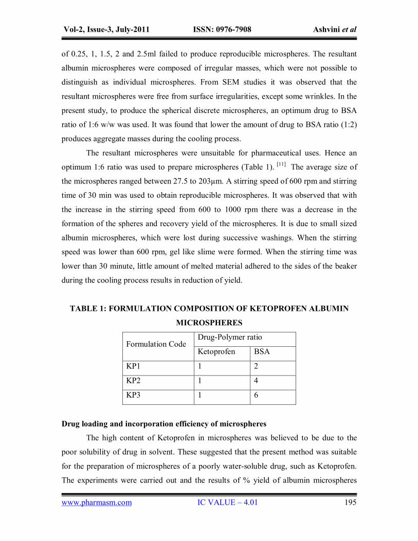
TABLE 1: FORMULATION COMPOSITION OF KETOPROFEN ALBUMIN
MICROSPHERES
Drug-Polymer ratio Formulation Code
Ketoprofen BSA
KP1 1 2
KP2 1 4
KP3 1 6
Drug loading and incorporation efficiency of microspheres
The high content of Ketoprofen in microspheres was believed to be due to the
poor solubility of drug in solvent. These suggested that the present method was suitable
for the preparation of microspheres of a poorly water-soluble drug, such as Ketoprofen.
The experiments were carried out and the results of % yield of albumin microspheres

Vol-2, Issue-3, July-2011 ISSN: 0976-7908 Ashvini et al
www.pharmasm.com IC VALUE – 4.01 196
were 83.86% to maximum of 96.99%. The maximum yield was obtained with
formulation KP3. The average diameter of the albumin microspheres were found to be in
the range of 27.5-203µm shown in fig 1.On further analysis of drug encapsulation of
albumin microspheres, the encapsulation efficiency was found to be between 48% - 79%
shown in fig 2. As the polymer concentration increases the drug encapsulation was found
to be increasing in albumin microspheres. Also percentage yield, average particle size
also increased along with increase in polymer concentration. The results of percentage
yield, drug entrapment, average particle size, and % cumulative drug release were shown
in table 2.
TABLE 2: DATA FOR % YIELD, DRUG ENCAPSULATION EFFICIENCY,
AVERAGE PARTICLE SIZE, % CUMMULATIVE DRUG RELEASE OF
KETOPROFEN ALBUMIN MICROSPHERES
Formula
Code
Percentage
yield (%)
Drug
encapsulation
efficiency (%)
Average
particle
size (µm)
% Cummulative
drug release
KP1 83.86 48.00±2.30 27.5±2.5 90.5
KP2 90.40 55.60±1.50 110.5±3.5 85.4
KP3 96.99 79.00±3.50 203.0±5.5 79.4
SD = Standard deviation (n=3)

Vol-2, Issue-3, July-2011 ISSN: 0976-7908 Ashvini et al
www.pharmasm.com IC VALUE – 4.01 197
Figure 1 Average diameter of Ketoprofen loaded albumin microspheres
Figure 2 Drug entrapment efficiency of Ketoprofen loaded albumin microspheres

Vol-2, Issue-3, July-2011 ISSN: 0976-7908 Ashvini et al
www.pharmasm.com IC VALUE – 4.01 198
Figure 3 Frequency distribution curves of Ketoprofen loaded albumin microspheres prepared by
Solvent evaporation method.
Scanning electron microscopy (SEM)
SEM photographs showed that the albumin microspheres were spherical in nature,
had a smooth surface with inward dents and shrinkage, which is due to the collapse of the
wall of the microspheres (Fig.4). Photographs reveal that as the concentration of the
polymer was increased the surface of the microspheres was found to be smooth and the
surface shrinkage reduced, indicating uniform distribution of the drug within the
microspheres. The rate of solvent removal from the microspheres exerts an influence on
the morphology of the final product. [12]

Vol-2, Issue-3, July-2011 ISSN: 0976-7908 Ashvini et al
www.pharmasm.com IC VALUE – 4.01 199
Figure 4 Scanning Electron Micrographs of Ketoprofen loaded albumin microspheres
(a) Microspheres prepared with 1:2 drug/polymer ratio (b) Microspheres prepared with 1:4 drug/polymer ratio (c) Microspheres prepared with 1:6 drug/polymer ratio
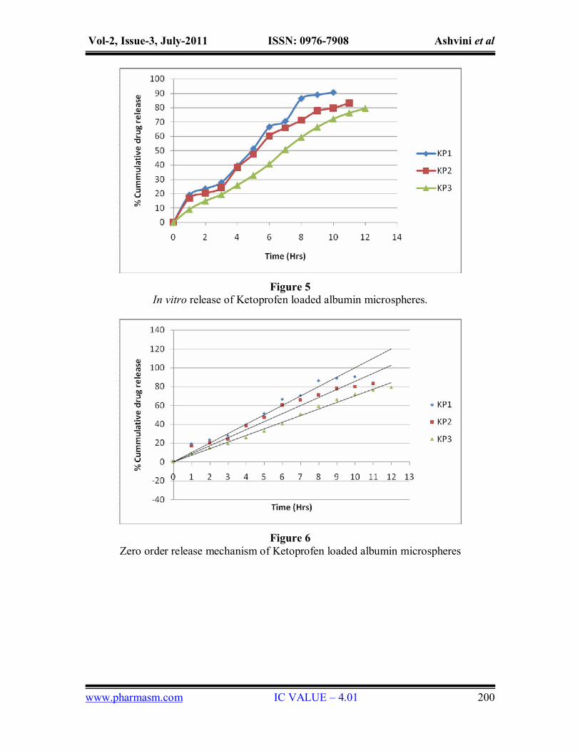
In vitro studies
Hence based on this study, the drug release profile of all the formulations were
carried out in Phosphate buffer pH 7. The dissolution studies revealed that albumin
microspheres released the drug completely within 10 h at lower drug to polymer ratio. At
ratio of more than 1:4, the drug release was sustained over a period of 12 h shown in fig
5. The data for In-vitro drug release profile of Ketoprofen albumin microspheres is shown
in table 3.
TABLE 3: REGRESSION CO-EFFICIENT (R2) VALUES OF DIFFERENT
KINETIC MODELS AND DIFFUSION EXPONENT (N) OF PEPPAS MODEL
FOR KETOPROFEN MICROSPHERES
Peppas plot Formulation
Zero order
First order Higuchi
Matrix r2 value ‘n’ value
KP1 0.9860 0.8572 0.9464 0.9717 0.6692
KP2 0.9827 0.9479 0.9536 0.9752 0.6522
KP3 0.9774 0.9759 0.9472 0.9769 0.6402
Vol-2, Issue-3, July-2011 ISSN: 0976-7908 Ashvini et al
www.pharmasm.com IC VALUE – 4.01 200
Figure 5 In vitro release of Ketoprofen loaded albumin microspheres.
Figure 6 Zero order release mechanism of Ketoprofen loaded albumin microspheres

Vol-2, Issue-3, July-2011 ISSN: 0976-7908 Ashvini et al
www.pharmasm.com IC VALUE – 4.01 201
Figure 7 Release kinetics profile of Ketoprofen albumin microspheres according to First order
Figure 8 Release kinetics profile of Ketoprofen albumin microspheres according to Higuchi matrix
diffusion

Vol-2, Issue-3, July-2011 ISSN: 0976-7908 Ashvini et al
www.pharmasm.com IC VALUE – 4.01 202
Figure 9 Release kinetics profile of Ketoprofen albumin microspheres according to Peppas model
CONCLUSION
The purpose of present work was to develop sustained release microspheres of
water insoluble drug, Ketoprofen, using bovine serum albumin as the drug carrier, by
solvent evaporation method. From the results of characterization and drug release studies
of microspheres it was concluded that this method was ideal for manufacturing sustained-
release microspheres. On the basis of release studies it was indicated that albumin
enhances the solubility of Ketoprofen from microspheres, hence presenting a suitable
method for preparing the sustained-release albumin microspheres for poorly water-
soluble drug Ketoprofen.
ACKNOWLEDGEMENT
Authors are thankful to Bharathi College of Pharmacy for their support and
encouragement in carrying out this work.
REFERENCES
1. Deore BV, Mahajan HS, Deore UV: Development and characterization of sustained
release microspheres by quassi emulsion solvent diffusion method. Int J Chem Tech
Res, 1(3), 2009: 634-42
2. Jeevana JB, Sunitha G.: Development and evaluation of Gelatin microspheres of
Tramadol Hydrochloride. J Young Pharma, 1, 2009: 24-27.
Vol-2, Issue-3, July-2011 ISSN: 0976-7908 Ashvini et al
www.pharmasm.com IC VALUE – 4.01 203
3. http://en.wikipedia.org/wiki/Ketoprofen
4. Thakkar H, Sharma RK, Mishra AK, Chuttani K, Murthy RR.: Albumin
microspheres as carriers for the antiarthritic drug Celecoxib. AAPS PharmSciTech, 6
(1), 2005: 65-73
5. Eroglu H, Suheyla KAS H, Oner L, Sargon M, Hincal A A.: Preparation of Bovine
Serum Albumin microspheres Containing Dexamethasone Sodium Phosphate and
the in Vitro evaluation. Turk J Med Sci, 30, 2000: 125–128.
6. Mathew TS, Gayathri Devi S, Sandhya KV.: Formulation and evaluation of
Ketorolac Tromethamine-loaded albumin microspheres for potential intramuscular
administration. AAPS PharmSciTech, 8(1), 2007: 1-9.
7. Tabassi SAS, Razavi N.: Preparation and characterization of albumin microspheres
encapsulated with Propranolol Hcl. DARU, 11(4), 2003: 137-141.
8. Jayaprakash S, Halith SM, Firthouse MPU, Kulaturanpillai K, Abhijith, Nagarajan
M.: Preparation and evaluation of biodegradable microspheres of Methotraxate.
Asian J Pharm, 2009: 26-29.
9. Zhao YL, Tian F, Liu CJ, Li F, Xing N.: Preparation and evaluation of Poly(3-
hydroxybutyrate) microspheres containing Bovine Serum Albumin for controlled
release. J App Polym Sci 2008: 3826-3835.
10. Brunete SJA, Dea M, Rama S, Bolas F, Alunda JM, Raposo R, Mendez MT,
Santiago TS, Torrado JJ.: Treatment of experimental visceral leishmaniasis with
Amphotericin B in stable albumin microspheres. Antimicrobial agents and
chemotherapy, 48, 2004: 3246–3252.
11. Tsaib M.J., Tsaia Y.H., Wua P.C., Huanga Y.B., Changa J.S.: Design and evaluation
of sustained release microspheres of Potassium Chloride prepared by eudragit, Eur. J
Pharm. Biopharm, 2003: 115-122.
12. Parul Trivedi, AML Verma, N Garud.: Preparation and characterization of
Aceclofenac microspheres, Asian J Pharm, 2008: 110-115.
For Correspondence: Ashvini Urs. V, Email: [email protected]