design and development of compact mini-beam collimators
TRANSCRIPT

Design and Development of compact Mini-beam Collimators for Macromolecular Crystallography at the
GMCA CAT
Shenglan Xu
GM/CA CATArgonne National Laboratory
Technical Working Group Meeting July 16, 2009

2
Sheila Trznadel Admin Specialist
Robert FischettiAssoc. Director
Janet SmithDirector
Administration
Steve CorcoranRich BennDerek YoderShenglan Xu
Engineering & TechnicalSupport
GM/CA CAT Staff
Mark HilgartOleg MakarovSergey Stepanov
ComputingSupport
Sudhir (Babu) Pothineni
Michael BeckerNukri SanishviliCraig Ogata Naga Venugopalan
CrystallographicSupport

3
Outline
Design history of compact mini beam collimators• Single collimator• Dual collimator• Triple collimator • Upgraded triple collimator • Quad collimator
Visualization test resultsBeam flux through collimator pinholes• Simulation and measurements
Material studyMounting and alignment Implementation into BluIce GUI

4
Design history of mini beam collimator
Feb. 2007: First use of single mini beam collimators, 5, 10 and 20μm defining apertures
Jun. 2007: Dual collimator5 μm beam defining and 300 μm scatter guard, and10 μm beam defining and 300 μm scatter guard
Feb. 2008: Triple collimator (5, 10, 300) with three forward scatter guard tubesApr. 2008: New “Uni-body”, triple channel, more robust, better alignment Feb. 2009: Two types of “Uni-body” Triple collimator installed on ID-stations
Type I: with 5, 10 and 300 μm aperturesType II: with 10, 20 and 300 μm apertures
Apr. 2009: Prototype of quad collimator designed and fabricatedwith 5, 10, 20 and 300 μm apertures

5
Components of the single mini-beam collimator
Cap –backscatter
guardBullet-shaped scatter guard
Micro-tube – forward scatter guard
X-ray beam
Pinhole -defining aperture (5, 10, 300 μm)

6
First mini-beam collimator installed Feb 2007
5, 10, 20 μm apertures
2.0 x 101325 x 70Full beam2.0 x 101110.5 x 10.8107.8 x 10105.0 x 5.15
Intensity(photons/sec/100mA)
Beam size, FWHM(VxH)
(μm)Pin hole diameter
(μm)
2.0 x 101325 x 70Full beam2.0 x 101110.5 x 10.8107.8 x 10105.0 x 5.15
Intensity(photons/sec/100mA)
Beam size, FWHM(VxH)
(μm)Pin hole diameter
(μm)

7
Advantages of Mini-beam
Background reduction due to the better size match of the beam and crystal
Collect useful data on projects that produce only small crystals
Select best part of crystal – mosaicity or macro twining
Rastering on large crystals during data collection reduces effects of radiation damage
8
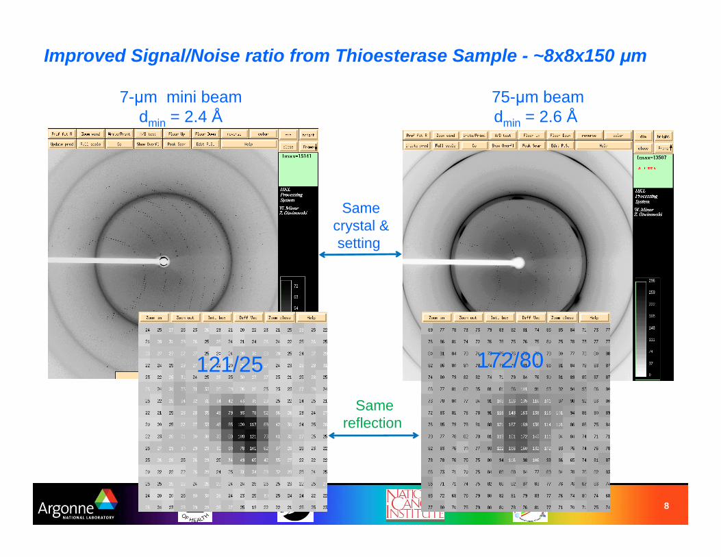
Improved Signal/Noise ratio from Thioesterase Sample - ~8x8x150 μm
75-μm beamdmin = 2.6 Å
Samereflection
Samecrystal &setting
121/25 172/80
7-μm mini beam dmin = 2.4 Å

9
Dual collimator installed June 2007Reliable user operations with mini-beam - quick switch
between full-beam and mini-beam
Double collimator can be used for optimal data collection from large crystals as well:
Loop area can be scanned with the larger beam to locate the crystal then finely scanned with the mini-beam to locate the best part of the crystal for data collection .
Visual obstruction of the double collimator is minimal
No collimator Double collimator
Dual collimator:• one click exchange• auto-align routines
5 or 10 μm 300 μm

10
Triple collimator with three tubes installed Feb 2008quick switch between full-baem and two mini-beams (5, 10 microns)
Cap-Back scatter guard
Scatter guard Holder
Micro-tube –Forward scatter guard
X-Ray beam
Pinhole-Defining aperture

11
Upgraded design of triple collimator
--- to overcome the assembly and alignment difficuties
Merge to One body
Scatter guard
Pinhole-defining apertureGlued on cap (back scatter guard)
Assemble 10 pieces and align 9 component
Assemble 4 pieces and align 3 component
Triple pathsScatter guard

12
“Uni-body” Triple collimator –significantly improves the robustness, ease of initial alignment, and reduction of background.
Cap - back scatter guard
Scatter guard body
Forward scatter guard Three beam path channels
X-ray beam
Pinhole - beam Defining aperture

13
“Uni-body” triple collimator installed Feb. 2009
User selectable via BluIce buttonsPrealignedHighly reproducible
Type I:5 and 10 micron mini-beam defining apertures, 300 micron scatter guard aperture
Type II:10 and 20 micron mini-beam defining apertures, 300 micron scatter guard aperture

14
Advantages of “Uni-body” Triple collimator • Easy to assemble• No tubes to pre-align using a microscope• Robust – no tubes to bend• Pinholes can be removed for cleaning• Easy to change pinhole size • Easy to align - machined forward scatter “tubes” all point in the same direction • Smaller exit aperture - Ø300μ• Reduced scatter around the beamstop
Solution: Designed quad-mini-beam collimator with 5, 10, 20 , 300 micron pinholes
Type I and II triple collimators had to be exchanged for 5 or 20micron beam

15
Prototype Quad Collimators - April 06, 2009
D - Quad Collimator T - Quad Collimator
5, 10 and 20 micron mini-beam defining apertures, 300 micron scatter guard apertures on one collimator
Back side views of Quad Collimators
D T

16
Testing at ID-D station. 2009_04_07Zoom 15, ring light : 3.0 V, Backlight: 4.000 V, Front light: 5.0V
Sample: crystal size: 18 X 18 microns approx
No Collimator Triple Collimator
Visualization test results:Visual obstruction of the triple collimator is marginal,While Visual obstruction for the top position of the quad collimator is minimal.
Quad Collimator I
Quad Collimator II

17
Beam flux through collimator pinholesSimulation and measurements

18
Beam properties for 23-ID-B and 23-ID-DBeam Size at
sample, FWHMμm
IntensityPhotons/sec
Flux density*Photons/sec/μm2
(Intensity / beam FWHM)
Convergenceμ-radians
Full 25 x 12020 x 65
1.0 x 10¹³
2.0 x 10133.3 x 109
1.5 x 101095 x 176
172 x 291
10- μm 10.6 x 11.610.5 x 10.8
1.3 x 10¹¹
5.2 x 10111.1 x 109
4.6 x 109103
5- μm 4.8 x 6.25.0 x 5.1
2.7 x 10¹⁰
5.4 x 10109.1 x 108
2.1 x 10⁹
1- μm 1.1 x 1.2 3.0 x 10⁹ 2.2 x 10⁹ 310
• Beam imaged on a YAG crystal mounted at the sample position. • The pinhole selects the central part of the focused beam.

19
Simulation of beam flux through triple collimator pinholes (with “shadow”):
Full focused beam 10μ beam 5μ beam
Flux through a 3.6mm x 1.3mm slit @ 60.98m 1.60E+14 (ph/s/0.1%BW)
Bandwidth defined by Si(1 1 1) (∆E/E=1.4E-4) 2.10E+13 (ph/s)
Reflectivity of Rh of K-B mirror. 0.85 1.78E+13 (ph/s)*
rays flux/(100mA) rays flux/(100mA) rays flux/(100mA) rays flux/(100mA)25μX 70μ at Sample position 20000 1.78E+13 (ph/s)* 2697 2.4E+12 (ph/s)* 714 6.35E+11 (ph/s)* 174 1.55E+11 (ph/s)*
65μX 90μ (Beam focus after Sample position 300mm) 20000 1.78E+13 (ph/s)* 906 8.06E+11 (ph/s)* 226 2.01E+11 (ph/s)* 47 4.18E+10 (ph/s)*
Flux through a 1.9mm x 0.6mm slit @ 28.35m 1.286E+14 (ph/s/0.1%BW)
Bandwidth defined by Si(1 1 1) (∆E/E=1.4E-4) 1.694E+13 (ph/s)
Reflectivity of Rh of K-B and HDM mirror. 0.7237 1.376E+13 (ph/s)*
rays flux/(100mA) rays flux/(100mA) rays flux/(100mA) rays flux/(100mA)25μX 120μ at Sample position 20000 1.376E+13 (ph/s)* 1606 1.1E+12 (ph/s)* 427 2.93E+11 (ph/s)* 111 7.6E+10 (ph/s)*
IntensityMeatured beam size at Sample positionno pinhole Ø10 μ Ø5 μ
ID_INØ20 μ
Meatured beam size at Sample position Intensityno pinhole Ø10 μ Ø5 μØ20 μ
ID_out

20
Material study

21
Material study of pinholePt Transmission
0.000
0.001
0.002
0.003
0.004
0.005
0.006
0.007
0.008
0.009
0.010
4000 6000 8000 10000 12000 14000 16000 18000 20000 22000 24000 26000 28000 30000
Photon Energy [eV]
Tran
smis
sion
Pt 50µ
Pt 60µ
Pt 70µ
Pt 80µ
Pt 90µ
Pt 100µ
Pt 110µ
The mini beam size is defined by a 2mm diameter platinum disk with a pinhole in the center. The disk is 600 μm thick and tapers to 80-150 μm at the position of the aperture. The calculation results on the top, show that the transmission is negligible at 12 keV.
Source: Tedpella Inc.
5μ pinholes only in PtBigger ones in Mo

22
Material study of Molybdenum
Transmission of Mo at 12Kev: 0.51E-6, is negligible
The minimal wall thickness of “Uni-body”is 0.28 mm
12Kev

23
Inspection of aperture size Nikon VMR – 3020 (CNC Video Measuring System NEXIV)
5μ A
10μ E1 2 3
Ø5 5-A 6-7 Ø4.2 μ Ø4.3 μ Ø4.3 μØ5 5-E 6-7 Ø4.1 μ Ø4.1 μ Ø4.2 μØ10 10-A 10-11 Ø8.5 μ Ø8.5 μ Ø8.4 μØ10 10-C 10-11 Ø9.0 μ Ø9.0 μ Ø9.0 μ
Measurement resultsPinhole (μ) Number
Aperture tolerance: +/- 1 μm for 5μm aperture+/- 1.5 μm for 10μm aperture

24
Mounting and alignment - collimator mounting on on-axis sample-visualization (OAV) system
High ResolutionMicro-Translation Stages
Triple Collimator
Goniometer
Sample
High ResolutionMicro-Translation Stages
Kinematic Mount

25
XY Stages, Physik Instrumete
M110, M-111 can be combined to xy and xyz systems for multiaxisAlignment applications

26
Technical Data

27
Pre-alignment jig
Pre-alignment jig: Align tubes and guard holder relative to Kinematic base

Kinematic mounting with High repeatability and stability
The positional reproducibility of the mini-beam collimator on the kinematic mount was measured by optical metrology. The RMS deviation from the mean position was 0.24 μm in both the X- and Z-directions for 34 repeated manual mount and dismount operations. The stability of the assembled mount was monitored in the X-direction once per minute over a period of 20 minutes. The RMS deviation from the mean X-position was 0.06 microns. The stability in the Z-direction was not measured, but is expected to be smaller than the X-direction.
Measurement by Keyence Surface Scanning Lase Confcoal Displacement Meter (In Profile Mode)
29
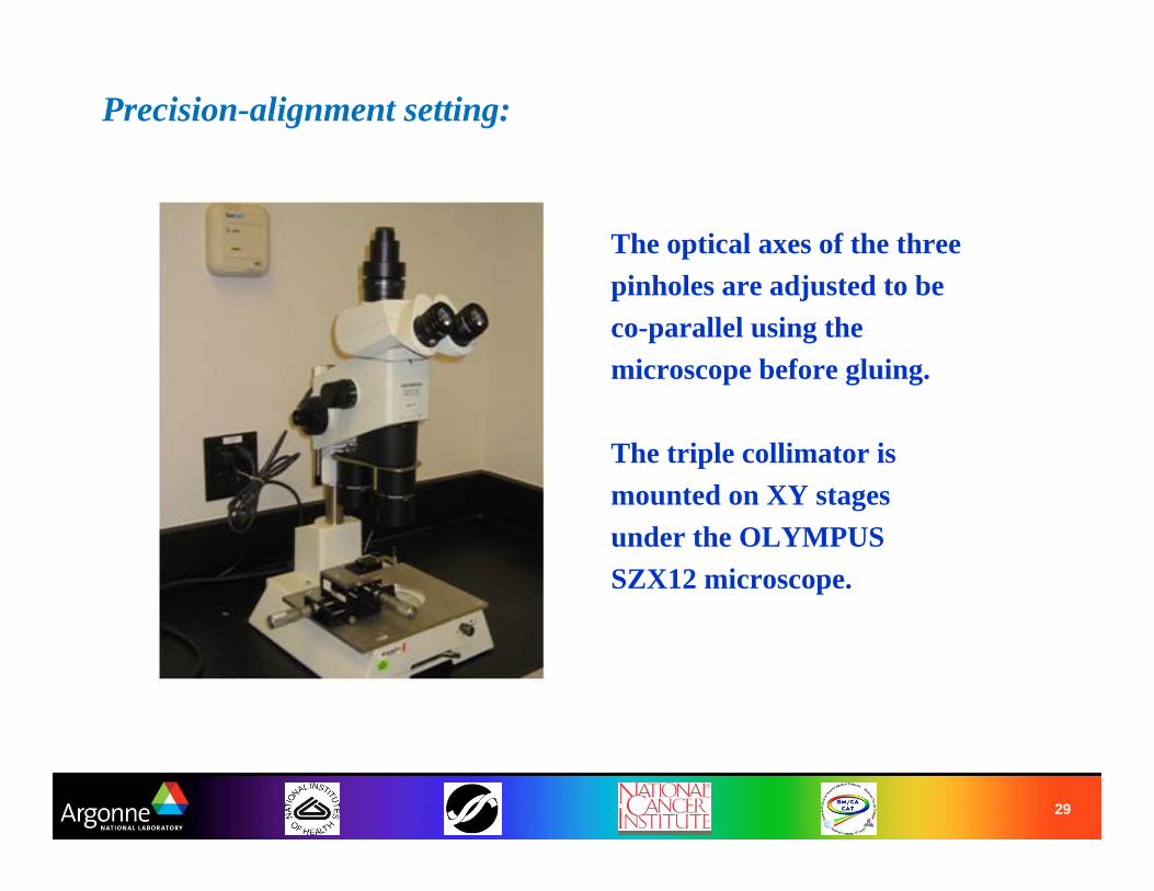
Precision-alignment setting:
The optical axes of the three pinholes are adjusted to be co-parallel using the microscope before gluing.
The triple collimator is mounted on XY stages under the OLYMPUS SZX12 microscope.
30
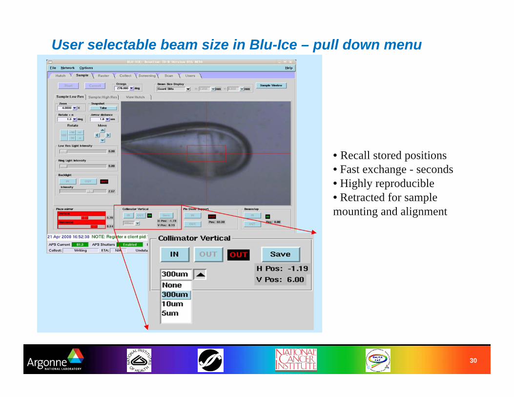
User selectable beam size in Blu-Ice – pull down menu
• Recall stored positions• Fast exchange - seconds• Highly reproducible• Retracted for sample mounting and alignment

31
User Comments on the Mini-beam
“Several problematic crystals were put on. Our worse crystal was a heavily twinned plate. Before the minibeam, we were getting a smeared diffraction pattern. With the minibeam, a discernable diffraction pattern was observed due to being able to section off a thick piece. We could even index and scale the data this time!” - Sacchettini group
“We used the 10u beam on small and high mosaicity crystals. This resulted in better data with lower background and higher resolution.” - Sylvie Doublie
“Perfect for our crystals. Data quality improved significantly by scanning the crystal using the 10 micron mini-beam” - Kornberg group
“We used the 10u beam. It worked very well, and many of our datasets (on small crystals, or crystals with good and bad spots) would not have been possible without it.” - Kate Ferguson
“We found that being able to use the minibeam as needed from the Blu-Ice tab was great. We had some crystals that responded better to a larger beam, and some where we got markedly improved signal-to-noise with the minibeam.” - Petsko group
Quotes from end-of-run reports.

32
ResultsMini-beam has been a huge success at GM/CA CAT. About 30% of our users used the mini-beam when the collimators were single, after the implementation of triple collimator about 80% of users use the mini-beam for data collection. A few users come to GM/CA CAT exclusively to use the mini-beam and have successfully solved structures which might not have been otherwise possible
References:
1. Robert F. Fischetti, Shenglan Xu, Derek Yoder, Michael Becker, Venugopalan Nagarajan, Ruslan Sanishvili, Mark C. Hilgart, Sergey Stepanov, and Janet L.Smith “Mini-beam collimator enables micro-crystallography experiments on standard beamlines” J. Synchrotron Rad. (2009). 16, P217–225
2. S. Xu and R.F. Fischetti “Design and performance of a compact collimator at GM/CA-CAT for macromolecular crystallography” SPIE 2007. Proc. SPIE Vol. 6665-66650X_ P1-8. (Published on May, 2008)
4. R. Sanishvili Ruslan Sanishvili, Venugopalan Nagarajan, Derek Yoder, Michael Becker, Shenglan Xu, Stephen Corcoran, David L.Akey, Janet L. Smith and Robert F. Fischetti “A 7 um mini-beam improves diffraction data from small or imperfect crystals of macromolecules” 2008 Biological Crystallography, Vol. D64, Part 4, P425-435.

33
Thanks!