design and characterization of submicron ccds in...
TRANSCRIPT

Design and Characterization of Submicron CCDs in CMOSKeith Fife, Abbas El Gamal and H.-S. Philip Wong
Department of Electrical Engineering, Stanford University, Stanford, CA 94305Phone: 650-725-9696, Fax: 650-724-3648, E-mail: [email protected]
AbstractThree types of submicron CCDs are implemented in single-poly 0.11µm CMOS technology to demonstrate the feasibility ofmulti-aperture imaging systems that produce data from distributedarrays of CCDs integrated across a monolithic substrate. Teststructures comprising16 × 16 pixel Frame-Transfer (FT)-CCDswith 0.5−0.7µm pixels are fabricated under various process con-ditions to implement devices which operate as surface-channel,buried-channel and pinned phase buried-channel. Ripple chargetransfer and single electrode charge confinement are implementedto minimize pixel pitch.
IntroductionAs pixel size is approaching the limits of conventional optics,improvements in resolution are diminishing. Scaling pixels be-yond these limits, however, can provide new imaging capabilitiesbeyond merely attempting to increase spatial resolution. In [1],we describe a multi-aperture approach to imaging, whereby theimage sensor is partitioned into an array of apertures, eachwith itsown local subarray of pixels and image-forming optics. A virtualimage is focused a certain distance above the sensor such that theapertures capture overlapping views of the scene. The subimagesare post-processed to obtain both a high resolution 2D imageand a depth map. A key feature of this design is in the use ofsubmicron pixels to obtain accurate depth measurements derivedfrom the localization of features within adjacent subarrays.
This paper presents the design and characterization of 3 typesof CCD structures implemented in 0.11µm CMOS technol-ogy: surface-channel, buried-channel, and pinned phase buried-channel. Each CCD structure differs in the location of chargestorage during the integration time and in the sequencing oftheelectrodes during charge transfer. Our surface-channel design wasreported in [2]. We used the buried-channel CCD design in amulti-aperture image sensor reported in [3]. A new pinned phaseburied-channel design is implemented to improve dark currentand charge transfer efficiency.
Design, Fabrication, and OperationWe use an FT-CCD architecture to minimize pixel pitch andto eliminate metal layers in the active imaging area. Each teststructure consists of a pixel array, a storage array, a horizontal(H)-CCD, and a source follower readout circuit (see Fig. 1).Thestorage array is covered by metal layers that are also used todistribute global control lines (see Fig. 2). A photomicrograph isshown in Fig. 3. Each pixel consists of a single poly electrode, achannel, and a channel stop. The channels and stops are showninFig. 4. The electrodes are patterned with non-silicided polysiliconas shown in Fig. 5. Each pixel array is separated by a wall of 4metal layers. The first 2 metal layers are shown in Fig. 6.
In all designs, the polysilicon is doped by masking out thechannels as shown in Fig. 7. The polysilicon for the surface-channel device is doped N+ and the buried-channel designs aredoped P+ to shift the workfunction closer to the operating rangeof CMOS circuits. The IOs on the test chip were designed toallow both positive and negative voltage sequencing. SEM imagesfor each cross section of the surface-channel device are shownin Fig. 8 and Fig. 9. Electrode spacing of 180nm was used inall designs. The polysilicon is 130nm thick with gate oxide of8nm. The channel stop for the surface-channel device is STI.The
channel stop for the buried-channel is formed by a p-type implant(BF2, 75keV, 4.0E13/cm2). The SEM for the H-CCD with fill-and-spill input and floating diffusion is shown in Fig. 10. Therequired sequencing for this design is described in [3].
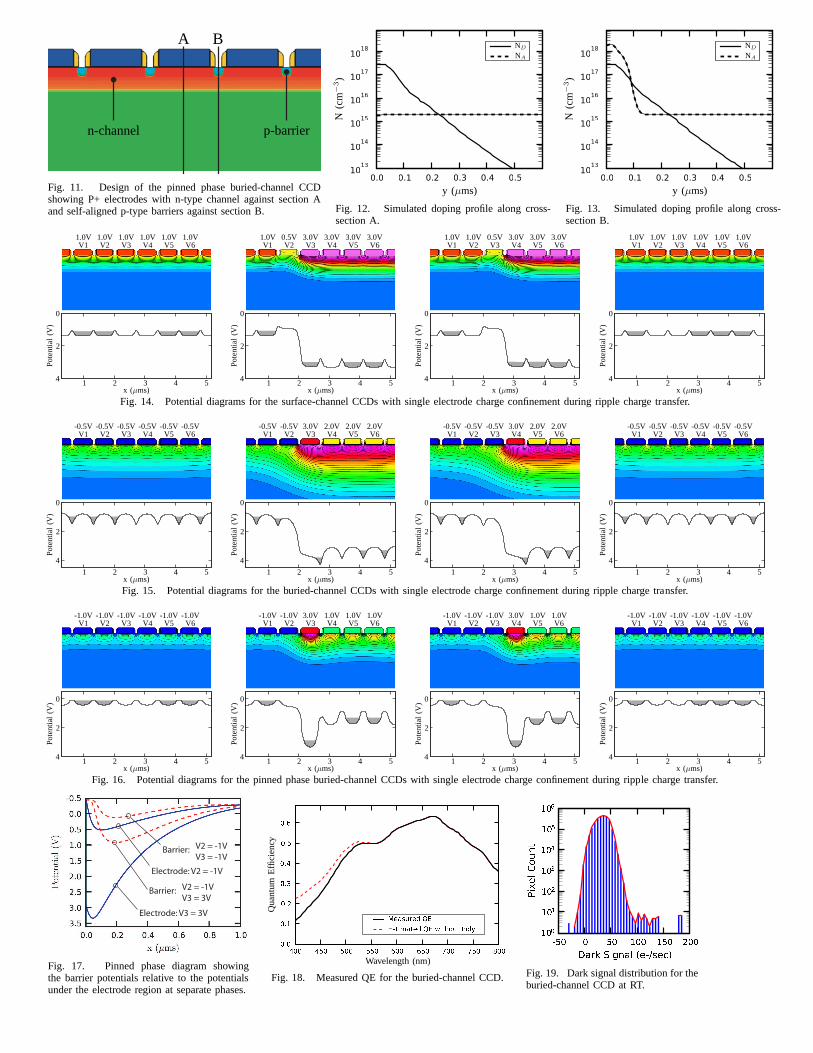
The pinned phase buried-channel design is shown in Fig. 11with doping profiles plotted in Fig. 12-13. This design is similar tothe open-pinned phase CCD described in [4] where the channelisinverted during the integration time. Instead of integrating chargeunder the p-type implant, we integrate charge under the electrodewith an inverted surface. During the integration time, the entiresurface is pinned with a large concentration of holes provided bythe channel stops, which reduces the dark current at the interface.
An image is captured by integrating photocharge at eachelectrode or at every other electrode for higher well capacity. Theintegration begins by depleting the CCDs of charge via transferto the upper diffusion V0. During integration, the pixel arrayelectrodes are held at an intermediate voltage. At the end ofintegration, the accumulated charge is ripple transferredrow-by-row to the storage array and then into the H-CCD one pixel ata time until every pixel has been double sampled at the floatingdiffusion and buffered by the source follower transistor.
Simulation and Measurement ResultsSimulated potential diagrams along the channel (wherever themax potential occurs) for several phases are shown for all 3designs in Fig. 14-16. Single electrode charge confinement isachieved in the surface-channel device due to the barriers createdby the poly gap spacing, whereas it is achieved in the buried-channel device due to the induced pockets. The pinned phasedesign uses the self-aligned p-type implants as barriers toconfinethe charge. Although the surface potential is pinned to the channelstop potential, the depleted channel under the electrode remains ata higher potential. With sufficient gate voltage, each of thedesignsovercomes the pocket or barrier that creates the confinementandcharge is transfered away from one region and then packed againat single electrode pitch into another.
Charge transfer efficency (CTE) is measured the highest for thesurface-channel device at just above 99.9%. We also fabricated thesurface-channel device with p-type channel stops and foundthatthe CTE degrades. When the surface channel electrodes are firstaccumulated with holes, the CTE drops to as low as 85% whereasthe buried-channel devices don’t degrade under this condition.The conversion gain for the 0.5µm pixel is 193µV/e- and165µV/e- for the 0.7µm pixel. There is no significant differencebetween the 3 types of CCDs because the readout transistors areidentical. Despite the use of poly electrodes, the QE is reasonablefor short wavelengths as shown in Fig. 18. This is due to the thinthe poly layer and the open space in between each electrode. Thedark current is about 35e-/sec for both the surface and buried-channel devices (See Fig. 19). The dark current improves by afactor of 15 for the pinned phase device.
References[1] K. Fife, A. El Gamal, and H. Wong, “A 3D multi-aperture image sensor
architecture,” Custom Integrated Circuits Conference, pp. 281–284, Sep.2006.
[2] ——, “A 0.5µm pixel frame-transfer CCD image sensor in 110nm CMOS,”IEDM, pp. 1003–1006, Dec. 2007.
[3] ——, “A multi-aperture image sensor with 0.7µm pixels in 0.11µm CMOStechnology,”IEEE J. Solid-State Circuits, pp. 2990–3005, Dec. 2008.
[4] J. Janesick, “Open-pinned phase CCD technology,”Proc.SPIE 1159, 1989.

pixel
pixel
array
frame
buffer
follower
H-CCD
V0
V1
V2
V16
V17
V18
V19
V33
V34
V35
TS H15 H14 H13 H0 TX RTRS
VPVP COLB
Fig. 1. FT-CCD schematic showing the pixel array, frame buffer,H-CCD and follower readout. Fig. 2. CAD layout of the16×16 FT-CCD.
Fig. 3. Photomicrograph of a fabricated16× 16 FT-CCD. Two photos are combinedto simultaneously focus on the pixels and thetop metal.
Fig. 4. Channel and channel stops for the FT-CCD. Fig. 5. Placement of the polysilicon electrodes. Fig. 6. Metal routing and isolation between arrays.
Fig. 7. Method for doping the polysilicon such thatthe channel region is protected.
Fig. 8. Cross-section of the surface-channel CCDalong the channel.
Fig. 9. Cross-section of the surface-channel CCDagainst the channel stops.
VP (Fill/Spill Input) FD (Floating Diffusion) VP
N+ diffusion Bulk (Epi) N+ Polysilicon Electrode P+ Transfer gate N+ Reset gateMetal1 500nm
Fig. 10. SEM of 16-stage H-CCD showing fill/spill input for electrical testing, floating diffusion for output charge-to-voltage conversion, and the reset gate.
n-channel p-barrier
A B
Fig. 11. Design of the pinned phase buried-channel CCDshowing P+ electrodes with n-type channel against section Aand self-aligned p-type barriers against section B.
0.0 0.1 0.2 0.3 0.4 0.510
13
1014
1015
1016
1017
1018
N(c
m−
3)
y (µms)
ND
NA
Fig. 12. Simulated doping profile along cross-section A.
0.0 0.1 0.2 0.3 0.4 0.510
13
1014
1015
1016
1017
1018
N(c
m−
3)
y (µms)
ND
NA
Fig. 13. Simulated doping profile along cross-section B.
Pot
entia
l(V
)
x (µms)
V1 V2 V3 V4 V5 V61.0V1.0V1.0V1.0V1.0V1.0V
0
1
2
2 34 4 5
Pot
entia
l(V
)
x (µms)
V1 V2 V3 V4 V5 V60.5V1.0V 3.0V3.0V3.0V3.0V
0
1
2
2 34 4 5
Pot
entia
l(V
)
x (µms)
V1 V2 V3 V4 V5 V60.5V1.0V1.0V 3.0V3.0V3.0V
0
1
2
2 34 4 5
Pot
entia
l(V
)
x (µms)
V1 V2 V3 V4 V5 V61.0V1.0V1.0V1.0V1.0V1.0V
0
1
2
2 34 4 5
Fig. 14. Potential diagrams for the surface-channel CCDs with single electrode charge confinement during ripple charge transfer.
Pot
entia
l(V
)
x (µms)
V1 V2 V3 V4 V5 V6-0.5V-0.5V-0.5V-0.5V-0.5V-0.5V
0
1
2
2 3
4
4 5
Pot
entia
l(V
)
x (µms)
V1 V2 V3 V4 V5 V6-0.5V-0.5V 2.0V2.0V2.0V3.0V
0
1
2
2 3
4
4 5
Pot
entia
l(V
)
x (µms)
V1 V2 V3 V4 V5 V6-0.5V-0.5V-0.5V 2.0V2.0V3.0V
0
1
2
2 3
4
4 5
Pot
entia
l(V
)
x (µms)
V1 V2 V3 V4 V5 V6-0.5V-0.5V-0.5V-0.5V-0.5V-0.5V
0
1
2
2 3
4
4 5
Fig. 15. Potential diagrams for the buried-channel CCDs withsingle electrode charge confinement during ripple charge transfer.
Pot
entia
l(V
)
x (µms)
V1 V2 V3 V4 V5 V6-1.0V-1.0V-1.0V-1.0V-1.0V-1.0V
0
1
2
2 34 4 5
Pot
entia
l(V
)
x (µms)
V1 V2 V3 V4 V5 V6-1.0V-1.0V 1.0V1.0V1.0V3.0V
0
1
2
2 34 4 5
Pot
entia
l(V
)
x (µms)
V1 V2 V3 V4 V5 V6-1.0V-1.0V-1.0V 1.0V1.0V3.0V
0
1
2
2 34 4 5
Pot
entia
l(V
)
x (µms)
V1 V2 V3 V4 V5 V6-1.0V-1.0V-1.0V-1.0V-1.0V-1.0V
0
1
2
2 34 4 5
Fig. 16. Potential diagrams for the pinned phase buried-channel CCDs with single electrode charge confinement during ripple charge transfer.
Electrode: V3 = 3V
Electrode: V2 = -1V
V2 = -1V
V3 = 3VBarrier:
V2 = -1V
V3 = -1VBarrier:
Fig. 17. Pinned phase diagram showingthe barrier potentials relative to the potentialsunder the electrode region at separate phases.
4 0 0 4 5 0 5 0 0 5 5 0 6 0 0 6 5 0 7 0 0 7 5 0 8 0 00 . 00 . 10 . 20 . 30 . 40 . 50 . 6M e a s u r e d Q EE s t i m a t e d Q E w i t h o u t P o l yQ
uant
umE
ffici
ency
Wavelength (nm)
Fig. 18. Measured QE for the buried-channel CCD.
� 5 0 0 5 0 1 0 0 1 5 0 2 0 0D a r k S i g n a l ( e , / s e c )1 0 01 0 11 0 21 0 31 0 41 0 51 0 6Pi xelC ountFig. 19. Dark signal distribution for theburied-channel CCD at RT.