dermoscopy in general dermatology: a practical overview · pdf filereview dermoscopy in...
TRANSCRIPT

REVIEW
Dermoscopy in General Dermatology: A PracticalOverview
Enzo Errichetti . Giuseppe Stinco
Received: July 12, 2016 / Published online: September 9, 2016� The Author(s) 2016. This article is published with open access at Springerlink.com
ABSTRACT
Over the last few years, dermoscopy has been
shown to be a useful tool in assisting the
noninvasive diagnosis of various general
dermatological disorders. In this article, we
sought to provide an up-to-date practical
overview on the use of dermoscopy in general
dermatology by analysing the dermoscopic
differential diagnosis of relatively common
dermatological disorders grouped according to
their clinical presentation, i.e. dermatoses
presenting with erythematous-desquamative
patches/plaques (plaque psoriasis, eczematous
dermatitis, pityriasis rosea, mycosis fungoides
and subacute cutaneous lupus erythematosus),
papulosquamous/papulokeratotic dermatoses
(lichen planus, pityriasis rosea,
papulosquamous sarcoidosis, guttate psoriasis,
pityriasis lichenoides chronica, classical
pityriasis rubra pilaris, porokeratosis,
lymphomatoid papulosis, papulosquamous
chronic GVHD, parakeratosis variegata, Grover
disease, Darier disease and
BRAF-inhibitor-induced acantholytic
dyskeratosis), facial inflammatory skin diseases
(rosacea, seborrheic dermatitis, discoid lupus
erythematosus, sarcoidosis, cutaneous
leishmaniasis, lupus vulgaris, granuloma
faciale and demodicidosis), acquired
keratodermas (chronic hand eczema, palmar
psoriasis, keratoderma due to mycosis
fungoides, keratoderma resulting from
pityriasis rubra pilaris, tinea manuum, palmar
lichen planus and aquagenic palmar
keratoderma), sclero-atrophic dermatoses
(necrobiosis lipoidica, morphea and cutaneous
lichen sclerosus), hypopigmented macular
diseases (extragenital guttate lichen sclerosus,
achromic pityriasis versicolor, guttate vitiligo,
idiopathic guttate hypomelanosis, progressive
macular hypomelanosis and postinflammatory
hypopigmentations), hyperpigmented
maculopapular diseases (pityriasis versicolor,
lichen planus pigmentosus, Gougerot-Carteaud
syndrome, Dowling-Degos disease, erythema ab
igne, macular amyloidosis, lichen amyloidosus,
Enhanced content To view enhanced content for thisarticle go to http://www.medengine.com/Redeem/2295F06011919714.
E. Errichetti (&) � G. StincoDepartment of Experimental and Clinical Medicine,Institute of Dermatology, University of Udine,Udine, Italye-mail: [email protected]
Dermatol Ther (Heidelb) (2016) 6:471–507
DOI 10.1007/s13555-016-0141-6

friction melanosis, terra firma-forme
dermatosis, urticaria pigmentosa and
telangiectasia macularis eruptiva perstans),
itchy papulonodular dermatoses (hypertrophic
lichen planus, prurigo nodularis, nodular
scabies and acquired perforating dermatosis),
erythrodermas (due to psoriasis, atopic
dermatitis, mycosis fungoides, pityriasis rubra
pilaris and scabies), noninfectious balanitis
(Zoon’s plasma cell balanitis, psoriatic
balanitis, seborrheic dermatitis and
non-specific balanitis) and erythroplasia of
Queyrat, inflammatory cicatricial alopecias
(scalp discoid lupus erythematosus, lichen
planopilaris, frontal fibrosing alopecia and
folliculitis decalvans), nonscarring alopecias
(alopecia areata, trichotillomania,
androgenetic alopecia and telogen effluvium)
and scaling disorders of the scalp (tinea capitis,
scalp psoriasis, seborrheic dermatitis and
pityriasis amiantacea).
Keywords: Dermatitis; Dermatoscopy;
Dermoscopy; Differential diagnosis;
Inflammoscopy; Trichoscopy
INTRODUCTION
Over the last few years, several studies have
shown that dermoscopy may come in very
handy for assisting the noninvasive diagnosis
of various general dermatological disorders
[1–6], including scalp/hair diseases
(trichoscopy) [7], nail/nailfold abnormalities
(onychoscopy) [8], cutaneous infections/
infestations (entomodermoscopy) [5] and
inflammatory dermatoses (inflammoscopy) [1].
Indeed, such a technique provides additional
information at a submacroscopic level that may
help the dermatologist differentiate between
two or more conditions that are hardly
distinguishable with the naked eye [1]. The
most important criteria to be considered when
using dermoscopy in general dermatology are:
(1) the morphology/arrangement of vascular
structures, (2) scaling patterns, (3) colours, (4)
follicular abnormalities and (5) specific features
(clues) [1, 2]. Obviously, dermoscopic findings
must be interpreted within the overall clinical
context of the patient (personal/family history,
number, location, morphology and distribution
of the lesions, etc.) because only the
combination between such data can really
enhance the diagnostic accuracy in the field of
general dermatological disorders [1–6]. In fact,
even though it has been demonstrated that
some skin diseases may display ‘‘specific’’
dermoscopic criteria, there are others featuring
just ‘‘nonspecific’’ findings, which may be
considered useful only if coupled with proper
and accurate clinical and anamnestic
information [1–6]. Another crucial factor that
must be taken into account in dermoscopic
examination of most dermatoses is the choice of
the equipment [1–6]. In particular, polarised
light noncontact dermoscopy is usually
preferred over conventional nonpolarised light
contact dermoscopy as the latter may reduce the
vessels (due to pressure) and/or scaling (when
using a liquid interface) visibility, even though
some clues are better seen with non-polarised
light devices (i.e. more superficial findings, such
as comedo-like structures) [1, 3].
The purpose of this article is to provide an
up-to-date practical overview on the use of
dermoscopy in general dermatology by
analysing the dermoscopic differential
diagnosis of several groups of relatively
common dermatological disorders sharing the
same (or similar) clinical presentation
(erythematous-desquamative dermatoses,
papulosquamous/papulokeratotic dermatoses,
etc.) according to the available literature data
472 Dermatol Ther (Heidelb) (2016) 6:471–507

and our personal experience. All published
information about the dermoscopy of the
conditions considered in the present article was
retrieved by a comprehensive search of the
literature using the PubMed electronic database
(including all publications describing at least one
instance); the search termswere the names of the
diseases and the words ‘‘epiluminescence
microscopy’’, ‘‘dermatoscopy’’ and
‘‘dermoscopy’’. A manual search was also carried
out by analysing the reference sections of all
relevant studies or reviews about such a topic.
For each clinical category,wewill first describe
the diseases for which there is good evidence (if
any) and afterwards mention those having
weaker evidence, specifying the highest level of
evidence available for each considered
dermatosis, according to the most recent
guidelines for evidence-based medicine, The
Oxford 2011 Levels of Evidence: [9] level of
evidence I, systematic review of cross sectional
studies with consistently applied reference
standard and blinding; II, individual cross
sectional studies with consistently applied
reference standard and blinding; III,
non-consecutive studies or studies without
consistently applied reference standards; IV,
case-control studies or ‘‘poor or
non-independent reference standard’’; V,
mechanism-based reasoning. All the retrieved
studies were classified according to standard
definitions for diagnostic accuracy studies
[10–12]. Importantly, blinded cross-sectional
studies not mentioning the sampling method
(consecutive or non-consecutive) were
considered as non-consecutive studies (level of
evidence III),while case series studies (CSS), single
case reports (SCR) and personal observations (PO)
were labelled as level of evidence V. To be more
accurate, we will also specify the information
source type (CSS, SCR and/or PO) in case the level
V turns out to be the best evidence available.
Tables 1, 2, 3, 4, 5 and 6 provide a summary
of the dermoscopic clues of all the
dermatological disorders considered, divided
according to their clinical pattern.
The article is based on previously conducted
studies and does not contain any new studies
with human or animal subjects performed by
any of the authors.
DERMATOSES PRESENTINGWITH ERYTHEMATOUS-DESQUAMATIVE PATCHES/PLAQUES
Plaque Psoriasis (Level of Evidence: II)
Dermoscopy of plaque psoriasis typically shows
a characteristic pattern consisting of diffuse
white scales and symmetrically and regularly
distributed dotted vessels on a light or dull red
background (Fig. 1a) [13–24]. When the
presence of marked hyperkeratosis impedes the
view of underlying features, scale removal may
be useful to display the above-mentioned
vascular pattern as well as possible tiny red
blood drops (dermoscopic ‘‘Auspitz sign’’) [21].
The ‘‘red globular ring’’ pattern is another less
common (but specific) vascular pattern visible
in plaque psoriasis lesions, while other patterns
of vessel distribution are extremely rare [25].
Eczematous Dermatitis (Level of Evidence:
II)
The most important dermoscopic features of
eczematous dermatitis include dotted vessels in
a patchy distribution and yellow serocrusts/
scaling (Fig. 1b) [13, 26–28]. Focal whitish scales
are sometimes visible, but they are always
associated with the aforementioned ‘‘yellowish
findings’’ [13, 26–28]. According to the disease
stage, eczematous dermatitis may display some
Dermatol Ther (Heidelb) (2016) 6:471–507 473

Table1
Summaryof
thederm
oscopiccluesof
allthederm
atologicaldisordersconsidered,d
ivided
accordingto
theirclinicalpattern(PartI)
Clin
ical
pattern
Dermatoses
presenting
with
erythematou
s-desquamative
patchesplaques(I)
Dermatoses
presenting
with
erythematou
s-desquamativepatches
plaques(II)
Papulosqu
amou
s—papu
lokeratoticderm
atoses
(I)
Papulosqu
amou
s—papu
lokeratotic
derm
atoses
(II)
Dermoscopic
cluesof
each
derm
atosis
Plaqu
epsoriasis:
•White
scales
•Symmetrically
andregularly
distributed,
dotted
vesselson
a
light
ordullredbackground
Eczem
atou
sderm
atitis:
•Yellowserocrustsa
•Dottedvesselsin
apatchy
distribution
b
Pityriasisrosea:
•Peripheralwhitish
scales
(‘‘collarette’’
sign)
•Irregularor
patchy
dotted
vessels
Mycosisfangoides:
•Orange-yellowishpatchy
areas
•Linearvesselswithor
without
reddots
form
ingpeculiar‘‘sperm
atozoon-like’’
structures
Subacute
cutaneou
slupu
s
erythematosus:
•Whitish
scale
•Mixed
vascular
pattern(atleasttwo
typesam
ongdotted,linear-irregular,
linearandbranchingvessels)
Lichenplanus:
•Wickham
striae
Papulosqu
amou
ssarcoido
sis:
•See‘‘Facialinflammatoryskin
diseases
(II)’’
Pityriasisrosea:
•See‘‘D
ermatoses
presenting
with
erythematous-desquamative
patchesplaques(I)’’
Guttate
psoriasis:
•Diffuselydistributed
dotted
vessels
Pityriasislicheno
ides
chronica:
•Nondotted
vessels
•Focally
distributed
dotted
vessels
•Orange-yellowish
structurelessareas
Classic
pityriasisrubra
pilaris:
•Round
/ovalyellowish
areassurround
edby
lineardotted
vessels
•Centralkeratinplugs
aMorecommon
inacuteexudativelesions
bMorecommon
inchronicandlichenifiedlesions
474 Dermatol Ther (Heidelb) (2016) 6:471–507

Table 2 Summary of the dermoscopic clues of all the dermatological disorders considered, divided according to theirclinical pattern (Part II)
Clinicalpattern
Papulosquamous—papulokeratotic dermatoses(III)
Papulosquamous—papulokeratotic dermatoses(IV)
Facialinflammatoryskin diseases (I)
Facialinflammatoryskin diseases (II)
Dermoscopic
clues of
each
dermatosis
Disseminated forms of
porokeratosis:
• Peripheral ‘‘cornoid
lamella’’
Lymphomatoid papulosis:
• Diffuse tortuous irregular (or
dotted at low magnification)
vessels (early lesions)
• Central whitish-yellowish(hyperkeratotic lesions) or
brown-grey (necrotic lesions)
structureless area
Papulosquamous chronic
GVHD:
•Whitish scales
• Dotted and linear vessels
Poikiloderma vasculare
atrophicans:
• Sparse whitish scales
• Blurred branched vessels on a
reddish/orangish background
Grover disease, Darier disease
and BRAF-inhibitor-
induced acantholytic
dyskeratosis:
• Central star-shaped/branchedpolygonal/roundish-oval
brownish area surrounded by
a whitish haloa
Rosacea:
• Linear vesselsarranged in a
polygonal
network
Seborrheic
dermatitis:
• Dotted vessels
in a patchy
distribution
• Fine yellowishscales
Discoid lupus
erythematosus:
• Perifollicularwhitish halo
(early lesions)
• Follicularkeratotic plugs,
red dots (early
lesions)
•White scaling
(early lesions)
•Whitish
structureless
areas (late
lesions)
• Blurred linear
branching (late
lesions)
Sarcoidosis,
cutaneous
leishmaniasis
and lupus
vulgaris:
• Diffuse or
localised,
structureless,
orange yellowish
areas
• Focussed linear
or branching
vessels
Granuloma
faciale:
• Dilated follicular
openings
• Linear branchingvessels
Demodicidosis:
• ‘‘Demodex tails’’b
• ‘‘Demodex
follicular
openings’’b
a Grover disease (spongiotic variant) may also display whitish scaling over a reddish-yellowish backgroundb ‘‘Demodex follicular openings’’ appear as round and coarse follicular openings containing light brown/greyish plugssurrounded by an erythematous halo, while ‘‘demodex follicular openings’’ appear as round and coarse follicular openingscontaining light brown/greyish plugs surrounded by an erythematous halo
Dermatol Ther (Heidelb) (2016) 6:471–507 475

differences, with acute exudative lesions mainly
showing yellow scale/crusts (‘‘yellow clod sign’’)
and chronic and lichenified lesions
predominantly displaying dotted vessels in a
patchy distribution and scaling [1, 13, 29, 30].
Pityriasis Rosea (Level of Evidence: II)
Both the herald patch and the secondary lesions
of pityriasis rosea typically show a characteristic
peripheral whitish scaling (‘‘collarette’’ sign) as
well as dotted vessels, which, differently from
psoriasis, are distributed in an irregular or focal
pattern; diffuse or localised yellowish-orange
structureless areas may be visible as well
(Fig. 1c) [13, 31, 32].
Mycosis Fungoides (Level of Evidence: III)
The most common dermoscopic aspect of
mycosis fungoides consists of a combination
of fine short linear vessels with
Table 3 Summary of the dermoscopic clues of all the dermatological disorders considered, divided according to theirclinical pattern (Part III)
Clinicalpattern
Acquiredkeratodermas (I)
Acquiredkeratodermas (II)
Sclero-atrophic dermatoses Hypopigmentedmacular diseases(I)
Dermoscopic
clues of
each
dermatosis
Palmar psoriasis:
• Diffuse white
scaling
Chronic hand
eczema:
• Brownish-orangedots/globules
• Yellowish scales/
crusts
Keratoderma due
to mycosis
fungoides:
• Relatively large,amber scales over a
white-to-pinkish
background
Keratoderma due
to pityriasis rubra
pilaris:
• Patchilydistributed,
homogeneous,
structureless,
orange areas
Tinea manuum:
•Whitish scales
mainly localised in
the creases
Palmar lichen
planus:
• Roundish,yellowish
areas often having
peripheral
projections
Aquagenic palmar
keratoderma:
• Yellowish-whitishwell-defined
globules
• Enlargement of
the sweat duct
pores
Morphea:
• Fibrotic beams
Lichen sclerosus:
• ‘‘Comedo-like openings’’
•Whitish patches
Necrobiosis lipoidica:
• Yellowish-orange/whitish-pinkishbackground
• Comma-shaped (incipient lesions),
network-shaped/hairpin-like (more
developed lesions), or elongated,
branching and focussed serpentine
(advanced lesions) vessels
Extragenital
guttate lichen
sclerosus:
• See‘‘Sclero-atrophic
dermatoses’’
Achromic pityriasis
versicolor:
• Fairly demarcated
white area. Fine
scales in the skin
furrows
Guttate vitiligo:
• Well-demarcated,
dense/glowing,
white area
• Perifollicularhyperpigmentation
476 Dermatol Ther (Heidelb) (2016) 6:471–507
orange-yellowish patchy areas (Fig. 1d) [33]; a
peculiar vascular structure resembling
spermatozoa (composed of a dotted and a
short curved linear vessel) is also quite
frequently visible [33]. Additional dermoscopic
features are represented by fine white scaling,
dotted vessels and purpuric dots [33].
Subacute Cutaneous Lupus
erythematosus (Level of Evidence:
V—CSS)
Subacute cutaneous lupus erythematosus is
characterised by two constant dermoscopic
findings, namely whitish scales (diffusely or
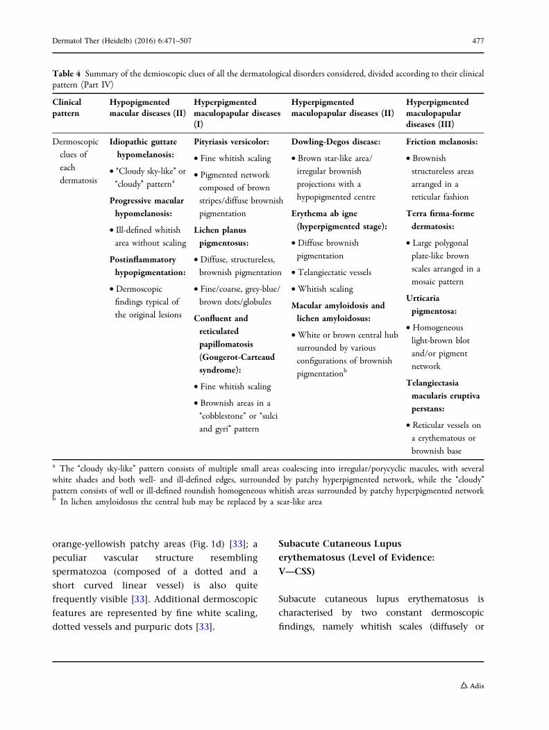
Table 4 Summary of the demioscopic clues of all the dermatological disorders considered, divided according to their clinicalpattern (Part IV)
Clinicalpattern
Hypopigmentedmacular diseases (II)
Hyperpigmentedmaculopapular diseases(I)
Hyperpigmentedmaculopapular diseases (II)
Hyperpigmentedmaculopapulardiseases (III)
Dermoscopic
clues of
each
dermatosis
Idiopathic guttate
hypomelanosis:
• ‘‘Cloudy sky-like’’ or‘‘cloudy’’ patterna
Progressive macular
hypomelanosis:
• Ill-defined whitish
area without scaling
Postinflammatory
hypopigmentation:
• Dermoscopic
findings typical of
the original lesions
Pityriasis versicolor:
• Fine whitish scaling
• Pigmented network
composed of brown
stripes/diffuse brownish
pigmentation
Lichen planus
pigmentosus:
• Diffuse, structureless,
brownish pigmentation
• Fine/coarse, grey-blue/brown dots/globules
Confluent and
reticulated
papillomatosis
(Gougerot-Carteaud
syndrome):
• Fine whitish scaling
• Brownish areas in a
‘‘cobblestone’’ or ‘‘sulci
and gyri’’ pattern
Dowling-Degos disease:
• Brown star-like area/
irregular brownish
projections with a
hypopigmented centre
Erythema ab igne
(hyperpigmented stage):
• Diffuse brownish
pigmentation
• Telangiectatic vessels
•Whitish scaling
Macular amyloidosis and
lichen amyloidosus:
•White or brown central hub
surrounded by various
configurations of brownish
pigmentationb
Friction melanosis:
• Brownishstructureless areas
arranged in a
reticular fashion
Terra firma-forme
dermatosis:
• Large polygonalplate-like brown
scales arranged in a
mosaic pattern
Urticaria
pigmentosa:
• Homogeneous
light-brown blot
and/or pigment
network
Telangiectasia
macularis eruptiva
perstans:
• Reticular vessels ona erythematous or
brownish base
a The ‘‘cloudy sky-like’’ pattern consists of multiple small areas coalescing into irregular/porycyclic macules, with severalwhite shades and both well- and ill-defined edges, surrounded by patchy hyperpigmented network, while the ‘‘cloudy’’pattern consists of well or ill-defined roundish homogeneous whitish areas surrounded by patchy hyperpigmented networkb In lichen amyloidosus the central hub may be replaced by a scar-like area
Dermatol Ther (Heidelb) (2016) 6:471–507 477

peripherally distributed) and a mixed vascular
pattern (at least two types of vessels among
dotted, linear-irregular, linear and branching
vessels) over a pinkish-reddish background [34].
Focally distributed orange-yellowish structureless
areas may also be seen less commonly [34].
Table 5 Summary of The dermoscopic clues of all the dermatological disorders considered, divided according to theirclinical pattern (Part V)
Clinicalpattern
Itchypapulonodulardermatoses
Erythrodermas (I) Erythrodermas (II) Noninfectiousbalanitis—erythroplasiaof Queyrat
Dermoscopic
clues of
each
dermatosis
Hypertrophic
lichen planus:
• Rippled surface
• ‘‘Comedo-like’’
structures
• Round corneal
structures
(‘‘corn pearls’’)
Prurigo
nodularis:
• ‘‘White
starburst’’
patterna
Nodular
scabies:
•Mites (‘‘hang
glider sign’’)
• Burrows (‘‘jetwith
condensation
trails’’)
Acquired
perforating
dermatosis:
• Threeconcentric
areas with
different
aspect/colour
Erythrodermic
psoriasis:
• Diffusely
distributed whitish
scales
• Regularly arrangeddotted/glomerular
vessels
Erythrodermic
atopic dermatitis:
• Yellowish scales/
sero crusts
• Patchily distributeddotted vessels
Erythrodermic
mycosis fungoides:
• Linear vessels(including
spermatozoon-like
vessels) and dotted
vessels
Erythrodermic pityriasis rubra
pilaris:
• Orange blotches
• Islands of nonerythematous
(spared) skin displaying reticular
vessels
Erythrodermic scabies:
• Dark-brown triangular structures
located at the end of whitish
structureless wavy lines
(delta-wing jets with contrail)
Zoon’s plasma cell
balanitis:
• Focal/diffuseorange-yellowish
structureless areas
• Fairly focussed curved
vessels (including
serpentine, convoluted
and chalice-shaped)
Psoriatic balanitis:
• Regularly distributeddotted/glomerular
vessels
Seborrheic dermatitis
and non-specific
balanitis:
• Linear irregularunspecific blurry vessels
Erythroplasia of
Queyrat:
• Scattered glomerular
vessels
a Consists of radially arranged whitish lines or a peripheral whitish halo with some centrifugal coarse projections on abrownish and/or reddish background, which may surround brown-reddish/brown-yellowish crust(s), erosion(s) and/orhyperkeratosis/scales
478 Dermatol Ther (Heidelb) (2016) 6:471–507

Table6
Summaryof
thederm
oscopiccluesof
allthederm
atologicaldisordersconsidered,d
ivided
accordingto
theirclinicalpattern(PartVI)
Clin
ical
pattern
Inflammatorycicatricialalop
ecia
Non
scarring
alop
ecias
Scalingdisordersof
thescalp
Dermoscopic
cluesof
each
derm
atosis
Discoid
lupu
serythematosus:
•Follicularkeratonc
plugs,thick
arborising
vesselsandreddots(acute
lesions)
•Thinarborising
vesselsem
erging
from
yellowdots(latelesions)
•White
areasandbranchingvessels
(long-lastinglesions)
Lichenplanop
ilaris:
•Perifollicularscales
Fron
talfib
rosing
alop
ecia:
•Minor
perifollicularscaling
•Lonelyhair/predominance
of
follicularopenings
withonlyonehair
atthehair-bearing
margin
Folliculitisdecalvans:
•Follicularpustules
•Yellowdischarge/crusts
•Hairtuftsthat
contain[10
hairs
Shafts
Alopeciaareata:
•Black
dots,m
icro-exclamationmarkhairs,broken
hairs,taperedhairs,monilethrix-likehairsand
trichorrhexisnodosa
(acute
form
s)
•Regular
yellowdots(inactivelesions);
•Circleand/or
pigtailhairs(regrowingphases)
Trichotillom
ania:
•Hairsbroken
atdifferentlengths
•Shorthairswithtrichoptilosis(‘‘split
ends’’)
•Other:irregularcoiledhairs,am
orphoush
airresidues,
blackdots,fl
ame-likehairs,tulip-like
hairsand
V-signa
And
rogeneticalop
ecia:
•Hairshaftthicknessheterogeneity
•Increasedproportion
ofthin
andvellushairs([
10%
ofthehairs)
Telogen
efflu
vium
:
•Lackof
features
typicalof
otherdiseasesb
Tinea
capitis:
•‘‘C
omma’’
hair,‘‘corkscrew
’’hair,‘‘zigzag’’
hairand‘‘M
orse
code’’hair
Scalppsoriasis:
•Red
dots/globules
•Signet
ring
vessels,redloops,white
scales,
punctate
hemorrhages
andhidden
hairs
(withalower
specificity)
Sebo
rrheic
derm
atitis:
•Arborizingvessels
•Yellowishscaling,structurelessredareas,
honeycom
bpigm
entandcommavessels
(withalower
specificity)
Pityriasisam
iantacea:
•Com
pact
whitekeratoticmaterialadhering
toatuftof
hair(asbestos-likescale)
aTwoor
morehairsem
erging
from
onefollicularun
itthat
arebroken
atthesamelevel
bCom
mon,but
nonspecific,find
ingsincludethepresence
ofem
ptyhairfollicles,a
predom
inance
offollicularun
itswithonlyonehair,perifo
lliculardiscolouration
(the
peripilarsign),uprightregrow
inghairs(m
ainlyacuteform
s)andprogressiveun
iform
hairthinning
(chrom
eform
s).Im
portantly,thereisno
significant
difference
betweenthefin
dingsin
thefrontalarea
andthosein
theoccipitalarea,w
hich
differentiates
telogeneffluvium
from
androgeneticalopecia
Dermatol Ther (Heidelb) (2016) 6:471–507 479
PAPULOSQUAMOUS/PAPULOKERATOTIC DERMATOSES
Classical Lichen Planus (Level of Evidence:
II)
The dermoscopic hallmark of classical lichen
planus is represented by Wickham striae
(Fig. 2a), which may appear as pearly-whitish
(and less commonly yellow or blue-white)
structures possibly displaying several
morphological patterns, including reticular
(the most common), linear, ‘‘radial
streaming’’, annular, round, ‘‘leaf venation’’
(delicate secondary striae branching from the
centred whitish venation, linked together at
either end, mimicking the crystal structure of
snow) and ‘‘starry sky’’ (clustered, follicular
white dots) aspect [13–15, 35–39]. Dotted,
globular and/or linear vessels, mainly
localised at the periphery of the lesion (and
less commonly showing a perifollicular or
diffuse pattern), violet, reddish, pink, brown
or yellow background, white/yellow dots and
some pigmented structures (dots, globules and/
or reticular or cloud-like areas) are other
additional dermoscopic findings of active
lesions [13–15, 35–39].
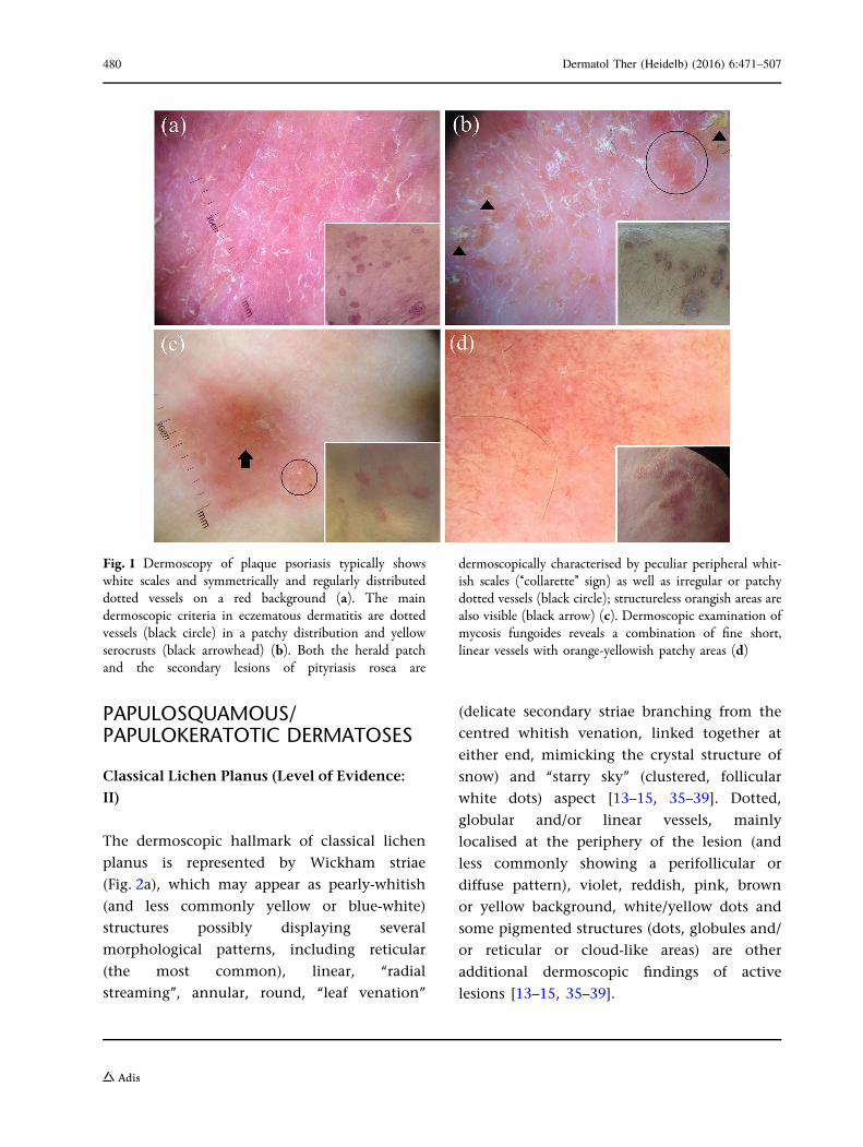
Fig. 1 Dermoscopy of plaque psoriasis typically showswhite scales and symmetrically and regularly distributeddotted vessels on a red background (a). The maindermoscopic criteria in eczematous dermatitis are dottedvessels (black circle) in a patchy distribution and yellowserocrusts (black arrowhead) (b). Both the herald patchand the secondary lesions of pityriasis rosea are
dermoscopically characterised by peculiar peripheral whit-ish scales (‘‘collarette’’ sign) as well as irregular or patchydotted vessels (black circle); structureless orangish areas arealso visible (black arrow) (c). Dermoscopic examination ofmycosis fungoides reveals a combination of fine short,linear vessels with orange-yellowish patchy areas (d)
480 Dermatol Ther (Heidelb) (2016) 6:471–507

Pityriasis Rosea (Level of Evidence: II)
See the section ‘‘Dermatoses presenting with
erythematous-desquamative patches/plaques’’.
Papulosquamous Sarcoidosis (Level
of Evidence: II)
See the section ‘‘Common facial inflammatory
skin diseases’’ (Fig. 2b).
Guttate Psoriasis (Level of Evidence: III)
Guttate psoriasis classically displays a
distinctive monomorphic dermoscopic aspect
consisting of dotted vessels regularly distributed
all over the lesion, which are often associated
with whitish scales (Fig. 2c) [1, 40], similarly to
that seen in plaque-type psoriasis [1–6, 41];
orange-yellowish structureless areas may also be
present, but they are quite uncommon [40].
Pityriasis Lichenoides Chronica (Level
of Evidence: III)
The most peculiar dermoscopic findings of
pityriasis lichenoides chronica include
nondotted vessels (i.e. milky red
areas/globules, linear irregular and branching
vessels), focally distributed dotted vessels and
orange-yellowish structureless areas (Fig. 2d)
[40]. Interestingly, whitish areas may
Fig. 2 The dermoscopic analysis of classical lichen planustypically shows the Wickham striae over a purplishbackground (a). Dermoscopy of papulosquamous sarcoido-sis shows the characteristic orange-yellowish background,in combination with in-focus fine linear vessels (blackarrowhead); whitish lines and white scales are also evidentin the centre (b). Guttate psoriasis lesions typically show adistinctive monomorphic dermoscopic picture, with dottedvessels distributed in a diffuse pattern (c). Dermoscopy of
pityriasis lichenoides chronica frequently displays nondot-ted vessels, e.g. linear irregular vessels (black arrowhead),focally distributed dotted vessels (black circle) andorange-yellowish structureless areas (d). Dermoscopicexamination of a case of disseminated superficial actinicporokeratosis displays the peculiar ‘‘cornoid lamella’’ at theperiphery of the lesion (e). Dermoscopy of a necroticlesion of lymphomatoid papulosis shows a centralbrown-grey structureless area (f)
Dermatol Ther (Heidelb) (2016) 6:471–507 481
sometimes be present in the context of
clinically active lesions as a result of focal
post-inflammatory hypopigmentation [1, 40].
Classical Pityriasis Rubra Pilaris (Level
of Evidence: V—CSS, CR)
Dermoscopy of classical pityriasis rubra pilaris
papules may show round/oval yellowish areas
surrounded by vessels of mixed morphology,
namely linear and dotted [1, 16].
Additionally, central keratin plugs may also
be observed [1].
Disseminated Forms of Porokeratosis
(Level of Evidence: V—CSS, CR)
The most peculiar dermoscopic feature of all
variants of porokeratosis is the ‘‘cornoid
lamella’’, which appears as a well-defined,
thin, white-yellowish, annular peripheral
hyperkeratotic structure (‘‘white track’’) similar
to the outlines of a volcanic crater as observed
from a high point, which may be
hyperpigmented in disseminated superficial
actinic porokeratosis (Fig. 2e); the centre of the
lesions is usually whitish or brownish and may
exhibit circular and/or linear whitish and/or
hyperpigmented tracks, blue-grey dots and
dotted, linear or globular vessels (Fig. 2e)
[17, 42–49].
Lymphomatoid Papulosis (Level
of Evidence: V—CSS)
Dermoscopic pattern of lymphomatoid
papulosis varies according to the disease stage.
The initial inflammatory papules usually
display a vascular pattern of tortuous irregular
(or dotted at low magnification) vessels,
surrounded by white structureless areas,
radiating from the centre to the periphery of
the lesion, while in more mature papules, such a
vascular pattern is less evident and generally
detectable only at the periphery of the lesion as
the centre is occupied by a whitish-yellowish
(hyperkeratotic lesions) or brown-grey (necrotic
lesions) structureless area (Fig. 2f) [50].
Papulosquamous Chronic GVHD (Level
of Evidence: V—CSS)
The dermoscopic aspect of papulosquamous
chronic GVHD consists of whitish scales
associated with vessels of mixed morphology,
namely dotted and linear [1]. Although such a
pattern is quite unspecific, it might be useful in
assisting the clinical differential diagnosis with
the other above-mentioned papulosquamous
disorders as they typically show a different
appearance [1].
Poikiloderma Vasculare Atrophicans/
Parakeratosis Variegata (Level of Evidence:
V—CSS)
This condition typically shows a monomorphic
pattern consisting of relatively blurred
branched vessels on a reddish or
orangish-brown background, associated with
sparse whitish scales [51].
Acantholytic and Dyskeratotic Papular
Disorders (Grover Disease, Darier Disease
and BRAF-Inhibitor-Induced Acantholytic
Dyskeratosis; Level of Evidence: V—CSS,
CR)
Grover disease may display different features
according to the histological subtype, with a
central star-shaped/branched polygonal/
roundish-oval brownish area surrounded by a
482 Dermatol Ther (Heidelb) (2016) 6:471–507
whitish halo being characteristic of the
Darier-like histological subtype (Fig. 3a) and
whitish scaling over a reddish-yellowish
background being characteristic of the
spongiotic histological subtype (Fig. 3b);
dotted and/or linear/irregular vessels may be
found in both such forms (Fig. 3a, b) [52–55].
Importantly, the dermoscopic pattern of
Darier-like Grover disease overlaps with that
detectable in both Darier disease and
BRAF-inhibitor-induced acantholytic
dyskeratosis (Fig. 3c, d) [55–58].
COMMON FACIALINFLAMMATORY SKIN DISEASES
Rosacea (Level of Evidence: III)
The dermoscopic hallmark of rosacea is
represented by the presence of linear vessels
characteristically arranged in a polygonal
network (vascular polygons) [26, 59] (Fig. 4a).
Additional features include rosettes [60],
follicular plugs, white/yellowish scales,
orange-yellowish areas, pigmentation
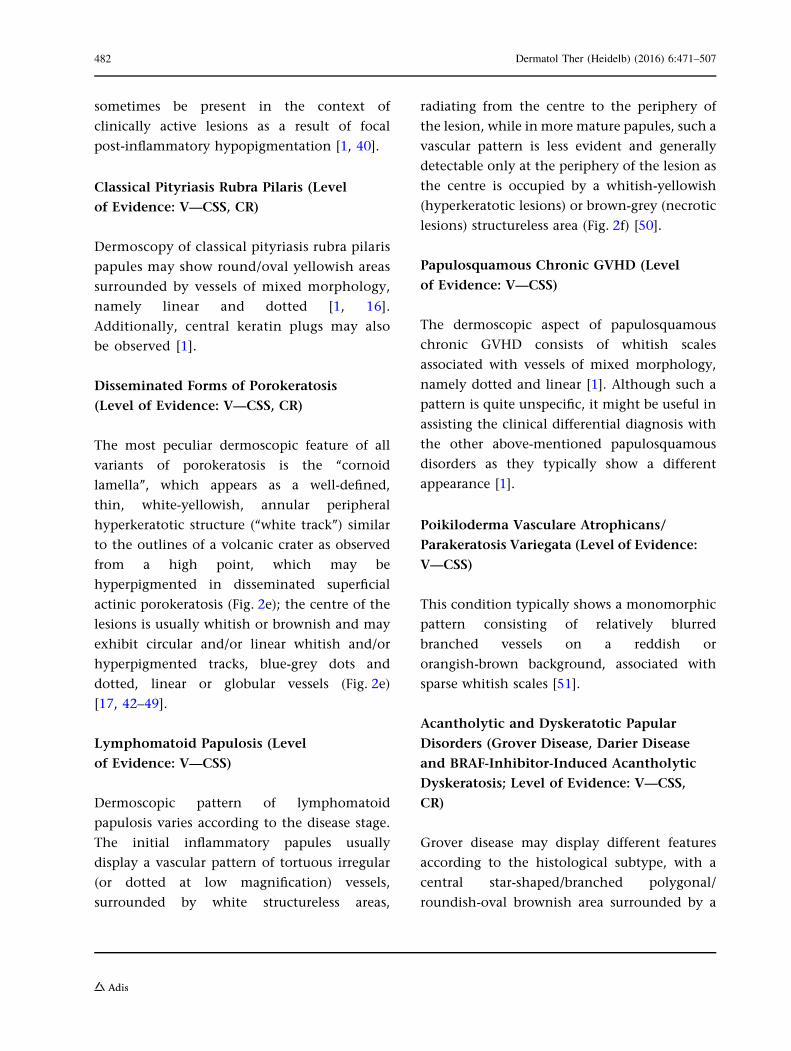
Fig. 3 Dermoscopy of Darier-like Grover disease displaysa central branched polygonal brownish area surrounded bya thin whitish halo with peripheral dotted vessels (blackcircle) (a), while spongiotic Grover disease presents withwhitish scaling over a reddish-yellowish background andirregular vessels (black circle) (b). Dermoscopic examina-tion of Darier disease (c) and BRAF-inhibitor-induced
acantholytic dyskeratosis (d) shows a pattern similar tothat observed in Darier-like Grover disease, with a centrallylocated polygonal brownish area surrounded by a whitishhalo and linear vessels (black arrow) in Darier disease(c) and a central branched polygonal brownish areasurrounded by a thin whitish halo in the latter condition(d)
Dermatol Ther (Heidelb) (2016) 6:471–507 483

structures, dilated follicles and follicular
pustules (papulopustular rosacea) [26, 59].
Seborrheic Dermatitis (Level of Evidence:
III)
The most typical dermoscopic findings of
seborrheic dermatitis include dotted vessels
in a patchy distribution and fine yellowish
scales (in combination or not with white
scales) (Fig. 4b); follicular plugs,
orange-yellowish areas, whitish structureless
areas and linear branching vessels are less
common features [26].
Discoid Lupus erythematosus (Level
of Evidence: III)
Dermoscopy of facial (and extra-scalp in general)
discoid lupus erythematosus shows different
features according to the stage of disease, with
erythema, perifollicular whitish halo, follicular
keratotic plugs, red dots and white scaling being
themost common/characteristic features of early
lesions, and whitish structureless areas,
hyperpigmentation (honeycomb network,
perifollicular pigmentation, radial pigment
streaks or pigmentation arranged in unspecified
pattern) and blurred telangiectasias (mainly
Fig. 4 The main dermoscopic feature of rosacea is thepresence of linear vessels, which are characteristicallyarranged in a polygonal network (a). The most typicaldermoscopic finding of seborrheic dermatitis is representedby the presence of dotted vessels in a patchy distribution(black circle) and yellowish scales (black arrows); blurrylinear branching vessels (black arrowheads) and whitishscales are also not uncommonly present (b). Dermoscopyof an intermediate-stage lesion of facial discoid lupuserythematosus reveals follicular white/yellowish rings/keratotic plugs, whitish scaling and blurred branchingvessels (black arrow) over a reddish background (c).
Dermoscopic examination of facial sarcoidosis displays astructureless orange-yellowish background with focussedlinear vessels (d), while granuloma faciale features dilatedfollicular openings (black arrows) associated with linear/branching vessels (black circles) over a pinkish background(e). Dermoscopy of demodicidosis shows the so-called‘‘Demodex tails’’, which are visualised as creamy/whitishgelatinous threads protruding out of follicular openings(black arrow), and ‘‘Demodex follicular openings’’, whichappear as round and coarse follicular openings containinglight brown/greyish plugs surrounded by an erythematoushalo (black arrowhead) (f)
484 Dermatol Ther (Heidelb) (2016) 6:471–507

linear branching vessels and less commonly
dotted/polymorphous vessels) representing the
most frequent findings of late phases [26, 61–65];
intermediate-stage lesionsmay display amixture
of the aforementioned features (Fig. 4c)
[26, 61–65]. Less common dermoscopic findings
include diffuse hyperkeratosis (hypertrophic
discoid lupus erythematosus) [65], dilated
follicles and yellowish scales [26, 61–65].
Granulomatous Skin Diseases (Sarcoidosis,
Cutaneous Leishmaniasis and Lupus
Vulgaris; Level of Evidence: III)
The dermoscopic signature of all these
granulomatous facial dermatoses consists of
structureless orange-yellowish areas (diffuse or
localised—often described as ‘‘grains of sand’’ in
lupus vulgaris and teardrop-like areas in
leishmaniasis), commonly associated with
focussed linear or branching vessels (Fig. 4d)
[26, 66–80]. Other possible findings include
milia-like cysts, erythema, whitish lines or
structureless areas, follicular plugs, dilated
follicles, pigmentation structures, and white
and/or yellow scales [26, 66–80]. Additionally,
leishmaniasis has been reported to show
hyperkeratosis, further vascular features
(hairpin, comma-shaped, glomerular-like and/
or corkscrew vessels), central ulcerations and
white peripheral projections (white starburst
pattern) [73–80]. Nevertheless, dermoscopy
may not be considered as a reliable tool in
differentiating such granulomatous diseases
and therefore histological assessment is needed
to reach a definitive diagnosis [26, 66–80].
Granuloma Faciale (Level of Evidence: III)
The dermoscopic hallmark of granuloma faciale
is representedby thepresenceof dilated follicular
openings associated with linear branching
vessels (which sometimes appear as focussed
elongated telangiectasias) over a pinkish
background (Fig. 4e) [26, 81–83]; additional
findings include perifollicular whitish halo,
whitish streaks, follicular plugs, yellowish scales
and pigmentation structures [26, 81–83].
Demodicidosis (Level of Evidence: V—CSS)
The most indicative dermoscopic features of all
types of demodicidosis are the so-called
‘‘Demodex tails’’, which are creamy/whitish
gelatinous threads (representing the presence of
the mite itself under magnification) protruding
out of follicular openings, and ‘‘Demodex
follicular openings’’, which appear as round and
coarse follicular openings containing light
brown/greyish plugs surrounded by an
erythematous halo (Fig. 4f) [84]. Other
unspecific dermoscopic findings (whose
prevalence varies according to the subtypes of
demodicidosis) include diffuse erythema, scaling,
pustules and reticular dilated vessels [84].
ACQUIRED KERATODERMAS
Chronic Hand Eczema (Level of Evidence:
III)
The most specific dermoscopic features of
chronic hand eczema include brownish-orange
dots/globules (corresponding to tiny spongiotic
vesicles), yellowish scales and yellowish-orange
crusts [28, 85]; other less common findings are
focally distributed whitish scaling and dotted
vessels (Fig. 5a) [28, 85].
Palmar Psoriasis (Level of Evidence: III)
The main dermoscopic finding of palmar
psoriasis is represented by the presence of
Dermatol Ther (Heidelb) (2016) 6:471–507 485

white scales typically distributed in a diffuse
pattern (and only infrequently showing
patchily or central distribution) (Fig. 5b)
[23, 28, 85]. Dotted vessels, which are
regularly distributed (and only rarely in rings
or patchy-distributed), may also be visible quite
commonly when using a fluid interface (which
reduce the scaling) [23]; focal yellowish scales
are an additional but very rare finding [23, 28].
Keratoderma due to Mycosis Fungoides
(Level of Evidence: V—CR)
The most characteristic dermoscopic finding of
keratoderma due to mycosis fungoides consists
of relatively large amber scales over a
white-to-pinkish background; sparse whitish
scales and several non-specific reddish fissures
are also visible [85].
Keratoderma due to Pityriasis Rubra Pilaris
(Level of Evidence: V—CR)
The dermoscopic hallmark of keratoderma
resulting from pityriasis rubra pilaris is the
presence of patchily distributed,
homogeneous, structureless orange areas
presenting different sizes; unspecific whitish
scaling may also be observed [85].
Fig. 5 Dermoscopy of chronic hand eczema typicallyreveals sparse whitish scales, yellowish scaling (blackcircles) and orangish dots/globules (black arrowheads),while palmar psoriasis and tinea manuum respectivelydisplay diffuse white scaling (b) and white scales mainly
localised in the skin furrows (c). Dermoscopic examinationof a case of palmar lichen planus shows roundish yellowishareas, some of which display peripheral projections in astar-like appearance (black arrowheads) over a purplishbackground (d)
486 Dermatol Ther (Heidelb) (2016) 6:471–507

Tinea Manuum (Level of Evidence: V—PO)
From a dermoscopic point of view, tinea
manuum displays whitish scaling distributed
in a characteristic pattern, i.e. mainly localised
in the physiologic palmar creases (Fig. 5c)
(personal observations).
Palmar Lichen Planus (Level of Evidence:
V—PO)
Palmar lichen planus is typically characterised
by roundish yellowish areas often having
peripheral projections that may create a
star-like appearance; a purplish background is
sometimes visible (Fig. 5d) (personal
observations).
Aquagenic Palmar Keratoderma (Level
of Evidence: V—CR)
Dermoscopy of aquagenic palmar keratoderma
shows large yellow well-defined globules not
affecting dermatoglyphs [86] or simply
enlargement of the sweat duct pores when
compared with a normal-looking palmar skin
area [87, 88].
SCLERO-ATROPHIC DERMATOSES
Necrobiosis Lipoidica (Level of Evidence:
III)
Dermoscopy of necrobiosis lipoidica lesions
typically shows comma-shaped (incipient
lesions), network-shaped/hairpin-like (more
developed lesions) or elongated, branching
and focussed serpentine (advanced lesions)
vessels over a yellowish-orange/
whitish-pinkish background (with or without
reddish areas) (Fig. 6a) [89–93]. Additional
findings include patchy pigmented
reticulum, yellow crusting and ulceration
[89–93].
Morphea (Level of Evidence: IV)
The most specific dermoscopic feature of
morphea consists of whitish fibrotic beams,
which are frequently crossed by linear
branching vessels (Fig. 6b) [94–96]; pigment
network-like structures are also often
evident, while ‘‘comedo-like openings’’ and
whitish patches are less commonly seen
[94–96].
Fig. 6 Dermoscopic examination of an advanced lesion ofnecrobiosis lipoidica reveals elongated, branching andfocussed serpentine vessels over a yellowish-orange/whitishbackground (a). Dermoscopy of morphea shows the typicalfibrotic beams (black arrows) associated with linear
branching vessels (b), while cutaneous lichen sclerosusdisplays several ‘‘comedo-like openings’’ (follicular keratoticplugs), whitish patches, dotted vessels (black circle) anddelicate linear branching vessels (c)
Dermatol Ther (Heidelb) (2016) 6:471–507 487
Cutaneous Lichen Sclerosus (Level
of Evidence: IV)
The dermoscopic hallmarks of cutaneous lichen
sclerosus include ‘‘comedo-like openings’’
(follicular keratotic plugs) and whitish patches
(Fig. 6c) [94, 95, 97–100]; less common/less
specific findings are represented by delicate
linear branching vessels, fibrotic beams, grey
dots, purpuric spots, pigment network-like
structures, non-branching vessels (comma-like,
hairpin and/or dotted), fine whitish scaling and
chrysalis structures [94, 95, 97–100].
HYPOPIGMENTED MACULARDISEASES
Extragenital Guttate Lichen Sclerosus
(Level of Evidence: IV)
See the section ‘‘Sclero-atrophic dermatoses’’.
Achromic Pityriasis Versicolor (Level
of Evidence: V—PO)
Dermoscopy of achromic/hypochromic lesions
of pityriasis versicolor usually shows a fairly
demarcated white area with fine scales that are
commonly localised in the skin furrows
(Fig. 7a), similarly to hyperpigmented lesions
[101].
Guttate Vitiligo (Level of Evidence:
V—CSS)
The most common/typical dermoscopic
features of guttate vitiligo include a
well-demarcated, dense/glowing, white area
and perifollicular hyperpigmentation (which is
more frequently seen in repigmenting or
progressing lesions than stable lesions)
(Fig. 7b) [102–104]. Other possible findings
include perilesional hyperpigmentation, a
reversed pigmentary network, reticular
pigmentation and telangiectasias [102–104].
Idiopathic Guttate Hypomelanosis (Level
of Evidence: V—CSS)
Dermoscopic examination of idiopathic guttate
hypomelanosis displays two main aspects, i.e.
the ‘‘cloudy sky-like’’ pattern (multiple small
areas coalescing into irregular/polycyclic
macules, with several white shades and both
well- and ill-defined edges, surrounded by
patchy hyperpigmented network) and the
‘‘cloudy’’ pattern (well or ill-defined roundish
homogeneous whitish areas surrounded by
patchy hyperpigmented network) (Fig. 7c)
[101, 105].
Progressive Macular Hypomelanosis (Level
of Evidence: V—PO)
Progressive macular hypomelanosis is
dermoscopically characterised by an ill-defined
whitish area without scaling [101].
Postinflammatory Hypopigmentation
(Level of Evidence: V—CSS, PO)
Postinflammatory macular hypopigmentations
often present some dermoscopic findings
typical of the original lesions, e.g. non-dotted
vessels/orangish structureless areas in pityriasis
lichenoides [1, 40], dotted vessels in guttate
psoriasis [1, 40] and star-like depigmentation
in prurigo nodularis [103] (Fig. 7d), thereby
assisting the retrospective diagnosis
[1, 40, 106].
488 Dermatol Ther (Heidelb) (2016) 6:471–507

HYPERPIGMENTEDMACULOPAPULAR DISEASES
Pityriasis Versicolor (Level of Evidence:
V—CR, PO)
Dermoscopy of hyperpigmented lesions of
pityriasis versicolor shows fine whitish scaling
(often localised in the skin furrows) associated
with a pigmented network composed of brown
stripes [107] or a diffuse, more or less
homogeneous, brownish pigmentation
(Fig. 8a) (personal observations).
Lichen Planus Pigmentosus (Level
of Evidence: V—CSS)
The main dermoscopic patterns of lichen
planus pigmentosus are represented by a
diffuse, structureless, brownish pigmentation
and/or fine/coarse, grey-blue/brown
dots/globules (Fig. 8b); perifollicular/annular
Fig. 7 Dermoscopy of achromic/hypochromic lesions ofpityriasis versicolor usually shows a fairly demarcated whitearea with fine scales that are commonly localised in theskin furrows (a), while active lesions of guttate vitiligotypically display a well-demarcated, dense/glowing, oftenassociated with perifollicular hyperpigmentation (blackarrowheads) (b). Dermoscopic examination of idiopathicguttate hypomelanosis may show multiple small areas
coalescing into irregular/polycyclic macules, with severalwhite shades and both well- and ill-defined edges,surrounded by patchy hyperpigmented network (‘‘cloudysky-like’’ pattern) (c), whilst postinflammatory hypopig-mentation often presents with some dermoscopic findingstypical of the original lesions (in this case, the star-likearrangment typical of prurigo nodularis) (d)
Dermatol Ther (Heidelb) (2016) 6:471–507 489

pigmentation and white dots are other less
common findings [38, 39, 108, 109].
Confluent and Reticulated Papillomatosis
(Gougerot-Carteaud Syndrome; Level
of Evidence: V—CSS, CR)
Confluent and reticulated papillomatosis
typically displays fine whitish scaling
associated with brownish, homogeneous, more
or less defined, polygonal, flat globules
separated by whitish/pale striae creating a
cobblestone appearance [110] (Fig. 8c) or
brownish areas presenting a ‘‘sulci and gyri’’
pattern [111].
Dowling-Degos Disease (Level of Evidence:
V—CR)
The dermoscopic aspect of Dowling-Degos
disease consists of a brown star-like area/
irregular brownish projections with a
hypopigmented centre over a brownish/
reddish-brown background [112–114].
Erythema Ab Igne (Hyperpigmented Stage;
Level of Evidence: V—PO)
The pigmentary stage of erythema ab igne is
typically characterised by diffuse brownish
pigmentation with or without telangiectatic
vessels/whitish scaling (Fig. 8d) [110].
Fig. 8 Dermoscopy of hyperpigmented lesions of pityriasisversicolor often shows fine whitish scaling localised in theskin furrows associated with a diffuse brownish pigmen-tation (a). The most common dermoscopic finding oflichen planus pigmentosus is represented by fine/coarse,grey-blue/brown dots over a brownish background (b),while confluent and reticulated papillomatosis (Gouger-ot–Carteaud syndrome) displays fine whitish scaling andbrownish, homogeneous, more or less defined, polygonal,
flat globules separated by whitish/pale striae creating acobblestone pattern (c). Dermoscopic examination ofpigmented lesions of erythema ab igne may reveal diffusebrownish pigmentation with telangiectatic vessels/finewhitish scaling, while friction melanosis and urticariapigmentosa typically display brownish structureless areasarranged in a reticular fashion (e) and a homogeneouslight-brown blot with a pigment network (f), respectively
490 Dermatol Ther (Heidelb) (2016) 6:471–507

Primary Cutaneous Amyloidosis (Macular
Amyloidosis and Lichen Amyloidosus;
Level of Evidence: V—CSS)
The most common dermoscopic finding of both
macular amyloidosis and lichen amyloidosus is
a central hub (which is either white or brown in
the former and white in the latter) surrounded
by various configurations of brownish
pigmentation, including fine radiating streaks,
dots, leaf-like projections and bulbous
projections [115]. Additionally, in lichen
amyloidosus the central hub may be replaced
by a scar-like area (which may be the only
feature in larger and thicker lesions) and a rim
of white collarette (resembling a volcanic crater)
may sometimes be appreciated [115].
Friction Melanosis (Level of Evidence:
V—CR)
The dermoscopic examination of friction
melanosis typically reveals brownish
structureless areas arranged in a reticular
fashion (Fig. 8e) [115].
Terra Firma-Forme Dermatosis (Level
of Evidence: V—CSS)
Dermoscopy of terra firma-forme dermatosis
classically shows large polygonal plate-like
brown scales arranged in a mosaic pattern [116].
Maculopapular Cutaneous Mastocytosis
(Urticaria Pigmentosa, UP,
and Telangiectasia Macularis Eruptiva
Perstans, TMEP; Level of Evidence:
V—CSS, CR)
The most common dermoscopic features of
UP consist of a homogeneous light-brown
blot and/or pigment network (Fig. 8f), while
TMEP is mainly characterised by reticular
vessels on an erythematous/brownish base
(‘‘reticular vascular’’ pattern), sometimes
associated with a brownish network
[117–121]. However, dermoscopy cannot
guarantee a reliable distinction of such
conditions as, albeit uncommonly, UP may
display the reticular vascular pattern as well
[117]. Other less frequent vascular findings
visible in both UP and TMEP include sparse
dotted vessels and thin and tortuous linear
vessels [117–121].
ITCHY PAPULONODULARDERMATOSES
Hypertrophic Lichen Planus (Level
of Evidence: V—CR)
Dermoscopic examination of hypertrophic
lichen planus lesions displays a characteristic
pattern consisting of a rippled surface with
comedo-like structures filled with yellow
keratinous plugs and/or round corneal
structures (‘‘corn pearls’’) (Fig. 9a)
[14, 15, 35–37, 108, 122]; less common
features include Wickham striae, unspecific
vascular findings (red globules, linear and
dotted vessels), chalk-white structureless areas,
scaling and central hyperpigmentation (Fig. 9a)
[14, 15, 35–37, 108, 122].
Prurigo Nodularis (Level of Evidence:
V—CSS)
The dermoscopic hallmark of prurigo nodularis
(both hyperkeratotic and excoriated lesions) is
represented by the presence of the so-called
‘‘white starburst pattern’’, consisting of radially
arranged whitish lines or peripheral whitish
halo with some centrifugal coarse projections
on a brownish and/or reddish background,
Dermatol Ther (Heidelb) (2016) 6:471–507 491
which may surround brown-reddish/
brown-yellowish crust(s), erosion(s) and/or
hyperkeratosis/scales (Fig. 9b) [106].
Nodular Scabies (Level of Evidence:
V—CSS)
The distinctive dermoscopic sign of nodular
lesions of scabies is the presence of mites (‘‘hang
glider sign’’) and/or burrows (‘‘jet with
condensation trails’’) [123]. According to a
recent study on ten patients with nodular
scabies, the latter dermoscopic finding would
be constantly present in such a type of scabies
[123], but in our experience it may be missing
(especially in extragenital sites) and unspecific
vascular features (mainly dotted vessels) may be
the only detectable findings (Fig. 9c) [1].
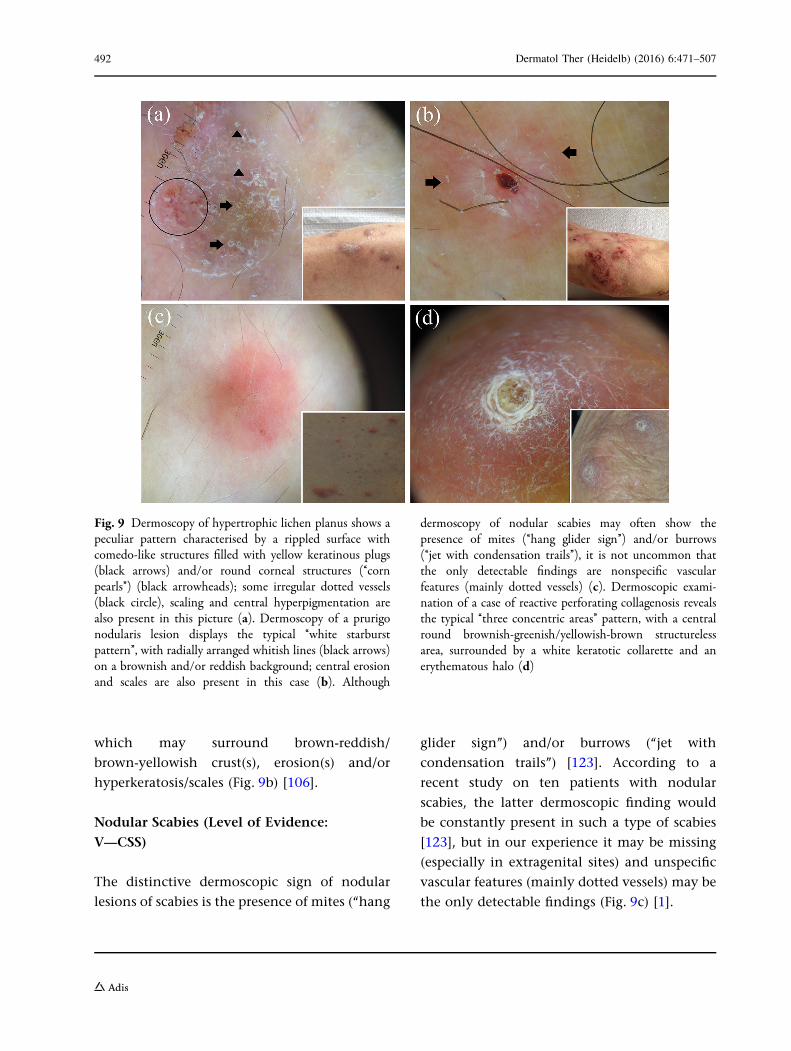
Fig. 9 Dermoscopy of hypertrophic lichen planus shows apeculiar pattern characterised by a rippled surface withcomedo-like structures filled with yellow keratinous plugs(black arrows) and/or round corneal structures (‘‘cornpearls’’) (black arrowheads); some irregular dotted vessels(black circle), scaling and central hyperpigmentation arealso present in this picture (a). Dermoscopy of a prurigonodularis lesion displays the typical ‘‘white starburstpattern’’, with radially arranged whitish lines (black arrows)on a brownish and/or reddish background; central erosionand scales are also present in this case (b). Although
dermoscopy of nodular scabies may often show thepresence of mites (‘‘hang glider sign’’) and/or burrows(‘‘jet with condensation trails’’), it is not uncommon thatthe only detectable findings are nonspecific vascularfeatures (mainly dotted vessels) (c). Dermoscopic exami-nation of a case of reactive perforating collagenosis revealsthe typical ‘‘three concentric areas’’ pattern, with a centralround brownish-greenish/yellowish-brown structurelessarea, surrounded by a white keratotic collarette and anerythematous halo (d)
492 Dermatol Ther (Heidelb) (2016) 6:471–507

Acquired Perforating Dermatosis (Level
of Evidence: V—CR)
The dermoscopic pattern of acquired
perforating dermatosis is characterised by the
presence of three concentric areas
[1, 106, 124, 125], namely a central
round brownish-greenish/yellowish-brown
structureless area (I), surrounded by a white
keratotic collarette (II) and an erythematous
halo with or without dotted vessels (III)
(‘‘reactive perforating collagenosis’’
histological subtype) (Fig. 9d) [1, 106, 124] or
bright white clods (I), centred in a structureless
grey area (II), surrounded by reticular brown
lines (III) (‘‘perforating folliculitis’’ histological
subtype) [125].
ERYTHRODERMAS
Erythrodermic Psoriasis (Level
of Evidence: V—CR)
Dermoscopy of erythrodermic psoriasis
reveals a monomorphous pattern with
diffusely distributed whitish scales and
regularly arranged dotted/glomerular vessels
on a fairly homogeneous reddish background
[29, 126].
Erythrodermic Atopic Dermatitis (Level
of Evidence: V—CR)
As for other types of eczematous dermatitis, the
most important dermoscopic features of
erythrodermic atopic dermatitis consist of
yellowish scales/serocrusts and patchily
distributed dotted vessels on a pinkish
background; unspecific sparse whitish scales
may also be seen [29].
Erythrodermic Mycosis Fungoides (Level
of Evidence: V—CR)
The most characteristic dermoscopic finding of
erythrodermicmycosis fungoides is representedby
the combination of linear vessels (some of them
having a spermatozoon-like shape) and dotted
vessels over a whitish-pinkish background;
unspecific sparsewhitish scales are also visible [29].
Erythrodermic Pityriasis Rubra Pilaris
(Level of Evidence: V—CR)
Dermoscopy of erythrodermic pityriasis rubra
pilaris typically displays peculiar orange blotches
and islands of nonerythematous (spared) skin
displaying reticular vessels; additional features
include diffuse whitish scaling and scattered
dotted vessels over a reddish background [29].
Erythrodermic Scabies (Level of Evidence:
V—CR)
The main dermoscopic findings of erythrodermic
scabies include whitish scales and thousands of
characteristic dark-brown triangular structures
located at the end of whitish structureless wavy
lines (delta-wing jets with contrail) over a reddish
background [127].
COMMON FORMSOF NONINFECTIOUS BALANITISAND ERYTHROPLASIA OF QUEYRAT
Zoon’s Plasma Cell Balanitis (Level
of Evidence: V—CSS)
The dermoscopic hallmark of Zoon’s plasma cell
balanitis is the presence of focal/diffuse
orange-yellowish structureless areas and/or
fairly focussed curved vessels (including
serpentine, convoluted and chalice-shaped);
Dermatol Ther (Heidelb) (2016) 6:471–507 493

other possible findings include linear irregular
blurry vessels and dotted vessels [128].
Psoriatic Balanitis
From a dermoscopic point of view, psoriatic
balanitis is characterised by the presence of
regularly distributed dotted/glomerular vessels
[23, 129].
Seborrheic Dermatitis and Non-Specific
Balanitis (Level of Evidence: V—PO)
Seborrheic dermatitis and non-specific balanitis
usually show only linear irregular unspecific
blurry vessels [128].
Erythroplasia of Queyrat (Level
of Evidence: V—CR)
Erythroplasia of Queyrat has been reported to
show scattered glomerular vessels [130].
COMMON INFLAMMATORYCICATRICIAL ALOPECIA
Discoid Lupus erythematosus (Level
of Evidence: II)
The dermoscopic hallmarks of active discoid
lupus erythematosus of the scalp are represented
by follicular keratotic plugs (quite large yellowish/
whitish dots) and thick arborising vessels
Fig. 10 Dermoscopy of discoid lupus erythematosus of thescalp varies according to the disease stage: active lesionsmay be mainly characterised by red dots (a) or follicularkeratotic plugs (quite large yellowish/whitish dots) andthick arborising vessels (b), while long-lasting lesionscommonly display loss of follicular openings, white areasand thin vessels (c). The main dermoscopic hallmarks ofactive lichen planopilaris are perifollicular scales; charac-teristic (but not pathognomonic) white dots (fibroticwhite dots) (black arrowheads) and a reddish backgroundare also present in less active areas in this case (d).
Dermoscopic examination of a case of frontal fibrosingalopecia reveals minor perifollicular scaling with anaflegmasic (ivory white to ivory beige) surroundingbackground; follicular openings with only one hair at thehair-bearing margin (black arrows) and lonely hair (blackarrowhead) are also visible (e). Classic dermoscopicappearance of active folliculitis decalvans showing follicularpustules, yellow discharge, crusts and characteristic hairtufts that contain [10 hair shafts (white arrowhead);unspecific vessels and erythema are also evident in thepicture (f)
494 Dermatol Ther (Heidelb) (2016) 6:471–507

(Fig. 10a, b) [131–137]; additional findings
include fine interfollicular scaling, blue-grey
dots, scattered brown discolouration and red
dots (Fig. 10a) [131–137]. Thin arborising vessels
emerging from the yellow dots (‘‘red spider in a
yellow dot’’) are considered peculiar of late,
prefibrotic lesions [133], while pink areas, loss of
follicular openings, white areas and branching
vessels are typical of long-lasting lesions (Fig. 10c)
[131, 133–137].
Lichen Planopilaris (Level of Evidence: II)
The main dermoscopic features of active lichen
planopilaris are perifollicular scales, which
typically migrate along the hair shaft and form
a tubular structure covering the proximal
portion of the emerging hair shaft
(‘‘collar-like’’ or ‘‘tubular’’ perifollicular
hyperkeratosis) (Fig. 10d) [132, 133, 136–141,
149, 150]; other possible dermoscopic findings
of active lesions include violaceous or
violet-brown inter- or perifollicular violaceous
areas (Fig. 10d), perifollicular inflammation,
elongated linear blood vessels in concentric
arrangement and target ‘‘blue-grey dots’’
[132, 133, 136–141, 149, 150]. Inactive/late
lesions may show characteristic (but not
pathognomonic) irregular, large white dots
(fibrotic white dots) (Fig. 10d) as well as less
specific findings such as acquired pili torti, loss
of follicular openings, white areas, honeycomb/
scattered hyperpigmentation, milky red areas
(strawberry ice cream colour) and small hair
tufts of 5–9 hairs [132, 133, 136–141, 149, 150].
Frontal Fibrosing Alopecia (Level
of Evidence: II)
The most common dermoscopic findings in
frontal fibrosing alopecia include a lack of
follicular openings and minor perifollicular
scaling [131, 132, 137, 140–144]; additionally,
perifollicular erythema may be seen but the
surrounding background is usually aflegmasic
(ivory white to ivory beige) (Fig. 10e)
[131, 132, 137, 140–144]. Interestingly, there is
often a strong predominance of follicular
openings with only one hair at the
hair-bearing margin and lonely hair may be
observed (Fig. 10e) [131, 132, 137, 140–144].
Fine arborising vessels and perifollicular brown
or brown-violet areas may sometimes be visible
[131, 132, 137, 140–144].
Folliculitis Decalvans (Level of Evidence:
II)
The most characteristic dermoscopic feature of
folliculitis decalvans is the presence of hair tufts
that contain[10 hair shafts (Fig. 10f), which are
often surrounded by a band of yellowish scales
(yellowish tubular scaling) and by perifollicular
epidermal hyperplasia (which may be arranged
in a starburst pattern) at their base
[132, 133, 137, 145–148]; other peculiar
findings in active folliculitis decalvans include
follicular pustules and yellow discharge and
crusts (Fig. 10f) [132, 133, 137, 145–149]. A
perifollicular concentration of blood vessels
(elongated loops/coiled vessels) and a
perifollicular erythema arranged in a starburst
pattern may also be visible
[132, 133, 137, 145–147, 149]. In long-lasting
lesions, ivory-white and milky-red areas
without follicular orifices predominate [145].
COMMON NONSCARRINGALOPECIAS
Alopecia Areata (Level of Evidence: II)
The most characteristic findings of active
alopecia areata include black dots,
Dermatol Ther (Heidelb) (2016) 6:471–507 495
micro-exclamation mark hairs, broken hairs,
tapered hairs, monilethrix-like hairs and
trichorrhexis nodosa, while long-standing
inactive disease is mainly characterised by
yellow dots and vellus hairs (Fig. 11a)
[131, 139, 151–158]. The main signs of
regrowing consist of upright and regularly
coiled (circle and/or pigtail) hairs
[131, 139, 151–158]. Less specific/less common
features of active stages include tulip hairs and
zigzag hairs [151, 152].
Trichotillomania (Level of Evidence: II)
Dermoscopy of trichotillomania often reveals a
chaotic pattern of diverse findings related to
hair fracturing [151, 159]. The most peculiar
features include hairs broken at different
lengths, short hairs with trichoptilosis (‘‘split
ends’’), irregular coiled hairs, amorphous hair
residues, black dots, flame-like hairs, tulip-like
hairs (short hairs with darker, tulip-shaped
ends) and V-sign (two or more hairs emerging
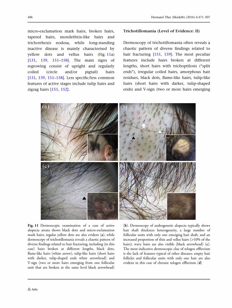
Fig. 11 Dermoscopic examination of a case of activealopecia areata shows black dots and micro-exclamationmark hairs; regular yellow dots are also evident (a), whiledermoscopy of trichotillomania reveals a chaotic pattern ofdiverse findings related to hair fracturing, including (in thiscase) hairs broken at different lengths, black dots,flame-like hairs (white arrow), tulip-like hairs (short hairswith darker, tulip-shaped ends white arrowhead) andV-sign (two or more hairs emerging from one follicularunit that are broken at the same level black arrowhead)
(b). Dermoscopy of androgenetic alopecia typically showshair shaft thickness heterogeneity, a large number offollicular units with only one emerging hair shaft, and anincreased proportion of thin and vellus hairs ([10% of thehairs); wavy hairs are also visible (black arrowhead) (c).The most indicative dermoscopic clue of telogen effluviumis the lack of features typical of other diseases; empty hairfollicles and follicular units with only one hair are alsoevident in this case of chronic telogen effluvium (d)
496 Dermatol Ther (Heidelb) (2016) 6:471–507
from one follicular unit that are broken at the
same level) (Fig. 11b) [131, 139, 151, 156,
159–163]. Less common/less specific findings
are tapered hairs, follicular microhemorrhages,
micro-exclamation mark hairs and upright
regrowing hairs [151, 159–163].
Androgenetic Alopecia (Level of Evidence:
IV)
The main dermoscopic features of androgenetic
alopecia include hair shaft thickness
heterogeneity, yellow dots (irregularly
distributed and with a remarkable variability
in size and shape), perifollicular discolouration
(the peripilar sign), an increased proportion of
thin and vellus hairs ([10 % of the hairs) and a
large number of follicular units with only one
emerging hair shaft (Fig. 11c)
[131, 132, 139, 164–168]. Thin wavy hair and
honeycomb hyperpigmentation often coexist as
additional, nonspecific features (Fig. 11c)
[131, 132, 139, 164–168].
Telogen Effluvium (Level of Evidence: IV)
The most indicative dermoscopic clue of
telogen effluvium is the lack of features typical
of other diseases (Fig. 11d) [131, 139, 169];
common, but nonspecific, findings include the
presence of empty hair follicles, a
predominance of follicular units with only one
hair, perifollicular discolouration (the peripilar
sign), upright regrowing hairs (mainly acute
forms) and progressive uniform hair thinning
(chronic forms) (Fig. 11d) [131, 139, 169]. There
is no significant difference between the findings
in the frontal area and those in the occipital
area, which differentiates telogen effluvium
from androgenetic alopecia; however, it is
important to underline that both disorders
may coexist [131, 139, 169].
COMMON SCALING DISORDERSOF THE SCALP
Tinea Capitis (Level of Evidence: II)
The main dermatoscopic features of tinea
capitis are represented by ‘‘comma’’ hair
(c-shaped hair shaft with a sharp, slanting end
and homogeneous thickness), ‘‘corkscrew’’ hair
(twisted or coiled, short, broken hair
fragments), ‘‘zigzag’’ hair (hair shaft bent at
multiple points) and ‘‘Morse code’’ hair
[presence of multiple transverse bands (gaps)
throughout the hair shaft] (Fig. 12a)
[151, 170–187]. Other nonspecific trichoscopic
findings in TC include broken and dystrophic
hairs, i-hair, black dots, yellowish dots,
erythema, scaling, pustules, elongated blood
vessels, tufted hair and large yellowish
wax-coloured perifollicular areas (favus)
[151, 170–187].
Scalp Psoriasis (Level of Evidence: III)
The most indicative dermoscopic features of
psoriasis of the scalp are represented by red dots
and red globules as well as (with a lower
specificity) signet ring vessels, red loops, white
scales, punctate haemorrhages and hidden hairs
(Fig. 12b) [138, 188–190]. Additional (but
unspecific) findings include other vascular
structures, pigmentations (perifollicular
pigmentation, honeycomb pigment pattern
and brown dots) and white/yellow dots
[138, 188–190] .
Seborrheic Dermatitis (Level of Evidence:
III)
The most characteristic dermoscopic findings of
seborrheic dermatitis of the scalp consist of
arborising vessels [138, 188–190]; additional
Dermatol Ther (Heidelb) (2016) 6:471–507 497

indicative features are yellowish scaling,
featureless areas (structureless red areas),
honeycomb pigment and comma vessels
(Fig. 12c) [138, 188–190]. Less specific finding
include other vascular structures,
pigmentations (perifollicular pigmentation,
honeycomb pigment pattern and brown dots)
and white/yellow dots [138, 188–190].
Pityriasis Amiantacea (Level of Evidence:
V—CR)
Dermoscopy of pityriasis amiantacea typically
displays diffuse white scaling and the
characteristic compact white keratotic material
adhering to a tuft of hair (asbestos-like scale)
(Fig. 12d) [191].
CONCLUSIONS
Dermoscopy may be a helpful auxiliary tool in
assisting the the noninvasive recognition/
differential diagnosis of several ‘‘general’’
dermatoses by magnifying both surface
structures and subsurface features that are
invisible to the unaided eye and reflect the
different histopathological background of
each condition. Importantly, this article
Fig. 12 Dermoscopic examination of a case of tinea capitisdisplays scaling and the peculiar ‘‘comma’’ hair (whitearrow), ‘‘corkscrew’’ hair (white circle), ‘‘zigzag’’ hair (blackarrow) and ‘‘Morse code’’ hair (black arrowhead) (a).Dermoscopy of scalp psoriasis reveals the typical dottedvessels (magnified in the upper-right box) and white scales;a haemorrhagic spot is also evident (b). Differently from
psoriasis, scalp seborrhoeic dermatitis shows yellowishscales and the characteristic arborising vessels (white circle)(c). Dermoscopic examination of pityriasis amiantaceadisplays diffuse white scaling and the characteristiccompact white keratotic material adhering to a tuft ofhair (asbestos-like scale) (d)
498 Dermatol Ther (Heidelb) (2016) 6:471–507

should be read with a critical eye as it presents
three limitations: (1) the comparative analysis
of several dermatoses is not the result of direct
comparative studies but has been made merely
considering the dermoscopic appearance of
each condition; (2) the dermoscopic
description of some considered diseases is
based on limited observations; (3) the level
of evidence assigned to each dermatosis is
based on the study/studies showing the best
evidence available, so some of the reported
dermoscopic findings might come from works
with a lower level of evidence. Of note, in this
analysis, we also considered studies lacking
strong evidence as there is a growing
recognition that observational studies (even
case series, case reports and anecdotes) may
provide worthy information, especially if they
are properly supported by mechanism-based
reasoning (e.g. dermoscopic-pathological
correlations) [192–194]. Anyway, further
high-quality, prospective, blinded, controlled
investigations are needed to better
characterise the use of dermoscopy in
general dermatology.
ACKNOWLEDGMENTS
We are extremely grateful to Dr. Angelo
Piccirillo for providing us with Fig. 4d and
Prof. Pasquale Patrone for the outstanding
encouragement to write this article. No
funding or sponsorship was received for this
study or publication of this article. All named
authors meet the International Committee of
Medical Journal Editors (ICMJE) criteria for
authorship for this manuscript, take
responsibility for the integrity of the work as a
whole and have given final approval for the
version to be published.
Disclosures. Enzo Errichetti and Giuseppe
Stinco declare no conflict of interest.
Compliance with Ethics Guidelines. The
article is based on previously conducted
studies and does not contain any new studies
with human or animal subjects performed by
any of the authors.
Open Access. This article is distributed
under the terms of the Creative Commons
Attribution-NonCommercial 4.0 International
License (http://creativecommons.org/licenses/
by-nc/4.0/), which permits any noncommercial
use, distribution, and reproduction in any
medium, provided you give appropriate credit
to the original author(s) and the source, provide
a link to the Creative Commons license, and
indicate if changes were made.
REFERENCES
1. Errichetti E, Stinco G. The practical usefulness ofdermoscopy in general dermatology. G ItalDermatol Venereol. 2015;150:533–46.
2. Lallas A, Giacomel J, Argenziano G, et al.Dermoscopy in general dermatology: practical tipsfor the clinician. Br J Dermatol. 2014;170:514–26.
3. Lallas A, Zalaudek I, Argenziano G, et al.Dermoscopy in general dermatology. DermatolClin. 2013;31:679–94.
4. Zalaudek I, Argenziano G, Di Stefani A, et al.Dermoscopy in general dermatology.Dermatology. 2006;212:7–18.
5. Micali G, Lacarrubba F, Massimino D, Schwartz RA.Dermatoscopy: alternative uses in daily clinicalpractice. J Am Acad Dermatol. 2011;64:1135–46.
6. Zalaudek I, Lallas A, Moscarella E, Longo C, SoyerHP, Argenziano G. The dermatologist’sstethoscope-traditional and new applications ofdermoscopy. Dermatol Pract Concept.2013;3:67–71.
7. Miteva M, Tosti A. Hair and scalp dermatoscopy.J Am Acad Dermatol. 2012;67:1040–8.
Dermatol Ther (Heidelb) (2016) 6:471–507 499
8. Lencastre A, Lamas A, Sa D, Tosti A. Onychoscopy.Clin Dermatol. 2013;31:587–93.
9. Oxford Centre for Evidence-Based Medicine. ‘‘TheOxford 2011 Levels of Evidence’’. Available at:http://www.cebm.net/index.aspx?o=5653. Accessed8 August 2016.
10. The Joanna Briggs Institute Reviewers’ Manual 2015.‘‘The systematic review of studies of diagnostic testaccuracy’’. Available at: http://www.joannabriggs.org/assets/docs/sumari/Reviewers-Manual_The-systematic-review-of-studies-of-diagnostic-test-accuracy.pdf. Accessed 8 August 2016.
11. Rutjes AW, Reitsma JB, Vandenbroucke JP, Glas AS,Bossuyt PM. Case-control and two-gate designs indiagnostic accuracy studies. Clin Chem.2005;51:1335–41.
12. Porta M, editor. A dictionary of epidemiology. 6thed. New York: Oxford University Press; 2014.
13. Lallas A, Kyrgidis A, Tzellos TG, et al. Accuracy ofdermoscopic criteria for the diagnosis of psoriasis,dermatitis, lichen planus and pityriasis rosea. Br JDermatol. 2012;166:1198–205.
14. Vazquez-Lopez F, Manjon-Haces JA,Maldonado-Seral C, Raya-Aguado C, Perez-Oliva N,Marghoob AA. Dermoscopic features of plaquepsoriasis and lichen planus: new observations.Dermatology. 2003;207:151–6.
15. Zalaudek I, Argenziano G. Dermoscopy subpatternsof inflammatory skin disorders. Arch Dermatol.2006;142:808.
16. Lallas A, Apalla Z, Karteridou A, Lefaki I.Dermoscopy for discriminating between pityriasisrubra pilaris and psoriasis. J Dermatol Case Rep.2013;7:20–2.
17. Moscarella E, Longo C, Zalaudek I, Argenziano G,Piana S, Lallas A. Dermoscopy and confocalmicroscopy clues in the diagnosis of psoriasis andporokeratosis. J Am Acad Dermatol.2013;69:e231–3.
18. Errichetti E, Maione V, Pegolo E, Stinco G.Dermoscopy: a useful auxiliary tool in thediagnosis of type 1 segmental Darier’s disease.Dermatol Pract Concept. 2016;6:53–5.
19. Vazquez-Lopez F, Kreusch J, Marghoob AA.Dermoscopic semiology: further insights intovascular features by screening a large spectrum ofnontumoral skin lesions. Br J Dermatol.2004;150:226–31.
20. Xu C, Liu J, Chen D, Liu Y, Sun Q. Roles ofdermoscopy in differential diagnosis of psoriasis
and eczema. Zhonghua Yi Xue Za Zhi.2014;94:2833–7.
21. Lallas A, Apalla Z, Tzellos T, Lefaki I. Dermoscopy inclinically atypical psoriasis. J Dermatol Case Rep.2012;6:61–2.
22. Vazquez Lopez F, Gonzalez-Lara L, Martin JS,Argenziano G. Dr K. Holubar (1936–2013).Teaching with dermoscopy: revealing thesubsurface morphology of Auspitz’s sign andpsoriasis. Int J Dermatol. 2014;53:e322–4.
23. Lallas A, Apalla Z, Argenziano G, et al. Dermoscopicpattern of psoriatic lesions on specific body sites.Dermatology. 2014;228:250–4.
24. Lacarrubba F, D’Amico V, Nasca MR, Dinotta F,Micali G. Use of dermatoscopy andvideodermatoscopy in therapeutic follow-up: areview. Int J Dermatol. 2010;49:866–73.
25. Lacarrubba F, Pellacani G, Gurgone S, Verzı AE,Micali G. Advances in non-invasive techniques asaids to the diagnosis and monitoring of therapeuticresponse in plaque psoriasis: a review. Int JDermatol. 2015;54:626–34.
26. Lallas A, Argenziano G, Apalla Z, et al. Dermoscopicpatternsof common facial inflammatory skindiseases.J Eur Acad Dermatol Venereol. 2014;28:609–14.
27. Vazquez-Lopez F, Zaballos P, Fueyo-Casado A,Sanchez-Martın J. A dermoscopy subpattern ofplaque-type psoriasis: red globular rings. ArchDermatol. 2007;143:1612.
28. Errichetti E, Stinco G. Dermoscopy in differentialdiagnosis of palmar psoriasis and chronic handeczema. J Dermatol. 2016;43:423–5.
29. Errichetti E, Piccirillo A, Stinco G. Dermoscopy asan auxiliary tool in the differentiation of the maintypes of erythroderma due to dermatologicaldisorders. Int J Dermatol. 2016;. doi:10.1111/ijd.13322.
30. Navarini AA, Feldmeyer L, Tondury B, et al. Theyellow clod sign. Arch Dermatol. 2011;147:1350.
31. Chuh AA. Collarette scaling in pityriasis roseademonstrated by digital epiluminescencedermatoscopy. Australas J Dermatol. 2001;42:288–90.
32. Chuh AA. The use of digital epiluminescencedermatoscopyto identify peripheral scaling inpityriasis rosea. Comput Med ImagingGraph. 2002;26:129–34.
33. Lallas A, Apalla Z, Lefaki I, et al. Dermoscopy ofearly stage mycosis fungoides. J Eur Acad DermatolVenereol. 2013;27:617–21.
500 Dermatol Ther (Heidelb) (2016) 6:471–507
34. Errichetti E, Piccirillo A, Viola L, Stinco G.Dermoscopy of subacute cutaneous lupuserythematosus. Int J Dermatol. 2016;. doi:10.1111/ijd.13331.
35. Vazquez-Lopez F, Alvarez-Cuesta C, Hidalgo-GarcıaY, Perez-Oliva N. The handheld dermatoscopeimproves the recognition of Wickham striae andcapillaries in lichen planus lesions. Arch Dermatol.2001;137:1376.
36. Vazquez-Lopez F, Vidal AM, Zalaudek I.Dermoscopic subpatterns of ashy dermatosisrelated to lichen planus. Arch Dermatol.2010;146:110.
37. Vazquez-Lopez F, Gomez-Dıez S, Sanchez J,Perez-Oliva N. Dermoscopy of active lichenplanus. Arch Dermatol. 2007;143:1092.
38. Gungor S, Topal IO, Goncu EK. Dermoscopicpatterns in active and regressive lichen planus andlichen planus variants: a morphological study.Dermatol Pract Concept. 2015;5:45–53.
39. Friedman P, Sabban EC, Marcucci C, Peralta R, CaboH. Dermoscopic findings in different clinicalvariants of lichen planus. Is dermoscopy useful?Dermatol Pract Concept. 2015;5:51–5.
40. Errichetti E, Lacarrubba F, Micali G, Piccirillo A,Stinco G. Differentiation of pityriasis lichenoideschronica from guttate psoriasis by dermoscopy.Clin Exp Dermatol. 2015;40:804–6.
41. Stinco G, Buligan C, Errichetti E, Valent F, PatroneP. Clinical and capillaroscopic modifications of thepsoriatic plaque during therapy: observations withoral acitretin. Dermatol Res Pract.2013;2013:781942.
42. Delfino M, Argenziano G, Nino M. Dermoscopy forthe diagnosis of porokeratosis. J Eur Acad DermatolVenereol. 2004;18:194–5.
43. Zaballos P, Puig S, Malvehy J. Dermoscopy ofdisseminated superficial actinic porokeratosis.Arch Dermatol. 2004;140:1410.
44. Panasiti V, Rossi M, Curzio M, Bruni F, Calvieri S.Disseminated superficial actinic porokeratosisdiagnosed by dermoscopy. Int J Dermatol.2008;47:308–10.
45. Pizzichetta MA, Canzonieri V, Massone C, Soyer HP.Clinical and dermoscopic features of porokeratosisof Mibelli. Arch Dermatol. 2009;145:91–2.
46. Oiso N, Kawada A. Dermoscopic features indisseminated superficial actinic porokeratosis. EurJ Dermatol. 2011;21:439–40.
47. Uhara H, Kamijo F, Okuyama R, Saida T. Open poreswith plugs in porokeratosis clearly visualized withthe dermoscopic furrow ink test: report of 3 cases.Arch Dermatol. 2011;147:866–8.
48. Hayashi Y, Ozeki M, Ishizaki S, et al. Inguinalporokeratosis in a Japanese man. Dermatol PractConcept. 2013;3:29–31.
49. Vargas-Laguna E, Nagore E, Alfaro A, et al.Monitoring the evolution of a localized type ofporokeratosis using dermatoscopy. ActasDermosifiliogr. 2006;97:77–8.
50. Moura FN, Thomas L, Balme B, Dalle S. Dermoscopyof lymphomatoid papulosis. Arch Dermatol.2009;145:966–7.
51. Errichetti E, Stinco G. Usefulness of dermoscopy inpoikiloderma vasculare atrophicans/parakeratosisvariegata. Eur J Dermatol. 2016;26:300–2.
52. Errichetti E, De Francesco V, Pegolo E, Stinco G.Dermoscopy of Grover’s disease: variabilityaccording to histological subtype. J Dermatol.2016;43:937–9.
53. Giacomel J, Zalaudek I, Argenziano G.Dermatoscopy of Grover’s disease and solitaryacantholytic dyskeratoma shows a brown, starlikepattern. Australas J Dermatol. 2012;53:315–6.
54. Sadayasu A, Maumi Y, Hayashi Y, et al.Dermoscopic features of a case of transientacantholytic dermatosis. Australas J Dermatol.2016;. doi:10.1111/ajd.12434.
55. Marchetti MA, Fonseca M, Dusza SW, et al.Dermoscopic clues to diagnose acantholyticdyskeratosis. Dermatol Pract Concept.2015;5:11–28.
56. Errichetti E, Stinco G, Lacarrubba F, Micali G.Dermoscopy of Darier’s disease. J Eur AcadDermatol Venereol. 2015;. doi:10.1111/jdv.13238.x.
57. Lacarrubba F, Verzı AE, Errichetti E, Stinco G, MicaliG. Darier disease: dermoscopy, confocalmicroscopy, and histologic correlations. J AmAcad Dermatol. 2015;73:e97–9.
58. Vazquez-Lopez F, Lopez-Escobar M,Maldonado-Seral C, Perez-Oliva N, Marghoob AA.The handheld dermoscope improves therecognition of giant pseudocomedones in Darier’sdisease. J Am Acad Dermatol. 2004;50:454–5.
59. Lallas A, Argenziano G, Longo C, et al. Polygonalvessels of rosacea are highlighted by dermoscopy.Int J Dermatol. 2014;53:e325–7.
Dermatol Ther (Heidelb) (2016) 6:471–507 501

60. Rubegni P, Tataranno DR, Nami N, Fimiani M.Rosettes: optical effects and not dermoscopicpatterns related to skin neoplasms. Australas JDermatol. 2013;54:271–2.
61. Lallas A, Apalla Z, Lefaki I, et al. Dermoscopy ofdiscoid lupus erythematosus. Br J Dermatol.2013;168:284–8.
62. Inui S, Itami S, Murakami M, Nishimoto N.Dermoscopy of discoid lupus erythematosus:report of two cases. J Dermatol. 2014;41:756–7.
63. Lopez-Tintos BO, Garcia-Hidalgo L, Orozco-TopeteR. Dermoscopy in active discoid lupus. ArchDermatol. 2009;145:358.
64. Lallas A, Apalla Z, Argenziano G, Moscarella E,Longo C, Zalaudek I. Clues for differentiatingdiscoid lupus erythematosus from actinickeratosis. J Am Acad Dermatol. 2013;69:e5–6.
65. Giacomel J, Zalaudek I, Argenziano G, Lallas A.Dermoscopy of hypertrophic lupus erythematosusand differentiation from squamous cell carcinoma.J Am Acad Dermatol. 2015;72:S33–6.
66. Pellicano R, Tiodorovic-Zivkovic D, Gourhant JY,et al. Dermoscopy of cutaneous sarcoidosis.Dermatology. 2010;221:51–4.
67. Bombonato C, Argenziano G, Lallas A, Moscarella E,Ragazzi M, Longo C. Orange color: a dermoscopicclue for the diagnosis of granulomatous skindiseases. J Am Acad Dermatol. 2015;72:S60–3.
68. Hadj I, Mernissi FZ. Dermoscopic features ofsarcoidosis. Pan Afr Med J. 2014;18:111.
69. Balestri R, La Placa M, Bardazzi F, Rech G.Dermoscopic subpatterns of granulomatous skindiseases. J Am Acad Dermatol. 2013;69:e217–8.
70. Vazquez-Lopez F, Palacios-Garcia L, Gomez-Diez S,Argenziano G. Dermoscopy for discriminatingbetween lichenoid sarcoidosis and lichen planus.Arch Dermatol. 2011;147:1130.
71. Huet P, Barneon G, Cribier B. Cutaneoussarcoidosis: correlation between dermatopathologyand dermoscopy. Ann Dermatol Venereol.2016;143:404–6.
72. Brasiello M, Zalaudek I, Ferrara G, et al. Lupusvulgaris: a new look at an old symptom—thelupoma observed with dermoscopy. Dermatology.2009;218:172–4.
73. Llambrich A, Zaballos P, Terrasa F, Torne I, Puig S,Malvehy J. Dermoscopy of cutaneous leishmaniasis.Br J Dermatol. 2009;160:756–61.
74. Yucel A, Gunasti S, Denli Y, Uzun S. Cutaneousleishmaniasis: new dermoscopic findings. Int JDermatol. 2013;52:831–7.
75. Taheri AR, Pishgooei N, Maleki M, et al.Dermoscopic features of cutaneous leishmaniasis.Int J Dermatol. 2013;52:1361–6.
76. Ramot Y, Nanova K, Alper-Pinus R, Zlotogorski A.Zosteriform cutaneous leishmaniasis diagnosedwith the help of dermoscopy. Dermatol PractConcept. 2014;4:55–7.
77. Ayhan E, Ucmak D, Baykara SN, Akkurt ZM, AricaM. Clinical and dermoscopic evaluation ofcutaneous leishmaniasis. Int J Dermatol.2015;54:193–201.
78. Caltagirone F, Pistone G, Arico M, et al. Vascularpatterns in cutaneous leishmaniasis: avideodermatoscopic study. Indian J DermatolVenereol Leprol. 2015;81:394–8.
79. Buljan M, Zalaudek I, Massone C,Hofmann-Wellenhof R, Fink-Puches R, ArzbergerE. Dermoscopy and reflectance confocal microscopyin cutaneous leishmaniasis on the face. Australas JDermatol. 2015;. doi:10.1111/ajd.12404.
80. Dobrev HP, Nocheva DG, Vuchev DI, GrancharovaRD. Cutaneous leishmaniasis—dermoscopicfindings and cryotherapy. Folia Med (Plovdiv).2015;57:65–8.
81. Caldarola G, Zalaudek I, Argenziano G, Bisceglia M,Pellicano R. Granuloma faciale: a case report onlong-term treatment with topical tacrolimus anddermoscopic aspects. Dermatol Ther.2011;24:508–11.
82. Lallas A, Sidiropoulos T, Lefaki I, Tzellos T, SotiriouE, Apalla Z. Photoletter to the editor: dermoscopy ofgranuloma faciale. J Dermatol Case Rep.2012;6:59–60.
83. Teixeira DA, Estrozi B, Ianhez M. Granulomafaciale: a rare disease from a dermoscopyperspective. An Bras Dermatol. 2013;88:97–100.
84. Segal R, Mimouni D, Feuerman H, Pagovitz O,David M. Dermoscopy as a diagnostic tool indemodicidosis. Int J Dermatol. 2010;49:1018–23.
85. Errichetti E, Stinco G. Dermoscopy as a supportiveinstrument in the differentiation of the main typesof acquired keratoderma due to dermatologicaldisorders. J Eur Acad Dermatol Venereol. 2016;.doi:10.1111/jdv.13566.
86. Fernandez-Crehuet P, Ruiz-Villaverde R.Dermoscopic features of aquagenic syringeal
502 Dermatol Ther (Heidelb) (2016) 6:471–507

acrokeratoderma. Int J Dermatol. 2016;. doi:10.1111/ijd.13247.
87. Sezer E, Erkek E, Duman D, Sahin S, Cetin E.Dermatoscopy as an adjunctive diagnostic tool inaquagenic syringeal acrokeratoderma.Dermatology. 2012;225:97–9.
88. Ghosh SK, Agarwal M, Ghosh S, Dey AK. Aquagenicpalmar wrinkling in two Indian patients withspecial reference to its dermoscopic pattern.Dermatol Online J. 2015;21:6.
89. Lallas A, Zaballos P, Zalaudek I, et al. Dermoscopicpatterns of granuloma annulare and necrobiosislipoidica. Clin Exp Dermatol. 2013;38:425–7.
90. Pellicano R, Caldarola G, Filabozzi P, Zalaudek I.Dermoscopy of necrobiosis lipoidica and granulomaannulare. Dermatology. 2013;226:319–23.
91. Conde-Montero E, Aviles-Izquierdo JA,Mendoza-Cembranos MD, Parra-Blanco V.Dermoscopy of necrobiosis lipoidica. ActasDermosifiliogr. 2013;104:534–7.
92. Bakos RM, Cartell A, Bakos L. Dermatoscopy ofearly-onset necrobiosis lipoidica. J Am AcadDermatol. 2012;66:e143–4.
93. Balestri R, La Placa M, Bardazzi F, Rech G.Dermoscopic subpatterns of granulomatous skindiseases. J Am Acad Dermatol. 2013;69:e217–8.
94. Shim WH, Jwa SW, Song M, et al. Diagnosticusefulness of dermatoscopy in differentiatinglichen sclerous et atrophicus from morphea. J AmAcad Dermatol. 2012;66:690–1.
95. Tiodorovic-Zivkovic D, Argenziano G, Popovic D,Zalaudek I. Clinical and dermoscopic findings of apatient with co-existing lichen planus, lichensclerosus and morphea. Eur J Dermatol.2012;22:143–4.
96. Campione E, Paterno EJ, Diluvio L, Orlandi A,Bianchi L, Chimenti S. Localized morpheatreated with imiquimod 5% and dermoscopicassessment of effectiveness. J Dermatol Treat.2009;20:10–3.
97. Garrido-Rıos AA, Alvarez-Garrido H, Sanz-Munoz C,Aragoneses-Fraile H, Manchado-Lopez P,Miranda-Romero A. Dermoscopy of extragenitallichen sclerosus. Arch Dermatol. 2009;145:1468.
98. Lacarrubba F, Dinotta F, Nasca MR, Fabbrocini G,Micali G. Localized vascular lesions of the glans inpatients with lichen sclerosus diagnosed bydermatoscopy. G Ital Dermatol Venereol.2012;147:510–1.
99. Horcajada-Reales C, Campos-Domınguez M,Conde-Montero E, Parra-Blanco V,Suarez-Fernandez R. Comedo-like openings indermoscopy: an essential diagnostic clue for lichensclerosus, even in children. J Am Acad Dermatol.2015;72:S4–5.
100. Lacarrubba F, Pellacani G, Verzı AE, Pippione M,Micali G. Extragenital lichen sclerosus: clinical,dermoscopic, confocal microscopy and histologiccorrelations. J Am Acad Dermatol. 2015;72:S50–2.
101. Errichetti E, Stinco G. Dermoscopy of idiopathicguttate hypomelanosis. J Dermatol.2015;42:1118–9.
102. Chuh AA, Zawar V. Demonstration of residualperifollicular pigmentation in localized vitiligo—areverse and novel application of digitalepiluminescence dermoscopy. Comput MedImaging Graph. 2004;28:213–7.
103. Meng R, Zhao G, Cai RK, Xiao M, Jiang Z.Application of polarized light dermoscopy in theearly diagnosis of vitiligo and its differentialdiagnosis from other depigmented diseases. Chin JDermatol. 2009;42:810–3.
104. Thatte SS, Khopkar US. The utility of dermoscopy inthe diagnosis of evolving lesions of vitiligo. Indian JDermatol Venereol Leprol. 2014;80:505–8.
105. Ankad BS, Beergouder SL. Dermoscopic evaluationof idiopathic guttate hypomelanosis: a preliminaryobservation. Indian Dermatol Online J.2015;6:164–7.
106. Errichetti E, Piccirillo A, Stinco G. Dermoscopy ofprurigo nodularis. J Dermatol. 2015;42:632–4.
107. Zhou H, Tang XH, De Han J, Chen MK. Dermoscopyas an ancillary tool for the diagnosis of pityriasisversicolor. J Am Acad Dermatol. 2015;73:e205–6.
108. Haldar SS, Khopkar U. Dermoscopy of LichenPlanus. In: Khopkar U, Valia A, editors. LichenPlanus. 1st ed. New Delhi: Jaypee Brothers MedicalPublishers; 2013. p. 148–62.
109. Murzaku EC, Bronsnick T, Rao BK. Axillary lichenplanus pigmentosus-inversus: dermoscopic clues ofa rare entity. Diagnosis: Lichen planus pigmentosus(LPP). J Am Acad Dermatol. 2014;71:e119–20.
110. Errichetti E, Maione V, Stinco G. Dermatoscopy ofconfluent and reticulated papillomatosis(Gougerot–Carteaud syndrome). J Dtsch DermatolGes. 2016 (in press).
111. Bernardes Filho F, Quaresma MV, Rezende FC, KacBK, Nery JA, Azulay-Abulafia L. Confluent and
Dermatol Ther (Heidelb) (2016) 6:471–507 503

reticulate papillomatosis of Gougerot–Carteaud andobesity: dermoscopic findings. An Bras Dermatol.2014;89:507–9.
112. Massone C, Hofmann-Wellenhof R. Dermoscopy ofDowling–Degos disease of the vulva. ArchDermatol. 2008;144:417–8.
113. Geissler S, Dyall-Smith D, Coras B, Guther S, PetersB, Stolz W. Unique brown star shape ondermatoscopy of generalized Dowling–Degosdisease. Australas J Dermatol. 2011;52:151–3.
114. Nirmal B, Dongre AM, Khopkar US. Dermatoscopicfeatures of hyper and hypopigmented lesions ofDowling Degos disease. Indian J Dermatol.2016;61:125.
115. Chuang YY, Lee DD, Lin CS, et al. Characteristicdermoscopic features of primary cutaneousamyloidosis: a study of 35 cases. Br J Dermatol.2012;167:548–54.
116. Abdel-Razek MM, Fathy H. Terra firma-formedermatosis: case series and dermoscopic features.Dermatol Online J. 2015;21:10.
117. Vano-Galvan S, Alvarez-Twose I, De las Heras E,et al. Dermoscopic features of skin lesions inpatients with mastocytosis. Arch Dermatol.2011;147:932–40.
118. Akay BN, Kittler H, Sanli H, Harmankaya K,Anadolu R. Dermatoscopic findings of cutaneousmastocytosis. Dermatology. 2009;218:226–30.
119. Miller MD, Nery NS, Gripp AC, Maceira JP,Nascimento GM. Dermatoscopic findings ofurticaria pigmentosa. An Bras Dermatol.2013;88:986–8.
120. Gutierrez-Gonzalez E, Ginarte M, Toribio J.Cutaneous mastocytosis with systemicinvolvement mimicking clinical anddermatoscopically multiple melanocytic nevi.Dermatol Online J. 2011;17:15.
121. Unterstell N, Lavorato FG, Nery NS, Mann D, AlvesMde F, Barcauı C. Dermatoscopic findings intelangiectasia macularis eruptiva perstans. An BrasDermatol. 2013;88:643–5.
122. de Coelho Sousa V, Oliveira A. Inflammoscopy inthe diagnosis of hypertrophic lichen planus. J AmAcad Dermatol. 2015;73:e171–3.
123. Suh KS, Han SH, Lee KH, et al. Mites and burrowsare frequently found in nodular scabies bydermoscopy and histopathology. J Am AcadDermatol. 2014;71:1022–3.
124. Kittisak P, Tanaka M. Dermoscopic findings in acase of reactive perforating collagenosis. DermatolPract Concept. 2015;5:75–7.
125. Ramirez-Fort MK, Khan F, Rosendahl CO, MercerSE, Shim-Chang H, Levitt JO. Acquired perforatingdermatosis: a clinical and dermatoscopiccorrelation. Dermatol Online J. 2013;19:18958.
126. Campione E, Diluvio L, Terrinoni A, et al. Severeerythrodermic psoriasis in child twins: fromclinical-pathological diagnosis to treatment ofchoice through genetic analyses: two case reports.BMC Res Notes. 2014;7:929.
127. Bollea Garlatti LA, Torre AC, Bollea Garlatti ML,Galimberti RL, Argenziano G. Dermoscopy aids thediagnosis of crusted scabies in an erythrodermicpatient. J Am Acad Dermatol. 2015;73:e93–5.
128. Errichetti E, Lacarrubba F, Micali G, Stinco G.Dermoscopy of Zoon’s plasma cell balanitis. J EurAcad Dermatol Venereol. 2015;. doi:10.1111/jdv.13538.
129. LacarrubbaF,NascaMR,MicaliG.Videodermatoscopyenhances diagnostic capability in psoriatic balanitis.J Am Acad Dermatol. 2009;61:1084–6.
130. Friedman BJ, Kohen LL. A case of pigmented penileintraepithelial neoplasia: dermoscopic andclinicohistopathologic analysis. J Am AcadDermatol. 2015;72:S71–2.
131. Ross EK, Vincenzi C, Tosti A. Videodermoscopy inthe evaluation of hair and scalp disorders. J AmAcad Dermatol. 2006;55:799–806.
132. Chiramel MJ, Sharma VK, Khandpur S, Sreenivas V.Relevance of trichoscopy in the differentialdiagnosis of alopecia: a cross-sectional study fromNorth India. Indian J Dermatol Venereol Leprol.2016;. doi:10.4103/0378-6323.183636.
133. Rakowska A, Slowinska M, Kowalska-Oledzka E,et al. Trichoscopy of cicatricial alopecia. J DrugsDermatol. 2012;11:753–8.
134. Lanuti E, Miteva M, Romanelli P, Tosti A.Trichoscopy and histopathology of follicularkeratotic plugs in scalp discoid lupuserythematosus. Int J Trichol. 2012;4:36–8.
135. Tosti A, Torres F, Misciali C, et al. Follicular red dots:a novel dermoscopic pattern observed in scalpdiscoid lupus erythematosus. Arch Dermatol.2009;145:1406–9.
136. Abraham LS, Pineiro-Maceira J, Duque-Estrada B,Barcaui CB, Sodre CT. Pinpoint white dots in the
504 Dermatol Ther (Heidelb) (2016) 6:471–507

scalp: dermoscopic and histopathologic correlation.J Am Acad Dermatol. 2010;63:721–2.
137. Abedini R, Kamyab Hesari K, Daneshpazhooh M,Ansari MS, Tohidinik HR, Ansari M. Validity oftrichoscopy in the diagnosis of primary cicatricialalopecias. Int J Dermatol. 2016;. doi:10.1111/ijd.13304.
138. Nikam VV, Mehta HH. A nonrandomized study oftrichoscopy patterns using nonpolarized (contact)and polarized (noncontact) dermatoscopy in hairand shaft disorders. Int J Trichol. 2014;6:54–62.
139. Lacarruba F, Dall’Oglio F, Rita Nasca M, Micali G.Videodermatoscopy enhances diagnostic capabilityin some forms of hair loss. Am J Clin Dermatol.2004;5:205–8.
140. Rudnicka L, Olszewska M, Rakowska A, SlowinskaM. Trichoscopy update 2011. J Dermatol Case Rep.2011;5:82–8.
141. Duque-Estrada B, Tamler C, Sodre CT, Barcaui CB,Pereira FB. Dermoscopy patterns of cicatricialalopecia resulting from discoid lupuserythematosus and lichen planopilaris. An BrasDermatol. 2010;85:179–83.
142. Rubegni P, Mandato F, Fimiani M. Frontal fibrosingalopecia: role of dermoscopy in differentialdiagnosis. Case Rep Dermatol. 2010;2:40–5.
143. Inui S, Nakajima T, Shono F, Itami S. Dermoscopicfindings in frontal fibrosing alopecia: report of fourcases. Int J Dermatol. 2008;47:796–9.
144. Miteva M, Whiting D, Harries M, Bernardes A, TostiA. Frontal fibrosing alopecia in black patients. Br JDermatol. 2012;167:208–10.
145. Fabris MR, Melo CP, Melo DF. Folliculitis decalvans:the use of dermatoscopy as an auxiliary tool inclinical diagnosis. An Bras Dermatol.2013;88:814–6.
146. Otberg N, Kang H, Alzolibani AA, Shapiro J.Folliculitis decalvans. Dermatol Ther.2008;21:238–44.
147. Baroni A, Romano F. Tufted hair folliculitis in apatient affected by pachydermoperiostosis: casereport and videodermoscopic features. Skinmed.2011;9:186–8.
148. Rudnicka L, Rakowska A, Olszewska M, et al. Hairshafts. In: Rudnicka L, Olszewska M, Rakowska A,editors. Atlas of trichoscopy. 1st ed. London:Springer; 2012. p. 11–46.
149. Qi S, Zhao Y, Zhang X, Li S, Cao H, Zhang X.Clinical features of primary cicatricial alopecia in
Chinese patients. Indian J Dermatol VenereolLeprol. 2014;80:306–12.
150. Kaliyadan F, Ameer AA. Localized and linear lichenplanopilaris over the face and scalp with associatedalopecia—clinical and dermoscopy pattern.Dermatol Online J. 2015;21:9.
151. Rakowska A, Slowinska M, Olszewska M, RudnickaL. New trichoscopy findings in trichotillomania:flame hairs, V-sign, hook hairs, hair powder, tuliphairs. Acta Derm Venereol. 2014;94:303–6.
152. Rudnicka L, Olszewska M, Rakowska A, Czuwara J.Alopecia areata. In: Rudnicka L, Olszewska M,Rakowska A, editors. Atlas of trichoscopy. 1st ed.London: Springer; 2012. p. 205–20.
153. Inui S, Nakajima T, Nakagawa K, Itami S. Clinicalsignificance of dermoscopy in alopecia areata:analysis of 300 cases. Int J Dermatol.2008;47:688–93.
154. Mane M, Nath AK, Thappa DM. Utility ofdermoscopy in alopecia areata. Indian J Dermatol.2011;56:407–11.
155. Karadag Kose O, Gulec AT. Clinical evaluation ofalopecias using a handheld dermatoscope. J AmAcad Dermatol. 2012;67:206–14.
156. Abraham LS, Torres FN, Azulay-Abulafia L.Dermoscopic clues to distinguish trichotillomaniafrom patchy alopecia areata. An Bras Dermatol.2010;85:723–6.
157. Inui S. Trichoscopy for common hair loss diseases:algorithmic method for diagnosis. J Dermatol.2011;38:71–5.
158. Inui S, Nakajima T, Itami S. Coudability hairs: arevisited sign of alopecia areata assessed bytrichoscopy. Clin Exp Dermatol. 2010;35:361–5.
159. Rudnicka L, Olszewska M, Rakowska A.Trichotillomania and traction alopecia. In:Rudnicka L, Olszewska M, Rakowska A, editors.Atlas of trichoscopy. 1st ed. London: Springer; 2012.p. 257–75.
160. Lee DY, Lee JH, Yang JM, Lee ES. The use ofdermoscopy for the diagnosis of trichotillomania.J Eur Acad Dermatol Venereol. 2009;23:731–2.
161. Ise M, Amagai M, Ohyama M. Follicularmicrohemorrhage: a unique dermoscopic sign forthe detection of coexisting trichotillomania inalopecia areata. J Dermatol. 2014;41:518–20.
162. Gallouj S, Rabhi S, Baybay H, et al.Trichotemnomania associated to trichotillomania:a case report with emphasis on the diagnostic value
Dermatol Ther (Heidelb) (2016) 6:471–507 505
of dermoscopy. Ann Dermatol Venereol.2011;138:140–1.
163. Peralta L, Morais P. Photoletter to the editor: theFriar Tuck sign in trichotillomania. J Dermatol CaseRep. 2012;6:63–4.
164. Inui S, Nakajima T, Itami S. Scalp dermoscopy ofandrogenetic alopecia in Asian people. J Dermatol.2009;36:82–5.
165. Rakowska A, Slowinska M, Kowalska-Oledzka E,Olszewska M, Rudnicka L. Dermoscopy in femaleandrogenic alopecia: method standardization anddiagnostic criteria. Int J Trichol. 2009;1:123–30.
166. de Lacharriere O, Deloche C, Misciali C, et al. Hairdiameter diversity: a clinical sign reflecting thefollicle miniaturization. Arch Dermatol.2001;137:641–6.
167. Kibar M, Aktan S, Bilgin M. Scalp dermatoscopicfindings in androgenetic alopecia and theirrelations with disease severity. Ann Dermatol.2014;26:478–84.
168. Hu R, Xu F, Han Y, Sheng Y, Qi S, Miao Y, Yang Q.Trichoscopic findings of androgenetic alopecia andtheir association with disease severity. J Dermatol.2015;42:602–7.
169. Rakowska A, Olszewska M, Rudnicka L. Telogeneffluvium. In: Rudnicka L, Olszewska M, RakowskaA, editors. Atlas of trichoscopy. 1st ed. London:Springer; 2012. p. 237–44.
170. Slowinska M, Rudnicka L, Schwartz RA, et al.Comma hairs: a dermatoscopic marker for tineacapitis: a rapid diagnostic method. J Am AcadDermatol. 2008;59:S77–9.
171. Sandoval AB, Ortiz JA, Rodrıguez JM, Vargas AG,Quintero DG. Dermoscopic pattern in tinea capitis.Rev Iberoam Micol. 2010;27:151–2.
172. Hughes R, Chiaverini C, Bahadoran P, Lacour JP.Corkscrew hair: a new dermoscopic sign fordiagnosis of tinea capitis in black children. ArchDermatol. 2011;147:355–6.
173. Mapelli ET, Gualandri L, Cerri A, Menni S. Commahairs in tinea capitis: a useful dermatoscopic signfor diagnosis of tinea capitis. Pediatr Dermatol.2012;29:223–4.
174. Pinheiro AM, Lobato LA, Varella TC. Dermoscopyfindings in tinea capitis: case report and literaturereview. An Bras Dermatol. 2012;87:313–4.
175. Vazquez-Lopez F, Palacios-Garcia L, Argenziano G.Dermoscopic corkscrew hairs dissolve aftersuccessful therapy of Trichophyton violaceum tinea
capitis: a case report. Australas J Dermatol.2012;53:118–9.
176. Hernandez-Bel P, Malvehy J, Crocker A,Sanchez-Carazo JL, Febrer I, Alegre V. Commahairs: a new dermoscopic marker for tinea capitis.Actas Dermosifiliogr. 2012;103:836–7.
177. Neri I, Starace M, Patrizi A, Balestri R. Corkscrewhair: a trichoscopy marker of tinea capitis in anadult white patient. JAMA Dermatol.2013;149:990–1.
178. Ekiz O, Sen BB, Rifaioglu EN, Balta I. Trichoscopy inpaediatric patients with tinea capitis: a usefulmethod to differentiate from alopecia areata. J EurAcad Dermatol Venereol. 2014;28:1255–8.
179. Rudnicka L, Rakowska A, Kerzeja M, Olszewska M.Hair shafts in trichoscopy: clues for diagnosis of hairand scalp diseases. Dermatol Clin.2013;31:695–708.
180. Neri I, Orgaz-Molina J, Ciabatti S, Ricci L, Balestri R.Tinea capitis and dermatoscopy. An Pediatr (Barc).2014;81:53–4.
181. Koch E, English JC 3rd. Diffuse alopecia in anadolescent female: tinea capitis. J Pediatr AdolescGynecol. 2014;27:45–7.
182. Lacarrubba F, Verzı AE, Micali G. Newly describedfeatures resulting from high-magnificationdermoscopy of tinea capitis. JAMA Dermatol.2015;151:308–10.
183. Wang HH, Lin YT. Bar code-like hair: dermoscopicmarker of tinea capitis and tinea of the eyebrow.J Am Acad Dermatol. 2015;72:S41–2.
184. Arrazola-Guerrero J, Isa-Isa R, Torres-Guerrero E,Arenas R. Tinea capitis. Dermoscopic findings in 37patients. Rev Iberoam Micol. 2015;32:242–6.
185. Lu M, Ran Y, Dai Y, et al. An ultrastructural study oncorkscrew hairs and cigarette-ash-shaped hairsobserved by dermoscopy of tinea capitis.Scanning. 2016;38:128–32.
186. Schechtman RC, Silva ND, Quaresma MV,Bernardes Filho F, Bucard AM, Sodre CT.Dermatoscopic findings as a complementary toolin the differential diagnosis of the etiological agentof tinea capitis. An Bras Dermatol. 2015;90:13–5.
187. Errichetti E, Stinco G. Dermoscopy as a usefulsupportive tool for the diagnosis of pityriasisamiantacea-like tinea capitis. Dermatol PractConcept. 2016;6:13.
188. Kim GW, Jung HJ, Ko HC, et al. Dermoscopy can beuseful in differentiating scalp psoriasis from
506 Dermatol Ther (Heidelb) (2016) 6:471–507
seborrhoeic dermatitis. Br J Dermatol.2011;164:652–6.
189. Kibar M, Aktan S, Bilgin M. Dermoscopic findings inscalp psoriasis and seborrheic dermatitis; two newsigns; signet ring vessel and hidden hair. Indian JDermatol. 2015;60:41–5.
190. Xu C, Chen D, Liu J, Liu Y, Sun Q. Roles ofdermoscopy in the diagnosis and differentialdiagnosis of scalp psoriasis and seborrheicdermatitis. Zhonghua Yi Xue Za Zhi.2014;94:3467–70.
191. Verardino GC, Azulay-Abulafia L, Macedo PM,Jeunon T. Pityriasis amiantacea:clinical-dermatoscopic features and microscopy ofhair tufts. An Bras Dermatol. 2012;87:142–5.
192. Oxford Centre for Evidence-Based Medicine.‘‘Explanation of the 2011 Oxford Centre forEvidence-Based Medicine (OCEBM) Levels ofEvidence (Background Document)’’. Oxford Centrefor Evidence-Based Medicine. Available at: http://www.cebm.net/index.aspx?o=5653. Accessed 8August 2016.
193. Aronson JK, Hauben M. Anecdotes that providedefiniKve evidence. BMJ. 2006;333:1267–9.
194. Glasziou P, Chalmers I, Rawlins M, McCulloch P.When are randomised trials unnecessary? Pickingsignal from noise. BMJ. 2007;334:349–51.
Dermatol Ther (Heidelb) (2016) 6:471–507 507