depression of hypoxic arousal response in adolescent mice following antenatal vasoactive intestinal...
TRANSCRIPT
Acute hypoxia disrupts the depth and duration of sleep,
increasing the frequency of arousals and proportion of
wakefulness, and reducing or even suppressing completely
slow wave (quiet) and rapid eye movement (active) sleep
(Pappenheimer, 1977; Baker & McGinty, 1979). The shift
towards waking activity patterns is accompanied by
powerful reflex cardiorespiratory and motor activation,
and is a strategy to improve oxygen supply. Arousal is
initiated by the peripheral arterial chemoreceptors (the
hypoxic sensors), which trigger, via pathways arising in the
brainstem and hypothalamus, diffuse cortical activation,
and the cascade of arousal-related events (Bowes et al.1981; Yardley & Hilton, 1986; Steriade et al. 1993). This is
an important defence against cardiorespiratory failure: a
delayed, impaired, or poorly co-ordinated arousal response
may exacerbate hypoxaemia, and increase morbidity and
mortality (Phillipson & Sullivan, 1978; Hunt, 1989; Berry
& Gleeson, 1997).
Damage to the arterial chemoreceptors and/or brainstem
cardiorespiratory centres, or physiological stresses such as
repetitive hypoxia, sleep fragmentation, or certain drugs,
may depress arousability to hypoxia (Bowes et al. 1981;
Fewell & Konduri, 1989; Filiano & Kinney, 1992; Hafström
et al. 2000). Physiological deficits may also be due to damage
sustained before birth (Barker, 1998), although linking a
functional deficit with a specific antenatal abnormality can
be difficult if neurological damage is widespread and
debilitating. In this study we have used a pharmacological
agent to produce a relatively circumscribed antenatal lesion.
Analysing the resulting behavioural changes may help
elucidate the functional consequences of subtle changes in
neural connectivity.
Maternal administration of a specific antagonist of the
neurotransmitter–neuromodulator vasoactive intestinal
polypeptide (anti-VIP) late in gestation disturbs the neural
architecture in the sensorimotor cortex of offspring mice.
This lesion is not associated with gross physical or
behavioural abnormalities (Zupan et al. 2000). Since
arousal from sleep is the result of brainstem-mediated
activation of the cortex and associated relay centres,
lesions in the cortex could conceivably impair the capacity
to pattern and propagate arousal. We have examined
whether the latency or patterning of the hypoxic arousal
response was impaired in adolescent mice following
Depression of hypoxic arousal response in adolescent micefollowing antenatal vasoactive intestinal polypeptideblockadeGary Cohen, Pierre Gressens, Jorge Gallego and Claude Gaultier
Laboratoire de Neurologie et de Physiologie du Développement, INSERM E9935, Hôpital Robert-Debré, 75019 Paris, France
Late-gestation blockade of vasoactive intestinal polypeptide (VIP) activity in pregnant mice
produces discrete morphological abnormalities in the somatosensory cortex of offspring. We
investigated the functional implications of this lesion on the behavioural arousal response to
moderate hypoxia. Pregnant mice received twice-daily injections of 200 ml saline (control), or
saline + 50 mg VIP antagonist (anti-VIP) on embryonic days 17 and 18. Offspring were studied
unrestrained at 6–7 weeks after birth, in a bias-flow whole-body plethysmograph during
behavioural quiet sleep. Arousal was defined by movement (MVT) lasting ≥1 s. Hypoxic ventilatory
(HVR) and arousal responses were measured during a 5 min exposure to 10 % O2–3 % CO2
(hypoxia); peripheral chemoreflex drive was estimated by transient hyperoxia administered at rest
and end-hypoxia (Dejours-type test). MVTs increased in all mice during hypoxia, but in anti-VIP
mice: (a) MVT onset was delayed (174 ± 90 vs. 108 ± 59 s from the start of hypoxia, anti-VIP vs.
control; P = 0.008); and (b) MVTs were less frequent, and total MVT time in hypoxia was less (8 ± 7
vs. 15 ± 9 %; P = 0.03). The HVR, and peripheral drive at rest and end-hypoxia were comparable in
control and anti-VIP mice. In conclusion, a significant arousal deficit was evident in anti-VIP mice.
This was not associated with obviously deranged peripheral or brainstem-mediated responses to
hypoxia during sleep. This may signal a general deficit in the way hypoxic distress is monitored and
processed, and arousal initiated and sustained in these mice.
(Resubmitted 22 November 2001; accepted 9 January 2002)
Corresponding author G. Cohen: David Read Laboratory, Department of Medicine D06, University of Sydney, Sydney 2006,Australia. Email: [email protected]
Journal of Physiology (2002), 540.2, pp. 691–699 DOI: 10.1113/jphysiol.2001.014464
© The Physiological Society 2002 www.jphysiol.org

late-gestation VIP blockade. Our findings indicate that
seemingly innocuous and discrete abnormalities in neural
development may result in long-term ‘reprogramming’ of
the hypoxic arousal reflex.
METHODS AnimalsEthical approval for these studies was granted by the FrenchMinistère de l’Agriculture et de la Forêt, and all proceduresconformed with the guidelines of the Institut National de laSanté et de la Recherche Médicale (INSERM). Swiss mice werehoused in a quiet room under a 12 h light–dark cycle, and wereprovided with water and dry food pellets ad libitum. The day ofconception (embryonic day (E) 0) was estimated by the presenceof a vaginal plug. Equal numbers of pregnant dams wereassigned to experimental and control groups. At 08.00–09.00 and18.00–19.00 h on E17 and E18, dams in the experimental groupreceived an intraperitoneal injection of 200 ml phosphate-bufferedsaline (PBS) containing 50 mg of a specific VIP antagonist (aneurotensin 1-6, VIP 7-28 hybrid; Gozes et al. 1991). Controldams were treated in the same way but received only PBS. Pups
which issued from these animals were sexed and separated at4 weeks old, prior to reaching sexual maturity, and were studied at6–7 weeks old (inclusive). Mice were killed by cervical dislocationat the end of the experiment.
Measurement of ventilationVentilation and the ventilatory responses to transient hyperoxiaand hypoxia were measured using whole-body flow-throughplethysmography, as described previously (Dauger et al. 1998).We reduced the volume of the measurement and referencechambers of the plethysmograph by ~75 % in this study in orderto improve the washout of gases within the measurement chamber(the chamber containing the mouse). In this configuration, asudden reduction in the fractional concentration of O2 (FI,O2
) ofthe gas flowing through the plethysmograph reached 90 % of itssteady-state value within 2 min. Measurement and referencechambers were connected to a differential pressure transducer(range, ±0.1 mbar, EFFA, Asnières, France) for the measurementof respiratory volumes and times. Gas was sampled at 100 ml min_1
from a port at the bottom of the measurement chamber (i.e. at thelevel of the mouse) for the continuous measurement of thefractional concentration of O2 and CO2 (FI,O2
and FI,CO2, Arelco
CO2/O2 analyser, Fontenay-sous-Bois, France). The delay inresponse of the gas analyser following a sudden change in FI,O2
atthe sampling port (= gas sample transit time +paramagnetic cellresponse time) was estimated to be 5 s. Volume calibration wasperformed at the beginning of each study by injecting 50 ml of airinto the measurement chamber with a precision syringe (HamiltonBonaduz AG, Switzerland). Analog voltage signals from the pressuretransducer and gas analyser were bandwidth filtered (0.4–15 Hz at_3 db), converted to digital signals at 100 Hz (MacAdios A/D12 bit converter, GW Instruments, Somerville, MA, USA), andwere displayed in real time and stored for later analysis using dataaquisition, analysis and presentation software (Superscope II; GWInstruments).
ProtocolMice were unrestrained in the plethysmograph, and were left toexplore the device, groom etc. for as long as was necessary for sleepto ensue. Recording commenced at sleep onset, and mice cycledspontaneously between different behavioural states. Continuousnotes were kept regarding the position and movements of eachmouse, as observed through the plethysmograph. These ‘on-line’observations were used (a) to identify the various phases of thesleep cycle to ensure that tests were commenced during definedsleep, and (b) to assist in later (‘off-line’) scoring of behaviouralstate.
The test protocol consisted of two phases (Fig. 1). During phase 1(duration, 25 min) the plethysmograph was perfused with air. Notests were administered during the initial 5 min, to permit sleep toconsolidate. Subsequently, three or four brief pulses of O2 (Dejours,1962) were added to the plethysmograph at 5 min intervals to testhyperoxic responses (chamber FI,O2
increased to ~48 % within 20 sand returned to 21 % prior to repeating the test). At the end ofphase 1, we commenced phase 2 (duration, 6 min). Here wemeasured the ventilatory and arousal responses to hypoxia (10 %O2 + 3 % CO2 in N2). The mixture was mildly hypercapnic tomoderate any change in arterial partial pressure of carbon dioxide(Pa,CO2
) during hyperpnoea, since this may depress ventilation(Pepelko & Dixon, 1975). Tests comprised 1 min baseline (air),followed by 5 min hypoxia, then 100 % O2. All gases werepremixed commercially, and fed directly to the plethysmographwithout prior warming or humidification. We assumed body
G. Cohen and others692 J. Physiol. 540.2
Figure 1. Experimental protocolA, tidal volume (VT) was calculated from pressure differencesbetween measurement (M) and reference (R) chambers of a bias-flow, body plethysmograph. Mice were unrestrained, and cycledspontaneously between various activity states. B, respiratorystimuli were administered during behavioural QS. During phase 1,three to four brief pulses of 100 % O2 were added, over a period of25 min, to the air stream perfusing both chambers. During phase 2,a 1 min control period (air) was followed by 5 min hypoxia, then100 % O2.
temperature was constant at 37 °C; the measurement chamber wasmaintained within the thermoneutral range for young, adult mice(26–28 °C; Cassin, 1963).
Data analysisBehavioural state. Sleep state was scored ‘off-line’ by anexperienced sleep investigator (G.C.) who reviewed all therecordings and study notes en bloc, unaware of gender, treatmentgroup, or when tests were started. State was scored using standardbehavioural criteria (Gramsbergen et al. 1970), and an epochlength of 5 s. Briefly, mice were deemed to be either awake (W:eyes open, gross body movements, such as crawling, groomingetc.), in quiet sleep (QS: eyes closed, recumbent with limbsadducted, regular respiratory rhythm, absence of movementsexcept for periodic sighs and intermittent brief startles), or inactive sleep (AS: recumbent, eyes closed, irregular respiratorypattern, frequent twitches of the whiskers, ears and extremities;eye movements were not used since it was difficult to see thesethrough the plethysmograph). If uncertain, state was recorded asindeterminate (IN). Sleep onset was taken to be the first period of≥ 3 min continuous QS. An arousal was defined as any movement(MVT) of ≥ 1 s duration as determined from the pressure artefact(loss of respiratory waveform). Epochs which consisted of ≥ 50 %movement artefact (below) were scored as W, otherwise as theprevailing state. Scored epochs were used to calculate sleepefficiency (SEF) during hypoxia:
SEF = 100 w (QS + AS)/(QS + AS + W + IN).
Ventilation. Recordings were scrutinised off-line, and parts of therecord where breaths were not clearly evident (i.e. MVTs) wereexcised. Data files consisted of breath-by-breath arrays of tidalvolume (VT), inspiratory (TI), expiratory (TE) and total (TTOT)breath time, and minute ventilation (◊E = VT w 1000/(TI + TE)).Augmented breaths (sighs) were defined as breaths with VT
greater than twice the mean VT of the five preceding breaths.
Ventilatory responses to hyperoxia. Only hyperoxic tests notdisturbed by a MVT or sigh within 30 s preceding the rise in FI,O2
were analysed. We compared breath-by-breath ◊E, VT and TTOT
during the 20 s preceding (control period), and the initial 20 sof hyperoxia. The ‘Dejours effect’ (D◊E-DJ) was the minimum of the10 breath moving average during the initial 20 s hyperoxia,expressed as a percentage of control; the mean value (all tests, eachmouse) was calculated. We took into account the delay in response
of the gas analyser when calculating D◊E-DJ. Baseline ventilation(air, QS, phase 1) was calculated from all prehyperoxic test controlperiods.
Hypoxic ventilatory response (HVR). Mean ◊E, VT and TTOT foreach half-minute preceding and following the switch to hypoxiawere calculated; data were expressed as a percentage of control(control was taken as the 60 s preceding the switch to hypoxia).Since arousal modulates breathing (Sullivan, 1980), it wasnecessary to take account of the differences in arousability ofcontrol and anti-VIP mice when comparing HVRs. To do this, weused arbitrary SEF cut-offs to match tests according to whethersleep during hypoxia remained relatively stable (SEF ≥ 90 %,‘quiet’ mice), or was unstable/fragmented (SEF < 90 %, ‘agitated’mice). Hyperoxic transitions at end-hypoxia were analysed asdescribed above for phase 1 hyperoxic tests (control = mean ◊E
during final 20 s hypoxia; D◊ E-DJ = minimum 10-point movingaverage during the initial 20 s hyperoxia). Only transitionsundisturbed by a MVT or sigh during the preceding 30 s, andwhich took place during QS, were analysed.
Statistics. Statistical analyses were performed using Superanova(Abacus Concepts, Inc., Berkeley, CA, USA). Baseline data (Table 1),and MVTs during hypoxia were analysed by ANOVA, with genderand treatment group the between-subject factors, and the variableof interest as the dependent variable. Hyperoxic responses and theHVR were compared using repeated-measures ANOVA to assessthe effects of gender, treatment group, FI,O2
(hyperoxia), andexposure time and SEF (hypoxia). Significant interactions werefollowed by post hoc pairwise comparisons. Fisher’s exact test wasused to determine whether the proportion of tests categorized bySEF (i.e. ‘quiet’ vs. ‘agitated’ mice) was similar across treatmentgroups. Data are presented as means ± S.D. in the text and table,and (for clarity) means ± S.E.M. in the figures. A P value of ≤ 0.05signified a statistical difference.
RESULTS Litter size and pup survival rates were similar across groups.
There were no fundamental differences between control
and anti-VIP mice at rest: females were consistently smaller
than males, took longer to establish sleep, and had a
slightly greater baseline VT and ◊E (Table 1). Behaviourally,
Arousal deficit in mice following VIP blockadeJ. Physiol. 540.2 693
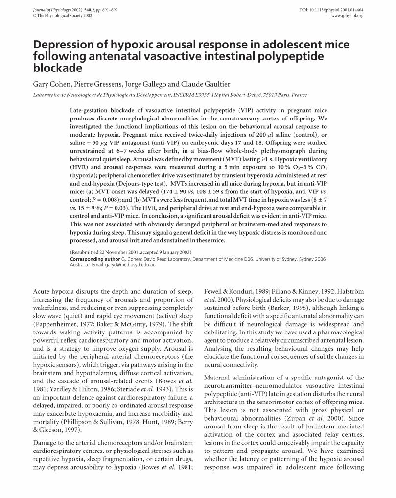
Table 1. Baseline data
Control mice Anti-VIP mice——————————— —————————— P
Male Female Male Female (ANOVA)
n 14 8 11 10 —Body weight (g) 29 ± 4 24 ± 2 26 ± 5 23 ± 3 0.0006Delay to sleep onset (min) 75 ± 35 113 ± 34 70 ± 23 91 ± 30 0.003
T I (ms) 169 ± 17 187 ± 19 181 ± 31 178 ± 32 0.37T TOT (ms) 362 ± 33 381 ± 36 377 ± 43 367 ± 61 0.76V T (ml g_1) 5.9 ± 1 7.3 ± 0.5 5.8 ± 0.6 6.6 ± 0.7 0.001◊E (ml s_1 g_1) 17 ± 3 20 ± 3 16 ± 3 19 ± 3 0.004
The number of mice studied (n), together with body weights and the delay to sleep onset (first period of≥ 3 min continuous QS) are indicated. Baseline ventilation in air (phase 1) is also shown. TI and TTOT,inspiratory and total breath times, respectively; VT and ◊E, tidal volume and minute ventilation, respectively.Significant gender differences are indicated in the final column; treatment group values are not significantlydifferent from control values. Data are means ± S.D.
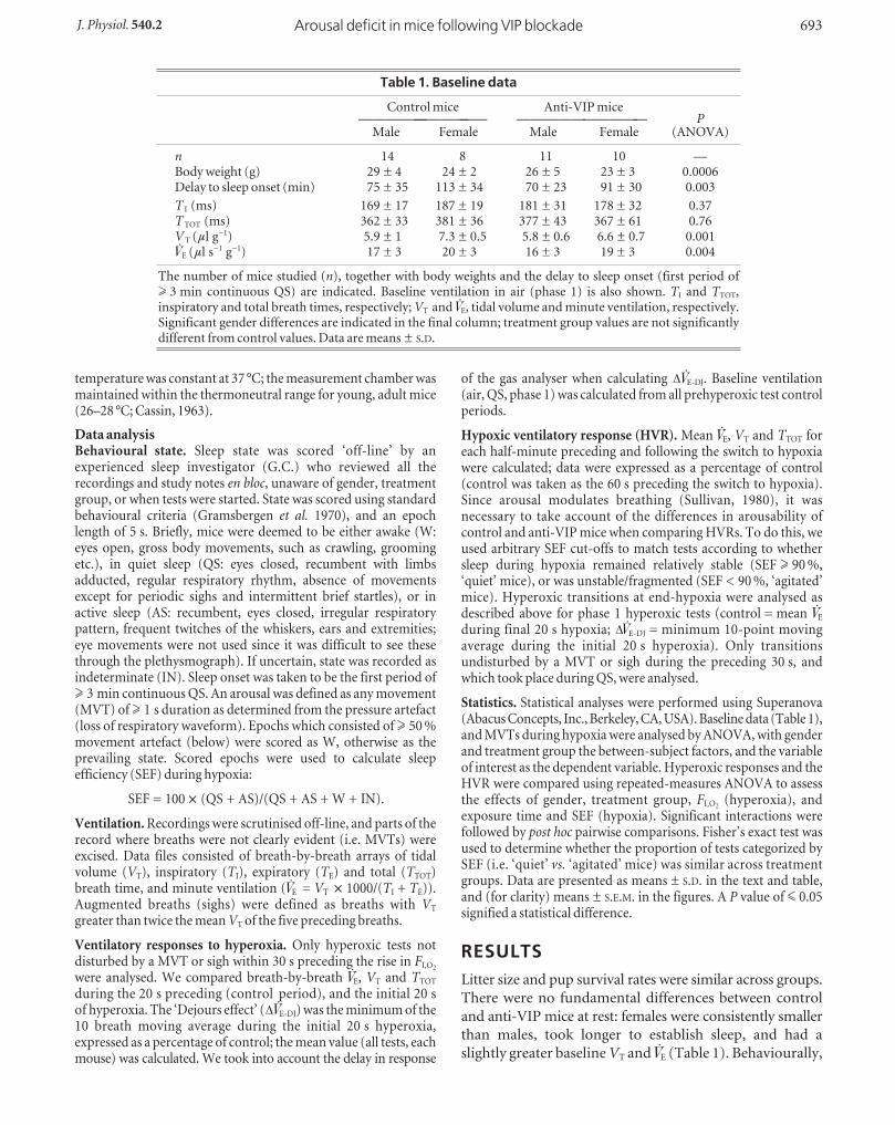
anti-VIP mice were unremarkable: exploratory behaviour
appeared normal, as was the delay to sleep onset; behavioural
correlates of QS and AS were easily identified in both
groups of mice (Fig. 2).
Attenuated hypoxic arousal response in anti-VIPmiceHypoxia normally increases arousal time and decreases
sleep quality and quantity (Pappenheimer, 1977). We
analysed MVTs to determine whether hypoxic arousal was
different in anti-VIP mice. Gender was not a significant
factor in any of these analyses, so only group data are
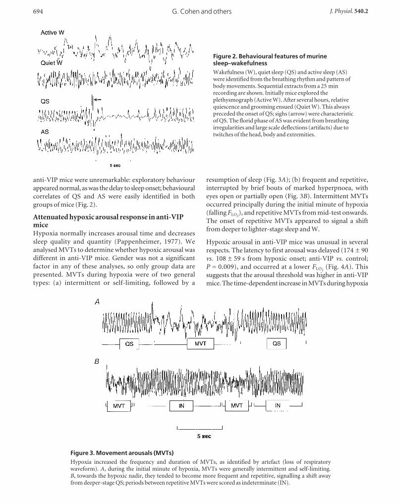
presented. MVTs during hypoxia were of two general
types: (a) intermittent or self-limiting, followed by a
resumption of sleep (Fig. 3A); (b) frequent and repetitive,
interrupted by brief bouts of marked hyperpnoea, with
eyes open or partially open (Fig. 3B). Intermittent MVTs
occurred principally during the initial minute of hypoxia
(falling FI,O2), and repetitive MVTs from mid-test onwards.
The onset of repetitive MVTs appeared to signal a shift
from deeper to lighter-stage sleep and W.
Hypoxic arousal in anti-VIP mice was unusual in several
respects. The latency to first arousal was delayed (174 ± 90
vs. 108 ± 59 s from hypoxic onset; anti-VIP vs. control;
P = 0.009), and occurred at a lower FI,O2(Fig. 4A). This
suggests that the arousal threshold was higher in anti-VIP
mice. The time-dependent increase in MVTs during hypoxia
G. Cohen and others694 J. Physiol. 540.2
Figure 2. Behavioural features of murinesleep–wakefulnessWakefulness (W), quiet sleep (QS) and active sleep (AS)were identified from the breathing rhythm and pattern ofbody movements. Sequential extracts from a 25 minrecording are shown. Initially mice explored theplethysmograph (Active W). After several hours, relativequiescence and grooming ensued (Quiet W). This alwayspreceded the onset of QS; sighs (arrow) were characteristicof QS. The florid phase of AS was evident from breathingirregularities and large scale deflections (artifacts) due totwitches of the head, body and extremities.
Figure 3. Movement arousals (MVTs)Hypoxia increased the frequency and duration of MVTs, as identified by artefact (loss of respiratorywaveform). A, during the initial minute of hypoxia, MVTs were generally intermittent and self-limiting.B, towards the hypoxic nadir, they tended to become more frequent and repetitive, signalling a shift awayfrom deeper-stage QS; periods between repetitive MVTs were scored as indeterminate (IN).
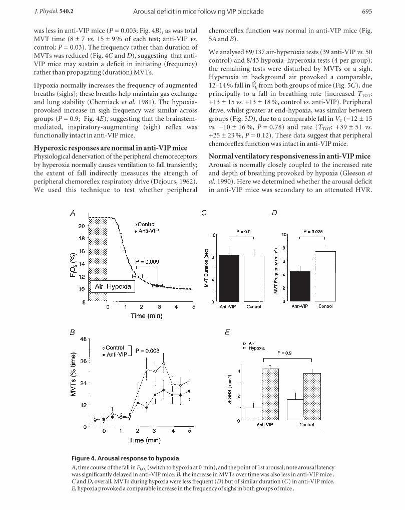
was less in anti-VIP mice (P = 0.003; Fig. 4B), as was total
MVT time (8 ± 7 vs. 15 ± 9 % of each test; anti-VIP vs.control; P = 0.03). The frequency rather than duration of
MVTs was reduced (Fig. 4C and D), suggesting that anti-
VIP mice may sustain a deficit in initiating (frequency)
rather than propagating (duration) MVTs.
Hypoxia normally increases the frequency of augmented
breaths (sighs); these breaths help maintain gas exchange
and lung stability (Cherniack et al. 1981). The hypoxia-
provoked increase in sigh frequency was similar across
groups (P = 0.9; Fig. 4E), suggesting that the brainstem-
mediated, inspiratory-augmenting (sigh) reflex was
functionally intact in anti-VIP mice.
Hyperoxic responses are normal in anti-VIP micePhysiological denervation of the peripheral chemoreceptors
by hyperoxia normally causes ventilation to fall transiently;
the extent of fall indirectly measures the strength of
peripheral chemoreflex respiratory drive (Dejours, 1962).
We used this technique to test whether peripheral
chemoreflex function was normal in anti-VIP mice (Fig.
5A and B).
We analysed 89/137 air-hyperoxia tests (39 anti-VIP vs. 50
control) and 8/43 hypoxia–hyperoxia tests (4 per group);
the remaining tests were disturbed by MVTs or a sigh.
Hyperoxia in background air provoked a comparable,
12–14 % fall in ◊E from both groups of mice (Fig. 5C), due
principally to a fall in breathing rate (increased TTOT:
+13 ± 15 vs. +13 ± 18 %, control vs. anti-VIP). Peripheral
drive, whilst greater at end-hypoxia, was similar between
groups (Fig. 5D), due to a comparable fall in VT (_12 ± 15
vs. _10 ± 16 %, P = 0.78) and rate (TTOT: +39 ± 51 vs.+25 ± 23 %, P = 0.12). These data suggest that peripheral
chemoreflex function was intact in anti-VIP mice.
Normal ventilatory responsiveness in anti-VIP miceArousal is normally closely coupled to the increased rate
and depth of breathing provoked by hypoxia (Gleeson etal. 1990). Here we determined whether the arousal deficit
in anti-VIP mice was secondary to an attenuted HVR.
Arousal deficit in mice following VIP blockadeJ. Physiol. 540.2 695
Figure 4. Arousal response to hypoxiaA, time course of the fall in FI,O2
(switch to hypoxia at 0 min), and the point of 1st arousal; note arousal latencywas significantly delayed in anti-VIP mice. B, the increase in MVTs over time was also less in anti-VIP mice .C and D, overall, MVTs during hypoxia were less frequent (D) but of similar duration (C) in anti-VIP mice.E, hypoxia provoked a comparable increase in the frequency of sighs in both groups of mice .
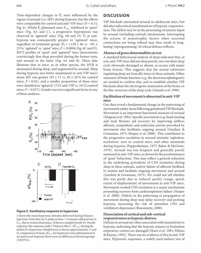
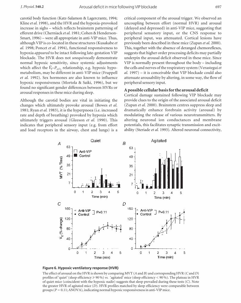
Time-dependent changes in ◊E were influenced by the
vigour of arousal (i.e. SEF) during hypoxia, but the effects
were comparable for control and anti-VIP mice (P = 0.11;
Fig. 6). Whilst ◊E plateaued once FI,O2stabilized in ‘quiet’
mice (Fig. 6A and C), a progressive hyperpnoea was
observed in ‘agitated’ mice (Fig. 6B and D). ◊E at end-
hypoxia was consequently greater in ‘agitated’ mice,
regardless of treatment group (◊E = +128 ± 46 vs. +81 ±
25 %, ‘agitated’ vs. ‘quiet’ mice, P = 0.0004; Fig. 6C and D).
MVT profiles of ‘quiet’ and ‘agitated’ mice demonstrate
convincingly that sleep prevailed during the former tests,
and arousal in the latter (Fig. 6A and B). These data
illustrate that in mice, as in other species, the HVR is
attenuated during sleep, and augmented by arousal. Sleep
during hypoxia was better maintained in anti-VIP mice:
mean SEF was greater (83 ± 17 vs. 65 ± 20 % for control
mice; P = 0.02), and a smaller proportion of these mice
were classified as ‘agitated’ (7/21 anti-VIP vs. 16/22 control
mice; P = 0.017). Gender was not a significant factor in any
of these analyses.
DISCUSSION VIP blockade attenuated arousal in adolescent mice, but
did alter subcortical manifestations of hypoxic responsive-
ness. The deficit may be in the processing of sensory input
by arousal (including cortical) mechanisms. Interrupting
the actions of neurotrophic factors when neuronal
connections are being refined may thus result in long-
lasting ‘reprogramming’ of critical defence reflexes.
Absence of gross abnormalities at restA standard behavioural analysis of sleep indicated that, at
rest, anti-VIP mice did not sleep poorly, nor was their sleep
cycle obviously deranged or absent, as occurs with major
brain lesions. This suggests that the neural substrates
regulating sleep are basically intact in these animals. Other
measures of brain function (e.g. the electroencephalogram)
are needed to confirm this, and to establish whether VIP
blockade alters the electrogenic maturation of the brain, or
the fine-structure of the sleep cycle (Schaub et al. 1998).
Facilitation of movement is abnormal in anti-VIPmiceOur data reveal a fundamental change in the patterning of
movements under stress following gestational VIP blockade.
Movement is an important functional measure of arousal
(Mograss et al. 1994). Specific movements (e.g. head-turning
and neck flexion) aid recovery by improving airflow;
afferent, sympathetic and endocrine activity provoked by
movement also facilitates ongoing arousal (Gardner &
Grossman, 1975; Harper et al. 2000). This contributes to
the progressive escalation in arousal intensity (agitation,
excitation) seen in control mice, and other mammals
during hypoxia (Pappenheimer, 1977; Baker & McGinty,
1979). Arousal was less frequent and generally poorly
sustained in anti-VIP mice, as shown by the predominance
of ‘quiet’ behaviour. This may reflect a general reduction
in the underlying periodicity of CNS excitation during
sleep in these animals, and/or failure of afferent feedback
to sustain and facilitate ongoing movement and arousal
(Gardner & Grossman, 1975). We could not tell whether
this was partly due to reduced quality (range, speed,
extent of displacement) of movements in anti-VIP mice.
Movement-evoked CNS excitation is a major mechanism
promoting recovery from cardiorespiratory failure (Harper
et al. 2000). Deficits in the patterning or propagation of
movement during sleep may delay recovery and prolong
hypoxia, increasing the risk of attendant CNS and
ventilatory depression (Bissonnette, 2000).
Dissociation of cortical and sub-corticalresponsiveness to hypoxic distressDeficits in arousal are often associated with insensitivity to
hypoxia, indicating that the hypoxic sensors or brainstem
respiratory centres are damaged (Hunt et al. 1981; Filiano
& Kinney, 1992). There was no evidence of this in anti-VIP
mice. Hyperoxic responses, a widely used indirect test of
G. Cohen and others696 J. Physiol. 540.2
Figure 5. Ventilatory response to hyperoxiaA shows the mean hyperoxic stimulus delivered during Dejours-type tests. Note that an O2 pulse at time = 0 caused a delayed rise inFI,O2
due to system dead space. B shows a sample breath-by-breathresponse; the response nadir (‘Dejours effect’, D◊E-DJ ) during theinitial 20 s hyperoxia (shaded area) is shown approximately. C andD, comparison of mean D◊E-DJ for hyperoxic tests administered inair and at end-hypoxia; there were no differences between groups(ANOVA).
carotid body function (Katz-Salamon & Lagercrantz, 1994;
Kline et al. 1998), and the HVR and the hypoxia-provoked
increase in sighs – which reflects brainstem patterning of
efferent drive (Cherniack et al. 1981; Cohen & Henderson-
Smart, 1996) – were all appropriate in anti-VIP mice. Thus,
although VIP is co-located in these structures (Kusakabe etal. 1998; Poncet et al. 1994), functional responsiveness to
hypoxia appeared to be intact following late-gestation VIP
blockade. The HVR does not unequivocally demonstrate
normal hypoxic sensitivity, since systemic adjustments
which affect the ◊E–Pa,O2relationship, e.g. hypoxic hypo-
metabolism, may be different in anti-VIP mice (Frappell
et al. 1992). Sex hormones are also known to influence
hypoxic responsiveness (Mortola & Saiki, 1996), but we
found no significant gender differences between HVRs or
arousal responses in these mice during sleep.
Although the carotid bodies are vital in initiating the
changes which ultimately provoke arousal (Bowes et al.1981; Ryan et al. 1983), it is the hyperpnoea (i.e. increased
rate and depth of breathing) provoked by hypoxia which
ultimately triggers arousal (Gleeson et al. 1990). This
indicates that peripheral sensory input (e.g. from effort
and load receptors in the airway, chest and lungs) is a
critical component of the arousal trigger. We observed an
uncoupling between effort (normal HVR) and arousal
(delayed and depressed) in anti-VIP mice, suggesting that
peripheral sensatory input, or the CNS response to
peripheral input, was attenuated. Cortical lesions have
previously been described in these mice (Zupan et al. 2000).
This, together with the absence of deranged chemoreflexes,
suggests that higher order processing deficits may partially
underpin the arousal deficit observed in these mice. Since
VIP is normally present throughout the body – including
the cells and nerves of the respiratory system (Verastegui etal. 1997) – it is conceivable that VIP blockade could also
attenuate arousability by altering, in some way, the flow of
peripheral sensory input.
A possible cellular basis for the arousal deficitCortical damage sustained following VIP blockade may
provide clues to the origin of the associated arousal deficit
(Zupan et al. 2000). Brainstem centres suppress sleep and
dramatically enhance forebrain activity (arousal) by
modulating the release of various neurotransmitters. By
altering neuronal ion conductances and membrane
potentials, this facilitates synaptic transmission and excit-
ability (Steriade et al. 1993). Altered neuronal connectivity,
Arousal deficit in mice following VIP blockadeJ. Physiol. 540.2 697
Figure 6. Hypoxic ventilatory response (HVR)The effect of arousal on the HVR is shown by comparing MVT (A and B) and corresponding HVR (C and D)profiles of ‘quiet’ (sleep efficiency ≥ 90 %) vs. ‘agitated’ mice (sleep efficiency < 90 %). The plateau in HVRof quiet mice (coincident with the hypoxic nadir) suggests that sleep prevailed during these tests (C). Notethe greater HVR of agitated mice (D). HVR profiles matched by sleep efficiency were comparable betweengroups (P = 0.11; ANOVA), indicating normal hypoxic responsiveness in anti-VIP mice.
ion channel structure, or expression of neuronal receptors
following antenatal VIP blockade may alter the sequence
or timing of these events. This may impair excitatory
transmission, or enhance inhibitory gating of input, delaying
the transition from sleep to arousal (Steriade et al. 1993).
Specific abnormalities in anti-VIP mice, e.g. altered
N-methyl-D-aspartate (NMDA) receptor expression, may
impair the potentiation and propagation of excitatory
input by cortical circuits (Steriade & McCarley, 1990).
Damage to intrinsic peptidergic expression or regulation
may also affect cortical excitability (Bayraktar et al. 2000).
Cortical damage may be a marker for more widespread
damage affecting other nuclei and transmitter systems
involved in arousal. This may include the carotid body and
brainstem cardiorespiratory centres; changes in cyto-
architecture or endogenous VIP expression may occur in
these structures following VIP blockade, even though
functional deficits were not revealed by the testing
protocol we used.
Developmental implicationsFactors which delay awakening may be particularly serious
when vulnerability to rapid and profound hypoxaemia is
greatest, i.e. in newborn and developing organisms.
Depression of arousal mechanisms delays recovery from
spontaneous apnoea, exacerbating hypoxaemia. This may
disturb growth and development, further exacerbate
sleep-disordered breathing, and contribute to respiratory
failure and sudden death during sleep (Hunt, 1989). Our
data suggest that one way this may occur is via antenatal
‘reprogramming’ of the underlying neural substrates. Here
we studied only adolescent mice. Longitudinal studies
using this model may establish whether significant deficits
are also present at earlier, more vulnerable stages of
development, as well as the extent to which underlying
deficits of this sort increase the susceptibility to external
modifiers of arousability.
REFERENCES BAKER, T. L. & MCGINTY, D. J. (1979). Sleep-waking patterns in
hypoxic kittens. Developmental Psychobiology 12, 561–575.
BARKER, D. J. (1998). In utero programming of chronic disease.
Clinical Science 95, 115–128.
BAYRAKTAR, T., WELKER, E., FREUND, T. F., ZILLES, K. & STAIGER, J. F.
(2000). Neurons immunoreactive for vasoactive intestinal
polypeptide in the rat primary somatosensory cortex : morphology
and spatial relationship to barrel-related columns. Journal ofComparative Neurology 420, 291–304.
BERRY, R. B. & GLEESON, K. (1997). Respiratory arousal from sleep:
mechanisms and significance. Sleep 20, 654–675.
BISSONNETTE, J. M. (2000). Mechanisms regulating hypoxic
respiratory depression during fetal and postnatal life. AmericanJournal of Physiology – Regulatory, Integrative and ComparativePhysiology 278, R1391–1400.
BOWES, G., TOWNSEND, E. R., KOZAR, L. F., BROMLEY, S. M. &
PHILLIPSON, E. A. (1981). Effect of carotid body denervation on
arousal response to hypoxia in sleeping dogs. Journal of AppliedPhysiology 51, 40–45.
CASSIN, S. (1963). Critical oxygen tensions in newborn, young and
adult mice. American Journal of Physiology 205, 325–330.
CHERNIACK, N. S., VON EULER, C., GLOGOWSKA, M. & HONMA, I.
(1981). Characteristics and rate of occurrence of spontaneous and
provoked augmented breaths. Acta Physiologica Scandinavica 111,
349–360.
COHEN, G. & HENDERSON-SMART, D. J. (1996). The characteristics
and frequency of augmented breaths during CO 2-induced
hyperpnoea of newborn infants. Journal of Physiology 490,
551–557.
DAUGER, S., NSEGEBE, E., VARDON, G., GAULTIER, C. & GALLEGO, J.
(1998). The effects of restraint on ventilatory responses to
hypercapnia and hypoxia in adult mice. Respiration Physiology112, 215–225.
DEJOURS, P. (1962). Chemoreceptors in breathing. PhysiologicalReviews 42, 335–358.
FEWELL, J. E. & KONDURI, G. G. (1989). Influence of repeated
exposure to rapidly developing hypoxaemia on the arousal and
cardiopulmonary response to rapidly developing hypoxaemia in
lambs. Journal of Developmental Physiology 11, 77–82.
FILIANO, J. J. & KINNEY, H. C. (1992). Arcuate nucleus hypoplasia in
the sudden infant death syndrome. Journal of Neuropathology andExperimental Neurology 51, 394–403.
FRAPPELL, P., LANTHIER, C., BAUDINETTE, R. V. & MORTOLA, J. P.
(1992). Metabolism and ventilation in acute hypoxia: a
comparative analysis in small mammalian species. AmericanJournal of Physiology 262, R1040–1046.
GARDNER, R. & GROSSMAN, W. I. (1975). Normal motor patterns in
sleep in man. In Advances in Sleep Research, ed. WEITZMAN, E. D.,
pp. 67–69. Spectrum Publications, New York.
GLEESON, K., ZWILLICH, C. W. & WHITE, D. P. (1990). The influence
of increasing ventilatory effort on arousal from sleep. AmericanReview of Respiratory Disease 142, 295–300.
GOZES, I., MCCUNE, S. K., JACOBSON, L., WARREN, D., MOODY, T. W.,
FRIDKIN, M. & BRENNEMAN, D. E. (1991). An antagonist to
vasoactive intestinal polypeptide affects cellular functions in the
central nervous system. Journal of Pharmacology and ExperimentalTherapeutics 257, 959–966.
GRAMSBERGEN, A., SCHWARTZE, P. & PRECHTL, H. F. (1970). The
postnatal development of behavioral states in the rat.
Developmental Psychobiology 3, 267–280.
HAFSTRÖM, O., MILERAD, J., ASOKAN, N., POOLE, H. S. D. & SUNDELL,
H. W. (2000). Nicotine delays arousal during hypoxemia in lambs.
Pediatric Research 47, 646–652.
HARPER, R. M., WOO, M. A. & ALGER, J. R. (2000). Visualization of
sleep influences on cerebeller and brainstem cardiac and
respiratory control mechanisms. Brain Research Bulletin 53,
125–131.
HUNT, C. E. (1989). Impaired arousal from sleep: relationship to
sudden infant death syndrome. Journal of Perinatology 9, 184–187.
HUNT, C. E., MCCULLOCH, K. & BROUILLETTE, R. T. (1981).
Diminished hypoxic ventilatory response in near-miss sudden
infant death syndrome. Journal of Applied Physiology 50,
1313–1317.
KATZ-SALAMON, M. & LAGERCRANTZ, H. (1994). Hypoxic ventilatory
defence in very preterm infants: attenuation after long-term
oxygen treatment. Archives of Disease in Childhood 70, F90–95.
G. Cohen and others698 J. Physiol. 540.2
KLINE, D. D., YANG, T., HUANG, P. L. & PRABHAKAR, N. R. (1998).
Altered respiratory responses to hypoxia in mutant mice deficient
in neuronal nitric oxide synthase. Journal of Physiology 511,
273–287.
KUSAKABE, T., HAYASHIDA, Y., MATSUDA, H., GONO, Y., POWELL, F. L.,
ELLISMAN, M. H., KAWAKAMI, T. & TAKENAKA, T. (1998). Hypoxic
adaptation of the peptidergic innervation in the rat carotid body.
Brain Research 806, 165–174.
MOGRASS, M. A., DUCHARME, F. M. & BROUILLETTE, R. T. (1994).
Movement/arousals. Description, classification, and relationship
to sleep apnea in children. American Review of Respiratory andCritical Care Medicine 150, 1690–1696.
MORTOLA, J. P. & SAIKI, C. (1996). Ventilatory response to hypoxia in
rats: gender differences. Respiration Physiology 106, 21–34.
PAPPENHEIMER, J. R. (1977). Sleep and respiration of rats during
hypoxia. Journal of Physiology 266, 191–207.
PASPALAS, C. D. & PAPADOPOULOS, G. C. (1999). Noradrenergic
innervation of peptidergic interneurons in the rat visual cortex.
Cerebral Cortex 9, 844–853.
PEPELKO, W. E. & DIXON, G. A. (1975). Arterial blood gases in
conscious rats exposed to hypoxia, hypercapnia, or both. Journal ofApplied Physiology 38, 581–587.
PHILLIPSON, E. A. & SULLIVAN, C. E. (1978). Arousal: the forgotton
response to respiratory stimuli. American Review of RespiratoryDisease 118, 807–809.
PONCET, L., DENOROY, L., DALMAZ, Y., PÉQUIGNOT, J. M. & JOUVET, M.
(1994). Chronic hypoxia affects peripheral and central vasoactive
intestinal peptide-like immunoreactivity in the rat. NeuroscienceLetters 176, 1–4.
RYAN, A. T., WARD, D. A. & MEGIRIAN, D. (1983). Sleep-waking
patterns of intact and carotid sinus nerve-transected rats during
hypoxic CO 2 breathing. Experimental Neurology 80, 337–348.
SCHAUB, C. D., TANKERSLEY, C., SCHWARTZ, A. R., SMITH, P. L.,
ROBOTHAM, J. L. & O’DONNELL, C. P. (1998). Effect of sleep/wake
state on arterial blood pressure in genetically identical mice.
Journal of Applied Physiology 85, 366–371.
STERIADE, M. & MCCARLEY, R. W. (1990). Brainstem Control ofWakefulness and Sleep, pp. 163–201. Plenum Press, New York.
STERIADE, M., MCCORMICK, D. A. & SEJNOWSKI, T. J. (1993).
Thalamocortical oscillations in the sleeping and aroused brain.
Science 262, 679–685.
SULLIVAN, C. E. (1980). Breathing in sleep. In Physiology in Sleep, ed.
OREM, J. & BARNES, D., pp. 213–271. Academic Press, New York.
VERASTEGUI, C., FERNANDEZ-VIVERO, J., PRADA, A., RODRIGUEZ, F.,
ROMERO, A., GONZALEZ-MORENO, M. & DE CASTRO, J. M. (1997).
Presence and distribution of 5HT, VIP, NPY, and
SP-immunoreactive structures in adult mouse lung. Histology andHistopathology 12, 909–918.
YARDLEY, C. P. & HILTON, S. M. (1986). The hypothalamic and
brainstem areas from which the cardiovascular and behavioral
components of the defence reaction are elicited in the rat. Journalof the Autonomic Nervous System 15, 227–244.
ZUPAN, V., NEHLIG, A., EVRARD, P. & GRESSENS, P. (2000). Prenatal
blockade of vasoactive intestinal polypeptide alters cell death and
synaptic equipment in the murine neocortex. Pediatric Research47, 53–63.
AcknowledgementsThis work was supported by INSERM and the National Healthand Medical Research Council of Australia.
Author’s present addressG. Cohen: David Read Laboratory, Department of Medicine D06,University of Sydney, Sydney 2006, Australia.
Arousal deficit in mice following VIP blockadeJ. Physiol. 540.2 699