department of pharmaceutics, atmiya institute of … of drug administration. 3 extensive first-pass...
TRANSCRIPT

International Journal of Pharma and Bio Sciences V1(2)2010
BUCCOADHESIVE DRUG DELIVERY SYSTEMS: A REVIEW
1 www.ijpbs.net Novel Drug Delivery System
Ravi Bhalodia*, Biswajit Basu, Kevin Garala, Bhavik Joshi and
Kuldeep Mehta
Department of Pharmaceutics, Atmiya Institute of Pharmacy, Yogidham Gurukul, Kalawad Road,
Rajkot-360 005, Gujarat State, India.
*Corresponding author [email protected]
ABSTRACT Delivery of the desired drug as buccoadhesive drug delivery systems has been subject of interest since last 3 decades. The various advantages associated with these systems made the buccal drug delivery as a novel route of drug administration. This review highlights the anatomy and nature of oral cavity, theories of mucoadhesion, classical and new generation mucoadhesive polymers, various types of mucoadhesive dosage forms and drugs which can be incorporated and various evaluation methods for the measurement of mucoadhesion potential. KEYWORDS
Buccoadhesive drug delivery; Mucoadhesion; Polymers 1. INTRODUCTION
For many decades, treatment of an acute disease or a chronic illness has been mostly accomplished by delivering drugs using various pharmaceutical dosage forms, including tablets, capsules, pills, suppositories, creams, ointments, liquids, aerosols, and injectables as carriers. Amongst various routes of drug delivery, oral route is perhaps the most preferred to the patient and the clinician alike. However, this route presents some problems for a few drugs. The enzymes in the GI fluids, GIT-pH
conditions, the enzymes bound to GIT membranes are a few factors responsible for the bioavailability problems. The blood that drains the GIT carries the drug directly to the liver leading to first-pass metabolism resulting in poor bioavailability. The inherent problems associated with the drug, in some cases, can be solved by modifying the formulation or by changing the routes of administration. Parenteral, mucosal, and transdermal routes circumvent hepatic first-pass metabolism and offer alternative routes for the systemic delivery of drugs.1
In recent years, the interest in novel routes of drug administration occurs from their ability to enhance the

International Journal of Pharma and Bio Sciences V1(2)2010
BUCCOADHESIVE DRUG DELIVERY SYSTEMS: A REVIEW
2 www.ijpbs.net Novel Drug Delivery System
bioavailability of drugs. Drug delivery via the buccal route, using bioadhesive dosage forms offers such a novel route of drug administration.2 Buccal delivery involves administration of desired drug through the buccal mucosal membrane lining of oral cavity. The mucosal lining of oral cavity offers some distinct advantages. It is richly vascularized and more accessible for the administration and removal of a dosage form. Additionally, buccal drug delivery has high patient acceptability compared to other non-oral routes of drug administration.3 Extensive first-pass metabolism and drug degradation in the harsh gastrointestinal environment can be circumvented by administering the drug via buccal route.2
Drug absorption through buccal mucosa is mainly by passive diffusion into the lipoidal membrane. After absorption, the drug is transported through facial vein which then drains into the general circulation via jugular vein, bypassing the liver and thereby sparing the drug from first-pass metabolism.4
Buccal route provides one of the potential routes for typically large, hydrophilic and unstable proteins, oligonucleotides and polysaccharides, as well as conventional small drug molecules.5
1.1 Advantages of Buccal Drug Delivery Systems Drug administration via buccal mucosa offers several distinct advantages3, 4
1. Ease of administration. 2. Termination of therapy is easy. 3. Permits localization of drug to the buccal cavity
for a prolonged period of time. 4. Can be administered to unconscious patients. 5. Offers an excellent route, for the systemic
delivery of drugs which undergo extensive first-pass metabolism or degradation in harsh gastrointestinal environment.
6. A significant reduction in dose can be achieved thereby reducing dose related side effects.
7. Drugs, which show poor bioavailability via the oral route, can be administered conveniently.
8. It offers a passive system of drug absorption and does not require any activation.
9. The presence of saliva ensures relatively large amount of water for drug dissolution unlike in case of rectal or transdermal routes.
10. Systemic absorption is rapid as buccal mucosa is thin and highly perfused with blood.
11. Provides an alternative route for the administration of various hormones, narcotic analgesics, steroids, enzymes, cardiovascular agents etc.
12. It allows the local modification of tissue permeability, inhibition of protease activity and reduction in immunogenic response. Thus, delivery of therapeutic agents like peptides, proteins and ionized species can be done easily.
1.2 Disadvantages of Buccal Drug Delivery Systems Drug administration via buccal mucosa has certain limitations3, 4
1. Drugs, which irritate the oral mucosa, have a bitter or unpleasant taste or odour; can not be administered by this route.
2. Drugs, which are unstable at buccal pH, cannot be administered by this route.
3. Only drugs with small dose requirements can be administered.
4. Drugs may get swallowed with saliva and loses the advantages of buccal route.
5. Only those drugs, which are absorbed by passive diffusion, can be administered by this route.
6. Over hydration may lead to the formation of slippery surface and structural integrity of the

International Journal of Pharma and Bio Sciences V1(2)2010
BUCCOADHESIVE DRUG DELIVERY SYSTEMS: A REVIEW
3 www.ijpbs.net Novel Drug Delivery System
formulation may get disrupted by the swelling and hydration of the bioadhesive polymers.
7. Surface area available for absorption is less. 8. The buccal mucosa is relatively less permeable
than the small intestine, rectum, etc.
2. ANATOMY AND NATURE OF ORAL CAVITY 2.1 Oral Cavity The oral cavity may be divided into two regions, the outer oral vestibule, bounded by the lips and cheeks and the oral cavity itself the borders being, and formed by the hard and soft palates, the floor of the mouth and tonsils. 2.2 Oral Mucosa 2.2.1 Anatomy of the oral mucosa The mucosa that lines the oral cavity may be divided into three types, classified according to their function as; 1. Masticatory mucosa: Which includes the mucosa
around the teeth and on the hard palate and these regions have keratinized epithelium.
2. Lining mucosa: Which covers the lips, cheeks, fornix, base of the oral cavity, lower part of tongue, buccal mucosa and the soft palate and these regions have non-keratinized epithelium.
3. Specialized mucosa: covering the dorsum of the tongue with highly keratinization. Light microscopy reveals several distinct patterns
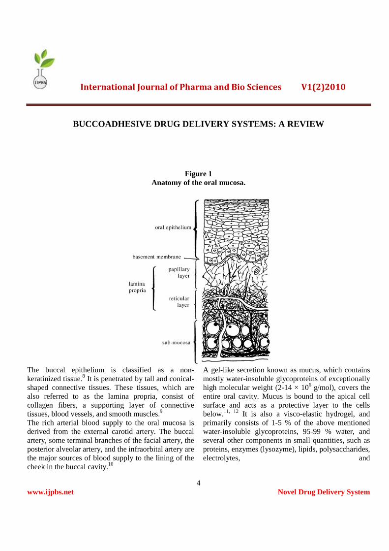
of maturation in the epithelium of the human oral mucosa based on various regions of the oral cavity. Three distinctive layers of the oral mucosa are the epithelium, basement membrane and connective tissues. The oral cavity is lined with the epithelium, below which lies the supporting basement membrane. The basement membrane is, in turn, supported by connective tissues (Fig. 1).6
The epithelial cells, originating from the basal cells, mature, change their shape, and increase in size while moving towards the surface. The thickness of buccal epithelium in humans, dogs, and rabbits has been determined to be approximately 500–800 µm.7
The basement membrane forms a distinctive layer between the connective tissues and the epithelium. It provides the required adherence between the epithelium and the underlying connective tissues, and functions as a mechanical support for the epithelium. The underlying connective tissues provide many of the mechanical properties of oral mucosa.
International Journal of Pharma and Bio Sciences V1(2)2010
BUCCOADHESIVE DRUG DELIVERY SYSTEMS: A REVIEW
4 www.ijpbs.net Novel Drug Delivery System
Figure 1 Anatomy of the oral mucosa.
The buccal epithelium is classified as a non-keratinized tissue.8 It is penetrated by tall and conical-shaped connective tissues. These tissues, which are also referred to as the lamina propria, consist of collagen fibers, a supporting layer of connective tissues, blood vessels, and smooth muscles.9
The rich arterial blood supply to the oral mucosa is derived from the external carotid artery. The buccal artery, some terminal branches of the facial artery, the posterior alveolar artery, and the infraorbital artery are the major sources of blood supply to the lining of the cheek in the buccal cavity.10
A gel-like secretion known as mucus, which contains mostly water-insoluble glycoproteins of exceptionally high molecular weight (2-14 × 106 g/mol), covers the entire oral cavity. Mucus is bound to the apical cell surface and acts as a protective layer to the cells below.11, 12 It is also a visco-elastic hydrogel, and primarily consists of 1-5 % of the above mentioned water-insoluble glycoproteins, 95-99 % water, and several other components in small quantities, such as proteins, enzymes (lysozyme), lipids, polysaccharides, electrolytes, and

International Journal of Pharma and Bio Sciences V1(2)2010
BUCCOADHESIVE DRUG DELIVERY SYSTEMS: A REVIEW
5 www.ijpbs.net Novel Drug Delivery System
nucleic acids. This composition can vary based on the origin of the mucus secretion in the body. Some of these non-mucin components are believed to be responsible for the bacteriostatic action observed in mucus.12-14
A thorough understanding of the glycoprotein mucin component is very important with regard to understanding the properties of mucus (Fig. 2). Mucin glycoprotein may be described as consisting of a basic unit made from a single chain polypeptide backbone with two distinct regions.15
1. attached, predominantly via O-glycosidic linkage.
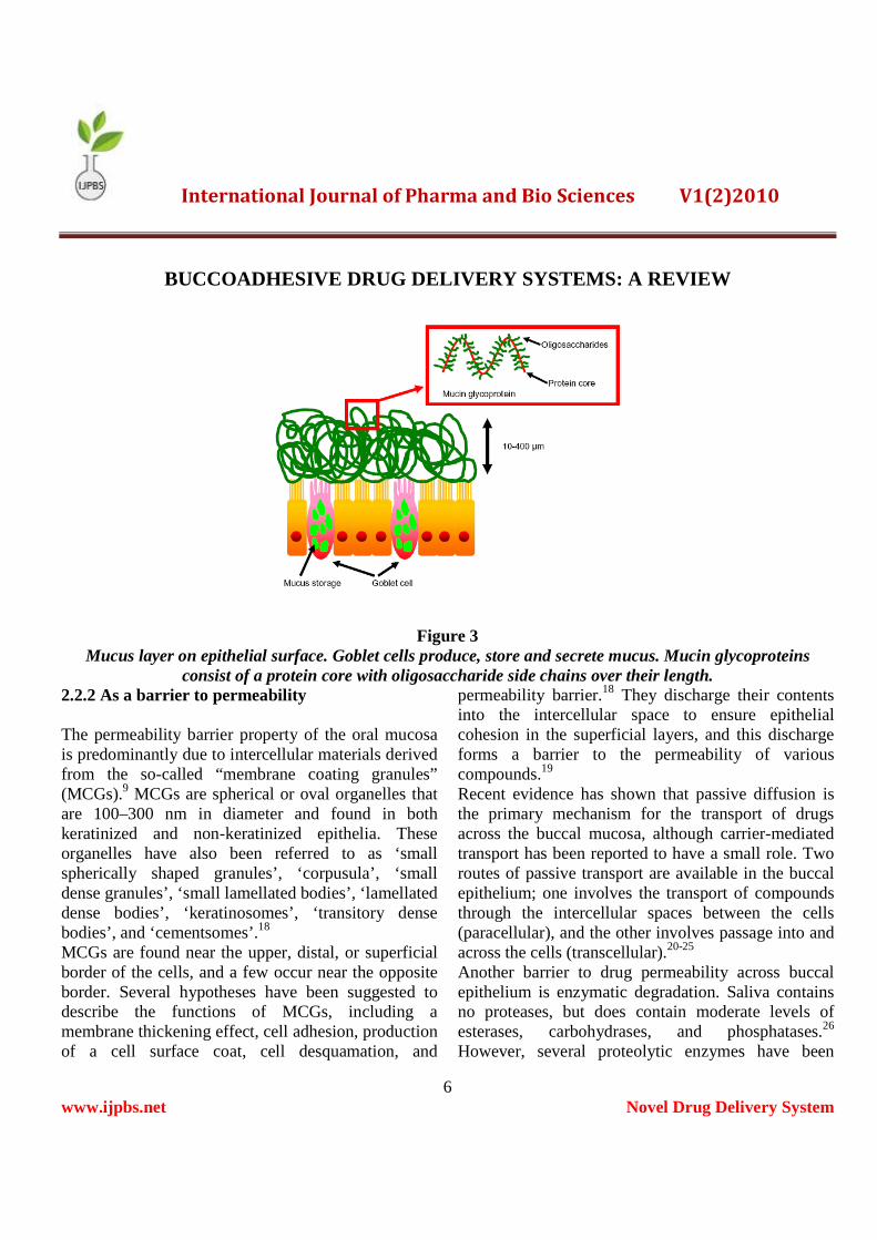
2. One or two terminal peptide regions where there is little glycosylation. These regions are often referred to as ‘naked protein regions’. Mucin itself is stored in both submucosal and goblet cells (Fig. 3)16, wherein the negative charges of the mucin glycoprotein are shielded by calcium ions, this allows for the compact packing ofsuch molecules 17
Figure 2: The composition and interaction of glycoprotein chains within mucus.
International Journal of Pharma and Bio Sciences V1(2)2010
BUCCOADHESIVE DRUG DELIVERY SYSTEMS: A REVIEW
6 www.ijpbs.net Novel Drug Delivery System
Figure 3 Mucus layer on epithelial surface. Goblet cells produce, store and secrete mucus. Mucin glycoproteins
consist of a protein core with oligosaccharide side chains over their length. 2.2.2 As a barrier to permeability The permeability barrier property of the oral mucosa is predominantly due to intercellular materials derived from the so-called “membrane coating granules” (MCGs).9 MCGs are spherical or oval organelles that are 100–300 nm in diameter and found in both keratinized and non-keratinized epithelia. These organelles have also been referred to as ‘small spherically shaped granules’, ‘corpusula’, ‘small dense granules’, ‘small lamellated bodies’, ‘lamellated dense bodies’, ‘keratinosomes’, ‘transitory dense bodies’, and ‘cementsomes’.18
MCGs are found near the upper, distal, or superficial border of the cells, and a few occur near the opposite border. Several hypotheses have been suggested to describe the functions of MCGs, including a membrane thickening effect, cell adhesion, production of a cell surface coat, cell desquamation, and
permeability barrier.18 They discharge their contents into the intercellular space to ensure epithelial cohesion in the superficial layers, and this discharge forms a barrier to the permeability of various compounds.19
Recent evidence has shown that passive diffusion is the primary mechanism for the transport of drugs across the buccal mucosa, although carrier-mediated transport has been reported to have a small role. Two routes of passive transport are available in the buccal epithelium; one involves the transport of compounds through the intercellular spaces between the cells (paracellular), and the other involves passage into and across the cells (transcellular).20-25
Another barrier to drug permeability across buccal epithelium is enzymatic degradation. Saliva contains no proteases, but does contain moderate levels of esterases, carbohydrases, and phosphatases.26 However, several proteolytic enzymes have been

International Journal of Pharma and Bio Sciences V1(2)2010
BUCCOADHESIVE DRUG DELIVERY SYSTEMS: A REVIEW
7 www.ijpbs.net Novel Drug Delivery System
found in the buccal epithelium.27 Walker et al. reported that endopeptidases and carboxypeptidases were not present on the surface of porcine buccal mucosa, whereas aminopeptidases appeared to be the major enzymatic barrier to the buccal delivery of peptide drugs.28 Aminopeptidase N and A (plasma membrane-bound peptidases) and aminopeptidase B (cytosolic enzyme) have been found in the buccal tissue.29 The use of mucoadhesive polymers as enzyme inhibitor agents has been developed to overcome this obstacle in peptide and protein delivery. 2.3 Drug Delivery through Oral Mucosa In general, the oral mucosa is classified as a somewhat leaky epithelium with a permeability rank order of sublingual > buccal > palatal, based on the thickness and degree of keratinization of the tissues.7
Different regions of the oral cavity vary greatly in terms of their composition and their potential utility in drug delivery. The thin and highly permeable membrane of the sublingual tissue is a perfect target if a prompt onset is desired. Considerable surface area and high blood flow to this region provide a means for rapid access to the systemic circulation. However, if a retentive, sustained-release system is desired, the sublingual membrane fails to be an appropriate target tissue. Sustained-release systems, which are able to provide sustained drug concentrations in the systemic circulation due to delayed release of the drug from the formulation, are suitable dosage forms for the buccal region of the oral cavity. The lower permeability of this region compared to the sublingual site is deal for controlled-release systems. Additionally, drug delivery via this site avoids extensive enzyme degradation and first-pass metabolism seen with oral administration, which are desired outcomes for the
delivery of therapeutic proteins and peptides. However, the low permeability of this site is not always an attractive feature and, depending on the choice of drug, can be a major limitation. Use of sub-toxic levels of penetration enhancers and targeted delivery may potentially overcome this problem in the buccal region of the oral cavity. Local delivery in the oral cavity has had particular applications in the treatment of toothache, periodontal diseases, and bacterial infections. However, because of its specificity, local delivery does not have the broad range of applications that sublingual and buccal drug administration provides.3
3. MUCOADHESION 3.1 Definition Longer and Robinson defined the term “bioadhesion” as the “attachment of a synthetic or natural macromolecule to mucus and/or an epithelial surface”.30 The general definition of adherence of a polymeric material to biological surfaces (bioadhesives) or to the mucosal tissue (mucoadhesives) still holds.3
3.2 Theories of Mucoadhesion The mucosal surfaces are covered with a mucus layer, in which mucins are the major component. Mucins are highly glucosylated glycoproteins with a large peptide backbone and oligosaccharides as side chains. Their protein backbone is characterized by the presence of repeating sequences rich in serine, threonine, and proline residues. Many of the O-linked oligosaccharide side chains are often terminated in sialic acid, sulfonic acid, or L-fructose.9 As a result, mucins are negatively charged at physiological pH. Mechanisms of polymer
International Journal of Pharma and Bio Sciences V1(2)2010
BUCCOADHESIVE DRUG DELIVERY SYSTEMS: A REVIEW
8 www.ijpbs.net Novel Drug Delivery System
attachment to mucosal surfaces are not yet fully understood. However, certain theories of bioadhesion have suggested that it might occur via physical entanglement (diffusion theory) and/or chemical interactions, such as electrostatic, hydrophobic, hydrogen bonding, and van der Waals’ interactions (adsorption and electronic theories).3
Four theories have been suggested to play a major role in bioadhesion, namely, adsorption, diffusion, electronic, and wetting theories.31-36
3.2.1 Adsorption theory In the “adsorption theory”, primary and secondary chemical bonds of the covalent and non-covalent (electrostatic and van der Waals’ forces, hydrogen, and hydrophobic bonds) types are formed upon initial contact between the mucus and the mucoadhesive polymer. Most of the initial interfacial bonding forces are attributed to non-covalent forces. The formation of
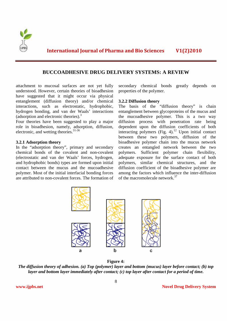
secondary chemical bonds greatly depends on properties of the polymer. 3.2.2 Diffusion theory The basis of the “diffusion theory” is chain entanglement between glycoproteins of the mucus and the mucoadhesive polymer. This is a two way diffusion process with penetration rate being dependent upon the diffusion coefficients of both interacting polymers (Fig. 4).12 Upon initial contact between these two polymers, diffusion of the bioadhesive polymer chain into the mucus network creates an entangled network between the two polymers. Sufficient polymer chain flexibility, adequate exposure for the surface contact of both polymers, similar chemical structures, and the diffusion coefficient of the bioadhesive polymer are among the factors which influence the inter-diffusion of the macromolecule network.37
Figure 4: The diffusion theory of adhesion. (a) Top (polymer) layer and bottom (mucus) layer before contact; (b) top
layer and bottom layer immediately after contact; (c) top layer after contact for a period of time.

International Journal of Pharma and Bio Sciences V1(2)2010
BUCCOADHESIVE DRUG DELIVERY SYSTEMS: A REVIEW
9 www.ijpbs.net Novel Drug Delivery System
3.2.3 Electronic theory Because of different electronic properties of the mucoadhesive polymer and the mucus glycoprotein, electron transfer between these two surfaces occurs. Electron transfer contributes to formation of a charged double layer at the interface of the mucus and the polymer, which results in forces of attraction in this region and inter-diffusion of the two surfaces.38 3.2.4 Wetting theory The “wetting theory” describes the ability of a bioadhesive polymer to spread on biological surfaces (Fig. 5).12 This theory postulates that the adhesive components penetrates surface irregularities, hardens and anchors itself to the surface. This process defines the energy required to counter the surface tension at
the interface between the two materials allowing for a good mucoadhesive spreading and coverage of the biological substrate.39 Therefore the contact angle (�), which may be easily determined experimentally is related to interfacial tension () of both components using
…….. (1)
…….(2) where, is liquid-gas surface tension, is solid-
liquid surface tension and is solid-gas surface tension. This theory is predominantly applicable to liquid bioadhesive systems. Moderately wettable polymers have been shown to exhibit optimal adhesion to human endothelial cells.40
Figure 5 The interfacial forces involved in polymer spreading, where θ is angle of contact, is liquid-gas surface
tension, is solid-liquid surface tension and is solid-gas surface tension.

International Journal of Pharma and Bio Sciences V1(2)2010
BUCCOADHESIVE DRUG DELIVERY SYSTEMS: A REVIEW
10 www.ijpbs.net Novel Drug Delivery System
3.3 Factors Affecting Mucoadhesion Mucoadhesive characteristics are a factor of both the bioadhesive polymer and the medium in which the polymer will reside. A variety of factors affect the mucoadhesive properties of polymers. 3.3.1 Polymer related factors 3.3.1.1 Molecular weight In general, it has been shown that the bioadhesive strength of a polymer increases with molecular weights above 100,000.41
3.3.1.2 Flexibility Bioadhesion starts with the diffusion of the polymer chains in the interfacial region. Therefore, it is important that the polymer chains contain a substantial degree of flexibility in order to achieve the desired entanglement with the mucus. In general, mobility and flexibility of polymers can be related to their viscosities and diffusion coefficients, where higher flexibility of a polymer causes greater diffusion into the mucus network.31
3.3.1.3 Hydrogen bonding capacity Hydrogen bonding is another important factor in mucoadhesion of a polymer. For mucoadhesion to occur, desired polymers must have functional groups that are able to form hydrogen bonds.42 It was also confirmed that flexibility of the polymer is important to improve its hydrogen bonding potential. Polymers such as poly(vinyl alcohol), hydroxylated methacrylate, and poly(methacrylicacid), as well as all their copolymers, are polymers with good hydrogen bonding capacity.32
3.3.1.4 Cross-linking density
The average pore size, the number average molecular weight of the cross-linked polymers, and the density of cross-linking are three important and interrelated structural parameters of a polymer network. Therefore, it seems reasonable that with increasing density of cross-linking, diffusion of water into the polymer network occurs at a lower rate which, in turn, causes an insufficient swelling of the polymer and a decreased rate of interpenetration between polymer and mucin.31 It was reported that, this general property of polymers, in which the degree of swelling at equilibrium has an inverse relationship with the degree of cross-linking of a polymer.43
3.3.1.5 Charge Some generalizations about the charge of bioadhesive polymers have been made previously, where nonionic polymers appear to undergo a smaller degree of adhesion compared to anionic polymers. Peppas and Buri have demonstrated that strong anionic charge on the polymer is one of the required characteristics for mucoadhesion.32 It has been shown that some cationic polymers are likely to demonstrate superior mucoadhesive properties, especially in a neutral or slightly alkaline medium.44 Additionally, some cationic high-molecular-weight polymers, such as chitosan, have shown to possess good adhesive properties.45
3.3.1.6 Concentration The importance of this factor lies in the development of a strong adhesive bond with the mucus, and can be explained by the polymer chain length available for penetration into the mucus layer. When the concentration of the polymer is too low, the number of penetrating polymer chains per unit volume of the mucus is small, and the interaction between polymer
International Journal of Pharma and Bio Sciences V1(2)2010
BUCCOADHESIVE DRUG DELIVERY SYSTEMS: A REVIEW
11 www.ijpbs.net Novel Drug Delivery System
and mucus is unstable.32 In general, the more concentrated polymer would result in a longer penetrating chain length and better adhesion. However, for each polymer, there is a critical concentration, above which the polymer produces an “unperturbed” state due to a significantly coiled structure. As a result, the accessibility of the solvent to the polymer decreases, and chain penetration of the polymer is drastically reduced. Therefore, higher concentrations of polymers do not necessarily improve and, in some cases, actually diminish mucoadhesive properties. One of the studies addressing this factor demonstrated that high concentrations of flexible polymeric films based on polyvinylpyrrolidone or poly(vinyl alcohol) as film-forming polymers did not further enhance the mucoadhesive properties of the polymer. On the contrary, it decreased the desired strength of mucoadhesion.46
3.3.1.7 Hydration (swelling) Hydration is required for a mucoadhesive polymer to expand and create a proper “macromolecular mesh” of sufficient size, and also to induce mobility in the polymer chains in order to enhance the interpenetration process between polymer and mucin. Polymer swelling permits a mechanical entanglement by exposing the bioadhesive sites for hydrogen bonding and/or electrostatic interaction between the polymer and the mucous network.31 However, a critical degree of hydration of the mucoadhesive polymer exists where optimum swelling and bioadhesion occurs.32
3.3.2 Environmental factors The mucoadhesion of a polymer not only depends on its molecular properties, but also on the environmental factors adjacent to the polymer. Saliva, as a
dissolution medium, affects the behavior of the polymer. Depending on the saliva flow rate and method of determination, the pH of this medium has been estimated to be between 6.5 and 7.5.47 The pH of the microenvironment surrounding the mucoadhesive polymer can alter the ionization state and, therefore, the adhesion properties of a polymer. Mucin turnover rate is another environmental factor. The residence time of dosage forms is limited by the mucin turnover time, which has been calculated to range between 47 and 270 min in rats and 12–24 h in humans.48, 49
Movement of the buccal tissues while eating, drinking, and talking, is another concern which should be considered when designing a dosage form for the oral cavity. Movements within the oral cavity continue even during sleep, and can potentially lead to the detachment of the dosage form. Therefore, an optimum time span for the administration of the dosage form is necessary in order to avoid many of this interfering factors.50
4. MUCOADHESIVE POLYMERS 4.1 Desired Structural Characteristics The necessary structural characteristics for bioadhesive polymers include strong hydrogen bonding groups, strong anionic or cationic charges, high molecular weight, chain flexibility, and surface energy properties favoring spreading on a mucus layer.37
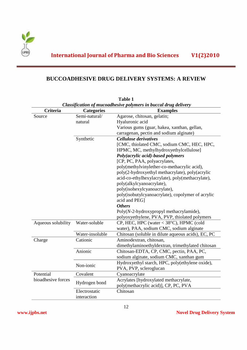
4.2 Classification In general, adhesive polymers can be classified as synthetic vs. natural, water-soluble vs. water-insoluble, and charged vs. uncharged polymers. Examples of the recent polymers classified in these categories are listed in below Table 1.3, 5, 31, 37
International Journal of Pharma and Bio Sciences V1(2)2010
BUCCOADHESIVE DRUG DELIVERY SYSTEMS: A REVIEW
12 www.ijpbs.net Novel Drug Delivery System
Table 1 Classification of mucoadhesive polymers in buccal drug delivery
Criteria Categories Examples Source Semi-natural/
natural Agarose, chitosan, gelatin; Hyaluronic acid Various gums (guar, hakea, xanthan, gellan, carragenan, pectin and sodium alginate)
Synthetic Cellulose derivatives [CMC, thiolated CMC, sodium CMC, HEC, HPC, HPMC, MC, methylhydroxyethylcellulose] Poly(acrylic acid)-based polymers [CP, PC, PAA, polyacrylates, poly(methylvinylether-co-methacrylic acid), poly(2-hydroxyethyl methacrylate), poly(acrylic acid-co-ethylhexylacrylate), poly(methacrylate), poly(alkylcyanoacrylate), poly(isohexylcyanoacrylate), poly(isobutylcyanoacrylate), copolymer of acrylic acid and PEG] Others Poly(N-2-hydroxypropyl methacrylamide), polyoxyethylene, PVA, PVP, thiolated polymers
Aqueous solubility Water-soluble CP, HEC, HPC (water < 38°C), HPMC (cold water), PAA, sodium CMC, sodium alginate
Water-insoluble Chitosan (soluble in dilute aqueous acids), EC, PC Charge Cationic Aminodextran, chitosan,
dimethylaminoethyldextran, trimethylated chitosan Anionic Chitosan-EDTA, CP, CMC, pectin, PAA, PC,
sodium alginate, sodium CMC, xanthan gum
Non-ionic Hydroxyethyl starch, HPC, poly(ethylene oxide), PVA, PVP, scleroglucan
Potential bioadhesive forces
Covalent Cyanoacrylate
Hydrogen bond Acrylates [hydroxylated methacrylate, poly(methacrylic acid)], CP, PC, PVA
Electrostatic interaction
Chitosan
International Journal of Pharma and Bio Sciences V1(2)2010
BUCCOADHESIVE DRUG DELIVERY SYSTEMS: A REVIEW
13 www.ijpbs.net Novel Drug Delivery System
4.3 New Generation Mucoadhesive Polymers
New generation polymers are capable of forming covalent bonds with the mucus and the underlying cell layers, and hence, exhibit improved chemical interactions. The new generation of mucoadhesives (with the exception of thiolated polymers) can adhere directly to the cell surface, rather than to mucus. They interact with the cell surface by means of specific receptors or covalent bonding instead of non-specific mechanisms, which are characteristic of the previous polymers. Examples of such are the incorporation of l-cysteine into thiolated polymers and the target-specific, lectin-mediated adhesive polymers. These classes of polymers hold promise for the delivery of a wide variety of new drug molecules, particularly macromolecules, and create new possibilities for more specific drug–receptor interactions and improved targeted drug delivery.3
4.3.1 Thiolated polymers
Through a covalent attachment between a cysteine (Cys) residue and a polymer of choice, such as polycarbophil, poly(acrylic acid), and chitosan, a new generation of mucoadhesive polymers have been created. The modified polymers, which contain a carbodiimide-mediated thiol bond, exhibit much-improved bioadhesive properties. Investigations of the GI epithelial mucus have clarified the structure of this gel-like biopolymer. With more than 4500 amino acids, the enormous polypeptide backbone of mucin protein is divided into three major subunits; tandem repeat array, carboxyl-and amino-terminal domains. The carboxyl-terminal domain contains more than 10 % of cysteine residues. The amino-terminal domain also contains Cys-rich regions. The Cys-rich sub-
domains are responsible for forming the large oligomers of mucin through disulfide bonds. Based on the disulfide exchange reaction, disulfide bonds between the mucin glycoprotein and the thiolated mucoadhesive polymer can potentially be formed, which results in a strong covalent interaction. Other improved mucoadhesive properties of the thiolated polymers, such as improved tensile strength, high cohesive properties, rapid swelling, and water uptake behavior, have made them an attractive new generation of bioadhesive polymers.51-55
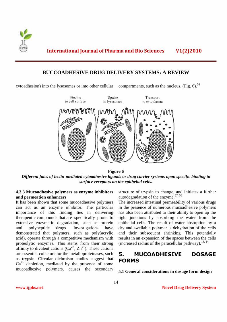
4.3.2 Target-specific, lectin-mediated bioadhesive polymers The possibility of developing a bioadhesive polymer which is able to selectively create specific molecular interactions with a particular target, such as a receptor on the cell membrane of a specific tissue, is a very attractive potential for targeted delivery. The potential of a specific receptor–bioadhesive polymer interaction can circumvent the limiting factors of rapid mucus turnover and short residence time. Unlike general mucoadhesive polymers, which bind to the mucosal surface ubiquitously, a specific receptor-mediated interaction with the mucosal surface could allow for direct binding to the cell surface, rather than only the mucus layer. Specific proteins or glycoproteins, such as lectins, which are able to bind certain sugars on the cell membrane, can increase bioadhesion and potentially improve drug delivery via specific binding and increase the residence time of the dosage form. This type of bioadhesion should be more appropriately termed as cytoadhesion. A site-specific interaction with the receptor could potentially trigger intercellular signaling for internalization of the drug or the carrier system (endocytosis through
International Journal of Pharma and Bio Sciences V1(2)2010
BUCCOADHESIVE DRUG DELIVERY SYSTEMS: A REVIEW
14 www.ijpbs.net Novel Drug Delivery System
cytoadhesion) into the lysosomes or into other cellular compartments, such as the nucleus. (Fig. 6).56
Figure 6 Different fates of lectin-mediated cytoadhesive ligands or drug carrier systems upon specific binding to
surface receptors on the epithelial cells. 4.3.3 Mucoadhesive polymers as enzyme inhibitors and permeation enhancers It has been shown that some mucoadhesive polymers can act as an enzyme inhibitor. The particular importance of this finding lies in delivering therapeutic compounds that are specifically prone to extensive enzymatic degradation, such as protein and polypeptide drugs. Investigations have demonstrated that polymers, such as poly(acrylic acid), operate through a competitive mechanism with proteolytic enzymes. This stems from their strong affinity to divalent cations (Ca2+, Zn2+). These cations are essential cofactors for the metalloproteinases, such as trypsin. Circular dichroism studies suggest that Ca2+ depletion, mediated by the presence of some mucoadhesive polymers, causes the secondary
structure of trypsin to change, and initiates a further autodegradation of the enzyme.57, 58
The increased intestinal permeability of various drugs in the presence of numerous mucoadhesive polymers has also been attributed to their ability to open up the tight junctions by absorbing the water from the epithelial cells. The result of water absorption by a dry and swellable polymer is dehydration of the cells and their subsequent shrinking. This potentially results in an expansion of the spaces between the cells (increased radius of the paracellular pathway).13, 14
5. MUCOADHESIVE DOSAGE
FORMS 5.1 General considerations in dosage form design

International Journal of Pharma and Bio Sciences V1(2)2010
BUCCOADHESIVE DRUG DELIVERY SYSTEMS: A REVIEW
15 www.ijpbs.net Novel Drug Delivery System
5.1.1 Physiological aspects Constant flow of saliva and mobility of the involved tissues challenge drug delivery to the oral cavity. The residence time of drugs delivered to the oral cavity is typically short; in the range of < 5–10 min. Buccal mucoadhesive formulations are expected to overcome this problem. Bioadhesive polymers offer a means by which a delivery system is attached to the buccal mucosa, and hence, provide substantially longer retention times at the absorption site. They also provide a means to confine and maintain high local concentrations of the drug and/or excipient(s) to a defined, relatively small region of the mucosa in order to minimize loss to other regions and limit potential side effects.3, 37
The buccal mucosa is a very suitable region for bioadhesive system application because of its smooth and relatively immobile surface, as well as direct accessibility. However, there are some inherent limitations associated with buccal drug delivery, including short residence time, small absorption area, and barrier properties of the buccal mucosa. The size of a buccal dosage form is restricted by the very limited area available for application of the delivery system. This size restriction, in turn, limits the amount of drug that can be incorporated in the dosage forms. In general, a buccal delivery device that is 1–3 cm2 in size and a drug with a daily dose requirement of 25 mg or less would be preferred. In addition, an ellipsoid shape appears to be most acceptable, and the thickness of buccal delivery devices is usually limited to a few millimeters.3,9,47,59
5.1.2 Pathological aspects Many diseases can affect the thickness of the epithelium, resulting in alteration of the barrier property of the mucosa. Some diseases or treatments may also influence the secretion and properties of the
mucus, as well as the saliva.60 Changes at the mucosal surface due to these pathological conditions may complicate the application and retention of a bioadhesive delivery device. Therefore, understanding the nature of the mucosa under relevant disease conditions is necessary for designing an effective buccal delivery system. In addition, drugs with the potential of changing the physiological conditions of the oral cavity may not be suitable for buccal delivery.3
5.1.3 Pharmacological aspects A buccal dosage form may be designed to deliver a drug to the systemic circulation, or merely indicated for local therapy of the oral mucosa. Selection of dosage forms is affected by the intended application, target site of action, drug characteristics, and the site to be treated (periodontal pockets, gingival, teeth, buccal mucosa, or systemic).3
5.1.4 Pharmaceutical aspects Regardless of dosage form types, the drug must be released from the delivery system and subsequently taken up by the oral mucosa. Poor drug solubility in saliva could significantly retard drug release from the dosage form. Cyclodextrin has been used to solubilize and increase the absorption of poorly water-soluble drugs delivered via the buccal mucosa.61 Other factors affecting both drug release and penetration through buccal mucosa must also be considered in the formulation design. In addition to the physicochemical characteristics required for desirable drug release and absorption, organoleptic properties of the drug or the delivery device should also be considered, since the buccal delivery systems are to be exposed to a highly developed sensory organ.
Some excipients may be incorporated to enhance the effectiveness and acceptability of the dosage forms.

International Journal of Pharma and Bio Sciences V1(2)2010
BUCCOADHESIVE DRUG DELIVERY SYSTEMS: A REVIEW
16 www.ijpbs.net Novel Drug Delivery System
Selection of formulation excipients is yet another important consideration, since acidic compounds can stimulate the secretion of saliva, which enhances not only drug dissolution, but also drug loss by involuntary swallowing. Besides, addition of a separate additive for each function could complicate and enlarge the dosage form, which might be problematic for buccal applications. Therefore, as mentioned previously, polymers with multiple functions seem promising. Permeability characteristics of the buccal mucosa may be continually changed by the rapid turnover of the buccal epithelium (3–8 days compared to about 30 days for the skin).9 Generally, the buccal mucosa is considerably less permeable, and hence, does not provide rapid absorption and good bioavailability seen with sublingual administration. Permeability of the buccal mucosa can be increased by various penetration enhancers capable of increasing cell membrane fluidity, extracting the structural intercellular and/or intracellular lipids, altering cellular proteins, or altering mucus structure and rheology.27 At present, bile salts, fatty acids, and sodium lauryl sulfate are the most commonly investigated penetration enhancers.
Even though the enzyme activity in the buccal mucosa is relatively low and, as a result, drug inactivation is slower and less extensive than in other mucosal routes, susceptible drugs, especially peptides and proteins, can still be degraded by the enzymes in saliva and buccal mucosa.62 Therefore, enzyme inhibitors may be incorporated in the dosage forms to increase drug bioavailability. As previously mentioned, some bioadhesive polymers, such as poly(acrylic acid), polycarbophil, and carbopol, can also inhibit certain proteolytic enzymes (trypsin, a-chymotrypsin, carboxypeptidases A and B, and leucine aminopeptidase). However, cysteine
protease (pyroglutamyl aminopeptidase) may not be inhibited by polycarbophil and carbopol.63 The pH-partitioning theory characteristic of passive diffusion also governs the transcellular permeability of ionizable drugs across the buccal mucosa, similar to other epithelial membranes. Maximal permeation occurs at the pH at which these drugs are predominantly in the unionized form. Control of pH is critical for successful buccal delivery of ionizable drugs. Saliva has a weak buffering capacity to maintain pH value within local regions. It might be desirable to include some pH modifiers in the formulation in order to temporarily modulate the microenvironment at the application site for better drug absorption. It is worth noting that pH can also influence the charge on the surface of the mucus, as well as certain ionizable groups of the polymers, which might affect the strength of mucoadhesion. In addition, it has been shown that the pH of the medium influences the degree of hydration of cross-linked poly(acrylic acid), e.g. polycarbophil. Therefore, the pH needs to be carefully chosen to optimize both drug permeation and mucoadhesion.64, 65
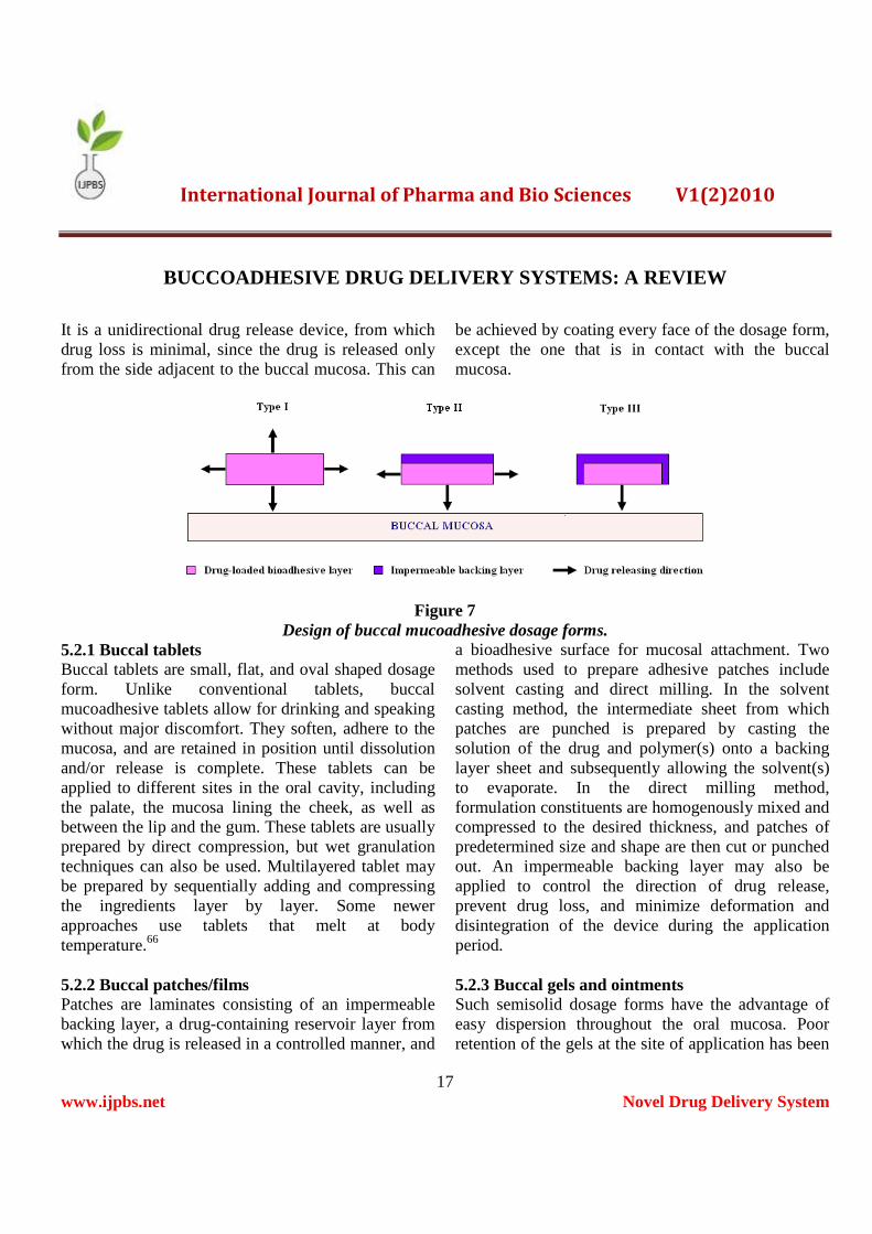
5.2 Buccal Mucoadhesive Dosage Forms Buccal mucoadhesive dosage forms can be categorized into three types based on their geometry (Fig. 7). Type I: It is a single layer device with multidirectional drug release. This type of dosage form suffers from significant drug loss due to swallowing. Type II: It is a device in which an impermeable backing layer is superimposed on top of the drug loaded bioadhesive layer, creating a double-layered device and preventing drug loss from the top surface into the oral cavity. Type III:
International Journal of Pharma and Bio Sciences V1(2)2010
BUCCOADHESIVE DRUG DELIVERY SYSTEMS: A REVIEW
17 www.ijpbs.net Novel Drug Delivery System
It is a unidirectional drug release device, from which drug loss is minimal, since the drug is released only from the side adjacent to the buccal mucosa. This can
be achieved by coating every face of the dosage form, except the one that is in contact with the buccal mucosa.
Figure 7 Design of buccal mucoadhesive dosage forms.
5.2.1 Buccal tablets Buccal tablets are small, flat, and oval shaped dosage form. Unlike conventional tablets, buccal mucoadhesive tablets allow for drinking and speaking without major discomfort. They soften, adhere to the mucosa, and are retained in position until dissolution and/or release is complete. These tablets can be applied to different sites in the oral cavity, including the palate, the mucosa lining the cheek, as well as between the lip and the gum. These tablets are usually prepared by direct compression, but wet granulation techniques can also be used. Multilayered tablet may be prepared by sequentially adding and compressing the ingredients layer by layer. Some newer approaches use tablets that melt at body temperature.66 5.2.2 Buccal patches/films Patches are laminates consisting of an impermeable backing layer, a drug-containing reservoir layer from which the drug is released in a controlled manner, and
a bioadhesive surface for mucosal attachment. Two methods used to prepare adhesive patches include solvent casting and direct milling. In the solvent casting method, the intermediate sheet from which patches are punched is prepared by casting the solution of the drug and polymer(s) onto a backing layer sheet and subsequently allowing the solvent(s) to evaporate. In the direct milling method, formulation constituents are homogenously mixed and compressed to the desired thickness, and patches of predetermined size and shape are then cut or punched out. An impermeable backing layer may also be applied to control the direction of drug release, prevent drug loss, and minimize deformation and disintegration of the device during the application period. 5.2.3 Buccal gels and ointments Such semisolid dosage forms have the advantage of easy dispersion throughout the oral mucosa. Poor retention of the gels at the site of application has been

International Journal of Pharma and Bio Sciences V1(2)2010
BUCCOADHESIVE DRUG DELIVERY SYSTEMS: A REVIEW
18 www.ijpbs.net Novel Drug Delivery System
overcome by using bioadhesive formulations. Certain bioadhesive polymers undergo a phase change from a liquid to a semisolid; this change enhances the viscosity, which results in sustained and controlled release of drugs. Hydrogels are also a promising dosage form, which are formed from polymers that are hydrated in an aqueous environment and physically entrap drug molecules for subsequent slow release by diffusion or
erosion. These dosage forms provide an extended retention time, adequate drug penetration, as well as high efficacy and patient acceptability.67-72 5.2.4 Drugs investigated for buccal drug delivery Various drugs have been investigated for their delivery through the buccal mucosa (Table 2) by formulating them in the above mentioned suitable dosage forms.
Table 2
List of drugs investigated for buccal drug delivery.
1 †Acitretin73 34 ‡Metoprolol tartrate68
2 ‡Acyclovir74-76 35 †Metronidazole78, 116, 117
Arecoline77 36 †‡Miconazole nitrate118, 119-122, 123٭ 3
4 †Benzydamine78 37 †Morphine124
5 ‡Buprenorphine79, 80 38 †Morphine sulfate125
6 †Carbamazepine81 39 †Nalbuphine126
7 †‡Cetylpyridium chloride82 40 †Nicotine127, 128
Chitosan83 41 †‡Nifedipine129-131٭‡ 8
Chlorhexidine84-86 42 ‡Ofloxacin103٭ 910 †‡Chlorhexidine diacetate87, 88 43 †Omeprazole132, 133
Chlorhexidine digluconate89 44 ‡Oxytocin134, 135٭‡ 11
12 †Chlorpheniramine maleate90 45 †Pentazocine136
13 †Cyanocobalamine91 46 †Pindolol137
14 †Danazol61 47 †Piroxicam138
Propolis139٭ Denbufylline72 48٭ 15
Diclofenac sodium92 49 †Propranolol140٭ 1617 †Diltiazem HCl93, 94 50 †‡Propranolol HCl141
18 ‡Dipotassium glycyrrhizate95 51 †Prosidol142
Ergotamine tartrate96 52 ‡Protirelin (TRH)59٭† 19
20 †Fluoride97, 98 53 Recombinant human epidermal growth٭factor (rhEGF)143
Flurbiprofen99 54 †‡Salmon calcitonin144, 145٭ 21
22 ‡Glibenclamide100 55 †Sodium fluoride146
23 †Glucagon-like peptide (GLP)-1101, 102 56 ‡Terbutaline sulfate147
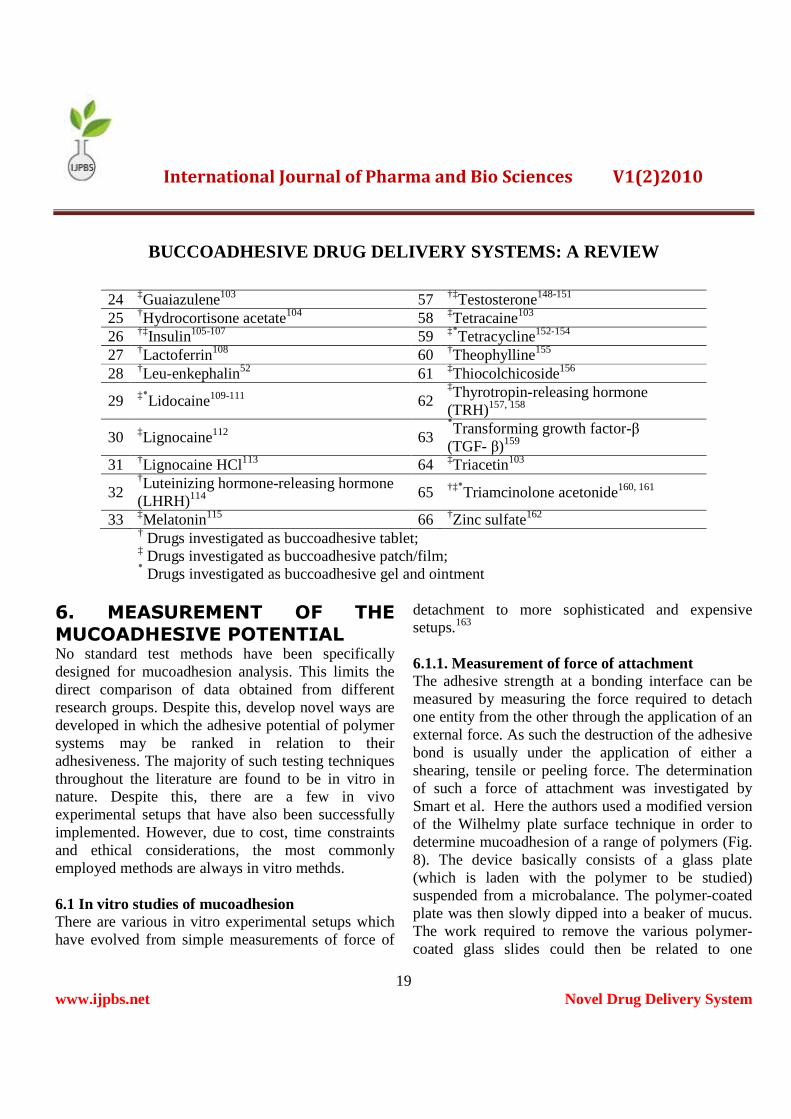
International Journal of Pharma and Bio Sciences V1(2)2010
BUCCOADHESIVE DRUG DELIVERY SYSTEMS: A REVIEW
19 www.ijpbs.net Novel Drug Delivery System
24 ‡Guaiazulene103 57 †‡Testosterone148-151
25 †Hydrocortisone acetate104 58 ‡Tetracaine103 26 †‡Insulin105-107 59 ‡٭Tetracycline152-154 27 †Lactoferrin108 60 †Theophylline155
28 †Leu-enkephalin52 61 ‡Thiocolchicoside156
Lidocaine109-111 62٭‡ 29‡Thyrotropin-releasing hormone (TRH)157, 158
30 ‡Lignocaine112 63 Transforming growth factor-β٭(TGF- β)159
31 †Lignocaine HCl113 64 ‡Triacetin103
32 †Luteinizing hormone-releasing hormone (LHRH)114 65 †‡٭Triamcinolone acetonide160, 161
33 ‡Melatonin115 66 †Zinc sulfate162
† Drugs investigated as buccoadhesive tablet; ‡ Drugs investigated as buccoadhesive patch/film; Drugs investigated as buccoadhesive gel and ointment ٭
6. MEASUREMENT OF THE
MUCOADHESIVE POTENTIAL No standard test methods have been specifically designed for mucoadhesion analysis. This limits the direct comparison of data obtained from different research groups. Despite this, develop novel ways are developed in which the adhesive potential of polymer systems may be ranked in relation to their adhesiveness. The majority of such testing techniques throughout the literature are found to be in vitro in nature. Despite this, there are a few in vivo experimental setups that have also been successfully implemented. However, due to cost, time constraints and ethical considerations, the most commonly employed methods are always in vitro methds. 6.1 In vitro studies of mucoadhesion There are various in vitro experimental setups which have evolved from simple measurements of force of
detachment to more sophisticated and expensive setups.163
6.1.1. Measurement of force of attachment The adhesive strength at a bonding interface can be measured by measuring the force required to detach one entity from the other through the application of an external force. As such the destruction of the adhesive bond is usually under the application of either a shearing, tensile or peeling force. The determination of such a force of attachment was investigated by Smart et al. Here the authors used a modified version of the Wilhelmy plate surface technique in order to determine mucoadhesion of a range of polymers (Fig. 8). The device basically consists of a glass plate (which is laden with the polymer to be studied) suspended from a microbalance. The polymer-coated plate was then slowly dipped into a beaker of mucus. The work required to remove the various polymer-coated glass slides could then be related to one

International Journal of Pharma and Bio Sciences V1(2)2010
BUCCOADHESIVE DRUG DELIVERY SYSTEMS: A REVIEW
20 www.ijpbs.net Novel Drug Delivery System
another and their adhesiveness could be ranked.164, 165 The lack of biological tissue in such a setup maynot represents true mucoadhesion. Most of the mucoadhesive delivery systems will tend to exhibit other mechanical forces, such as the shear stresses
exhibited within the buccal cavity. The effect of both these important forces on measuring the adhesive bond was made possible via the use of a dual tensiometer. (Fig. 9).166-168
Figure 8 Modified plate surface technique, for mucoadhesion determination.
International Journal of Pharma and Bio Sciences V1(2)2010
BUCCOADHESIVE DRUG DELIVERY SYSTEMS: A REVIEW
21 www.ijpbs.net Novel Drug Delivery System
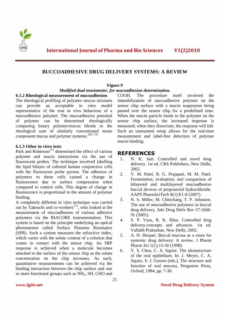
Figure 9 Modified dual tensiometer, for mucoadhesion determination.
6.1.2 Rheological measurement of mucoadhesion The rheological profiling of polymer–mucus mixtures can provide an acceptable in vitro model representative of the true in vivo behaviour of a mucoadhesive polymer. The mucoadhesive potential of polymer can be determined rheologically comparing binary polymer/mucus blends to the rheological sum of similarly concentrated mono component mucus and polymer systems.169, 170
6.1.3 Other in vitro tests Park and Robinson171 determined the effect of various polymer and mucin interactions via the use of fluorescent probes. The technique involved labelling the lipid bilayer of cultured human conjunctiva cells with the fluorescent probe pyrene. The adhesion of polymers to these cells caused a change in fluorescence due to surface compression when compared to control cells. This degree of change in fluorescence is proportional to the amount of polymer binding. A completely different in vitro technique was carried out by Takeuchi and co-workers172, who looked at the measurement of mucoadhesion of various adhesive polymers via the BIACORE instrumentation. This system is based on the principle underlying an optical phenomenon called Surface Plasmon Resonance (SPR). Such a system measures the refractive index, which varies with the solute content of a solution that comes in contact with the sensor chip. An SRP response is achieved when a molecule becomes attached to the surface of the sensor chip as the solute concentration on the chip increases. As such, quantitative measurements can be achieved via the binding interaction between the chip surface and one or more functional groups such as NH2, SH, CHO and
COOH. The procedure itself involved the immobilization of mucoadhesive polymer on the sensor chip surface with a mucin suspension being passed over the sensor chip for a predefined time. When the mucin particle binds to the polymer on the sensor chip surface, the increased response is measured; when they dissociate, the response will fall. Such an instrument setup allows for the real-time measurement and label-free detection of polymer mucin binding.
REFERENCES 1. N. K. Jain. Controlled and novel drug
delivery. 1st ed. CBS Publishers, New Delhi, 2002.
2. V. M. Patel, B. G. Prajapati, M. M. Patel. Formulation, evaluation, and comparison of bilayered and multilayered mucoadhesive buccal devices of propranolol hydrochloride. AAPS PharmSciTech 8(1):E1-8 (2007).
3. N. S. Miller, M. Chittchang, T. P. Johnston. The use of mucoadhesive polymers in buccal drug delivery. Adv Drug Deliv Rev 57:1666-91 (2005).
4. S. P. Vyas, R. K. Khar. Controlled drug delivery-concepts and advances. 1st ed. Vallabh Prakashan, New Delhi, 2002.
5. A. H. Shojaei. Buccal mucosa as a route for systemic drug delivery: A review. J Pharm Pharm Sci 1(1):15-30 (1998).
6. Y. S. Chen, C. A. Squier. The ultrastructure of the oral epithelium. In: J. Meyer, C. A. Squier, S. J. Gerson (eds.), The structure and function of oral mucosa, Pergamon Press, Oxford, 1984, pp. 7-30.

International Journal of Pharma and Bio Sciences V1(2)2010
BUCCOADHESIVE DRUG DELIVERY SYSTEMS: A REVIEW
22 www.ijpbs.net Novel Drug Delivery System
7. D. Harris, J. R. Robinson. Drug delivery via the mucous membranes of the oral cavity. J Pharm Sci 81:1-10 (1992).
8. J. Meyer, S. J. Gerson. A comparison of human palatal and buccal mucosa. Periodontics 2:284-91 (1964).
9. R. B. Gandhi, J. R. Robinson. Oral cavity as a site for bioadhesive drug delivery. Adv Drug Deliv Rev 13:43-74 (1994).
10. M. J. Stablein, J. Meyer. The vascular system and blood supply. In: J. Meyer, C. A. Squier, S. J. Gerson (eds.), The structure and function of oral mucosa, Pergamon Press, Oxford, 1984, pp. 237-56.
11. A. Allen, A. Bell, S. McQueen. Mucus and mucosal protection. In: A. Allen, G. Flemström, A. Garner, W. Silen, L. A. Turnberg (eds.), Mechanisms of mucosal protection in the upper gastrointestinal tract, Raven Press, New York, 1984, pp. 195-202.
12. G. P. Andrews, T. P. Laverty, D. S. Jones. Mucoadhesive polymeric platforms for controlled drug delivery. Eur J Pharm Biopharm 71:505-18 (2009).
13. C. M. Lehr. From sticky stuff to sweet receptors-achievements, limits and novel approaches to bioadhesion. Eur J Drug Metab Pharmacokinet 21:139-48 (1996).
14. J. Haas, C. M. Lehr. developments in the area of bioadhesive drug delivery systems. Expert Opin Biol Ther 2:287-98 (2002).
15. I. Fiebrig, S. Harding, A. Rowe, S. Hyman, S. Davis. Transmission electron microscopy studies on pig gastric mucin and its interactions with chitosan. Carbohydr Polym 28:239-44 (1995).
16. L. Serra, J. Doménech, N. A. Peppas. Engineering design and molecular dynamics
of mucoadhesive drug delivery systems as targeting agents. Eur J Pharm Biopharm 71:519-28 (2009).
17. R. Willits, W. M. Saltzman. Synthetic polymers alter the structure of cervical mucus. Biomaterials 22:445-52 (2001).
18. A. F. Hayward. Membrane-coating granules. Int Rev Cyt 59:97-127 (1979).
19. C. A. Squier, R. A. Eady, R. M. Hopps. The permeability of epidermis lacking normal membrane-coating granules: an ultrastructural tracer study of Kyrle-Flegel disease. J Invest Dermatol 70:361-64 (1978).
20. I. A. Siegel, S. H. Hall, R. Stambaugh. Permeability of the oral mucosa. In: C. A. Squier, J. Meyer (eds.), Current concepts of the histology of oral mucosa, Springfield, IL: Carles Thomas; 1971, pp. 274-86.
21. Y. Oyama, H. Yamano, A. Ohkuma, K. Ogawara, K. Higaki, T. Kimura. Carrier-mediated transport systems for glucose in mucosal cells of the human oral cavity. J Pharm Sci 88:830-34 (1999).
22. N. Utoguchi, Y. Watanabe, T. Suzuki, J. Maehara, Y. Matsumoto, M. Matsumoto. Carrier-mediated transport of monocarboxylic acids in primary cultured epithelial cells from rabbit oral mucosa. Pharm Res 14:320-24 (1997).
23. N. Utoguchi, Y. Watanabe, Y. Takase, T. Suzuki, Matsumoto M. Carrier-mediated absorption of salicylic acid from hamster cheek pouch mucosa. J Pharm Sci 88:142-46 (1999).
24. D. F. Evered, J. V. Vadgama. Absorption of amino acids from the human buccal cavity. Biochem Soc Trans 9:132-33 (1981).
International Journal of Pharma and Bio Sciences V1(2)2010
BUCCOADHESIVE DRUG DELIVERY SYSTEMS: A REVIEW
23 www.ijpbs.net Novel Drug Delivery System
25. D. F. Evered, C. Mallett. Thiamine absorption across human buccal mucosa in vivo. Life Sci 32:1355-58 (1983).
26. J. R. Robinson, X. Yang. Absorption enhancers. In: J. Swarbrick, J. C. Boylan (eds.), Encyclopedia of pharmaceutical technology, Marcel Dekker Inc., New York, 2001, vol 18, pp. 1-27.
27. F. Veuillez, Y. N. Kalia, Y. Jacques, J. Deshusses, P. Buri. Factors and strategies for improving buccal absorption of peptides. Eur J Pharm Biopharm 51:93-109 (2001).
28. G. F. Walker, N. Langoth, A. Bernkop-Schnürch. Peptidase acitivity on the surface of the porcine buccal mucosa. Int J Pharm 233:141-47 (2002).
29. S. D. Kashi, V. H. L. Lee. Enkephalin hydrolysis in homogenates of various absorptive mucosae of the albino rabbit: similarities in rates and involvement of aminopeptidases. Life Sci 38:2019-28 (1986).
30. M. A. Longer, J. R. Robinson. Fundamental aspects of bioadhesion. Pharm. Int. 7:114-17 (1986).
31. J. M. Gu, J. R. Robinson, S. H. S. Leung. Binding of acrylic polymers to mucin/epithelial surfaces: structure-property relationships. Crit Rev Ther Drug Carr Syst 5:21-67 (1998).
32. N. A.. Peppas, P. A. Buri. Surface, interfacial and molecular aspects of polymer bioadhesion on soft tissues. J Control Release 2:257-75 (1985).
33. A. Ahuja, R. K. Khar, J. Ali. Mucoadhesive drug delivery systems. Drug Dev Ind Pharm 23:489-515 (1997).
34. Y. Huang, W. Leobandung, A. Foss, N. A. Peppas. Molecular aspects of muco- and bioadhesion: tethered structures and site-specific surfaces. J Control Release 65:63-71 (2000).
35. A. Ahagon, A. N. Gent. Effect of interfacial bonding on the strength of adhesion. J Polym Sci Polym Phys 13:1285-1300 (1975).
36. A. J. Kinloch. The science of adhesion. J Mater Sci 15:2141-66 (1980).
37. J. W. Lee, J. H. Park, J. R. Robinson. Bioadhesive-based dosage forms: the next generation. J Pharm Sci 89:850-66 (2000).
38. D. Dodou, P. Breedveld, P. Wieringa. Mucoadhesives in the gastrointestinal tract: revisiting the literature for novel applications. Eur J Pharm Biopharm 60:1-16 (2005).
39. M. I. Ugwoke, R. U. Agu, N. Verbeke, R. Kinget. Nasal mucoadhesive drug delivery: background, applications, trends and future perspectives. Adv Drug Deliv Rev 57:1640-1665 (2005).
40. P. B. van Wachem, T. Beugeling, J. Feijen, A. Bantjes, J. P. Detmers, W. G. van Aken. Interaction of cultured human endothelial cells with polymeric surfaces of different wettabilities. Biomaterials 6:403-8 (1985).
41. J. L. Chen, G. N. Cyr. Compositions producing adhesion through hydration. In: R. S. Manly (ed.), Adhesion in biological systems, Academic Press, New York, 1970, pp. 163-80.
42. H. Park, J. R. Robinson. Mechanisms of mucoadhesion of poly(acrylic acid) hydrogels. Pharm Res 4:457-64 (1987).
International Journal of Pharma and Bio Sciences V1(2)2010
BUCCOADHESIVE DRUG DELIVERY SYSTEMS: A REVIEW
24 www.ijpbs.net Novel Drug Delivery System
43. P. J. Flory. Principle of polymer chemistry. Cornell University Press, New York, 1953, pp. 541.
44. H. Park, M. Amiji, K. Park. Mucoadhesive hydrogels effective at neutral pH. Proc Int Symp Control Release Bioact Mater 16:217-18 (1989).
45. C. M. Lehr, J. A. Bouwstra, E. H. Schacht, H. E. Junginger. In vitro evaluation of mucoadhesive properties of chitosan and some other natural polymers. Int J Pharm 78:43-48 (1992).
46. D. Solomonidou, K. Cremer, M. Krumme, J. Kreuter. Effect of carbomer concentration and degree of neutralization on the mucoadhesive properties of polymer films. J Biomater Sci Polym Ed 12:1191-1205 (2001).
47. M. J. Rathbone, B. K. Drummond, I. G. Tucker. The oral cavity as a site for systemic drug delivery. Adv Drug Deliv Rev 13:1-22 (1994).
48. C. M. Lehr, F. G. J. Poelma, H. E. Junginger, J. J. Tukker. An estimate of turnover time of intestinal mucus gel layer in the rat in situ loop. Int J Pharm 70:235-40 (1991).
49. J. F. Forstner. Intestinal mucins in health and disease. Digestion 17:234-63 (1978).
50. N. F. H. Ho, C. L. Barsuhn, P. S. Burton, H. P. Merkle. (D) Routes of delivery: case studies. (3) Mechanistic insights to buccal delivery of proteinaceous substances. Adv Drug Deliv Rev 8:197-235 (1992).
51. A. Bernkop-Schnürch, V. Schwarz, S. Steininger. Polymers with thiol groups: a new generation of mucoadhesive polymers. Pharm Res 16:876-81 (1999).
52. N. Langoth, J. Kalbe, A. Bernkop-Schnürch. Development of buccal drug delivery systems based on a thiolated polymer. Int J Pharm 252:141-48 (2003).
53. M. K. Marschütz, A. Bernkop-Schnürch. Thiolated polymers: self-crosslinking properties of thiolated 450 kDa poly(acrylic acid) andtheir influence on mucoadhesion. Eur J Pharm Sci 15:387-94 (2002).
54. C. E. Kast, A. Bernkop-Schnürch. Thiolated polymers-thiomers: development and in vitro evaluation of chitosan-thioglycolic acid conjugates. Biomaterials 22:2345-52 (2001).
55. J. R. Gum Jr, J. W. Hicks, N. W. Toribara, E. M. Rothe, R. E. Lagace, Y. S. Kim. The human MUC2 intestinal mucin has cycteine-rich subdomains located both upstream and downstream of its central repetitive region. J Biol Chem 267:21375-83 (1992).
56. C. M. Lehr. Lectin-mediated drug delivery: the second generation of bioadhesives. J Control Release 65:19-29 (2000).
57. H. L. Lueßen, C. M. Lehr, C. O. Rentel, A. B. J. Noach, A. G. de Boer, J. C. Verhoef, H. E. Junginger. Bioadhesive polymers for the peroral delivery of peptide drugs. J Control Release 29:329-38 (1994).
58. H. L. Lueßen, J. C. Verhoef, G. Borchard, C. M. Lehr, A. G. de Bore, H. E. Junginger. Mucoadhesive polymers in peroral peptide drug delivery: II. Carbomer and polycarbophil are potent inhibitors of the intestinal proteolytic enzyme trypsin. Pharm Res 12:1293-98 (1995).
59. R. Anders, H. P. Merkle. Evaluation of laminated mucoadhesive patches for buccal drug delivery. Int J Pharm 49:231-40 (1989).
International Journal of Pharma and Bio Sciences V1(2)2010
BUCCOADHESIVE DRUG DELIVERY SYSTEMS: A REVIEW
25 www.ijpbs.net Novel Drug Delivery System
60. K. Khanvilkar, M. D. Donovan, D. R. Flanagan. Drug transfer through mucus. Adv Drug Deliv Rev 48:173-93 (2001).
61. A. C. Jain, B. J. Aungst, M. C. Adeyeye. Development and in vivo evaluation of buccal tablets prepared using danazol-sulfobutylether 7 β-cyclodextrin (SBE 7) complexes. J Pharm Sci 91:1659-68 (2002).
62. M. E. de Vries, H. E. Boddé, J. C. Verhoef, H. E. Junginger. Developments in buccal drug delivery. Crit. Rev Ther Drug Carr Syst 8:271-303 (1991).
63. H. L. Lueßen, J. C. Verhoef, A. G. de Bore, H. E. Junginger, B. J. de Leeuw, G. Borchard, C. M. Lehr. Multifunctional polymers for the peroral delivery of peptide drugs. In: E. Mathiowitz, D. E. Chickering III, C. M. Lehr (eds.), Bioadhesive drug delivery systems. Fundamentals, novel approaches, and development. Marcel Dekker, New York, 1999, pp. 299-339.
64. H. S. Ch’ng, H. Park, P. Kelly, J. R. Robinson. Bioadhesive polymers as platforms for oral controlled drug delivery: II. Synthesis and evaluation of some swelling, water-insoluble bioadhesive polymers. J Pharm Sci 74:399-405 (1985).
65. H. Park, J. R. Robinson. Physico-chemical properties of water insoluble polymers important to mucin epithelial adhesion. J Control Release 2:47-57 (1985).
66. E. M. Rudnic, J. D. Schwartz. Oral solid dosage forms. In: A. R. Gennaro (editor). Remington: the science and practice of pharmacy, 20th ed. Lippincott Williams & Wilkins, Baltimore, MD, 2000, pp. 858-59.
67. S. C. Miller, M. D. Donovan. Effect of poloxamer 407 gel on the miotic activity of
pilocarpine nitrate in rabbits. Int J Pharm 12:147-52 (1982).
68. C. F. Wong, K. H. Yuen, K. K. Peh. Formulation and evaluation of controlled release Eudragit buccal patches. Int J Pharm 178:11-22 (1999).
69. S. Kumar, B. O. Haglund, K. J. Himmelstein. In situ-forming gels for ophthalmic drug delivery. J Ocul Pharmacol 10:47-56 (1994).
70. R. Gurny, J. E. Ryser, C. Tabatabay, M. Martenet, P. Edman, O. Camber. Precorneal residence time in humans of sodium hyaluronate as measured by gamma scintigraphy. Graefe Arch Clin Exp Ophthalmol 228:510-12 (1990).
71. G. Meseguer, R. Gurny, P. Buri. Gamma scintigraphic evaluation of precorneal clearance in human volunteers and in rabbits. Eur J Drug Metab Pharmacokinet 18:190-94 (1993).
72. L. Martin, C. G. Wilson, F. Koosha, I. F. Uchegbu. Sustained buccal delivery of the hydrophobic drug denbufylline using physically cross-linked palmitoyl glycol chitosan hydrogels. Eur J Pharm Biopharm 55:35-45 (2003).
73. G. M. Gaeta, F. Gombos, F. Femiano, C. Battista, P. Minghetti, L. Montanari, R. A. Satriano, G. Argenziano. Acitretin and treatment of the oral leucoplakias. A model to have an active molecules release. J Eur Acad Dermatol Venereol 14:473-478 (2000).
74. A. H. Shojaei, B. Berner, X. Li. Transbuccal delivery of acyclovir: I. In vitro determination of routes of buccal transport. Pharm Res 15:1182-88 (1998).
75. A. H. Shojaei, S. L. Zhuo, X. Li. Transbuccal delivery of acyclovir: II. Feasibility, system
International Journal of Pharma and Bio Sciences V1(2)2010
BUCCOADHESIVE DRUG DELIVERY SYSTEMS: A REVIEW
26 www.ijpbs.net Novel Drug Delivery System
design, and in vitro permeation studies. J Pharm Pharm Sci 1:66-73 (1998).
76. S. Rossi, G. Sandri, F. Ferrari, M. C. Bonferoni, C. Caramella. Buccal delivery of acyclovir from films based on chitosan and polyacrylic acid. Pharm Dev Technol 8:199-208 (2003).
77. S. S. Strickland, G. V. Veena, P. J. Houghton, S. C. Stanford, A. V. Kurpad. Areca nut, energy metabolism and hunger in Asian men. Ann Hum Biol 30:26-52 (2003).
78. B. Parodi, E. Russo, P. Gatti, S. Cafaggi, G. Bignardi. Development and in vitro evaluation of buccoadhesive tablets using a new model substrate for bioadhesion measures: the eggshell membrane. Drug Dev Ind Pharm 25:289-95 (1999).
79. J. H. Guo. Bioadhesive polymer buccal patches for buprenorphine controlled delivery: formulation, in-vitro adhesion and release properties. Drug Dev Ind Pharm 20:2809-21 (1994).
80. J. H. Guo, K. M. Cooklock. The effects of backing materials and multilayered systems on the characteristics of bioadhesive buccal patches. J Pharm Pharmacol 48:255-57 (1996).
81. G. Đkinci, Y. Çapan, S. Şenel, E. Alaaddinoğlu, T. Dalkara, A. A. Hıncal. In vitro/in vivo studies on a buccal bioadhesive tablet formulation of carbamazepine. Pharmazie 55:762-65 (2000).
82. N. A. Nafee, N. A. Boraie, F. A. Ismail, L. M. Mortada. Design and characterization of mucoadhesive buccal patches containing cetylpyridinium chloride. Acta Pharm 53:199-212 (2003).
83. G. Đkinci, S. Şenel, H. Akıncıbay, S. Kaş, S. Erciş, C. G. Wilson, A. A. Hıncal. Effect of chitosan on a periodontal pathogen Porphyromonas gingivalis. Int J Pharm 235:121-27 (2002).
84. A. H. C. Vinholis, L. C. de Figueiredo, E. Marcantonio Jr, R. A. C. Marcantonio, S. L. S. Salvador, G. Goissis. Subgingival utilization of a 1 % chlorhexidine collagen gel for the treatment of periodontal pockets. A clinical and microbiological study. Braz Dent J 12:209-13 (2001).
85. D. S. Jones, A. D. Woolfson, A. F. Brown. Textural analysis and flow rheometry of novel, bioadhesive antimicrobial oral gels. Pharm Res 14:450-57 (1997).
86. D. S. Jones, A. F. Brown, A. D. Woolfson, A. C. Dennis, L. J. Matchett, S. E. J. Bell. Examination of the physical state of chlorhexidine within viscoelastic, bioadhesive semisolids using raman spectroscopy. J Pharm Sci 89:563-71 (2000).
87. P. Giunchedi, C. Juliano, E. Gavini, M. Cossu, M. Sorrenti. Formulation and in vivo evaluation of chlorhexidine buccal tablets prepared using drug-loaded chitosan microspheres. Eur J Pharm Biopharm 53:233-39 (2002).
88. D. S. Jones, N. J. Medlicott. Casting solvent controlled release of chlorhexidine from ethylcellulose films prepared by solvent evaporation. Int J Pharm 114:257-61 (1995).
89. S. Şenel, G. Đkinci, S. Kaş, A. Yousefı-Rad, M. F. Sargon, A. A. Hıncal. Chitosan films and hydrogels of chlorhexidine gluconate for oral mucosal delivery. Int J Pharm 193:197-203 (2000).
International Journal of Pharma and Bio Sciences V1(2)2010
BUCCOADHESIVE DRUG DELIVERY SYSTEMS: A REVIEW
27 www.ijpbs.net Novel Drug Delivery System
90. D. Tiwari, D. Goldman, R. Sause, P. L. Madan. Evaluation of polyoxyethylene homopolymers for buccal bioadhesive drug delivery device formulations. AAPS PharmSci 1:E13 (1999).
91. D. Tiwari, D. Goldman, C. Town, R. Sause, P. L. Madan. In vitro-in vivo evaluation of a controlled release buccal bioadhesive device for oral drug delivery. Pharm Res 16:1775-80 (1999).
92. J. Cassidy, B. Berner, K. Chan, V. John, S. Toon, B. Holt, M. Rowland. Human transbuccal absorption of diclofenac sodium from a prototype hydrogel delivery device. Pharm Res 10:126-29 (1993).
93. B. Singh, N. Ahuja. Development of controlled-release buccoadhesive hydrophilic matrices of diltiazem hydrochloride: optimization of bioadhesion, dissolution, and diffusion parameters. Drug Dev Ind Pharm 28:431-42 (2002).
94. A. Ahuja, M. Dogra, S. P. Agarwal. Development of buccal tablets of diltiazem hydrochloride. Indian J Pharm Sci 57:26-30 (1995).
95. G. J. Rhee, D. K. Lee, K. H. Sin, C. B. Park. Formulation and pharmaceutical properties of mucoadhesive films containing dipotassium glycyrrhizate. J Korean Pharm Sci 29:127-36 (1999).
96. K. Tsutsumi, Y. Obata, T. Nagai, T. Loftsson, K. Takayama. Buccal absorption of ergotamine tartrate using the bioadhesive tablet system in guinea-pigs. Int J Pharm 238:161-70 (2002).
97. P. Bottenberg, R. Cleymaet, K. Röhrkasten, F. Lampert. Microhardness changes in surface enamel after application of
bioadhesive fluoride tablets in situ. Clin. Oral Investig 4:153-156 (2000).
98. N. Vivien-Castioni, R. Gurny, P. Baehni, V. Kaltsatos. Salivary fluoride concentrations following applications of bioadhesive tablets and mouthrinses. Eur J Pharm Biopharm 49:27-33 (2000).
99. D. S. Jones, C. R. Irwin, A. D. Woolfson, J. Djokic, V. Adams. Physicochemical characterization and preliminary in vivo efficacy of bioadhesive, semisolid formulations containing flurbiprofen for the treatment of gingivitis. J Pharm Sci 88:592-98 (1999).
100. R. Ilango, S. Kavimani, A. R. Mullaicharam, B. Jayakar. In vitro studies on buccal strips of glibenclamide using chitosan. Indian J Pharm Sci 59:232-35 (1997).
101. M. K. Gutniak, H. Larsson, S. J. Heiber, O. T. Juneskans, J. J. Holst, B. Ahrén. Potential therapeutic levels of glucagon-like peptide I achived in humans by a buccal tablet. Diabetes Care 19:843-48 (1996).
102. M. K. Gutniak, H. Larsson, S. W. Sanders, O. Juneskans, J. J. Holst, B. Ahrén. GLP-1 tablet in type 2 diabetes in fasting and postprandial conditions. Diabetes care 20:1874-79 (1997).
103. M. Oguchi, N. Shikama, S. Sasaki, K. Gomi, Y. Katsuyama, S. Ohta, M. Hori, K. Takei, K. Arakawa, S. Sone. Mucoadhesive water-soluble polymer film for treatment of acute radiation-induced oral mucositis. Int J Radiat Oncol Biol Phys 40:1033-37 (1998).
104. G. C. Ceschel, P. Maffei, S. Lombardi Biogia, C. Ronchi. Design and evaluation of buccal adhesive hydrocortisone acetate (HCA) tablets. Drug Deliv 8:161-71 (2001).
International Journal of Pharma and Bio Sciences V1(2)2010
BUCCOADHESIVE DRUG DELIVERY SYSTEMS: A REVIEW
28 www.ijpbs.net Novel Drug Delivery System
105. W. A. Ritschel, G. B. Ritschel, H. Forusz, M. Kraeling. Buccal absorption of insulin in the dog. Res Commun Chem Pathol Pharmacol 63:53-67 (1989).
106. E. A. Hosny, S. A. Elkheshen, S. I. Saleh. Buccoadhesive tablets for insulin delivery: in-vitro and in-vivo studies. Boll Chim Farm 141:210-17 (2002).
107. M. Ishida, Y. Machida, N. Nambu, T. Nagai. New mucosal dosage form of insulin. Chem Pharm Bull (Tokyo) 29:810-16 (1981).
108. M. E. Kuipers, J. Heegsma, H. I. Bakker, D. K. F. Meijer, P. J. Swart, E. W. Frijlink, A. C. Eissens, H. G. de Vries-Hospers, J. J. M. van den Berg. Design and fungicidal activity of mucoadhesive lactoferrin tablets for the treatment of oropharyngeal candidosis. Drug Deliv 9:31-38 (2002).
109. H. Okamoto, H. Taguchi, K. Iida, K. Danjo. Development of polymer film dosage forms of Lidocaine for buccal administration: I. Penetration rate and release rate. J Control Release 77:253-60 (2001).
110. H. Okamoto, T. Nakamori, Y. Arakawa, K. Iida, K. Danjo. Development of polymer film dosage forms of Lidocaine for buccal administration: II. Comparison of preparation methods. J Pharm Sci 91:2424-32 (2002).
111. Y. T. F. Tan, K. K. Peh, O. Al-Hanbali. Effect of carbopol and polyvinylpyrrolidone on the mechanical, rheological, and release properties of bioadhesive polyethylene glycol gels. AAPS PharmSciTech 1:E24 (2000).
112. I. M. Brook, G. T. Tucker, E. C. Tuckley, R. N. Boyes. A lignocaine patch for dental analgesia safety and early pharmacology. J Control Release 10:183-88 (1989).
113. N. Parvez, A. Ahuja, R. K. Khar. Development and evaluation of mucoadhesive buccal tablets of lignocaine hydrochloride. Indian J Pharm Sci 64:563-67 (2002).
114. S. Nakane, M. Kakumoto, K. Yukimatsu, Y. W. Chien. Oramucosal delivery of LHRH: pharmacokinetic studies of controlled and enhanced transmucosal permeation. Pharm Dev Technol 1:251-59 (1996).
115. L. Bénès, B. Claustrat, F. Horrière, M. Geoffriau, J. Konsil, K. A. Parrott, G. DeGrande, R. L. McQuinn, J. W. Ayres. Transmucosal, oral controlled-release, and transdermal drug administration in human subjects: a crossover study with melatonin. J Pharm Sci 86:1115-19 (1997).
116. A. Ahuja, R. K. Khar, R. Chaudhry. Evaluation of buccoadhesive metronidazole tablets: microbiological response. Pharmazie 53:264-67 (1998).
117. L. Perioli, V. Ambrogi, D. Rubini, S. Giovagnoli, M. Ricci, P. Blasi, C. Rossi. Novel mucoadhesive buccal formulation containing metronidazole for the treatment of periodontal disease. J Control Release 95:521-33 (2004).
118. F. A. Mohammed, H. Khedr. Preparation and in vitro/in vivo evaluation of the buccal bioadhesive properties of slow release tablets containing miconazole nitrate. Drug Dev Ind Pharm 29:321-37 (2003).
119. S. Bouckaert, H. Schautteet, R. A. Lefebvre, J. P. Remon, R. van Clooster. Comparison of salivary miconazole concentrations after administration of a bioadhesive slow-release buccal tablet and an oral gel. Eur J Clin Pharmacol 43:137-40 (1992).
International Journal of Pharma and Bio Sciences V1(2)2010
BUCCOADHESIVE DRUG DELIVERY SYSTEMS: A REVIEW
29 www.ijpbs.net Novel Drug Delivery System
120. S. Bouckaert, R. A. Lefebvre, J. P. Remon. In vitro/in vivo correlation of the bioadhesive properties of a buccal bioadhesive miconazole slow-release tablet. Pharm Res 10:853-56 (1993).
121. S. Bouckaert, R. A. Lefebvre, F. Colardyn, J. P. Remon. Influence of the application site on bioadhesion and slow-release characteristics of a bioadhesive buccal slow-release tablet of miconazole. Eur J Clin Pharmacol 44:331-35 (1993).
122. S. Bouckaert, J. P. Remon. In-vitro bioadhesion of a buccal, miconazole slow-release tablet. J Pharm Pharmacol 45:504-07 (1993).
123. J. Van Roey, M. Haxaire, M. Kamya, I. Lwanga, E. Katabira. Comparative efficacy of topical therapy with a slow-release mucoadhesive buccal tablet containing miconazole nitrate versus systemic therapy with ketoconazole in HIV-positive patients with oropharyngeal candidiasis. J Acquir Immune Defic Syndr 35:144-50 (2004).
124. E. Beyssac, F. Touaref, M. Meyer, L. Jacob, P. Sandouk, J. M. Aiache. Bioavailability of morphine after administration of a new bioadhesive buccal tablet. Biopharm Drug Dispos 19:401-05 (1998).
125. S. Anlar, Y. Çapan, O. Güven, A. Göğüs, T. Dalkara, A. A. Hıncal. Formulation and in vitro-in vivo evaluation of buccoadhesive morphine sulfate tablets. Pharm Res 11:231-36 (1994).
126. R. Y. Han, J. Y. Fang, K. C. Sung, O. Y. P. Hu. Mucoadhesive buccal disks for novel nalbuphine prodrug controlled delivery: effect of formulation variables on drug
release and mucoadhesive performance. Int J Pharm 177:201-09 (1999).
127. C. R. Park, D. L. Munday. Development and evaluation of a biphasic buccal adhesive tablet for nicotine replacement therapy. Int J Pharm 237:215-26 (2002).
128. G. Đkinci, S. Şenel, C. G. Wilson, M. Şumnu. Development of a buccal bioadhesive nicotine tablet formulation for smoking cessation. Int J Pharm 277:173-78 (2004).
129. T. Save, M. U. Shah, A. R. Ghamande, P. Venkitachalam. Comparative study of buccoadhesive formulations and sublingual capsules of nifedipine. J Pharm Pharmacol 46:192-95 (1994).
130. C. Remuñán-López, A. Portero, J. L. Vila-Jato, M. J. Alonso. Design and evaluation of chitosan/ethylcellulose mucoadhesive bilayered devices for buccal drug delivery. J Control Release 55:143-52 (1998).
131. S. Bouckaert, J. P. Remon. In-vitro bioadhesion of a buccal, miconazole slow-release tablet. J Pharm Pharmacol 45:504-07 (1993).
132. H. G. Choi, J. H. Jung, C. S. Yong, C. D. Rhee, M. K. Lee, J. H. Han, K. M. Park, C. K. Kim. Formulation and in vivo evaluation of omeprazole buccal adhesive tablet. J Control Release 68:405-12 (2000).
133. H. G. Choi, C. K. Kim. Development of omeprazole buccal adhesive tablets with stability enhancement in human saliva. J Control Release 68:397-404 (2000).
134. C. Li, P. P. Bhatt, T. P. Johnston. In vitro release and permeation of oxytocin from a mucoadhesive buccal patch. Pharm Dev Technol 1:357-64 (1996).

International Journal of Pharma and Bio Sciences V1(2)2010
BUCCOADHESIVE DRUG DELIVERY SYSTEMS: A REVIEW
30 www.ijpbs.net Novel Drug Delivery System
135. C. Li, P. P. Bhatt, T. P. Johnston. Transmucosal delivery of oxytocin to rabbits using a mucoadhesive buccal patch. Pharm Dev Technol 2:265-74 (1997).
136. V. Agarwal, B. Mishra. Design, development, and biopharmaceutical properties of buccoadhesive compacts of pentazocine. Drug Dev Ind Pharm 25:701-09 (1999).
137. B. Dortunç, L. Özer, N. Uyanik. Development and in vitro evaluation of a buccoadhesive pindolol tablet formulation. Drug Dev Ind Pharm 24:281-88 (1998).
138. M. Jug, M. Bećirević-Laćan. Influence of hydroxypropyl-β-cyclodextrin complexation on piroxicam release from buccoadhesive tablets. Eur J Pharm Sci 21:251-60 (2004).
139. G. C. Ceschel, P. Maffei, A. Sforzini, S. Lombardi Borgia, A. Yasin, C. Ronchi. In vitro permeation through porcine buccal mucosa of caffeic acid phenetyl ester (CAPE) from a topical mucoadhesive gel containing propolis. Fitoterapia 73(1):S44-52 (2002).
140. N. Çelebi, O. Kişlal. Development and evaluation of a buccoadhesive propranolol tablet formulation. Pharmazie 50:470-72 (1995).
141. J. Akbari, A. Nokhodchi, D. Farid, M. Adrangui, M. R. Siahi-Shadbad, M. Saeedi. Development and evaluation of buccoadhesive propranolol hydrochloride tablet formulations: effect of fillers. IL Farmaco 59:155-61 (2004).
142. N. A. Osipova, G. A. Novikov, M. S. Vetsheva, B. M. Prokhorov, V. A. Beresnev, N. A. Loseva, S. Yu. Zemskaya, T. A. Smolina. Prosidol, a new Russian narcotic
analgesic: the first results in oncology. Anesteziol Reanimatol 53-7 (1994).
143. J. S. Park, J. I. Yoon, H. Li, D. C. Moon, K. Han. Buccal mucosal ulcer healing effect of rhEGF/Eudispert hv hydrogel. Arch Pharm Res 26:659-65 (2003).
144. Z. Cui, R. J. Mumper. Buccal transmucosal delivery of calcitonin in rabbits using thin-film composites. Pharm Res 19:1901-06 (2002).
145. H. H. Alur, J. D. Beal, S. I. Pather, A. K. Mitra, T. P. Johnston. Evaluation of a novel, natural oligosaccharide gum as a sustained-release and mucoadhesive component of calcitonin buccal tablets. J Pharm Sci 88:1313-19 (1999).
146. M. Diarra, G. Pourroy, C. Boymond, D. Muster. Fluoride controlled release tablets for intrabuccal use. Biomaterials 24:1293-1300 (2003).
147. M. I. Mohamed, N. D. Mortada. Development and characterization of a buccoadhesive dosage form of terbutaline sulphate. Mansoura J Pharm Sci 16:69-81 (2000).
148. D. Ameye, J. Voorspoels, P. Foreman, J. Tsai, P. Richardson, S. Geresh, J. P. Remon. Ex vivo bioadhesion and in vivo testosterone bioavailability study of different bioadhesive formulations based on starch-g-poly(acrylic acid) copolymers and starch/poly(acrylic acid) mixtures. J Control Release 79:173-82 (2002).
149. J. Voorspoels, J. P. Remon, W. Eechaute, W. Sy De. Buccal absorption of testosterone and its esters using a bioadhesive tablet in dogs. Pharm Res 13:1228-32 (1996).

International Journal of Pharma and Bio Sciences V1(2)2010
BUCCOADHESIVE DRUG DELIVERY SYSTEMS: A REVIEW
31 www.ijpbs.net Novel Drug Delivery System
150. R. J. M. Ross, A. Jabbar, T. H. Jones, B. Roberts, K. Dunkley, J. Hall, A. Long, H. Levine, D. R. Cullen. Pharmacokinetics and tolerability of a bioadhesive buccal testosterone tablet in hypogonadal men. Eur J Endocrinol 150:57-63 (2004).
151. S. Jay, W. Fountain, Z. Cui, R. J. Mumper. Transmucosal delivery of testosterone in rabbits using novel bi-layer mucoadhesive wax-film composite disks. J Pharm Sci 91:2016-25 (2002).
152. M. Minabe, K. Takeuchi, T. Tamura, T. Hori, T. Umemoto. Subgingival administration of tetracycline on a collagen film. J Periodontol 60:552-56 (1989).
153. D. S. Jones, A. D. Woolfson, A. F. Brown, W. A. Coulter, C. McClelland, C. R. Irwin. Design, characterization and preliminary clinical evaluation of a novel mucoadhesive topical formulation containing tetracycline for the treatment of periodontal disease. J Control Release 67:357-68 (2000).
154. D. S. Jones, A. D. Woolfson, J. Djokic, W. A. Coulter. Development and mechanical characterization of bioadhesive semi-solid, polymeric systems containing tetracycline for the treatment of periodontal diseases. Pharm Res 13:1734-38 (1996).
155. S. Geresh, G. Y. Gdalevsky, I. Gilboa, J. Voorspoels, J. P. Remon, J. Kost. Bioadhesive grafted starch copolymers as platforms for peroral drug delivery: a study of theophylline release. J Control Release 94:391-99 (2004).
156. M. Artusi, P. Santi, P. Colombo, H. E. Junginger. Buccal delivery of thiocolchicoside: in vitro and in vivo
permeation studies. Int J Pharm 250:203-13 (2003).
157. C. Li, R. L. Koch, V. A. Raul, P. P. Bhatt, T. P. Johnston. Absorption of thyrotropin-releasing hormone in rats using a mucoadhesive buccal patch. Drug Dev Ind Pharm 23:239-46 (1997).
158. W. Schurr, B. Knoll, R. Ziegler, R. Anders, H. P. Merkle. Comparative study of intravenous, nasal, oral and buccal TRH administration among healthy subjects. J Endocrinol Invest 8:41-4 (1985).
159. S. Şenel, M. J. Kremer, S. Kaş, P. W. Wertz, A. A. Hıncal, C. A. Squier. Enhancing effect of chitosan on peptide drug delivery across buccal mucosa. Biomaterials 21:2067-71 (2000).
160. S. C. Shin, J. P. Bum, J. S. Choi. Enhanced bioavailability by buccal administration of triamcinolone acetonide from the bioadhesive gels in rabbits. Int J Pharm 209:37-43 (2000).
161. S. C. Shin, J. Y. Kim. Enhanced permeation of triamcinolone acetonide through the buccal mucosa. Eur J Pharm Biopharm 50:217-20 (2000).
162. M. Diarra, G. Pourroy, D. Muster, M. Zingraff, C. Boymond. Elaboration and evaluation of an intraoral controlled release delivering system. Biomaterials 19:1523-27 (1998).
163. D. Accili, G. Menghi, G. Bonacucina, P. Martino, G. Palmieri. Mucoadhesion dependence of pharmaceutical polymers on mucosa characteristics. Eur J Pharm Sci 22:225-34 (2004).

International Journal of Pharma and Bio Sciences V1(2)2010
BUCCOADHESIVE DRUG DELIVERY SYSTEMS: A REVIEW
32 www.ijpbs.net Novel Drug Delivery System
164. A. G. Mikos, N. A. Peppas. Systems for controlled release of drugs. V: Bioadhesive systems. STP Pharma Sci 19:705-15 (1986).
165. J. D. Smart, I. W. Kellaway, H. E. Worthington. An in-vitro investigation of mucosa-adhesive materials for use in controlled drug delivery. J Pharm Pharmacol 36:295-99 (1984).
166. C. Wong, K. Yuen, K. Peh. An in-vitro method for buccal adhesion studies: importance of instrument variables. Int J Pharm 180:47-57 (1999).
167. S. H. Leung, J. R. Robinson. The contribution of anionic polymer structural features to mucoadhesion. J Control Release 5:223-31 (1988).
168. S. A. Mortazavi, J. D. Smart. An investigation of some factors influencing the in vitro assessment of mucoadhesion. Int J Pharm 116:223-30 (1995).
169. R. Riley, J. Smart, J. Tsibouklis, P. Dettmar, F. Hampson, J. A. Davis, G. Kelly, W. Wilber. An investigation of mucus/polymer rheological synergism using synthesized and characterized poly(acrylic acid)s. Int J Pharm 217:87-100 (2001).
170. E. E. Hassan, J. M. Gallo. A simple rheological method for the in vitro assessment of mucin-polymer bioadhesive bond strength. Pharm Res 7:491-95 (1990).
171. K. Park, J. R. Robinson. Bioadhesive polymers as platforms for oral-controlled drug delivery: method to study bioadhesion. Int J Pharm 19:107-127 (1984).
172. H. Takeuchi, J. Thongborisute, Y. Matsui, H. Sugihara, H. Yamamoto, Y. Kawashima. Novel mucoadhesion tests for polymers and polymer-coated particles to design optimal
mucoadhesive drug delivery systems. Adv Drug Deliv Rev 57:1583-94 (2005).