deoxynivalenol as a new factor in the persistence of
TRANSCRIPT

Deoxynivalenol as a New Factor in the Persistence ofIntestinal Inflammatory Diseases: An EmergingHypothesis through Possible Modulation of Th17-Mediated ResponsePatricia M. Cano1,2., Julie Seeboth1,2., Francois Meurens3,6, Juliette Cognie4, Roberta Abrami1,2,5,
Isabelle P. Oswald1,2*, Laurence Guzylack-Piriou1,2*
1 INRA, UMR1331, Toxalim, Research Centre in Food Toxicology, Toulouse, France, 2 Universite de Toulouse, INPT, UMR1331, Toxalim, Toulouse, France, 3 INRA, UMR1282
Infectiologie et Sante Publique, Nouzilly, France, 4 INRA, Physiologie de la Reproduction et des Comportements, Nouzilly, France, 5 Universidade Estadual de Londrina, Lab
Patologia Animal, Londrina, Brazil, 6 Vaccine and Infectious Disease Organization, University of Saskatchewan, Saskatoon, Saskatchewan, Canada
Abstract
Background/Aims: Deoxynivalenol (DON) is a mycotoxin produced by Fusarium species which is commonly found intemperate regions worldwide as a natural contaminant of cereals. It is of great concern not only in terms of economic lossesbut also in terms of animal and public health. The digestive tract is the first and main target of this food contaminant and itrepresents a major site of immune tolerance. A finely tuned cross-talk between the innate and the adaptive immunesystems ensures the homeostatic equilibrium between the mucosal immune system and commensal microorganisms. Theaim of this study was to analyze the impact of DON on the intestinal immune response.
Methodology: Non-transformed intestinal porcine epithelial cells IPEC-1 and porcine jejunal explants were used toinvestigate the effect of DON on the intestinal immune response and the modulation of naive T cells differentiation.Transcriptomic proteomic and flow cytometry analysis were performed.
Results: DON induced a pro-inflammatory response with a significant increase of expression of mRNA encoding for IL-8, IL-1a and IL-1b, TNF-a in all used models. Additionally, DON significantly induced the expression of genes involved in thedifferentiation of Th17 cells (STAT3, IL–17A, IL-6, IL-1b) at the expenses of the pathway of regulatory T cells (Treg) (FoxP3,RALDH1). DON also induced genes related to the pathogenic Th17 cells subset such as IL–23A, IL-22 and IL-21 and notgenes related to the regulatory Th17 cells (rTh17) such as TGF-b and IL-10.
Conclusion: DON triggered multiple immune modulatory effects which could be associated with an increased susceptibilityto intestinal inflammatory diseases.
Citation: Cano PM, Seeboth J, Meurens F, Cognie J, Abrami R, et al. (2013) Deoxynivalenol as a New Factor in the Persistence of Intestinal Inflammatory Diseases:An Emerging Hypothesis through Possible Modulation of Th17-Mediated Response. PLoS ONE 8(1): e53647. doi:10.1371/journal.pone.0053647
Editor: Niels Olsen Saraiva Camara, Universidade de Sao Paulo, Brazil
Received May 29, 2012; Accepted December 3, 2012; Published January 10, 2013
Copyright: � 2013 Cano et al. This is an open-access article distributed under the terms of the Creative Commons Attribution License, which permitsunrestricted use, distribution, and reproduction in any medium, provided the original author and source are credited.
Funding: Patricia Cano, Julie Seeboth and Roberta Abrami were supported by a doctoral fellowships financed by INRA and ANSES (France), the Ministry forEducation and Research (France), CAPES-COFECUB (Brazil) respectively. This study was supported by the EU projects INTERPLAY (FP7-KBBE-KBBE-2008–2B),MycoRed (FP7-KBBE-2007–2A) and by a grant for newly recruited scientist from INRA-Division of animal health. The funders had no role in study design, datacollection and analysis, decision to publish, or preparation of the manuscript.
Competing Interests: The authors have declared that no competing interests exist.
* E-mail: [email protected] (LGP); [email protected] (IPO)
. These authors contributed equally to this work.
Introduction
Mycotoxins are fungal secondary metabolites that commonly
contaminate human food and animal feed. Given their global and
frequent occurrence, their stability through the food processing
chain [1] as well as their known toxic effects, mycotoxins have
become a major concern in Europe and North America. Their
effects on humans and domestic animals range from decreased
resistance to infectious diseases and growth impairment, to cancer
induction and death [2,3,4].
Deoxynivalenol (DON) is a mycotoxin of the trichothecenes
family mainly produced by Fusarium graminearum and F. culmorum.
Although it is not the most acutely toxic trichothecene, DON is
regarded as an important food safety issue since it is the most
prevalent trichothecene in wheat, corn, barley and their by-
products in Europe and North America [5,6]. Toxicity of DON
has been largely demonstrated for humans and all animal species
tested [7] but swine, which are readily exposed to this toxin
through their cereal-rich diet, are the most sensitive [8]. Therefore
they make an excellent research model for animal and even
human exposure to DON due to higher similarities concerning the
digestive and the immune system than other animal models [9,10].
Acute exposure to high doses of DON results in diarrhea,
PLOS ONE | www.plosone.org 1 January 2013 | Volume 8 | Issue 1 | e53647
vomiting, leukocytosis, gastrointestinal hemorrhage and ultimately
death whereas chronical exposure to low doses of this toxin
induces anorexia, reduced weight gain and altered nutritional
efficiency [7].
This toxin specifically targets dividing cells such as intestinal
epithelial cells and immune cells [11]. It can alter the expression of
transcription factors by readily binding to the ribosomes and
rapidly activating mitogen-activated protein kinases (MAPKs),
thus possibly affecting cytokines production and membrane
receptors [12].
The intestinal tract is the first physical barrier against food
contaminants, chemicals and intestinal pathogens and as such, it
plays an important role in the regulation of the immune response
to these intrusions. Previous studies have demonstrated that
intestinal absorbance of DON takes mainly place in the jejunum
both directly from the intestinal lumen to the apical side of the
intestinal epithelium and also indirectly through already absorbed
toxin reaching the intestinal epithelial cells (IECs) from the blood
stream to the basolateral side [13]. Low doses of this toxin can
induce morphological and histological impairments of IECs such
as atrophy and fusion of the villi [14,15]. It can also decrease the
number of goblet cells [14] and alter the intestinal permeability by
repressing the expression of tight junction proteins such as claudins
3-4, ZO-1 and occludin [14,16,17,18]. Basolateral exposure to
DON recently showed to have a more severe impact on the
intestinal barrier integrity and DNA fragmentation than apical
exposure [19].
As a defense mechanism, the intestinal epithelial cells are able to
produce several pro-inflammatory cytokines such as the tumor
necrosis factor-alpha (TNF-a), interleukin (IL)-6 and different
chemokines such as IL-8 and the CC-chemokine ligand (CCL)-20
that are crucial for the recruitment and activation of the
underlying immune cells of the lamina propria [20]. IECs also have
a particularly close interaction with dendritic cells (DCs) that play
an essential role in the initiation of the adaptive immune response
mediated for the most part by CD4+ T helper (Th) cells.
Depending on the local intestinal environment, different popula-
tions of DCs shape the immune response by activating naive T
cells and inducing their differentiation into specific effector T cell
populations [21]. Against intracellular infections, Th1 cells are
induced to secrete interferon-gamma (IFN-c) and IL-12 and
regulate cellular immunity. DCs are also strongly implicated in the
maintenance of tolerance by inducing T regulatory cells (Tregs)
that secrete IL-10 and the tumor growth factor beta (TGF-b).
Another subset of T helper lymphocytes, the Th17, was recently
described as an important mediator of mucosal immunity, defense
against extracellular pathogens and autoimmunity [22]. Interest-
ingly, Th17 cells have been related to both pathogenic and
regulatory responses, which are generated by two distinct
populations of Th17 with a common signature cytokine, IL-17A
[23,24]. The dichotomy of Th17 cells results from different
stimuli: IL-6, IL-1b and IL-23 promote the expression of pro-
inflammatory cytokines by the so-called pathogenic Th17 whereas
IL-6 and TGF-b restrain the pathogenic potential of these cells by
inducing the production of IL-10 by regulatory Th17 (rTh17)
[23,24]. The same plasticity allows induced Tregs to reverse their
phenotype and acquire the functions of pathogenic Th17 when
cultured with IL-6 [25]. Plasticity of T lymphocytes ensures the
adequacy of the immune response to an offensive situation and is
critical to maintain the balance between protection and homeo-
stasis. All of this relies on a well-established communication
between IECs, DCs and the different T helper cell subsets. The
damages caused to the intestinal epithelium by exposure to DON
could disrupt these interactions and thus lead to severe
disturbances of the intestinal immune system. Taken together,
all of this could lead to persistent tissue inflammation and
eventually, to chronic intestinal inflammatory diseases such as
intestinal bowel disease (IBD) characterized by a dysfunctional
intestinal barrier and autoimmune responses. DON could play a
role in the induction and/or persistence of such inflammatory
diseases [26]. However it is noteworthy that few publications have
paid attention to the intestinal effects of DON, and even less to the
effect of DON on the above mentioned balance between T cells
populations.
The aim of this study was to analyze the influence of a low but
relevant concentration of DON on the cytokines levels produced
by the different subsets of intestinal T lymphocytes using three
methodological approaches: in vitro, a non transformed porcine
intestinal epithelial cell line (IPEC-1) [16,17] and ex vivo porcine
intestinal explants [27].
Materials and Methods
ChemicalsPurified DON stock (Sigma Aldrich, Ayshire, UK) was dissolved
in dimethylsulfoxide (DMSO) and stored at 220uC before dilution
in complete cell culture media. Control samples were treated with
equivalent concentrations of DMSO, which were non cytotoxic.
Treatment of the Intestinal Epithelial Cell Line IPEC-1The Intestinal Porcine Epithelial cell line (IPEC-1) was derived
from the small intestine of a newborn unsuckled piglet [28]. IPEC-
1 cells are capable of differentiating into mature enterocytes to
form a uniform and polarized epithelial layer that suitably mimics
the intestinal epithelial barrier and its apico-basolateral discrim-
ination. They have been largely previously used to study bacterial
infections, intestinal epithelial integrity, and trans-epithelial
transport [18,29,30].
IPEC-1 cells were grown and differentiated as previously
described [31]. Briefly, cells were seeded into 4.2 cm2 polyethylene
terephtalate membrane inserts with 0.4 mm pore size (Beckon
Dickinson, Pont de Claix, France) at 2 x 10⁵ cells per well in 2 ml
of growth media. Treatments were applied to the apical
compartments of the inserts.
IPEC-1 cells were incubated for 1.5 h, 4 h, 8 h, 12 h or 24 h in
the presence of 10 mM of DON or DMSO at 39uC in a humidified
atmosphere of 5% CO2. After treatment, supernatants were
collected to evaluate cytokine production and cells were harvested
for transcriptional analysis.
Treatment of Explants CulturesAll animal experiments were carried out in accordance with
European Guidelines for the Care and Use of Animals for
Research Purposes. Jejunal tissue was obtained from six piglets
which were 5 week-old, 7–days after weaning. Animals were fed ad
libitum prior to the experiment. A 5 cm middle jejunum segment
was collected in pre-warmed Williams media (Sigma) supplement-
ed with 200 U/mL penicillin and 200 mg/mL streptomycin
(Eurobio, Courtaboeuf, France). It was washed twice and opened
longitudinally. Then, the external tunica muscularis was removed
and explants were made with punch trocards (centravet, Lapalisse,
France) and were placed in Williams culture media supplemented
with 1% of penicillin/streptomycin, 0.5% of gentamycine
(Eurobio), 4.5 g/L of glucose (Sigma), 10% FBS (Eurobio) and
30 mM of amino acid (Ala/Glu) (Eurobio). Pig jejunal explants
were incubated with 10 mM of DON or DMSO for 4 h, 8 h or
12 h at 39uC in a humidified atmosphere of 5% CO2. After
treatment, tissues were collected for transcriptional analysis.
Modulation of Th17 Response by Deoxynivalenol
PLOS ONE | www.plosone.org 2 January 2013 | Volume 8 | Issue 1 | e53647

IL-8, IL-1 Alpha and IL-17 Cytokine AssaysConcentrations of IL8 and IL-1 alpha were measured by
enzyme linked immuno-absorbent assays (ELISA) using specific
kits for porcine IL8 and IL-1 alpha (R&D Systems, Minneapolis,
MN, USA). Plates were washed with PBS/Tween 20 and then
blocked with PBS containing 1% BSA (w/v) for 1 h at room
temperature (RT). Supernatant samples were added to the ELISA
plate in duplicate and incubated for 2 h at RT. After washing,
wells were incubated with biotinylated specific detection antibodies
for porcine IL8 and porcine IL1-alpha (R&D Systems) for 2 h at
RT. Then, streptavidin-HRP-conjugated antibody (Thermo Fish-
er Scientific, Courtaboeuf, France) was added for 30 min at 37uC.
Positive reactions were revealed by 3,39,5,59-tetramethylbenzidine
(TMB) (Thermo Fisher Scientific) and reactions were stopped with
H2SO4 2 N. The optical density (OD) was read at 450 nm.
Cytokine IL-17 was measured using the swine IL-17A VetSetTM
ELISA Development kits (Kingfisher biotech, St.Paul, MN, USA).
RNA Extraction and Quantitative Real-Time PolymeraseChain Reaction (qRT-PCR) Analysis
Quantitative real-time PCR (qPCR) was performed to deter-
mine the relative mRNA expression levels of chemokines,
interleukins, chemokines receptors, enzymes, and several tran-
scription factors involved in the modulation of the intestinal
immune response. Total RNA from IPEC-1 cultures and jejunal
explants was extracted with Trizol Reagent (Extract all, Eurobio).
RNA concentration, integrity and quality were determined
spectrophotometrically using Nanodrop ND1000 (Labtech Inter-
national, Paris, France). RIN of these mRNA fluctuated between
6.80 and 4.70 for 4 hours and 12 hours of culture times,
respectively. Then, reverse transcription and real-time qPCR steps
were performed as previously described [32]. Non-reverse
transcripted RNA was used as non-template control for verifica-
tion of genomic DNA amplification signal. Specificity of qPCR
products was assessed at the end of the reactions by analyzing
dissociation curves. Primers were purchased from Invitrogen
(Invitrogen, Life Technologies Corporation, Paisley, UK). Specific
sequences were specified in tableo 1. Amplification efficiency and
initial fluorescence were determined by the DDCt method, then
obtained values were normalized using two reference genes,
ribosomal protein L32 (RPL32) and cyclophilline A [33]. Stability
of these genes was demonstrated previously in intestinal tissues
[34]. mRNA expression levels were expressed relative to the mean
of the control group at 4 h of exposure to DMSO alone.
Analysis of T Regulatory Cells Phenotype in LaminaPropria Cultured With DON
Leucocytes were isolated from jejunal lamina propria harvested
from two pigs by collagenase digestion as described previously
[35]. Briefly, pieces of small intestine were incubated for 2 times
45 min in several changes of calcium and magnesium-free Hank’s
balanced salt solution (HBSS), containing 1 mM of ethylenedi-
amine tetra-acetic acid (EDTA) at 37uC on a shaking platform.
This procedure largely removed the epithelial layer. The
remaining tissue was then digested in RPMI-1640 containing
0.36 g/ml of collagenase (Collagenase Grade V, Sigma), 10% of
FBS, 20 mM of Hepes (Eurobio, Paris, France) and 2000 U/ml of
Dnase1 (Roche diagnostics, Mannheim, Germany) at 37uC for 1 h
on a shaking platform. Cells were suspended in a 40% Percoll in
RPMI solution and underlaid with a 80% Percoll solution
(Pharmacia, Uppsala, sweden). Low-density cells were recovered
from the 40%/80% gradient interface. Cell suspensions isolated
from lamina propria were cultured with different concentration of
DON: 0.1 to 10 mM for 48 h at 39uC.
Following 2 days of culture, lymphocytes were evaluated for
their expression of CD4, CD25, and FoxP3 using flow cytometry.
Briefly, cells were first stained with mouse anti-porcine CD4 (clone
MIL17, ACRIS, Herford, Germany) and mouse anti-porcine
CD25 (AbD Serotec, Colmar, France). As secondary antibodies
isotype-specific fluorochrome- or biotin-labelled goat anti-mouse
antibodies (aIgG1-Alexa 488, aIgG2b-biotin (Invitrogen) were
used. Biotin-labelled secondary antibodies were further marked
with APC-Cy7 streptavidin conjugate (Invitrogen). For intracellu-
lar staining, cells were permeabilized with a FoxP3 permeabiliza-
tion/fixation buffer kit followed by staining with anti-mouse/rat
FoxP3-PE that reacts with porcine FoxP3 (eBioscience Inc., San
Diego, CA). Flow cytometric analysis was conducted on the
lymphocytes using a MACSQuant analyser (Miltenyi biotech,
Paris, France) and Venturi solfware. Dendritic cells were excluded
based on forward and side scatter.
StatisticsAfter checking of normal distribution of data, we performed
Fisher test and Student’s t-test to compare values between multiple
groups. The statistical analysis of the data was carried out with
Statview software (Statistical Analysis System; SAS for Windows 98;
SAS Institute Inc., Cary, NC, USA). Differences were considered to
be statistically significant when the p-value was lower than 0.05.
Results
DON potentiated the expression of immune genes and
increased protein concentration in differentiated IPEC-1 cells in
a time-dependent manner.
We initially aimed to investigate the effects of DON on gene
expression and protein production involved in the inflammatory
immune response. Characterization of DON immune modulatory
effects was first assessed by mRNA expression analysis in intestinal
epithelial cells (IPEC-1) (Fig.o 1). In untreated cells, no statistical
difference in mRNA expression was measured between the
different times of exposure, reflecting the absence of spontaneous
inflammation caused by extended culture time. Exposure to DON
significantly increased the expression of pro-inflammatory and
regulatory cytokines, chemokines and of the enzyme RALDH
(Fig.o 1A). Early up-regulation in response to DON treatment was
observed for IL-8, IL-1a and CCL20. Expression of these genes
reached a maximum level after 4 h of DON exposure (100, 14 and
18 fold increase, respectively). mRNA expression levels of these
three cytokines decreased in the course of time but remained
significantly up-regulated until 24 h of DON exposure. These
responses were correlated with IL-8 and IL-1a protein levels
which were significantly up-regulated after 8 h of DON exposure
(Fig.o 1B). In addition to these early expressed genes, TGF-b,
CX3CL1 and RALDH1 were also significantly increased but only
after 8 h of exposure to DON (Fig.o 1A).
Differentiated IPEC-1 cells were cultured in presence (black
squares/bars) or absence (white diamonds/gray bars) of 10 mM of
DON for 1.5 h, 4 h, 8 h, 12 h or 24 h. Gene expression was
analyzed by RT-qPCR and protein levels by ELISA. Gene
expressions were normalized by the mean of two reference genes
(Cyclophilin A and RPL32). Data are presented as means +/2
SEM of values obtained with three independent experiments.
mRNA values are expressed relative to the control group at 4 h.
Asterisks denote significant differences between groups: * P,0.05;
**P,0.01; *** P,0.001.
Modulation of Th17 Response by Deoxynivalenol
PLOS ONE | www.plosone.org 3 January 2013 | Volume 8 | Issue 1 | e53647

Table 1. List of genes, primers sequences (F: Forward; R: Reverse) and accession numbers and references.
Gene Symbol Gene name Primer sequence References
IL1-a Interleukin 1 - alpha F: TCAGCCGCCCATCCAA NC_010445.3
R: AGCCCCGGTGCCATGT
IL1-b Interleukin 1 - beta F: GAGCTGAAGGCTCTCCACCTC NM_001005149
R: ATCGCTGTCATCTCCTTGCAC
TNF-a Tumor necrosis factor - alpha F: ACTGCACTTCGAGGTTATCGG NM_214022
R: GGCGACGGGCTTATCTGA [32]
IL6 Interleukin 6 F: GGCAAAAGGGAAAGAATCCAG NM_214399
R: CGTTCTGTGACTGCAGCTTATCC [68]
IL8 Interleukin 8 F: GCTCTCTGTGAGGCTGCAGTTC NM_213867
R: AAGGTGTGGAATGCGTATTTATGC [14]
IL10 Interleukin 10 F: GGCCCAGTGAAGAGTTTCTTTC NM_214041
R: CAACAAGTCGCCCATCTGGT [14]
IL12-p40 Interleukin 12 p40 F: GGTTTCAGACCCGACGAACTCT NM_214013
R: CATATGGCCACAATGGGAGATG
IL17a Interleukin 17 - alpha F: CCAGACGGCCCTCAGATTAC AB102693
R: CACTTGGCCTCCCAGATCAC [69]
IL21 Interleukin 21 F : GGCACAGTGCCCCATAAATC MN_214415
R: GCAGCAATTCAGGGTCCAAG [70]
IL22 Interleukin 22 F: AAGCAGGTCCTGAACTTCAC AY937228
R: CACCCTTAATACGGCATTGG [69]
IL23A Interleukin 23 - alpha F: GAGAAGAGGGAGATGATGAGACTACA [66]
R: GGTGGATCCTTTGCAAGCA
TGF-b Transforming growth factor - beta F: GAAGCGCATCGAGGCCATTC X54859
R: GGCTCCGGTTCGACACTTTC [69]
CCL20/MIP3 alpha Chemokine (CCL20) F: GCTCCTGGCTGCTTTGATGTC NM_001024589
R: CATTGGCGAGCTGCTGTGTG [66]
IFN-c Interferon - gamma F: TGGTAGCTCTGGGAAACTGAATG NM_213948
R: GGCTTTGCGCTGGATCTG [66]
ROR-c like Nuclear receptor ROR-gamma-like F: CCTGGCCCTGGGCATGT NC_010446.3
R: TGTTCTAGCAGCGTCCGAAGT
FoxP3 Forkhead box P3 F: GGTGCAGTCTCTCTGGAACAA AY669812
R: GGTGCCAGTGGCTACAATAC [66]
CX3CL1/Fractalkine Chemokine (CX3CL1) F: GCAGCTCCTAGTCCATTAC EST CK464144
R: CACCATTCTGACCCAAAG [66]
XCR1 Chemokine (XCR1) F: TCTTCTGCAAGCTTCTCAACATC [66]
R: GGCTGACCACGGACAGGTA
CCR6 Chemokine (CCR6) F: CCTGCACTGCTGCCTCAA [71]
R: TTCAGAAAGTAGCTCCGGAA
STAT3 Signal transducer and activator of transcription 3 F: TGCAGCAGAAAGTGAGCTAC MN_001044580
R: CCGGTCTTGATGACTAATGG [22]
RALDH1 Retinaldehyde deshydrogenase 1 F: TGGAGTGTGTGGCCAGATCA N–010460–3
R: GCAGGCCCTATCTTCCAAATG [72]
T-bet T-cell beta chain Th17-Th22 like F: TTTGTGGCCTTTTGCATCCT Present study
R: CCTGTGTTTGTGATCTTGTTCCTT
Cyclo A Cyclophilin A F: CCCACCGTCTTCTTCGACAT MN_214353
R: TCTGCTGTCTTTGGAACTTTGTCT [22]
RPL32 Ribosomal Protein L32 F: AGTTCATCCGGCACCAGTCA MN_001001636
R: GAACCTTCTCCGCACCCTGT [16]
doi:10.1371/journal.pone.0053647.t001
Modulation of Th17 Response by Deoxynivalenol
PLOS ONE | www.plosone.org 4 January 2013 | Volume 8 | Issue 1 | e53647

DON increased relative mRNA Expression Levels of Bothpro-Inflammatory Cytokines and DC-RecruitingChemokines in an ex vivo Model of Porcine jejunalExplants
In order to get an insight on the impact of DON on the cross-
talk between epithelial cells and immune cells and its impact on
the emergence of the mucosal immune response, porcine jejunal
explants were exposed to 10 mM of DON. After 4 h, DON
exposure induced an up-regulation of mRNA expression of pro-
inflammatory cytokines (IL-8, IL-1a and TNF-a) as well as DC-
recruiting chemokines (CCL20, CCR6, CX3CL1 and XCR1)
(Fig.o 2). Concerning pro-inflammatory cytokines, two different
kinetics appeared again. The increase of both IL-8 and IL-1a was
progressive in a time dependent manner and these cytokines
reached their highest expression levels after 12 h (12 and 13 fold
induction, respectively) (Fig.o 2A). On the contrary, the significant
up-regulation of TNF-a kept more or less steady along the
different time points. An additional experiment involving jejunal
loops of a 5 week-old pig in vivo to 10 mM of DON, suggested a
decrease in the expression of these pro-inflammatory cytokines
after 24 h of exposure (Fig. S1).
The expression of DC-recruiting chemokines was also measured
in our explants model to analyze the effect of DON on CCL20/
CCR6-mediated signals which can induce chemotaxis of CCR6-
Figure 1. Deoxynivalenol induced cytokines and chemokines mRNA relative expression (A) and protein concentration (B) in IPEC-1cells.doi:10.1371/journal.pone.0053647.g001
Modulation of Th17 Response by Deoxynivalenol
PLOS ONE | www.plosone.org 5 January 2013 | Volume 8 | Issue 1 | e53647
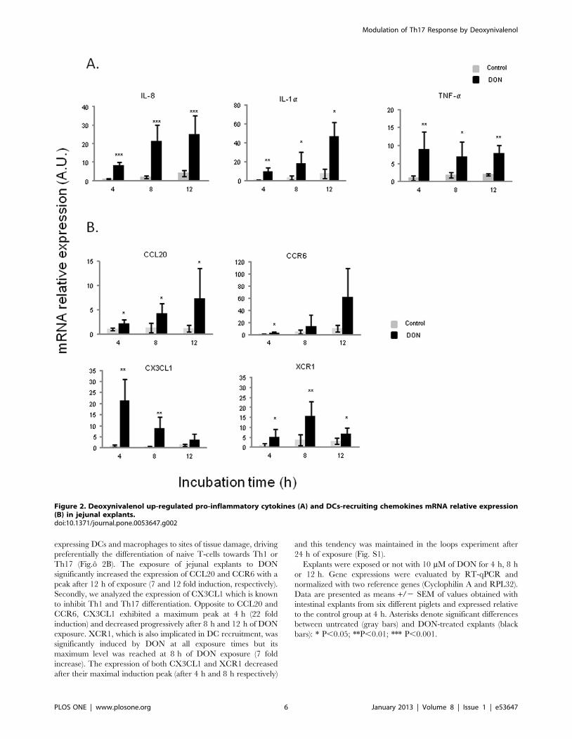
expressing DCs and macrophages to sites of tissue damage, driving
preferentially the differentiation of naive T-cells towards Th1 or
Th17 (Fig.o 2B). The exposure of jejunal explants to DON
significantly increased the expression of CCL20 and CCR6 with a
peak after 12 h of exposure (7 and 12 fold induction, respectively).
Secondly, we analyzed the expression of CX3CL1 which is known
to inhibit Th1 and Th17 differentiation. Opposite to CCL20 and
CCR6, CX3CL1 exhibited a maximum peak at 4 h (22 fold
induction) and decreased progressively after 8 h and 12 h of DON
exposure. XCR1, which is also implicated in DC recruitment, was
significantly induced by DON at all exposure times but its
maximum level was reached at 8 h of DON exposure (7 fold
increase). The expression of both CX3CL1 and XCR1 decreased
after their maximal induction peak (after 4 h and 8 h respectively)
and this tendency was maintained in the loops experiment after
24 h of exposure (Fig. S1).
Explants were exposed or not with 10 mM of DON for 4 h, 8 h
or 12 h. Gene expressions were evaluated by RT-qPCR and
normalized with two reference genes (Cyclophilin A and RPL32).
Data are presented as means +/2 SEM of values obtained with
intestinal explants from six different piglets and expressed relative
to the control group at 4 h. Asterisks denote significant differences
between untreated (gray bars) and DON-treated explants (black
bars): * P,0.05; **P,0.01; *** P,0.001.
Figure 2. Deoxynivalenol up-regulated pro-inflammatory cytokines (A) and DCs-recruiting chemokines mRNA relative expression(B) in jejunal explants.doi:10.1371/journal.pone.0053647.g002
Modulation of Th17 Response by Deoxynivalenol
PLOS ONE | www.plosone.org 6 January 2013 | Volume 8 | Issue 1 | e53647

DON Triggered Th17 Signature genes in Porcine jejunalExplants
In order to examine the effects of DON on naive Th cells
differentiation, we analyzed mRNA expression levels of transcrip-
tion factors and cytokine genes related to Th1 cells (Fig.o 3), Th17
cells (Fig.o 4) and Treg cells (Fig.o 5). DON treatment did not
affect expression of T-bet transcription factor nor IFN-c mRNA
levels (Fig.o 3). Significant increase of IL-12 expression levels was
observed at all exposure times (between 6 and 8 fold induction)
(Fig.o 3). DON treatment also induced a time-dependent increase
of expression of mRNA encoding for IL-17 A, IL-6 and STAT3
(Fig.o 4A). Accordingly the protein expression of IL-17A was
significantly increased after 12 h of exposure to DON (Fig.o 4B).
IL-1b expression levels were also significantly up-regulated but its
levels did not did not vary much in a time dependent manner (16
to 12 fold induction) (Fig.o 4A). Surprisingly, RORc-like, an
important Th17 transcription factor was negatively regulated by
DON treatment (Fig.o 4A). A different expression profile was
observed in genes related to Treg cells. FoxP3 and RALDH1 were
not affected by DON treatment after 4 h and 8 h of culture and
they were down regulated after 12 h of toxin exposure (Fig.o 5A).
This decrease of Treg cells was also found when analyzing the
effect of DON on CD4+ CD25+ FoxP3+ leukocytes extracted from
the intestinal lamina propria (Fig.o 5B). 10 mM of DON
significantly reduced the CD4+ CD25+ FoxP3+ cell population
of 40%. On the contrary, the preliminary in vivo results using the
loops experiment suggest an increase of expression of Treg cells
related genes (Fig. S1).
Relative mRNA expression of transcription factors and cyto-
kines related to Th1 cells were assessed by RT-qPCR. Explants
were exposed or not with 10 mM of DON for 4 h, 8 h or 12 h.
Values were normalized with two reference genes (Cyclophilin A
and RPL32). Data are presented as means +/2 SEM of values
obtained with intestinal explants from six different piglets and
expressed relative to the control group at 4 h. Asterisks denote
significant differences between untreated (gray bars) and DON-
treated explants (black bars): * P,0.05; **P,0.01; *** P,0.001.
Relative mRNA expression of transcription factors and cyto-
kines related to Th17 cells (A) and IL-17 A protein concentration
(B) were assessed by RT-qPCR and ELISA. Explants were
exposed or not with 10 mM of DON for 4 h, 8 h or 12 h. Values
were normalized with two reference genes (Cyclophilin A and
RPL32). Data are presented as means +/2 SEM of values
obtained with intestinal explants from six different piglets and
expressed relative to the control group at 4 h. Asterisks denote
significant differences between untreated (gray bars) and DON-
treated explants (black bars): * P,0.05; **P,0.01; *** P,0.001.
(A) Relative mRNA expression of transcription factors related to
T reg cells were assessed by RT-qPCR. Explants were exposed or
not with 10 mM of DON for 4 h, 8 h or 12 h. Values were
normalized with two reference genes (Cyclophilin A and RPL32).
Data are presented as means +/2 SEM of values obtained with
intestinal explants from six different piglets and expressed relative
to the control group at 4 h. (B) The frequency of
CD4+CD25+FoxP3+ Treg cells were evaluated by flow cytometry
after 2 days of co-culture with different concentration of DON (0.1
to 10 mM). Graphs show the mean 6 SEM from duplicates from
immune cells of jejunal lamina propria isolated 2 different pigs.
Data were calculated as relative percentage to control without
toxin. Asterisks denote significant differences between untreated
(gray bars) and DON-treated explants (black bars): * P,0.05;
**P,0.01; *** P,0.001.
DON Triggered Pathogenic Th17 Cells Subset inDetriment of Regulatory Th17 (rTh17) Cells in Porcinejejunal Explants
To understand which subset of Th17 cells is preferentially
induced by DON, pathogenic Th17-related genes (IL-23 A, IL-
22 and IL-21) and rTh17-related genes (TGF-b and IL-10) were
analyzed (Fig.o 6). DON significantly increased IL-23A mRNA
expression in a time-dependent manner (3, 17 and 31 fold
induction at 4, 8 and 12 hours respectively) (Fig.o 6A). mRNA
expression levels of both IL-22 and IL-21 were also significantly
induced but to a lesser extent (maximum levels of 10 and 3.5 fold
induction respectively) (Fig.o 6A). Interestingly, upregulation of
these genes did not show after 24 h of DON exposure (Fig. S1).
On the contrary, TGF-b mRNA expression was not induced by
DON treatment (Fig.o 6B). Interestingly, IL-10 which is produced
by both rTh17 and Treg cells showed a significant induction in the
presence of DON (6 fold increase) (Fig.o 6B).
Discussion
Mycotoxins have become an issue of major concern given their
global and frequent occurrence as well as their known toxic effects.
Among all, DON is of particular interest because it is one of the
most prevalent mycotoxins in Europe and North America. The
intestinal tract represents the first barrier against DON-contam-
Figure 3. Deoxynivalenol increased IL-12 but not IFN-c gene expression in jejunal explants.doi:10.1371/journal.pone.0053647.g003
Modulation of Th17 Response by Deoxynivalenol
PLOS ONE | www.plosone.org 7 January 2013 | Volume 8 | Issue 1 | e53647

inated food and feed. Many authors have already reported Th17
cells to be critical for protection against microbial infections like
bacteria, virus and fungi at mucosal surfaces [36,37,38]. The aim
of our work was to assess the impact of DON exposure onto the
intestinal homeostasis controlled by pro-inflammatory Th17 and
the regulatory Treg response. To the best of our knowledge this is
the first study showing that DON disrupts the intestinal
homeostasis and promotes the Th17 response over the Th1 and
T regulatory responses.
Intestinal epithelial cells and jejunal explants were used to
investigate the modulation of the intestinal immune response by
DON. The choice of using 10 mM of DON was based mainly on
the US Federal Grain Inspection Service and the European Union
surveys [6,39,40] which evaluated the naturally occurring
concentration of this mycotoxin to range between 19 and
50 mg/kg in grain cereals. Assuming that DON would be ingested
in one meal, diluted in 1 L of gastrointestinal fluid and would be
totally bioaccessible, 10 mM correspond approximately to 3 mg/
kg [39,40]. In addition to that, a previous study of our group
Figure 4. Deoxynivalenol triggered Th17 response in jejunal explants.doi:10.1371/journal.pone.0053647.g004
Modulation of Th17 Response by Deoxynivalenol
PLOS ONE | www.plosone.org 8 January 2013 | Volume 8 | Issue 1 | e53647

showed that this concentration does not affect cell viability up to
48 h after treatment in the porcine intestinal cell line, IPEC-1 [17]
but it does affect the morphology of the epithelial barrier
according to another study [27].
In the present experiment, in vitro and ex vivo exposure to DON
led to an early general state of inflammation depicted by mRNA
expression increase of IL-8, IL-1a and TNF- a, which is in
agreement with previous results of similar studies [41,42]. The
inflammation generated at the epithelial level could elicit the
activation of the population of Th17 cells by triggering further
communication with immune cells of the lamina propria. The mode
of action by which DON induces such inflammatory response has
been described by several authors and it was not the purpose of the
present study. Indeed, it has already largely been demonstrated
that DON activates ERK1/2 and p38 thus triggering MAPK
signaling cascades which upregulate COX-2 and PGE-2, major
inductors of the inflammatory response [11]. This molecular
mechanism most likely occurs in all immune cellular types,
including IECs. Therefore we can also soundly hypothesize that
DON-induced MAPK activation in IECs induces chemokine
secretion which then stimulates intestinal DCs, thus initiating the
classical immune response cascade and lymphocyte activation.
In this study, the exposure to DON induced IL-6, IL-23 and IL-
1b expression but did not affect the expression of TGF-b and
strongly repressed FoxP3 and RALDH1. These data suggest that
in our model DON mainly drives the intestinal immune system
towards a Th17 response. In agreement with that, DON strongly
induced IL-17 A which is the signature cytokine of these T helper
cells, but which can also be produced by other immune cells.
However to our knowledge, there is no available antibody for of
porcine IL-17 A intracellular labeling which would allow to clarify
the source of this cytokine. Despite that, the fact that STAT3, a
transcription factor expressed by Th17 cells, was also upregulated
in the intestinal explants is a strong indication of the increase of
this population in the intestinal tissues. Nevertheless, ROR-ct like,
which has been described as the other major transcription factor
governing Th17 differentiation in mice [23,43], was not induced
but repressed by the presence of DON in explants model.
Although this result is surprising, it could be possible that, unlike in
the mouse model, in the porcine specie the transcription factor
STAT3 plays a more important role than ROR-ct in Th17
differentiation. It would be very interesting to further investigate
this point.
The two lineages, Th17 and Tregs share a co-evolutionary
origin [44,45] which could explain why they share similarities at
Figure 5. Deoxynivalenol decreased T regulatory response in jejunal explants.doi:10.1371/journal.pone.0053647.g005
Modulation of Th17 Response by Deoxynivalenol
PLOS ONE | www.plosone.org 9 January 2013 | Volume 8 | Issue 1 | e53647

many levels [46,47,48,49]. In this way, CCR6 appears to be the
only chemokine receptor essentially implicated in driving Th17
migration in vivo but it is also highly expressed by Tregs [50].
CCL20, the ligand of this receptor drives the conversion of the
pathogenic Th17 into the regulatory subsets [23,51]. Our results
showed that CCR6 and CCL20 were induced by DON in a time-
dependent manner in the intestinal explants. It is therefore
tempting to speculate that along the overall pro-inflammatory
response both in the intestinal epithelial cells and the intestinal
explants, there is a regulatory rTh17 response setting up-rather
than the Treg response.
Plasticity of T cells is also reflected in the similarities between
rTh17 and Treg cells. Besides their common regulatory action,
these two populations share the importance of TGF-b and IL-10
[23,24,51]. TGF-b is involved in the differentiation of these two
populations. In this study, there was no significant effect of DON
on the expression of this cytokine in the intestinal explants. TGF-bwas however secreted by the intestinal epithelial cells after 8, 12
and 24 h of exposure to DON. Further studies will be important to
understand the exact role of TGF-b in the differentiation of Tregs
in swine but also the impact of DON on the intestinal secretion of
this cytokine. However, previous studies have also showed that
TGF-b presents minor or absent changes after PMA/Ionomycin
or CD3 stimulation [52,53] which might be an indication of the
fact that TGF-b might be of less importance in the regulatory
response compared to humans and mice. IL-10 is produced by
both Tregs and rTh17 to regulate inflammation and has the ability
of reversing the pathogenic phenotype of Th17 into rTh17 [24]. In
this study, this cytokine was up-regulated in a time independent
manner by the presence of DON on explants, which is in
contradiction with the absence of increase of FoxP3 and TGF-b.
However it is well described that many cells of the innate and
adaptive immune system other than Tregs and rTh17 cells express
IL-10, such as DCs, macrophages, Th1, Th2 and B cells [54].
Given our results in the intestinal explants on Th1 and Treg, it is
most likely for IL-10 to be produced by DCs, macrophages or B
cells in that experiment
Taken together, all these results suggest that DON preferentially
induces a pro-inflammatory response directed by pathogenic
Th17, when CCR6, CCL20 and IL-10 results point to a
concomitant protective response most likely lead by rTh17.
Supplementary in vivo data seems to indicate an increase in the
protective response after 24 h of exposure.
Determining whether immune activation or suppression should
occur in response to a given pathogen is a critical decision to be
taken by the adaptive immune system. Indeed, there is a fine line
between inflammation and pathogen elimination and excessive
inflammation and tissue damage. Numerous diseases such as
rheumatoid arthritis and multiple sclerosis arise from a failure to
restore the state of equilibrium [55]. In the intestine, inflammatory
bowel diseases (IBD) like ulcerative colitis (UC) and Crohn’s
disease (CD) are triggered by excessive inflammation of the colon
and/or the small bowel leading to recurrent diarrhea and pain
[56]. Until recently only excessive Th1 and Th2 responses were
accounted for these diseases. Nevertheless, the discovery of the
Th17 subset gave a new insight on the causes of these disorders as
Figure 6. Deoxynivalenol triggered pathogenic Th17 cells subset but not regulatory Th17 (rTh17) cells in porcine jejunal explants.doi:10.1371/journal.pone.0053647.g006
Modulation of Th17 Response by Deoxynivalenol
PLOS ONE | www.plosone.org 10 January 2013 | Volume 8 | Issue 1 | e53647

IL-17 producing cells were shown to be of major importance in
IBD [57]. Besides IL-17, IL-21, IL-22 and IL-23 have also been
reported to play an important role in the onset of these diseases
[57,58]. Interestingly, all of these cytokines were up-regulated by
the presence of DON in our experiments which could imply that
chronic exposure to this toxin could be a triggering or enhancing
factor of IBD. This hypothesis is supported by a recent study [26]
which draws a strong parallel between the alterations caused by
exposure to several mycotoxins including DON and the symptoms
observed during IBD. In addition to the induction of the above-
mentioned cytokines, intestinal permeability is also related to both
DON exposure and IBD. This mycotoxin specifically targets
claudin expression [16,17] which directly leads to an increase of
permeability of the intestinal barrier which could result in
increased bacterial translocation, one of the main causes of IBD
[56]. The same applies for toll-like receptors (TLRs) impairment,
associated with DON exposure [59]. Over activation of dendritic
cells (DCs) drawn by abnormal TLR activity could lead to inability
to detect bacterial components which is another major source of
IBD. Notably, TLR activation disruption has been detected after
exposure to T-2 toxin, another member of the trichothecene
family [60].
As depicted above, bacterial interactions with the immune cells
of the lamina propria are a central component of IBD, since
inflammation might arise from lack of tolerance to antigens
present in commensal bacteria [61]. Besides, loss of the
transcription factor T-bet, which regulates TNF-a production,
influences bacterial homeostasis by favoring colitogenic microbial
populations. The resulting increase of TNF-a production by DCs
could trigger breakdown of the intestinal epithelium and facilitate
bacterial translocation to the lamina propria, increasing risks of IBD
[62]. It has already been demonstrated that DON alters the
intestinal microbiota of pigs [63]. It thus comes as no surprise that
in this study, exposure to DON could possibly lead to induction of
colitogenic populations and to strong impairment of the epithelial
barrier by the lack of effect on T-bet and significant increase of the
expression levels of TNF-a. It is noteworthy that colonization of
the small intestine by segmented filamentous bacteria (SFB) of the
intestinal microbiota is directly associated with the appearance of
Th17 cells and that SFB promote the expression of inflammatory
genes linked to IBD [64].
In conclusion, exposure to the mycotoxin DON clearly induced
an early intestinal inflammatory response resulting from the
interplay of different intestinal cell types and leading to the
activation of Th17 cells. These results together with previous
observations strengthen the idea that chronical exposure to
deoxynivalenol could impair intestinal homeostasis and trigger
the appearance of IBD. However, the molecular mechanisms by
which this toxin specifically activates Th17 remain unclear.
Further research should therefore address the link between MAP
kinases activation by DON [11,12] and the herein presented
effects on lymphocyte populations. In addition to that, the cellular
interplay that takes play among immune cells (IECs – DCs – T
cells) upon DON exposure should be better characterized by
analyzing for instance cellular migration of DCs, macrophages and
T lymphocytes. Finally, more insight should be gained on the role
of commensal and pathogenic bacteria on modulating the Th17
response and the impact of DON on such modulation.
Supporting Information
Figure S1 Fold mRNA expression increase of cytokines and
transcription factors after DON exposure in an in vivo model of
jejunal loops. Following the guidelines provided by the French
Council for Animal Care (permit number 2011–07–1) and
previous published protocols [65,66,67], loops were performed
in a 5 week-old pig. Very briefly, 6 consecutive loops (10-–0 cm
long) were made by surgical ligation in a 2–4 m long segment of
the jejunum which was previously thoroughly washed with a
solution of metrodinazole, a common antibiotic. PBS was injected
in 3 loops as negative control and 10 mM of DON were injected in
3 other loops. The pig was euthanized by barbiturate overdose
24 h post surgery. Jejunal tissues were then collected and snap-
frozen in liquid nitrogen before RNA extraction. Relative mRNA
expression levels of immune genes related to pro-inflammatory
cytokines (A), DC-recrutment chemokines (B), Th1 (C), Th17 (D)
and Treg (E) signature and pathogenic/regulatory Th17 cytokines
(F) were assessed by RT-qPCR. Gene expressions were normalized
by the mean of two reference genes (Cyclophilin A and RPL32).
Data are presented as mean +/2 SEM of values obtained with
three different loops and expressed relative to the control group.
Significant differences between untreated loops (gray bars) and
treated loops (black bars) with 10 mM of DON are marked with
asterisks (* P,0.05).
(TIF)
Acknowledgments
The authors are also grateful to Philippe Pinton, Anne-Marie Cossalter,
Sophie Ausset and Claire Loiseau for their excellent technical assistance.
Author Contributions
Conceived and designed the experiments: LGP IPO. Performed the
experiments: PMC JS JC RA. Analyzed the data: PMC JS FM JC RA.
Contributed reagents/materials/analysis tools: JC. Wrote the paper: PMC
JS.
References
1. Doll S, Danicke S (2011) The Fusarium toxins deoxynivalenol (DON) and
zearalenone (ZON) in animal feeding. Prev Vet Med 102: 132–145.
2. Bryden WL (2007) Mycotoxins in the food chain: human health implications.Asia Paco Jo Clin Nutr 16 Suppl 1: 95–101.
3. Oswald IP, CC (1998) Immunotoxicity of mycotoxins. Revue Med Vet 149:
585–590.
4. Oswald IP, Marin DE, Bouhet S, Pinton P, Taranu I, et al. (2005)
Immunotoxicological risk of mycotoxins for domestic animals. Food Addit
Contam 22: 354–360.
5. CAST (2003) Mycotoxins: riks in plant, animal and human systems. Council Agr
Sc Technol Task Force Rep. 138: 136–142.
6. SCOOP (2003) Collection of occurrence data of Fusarium toxins in food andassessment of dietary intake by the population of EU member states.
Directorate-General Health and Cosummer Protection.
7. Pestka JJ, AT S (2005) Deoxynivalenol: Toxicology and Potential effects onHumans. Jo Toxicol Environ Health 8: 39–69.
8. Rotter BA, Prelusky DB, JJ P (1996) Toxicology of deoxynivalenol (vomitoxin).
Jo Toxicol Environ Health 48: 1–34.
9. Meurens F, Summerfield A, Nauwynck H, Saif L, Gerdts V (2011) The pig: amodel for human infectious diseases. Trends in Microbiology 20: 50–57.
10. Patterson JK, Lei XG, Miller DD (2008) The Pig as an Experimental Model for
Elucidating the Mechanisms Governing Dietary Influence on Mineral
Absorption. Exp Biol Med 233: 651–664.
11. Pestka JJ, Zhou H-R, Moon Y, Chung YJ (2004) Cellular and molecularmechanisms for immune modulation by deoxynivalenol and other trichothe-
cenes: unraveling a paradox. Toxicology Letters 153: 61–73.
12. Zhou HR, Islam Z, Pestka JJ (2003) Rapid, sequential activation of mitogen-
activated protein kinases and transcription factors precedes proinflammatorycytokine mRNA expression in spleens of mice exposed to the trichothecene
vomitoxin. Toxicol Sci 72: 130–142.
13. Avantaggiato G, Havenaar R, Visconti A (2004) Evaluation of the intestinal
absorption of deoxynivalenol and nivalenol by an in vitro gastrointestinal model,and the binding efficacy of activated carbon and other adsorbent materials. Food
Chem Toxicol 42: 817–824.
14. Bracarense A-PFL, Lucioli J, Grenier B, Drociunas Pacheco G, Moll W-D, et al.
(2012) Chronic ingestion of deoxynivalenol and fumonisin, alone or in
Modulation of Th17 Response by Deoxynivalenol
PLOS ONE | www.plosone.org 11 January 2013 | Volume 8 | Issue 1 | e53647

interaction, induces morphological and immunological changes in the intestine
of piglets. Bro Jo Nutr: 107: 1776–1786.15. Maresca M, Mahfoud R, Garmy N, Fantini J (2002) The Mycotoxin
Deoxynivalenol Affects Nutrient Absorption in Human Intestinal Epithelial
Cells. Jo Nutr 132: 2723–2731.16. Pinton P, Braicu C, Nougayrede JP, Laffitte J, Taranu I, et al. (2010)
Deoxynivalenol impairs porcine intestinal barrier function and decreases theprotein expression of claudin-4 through a mitogen-activated protein kinase-
dependent mechanism. Jo Nutr 140: 1956–1962.
17. Pinton P, Nougayrede J-P, Del Rio J-C, Moreno C, Marin DE, et al. (2009) Thefood contaminant deoxynivalenol, decreases intestinal barrier permeability and
reduces claudin expression. Toxicol Appl Pharmacol 237: 41–48.18. Pinton P, Tsybulskyy D, Lucioli J, Laffitte J, Callu P, et al. (2012) Toxicity of
deoxynivalenol and its acetylated derivatives on the intestine: differential effectson morphology, barrier function, tight junctions proteins and MAPKinases.
Toxicol Sci 130: 180–190.
19. Diesing A-K, Nossol C, Danicke S, Walk N, Post A, et al. (2011) Vulnerability ofPolarised Intestinal Porcine Epithelial Cells to Mycotoxin Deoxynivalenol
Depends on the Route of Application. PLoS ONE 6: e17472.20. Oswald IP (2006) Role of intestinal epithelial cells in the innate immune defence
of the pig intestine. Veterinary Research 37: 359–368.
21. Coombes JL, Powrie F (2008) Dendritic cells in intestinal immune regulation.Nat Rev Immunol 8: 435–446.
22. Curtis MM, Way SS (2009) Interleukin-17 in host defence against bacterial,mycobacterial and fungal pathogens. Immunology 126: 177–185.
23. Ghoreschi K, Laurence A, Yang XP, Tato CM, McGeachy MJ, et al. (2010)Generation of pathogenic T(H)17 cells in the absence of TGF-beta signalling.
Nature 467: 967-U144.
24. McGeachy MJ, Bak-Jensen KS, Chen Y, Tato CM, Blumenschein W, et al.(2007) TGF-beta and IL-6 drive the production of IL-17 and IL-10 by T cells
and restrain TH-17 cell-mediated pathology. Nature Immunol 8: 1390–1397.25. Zhu J, Paul WE (2009) Heterogeneity and plasticity of T helper cells. Cell Res
20: 4–12.
26. Maresca M, Fantini J (2010) Some food-associated mycotoxins as potential riskfactors in humans predisposed to chronic intestinal inflammatory diseases.
Toxicon 56: 282–294.27. Kolf-Clauw M, Castellote J, Joly B, Bourges-Abella N, Raymond-Letron I, et al.
(2009) Development of a pig jejunal explant culture for studying thegastrointestinal toxicity of the mycotoxin deoxynivalenol: Histopathological
analysis. Toxicol in Vitro 23: 1580–1584.
28. Gonzalez-Vallina R, Wang H, Zhan R, Berschneider HM, Lee RM, et al. (1996)Lipoprotein and apolipoprotein secretion by a newborn piglet intestinal cell line
(IPEC-1). Amo Jo Physiol 271: G249–259.29. Zanello G, Berri M, Dupont J, Sizaret P-Y, D’Inca R, et al. (2011) Saccharomyces
cerevisiae Modulates Immune Gene Expressions and Inhibits ETEC-Mediated
ERK1/2 and p38 Signaling Pathways in Intestinal Epithelial Cells. PLoS ONE6: e18573.
30. Gonzalez-Vallina R, Wang H, Zhan R, Berschneider HM, Lee RM, et al. (1996)Lipoprotein and apolipoprotein secretion by a newborn piglet intestinal cell line
(IPEC-1). Amo Jo Physiol 271: G249–G259.31. Bouhet S, Hourcade E, Loiseau N, Fikry A, Martinez S, et al. (2004) The
mycotoxin fumonisin B1 alters the proliferation and the barrier function of
porcine intestinal epithelial cells. Toxicol Sci 77: 165–171.32. Meissonnier GM, Pinton P, Laffitte J, Cossalter AM, Gong YY, et al. (2008)
Immunotoxicity of aflatoxin B1: impairment of the cell-mediated response tovaccine antigen and modulation of cytokine expression. Toxicol Appl Pharmacol
231: 142–149.
33. Bruel T, Guibon R, Melo S, Guillen N, Salmon H, et al. (2010) Epithelialinduction of porcine suppressor of cytokine signaling 2 (SOCS2) gene expression
in response to Entamoeba histolytica. Dev Comp Immunol 34: 562–571.34. Delgado-Ortega M, Melo S, Meurens F (2011) Expression of SOCS1–7 and CIS
mRNA in porcine tissues. Vet Immunol Immunopathol 144: 493–498.
35. Haverson K, Singha S, Stokes CR, Bailey M (2000) Professional and non-professional antigen-presenting cells in the porcine small intestine. Immunology
101: 492–500.36. Zelante T, Bozza S, De Luca A, D’Angelo C, Bonifazi P, et al. (2009) Th17 cells
in the setting of Aspergillus infection and pathology. Med Mycol 47: S162–S169.37. Khader SA, Gaffen SL, Kolls JK (2009) Th17 cells at the crossroads of innate
and adaptive immunity against infectious diseases at the mucosa. Mucosal
Immunol 2: 403–411.38. Kolls JK, Khader SA (2010) The role of Th17 cytokines in primary mucosal
immunity. uCytokine Growth Factor Rev 21: 443–448.39. Trucksess MW, Thomas F, Young K, Stack ME, Fulgueras WJ, et al. (1995)
Survey of deoxynivalenol in U.S. 1993 wheat and barley crops by enzyme-linked
immunosorbent assay. Jo AOAC Int 78: 631–636.40. Abouzied MM, Azcona JI, Braselton WE, Pestka JJ (1991) Immunochemical
assessment of mycotoxins in 1989 grain foods: evidence for deoxynivalenol(vomitoxin) contamination. Appl Environ Microbiol 57: 672–677.
41. Pestka JJ, Amuzie CJ (2008) Tissue distribution and proinflammatory cytokinegene expression following acute oral exposure to deoxynivalenol: comparison of
weanling and adult mice. Food Chem Toxicol 46: 2826–2831.
42. Amuzie CJ, Shinozuka J, Pestka JJ (2009) Induction of suppressors of cytokinesignaling by the trichothecene deoxynivalenol in the mouse. Toxicol Sci 111:
277–287.
43. Manel N, Unutmaz D, Littman DR (2008) The differentiation of human T(H)-
17 cells requires transforming growth factor-beta and induction of the nuclearreceptor RORgammat. Nat Immunol 9: 641–649.
44. Weaver CT, Harrington LE, Mangan PR, Gavrieli M, Murphy KM (2006)
Th17: An Effector CD4 T Cell Lineage with Regulatory T Cell Ties. Immunity24: 677–688.
45. Weaver CT, Hatton RD (2009) Interplay between the TH17 and TReg celllineages: a co-evolutionary perspective. Nat Rev Immunol 9: 883–889.
46. Kaser T, Gerner W, Saalmuller A (2011) Porcine regulatory T cells: mechanisms
and T-cell targets of suppression. Dev Comp Immunol 35: 1166–1172.47. Kaser T, Gerner W, Mair K, Hammer SE, Patzl M, et al. (2011) Current
knowledge on porcine regulatory T cells. Vet Immunol Immunopathol 148:136–138.
48. Kaser T, Gerner W, Hammer SE, Patzl M, Saalmuller A (2008) Detection ofFoxp3 protein expression in porcine T lymphocytes. Vet Immunol Immuno-
pathol 125: 92–101.
49. Kaser T, Gerner W, Hammer SE, Patzl M, Saalmuller A (2008) Phenotypic andfunctional characterisation of porcine CD4(+)CD25(high) regulatory T cells. Vet
Immunol Immunopathol 122: 153–158.50. Comerford I, Bunting M, Fenix K, Haylock-Jacobs S, Litchfield W, et al. (2010)
An immune paradox: how can the same chemokine axis regulate both immune
tolerance and activation?: CCR6/CCL20: a chemokine axis balancingimmunological tolerance and inflammation in autoimmune disease. Bioessays
32: 1067–1076.51. Esplugues E, Huber S, Gagliani N, Hauser AE, Town T, et al. (2011) Control of
TH17 cells occurs in the small intestine. Nature 475: 514–518.52. Ledger TN, Pinton P, Bourges D, Roumi P, Salmon H, et al. (2004)
Development of a Macroarray To Specifically Analyze Immunological Gene
Expression in Swine. Clin Diagn Lab Immunol 11: 691–698.53. Kaser T, Mullebner A, Hartl RT, Essler SE, Saalmuller A, et al. (2012) Porcine
T-helper and regulatory T cells exhibit versatile mRNA expression capabilitiesfor cytokines and co-stimulatory molecules. Cytokine 60: 400–409.
54. Paul G, Khare V, Gasche C (2011) Inflamed gut mucosa: downstream of
interleukin-10. Euro Jo Clin Invest 42: 95–109.55. Miossec P (2009) Diseases that may benefit from manipulating the Th17
pathway. Euro Jo Immunol 39: 667–669.56. Matricon J, Barnich N, Ardid D (2010) Immunopathogenesis of inflammatory
bowel disease. Self Nonself 1: 299–309.57. Caprioli F, Pallone F, Monteleone G (2008) Th17 immune response in IBD: A
new pathogenic mechanism. Jo Crohns Colitis 2: 291–295.
58. Ouyang W, Valdez P (2008) IL-22 in mucosal immunity. Mucosal Immunol 1:335–338.
59. Pestka J, Zhou HR (2006) Toll-like receptor priming sensitizes macrophages toproinflammatory cytokine gene induction by deoxynivalenol and other toxicants.
Toxicol Sci 92: 445–455.
60. Seeboth J, Solinhac R, Oswald I, Guzylack-Piriou L (2012) The fungal T-2 toxinalters the activation of primary macrophages induced by TLR-agonists resulting
in a decrease of the inflammatory response in the pig. Vet Res 43: 35.61. Chassaing B, Darfeuille-Michaud A (2011) The commensal microbiota and
enteropathogens in the pathogenesis of inflammatory bowel diseases. Gastroen-terology 140: 1720–1728.
62. Garrett WS, Lord GM, Punit S, Lugo-Villarino G, Mazmanian SK, et al. (2007)
Communicable ulcerative colitis induced by T-bet deficiency in the innateimmune system. Cell 131: 33–45.
63. Wache YJ, Hbabi-Haddioui L, Guzylack-Piriou L, Belkhelfa H, Roques C, et al.(2009) The mycotoxin Deoxynivalenol inhibits the cell surface expression of
activation markers in human macrophages. Toxicol 262: 239–244.
64. Ivanov, II, Atarashi K, Manel N, Brodie EL, Shima T, et al. (2009) Induction ofintestinal Th17 cells by segmented filamentous bacteria. Cell 139: 485–498.
65. Gerdts V, Uwiera RRE, Mutwiri GK, Wilson DJ, Bowersock T, et al. (2001)Multiple intestinal "loops" provide an in vivo model to analyse multiple mucosal
immune responses. Jo Immunol Methods 256: 19–33.
66. Meurens F, Berri M, Auray G, Melo S, Levast B, et al. (2009) Early immuneresponse following Salmonella enterica subspecies enterica serovar Typhimurium
infection in porcine jejunal gut loops. Vet Res 40: 05.67. Vandenbroucke V, Croubels S, Martel A, Verbrugghe E, Goossens J, et al.
(2011) The Mycotoxin Deoxynivalenol Potentiates Intestinal Inflammation bySalmonella Typhimurium in Porcine Ileal Loops. PLoS ONE 6: e23871.
68. Grenier B, Loureiro-Bracarense AP, Lucioli J, Pacheco GD, Cossalter AM, et al.
(2011) Individual and combined effects of subclinical doses of deoxynivalenoland fumonisins in piglets. Mol Nutr Food Res 55: 761–771.
69. Levast B, de Monte M, Chevaleyre C, Melo S, Berri M, et al. (2010) Ultra-earlyweaning in piglets results in low serum IgA concentration and IL17 mRNA
expression. Vet Immunol Immunopathol 137: 261–268.
70. Kiros TG, van Kessel J, Babiuk LA, Gerdts V (2011) Induction, regulation andphysiological role of IL-17 secreting helper T-cells isolated from PBMC, thymus,
and lung lymphocytes of young pigs. Vet Immunol Immunopathol 144: 448–454.
71. Royaee AR, Husmann RJ, Dawson HD, Calzada-Nova G, Schnitzlein WM, etal. (2004) Deciphering the involvement of innate immune factors in the
development of the host response to PRRSV vaccination. Vet Immunol
Immunopathol 102: 199–216.72. Saurer L, McCullough KC, Summerfield A (2007) In vitro induction of mucosa-
type dendritic cells by all-trans retinoic acid. Jo Immunol 179: 3504–3514.
Modulation of Th17 Response by Deoxynivalenol
PLOS ONE | www.plosone.org 12 January 2013 | Volume 8 | Issue 1 | e53647