dentistry, 3nd edition; summitt, et al chapters 5, 6 and 8...
TRANSCRIPT
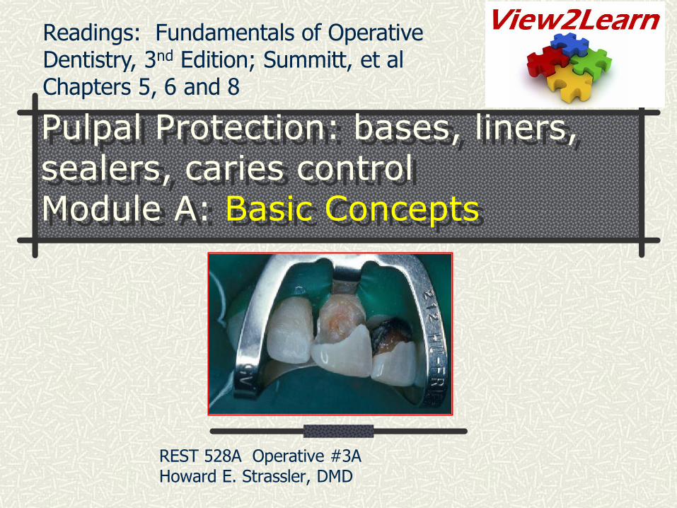
Pulpal Protection: bases, liners, sealers, caries control Module A: Basic Concepts
REST 528A Operative #3A Howard E. Strassler, DMD
Readings: Fundamentals of Operative Dentistry, 3nd Edition; Summitt, et al Chapters 5, 6 and 8

Considerations for pulpal health in the placement of restorative materials
Compromises to pulpal health (pulpal irritation)
Caries- bacterial infection causing pulpal inflammation
Rotary preparation- heat and trauma to odontoblastic processes
Goal- preservation of pulpal health
Goal-creation of barrier (seal of enamel and dentin with restoratives) to external irritation
Goal- seal any marginal gaps between tooth and restoration

To have pulpal health…
The pulp must be vital!

Pulpal vitality testing
Cold test with cotton applicator using ethyl chloride
Or Electric Pulp Test (EPT)

Cold test with cotton applicator using ethyl chloride
Or Electric Pulp Test (EPT)
Use of pulpal vitality testing to verify profound pulpal anesthesia
when doing restorative procedures
BONUS CLINICAL TIP

Before cavity preparation the radiograph provides a “window” looking in for what to
expect of cavity depth and probable Decisions regarding pulpal health!

Decision making in the use of sealers, liners and/or bases
Remaining dentin thickness in tooth preparation
Thermal conductivity of restorative material DISTAL- DEEP
AXIAL WALL

Pulpal health evaluation
Presence or absence of pulpal symptoms-pain to stimuli
Thermal
Sweets (osmotic changes)
Tactile touching of dentin
Occlusion
Duration of symptom
Spontaneous pain
Type of pain-sharp? Throbbing?
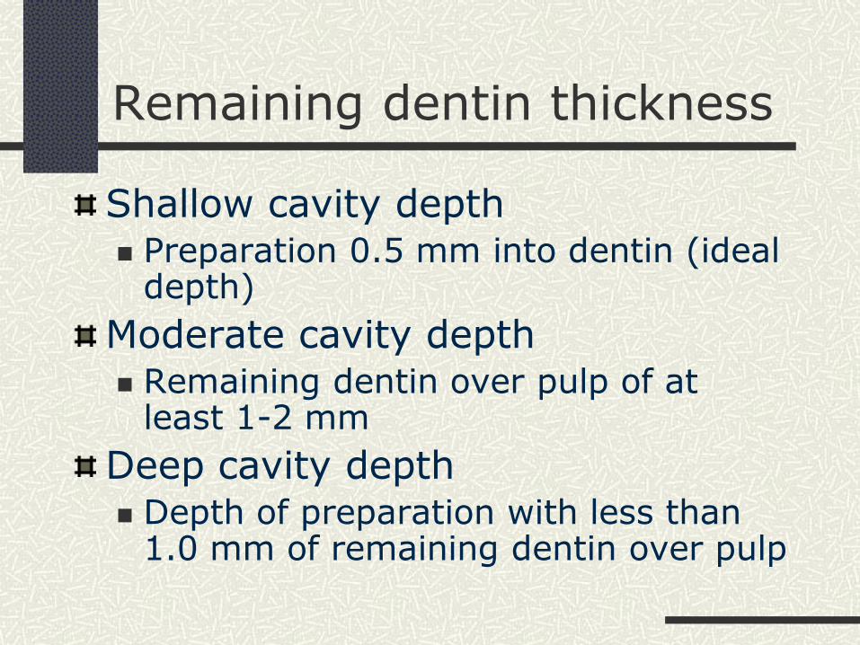
Remaining dentin thickness
Shallow cavity depth Preparation 0.5 mm into dentin (ideal
depth)
Moderate cavity depth Remaining dentin over pulp of at
least 1-2 mm
Deep cavity depth Depth of preparation with less than
1.0 mm of remaining dentin over pulp

Remaining dentin thickness
Shallow cavity depth Preparation 0.5 mm into dentin (ideal
depth)

Remaining dentin thickness
Moderate cavity depth Remaining dentin over pulp of at
least 1-2 mm

Remaining dentin thickness
Deep cavity depth Depth of preparation with less than
1.0 mm of remaining dentin over pulp
Base almost to the pulp

Causes of pulpal inflammation
Bacterial toxins penetrating the dentinal tubules (leading edge)
Bacteria can cause
Pulpal irritation-inflamation
Pulpal necrosis
Recurrent caries
Leakage at the restoration-tooth interface due to gaps at that interface
Trauma of tooth preparation

Bacterial penetration- pulpal inflammation
Mutans Streptococci
Bacterial invasion
at gap between Restorative-
tooth interface
Restorative material
inflammation
Bacteria penetrating gap, invading dentinal tubules
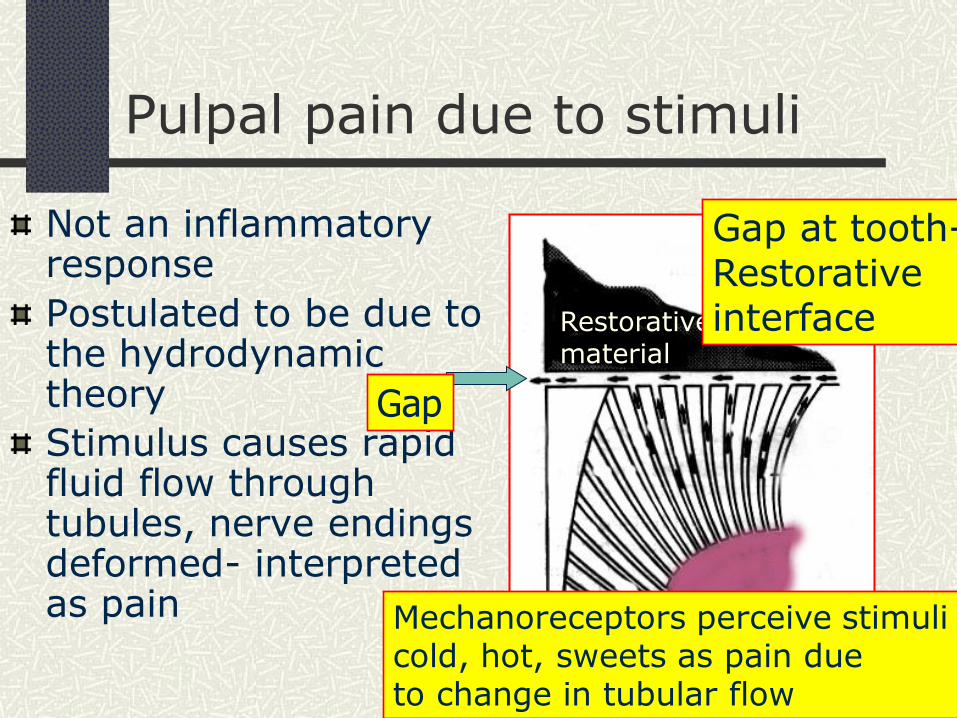
Not an inflammatory response
Postulated to be due to the hydrodynamic theory
Stimulus causes rapid fluid flow through tubules, nerve endings deformed- interpreted as pain
Pulpal pain due to stimuli
Restorative material
Gap at tooth- Restorative interface
Mechanoreceptors perceive stimuli cold, hot, sweets as pain due to change in tubular flow
Gap
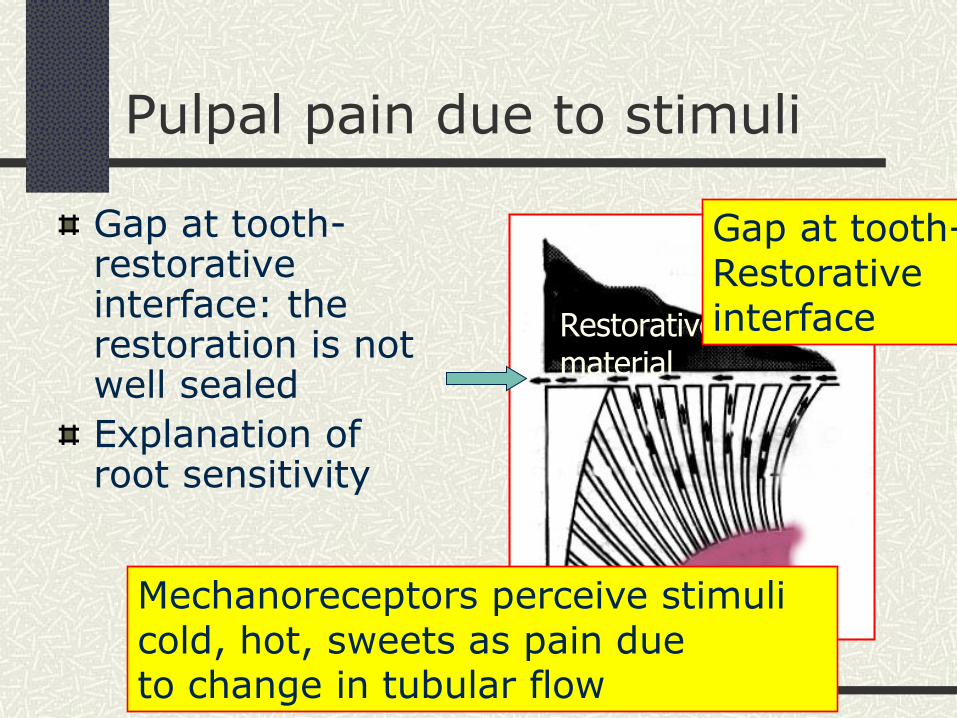
Gap at tooth-restorative interface: the restoration is not well sealed
Explanation of root sensitivity
Pulpal pain due to stimuli
Restorative material
Gap at tooth- Restorative interface
Mechanoreceptors perceive stimuli cold, hot, sweets as pain due to change in tubular flow

Marginal gaps at the restorative interface can lead to
Recurrent caries
Marginal staining (composite resin)
Tooth sensitivity
Microleakage
Use of sealers to avoid marginal gaps

Why are teeth sensitive to thermal shock?
Postulated that direct thermal shock transferred to pulp through thin dentin Metallic restorations more
thermoconductive
Liner-base thickness to prevent thermal transfer should be no thicker than 0.5-0.75 mm (thicker bases may weaken restoration)

Why are teeth sensitive to thermal shock?
Usually cold stimulates response
Air can trigger response
Air evaporates saliva-moisture from tooth creating a cooling effect
Cold sensitivity due to recently placed restoration being in hyperocclusion
Why are teeth sensitive to thermal shock?
Postulated that thermal sensitivity may be due to pulpal fluid flow hydrodynamics Cavity preparation cuts dentin leaving
more tubules exposed (desication of dentin leads to pulpal inflamation)
Deeper preparations have more dentinal tubules open
Sealing dentinal tubules reduces sensitivity to thermal shock, osmotic stimulation, changes in dentin fluid flow

Materials to seal the tooth for pulpal protection
Cavity sealers: materials placed in thin films to protective coating on the cavity walls creating a barrier to leakage Varnish (Barrier) (for amalgam) Etch and Rinse resin bonding systems
(Scotchbond MP and OptiBond Solo) (for composites)
Self-etch resin bonding systems (Xeno IV)

Materials to seal the tooth for pulpal protection
Cavity liners:cement or resin coating of minimal thickness (less than 0.5 mm) placed as a barrier to bacteria or to provide a therapeutic effect (pulpal sedative or antimicrobial effect). Applied to cavity walls adjacent to pulp (Dycal, VitreBond) Dycal (calcium hydroxide) is not
adhesive, very soluble VitreBond (resin modified glass ionomer)
is adhesive and would be placed to cover Dycal

Materials to seal the tooth for pulpal protection
Cavity bases:placed to replace missing dentin, placed in thicknesses of 0.5-1 mm; used to blockout undercuts in cavity preparations for indirect restorations (VitreBond, Fuji IILC, Fuji IX) (all glass ionomers are self-adhesive to dentin) Bases are not commonly used today for thermal insulation; predominent use to block out undercuts in preparations Inlay/onlay Crown preparations (usually cervical due
to tooth NCCL (tooth notching)

Pulpal Protection: bases, liners, sealers, caries control Module A: Basic Concepts
REST 528A Operative #3A Howard E. Strassler, DMD
Readings: Fundamentals of Operative Dentistry, 3nd Edition; Summitt, et al Chapters 5, 6 and 8