dental pulp of the third molar: a new source of ... · dental pulp of the third molar: a new source...
TRANSCRIPT

Dental Pulp of the Third Molar: A New Source of Pluripotent-likeStem Cells
Maher Atari1,5, Carlos Gil-Recio_, Marc Fabregat _, D.A. García-Fernández._, Miguel A Barajas2,Miguel Angel Carrasco3, Han-Sung Jung4, F. Hernández-Alfaro5, Nuria Casals 6, Felipe Prosper2, Eduard
Ferrés-Padró5, and Luis Giner1,5 **
_Laboratory for Regenerative Medicine, College of Dentistry, Universitat Internacional de Catalunya, Barcelona,Spain;
2Area of Hematology and Cell Therapy, University of Navarra, Pamplona, Spain;_Area of Pathology Universitat Internacional de Catalunya, Barcelona, Spain;
4Division in Anatomy and Developmental Biology, Department of Oral Biology, Oral Science Research Center,College of Dentistry, Yonsei University, Seoul, South Korea;
5Surgery and Oral Implantology Department, College of Dentistry, Universitat Internacional de Catalunya,Barcelona, Spain;
6Basic Sciences Department and CIBER Physiopathology of the Obesity and Nutrition (CIBEROBN), Faculty ofMedicine and Health Sciences, Universitat Internacional de Catalunya, Barcelona, Spain.
Author contributions:
Atari M: Conception and design. Collection and assembly of data. Data analysis and interpretation.Manuscript writing. Final approval of manuscript.Gil-Recio C: Collection and assembly of data. Manuscript writing.M. Fabregat: Collection and assembly of data.D. García-Fdez.: Collection and assembly of data.M. Barajas: Data analysis and interpretation.M. Carrasco: Collection and assembly of data.Han-Sung. Jung: Financial support. Administrative support.F. Hernandez-Alfaro: Provision of study patients.N. Casals: Data analysis and interpretation.F. Prosper:. Administrative supportE. Ferres Padro: Provision of study patients.L. Giner: Financial support. Administrative support. Final approval of manuscript.
**Address for Correspondence:Luis Giner. MD, DDS, Ph.D.Universitat Internacional de CatalunyaC/ Josep Trueta s/nSant Cugat del Vallès (Barcelona), 08195SpainEmail: [email protected]@csc.uic.es
Running Title: DPPSCs from human dental pulp.
Keywords: dental pulp, pluripotency, teratoma formation, embryonic markers. CGH technique
Abbreviations used: DPSC: Dental pulp stem cell. SHED: Stem cells from exfoliated deciduous teeth.PDLSC: Periodontal ligament stem cells. DFPC: Dental follicle progenitor cells. SCAP: Stem cells fromapical papilla DPPSC: Dental pulp pluripotent stem cell. DPMSC: Dental pulp mesenchymal stem cell.LIF: Leukaemia inhibitor factor. EGF: Epidermal growth factor. PDGF: Platelet-derived growth factor.NTERA: Embryonic carcinoma cells.
© 2012. Published by The Company of Biologists Ltd.Jo
urna
l of C
ell S
cien
ceA
ccep
ted
man
uscr
ipt
JCS online publication date 30 March 2012

Abstract
Dental pulp is particularly interesting in regenerative medicine because of theaccessibility and differentiation potential of the tissue. Dental pulp has an earlydevelopmental origin with multi-lineage differentiation potential due to its developmentduring childhood and adolescence. However, no study has previously identified thepresence of stem cell populations with embryonic-like phenotypes in human dental pulpfrom the third molar. In the present work, we describe a new population of pluripotent-like stem cells (DPPSCs) that were isolated from the dental pulp by culture in mediacontaining LIF, EGF and PDGF. These cells are SSEA4+, OCT3/4+, NANOG+, SOX2+,LIN28+, CD13+, CD105+, CD34-, CD45-, CD90+, CD29+, CD73+, STRO1+ and CD146-,and they show genetic stability in vitro based on genomic analysis with a newlydescribed CGH technique. Interestedly DPPSCs were able to form both embryoidbodies-like structutes (EBs) in vitro and teratom-like structures that contained tissuesderived from all three embryonic germ layers when injected in nude mice. We examinedthe capacity of DPPSCs to differentiate in vitro into tissues that have similarcharacteristics to mesoderm, endoderm and ectoderm layers in both 2D and 3D cultures.We performed a comparative RT-PCR analysis of GATA4, GATA6, MIXL1, NANOG,OCT3/4, SOX1 and SOX2 to determine the degree of similarity between DPPSCs, EBsand human induced pluripotent stem cells (hIPSC). Our analysis revealed that DPPSCs,hIPSC and EBs have the same gene expression profile. Because DPPSCs can bederived from healthy human molars from patients of different sexes and ages, theyrepresent an easily accessible source of stem cells, which opens a range of newpossibilities for regenerative medicine.
Jour
nal o
f Cel
l Sci
ence
Acc
epte
d m
anus
crip
t

Introduction
Stem cells have the ability to self-renew and to generate mature, differentiated cells(Fuchs and Segre, 2000). The main postnatal function of stem cells is to repair andregenerate the tissues in which they reside. As pluripotent stem cells have become amajor focus of scientific research, many techniques have been developed to determinethe actual pluripotency of embryonic stem (ES) cells or induced pluripotent stem (iPS)cells. The pluripotency of human stem cells can be tested in two different ways:teratoma formation by cells injected subcutaneously and the aggregation and generationof embryoid bodies (EBs) from cells cultured in vitro (O'Connor et al., 2008;Papapetrou et al., 2009). These techniques demonstrate a multilineage differentiationcapability by which stem cells can give rise to cells of all three germ layers (Itskovitz-Eldor et al., 2000; Martin and Evans, 1975).
Dental pulp tissue is thought to be derived from migratory neural crest cells duringdevelopment (Peters and Balling, 1999; Thesleff and Aberg, 1999), and it has beenshown to harbour various populations of multipotent stem/progenitor cells (Miura et al.,2003; Nosrat et al., 2001). To date, multiple human dental stem/progenitor cells havebeen isolated, characterised, and classified as a group designated “dental pulp stemcells” (DPSCs). These include stem cells from exfoliated deciduous teeth (SHEDs),periodontal ligament stem cells (PDLSCs), dental follicle progenitor cells (DFPCs) andstem cells from apical papilla (SCAPs). These post-natal populations havemesenchymal stem cell (MSC)-like qualities, namely the capacity for self-renewal, thepotential to differentiate into multiple lineages, including osteoblasts and chondroblasts,and a potential for in vitro differentiation into cell types from various embryonic layers,including adipose, bone, endothelial and neural-like tissues (Arthur et al., 2008; Chenget al., 2008; Cordeiro et al., 2008; Fujii et al., 2008; Gay et al., 2007; Harada et al.,1999; He et al., 2009; Honda et al., 2008; Huo et al., 2010). Many researchers haveproposed that DPMSCs are promising candidates for the repair and regeneration of avariety of mesenchymal tissues, such as bone, cartilage and muscle (Dezawa et al.,2005; Noel et al., 2002). These findings, together with those of other studies, alsosuggest that cells from the dental pulp may represent a unique population based on theirregenerative potential (About et al., 2000; Gronthos et al., 2000; Iohara et al., 2004;Mina and Braut, 2004; Zhang et al., 2005). However, no optimal culture medium thatallows adult stem cell amplification without self-differentiation has yet been reported(d'Aquino et al., 2007; Stevens et al., 2008).
No previous study to date has described the presence and isolation of human dental pulpstem cells with pluripotent-like characteristics at a single-cell level, nor the use ofculture media containing LIF (leukaemia inhibitor factor), EGF (epidermal growthfactor) and PDGF (platelet derived growth factor) for isolation of these cells. In thepresent study, we describe the isolation and identification of a subpopulation ofpluripotent-like stem cells from the dental pulp of third molars (termed DPPSCs) thatshow greater regenerative power than currently used mesenchymal stem cells. TheseDPPSCs are SSEA4+, OCT4+, NANOG+, SOX2+, LIN28+, CD13+, CD105+, CD34-,
Jour
nal o
f Cel
l Sci
ence
Acc
epte
d m
anus
crip
t
CD45-, CD90+, CD29+, CD73-, STRO1+ and CD146-. We investigated the capacity ofDPPSCs to differentiate in vitro into tissues that have similar characteristics toembryonic mesoderm, endoderm and ectoderm layers in 2D and 3D, as well as theirability to generate EB-like structures and to develop teratoma-like structures wheninjected into nude mice. We also performed a comparative analysis of GATA4,GATA6, MIXL1, NANOG, OCT3/4, SOX1 and SOX2 by RT-PCR to determine thedegree of similarity between DPPSCs, EBs and hIPS. DPPSCs are derived from aneasily accessible source, and they can be used in future protocols for the regeneration oftissues from the three embryonic layers.
Results
Characterisation and isolation of dental pulp pluripotent stem cells
These studies were carried out with the goal of isolating and purifying a population ofpluripotent-like stem cells derived from dental pulp (DPPSCs). We analysed thephenotypes of all populations, each of which corresponds to a dental pulp donor,cultivated at a density of 80-100 cells per cm2 and expanded at different passages whenthe cultures reached 60% confluence. Colony formation was observed, specially whenthe cells were cultured by hanging drop method. When colonies were seeded intoadherent surfaces cells tended to migrate (Supplementary Figure 1). We performedFACS, qRT-PCR, immunophenotype analysis and cytogenetic analysis. Figure 1Ashows representative images of DPPSCs morphology at P5, P10 and P15. DPPSCs aresmall-sized cells with large nuclei and low cytoplasm content, without the typical flatand elongated MSC appearance. The morphology of DPPSCs cultured in a Cell Carrier3D glass scaffold was also examined using scanning electronic microscopy(Supplementary Figure 2). Immunofluorescence assays for SSEA4, OCT3/4 andNANOG showed that cells were positive for all three markers and that SSEA4 localisedin the cytoplasm, whereas embryonic transcription factors were located in the nucleus(Figure 1B).
The development of therapeutic strategies depends on the ability of stem cells toundergo large scale in vitro amplification, which can be associated with geneticinstability. 85% of DPPSCs exhibited a normal karyotype with no presence of anyaneuploidy, polyploidy or any chromosome structural abnormality in metaphases aftermore than 65 passages (Figure 1C).
Using transmission electron microscopy (TEM), we evaluated the morphology andintegrity of the cells (Figure 1D). A notable feature of DPPSCs is that they possesslarge nuclei relative to cytoplasm volume, which is also a characteristic of ES cells.
Short-CGH analysis demonstrate genetic stability of DPPSCs as they showed the samegenetic dose as a healthy control sample. A gain in X and a loss of Y chromosome dosecan be seen, where this is due to sex differences: DPPSCs were extracted from a female
Jour
nal o
f Cel
l Sci
ence
Acc
epte
d m
anus
crip
t
patient, whereas the cells onto which the hybridisation was performed were from a maledonor (Figure 1E).
In order to further characterise the population, we analysed the cells by flow cytometryand found that the population was CD105+, CD146+ , CD45- , CD34- , STRO1+ , TRA1-60- , OCT3/4+ and NANOG+ (Figure 2A-B). Double staining for OCT3/4 and NANOGwas also carried out showing a 19.55% of double positive cells (Figure 2C). Somedifferences in the expression level were found between different passages. Interestingly,percentage of SSEA4+, OCT3/4+ and NANOG+ increased with passages in DPPSCswhile in DPMSCs remained negative (Supplementary Table 2). We also looked forcells expressing embryonic markers in pulp tissues from donors of different ages on theday of extraction (Supplementary Figure 3). The percentage of SSEA4+ cellsincreased with age, whereas the number of OCT3/4+ and CD13+ cells decreased withage. In addition, we found that embryonic markers were still expressed in DPPSCsfrom 58-year-old patients.
RNA was isolated from DPPSCs at P15, and expression of OCT3/4, SOX2 and TERTwas analysed by RT-PCR (Figure 2D). Western blot analysis of OCT3/4 expressionwas performed in DPPSCs at P5, P10, P15 and P20. NTERA cells and bone marrow-multipotent adult progenitor cells (BM-MAPCs) were used as positive controls, andSchwann cells and DPMSCs were used as negative controls (Figure 2E). Expression ofOCT3/4 in DPPSCs was maintained until at least P20
The pluripotency of DPPSCs was assessed in vivo by teratoma formation. The injectionof DPPSCs (P15) into nude mice resulted in the formation of teratoma-like structuresthat contained tissues derived from all three embryonic germ layers. DPPSCs from twodifferent donors gave similar results. DPMSCs from the same donors were used asnegative controls, and did not give rise to teratoma formation (Figure 2F). Weperformed the teratoma assays with 4 groups with a total number of 7 Eight-week-oldnude mice (Samtako Bio Korea, Seoul, Korea) were anesthetized with diethyl ether.Group 1: 2 mice injected with cells from the 14 years donor. Group 2: 2 mice injectedwith cells from the 17 years donor. Group 3: 2 mice injected with cells from the 28years donor. Group 4: 1 mice injected with Matrigel (BD). Four out of seven miceinjected developed teratoma-like forms with sizes between 0.6 to 1 cm just in the leftside, where DPPSCs were injected (two mice from group 1, and two from group 3), Incontrast, when DPMSCs from the same donor were injected, no teratoma formation wasobserved (Figure 2E). All the mice from group 2 died at 8, 12 day after injection. Itresulted unexpected and no obvious explanation occurred to us.
Staining with H&E showed the formation of multiple adult structures with origins indifferent embryonic layers (Figure 2G, Supplementary Figure 4) such as chondroidtissue, chondroid matrix, fibroblasts and collagen fibres, adipose tissue and endothelium(Figure 2L), gut-like epithelium (Figure 2M), and neural-like tissue such as nerve andkeratin (Figure 2N). Immunohistochemical staining was performed to evaluate theexpression of embryonic markers after 3 weeks (Figure 2H-K). Although DPPSCs
Jour
nal o
f Cel
l Sci
ence
Acc
epte
d m
anus
crip
t
expressed embryonic markers when they were undifferentiated, expression of thesegenes was lost during differentiation, and very few cells were positive for embryonicmarkers at 3 weeks. Antihuman antibodies were used to confirm that the tissues formedwere of human origin.
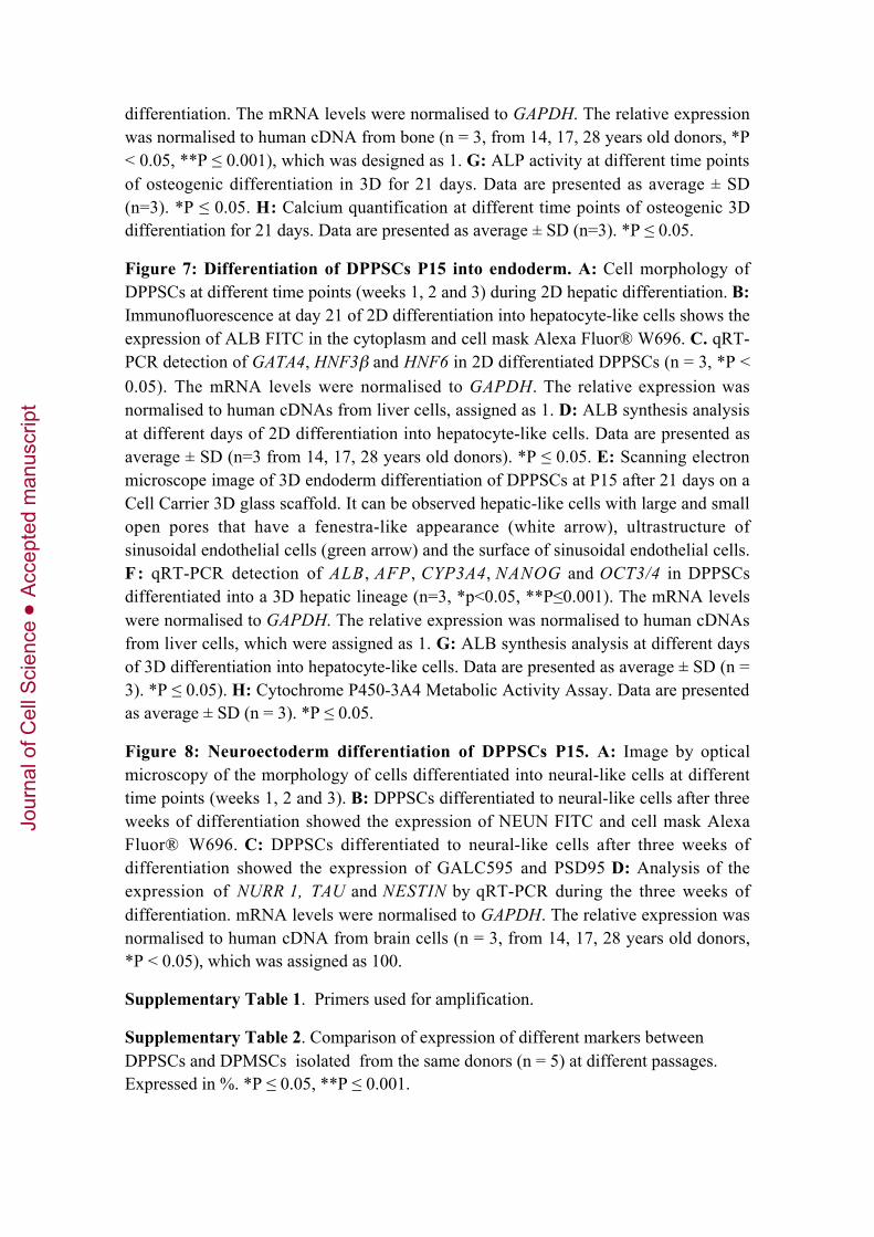
The ability of DPPSCs to form EBs was studied using a micro-patterned culture surfaceand centrifugal force (Figure 3A). After 5 days of culture, the morphology of EBs wasevaluated by light microscopy (Figure 3B). EBs exhibited the typical spherical andwell-limited appearance of EBs formed from ES cells. Alkaline phosphatase (ALP)staining was performed to confirm the stem-like properties of the EBs (Figure 3C).Furthermore, the EBs continued to express embryonic markers such as OCT3/4 andNANOG at day 5, as observed by immunofluorescence (Figure 3D-E). The expressionof embryonic markers and lineage specific markers was studied by RT-PCR. The resultsshowed that DPPSCs and EBs from DPPSCs expressed embryonic markers such asOCT3/4, NANOG and SOX2 as well as other lineage markers as SOX1, BDNF,MIXL1, GATA4 and GATA6 with levels comparable to iPS cells. DPMSCs did notexpress these markers (Figure 3F). To confirm the results, a qRT-PCR was performedto check the expression of the same genes, using iPS cells as a positive control. Levelsof OCT3/4 and NANOG were higher in DPPSCs than in EBs whereas lineage markersas GATA 4, GATA6 and MIXL1 were higher in EBs. (Figure 3G).
The protein expression profile of DPMSCs (Figure 4A) was substantially different fromthat of DPPSCs. Specifically, levels of embryonic markers (OCT3/4, NANOG andSSEA4) were very low, whereas levels of CD73 and STRO1 were high in DPMSCs.Comparative FACS analysis was carried out on different passages of populations fromboth cell types isolated from 14-, 17-, 18-, 28-, and 38-year-old donors(Supplementary Table 2). The two cell types were easily distinguishable by theirmorphology. DPMSCs showed the typical flat and elongated appearance ofmesenchymal cells, whereas DPPSCs were smaller and more spherical in shape (Figure4B). Cell diameters ranged from 8-12 µm for DPPSCs and 12-19 µm for DPMSCs(Figure 4C).
Multiscreen-MIC plate carbonate filters were used to evaluate the migratory capacity ofDPPSCs. Although they came from the same donor and were from the same passage,more DPPSCs migrated across the filters than DPMSCs (Figure 4D). To compare theadhesive ability of DPPSCs and DPMSCs, the expression of integrin CD29 wasevaluated by FACS analysis. The results showed that 99.6% of DPPSCs expressedintegrin CD29, compared to only 82.3% of DPMSCs (Figure 4E).
Although they were the main population, DPPSCs coexisted in culture with other celltypes, such as mesenchymal stem cells from the dental pulp (DPMSCs), due to the lackof a stringent selection when performing primary culture and successive cultures withDPPSC medium. The two populations were indistinguishable from each other by FACSanalisys, although size was known to be different. The percentages of OCT3/4,NANOG and CD73 in different gates depending on size and complexity were
Jour
nal o
f Cel
l Sci
ence
Acc
epte
d m
anus
crip
t

insufficient to distinguish the two populations as well. (Figure 4F-G, SupplementaryTable 3),
To further characterise the two cell populations, we performed magnetic separationusing a human PE selection kit. Cells positive for CD73 were stained with a PE-conjugated antibody, and we extracted RNA from both the CD73- and CD73+
populations after separation. RT-PCR was performed to determine embryonic geneexpression. The CD73- population expressed TERT, OCT3/4 and SOX2, but the CD73+
cells did not (Figure 4H). This confirmed our previous assumption that DPPSCscoexisted with DPMSCs when cultured in vitro.
We also observed a correlation between the embryonic development stages of the thirdmolar and the percentage of OCT3/4 and NANOG expression by FACS analysis in pulptissues from donors of different nolla stages (6-10) and different ages on the same dayof extraction (n = 14 samples) (Supplementary Table 4). To determine the relationshipbetween specific DPPSCs markers and tooth nolla stages, the expression profiles weresubjected to regression analysis using nolla stage as the independent variable todetermine the subpopulation of DPPSCs. All the samples showed the presence ofDPPSCs and low expression of the markers OCT3/4 and NANOG.
To see whether the culture conditions were a key aspect for the maintenance of thedifferent phenotypes between DPPSCs and DPMSCs, we cultured each type of cell withthe medium of the other one. After one week changing the medium every 2 or 3 days weobserved some phenotypic changes (Figure 5A). DPPSCs acquired a longer andflattered shape whereas some of the DPMSCs became smaller and with a morphologyresembling DPPSCs. Changes were easier to see in DPPSCs culture in mesenchymalmedia than in the other way. We checked the expression of the embryonic markers thatdiffer between the two cell types. We observed that DPPSCs cultured in mesenchymalmedium lost the expression of NANOG, whereas the OCT3/4 levels only decreased. Inthe case of DPMSCs cultured in DPPSC medium, cells gained the expression of bothOCT3/4 and NANOG (Figure 5B).
To confirm the pluripotent capacity of DPPSCs we performed three in vitrodifferentiation assays in which DPPSCs were induced to give rise tissues from all threegerm layers.
Mesoderm differentiation
We analysed the ability of DPPSCs to differentiate into osteoblasts in 2D byimmunofluorescence and qRT-PCR. The morphology of DPPSCs cultured withosteogenic media changed over three weeks of differentiation, resulting in cells with abone-like appearance (Figure 6A) that expressed the osteoblast markerOSTEOCALCIN (Figure 6B). The differentiated cells showed upregulated expressionof specific bone tissue genes, such as ALP, OSTEONECTIN, OSTECALCIN (Figure6C), OSTEOPONTIN, COLLAGEN I, COLLAGEN III and BMP2, whereas NANOGwas downregulated (Supplementary Table 5). Human bone cDNAs were used to
Jour
nal o
f Cel
l Sci
ence
Acc
epte
d m
anus
crip
t

normalise the data, G A P D H was used as a housekeeping gene and DPPSCundifferentiated dental pulp cells were used as a negative control (data not shown).ALP, OSTEONECTIN and OSTEOCALCIN expression increased in the first, second andthird weeks of differentiation. We also observed a decrease in NANOG expression. Toconfirm bone-like cell differentiation, we used Alizarin Red to stain extracellular matrixdeposits consisting of hydroxyapatite, calcium and magnesium salts (Figure 6D). Takentogether, these assays demonstrate that DPPSCs could efficiently differentiate intobone-like tissue and express specific bone tissue genes.
Differentiation was also performed using a Cell Carrier 3D glass scaffold. Whencultured in osteogenic differentiation medium, DPPSCs derived into bone-like tissuethat was able to synthesise typical bone structures, such as collagen and corticalstructures that were detectable by SEM analysis (Figure 6E, Supplementary Figure5). The 3D differentiation was also confirmed by qRT-PCR. During three weeks ofdifferentiation, ALP, COLLAGEN I and COLLAGEN III expression steadily increased,whereas the embryonic markers NANOG and OCT3/4 decreased each week (Figure6F). Functional activity was determined by quantifying ALP activity (Figure 6G) andthe calcium secretion (Figure 6H) every week for three weeks. Both ALP activity andthe calcium secretion increased significantly on days 7, 14 and 21 of osteogenicdifferentiation.
To demonstrate the capacity of DPPSC to differentiate into other tissues of themesoderm cap, we performed a Vessel-Derived Endothelial Cell Differentiation, inwhich DPPSCs were cultured with basal media (2% FBS, 50 ng/mL VEGF, 10 ng/mLbFGF) for three week. We observed an increase in the expression of specific endothelialgenes, such as FLK1 and CD14 (by qRT-PCR) and FLK1 (by immunofluorescence)(Supplementary Figure 6).
Endoderm differentiation
Another differentiation assay was performed to induce DPPSCs to generate cells fromthe endodermal lineage, specifically hepatocyte-like cells. DPPSCs cultured in 2D withhepatogenic media for the three weeks showed altered morphologies, with some cellsadopting a polygonal shape similar to hepatocytes (Figure 7A). Expression of thetypical hepatic protein albumin (ALB) was detected by immunofluorescence (Figure7B). Using qRT-PCR, we demonstrated that differentiated cells expressed the hepaticnuclear factors HNF3β, HNF6, and GATA4 (Figure 7C). The expression levels of _-
fetoprotein (AFP), ALB, CEBPA and NANOG were also determined (SupplementaryTable 6). We also observed a decrease in NANOG during the 3 weeks of differentiation(data not shown). Human liver cDNAs (Ambion) were used to normalise the data andGAPDH was used as the housekeeping control (data not shown). The levels of secretedALB increased significantly over three weeks of differentiation (Figure 7D). Theseresults indicate DPPSCs can upregulate endoderm markers and differentiate intohepatic-like cells, both genetically and functionally, upon hepatogenic induction.
Jour
nal o
f Cel
l Sci
ence
Acc
epte
d m
anus
crip
t
Cell Carrier 3D glass scaffolds were also used to perform the endoderm differentiation.After 21 days of 3D culture with hepatogenic media, some hepatic structures were seenby SEM analysis, including large and small pores with a fenestra-like appearance, thesurfaces of sinusoidal endothelial cells and ultra-structures of sinusoidal endothelialcells (Figure 7E, Supplementary figure 7). qRT-PCR was performed to evaluate theexpression of hepatic specific genes ALB, AFP and CYP3A4. Expression of thesemarkers increased each week, whereas the embryonic markers NANOG and OCT3/4expression decreased (Figure 7F). Functional activity was determined by quantifyingALB secretion (Figure 7G) and P450 CYP3A4 activity (luciferin-PFBE) (Figure 7H).As in 2D differentiation cultures, the levels of secreted ALB increased with time. Theactivity of CYP3A4 in differentiated cells was higher than in undifferentiated controlcells. Both experiments were performed in triplicate.
Ectoderm differentiation
Finally, DPPSCs were able to follow a neuroectodermal differentiation pathway in 2Dcultures. The morphology of differentiated cells was phenotypically clearly similar toneuronal cells (Figure 8A). Immunofluorescence staining showed that differentiatedDPPSCs expressed neuronal tissue-specific proteins such as NEUN (Figure 8B), GALC595 and PSD95 (Figure 8C). qRT-PCR analysis showed that expression levels of TAUincreased with time, and that the levels of NURR1 and NESTIN increased during weeksone and two and then plateaued at week three (Figure 8D). GFAP, MBP and SOX1 alsoincreased during the first, second and third weeks, whereas NANOG expressiondecreased during differentiation (Supplementary Table 7). Human brain cDNA(Ambion) was used to normalise the data and GAPDH was used as the housekeepingcontrol (data not shown).
Discussion
Several populations of stem cells have been isolated from different parts of the humantooth and all of them have been shown to have generic mesenchymal stem cell-likeproperties (Laino et al., 2005). These cells express markers associated with theendothelium and/or the smooth muscle, such as the Stromal-derived factor 1 (STRO1),Vascular cell adhesion molecule 1 (VCAM1), Melanoma-associated antigen MUC18(MUC18) and smooth muscle actin (Tecles et al., 2005). Other reports have describedthe presence of a lateral population of stem cells in the dental pulp (Casagrande et al.,2006; Liu et al., 2006; Rizzino, 2009; Sloan and Smith, 2007; Volponi et al., 2010;Zhang et al., 2006b). However, there has been no previously published reference to thepresence of a population of cells in dental pulp with the protein profile SSEA4+,OCT3/4+, NANOG+, Nestin+, SOX2+, LIN28+, CD13+, CD105+, CD34-, CD45-,CD90low, CD29+, +, CD73low, STRO1low and CD146- (Oda et al., 2010), as presented inthis work. OCT3/4, NANOG and SOX2 are indispensable for indefinite stem celldivision, without affecting differentiation potential or the capacity for self-renewal. Thefunctional importance of SOX2 and NANOG genes in altering the progenitor status hasalso been clearly demonstrated (Hanna et al., 2010; Ratajczak et al., 2008; Takahashi
Jour
nal o
f Cel
l Sci
ence
Acc
epte
d m
anus
crip
t
and Yamanaka, 2006; Yu et al., 2007; Zuba-Surma et al., 2009). NANOG has beenreported to be a key gene for maintaining pluripotency, as shown by the capacity formultilineage differentiation and perpetual self-renewal of cells expressing this gene.Although DPPSCs share some features with other populations of stem cells in the tooth,they differ in other aspects, such as gene expression and differentiation potential (Hirataet al., 2010) due to their embryonic-like properties. In this work, we observed theformation of EB-like structures with characteristics similar to embryonic stem cells andMAPCs (Braccini et al., 2005; Verfaillie, 2005).
Both DPMSCs and DPPSCs are obtained from dental pulp using the same isolationprotocol. Distinction of these different cell populations is dependent on the density atwhich the cells are seeded and the culture medium. The use of media that containgrowth factors EGF, PDGF and LIF, allows maintenance of the pluripotent state ofDPPSCs. There has been no other report of the use of a similar medium to culture stemcells from the dental pulp, so it seems likely that this media formulation is key formaintaining DPPSCs with typical stem cell properties. In culture, however, DPPSCsdisplay heterogeneity that can be explained as a consequence of their spontaneousdifferentiation and the lack of clonality in their obtention. As previously shown, otherstem cell cultures undergo the same processes; for example, MAPC cultures areheterogeneous, and two cell populations with different phenotypes (large and small)coexist in the same culture (Verfaillie, 2005). Here, we have demonstrated that theDPPSC population coexists in culture with other cell types, such as DPMSC, and thatthe populations were different of one another, as they differed in pluripotency geneexpression after magnetic separation by CD73. It seems plausible that both cell typeshave a common progenitor, but the relationship between them is still unclear. The factthat DPPSCs express some embryonic markers but the other population does notsuggests that these populations may be at a different level of the differentiationhierarchy, and that DPPSCs may be the progenitors that gave rise to the otherpopulations. More investigation is necessary to test this hypothesis.
The third molars are the most common source of dental stem cells, because wisdomtooth extraction is widely performed and the teeth are usually considered to be medicalwaste (Otaki et al., 2007; Yang et al., 2007). Because the third molar is the last tooth todevelop in humans, it is normally in an early stage of development and is capable ofyielding an optimum quantity of dental pulp tissue for the isolation of DPSCs (Gandiaet al., 2008; Laino et al., 2006; Leeb et al., 2010; Morsczeck et al., 2009; Smith et al.,2009; Atari et al., 2011; Zhang et al., 2006a). Although the percentage of DPPSCsdecreases with age, a population of these cells was always present, even in olderpatients. Multipotent cells that have a certain degree of pluripotency, because they arederived from early embryonic cells and maintained in the adult stage (Ebert et al.,2009), have been found in adult tissues, such as very small embryonic-like (VSEL) stemcells in the bone marrow. These studies, together with the data shown here, indicate thatthe dental pulp could be an important source of cells with pluripotent characteristics.We may speculate that DPPSCs could be derived from residual undifferentiated cells in
Jour
nal o
f Cel
l Sci
ence
Acc
epte
d m
anus
crip
t

the dental pulp.. However, this hypothesis requires further testing. The characteristicsunique to these cells are still under investigation, but the current evidence opens to theway for future comparative studies of the regenerative potency of DPPSCs and stemcells from other sources. In addition, the possibility of freezing these cells followingmolar extraction seems feasible, as is currently performed with cells from the umbilicalcord.
In this paper, we have shown that DPPSCs have pluripotent-like properties that have notbeen found in cells of any other adult source to date. The ability to form EB-like orteratoma-like structures has been thought to be exclusive to ES or iPS cells (Benton etal., 2009; Maltman and Przyborski, 2010). With this finding, a new field ofinvestigation can be opened. If one population of cells found in adult individuals canachieve true pluripotency, there may be other populations with the same or similarproperties that have yet to be discovered. The relationship between DPPSCs and iPScells should also be investigated. The induction of iPS cells seems to be easier fromstem cells than from differentiated cells (Illich et al., 2011). It could be that areprogramming process occurs in DPPSCs, but not in differentiated dermal fibroblasts.Thus, only DPPSCs could selectively expand when cultured in DPPSC media, but othercells from the dental pulp could not undergo the reprogramming process needed toacquire pluripotency. Importantly, the number of reprogramming factors needed forinduced pluripotency could be reduced when using DPPSCs. Further studies are neededto answer these questions.
For therapeutic purposes, the reliability and safety of putative clinical applications forDPPSCs must be considered, especially the issue of genetic stability. We havedemonstrated that DPPSCs show no chromosome abnormalities when cultured in vitro,such that we propose that DPPSCs are safe to use for clinical therapies. We propose thatshort-CGH be used in stem cell research to determine genetic stability when cells arecultured in vitro, because s-CGH allows the detection of genetic abnormalities thatcould remain hidden with the current protocols, such as karyotype or FISH techniques.In addition, stem cells for therapeutic applications must be able to differentiate intodifferent tissues. As we have shown, DPPSCs are capable of giving rise to mesodermal,endodermal and ectodermal tissues that express markers typical of osteoblasts (Atari etal., 2012), hepatocytes and neurons, respectively, as shown by qRT-PCR assays, inwhich all data were normalised to human cDNAs of the respective tissues. The use of3D culture systems to carry out differentiation protocols represents an improved systemthat simulates the physiological in vivo environment (Dhawan et al., 2010; Undale et al.,2009). Based on images obtained by inverted optical microscopy (2D differentiation)and the pictures taken by SEM (3D differentiation), we observed tissue-specificstructures, such as collagen fibres and fenestra-like structures, in 3D that were not seenin 2D.
These cells have properties that are not observed in other cells obtained from adulttissues, which could open a new range of possibilities for regenerative medicine. The
Jour
nal o
f Cel
l Sci
ence
Acc
epte
d m
anus
crip
t

results presented here suggest that DPPSCs may be useful for treating various disorders,such as those related to the loss of bone or some of the proteins synthesised related withthe malfunction of liver cells. Further investigations are needed to fully characterisethese cells.
Materials and methods
Patient Selection
Healthy human third molars extracted for orthodontic and prophylactic reasons wereselected from 20 different patients of different sexes and ages (14-60 years old). Theextraction procedure was kept simple to prevent tooth damage.
Primary cells obtained from human molar samples
Immediately after extraction, the third molars were washed using gauze soaked in 70%ethanol, followed by a wash with sterile distilled water. Holding the tooth with upperincisor forceps, an incision was made between the enamel and the cement using acylindrical turbine bur. A fracture was made on the same line, and fragments of thetooth were placed in a Falcon flask containing sterile 1X PBS. The samples wererapidly transported to the laboratory and placed in Petri dishes in a laminar flow hood.Tissues were isolated from the dental pulp using a sterile nerve-puller file 15 andforceps. Cellular separation was completed by digesting the divided pulp tissue withcollagenase type I (3 mg/ml) (Sigma) for 60 minutes at 37ºC. Cells were then separatedusing an insulin syringe and centrifuged for 10 minutes at 275 g (RCF). The cellfraction was washed twice with sterile 1X PBS and centrifuged again for 10 minutes at275 g (RCF) at room temperature. Once collected, the cells were counted and seeded inDPPSC medium. In order to establish the primary culture, the cells were grown in 96-,24- and 6-well culture dishes and in 150-ml flasks coated with 100 ng/ml humanFibronectin inside a 5% CO2 humidified chamber for three weeks. The medium waschanged every four days. During the splitting/passaging of DPPSCs, cell density wasmaintained at 80-100 cells/cm2 by detaching cells with 0.25% trypsin (Cellgro) andreplating every 36-48 hours, when cells were 60% confluent.
DPPSC Culture Medium
The cell expansion medium consisted of 60% DMEM-low glucose (Sigma) and 40%MCDB-201 (Sigma) supplemented with 1X Insulin-Transferrin-Selenium (ITS)(Sigma), 1X linoleic acid-bovine serum albumin (LA-BSA) (Sigma), 10-9 Mdexamethasone (Sigma), 10-4 M ascorbic acid 2-phosphate (Sigma), 100 units ofpenicillin, 1000 units of streptomycin (PAA), 2% foetal bovine serum (Sigma), 10ng/ml hPDGF-BB (R&D Systems), 10 ng/ml EGF (R&D Systems), 1,000 units/ml hLIF(Chemicon), Chemically Defined Lipid Concentrate (Gibco), 0.8 mg/ml BSA (Sigma)and 55 _M _-mercaptoethanol (_- ME, Sigma).
Jour
nal o
f Cel
l Sci
ence
Acc
epte
d m
anus
crip
t
Base medium
The base medium consisted of 60% DMEM-low glucose (Gibco), 40% MCDB-201(Sigma) with 1X Insulin-Transferrin-Selenium, 1X linoleic acid BSA, 10-9 Mdexamethasone (Sigma), 10-4 M ascorbic acid 2-phosphate (Sigma), 100 units ofpenicillin and 1,000 units of streptomycin (Gibco).
Isolation and culture of human dental pulp mesenchymal stem cells (DPMSCs)
Human adult DPMSCs were isolated from the dental pulp of third molars and weresuspended in Dulbecco’s Modified Eagle Medium (DMEM, Biochrom) containing 2ng/ml basic fibroblast growth factor (bFGF) and 10% foetal bovine serum (FBS,Hyclone). Cells were plated at a density of 300,000 cells/cm2. The medium waschanged after 72 h and every two days thereafter. To propagate DPMSCs, the cells weredetached at 90% confluence by the addition of phosphate buffered saline (PBS,Biochrom) containing 0.05% trypsin-ethylenediaminetetraacetic acid (EDTA;Biochrom) and replated at a density of 4,000 cells/cm2.
Human NTERA-2 and Human IPS
NTERA-2 cells were obtained from the ATCC®. Cells were maintained and cultured inDulbecco's Modified Eagle's Medium supplemented with 10% foetal bovine serum and1% penicillin-streptomycin at 37ºC humidified atmosphere at 5% CO2. hIPS wirekindly donated by our collaborators at University of Navarra.
Flow Cytometry
FACS analysis was carried out the same day of the extraction and again after two andthree weeks of culture initiation. The following fluorochrome-labelled monoclonalantibodies were used: CD13 FITC (eBioscience), SSEA4 PE (eBioscience), OCT3/4FITC (RD SYSTEMS), CD45 PE-Cy5 (BD Pharmingen),CD105 FITC(BDPharmingen), CD34 PE-Cy5 (BD Pharmingen), CD73 PE (BD Pharmingen), CD146FITC (BD Pharmingen), CD90 FITC (eBioscience), CD29 PE (BD Pharmingen),STRO1 FITC (BD Pharmingen), LIN28, SSEA1 PE , SOX2 PE and NANOG FITC(Abcam). For the analysis of control samples, different IgG isotypes coupled to FITC,PE and PE-Cy5 fluorochromes (BD Pharmingen) were used. The cell suspension (inPBS plus 2% FBS) was incubated for 45 minutes at 4ºC in the dark. Later, cells werewashed twice with PBS containing 2% FBS and centrifuged for 6 minutes at 275 g(RCF). Depending on the number, cells were resuspended in 300 to 600 _l of PBS and2% FBS. All flow cytometry measurements were made using a FACScan cytometer(FACSCalibur) and analysed using the winMDI 2.8 program.
Karyotype
The cells were trypsinised and centrifuged at 250 g (RCF). The cell pellet was re-suspended in a volume less than or equal to 500 µl of DMEM-LG and was thensubjected to a hypotonic shock with 0.075 M KCl that had been pre-heated to 37ºC (the
Jour
nal o
f Cel
l Sci
ence
Acc
epte
d m
anus
crip
t

saline solution was added drop by drop under continuous agitation). After 30 min ofincubation in the presence of KCl at 37ºC, the cells were centrifuged at 300 g (RCF) for10 min. The nucleus suspension was fixed twice consecutively in methanol:acetic acid(3:1). After the final centrifugation step, the final cell pellet was re-suspended infixation liquid and extensions were performed on glass slides that were pre-cooled to4ºC. The extensions were stained using GIEMSA stain and the number of cells inmetaphase was counted using a light microscope at a 100x magnification with oilimmersion. A minimum of 50 metaphases were counted per sample.
RNA Isolation and qRT-PCR
Total cellular RNA samples were extracted using Trizol (Invitrogen) from the followingcell types: DPPSCs at passages 5, 10 and 15 (P5, P10 and P15), H-NTERA, DPMSCsand differentiated cells. RNA was extracted weekly from the differentiated cells. Twoµg of RNA was treated with DNase I (Invitrogen) and reverse-transcribed using M-MLV Reverse transcriptase (Invitrogen). We analysed the efficacy of the cDNA (1, 0.1,0.01, 0.001, 0.0001 dilutions) at different concentrations for all primers of pluripotentgenes using NTERA cells as positive controls. Additionally, we tested the followingsamples as positive controls: hepatocyte markers from human liver cDNA samples,osteoblast markers from human bone cDNA and neuroectoderm markers from humanbrain cDNA (Ambion). Quantitative RT-PCR was performed using the CFX96thermocycler (Bio-Rad). Quantitative RT-PCR was performed using 50 ng of cDNAand SYBR Green Supermix (Bio-Rad Laboratories, Inc.). cDNA samples wereamplified using specific primers with the following conditions for 40 cycles. Theexpression levels of genes of interest (Supplementary Table 1) were normalisedagainst the housekeeping gene GAPDH. The relative expression levels were normalisedto human cDNAs (positive controls), which were assigned as 1. The results wereanalysed using the 2__Ct method.
Western blotting
Total protein was extracted from the following samples: DPPSCs collected at differentpassages (P5, P10, P15 and P20), DPMSCs, NTERA-2, HEK 293 and bone marrowderived-MAPCs. Cell lysates with equal protein concentrations (20 µg/_l) wereseparated by SDS-PAGE on 12% polyacrylamide gels and transferred ontonitrocellulose membranes. The membranes were blocked with 1% (wt/vol) BSA in PBScontaining 0.1% Tween 20, blotted with OCT3/4 and GAPDH primary antibodies(1:5000) and blotted with secondary antibodies (1:15000). All antibodies werepurchased from Abcam.
Immunofluorescence Analysis
Samples were fixed with 4% paraformaldehyde (Sigma) for 4 min at room temperaturefollowed by methanol (Sigma) for 2 min at -20°C. For nuclear ligands, cells werepermeabilised with 0.1 M Triton X-100 (Sigma) for 10 min. Slides were incubatedsequentially for 30 min each with primary antibody and FITC, PE or PE-Cy5-coupled
Jour
nal o
f Cel
l Sci
ence
Acc
epte
d m
anus
crip
t

anti-mouse IgG antibodies. Between each step, the slides were washed with 1% BSA(Sigma) in PBS. Cells were examined using confocal fluorescence microscopy(Confocal 1024 microscope, Olympus AX70, Olympus Optical, Tokyo).
Teratoma Formation and Histological Analysis
Eight-week-old nude mice (Samtako Bio Korea, Seoul, Korea) were anaesthetised withdiethyl ether. Fifty microliters of a P15 DPPSC or DPMSC cell suspension (4x10⁷
cells/ml), from three different donors ( 14, 17, 28 years old donors), mixed with 50 µlof Matrigel (BD) was injected subcutaneously into the dorsal flanks of the mice, whichwere then housed with free access to water and food under specific pathogen-freeconditions. After 3 or 5 weeks, the teratoma-like structures were surgically dissectedfrom the mice followed by fixation with 4% paraformaldehyde and 1.25%glutaraldehyde, and the mice were subjected to histological analysis. Specimens wereembedded in paraffin, cut into 3 µm sections and stained with haematoxylin and eosin(H&E).
Immunohistochemistry
Samples were cut into 7-µm thick sections for immunohistochemical staining. Slideswere placed in PBS for 30 min to remove gelatin. After being washed twice withdistilled water for 5 min, the sections were blocked against endogenous peroxidase in0.3% hydrogen peroxide for 15 min and 10% normal goat serum in PBS for 1 hour atroom temperature to reduce nonspecific antibody interactions. The slides wereincubated with mouse monoclonal primary antibodies against human OCT3/4 (1:400,BD), NANOG (1:400, BD), SSEA4 (1:400, BD) and LIN28 (1:400, BD) at 4ºCovernight. After washing with PBS, the specimens were incubated with biotinylatedgoat anti-mouse secondary antibody (Zymed) and streptavidin peroxidase (Zymed) atroom temperature for 10 min each. Finally, the specimens were visualised using adiaminobenzidine reagent kit (Zymed). The immunostained sections werecounterstained with haematoxylin.
Transmission Electronic Microscopy
A piece of the cell pellet measuring 1 mm_ was fixed in a solution of 2% formaldehyde,2.5% glutaraldehyde and Karnoski buffer with cacodylate (0.2 mol/L, pH 7.4). After 48hours, the samples were soaked in araldite. The ultra-fine sections were stained forcontrast with citrate and then observed using an electronic microscope (Zeiss EM900).
Scanning Electron Microscopy
For SEM analysis, samples were fixed in 2.5% glutaraldehyde (Ted Pella Inc.) in 0.1 MNa-cacodylate buffer (EMS, Electron Microscopy Sciences, Hatfield, PA) (pH 7.2) for1 h on ice. After fixation, the samples were treated with 1% osmium tetroxide (OsO4)for 1 h. The samples were dehydrated in serial solutions of acetone (30–100%) with the
Jour
nal o
f Cel
l Sci
ence
Acc
epte
d m
anus
crip
t

scaffolds mounted on aluminium stubs. The samples were then examined using a Zeiss940 DSM scanning electron microscope.
Cell Migration Assay
The cell migration capacity of DPPSCs and DPMSCs from the same donor, as well ashNTERA cells, which were used as a positive control, was tested using a Multiscreen-MIC Plate polycarbonate filter (8 _M pore size) (Millipore). Approximately 1x10_ cellsin 150 _L were added to the bottom wells of the filter plate and incubated at 37ºC in 5%CO2 for 6 h. After the incubation, the filters were removed and the topside of themembrane was scraped to remove non-migrated cells. The filters were then stained withToluidine Blue. The cells were counted using a light microscope. Experiments wereperformed in triplicate and the data were pooled.
Short-chromosome genomic hybridisation (short-CGH)
DPPSCs were isolated one by one by manual catching and short-CGH technique wasdeveloped (Rius et al., 2010) (n = 15). Hybridisation of control and DPPSC sampleswas carried out against a masculine cell preparation.
Mesoderm differentiation
For bone differentiation, cells were seeded in 6-well plates and in a Cell Carrier 3Dglass scaffold (Orla protein) on a 24-well plate with culture medium at a density of 3x10³
cells P15 per cm2. After 24 hours, differentiation was initiated by the followingmedium: _-MEM containing 10% heat inactivated FBS, 10 mM _-glycerol phosphate(Sigma), 50 _M of L-ascorbic acid (Sigma), 0.01 _M dexamethasone and 1% penicillinand streptomycin. The medium was changed every 3 days for 21 days. Differentiatedcultures were evaluated by qRT-PCR for ALP, OSTEONECTIN, OSTEOCALCIN,OSTEOPONTIN, COLLAGEN I, COLLAGEN III, BMP-2 and NANOG every week. Thecultures were also analysed via immunofluorescence for OSTEOCALCIN and AlizarinRed staining after three weeks of differentiation.
Vessel-Derived Endothelial Cells Differentiation
Undifferentiated DPPSCs were cultured at a density of 3x10³ cells per cm2 with basalmedia for one day. Culture medium was then exchanged for differentiation media(Basal Media, FBS 2%, VEGF 50 ng/mL, bFGF 10 ng/mL). Cells were grown oncoverslips and 6-well plates treated with Fibronectin and incubated at 37ºC at 5% CO2.The medium was changed every 3 days for 21 days. Differentiated cultures wereevaluated by qRT-PCR and immunofluorescence. RNA was obtained at day 0 and thenweekly and tested for endothelial gene Flk1 and CD14 expression by qRT-PCR.
Jour
nal o
f Cel
l Sci
ence
Acc
epte
d m
anus
crip
t
Endoderm differentiation
For 2D differentiation, DPPSCs P15 were seeded in 6-well plates with culture mediumat a density of 5x10⁴ cells/cm2. The following day, the culture medium was exchangedfor differentiation medium consisting of base medium containing 100 ng/ml HGF and100 ng/ml FGF-4 (R&D Systems). The medium was changed every 3 days for 3 weeks.Differentiated cultures were evaluated by qRT- PCR for GATA4, HNF3β, HNF6, AFP,
ALB, CEBPA and NANOG expression. Differentiated cells were identified usingimmunofluorescence microscopy for albumin protein expression and albumin secretionanalysis at different days of differentiation.
For 3D differentiation, 5 x 10⁴ cells P15 were seeded in a Cell Carrier 3D glass scaffold(Orla protein) pre-coated with 2% Matrigel and placed in 24-well plates with RPMImedium (Mediatech) supplemented with GlutaMAX and penicillin/streptomycin andcontaining 0.5% defined foetal bovine serum (FBS; HyClone) and 100 ng/ml Activin A(R&D Systems). Three days post-induction, the medium was refreshed using the sameRPMI-based medium with 100 ng/ml Activin A but replacing FBS by KOSR 2%. After2 days, definitive endoderm cultures were refreshed with RPMI medium supplementedwith GlutaMAX and penicillin/streptomycin and containing 2% KOSR, 10 ng/ml FGF-4 (R&D Systems) and 10 ng/ml HGF (R&D Systems). Three days later, the cells wereswitched to minimal MDBK-MM medium (Sigma-Aldrich) supplemented withGlutaMAX and penicillin/streptomycin and containing 0.5 mg/ml bovine serumalbumin (BSA) (Sigma-Aldrich), 10 ng/ml FGF-4 and 10 ng/ml HGF. After another 3days, the cells were switched to complete hepatocyte culture medium (HCM)supplemented with SingleQuots (Lonza) and containing 10 ng/ml FGF-4, 10 ng/mlHGF, 10 ng/ml oncostatin M (R&D Systems) and 10-7 M dexamethasone (Sigma-Aldrich). Differentiation was continued for another 9 days. At each stage, the mediumwas refreshed every 2-3 days.
Neuroectoderm differentiation
For neural differentiation, cells P15 were seeded in 6-well plates and in 75-cm2 flasks inbase culture medium at a density of 3x10³/cm2. The following day, the culture mediumwas exchanged for differentiation medium; the differentiation medium differed fromweek to week. During the first week, the medium consisted of base medium and bFGF(100 ng/ml). During the second week, the medium consisted of base medium, FGF-8(10 ng/ml) and SHH (100 ng/ml). During the third week, the medium consisted of basemedium, BDNF (10 ng/ml) and GDNF (10 ng/ml) + N2 (R&D Systems). The mediumwas changed every 3 days. Neural differentiation was evaluated via qRT-PCR for TAU,NURR1, NESTIN, GFAP, MBP, SOX1 and NANOG expression. Cultures were alsoanalysed via immunofluorescence for PSD95-FITC and GALC-595 protein expression.
Jour
nal o
f Cel
l Sci
ence
Acc
epte
d m
anus
crip
t
Alizarin red staining
Cells were fixed in a 2.5% glutaraldehyde mixture that was freshly prepared in 1X PBSbuffer for 10-15 minutes at room temperature. Cells were then washed with 1X PBS and2% Alizarin Red Solution (Millipore) was added to the fixed cells. Followingincubation at 37ºC for 20 minutes, cells were then observed through microphotography(positively stained nodules are in orange-red).
Albumin secretion
The production of albumin was determined using the Albumin Fluorescence Assay Kit(Sigma) according to the manufacturer’s instructions.
Alkaline Phosphatase Staining
For alkaline phosphatase (ALP) staining, EBs were fixed in a solution of 4%paraformaldehyde in PBS for 20 min. After extensive washing in PBS, cells wereincubated in NTMT solution (10 mM NaCl, 100 mM Tris- HCl (pH 9), 50 mM MgCl2,supplemented with 0.1% Tween-20) for 5 min and then in NTMT solutionsupplemented with NBT (Nitro-Blue Tetrazolium Chloride) and BCIP (5-Bromo-4-Chloro-3'-Indolyphosphate p-Toluidine Salt) in darkness until the staining developed.
ALP activity
During the 3D osteoblast differentiation of DPPSCs and DPMSCs from passage number15, ALP activity was quantified every week by spectrophotometry using a Cromatest kit(Linear), in accordance with the manufacturer’s instructions. We measured theabsorbance of each sample at 1, 2, 3, 5 and 10 minutes.
Calcium quantification
Differentiated cells were washed twice with 1X PBS. Accumulated calcium wasremoved from the cellular components using lysis solutions contained in the Sigma Kitfor the analysis of calcium accumulation, according to the manufacturer's instructions.The total calcium was calculated using standard solutions and the absorbance wasmeasured at 575 nm.
Cytochrome P450 3A4 Metabolic Activity Assay
Cytochrome P450 (CYP) 3A4 enzyme activity assay was assessed by measurement ofluciferase activity with the P450-Glo CYP3A4 assay, according to the manufacturer’sinstructions. Differentiated cells were incubated at 37°C in DMSO plus ethanolsupplemented with 50 µmol/L luciferin PFBE (150 µL/well) and without DMSO;undifferentiated DPPSCs were used as negative control. After 3 h incubation, 50 µL ofmedium was transferred in a 48-well and mixed with 50 µL of luciferin detectionreagent to initiate luminescent reaction. After 20 m incubation at room temperature,luminescence was measured with a Victor3 luminometer (PerkinElmer).
Jour
nal o
f Cel
l Sci
ence
Acc
epte
d m
anus
crip
t

Statistical analysis
To assess the percentages of specific markers for DPPSCs, data were subjected to aregression analysis, which considered the independent variable (age) and the dependentvariable (different markers). We established statistical significance at a p-value less than0.1 (90% confidence level). For all other data, the statistical test applied was the pairedsamples T-test, with statistical significance set at p < 0.05. Data were analysed withSPSS Version 16.0 software. The values are expressed as the mean ± standarddeviation.
Acknowledgements
We thank M. Costa for help with FACS analysis, as well as J. Navarro and J. del Reyfor their dedication in cytogenetic analysis using a newly developed CGH technique wasperformed in the Unitat de Biologia Cel.lular i Genètica Mèdica UAB , with the economical suport of FIS PI080012 and within the framework of the Càtedra deRecerca Eugin-UAB . The International University of Catalonia supported all this work.
Ethical regulations
Dental pulp tissues used for these experiments were obtained with informed consentfrom donors. All experiments were performed in accordance with the guidelines onhuman stem cell research issued by the Committee on Bioethics of the InternationalUniversity of Catalonia.
Bibliography
1-About, I., Bottero, M. J., de Denato, P., Camps, J., Franquin, J. C.,Mitsiadis, T. A. (2000). Human dentin production in vitro. Exp. Cell Res. 258, 33-41.
2-Arthur, A., Rychkov, G., Shi, S., Koblar, S. A., Gronthos, S. (2008). Adulthuman dental pulp stem cells differentiate toward functionally active neurons underappropriate environmental cues. Stem Cells 26, 1787-1795.
3-Atari, M., Barajas, M., Hernández-Alfaro F., Gil, C., Fabregat, M., FerrésPadró E., Giner, L., Casals, N. (2011). Isolation of pluripotent stem cells fromhuman third molar dental pulp. Histol. Histopathol. 26, 1057-1070.
4-Atari, M., Caballé-Serrano, J., Gil-Recio, C., Giner-Delgado, C., Martínez-Sarrà, E., García-Fernández, D. A., Barajas, M., Hernández-Alfaro, F.,
Jour
nal o
f Cel
l Sci
ence
Acc
epte
d m
anus
crip
t

Ferrés-Padró, E., Giner-Tarrida, L. (2012). The enhancement of osteogenesisthrough the use of dental pulp pluripotent stem cells in 3D. Bone (in press).
5-Benton, G., George, J., Kleinman, H. K., Arnaoutova, I. P. (2009).Advancing science and technology via 3D culture on basement membrane matrix. J.Cell. Physiol. 221, 18-25.
6-Braccini, A., Wendt, D., Jaquiery, C., Jakob, M., Heberer, M., Kenins, L.,Wodnar-Filipowicz, A., Quat¡rto, R., Martin, I. (2005). Three-dimensionalperfusion culture of human bone marrow cells and generation of osteoinductivegrafts. Stem Cells 23, 1066-1072.
7-Casagrande, L., Mattuella, L. G., de Araujo, F. B., Eduardo, J. (2006). Stemcells in dental practice: perspectives in conservative pulp therapies. J. Clin. Pediatr.Dent. 31, 25-27.
8-Cheng, P. H., Snyder, B., Fillos, D., Ibegbu, C. C., Huang, A. H., Chan, A.W. (2008). Postnatal stem/progenitor cells derived from the dental pulp of adultchimpanzee. BMC Cell Biol. 9, 20.
9-Cordeiro, M. M., Dong, Z., Kaneko, T., Zhang, Z., Miyazawa, M., Shi, S.,Smith, A. J., Nor, J. E. (2008). Dental pulp tissue engineering with stem cells fromexfoliated deciduous teeth. J. Endodont. 34, 962-969.
10-d'Aquino, R., Graziano, A., Sampaolesi, M., Laino, G., Pirozzi, G., DeRosa, A., Papaccio, G. (2007). Human postnatal dental pulp cells co-differentiateinto osteoblasts and endotheliocytes: a pivotal synergy leading to adult bone tissueformation. Cell Death Differ. 14, 1162-1171.
11-Dezawa, M., Ishikawa, H., Hoshino, M., Itokazu, Y., Nabeshima, Y.(2005). Potential of bone marrow stromal cells in applications for neuro-degenerative, neuro-traumatic and muscle degenerative diseases. Curr.Neuropharmacol. 3, 257-266.
12-Dhawan, A., Strom, S. C., Sokal, E., Fox, I. J. (2010). Human hepatocytetransplantation. Meth. Mol. Biol. 640, 525-534.
13-Ebert, A. D., Yu, J., Rose, F. F., Jr., Mattis, V. B., Lorson, C. L., Thomson,J. A., Svendsen, C. N. (2009). Induced pluripotent stem cells from a spinalmuscular atrophy patient. Nature 457, 277-280.
14-Fuchs, E., Segre, J. A. (2000). Stem cells: a new lease on life. Cell 100, 143-155.
15-Fujii, S., Maeda, H., Wada, N., Tomokiyo, A., Saito, Akamine, A. (2008).Investigating a clonal human periodontal ligament progenitor/stem cell line in vitroand in vivo. J. Cell. Physiol. 215, 743-749.
16-Gandia, C., Arminan, A., Garcia-Verdugo, J. M., Lledo, E., Ruiz, A.,Minana, M. D., Sanchez-Torrijos, J., Paya, R., Mirabet, V., Carbonell-Uberos,F., et al. (2008). Human dental pulp stem cells improve left ventricular function,induce angiogenesis, and reduce infarct size in rats with acute myocardialinfarction. Stem Cells 26, 638-645.
Jour
nal o
f Cel
l Sci
ence
Acc
epte
d m
anus
crip
t
17-Gay, I. C., Chen, S., MacDougall, M. (2007). Isolation and characterization ofmultipotent human periodontal ligament stem cells. Orthod. Craniofac. Res. 1 0,149-160.
18-Gronthos, S., Mankani, M., Brahim, J., Robey, P. G., Shi, S. (2000).Postnatal human dental pulp stem cells (DPSCs) in vitro and in vivo. Proc. Natl.Acad. Sci. U. S. A. 97, 13625-13630.
19-Hanna, J. H., Saha, K., Jaenisch, R. (2010). Pluripotency and cellularreprogramming: facts, hypotheses, unresolved issues. Cell 143, 508-525.
20-Harada, H., Kettunen, P., Jung, H. S., Mustonen, T., Wang, Y. A.,Thesleff, I. (1999). Localization of putative stem cells in dental epithelium andtheir association with Notch and FGF signaling. J. Cell Biol. 147, 105-120.
21-He, F., Yang, Z., Tan, Y., Yu, N., Wang, X., Yao, N., Zhao, J. (2009). Effectsof Notch ligand Delta1 on the proliferation and differentiation of human dental pulpstem cells in vitro. Arch. Oral. Biol. 54, 216-222.
22-Hirata, T. M., Ishkitiev, N., Yaegaki, K., Calenic, B., Ishikawa, H.,Nakahara, T., Mitev, V., Tanaka, T., Haapasalo, M. (2010). Expression ofmultiple stem cell markers in dental pulp cells cultured in serum-free media. J.Endodont. 36, 1139-1144.
23-Honda, M. J., Fong, H., Iwatsuki, S., Sumita, Y., Sarikaya, M. (2008).Tooth-forming potential in embryonic and postnatal tooth bud cells. Med. Mol.Morphol. 41, 183-192.
24-Huo, N., Tang, L., Yang, Z., Qian, H., Wang, Y., Han, C., Gu, Z., Duan, Y.,Jin, Y. (2010). Differentiation of dermal multipotent cells into odontogenic lineageinduced by embryonic and neonatal tooth germ cell-conditioned medium. StemCells Dev. 19, 93-104.
25-Illich, D. J., Demir, N., Stojkovic, M., Scheer, M., Rothamel, D.,Neugebauer, J., Hescheler, J., Zoller, J. E. (2011). Concise review: inducedpluripotent stem cells and lineage reprogramming: prospects for bone regeneration.Stem Cells 29, 555-563.
26-Iohara, K., Nakashima, M., Ito, M., Ishikawa, M., Nakasima, A.,Akamine, A. (2004). Dentin regeneration by dental pulp stem cell therapy withrecombinant human bone morphogenetic protein 2. J. Dent. Res. 83, 590-595.
27-Itskovitz-Eldor, J., Schuldiner, M., Karsenti, D., Eden, A., Yanuka,O.,Amit, M., Soreg, H., Benvenisty, N. (2000). Differentiation of human embryonicstem cells into embryoid bodies compromising the three embryonic germ layers.Mol. Med. 6, 88-95.
28-Laino, G., d'Aquino, R., Graziano, A., Lanza, V., Carinci, F., Naro, F.,Pirozzi, G., Papaccio, G. (2005). A new population of human adult dental pulpstem cells: a useful source of living autologous fibrous bone tissue (LAB). J. BoneMiner. Res. 20, 1394-1402.
29-Laino, G., Carinci, F., Graziano, A., d'Aquino, R., Lanza, V., De Rosa, A.,Gombos, F., Caruso, F., Guida, L., Rullo, R., et al. (2006). In vitro bone
Jour
nal o
f Cel
l Sci
ence
Acc
epte
d m
anus
crip
t

production using stem cells derived from human dental pulp. J. Craniofac. Surg.17, 511-515.
30-Leeb, C., Jurga, M., McGuckin, C., Moriggl, R., Kenner, L. (2010).Promising new sources for pluripotent stem cells. Stem Cell Rev. 6, 15-26.
31-Liu, H., Gronthos, S., Shi, S. (2006). Dental pulp stem cells. Method.Enzymol. 419, 99-113.
32-Maltman, D. J., Przyborski, S. A. (2010). Developments in three- dimensionalcell culture technology aimed at improving the accuracy of in vitro analyses.Biochem. Soc. T. 38, 1072-1075.
33-Martin, G. R., Evans, M. J. (1975). Differentiation of clonal lines ofteratocarcinoma cells: formation of embryoid bodies in vitro. Proc. Natl. Acad. Sci.U. S. A. 72, 1441-1445.
34-Mina, M., Braut, A. (2004). New insight into progenitor/stem cells in dentalpulp using Col1a1-GFP transgenes. Cells Tissues Organs 176, 120-133.
35-Miura, M., Gronthos, S., Zhao, M., Lu, B., Fisher, L. W., Robey, P. G., Shi,S. (2003). SHED: stem cells from human exfoliated deciduous teeth. Proc. Natl.Acad. Sci. U. S. A. 100, 5807-5812.
36-Morsczeck, C., Frerich, B., Driemel, O. (2009). Dental stem cell patents.Recent Pat. DNA Gene Seq. 3, 39-43.
37-Noel, D., Djouad, F., Jorgense, C. (2002). Regenerative medicine throughmesenchymal stem cells for bone and cartilage repair. Curr. Opin. Invest. Dr. 3,1000-1004.
38-Nosrat, I. V., Widenfalk, J., Olson, L., Nosrat, C. A. (2001). Dental pulpcells produce neurotrophic factors, interact with trigeminal neurons in vitro, andrescue motoneurons after spinal cord injury. Dev. Biol. 238, 120-132.
40-O'Connor, M. D., Kardel, M. D., Iosfina, I., Youssef, D., Lu, M., Li, M. M.,Vercauteren, S., Nagy, A., Eaves, C. J. (2008). Alkaline phosphatase-positivecolony formation is a sensitive, specific, and quantitative indicator ofundifferentiated human embryonic stem cells. Stem Cells 26, 1109-1116.
41-Oda, Y., Yoshimura, Y., Ohnishi, H., Tadokoro, M., Katsube ,Y., Sasao,M., Kubo,Y., Hattori, K., Saito, S., Horimoto, K., et al. (2010). Induction ofpluripotent stem cells from human third molar mesenchymal stromal cells. J. Biol.Chem. 285, 29270-29278.
42-Otaki, S., Ueshima, S., Shiraishi, K., Sugiyama, K., Hamada, S.,Yorimoto, M., Matsuo, O. (2007). Mesenchymal progenitor cells in adult humandental pulp and their ability to form bone when transplanted intoimmunocompromised mice. Cell Biol. Int. 31, 1191-1197.
43-Papapetrou, E. P., Tomishima, M. J., Chambers, S. M., Mica, Y., Reed, E.,Menon, J., Tabar, V., Mo, Q., Studer, L., Sadelain, M. (2009). Stoichiometricand temporal requirements of Oct4, Sox2, Klf4, and c-Myc expression for efficienthuman iPSC induction and differentiation. Proc. Natl. Acad. Sci. U. S. A. 106 ,12759-12764.
Jour
nal o
f Cel
l Sci
ence
Acc
epte
d m
anus
crip
t

44-Peters, H., Balling, R. (1999). Teeth. Where and how to make them. TrendsGenet. 15, 59-65.
45-Ratajczak, M. Z., Zuba-Surma, E. K., Machalinski, B., Ratajczak, J.,Kucia, M. (2008). Very small embryonic-like (VSEL) stem cells: purification fromadult organs, characterization, and biological significance. Stem Cell Rev. 4, 89-99.
46-Rius, M., Obradors, A., Daina, G., Cuzzi, J., Marques, L., Calderon, G.,Velilla, E., Martinez-Passarell, O., Oliver-Bonet, M., Benet, J., et al. (2010).Reliability of short comparative genomic hybridization in fibroblasts andblastomeres for a comprehensive aneuploidy screening: first clinical application.Hum. Reprod. 25, 1824-1835.
47-Rizzino, A. (2009). Sox2 and Oct-3/4: a versatile pair of master regulators thatorchestrate the self-renewal and pluripotency of embryonic stem cells. WileyInterdiscip. Rev. Syst. Biol. Med. 1, 228-236.
48-Sloan, A. J., Smith, A. J. (2007). Stem cells and the dental pulp: potentialroles in dentine regeneration and repair. Oral Dis. 13, 151-157.
49-Smith, K. P., Luong, M. X., Stein, G. S. (2009). Pluripotency: toward a goldstandard for human ES and iPS cells. J. Cell. Physiol. 220, 21-29.
50-Stevens, A., Zuliani, T., Olejnik, C., LeRoy, H., Obriot, H., Kerr-Conte, J.,Formstecher, P., Bailliez, Y., Polakowska, R. R. (2008). Human dental pulpstem cells differentiate into neural crest-derived melanocytes and have label-retaining and sphere-forming abilities. Stem Cells Dev. 17, 1175-1184.
51-Takahashi, K., Yamanaka, S. (2006). Induction of pluripotent stem cells frommouse embryonic and adult fibroblast cultures by defined factors. Cell 126, 663-676.
52-Tecles, O., Laurent, P., Zygouritsas, S., Burger, A. S., Camps, J., Dejou,J., About, I. (2005). Activation of human dental pulp progenitor/stem cells inresponse to odontoblast injury. Arch. Oral Biol. 50, 103-108.
53-Thesleff, I., Aberg, T. (1999). Molecular regulation of tooth development.Bone 25, 123-125.
54-Undale, A. H., Westendorf, J. J., Yaszemski, M. J., Khosla, S. (2009).Mesenchymal stem cells for bone repair and metabolic bone diseases. Mayo Clin.Proc. 84, 893-902.
55-Verfaillie, C. M. (2005). Multipotent adult progenitor cells: an update. NovartisFound Symp. 265, 55-61; discussion: 61-5, 92-7.
56-Volponi, A. A., Pang, Y., Sharpe, P. T. (2010). Stem cell-based biologicaltooth repair and regeneration. Trends Cell. Biol. 20, 715-722.
57-Yang, X., van der Kraan, P. M., van den Dolder, J., Walboomers, X. F.,Bian, Z., Fan, M., Jansen, J. A. (2007). STRO-1 selected rat dental pulp stemcells transfected with adenoviral-mediated human bone morphogenetic protein 2gene show enhanced odontogenic differentiation. Tissue Eng. 13, 2803-2812.
Jour
nal o
f Cel
l Sci
ence
Acc
epte
d m
anus
crip
t
58-Yu, J., Vodyanik, M. A., Smuga-Otto, K., Antosiewicz-Bourget, J., Frane,J. L., Tian, S., Nie, J., Jonsdottir, G. A., Ruotti, V., Stewart, R. et al. (2007).Induced pluripotent stem cell lines derived from human somatic cells. Science 318,1917-1920.
59-Zhang, W., Walboomers, X. F., Wolke, J. G., Bian, Z., Fan, M. W., Jansen,J. A. (2005). Differentiation ability of rat postnatal dental pulp cells in vitro. TissueEng. 11, 357-368.
60-Zhang, W., Walboomers, X. F., Shi, S., Fan, M., Jansen, J. A. (2006a).Multilineage differentiation potential of stem cells derived from human dental pulpafter cryopreservation. Tissue Eng. 12, 2813-2823.
61-Zhang, W., Walboomers, X. F., van Kuppevelt, T. H., Daamen, W. F.,Bian, Z., Jansen, J. A. (2006b). The performance of human dental pulp-stem cellson different three-dimensional scaffold materials. Biomaterials 27, 5658-5668.
62-Zuba-Surma, E. K., Kucia, M., Ratajczak, J., Ratajczak, M. Z. (2009)."Small stem cells" in adult tissues: very small embryonic-like stem cells stand up!.Cytom. Part A 75, 4-13.
Jour
nal o
f Cel
l Sci
ence
Acc
epte
d m
anus
crip
t
Figure legends
Figure 1: Characterisation and cellular morphology of pluripotent stem cellsobtained from dental pulp (DPPSCs) by in vitro expansion. A: Morphology ofDPPSCs at different passages (P5, P10 and P15). B: Analysis of DPPSC immuno-phenotype by confocal microscopy shows the expression of SSEA4 PE (28% ±1,13)together with OCT3/4 FITC (60,3% ± 5,3) or NANOG FITC (21,3% ± 3,6). Average of3 independent experiments. C: Cytogenetic analysis of undifferentiated DPPSCs (P15)show 46 XY without aneuploidy or polyploidy; chromosome structural abnormalitieswere not detected. D: Cells examined by transmission electron microscopy show largenuclei and small cytoplasmic volume. Scale bar: 10 µm. F: Short-CGH analysisshowing genetic stability of DPPSCs from a female donor (N=3).
Figure 2: FACS characterisation, gene expression of DPPSCs P15 and teratomaformation. A: FACS analysis of DPPSC P15 for membrane markers: CD105 (90.77%± 2,28), CD146 (13.17%± 0,68), CD45 (0.02%±0,39) and CD34 (0.06% ±0,05); B:FACS analysis of DPPSC P15 for nuclear markers: STRO1 (4.42%±1,23), TRA1(0.00%±0), OCT3/4 (62.98%±5,22) and NANOG (22.96%±2,58). C: Analysis byFACS of DPPSCs with double staining (19.55%) for OCT3/4 FITC (27.08%) andNANOG PE (25.94%±2,51). Expressed as average of 3 independent experiments. D:RT-PCR of OCT3/4, SOX2, TERT and GAPDH of DPPSC P15 in 2D culture. E:OCT3/4 detection by western blot of DPPSCs at different passages compared toDPMSCs. NTERA cells and BM-MAPCs were used as positive controls, and Schwanncells and DPMSCs were used as negative controls. F: Transplantation of DPPSCs P15and DPMSC P15 isolated from the same donor from different ages (14, 17, 28 yearsold) into immunodeficient mice resulted in apparent teratoma-like formation (left flank)after 5 weeks; DPMSCs P15 were used as a negative control (right flank). H&E stainingof teratoma-like structures induced by DPPSCs show the presence of tissues frommesoderm, endoderm and ectoderm G: H&E staining of teratoma-like structure after 3weeks. Circle points the zone that is amplified in following pictures. H :Immunohistochemical staining for OCT3/4. I: Immunohistochemical staining forSSEA4. J: Immunohistochemical staining for NANOG. K: Immunohistochemicalstaining for LIN28. Immunohistochemical staining shows embryonic markers SSEA4,LIN28, OCT3/4 and NANOG (black arrows). L: H&E staining showing chondroidtissue, chondroid matrix, fibroblasts and collagen fibres. M: H&E staining showing gut-like epithelium. N: H&E staining showing nerve-l ike tissue and keratin.
Figure 3: Generation of DPPSC P15 embryoid bodies. A: AggreWell system (StemCell Technologies), which utilises a micropatterned culture surface and centrifugalforced aggregation to direct the formation of EBs by DPPSCs for 5 days. Scale bar: 100µm. B: Morphology of DPPSC embryoid bodies examined by light microscope. Scalebar: 100µm. C: Alkaline phosphatase staining of DPPSC embryoid bodies. Scale bar:100µm. D: Immuno-phenotype analysis by fluorescence microscopy shows the
Jour
nal o
f Cel
l Sci
ence
Acc
epte
d m
anus
crip
t
expression of OCT3/4 FITC. E: Immunostaining of DPPSC EBs generated byAggreWell system with NANOG FITC. F: RT-PCR analysis of OCT3/4, NANOG,SOX2, SOX1, BDNF, MIXL1, GATA4, GATA6 and GAPDH, comparing expression ofhIPSC, EBs from DPPSCs P15, DPMSCs P15 and DPPSCs P15. G: qRT-PCR analysisof GATA4, GATA6, MIXL1, NANOG, OCT3/4, SOX1 in DPPSCs P15, EBs fromDPPSCs and hiPSC. Expression levels were normalized to GAPDH. Data representedas average of 3 independet experiments
Figure 4: Comparison of morphology and protein profiles in DPPSCs andDPMSCs. A: Immuno-phenotype by FACS analysis of P15 DPMSCs isolated fromdental pulp. Expression of STRO-1 (84,12% ± 1,67), NANOG (0,28 ± 0,33), SSEA4(0,09 ± 0,12), OCT3/4 (0,2 ± 0,2), CD73 (72% ± 2,69) is shown as the average of 3independent experiments. B: Comparison of morphology of DPPSCs and DPMSCsfrom the same donor and the same passage (P15). C: Cell diameters of DPPSCs andDPMSCs measured by Scepter Millipore. D: Cell migration capacity of DPPSCs at P15,DPMSCs at P15 from the same donor and hNTERA cells, which were used as a positivecontrol, was determined using a Multiscreen-MIC Plate polycarbonate filter (8 _M poresize), incubated at 37ºC, 5% CO2 for 6 h. Significance was set at *P ≤ 0.05, (n = 5). E:Analysis by FACS for CD29 expression in a 3D culture of DPPSCs at P15 (99.6% ±0,61) and DPMSCs at P15 (82.3% ± 7,88). F-G: Analysis of the different populationscoexisting in DPPSC cell culture in terms of their cell size (FSC) and their complexity(SSC). Percentage of OCT3/4, NANOG or CD73 positive cells in every gate analysedrespect the total is given in the respective table. H: RT-PCR for embryonic genes TERT,OCT3/4 and SOX2 of the two separated cell populations from the culture: DPPSC(CD73-) and DPMSCs (CD73+).
Figure 5: Changes in expression of embryonic markers upon changing the cultureconditions. A: Changes in morphology in DPPSCs and DPMSCs when they werecultured in each other medium for a week. B: Changes in expression of embryonicmarkers in DPPSCs P10 and DPMSCs P10 when cultured in each other medium for aweek (2 passages). Expression of OCT3/4 and NANOG was analysed by RT-PCR.
Figure 6: Differentiation of DPPSCs P15 into mesoderm. A: Cell morphology duringosteogenic 2D differentiation at weeks 1, 2 and 3 (W1, W2 and W3). B:Immunofluorescence shows the expression of OSTEOCALCIN FITC in the cytoplasmand cell mask Alexa Fluor® W696. C: ALP, OSTEONECTIN and OSTEOCALCINdetection by qRT-PCR at different time points during 2D osteogenic differentiation ofDPPSCs (n = 3, from 14, 17, 28 years old donors, *p < 0.05). The mRNA levels werenormalised to GAPDH (a housekeeping gene). The relative expression was normalisedto human cDNAs, which is normalised to 1. D: Alizarin Red staining of DPPSCs afterthree weeks of 2D osteogenic differentiation. E: Scanning electron microscopy image of3D differentiation of DPPSCs at P15 using a Cell Carrier 3D glass scaffold for 21 daysinto mesoderm tissues. Bone-like tissue, collagen (white arrow) and cortical (greenarrow) structures were observed. F: Analysis of the expression of ALP, COLLAGEN I,COLLAGEN II, NANOG and OCT3/4 by qRT-PCR during three weeks of 3D
Jour
nal o
f Cel
l Sci
ence
Acc
epte
d m
anus
crip
t
differentiation. The mRNA levels were normalised to GAPDH. The relative expressionwas normalised to human cDNA from bone (n = 3, from 14, 17, 28 years old donors, *P< 0.05, **P ≤ 0.001), which was designed as 1. G: ALP activity at different time pointsof osteogenic differentiation in 3D for 21 days. Data are presented as average ± SD(n=3). *P ≤ 0.05. H: Calcium quantification at different time points of osteogenic 3Ddifferentiation for 21 days. Data are presented as average ± SD (n=3). *P ≤ 0.05.
Figure 7: Differentiation of DPPSCs P15 into endoderm. A: Cell morphology ofDPPSCs at different time points (weeks 1, 2 and 3) during 2D hepatic differentiation. B:Immunofluorescence at day 21 of 2D differentiation into hepatocyte-like cells shows theexpression of ALB FITC in the cytoplasm and cell mask Alexa Fluor® W696. C. qRT-PCR detection of GATA4, HNF3β and HNF6 in 2D differentiated DPPSCs (n = 3, *P <0.05). The mRNA levels were normalised to GAPDH. The relative expression wasnormalised to human cDNAs from liver cells, assigned as 1. D: ALB synthesis analysisat different days of 2D differentiation into hepatocyte-like cells. Data are presented asaverage ± SD (n=3 from 14, 17, 28 years old donors). *P ≤ 0.05. E: Scanning electronmicroscope image of 3D endoderm differentiation of DPPSCs at P15 after 21 days on aCell Carrier 3D glass scaffold. It can be observed hepatic-like cells with large and smallopen pores that have a fenestra-like appearance (white arrow), ultrastructure ofsinusoidal endothelial cells (green arrow) and the surface of sinusoidal endothelial cells.F: qRT-PCR detection of ALB, AFP , CYP3A4, NANOG and OCT3/4 in DPPSCsdifferentiated into a 3D hepatic lineage (n=3, *p<0.05, **P≤0.001). The mRNA levelswere normalised to GAPDH. The relative expression was normalised to human cDNAsfrom liver cells, which were assigned as 1. G: ALB synthesis analysis at different daysof 3D differentiation into hepatocyte-like cells. Data are presented as average ± SD (n =3). *P ≤ 0.05). H: Cytochrome P450-3A4 Metabolic Activity Assay. Data are presentedas average ± SD (n = 3). *P ≤ 0.05.
Figure 8: Neuroectoderm differentiation of DPPSCs P15. A: Image by opticalmicroscopy of the morphology of cells differentiated into neural-like cells at differenttime points (weeks 1, 2 and 3). B: DPPSCs differentiated to neural-like cells after threeweeks of differentiation showed the expression of NEUN FITC and cell mask AlexaFluor® W696. C: DPPSCs differentiated to neural-like cells after three weeks ofdifferentiation showed the expression of GALC595 and PSD95 D: Analysis of theexpression of NURR 1, TAU and NESTIN by qRT-PCR during the three weeks ofdifferentiation. mRNA levels were normalised to GAPDH. The relative expression wasnormalised to human cDNA from brain cells (n = 3, from 14, 17, 28 years old donors,*P < 0.05), which was assigned as 100.
Supplementary Table 1. Primers used for amplification.
Supplementary Table 2. Comparison of expression of different markers betweenDPPSCs and DPMSCs isolated from the same donors (n = 5) at different passages.Expressed in %. *P ≤ 0.05, **P ≤ 0.001.
Jour
nal o
f Cel
l Sci
ence
Acc
epte
d m
anus
crip
t

Supplementary Table 3. Data obtained from FACS. Expression of OCT3/4, NANOGand CD73 in different ranges of FSC and SSC.
Supplementary Table 4. Correlation between age, nolla stages and expression ofembryonic markers (N=14).
Supplementary Table 5. Quantitative RT-PCR (OSTEOPONTIN, COLLAGEN I,COLLAGEN III, BMP2, and NANOG) of mesoderm differentiation markers. mRNAlevels were analysed on day 0 and weeks 1, 2 and 3 of DPPSC differentiation into bone-like cells (n = 3). mRNA levels were normalised to the housekeeping gene GAPDH andcompared with levels in human bone cDNAs.
Supplementary Table 6. Quantitative RT-PCR (AFP, ALB, CEBPA and NANOG) ofmesoderm differentiation markers; mRNA levels were analysed on day 0 and weeks 1,2 and 3 of DPPSC differentiation into bone-like cells (n = 3). mRNA levels werenormalised to the housekeeping gene GAPDH and compared with levels in human livercDNAs.
Supplementary Table 7. Quantitative RT-PCR (GFAP, MBP , SOX1 , NANOG) ofneurectoderm differentiation markers. mRNA levels were analysed on day 0 and weeks1, 2 and 3 of DPPSC differentiation into bone-like cells (n = 3). mRNA levels werenormalised to the housekeeping gene GAPDH and compared with levels in human braincDNAs.
Supplementary Figure 1. Images of DPPSCs colonies. Colony formation wasobserved specially when the cells were cultured by the hanging drop method. Whencolonies were seeded into adherent surfaces cells tended to migrate.
Supplementary Figure 2. Images from the SEM of DPPSCs seeded in the Cell Carrier3D glass scaffold.
Supplementary Figure 3. Correlation between age and percentage of expression ofembryonic markers in dental pulps extracted from patients with different ages.
Supplementary Figure 4. Teratoma-like formation 5 weeks after of injection ofDPPSCs into nude mice. showed the formation of multiple adult structures withorigins in different embryonic layers, such as chondroid tissue, chondroid matrix,fibroblasts and collagen fibres, adipose tissue and endothelium (mesoderm A-D), gut-like epithelium (endoderm, E-H), and neural-like tissue such as nerve and keratin(ectoderm, I-L), (n=3, from 14, 17, 28 years old donors).
Supplementary Figure 5. Images from the SEM analysis of DPPSCs induced intoosteogenic differentiation after 3 weeks.
Supplementary Figure 6. A: Immunofluorescence analysis of FLK1 FITC for cellsdifferentiated into endothelium. B: FLK1 and CD14 detection by qRT-PCR at differenttime points during differentiation of DPPSCs (n = 3, *P < 0.05). mRNA levels were
Jour
nal o
f Cel
l Sci
ence
Acc
epte
d m
anus
crip
t

normalised to GAPDH (a housekeeping gene). The relative expression was normalisedto Day 0 cDNAs, which is normalised to 0.
Supplementary Figure 7. Images from the SEM analysis of DPPSCs induced intohepatic differentiation after 3 weeks.
Jour
nal o
f Cel
l Sci
ence
Acc
epte
d m
anus
crip
t

Jour
nal o
f Cel
l Sci
ence
Acc
epte
d m
anus
crip
t

Jour
nal o
f Cel
l Sci
ence
Acc
epte
d m
anus
crip
t
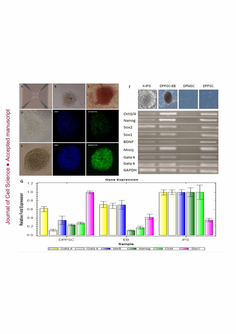
Jour
nal o
f Cel
l Sci
ence
Acc
epte
d m
anus
crip
t
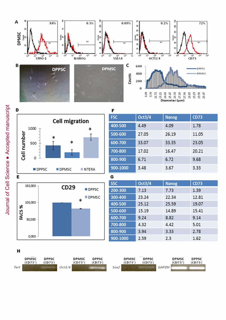
Jour
nal o
f Cel
l Sci
ence
Acc
epte
d m
anus
crip
t

Jour
nal o
f Cel
l Sci
ence
Acc
epte
d m
anus
crip
t

Jour
nal o
f Cel
l Sci
ence
Acc
epte
d m
anus
crip
t
Jour
nal o
f Cel
l Sci
ence
Acc
epte
d m
anus
crip
t
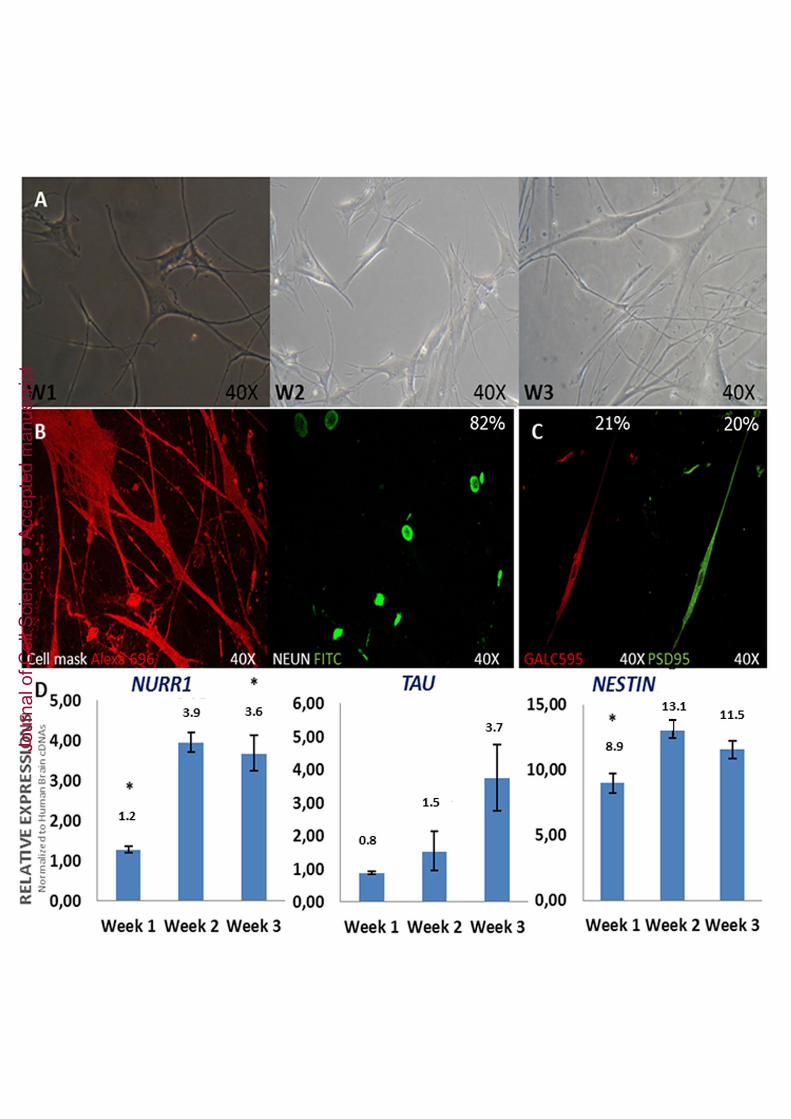
Jour
nal o
f Cel
l Sci
ence
Acc
epte
d m
anus
crip
t

Jour
nal o
f Cel
l Sci
ence
Acc
epte
d m
anus
crip
t

Jour
nal o
f Cel
l Sci
ence
Acc
epte
d m
anus
crip
t
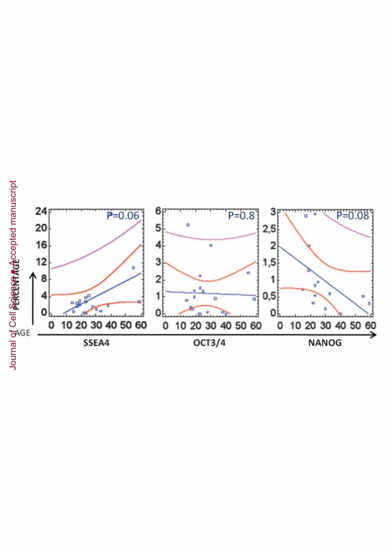
Jour
nal o
f Cel
l Sci
ence
Acc
epte
d m
anus
crip
t

Jour
nal o
f Cel
l Sci
ence
Acc
epte
d m
anus
crip
t

Jour
nal o
f Cel
l Sci
ence
Acc
epte
d m
anus
crip
t

Jour
nal o
f Cel
l Sci
ence
Acc
epte
d m
anus
crip
t
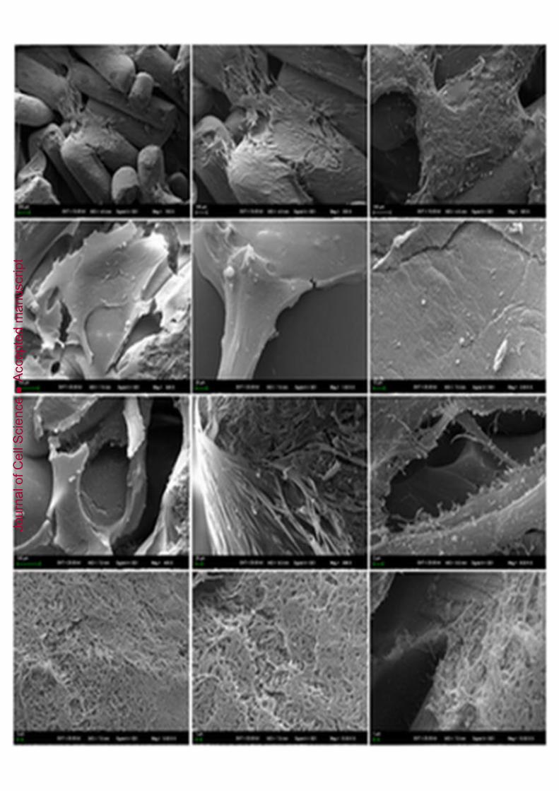
Jour
nal o
f Cel
l Sci
ence
Acc
epte
d m
anus
crip
t

Jour
nal o
f Cel
l Sci
ence
Acc
epte
d m
anus
crip
t

Jour
nal o
f Cel
l Sci
ence
Acc
epte
d m
anus
crip
t
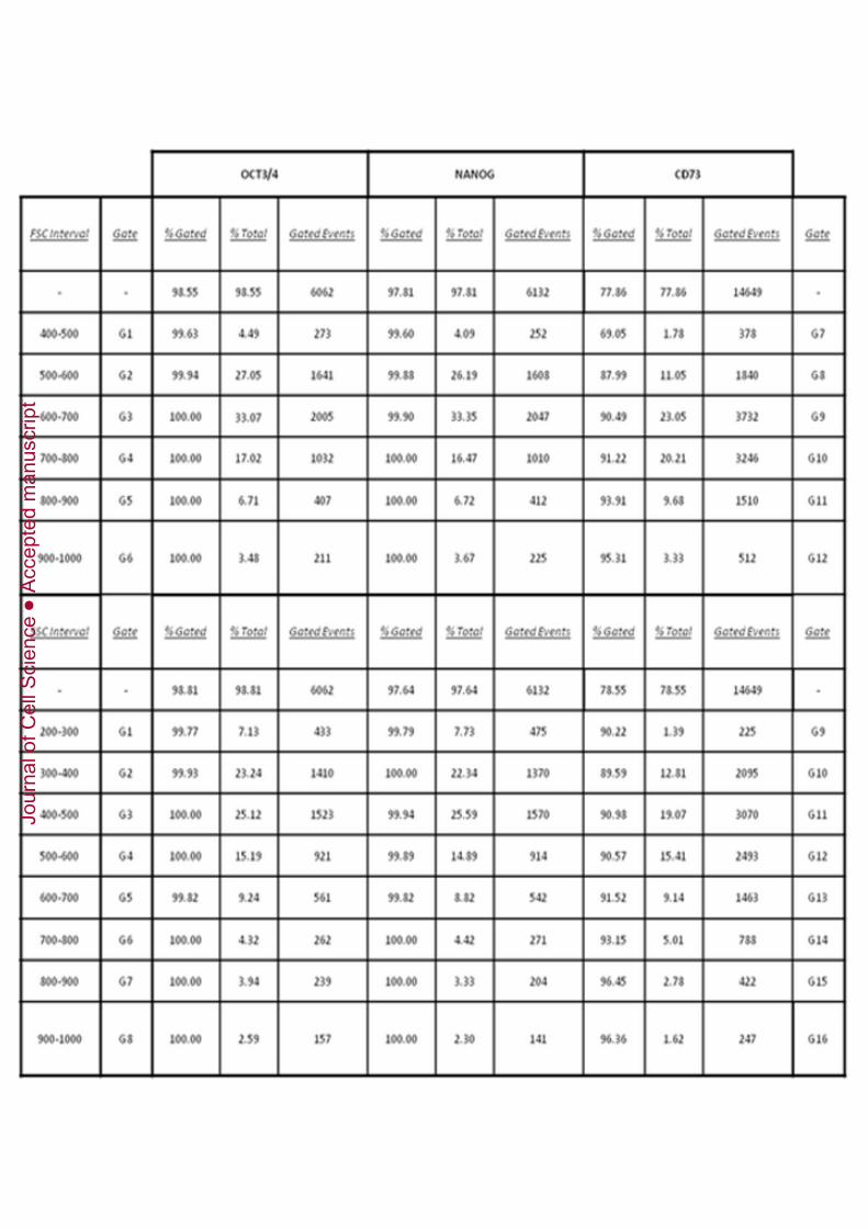
Jour
nal o
f Cel
l Sci
ence
Acc
epte
d m
anus
crip
t

Jour
nal o
f Cel
l Sci
ence
Acc
epte
d m
anus
crip
t
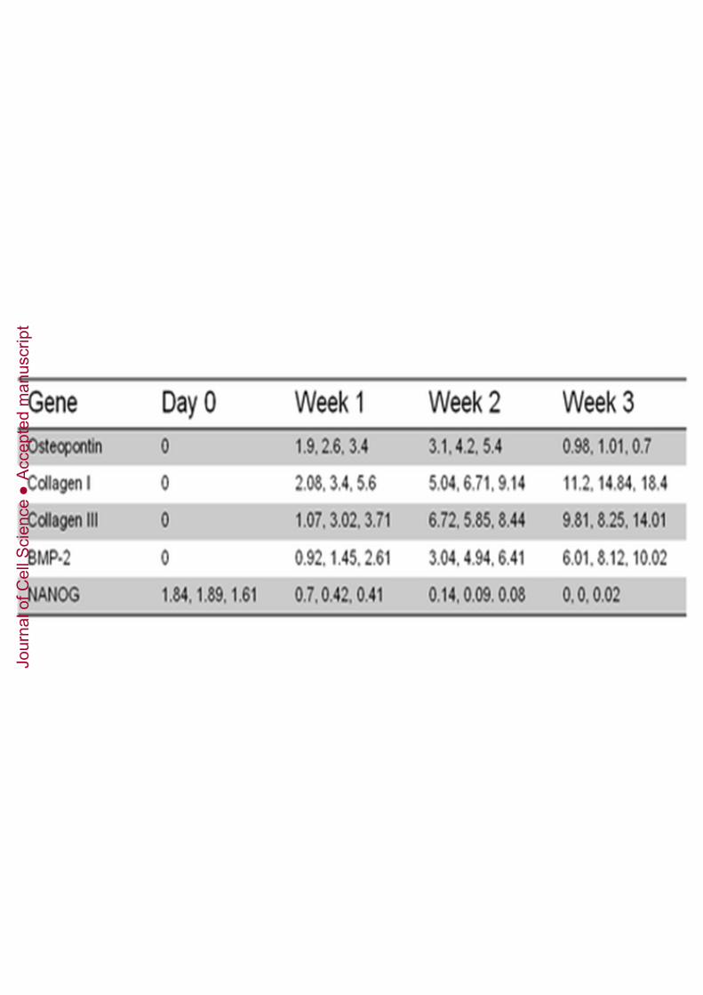
Jour
nal o
f Cel
l Sci
ence
Acc
epte
d m
anus
crip
t

Jour
nal o
f Cel
l Sci
ence
Acc
epte
d m
anus
crip
t
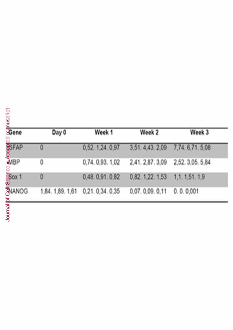
Jour
nal o
f Cel
l Sci
ence
Acc
epte
d m
anus
crip
t