dental implants
TRANSCRIPT
The Biology of Dental The Biology of Dental ImplantsImplants
James Q. Swift, DDSDivision of Oral and Maxillofacial Surgery
University of Minnesota


Concepts of Connective Concepts of Connective Tissue Healing Around Tissue Healing Around
ImplantsImplants Osseous Integration / Osseointegration
Fibrous Integration Fibro-osseous Integration


Bone HealingBone Healing
Phase I: Inflammatory Phase Phase II: Proliferative Phase Phase III: Maturation Phase

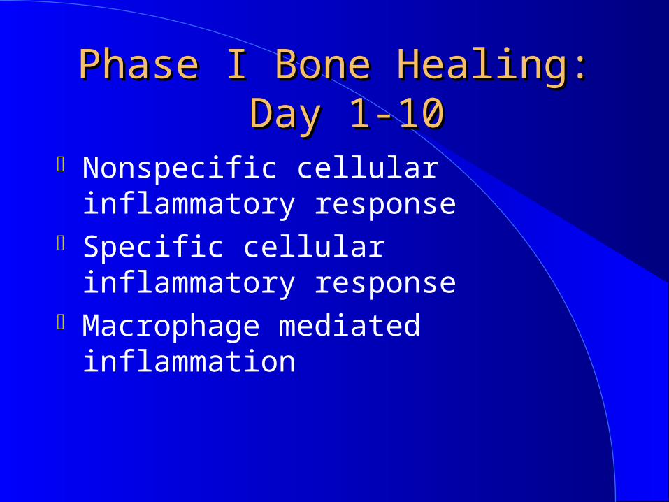
Phase I Bone Healing:Phase I Bone Healing: Day 1-10 Day 1-10
Absorption of plasma proteins Platelet aggregation and activation
Clotting cascade activation Cytokine release
Phase I Bone Healing:Phase I Bone Healing: Day 1-10 Day 1-10
Nonspecific cellular inflammatory response
Specific cellular inflammatory response
Macrophage mediated inflammation


Phase II Bone Healing:Phase II Bone Healing: Day 3-42 Day 3-42
Neovascularization Differentiation, proliferation and activation of cells
Production of immature connective tissue matrix
Phase III Bone Phase III Bone Healing:Healing:
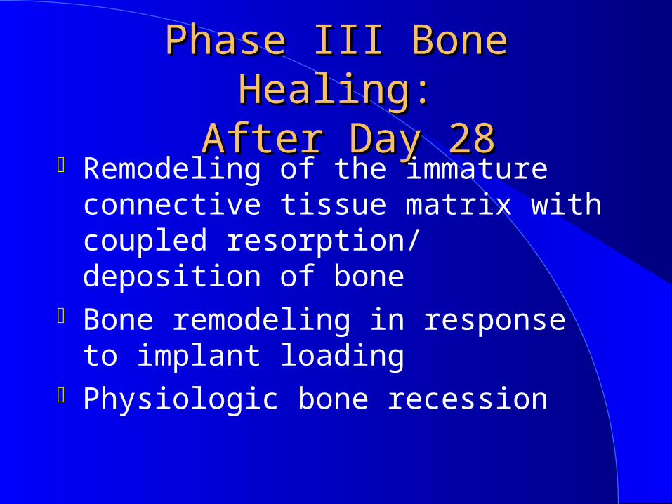
After Day 28 After Day 28 Remodeling of the immature connective tissue matrix with coupled resorption/ deposition of bone
Bone remodeling in response to implant loading
Physiologic bone recession
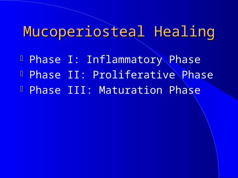
Mucoperiosteal HealingMucoperiosteal Healing
Phase I: Inflammatory Phase Phase II: Proliferative Phase Phase III: Maturation Phase

Phase I Mucoperiosteal Phase I Mucoperiosteal Healing Inflammatory Healing Inflammatory
Phase: Day 1-10Phase: Day 1-10 Platelet aggregation and activation
Clotting cascade activation Cytokine release Nonspecific cellular inflammatory response
Macrophage mediated inflammation

Phase II Mucoperiosteal Phase II Mucoperiosteal Healing Proliferative Healing Proliferative
Phase: Day 2-42Phase: Day 2-42 Neovascularization Differentiation, proliferation and activation of cells
Deposition of immature collagen, elastin, and ground substance

Phase III Mucoperiosteal Phase III Mucoperiosteal Healing Maturation Healing Maturation Phase: After Day 21Phase: After Day 21
Connective Tissue Remodeling
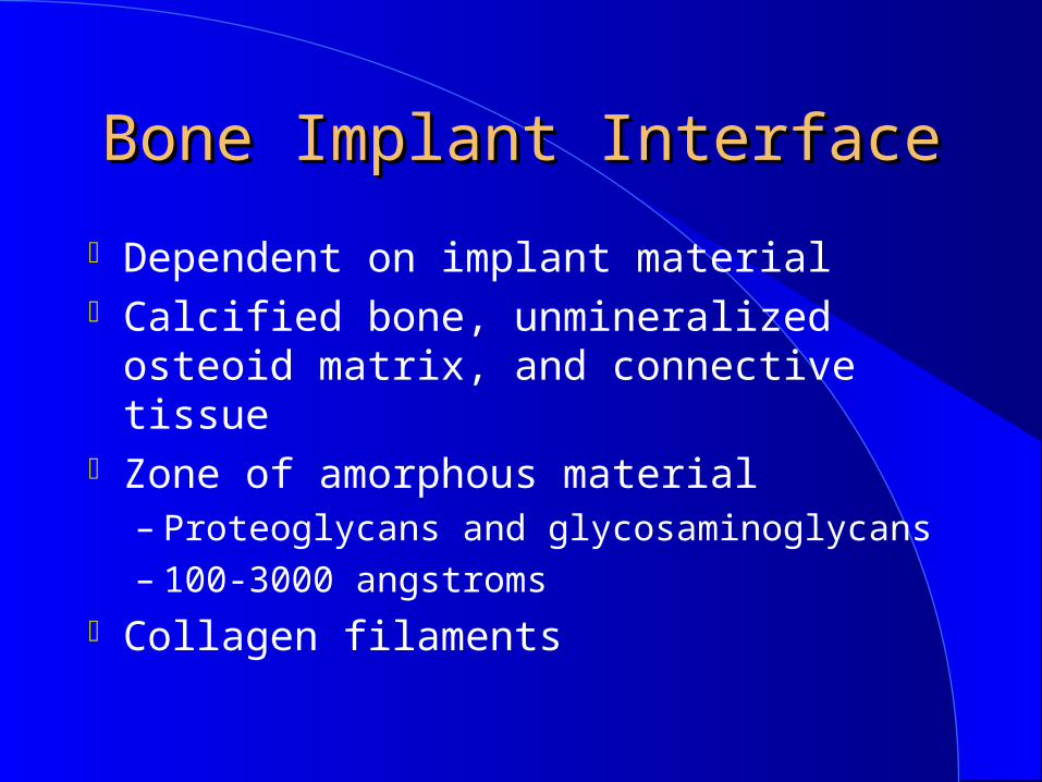
Bone Implant InterfaceBone Implant Interface
Dependent on implant material Calcified bone, unmineralized osteoid matrix, and connective tissue
Zone of amorphous material– Proteoglycans and glycosaminoglycans– 100-3000 angstroms
Collagen filaments

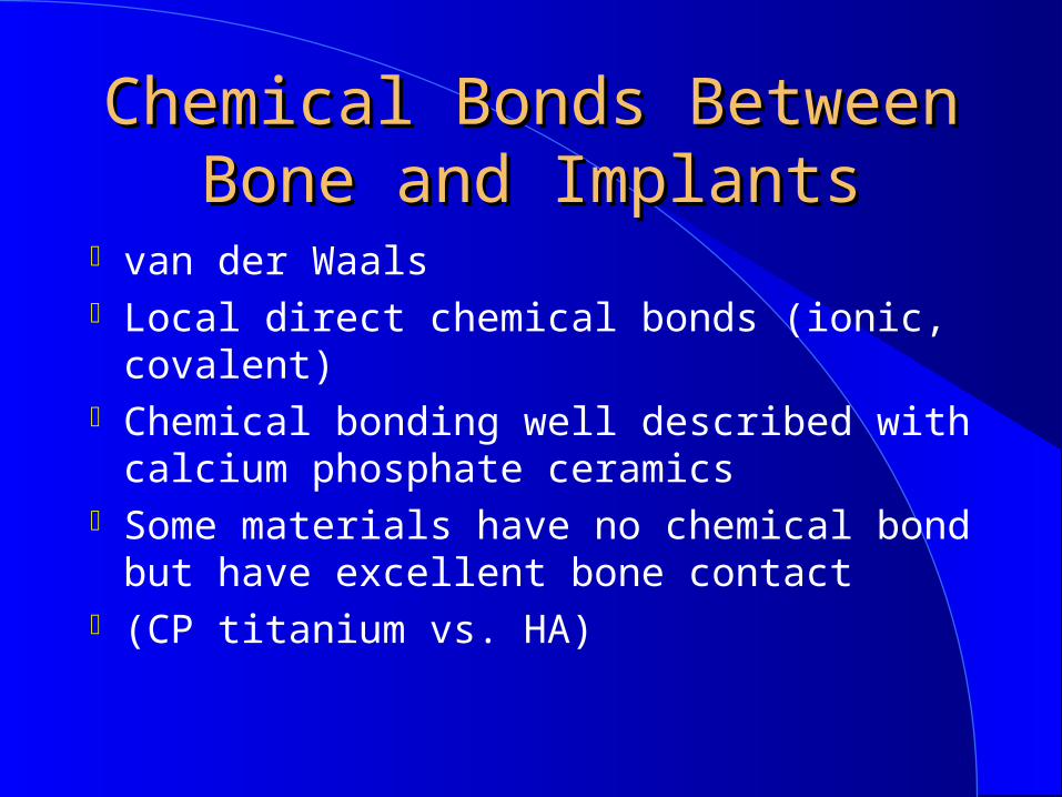
Chemical Bonds Between Chemical Bonds Between Bone and ImplantsBone and Implants
van der Waals Local direct chemical bonds (ionic, covalent)
Chemical bonding well described with calcium phosphate ceramics
Some materials have no chemical bond but have excellent bone contact
(CP titanium vs. HA)


Factors Affecting Factors Affecting HealingHealing
Surgical technique Premature loading Surgical fit Extraction sites

Factors Affecting Factors Affecting HealingHealing
Peri-implantitis Bone quality Staged healing


Logic CONTROLLogic CONTROL
-10
0
10
20
30
40
50
60
0 200 400 600 800

ExperimentExperiment ControlControl12 Pigs12 Pigs 6 Pigs6 Pigs
5 Months 5 Months Controlled Controlled
LoadingLoading
1 Month1 Month
HealingHealing
4 Month4 Month2 Month2 Month
5 Months 5 Months Non-LoadingNon-Loading
HealingHealing
(4)(4)
(4)(4)(4)(4)
1 Month1 Month
4 Month4 Month2 Month2 Month (4)(4)
(4)(4)(4)(4)
3 Healing Groups
One Month Healing
Four Months Healing
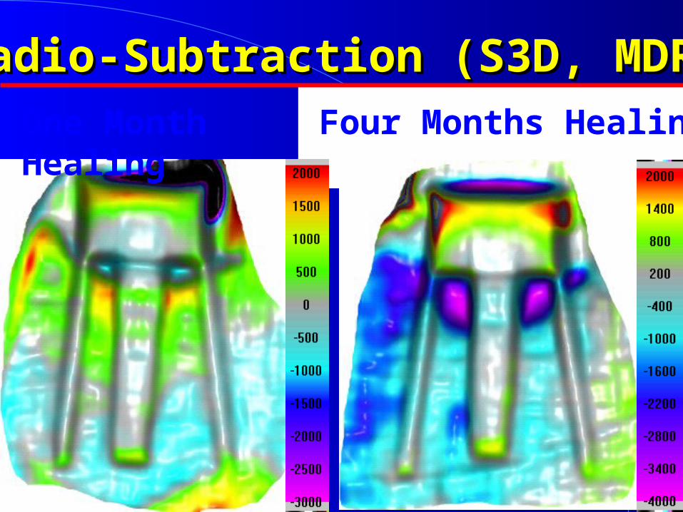
Radio-Subtraction (S3D, MDRCBB)Radio-Subtraction (S3D, MDRCBB)

One MonthTwo MonthsFour Months
Micro-CT ImagesMicro-CT Images
One MonthTwo MonthsFour Months
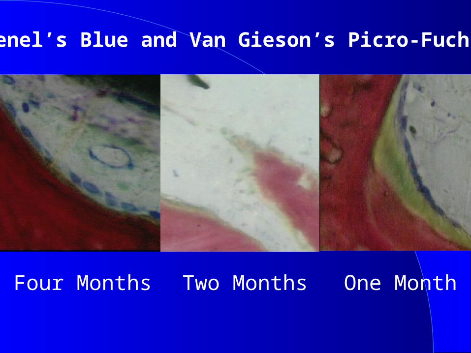
Stevenel’s Blue and Van Gieson’s Picro-Fuchsin Stain

Issues Relating to Issues Relating to Physical Status of Physical Status of
RecipientRecipient Nutritional status Age Hematologic issues

Issues Relating to Issues Relating to Physical Status of Physical Status of
RecipientRecipient Diabetes Corticosteroids Radiation

51 year old man51 year old man
CC- “I ‘ve been referred for evaluation and treatment for a growth in my jaw”
HPI– 2 month duration– mobile teeth– facial swelling

51 year old man51 year old man
PMH– Hypertension-well controlled
PDH Imaging findings
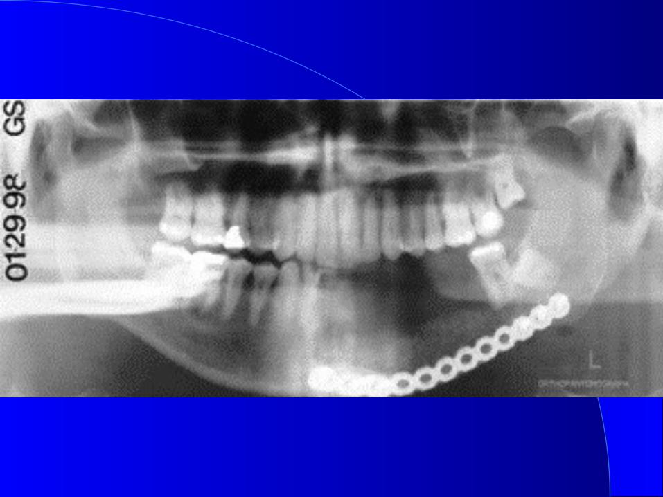
– 4 cm x 2 cm radiolucent lesion left mandibular body causing tooth displacement


51 year old man51 year old man
Problem list– Radiolucent lesion– Teeth vital– Buccal plate perforation– No paresthesias– Differential diagnosis
ameloblastoma central giant cell lesion
odontogenic myxoma


51 year old man51 year old man
Treatment plan– Incisional biopsy for histologic typing
– Prosthodontic consultation– Definitive imaging (CT scan)
Biopsy results– Acanthomatous ameloblastoma

51 year old man51 year old man
Treatment– Arch bars/surgical stent construction
– Transcervical approach for exposure and lateralization of IAN/Mental nerve
– Transoral marginal resection preserving inferior border
– Reconstruction plate applied for stabiliation


51 year old man51 year old man
Treatment– Healing interval 6 weeks– Mandibular reconstruction of 4cm x 2cm x1.5 cm defect with transcervical approach and AICBG (PCMB)
– Healing interval 3 months


51 year old man51 year old man
Treatment– Placement of 5 osseointegrated dental implants left parasymphysis and body with prosthodontic guidance
– Transoral removal of reconstruction plate
– Healing interval 5 months


51 year old man51 year old man
Treatment– Second stage implant surgery with temporary healing abutments placed and preservation of attached gingiva
– Prosthodontic reconstruction with fixed detachable prosthesis


51 year old man51 year old man
Results– Minimal paresthesia– No recurrence of lesion– Fixed prosthesis with lip support and excellent masticatory function
51 year old man51 year old man
Rationale for treatment– Alternative reconstructive surgery harbors greater potential morbidity
– Fixed partial denture not ideal span too great
– Removable partial denture not ideal blunting of vestibular and sulcular depths
16 year old woman16 year old woman
CC- “I was in a car accident and hit the steering wheel.”
HPI– Patient referred from emergency department
– Injury had occurred > 16 hours prior to presentation
– No other injuries identified (C-spine negative)

16 year old woman16 year old woman
PMH– Non contributory
PDH– Had just completed orthodontic therapy and was in retention phase


16 year old woman16 year old woman
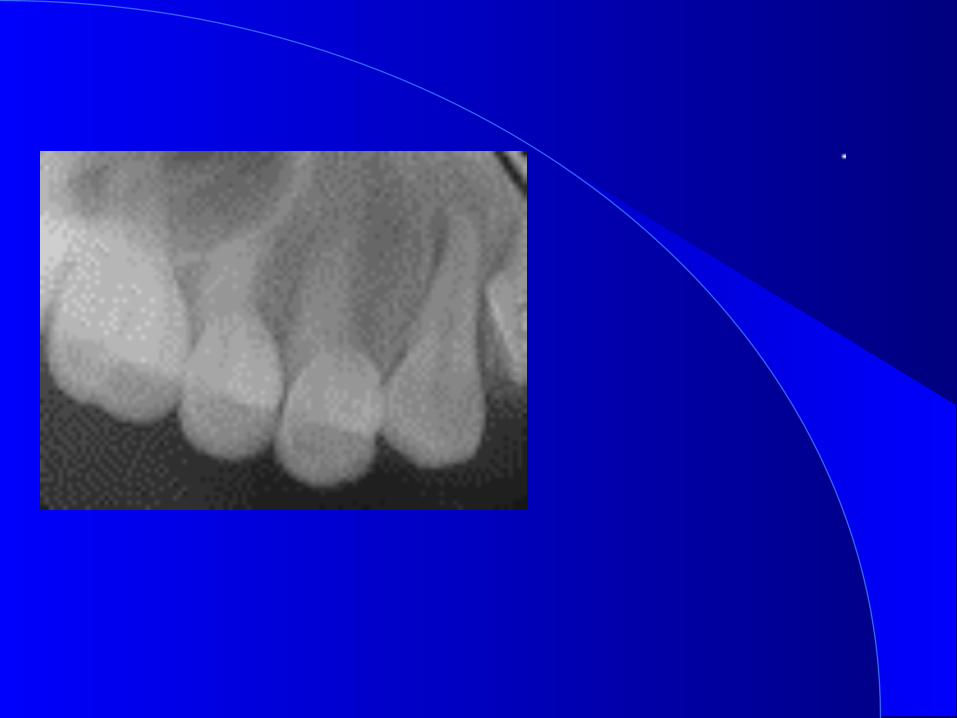
Clinical findings– Lip laceration– # 6 , 7 and 8 not visible– Palpable step defect of alveolus– Coagulum in socket sites of 6 & 7


16 year old woman16 year old woman
Imaging findings– #6 & 7 superiorly displaced and rotated
– #8 absent– 9 abnormalities in the PDL– Radiolucent transverse fracture line superior to apices of anterior teeth
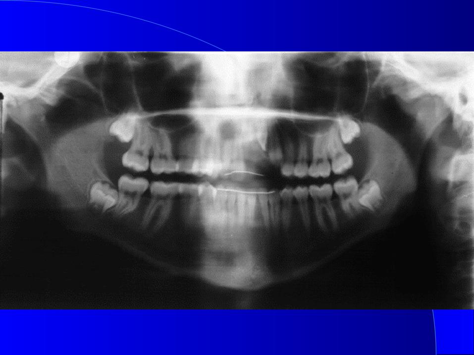


16 year old woman16 year old woman
Diagnosis– Trauma to the anterior maxilla
Lip abrasion/ laceration Maxillary alveolar fracture Avulsion # 8 Traumatic displacement # 6, 7, & 9 Incisal edge fractures # 6, 7 & 9

16 year old woman16 year old woman
Treatment– IV sedation– Surgical exploration of injured area with preservation of alveolar bone
– Reduction of alveolar fractures– Repositioning of #6, 7 & 9 into normal dental arch relationship and stabilization with splint and pontic placed
– Closure of laceration and surgical wound


16 year old woman16 year old woman
Treatment– Referral to orthodontist
Replaced stabilization wire with arch wire
– Referral to endodontist RCT # 6 & 7


16 year old woman16 year old woman
Referral to prosthodontist– Consultation for dental reconstruction
– Options FPD RPD Implant # 8 area
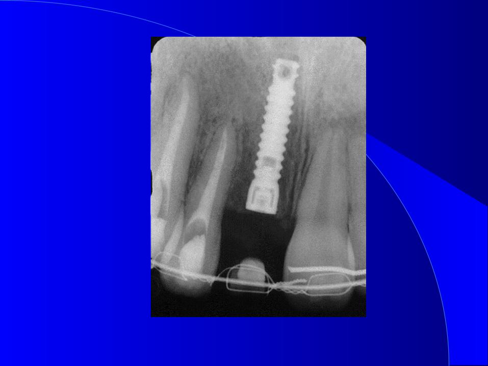

16 year old woman16 year old woman
Treatment– After stabilization for 4 months
Implant inserted # 8 area (internal hex)
Presevation of attached tissue Preservation of bone Osteotome technique

16 year old woman16 year old woman
Treatment– Integration time 5 months– Second stage surgery with preservation of soft tissue
– Temporary healing abutment and temporary crown
– Implant crown constructed


16 year old woman16 year old woman
Results– Acceptable cosmetic outcome
Rationale for treatment– Preservation of natural teeth, bone and soft tissue
– FPD would require preparing endodontically treated adjacent teeth with uncertain prognosis
– Patient and parents desires

Thank youThank you