dental health and need for non-operative treatment among
TRANSCRIPT

Faculty of Health Sciences
Department of Clinical Dentistry
Dental health and need for non-operative treatment
among 16-year-olds in Northern Norway
—
Ioanna Dallari Jacobsen
A dissertation for the degree of Philosophiae Doctor – February 2018

1

2
This thesis is dedicated to my beloved parents, who have always loved me unconditionally and
whose good examples have taught me to work hard for the things that I aspire to achieve.
We do not know a truth without knowing its cause.
Aristotle, Nicomacheian Ethics, I.1.

3
AKNOWLEDGEMENTS
The work included in this thesis has been carried out during my years as PhD student at the
Department of Clinical Dentistry at UiT The Arctic University of Norway. I am grateful to the
Dental faculty for the research facilities that have been at my disposal. I want also to express
my gratitude to all those who helped me in this project, with special thanks to:
My supervisors. Without continuous encouragement and support and endless patience from
my supervisors, what I have achieved could never have happened.
Professor Claes-Göran Crossner for introducing me to the Fit Futures studies his encouraging
and inspiring guidance. I am greatly indebted to him for all the support, but also for the
freedom to work independently.
Professor Harald M. Eriksen, for his patience and for sharing his knowledge and expertise
with me during long, invaluable hours discussing my drafts.
Department leader Dr Christer Ullbro, for his enthusiasm, positive attitude, valuable advices
and support.
Professor Ivar Espelid for being available and, more specifically, for his insightful response
on various statistical issues.
My co-author, Professor Anne Bjørg Tveit, for offering opportunities and being supporting.
My colleagues, former PhD candidates, doctors Linda, Lina, Natalia, for their support,
inspiration, good advice.
My co-authors, Aida Mulic, Andreas Schmalfuss, Øystein Fredriksen, for their valuable
support.
My former and current colleagues at the Department of Clinical Dentistry, for informative and
inspiring conversations and discussions, and other pleasant gatherings.
The administrative staff at the Department of Clinical Dentistry for being smiling, positive
and solution oriented.
The dental assistants and other staff at the University Dental Clinic, for working hard for the
success of Fit Futures project. It was a very positive experience to work with them.
My family and friends; to those who have been there with me and for me and in various ways
contributed to this thesis. There is not enough space, to acknowledge everyone to the extent
they deserve.

4
CONTENTS
CONTENTS .............................................................................................................................. 4
LIST OF ABREVIATIONS ....................................................................................................... 7
LIST OF PAPERS .................................................................................................................. 9
1. ABSTRACT ........................................................................................................................ 10
2. INTRODUCTION ............................................................................................................... 11
Dental caries ......................................................................................................................... 11
Risk factors ....................................................................................................................... 12
International trends ........................................................................................................... 13
Norwegian trends .............................................................................................................. 14
Enamel caries .................................................................................................................... 15
Treatment options ............................................................................................................. 15
Quality and longevity of fillings ....................................................................................... 18
Dental erosion ....................................................................................................................... 20
International trends ........................................................................................................... 22
Norwegian trends .............................................................................................................. 23
Treatment options ............................................................................................................. 23
3. AIMS .................................................................................................................................... 24
4. MATERIALS AND METHODS ........................................................................................ 24
Study sample ......................................................................................................................... 24
Clinical examination ............................................................................................................. 25
Study design ......................................................................................................................... 25
Paper I ............................................................................................................................... 25
Paper II .............................................................................................................................. 26
Paper III ............................................................................................................................ 26
Registration of variables ....................................................................................................... 26
Paper I and II ..................................................................................................................... 26
5
Paper III ............................................................................................................................ 28
Calibration ............................................................................................................................ 28
Paper I and II ..................................................................................................................... 28
Paper III ............................................................................................................................ 29
Data Analysis ........................................................................................................................ 29
Ethical approval .................................................................................................................... 30
5. RESULTS ............................................................................................................................. 30
Paper I ................................................................................................................................... 30
Dependent variable - dentinal caries ................................................................................. 30
Independent variables - Bivariate model .......................................................................... 30
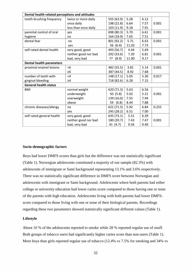
Paper II ................................................................................................................................. 35
Proximal enamel lesions ................................................................................................... 35
Need for non-operative caries treatment ........................................................................... 36
Quality of dental restorations ............................................................................................ 36
Paper III ................................................................................................................................ 37
Prevalence, distribution and severity of dental erosive wear ............................................ 37
6. GENERAL DISCUSSION ................................................................................................... 40
Methodological considerations (Paper I) .............................................................................. 40
Representativity ................................................................................................................ 40
The questionnaires ............................................................................................................ 41
Ethnicity ............................................................................................................................ 41
Caries registration ............................................................................................................. 42
Calibration ......................................................................................................................... 43
Statistics ............................................................................................................................ 43
Methodological considerations (Paper II) ............................................................................ 43
Proximal enamel lesions ................................................................................................... 43
Active – arrested lesions ................................................................................................... 43

6
Quality of fillings .............................................................................................................. 44
Methodological considerations (Paper III) ........................................................................... 44
Erosive wear registration .................................................................................................. 44
The use of clinical photos ................................................................................................. 45
Calibration ......................................................................................................................... 45
Ethical considerations ........................................................................................................... 46
Main findings ........................................................................................................................ 47
Caries prevalence (Paper I) ............................................................................................... 47
Ethnicity and caries (Paper I) ............................................................................................ 48
Lifestyle factors and dental caries (Paper I)...................................................................... 49
Proximal enamel lesions (Paper II) ................................................................................... 49
Quality of restorations (Paper II) ...................................................................................... 49
Dental erosions (Paper III) ................................................................................................ 50
7. CONCLUDING REMARKS ............................................................................................... 51
REFERENCES ......................................................................................................................... 53

7
LIST OF ABREVIATIONS
ANOVA, Analysis of variance
BMI, Body Mass Index
BW, Bite-wing radiographs
CAPP, Country / Area Profile Project
CPITN, Community Periodontal Index of Treatment Needs
DEW, Dental erosive wear
DMFT/S, Decayed Missing and Filled teeth / surfaces
DS, Decayed surfaces
DFS, Decayed and Filled surfaces
EUTRO, Tromsø Study Database
FDI, World Dental Federation
FS, Filled surfaces
ICDAS, International Caries Detection and Assessment System
LCD, Liquid-Crystal Display
NOCT, Non-operative caries treatment
NSD, Norwegian Social Science Data Services
OR, Odds ratio
PDS, Public Dental Service
PEL, Proximal enamel lesions
REK, Regional Committee for Medical Research Ethics
USPHS, United States Public Health Service
VEDE, Visual Erosion Dental Examination

8
WHO, World Health Organization

9
LIST OF PAPERS
This thesis consists of the following three papers, referred to in the text by the corresponding
roman numerals.
I. JACOBSEN, I., ERIKSEN, H., ESPELID, I., SCHMALFUSS, A., ULLBRO, C.
& CROSSNER, C. (2016). Prevalence of dental caries among 16-year-olds in
Troms County, Northern Norway. Swed Dent J, 40(2):191-201.
II. JACOBSEN, I., CROSSNER, C., ERIKSEN, H., ESPELID, I. & ULLBRO, C.
Need of non-operative caries treatment in 16-year-olds. Accepted for publication
in Eur Arch Paediatr Dent (08.02.2017).
III. MULIC, A., FREDRIKSEN, Ø., JACOBSEN, I., TVEIT, A., ESPELID, I. &
CROSSNER, C. (2016). Dental erosion: Prevalence and severity among 16-year-
old adolescents in Troms, Norway. Eur J Paediatr Dent, 17(3):197-201.

10
1. ABSTRACT
Epidemiological data have disclosed a considerable reduction in caries prevalence among
children and adolescents in Western countries including Norway for over 40 years.
Concomitantly, enamel caries has received increased focus in order to give a better picture of
the complete need for dental treatment, non-operative as well as operative. More recently,
dental erosive wear seems to be a growing problem among the same age group.
The aims of the present thesis were:
• to determine the prevalence of dentinal caries and the variation in caries prevalence
related to selected independent variables (sociodemography, lifestyle) in a sample of 869 16-
year-olds from Northern Norway.
• to estimate the prevalence of proximal enamel lesions and the need for non-operative
caries treatment.
• to record the quality of dental restorations.
• to study the prevalence, distribution and severity of dental erosion.
The thesis is based on an oral- and general health cross-sectional study (Fit Futures), with an
attendance rate of 90%.
The DMFT/S-values were 4.2 / 6.1. The final multivariate regression analysis indicated that
use of smokeless tobacco, dental fear, self-rated dental health and proximal enamel caries
showed a strong independent association with prevalence of dentinal caries. Only 6 % of the
16-year-olds were completely caries-free. Eighty-four per cent of the participants presented
with proximal enamel lesions. A majority of them had either previously restored teeth (35%)
or both restored teeth and untreated dentinal caries lesions (34%). Over one third (35%) of the
participants with fillings presented with at least one restoration below acceptable quality level.
More than one third (38%) of the adolescents showed erosive wear on at least one tooth
surface, either limited to the enamel (18%), or extending into the dentine (20%).
Dental caries and erosive wear are challenging conditions among North Norwegian 16-year-
olds. The high prevalence of early signs of disease (proximal enamel lesions and cuppings)
entails a need for non-operative treatment interventions. The DMFS-score and the high
number of 16-year-olds with restorations in need of repair or replacement further indicate the
importance of a non-operative treatment strategy in order to reduce the need of traditional
restorative care.

11
2. INTRODUCTION
Dental caries
Dental caries is a chronic disease involving the localized destruction of dental hard tissues
(Fejerskov, 1997). Both the disease process and the resulting lesions are described by the term
“caries” (Koch and Poulsen, 2009). Cariogenic bacteria produce organic acids during
metabolism of fermentable carbohydrates in dental biofilm on the tooth surface. These acids
dissolve enamel and dentine minerals by reducing pH values locally (demineralization) and
this process may result in cavitation (Selwitz et al., 2007, Featherstone, 2008).
Demineralization can be arrested or reversed through precipitation of mineral ions (calcium,
phosphate and fluoride) derived from oral fluids and deposited in the demineralized tooth
structures. This process is called remineralization. Under healthy conditions, there is a
dynamic balance between demineralization and remineralization, maintaining a status quo at
the tooth surface. When demineralization outweighs remineralization, cavity formation is the
end result (Selwitz et al., 2007, Featherstone, 2008). In case of caries progression, the process
is usually slow, due to successive cycles of demineralization and remineralization (Takahashi
and Nyvad, 2011). The currently prevailing theory about the role of biofilm bacteria in the
etiology of dental caries and the demineralization-remineralization balance of the caries
process, is the ecological caries hypothesis (Marsh, 2003, Marsh, 2006), as extended by
Takahashi and Nyvad (Takahashi and Nyvad, 2008, Takahashi and Nyvad, 2011). According
to the extended caries ecological hypothesis, the changes in the demineralization /
remineralization status of caries lesions are associated with shifts in the composition of the
microflora caused by bacterial acid production. Acid production causes changes in the
composition of the oral microflora from a dynamic stability stage (dominated by non-mutans
streptococci and actinomyces), via acid induced adaptation and selection (dominated by low
pH non-mutans bacteria and actinomyces), to an aciduric stage (dominated by mutans
streptococci and other mutans bacteria, including lactovacilli and bifidobacterium). Whithin
this theory, a broad range of acidogenic / aciduric bacteria and not only the mutans
streprococci are involved in the caries process. Mutans streptococci are not the causative
factor per se, but the result of the microbial acid production.
Biofilm has a fundamental role in dental caries. Biofilm is structurally and metabolically
organized communities of interacting species on the oral surfaces in a dynamic equilibrium
with their environment (mutualistic symbiosis). The microbial homeostasis is very sensitive to
changes in the environment of the mouth and the lifestyle of the individual. Shifts in the
12
balance of the normal resident microbiota lead to dysbiosis. The microbial acid production
which has as result the carious process perturbs the mutualistic symbiosis in the microbial
ecosystem (Takahashi and Nyvad, 2011). Disease can be prevented by influencing the factors
that promote dysbiosis, such as saliva flow and buffering capacity, diet, oral hygiene, lifestyle
and the immune system (Marsh et al., 2015a). It is possible to intervene in the caries process
and arrest or reverse the progress of the caries lesion through environmental control of the
microflora. Preventing acidification of the dental biofilm (through biofilm control, sugary diet
control, pH-neutralization) may be more effective method than adopting antimicrobial
strategies against mutans streptococci (Takahashi and Nyvad, 2011, Marsh et al., 2015b). The
action of fluoride in the caries control is essential, as it both reduces demineralization and
promotes remineralization (Fejerskov et al., 1981, Groeneveld, 1985, Groeneveld et al., 1990,
Singh and Spencer, 2004).
The dental caries process is a continuum of disease states ranging from subclinical changes to
dentinal involvement with or without cavitation (Selwitz et al., 2007). A large number of very
early initial lesions in a dynamic state of progression-regression remain subclinical (Pitts,
2004b). The choice of diagnostic cut-off between sound and diseased tooth surfaces
determines the number of carious lesions detected (Pitts, 2004b).
Risk factors
Dental caries is a multifactorial disease. Concerning the microbial factor, mutans streptococci
have been considered as the main microbiological causative factor of caries. However, such a
role has not been extensively verified (Bowden, 1997, Aas et al., 2008). Caries occurrence can
neither be accurately anticipated in a person or at a tooth site, nor predicted following
presence of a particular bacterium. Known risk factors are previous caries experience; sugar
habits, amount of fluoride exposure, salivary flow, and socioeconomic status (Selwitz et al.,
2007). Socioeconomic factors, knowledge, behavior and attitudes are more distant
determinants of the carious process. Moreover, a social gradient has been shown in general
and oral health, present and persistent in different countries and contexts, indicating the
impact of broad social underlying factors in determining and shaping individual behaviors
(Watt, 2012).
Dental caries is a “transmissible” disease. Cariogenic bacteria are transmitted early in life
from mother / caregiver to child, and colonize teeth upon eruption (Featherstone, 2008, da
Silva Bastos et al., 2015). Maternal factors seem to influence bacterial acquisition, while

13
colonization may be mediated by oral health behaviors and practices and feeding habits
(Leong et al., 2013).
International trends
Most epidemiological data about caries come from studies of children and adolescents or
older people (Broadbent et al., 2013). Available data on caries prevalence at the international
level concern the indicator group of 12-year-olds and are collected by the World Health
Organization (WHO) within the Country / Area Profile Project (CAPP) (WHO, 2016). A
number of studies from many developed countries confirm the declining trend in caries
prevalence and in mean caries experience for permanent teeth in children and adolescents
(Marthaler, 2004, Hugoson et al., 2005, Christensen et al., 2010, Widström et al., 2011), and
also in adults (Norderyd et al., 2015a). This is a result of public health measures and well-
organized prevention, better living conditions and improved oral hygiene practices (Petersen
et al., 2005, Fontana et al., 2010). The change in the diagnostic and treatment criteria of caries
may also have played a role in this decline (Nadanovsky and Sheiham, 1995). The widespread
use of fluoride dentifrice may be the single most important reason for the decline (Bratthall et
al., 1996). The role of sugar in the diet seems to be weaker in the era of widespread fluoride
exposure (Burt and Szpunar, 1994, Burt and Pai, 2001, Mejàre et al., 2014).
However, not all share the positive changes in oral health. Despite the significant caries
reduction observed in high-income countries, disparities remain and poor and disadvantaged
population groups still present high levels of caries (Selwitz et al., 2007, WHO, 2012,
Schwendicke et al., 2015). The WHO reports that worldwide, 60-90% of school children and
nearly 100% of adults have experienced dental caries, while about 30% of people between 65
and 74 years old are edentulous (WHO, 2012). The prevalence of caries in later adult age
remains substantial. Demography is changing, people live longer and number of older people
retaining their teeth has increased. In USA, 91% of dentate adults older than 20 years have
caries experience (Fontana et al., 2010). Marcenes et al. (2013) found a 35% global
prevalence of untreated caries for all ages combined. In 2010, untreated caries in permanent
teeth was the most prevalent medical condition affecting 2.4 billion people worldwide
(Kassebaum et al., 2015). Bernabé and Sheiham (2014a) in their extensive analysis of age,
period and cohort trends of caries in permanent teeth in four developed countries (USA, UK,
Sweden and Japan) showed that there is still a gradual increase in DMFT/S-scores in the adult
population due to untreated caries and neglect of oral health promotion in adult life. They
generalized their findings to a larger set of developed and developing countries (Bernabé and

14
Sheiham, 2014b). Broadbent et al. (2013) also found (Dunedin study) that dental caries
disease continues in adulthood, and that rates of dental caries remain relatively constant over
time, with an average increase of 0.8 surfaces per year.
The comparison of international data about caries has many limitations. Diagnostic criteria
vary from one study to another and researchers collecting data are not calibrated. In addition,
caries lesion measurements in the various studies have variable diagnostic cut-offs, from
including all forms of lesions to only dentinal cavitation. Dental caries prevalence is mostly
measured at dentinal caries diagnostic threshold (Fontana et al., 2010) and the caries
prevalence shown may be underestimated to a certain degree, due to non-inclusion of
precavitated lesions and other factors inherent to the use of the DMFS index (Lagerweij and
van Loveren, 2015). Additionally, the available data from the WHO database which concern
the caries status of 12-year-olds in different countries and regions of the world cannot be used
for comparisons, due to variation in the internal and external validity and in the year of their
collection (WHO, 2012). Finally, the caries prevalence for this age group may be
underestimated in most children as the second molars are not erupted and premolars and
canines have been present only for a short period in the mouth (Meyer-Lueckel et al., 2013).
Norwegian trends
In Norway, statistical data on caries experience recorded at dentin level (DMFT) for index age
groups 5, 12 and 18 have been collected annually at county level since 1985 (Lyshol and
Biehl, 2009, Wilberg, 2012). These data confirm the internationally reported decrease in
caries prevalence. This positive development is, however, not shared by all. Dental health
varies with socio-economic background and dental caries still remains high in risk groups
(Norwegian Institute of Public Health, 2010). Parental migration and immigrant background
are associated with higher risk for caries in children and adolescents (Skeie et al., 2005,
Gimmestad et al., 2006, Wigen et al., 2011, Wigen and Wang, 2012). Furthermore, higher
caries prevalence and severity of caries have been observed among children and adolescents
in Northern Norway, in particular Finnmark, compared to the rest of the country (Helse- og
omsorgsdepartementet, 2007, Lyshol and Biehl, 2009, Widström et al., 2010, Adekoya and
Brustad, 2012, Skeie et al., 2012). Possible explanations for these findings are the high
frequency of rural population in this area, the large proportion of indigenous Sami population
living in Finnmark and the low dental service availability due to low dentist density (Adekoya
and Brustad, 2012). Compiled national data on caries among adolescents with Sami
background are, however, lacking.

15
Enamel caries
Lesions limited to the enamel constitute a considerable part of all carious lesions (Martignon
et al., 2010, Skeie and Klock, 2014). Alm et al. (2007) claim that over 80% of proximal caries
lesions diagnosed in adolescents are in the enamel only, indicating that the reduction in
prevalence of caries is overestimated and that the burden and the need for treatment of the
caries disease is underestimated (Amarante et al., 1998, Nyvad et al., 1999, Schwendicke et
al., 2014). Hugoson and Koch (2008) found that 80-90% of the proximal lesion in age groups
20-50 years were limited to enamel. As a consequence, valid caries diagnosis in populations
with low caries prevalence and slow caries progression may need more sensitive diagnostic
criteria including enamel lesions (Nyvad et al., 1999, Pitts, 2004a). As the prevalence of
dentinal caries has declined, enamel caries has received increased focus in order to give a
more comprehensive picture of dental heath in children and adolescents and consequently a
better picture of the complete need for dental treatment including non-operative as well as
operative treatment (Isaksson et al., 2014). The choice of diagnostic cut-off between sound
and diseased tooth substance determines the number of carious lesions detected and recorded,
and by choosing a cut-off at the level of dentinal cavitation and recording only dentinal
lesions, the caries disease is underestimated (Pitts, 2004b). Early detection and treatment of
caries lesions preserves more dental tissues and is compatible with the principles of the
minimally invasive dentistry (Araújo et al., 2014). Treatment objectives for enamel lesions are
to slow down, arrest or reverse the progression of the lesions by non-operative treatment
procedures and thereby reduce the need for restorative treatment (Ekstrand and Christiansen,
2005, Hausen et al., 2007). It seems that, if caries is controlled during childhood and
adolescence, the benefits are maintained later in life, as most lesions develop before the age of
20 years, as shown by Crossner and Unell (2007) and Norderyd et al. (2015a). Enamel caries
is not recorded and monitored in the Official Statistics of Norway (Skeie and Klock, 2014).
Treatment options
Caries treatment has traditionally been associated with restorations. The term “treatment”
should be used with caution for caries, as “dental caries” is a dynamic process without a
generally agreed start point. In the past, treatment of dental caries was symptomatic, aiming to
relieve symptoms and restore function. The treatment included removal of the carious tissues
and restoration of the deriving cavity. Modern dentistry has moved away from operative
intervention, towards a causal, biologically grounded approach to caries management, based
on prevention and preservation of the dental tissues (Selwitz et al., 2007). This shift
16
happened following considerations of tooth integrity – avoidance of the retreatment spiral -,
but also cost, and the fact that prevention proved effective (Tyas et al., 2000, Pitts, 2004a,
Qvist, 2012, Schwendicke et al., 2014). Additionally, due to slow caries progression in most
cases (Mejàre et al., 2004), unnecessary invasive treatment can be delayed considerably.
Modern treatment philosophy focuses on caries control through elimination of the causes of
the disease, by altering the unfavorable oral milieu and by restoring the ecological equilibrium
in the oral cavity. Initiation and progression of the disease can thus be controlled lifelong for
individual patients (Selwitz et al., 2007). The environmental control of the microflora
involves mechanical biofilm control, control of sugary diet and pH-neutralizing techniques.
This approach necessitates accurate diagnosis of any disease or lesions at individual
(etiological risk factors) and tooth (location, severity, activity of lesion) level. Disease
prevention, just-in-time restoration, choice of minimally invasive restorative procedures, and
prevention of recurrence are essential measures and should be based on patient’s compliance
(Selwitz et al., 2007, Young and Featherstone, 2013).
Non-operative caries treatment (NOCT)
In non-operative treatment, hard tissues of the tooth are not removed. This approach is fully
justified in the earlier stages of the disease. The clinician faces the challenge to detect caries
lesions early, before progression into dentine and cavitation occurs, and also to monitor
eventual changes in severity, extent or activity of the lesion (Selwitz et al., 2007, Pretty and
Ekstrand, 2015). Generally, non-cavitated lesions may be treated with non-operative
interventions (Meyer-Lueckel et al., 2013). Moreover, only active lesions need treatment
(Nyvad et al., 1999), and in case of doubt concerning the activity of the lesion, it is suggested
to consider it as active (Kidd, 2011). Additionally, caries risk assessment is necessary, in
order to take measures for preventing new lesions in individuals at risk (Pretty and Ekstrand,
2015).
Components of caries control by non-operative treatment are efficient mechanical removal of
dental biofilm, fluoride application, diet considerations, and considerations of social and
behavioral factors for motivation and active compliance of the individual patient (Kidd,
2011). Fluoride acts at the point of acid attack, promotes remineralization, and inhibits further
demineralization and progression of the lesion (Fejerskov et al., 1981). It seems that frequent,
daily exposure of the teeth to fluoride is more effective against caries than incorporation of
fluoride into the dental tissues, achieved in semiannual topical fluoride applications (Ten
Cate, 2013). Daily use of fluoride toothpaste in the permanent dentition is reported to be

17
effective with high quality of evidence for primary caries prevention (Marinho, 2014,
Twetman, 2015, Kay et al., 2016). Twetman (2015), in a conference paper, rated the quality
of evidence for primary prevention of caries concerning a number of interventions - other than
fluoride toothpaste - from low or very low (fluoride supplements, xylitol, antibacterial
preparations, interdental cleaning and oral health promotion) to moderate (fluoride varnish,
fissure sealants), whereas the role of diet counseling was found unclear. In the same paper, the
quality of evidence for secondary prevention of caries or caries control (re-mineralization or
arrestment of existing early, non-cavitated lesions) was rated, as low (for fluoride
interventions) or very low, based on few systematic reviews with few studies.
In most cases, the first clinical visually detected sign of caries activity is white spot lesions, a
rather advanced stage of the disease (Twetman, 2015). Similarly, Kay et al. (2016) found no
effect for dietary counseling, and only short term effect for changes in oral hygiene behavior.
Although the preventive caries management has focus on children and adolescents, the caries
process needs to be managed over a person’s lifetime, and the components of the non-
operative treatment can be used with benefits at any stage of the disease and at any age
(Fejerskov et al., 2013).
The caries control concept has been applied successfully in three intervention studies: the
Nexø study (Ekstrand and Christiansen, 2005), followed by more projects applying the Nexø
method (Ekstrand and Qvist, 2014, Kuzmina and Ekstrand, 2015), the Pori clinical trial
(Hausen et al., 2007, Hietasalo et al., 2009) and the Odder municipality dental health-care
program (Fejerskov et al., 2013). These experiments had a non-operative treatment approach
in common, frequent recall intervals, a whole population approach, concrete treatment
protocol to follow, involvement of dental auxiliary personnel and active compliance of
patients or caregivers.
For non-cavitated lesions extending to the outer third of dentin and seeming to progress
despite the application of NOCT, often in the case of proximal lesions, the micro-invasive
intervention, which does not depend on the patient’s compliance, is reported to be more
effective compared to NOCT in order to arrest these initial lesions or reduce their progression
(Dorri et al., 2015, Meyer-Lueckel and Paris, 2016). The micro-invasive intervention
concerns the creation of a diffusion barrier for acids produced by the cariogenic bacteria
through sealing or resin infiltration of the lesion. A prior conditioning of the tooth surface is
required, causing a few micrometers loss of enamel (Kugel et al., 2009).
Restorative treatment

18
A prerequisite for the successful use of NOCT is the accessibility of the lesion to cleaning,
which is difficult in case of cavitation and depends on the cavitation level. In case of
cavitation, restorative treatment might be the best choice, however with respect to the
principles on minimally invasive therapy, which has evolved following progress in cariology,
diagnostics and dental materials, and changes in the approach to manage dental caries
(Murdoch-Kinch and McLean, 2003).
It is shown that restorations have limited longevity, and secondary caries may form at their
margins. Secondary caries and restoration fracture are reported to be major reasons for failure
of restorations (Mjör et al., 2000, Ástvaldsdóttir et al., 2015). Restorations may need
replacement several times, and result in larger restorations (Brantley et al., 1995). This re-
restoration circle places tooth survival at risk, may cause iatrogenic damages to the adjacent
tooth surface and secondary caries due to restoration quality (i.e. poor anatomical form, poor
marginal adaptation) and may necessitate costly interventions (Kuper et al., 2012, Kopperud
et al., 2015, Skudutyte-Rysstad et al., 2016). Despite its limitations, restorative treatment is
still the dominating treatment approach in dentistry (Selwitz et al., 2007). Staxrud et al.
(2016) report that 57% of the working day in the Public Dental Service (PDS) settings in
Norway are used in operative procedures and 45% among them are devoted to
replacement/repair of previously placed restorations. It is suggested to consider the
restorations in the management of caries as a mean to control biofilm, a complement to
preventive and non-operative treatment. Fillings should be placed only following cavitation,
in order to facilitate the removal of biofilm (Kidd, 2011).
Quality and longevity of fillings
The quality of dental fillings has been related to various aspects of restorative care and has
often been correlated to the technical excellence (Jokstad et al., 2001). The traditional quality
criteria were based to firm technical considerations, such as cavity design and accurate
reproduction of tooth anatomy. Currently, dental restorations are evaluated for their clinical
performance, based on specific criteria and considering tooth prognosis (Söderholm et al.,
1998, Jokstad et al., 2001).
Various systems have been applied for evaluation of the quality of dental restorations. Most
used are the United States Public Health Service (USPHS) and the World Dental Federation
(FDI) criteria. The USPHS system (Ryge criteria), was developed by Cvar and Ryge (1971)
for use in the United States Public Health Service. Five characteristics of restorations were
assessed, color match, cavo-surface marginal discoloration, anatomic form, marginal

19
adaptation and caries at margins (Cvar and Ryge, 2005). The USPHS guidelines have been
used in various modified versions (Bayne and Schmalz, 2005). The Ryge criteria were not
sensitive enough for the evaluation of various restorative materials used with various
operative techniques, and have undergone many modifications making comparison between
studies difficult (Demarco et al., 2015). Hickel et al. (2007) presented new clinical criteria,
approved by the FDI. The new criteria were flexible and adjustable to the needs of the
investigators, and comprised 3 groups, aesthetic, functional and biological, with 16 subgroups
of criteria. These "FDI clinical criteria for the evaluation of direct and indirect restorations”
were updated and instructions for training and calibration were given by Hickel et al. (2010).
According to these criteria, restorations with poor quality ratings are considered as failures (or
semi-failures) and should be repaired or replaced.
Fracture together with secondary caries, are the most common reasons for failure of dental
restorations (Demarco et al., 2012, Opdam et al., 2014). Despite the excellent mechanical and
physical properties of the existing restorative materials, their properties, as tested in
laboratories, correlate poorly with their clinical performance. Despite the numerous relevant
peer reviewed studies, factors influencing the clinical success of restorations are not identified
due to high variability in the reported data. The clinical success of dental restorations is
multifactorial and cannot be reliably predicted (Ferracane, 2013).
Longevity is a component of quality of products in general, but quality does not always imply
longevity (Cooper, 2012) and this applies also to dental restorations (Jokstad et al., 2001).
Longevity of dental restorations is desirable, as it prevents the vicious circle of retreatment
and is considered as an indicator of quality of the dental care provided (Laske et al., 2016a).
Longevity of restorations and reasons for their failure, including a large range of parameters
related to restorations, operators and patients, have been frequent research objects. The length
of survival of dental restorations is evaluated as a measure of their quality. Kaplan-Meier
survival analysis is the statistical method of choice for the evaluation of longevity of
restorations, allowing estimation of restoration survival over time. Despite the large amount
of relevant studies, a reliable measurement of longevity by Kaplan–Meier statistics is often
missing due to incomplete data. Many studies are cross-sectional or retrospective with
inherent methodological weaknesses. Prospective studies may not have adequate length of
observation time due to subjects’ attrition, and “failure” of restorations is evaluated based on
non-homogenous criteria (da Rosa Rodolpho et al., 2011, Loomans and Özcan, 2016). Due to
improved properties of dental materials and better dental health, restorations have generally
good survival with low annual failure rates (Laske et al., 2016b). Kubo (2011) suggest

20
longevity more than 10 years for over 60% of composite restorations, “when proper materials
are applied correctly”. Pallesen et al. (2013) found that posterior composite restorations
placed in PDS clinical settings in Denmark had 15.7 % failure after 8 years, while Kopperud
et al. (2012) reported 11% failure in the restorations placed in a large sample of adolescents in
PDS in Norway, after an average follow up period of 4.6 years. The findings from general
practice show high heterogeneity (Laske et al., 2016a, Laske et al., 2016b). Research carried
out in university clinical settings, where many factors can be controlled, differs from that
carried out in general dental practice. However research from the general dental practice is
also needed, as it reflects the real-life situation (Wilson et al., 2002, da Rosa Rodolpho et al.,
2006, Kopperud et al., 2012, Laske et al., 2016b). It seems that longevity of restorations is
related to numerous factors depending on the patient, the tooth and the operator (Wilson et al.,
2016). Operative treatment and re-restoration is an important part of general practice and
criteria for placing, repairing and replacing restorations vary a lot among dentists and are not
based on standardized criteria but on the clinical experience and attitude of the dentist (Laske
et al., 2016a). It is not clear which quality factors are the most important contributors to
longevity of restorations.
The prevailing philosophy is to maintain existing restorations as long as possible, through
prevention of caries disease and by repairing them instead of replacing them, according to the
principles of minimal intervention dentistry (Opdam et al., 2012, Hickel et al., 2013, Lynch et
al., 2014, Wilson et al., 2016).
Considering the documented limitations in longevity of restorations, the most important
aspect of caries treatment is to intervene early with non-operative treatment modalities
thereby avoiding restorations. This aspect is considered in the present thesis.
Dental erosion
Dental erosion is the progressive dissolution of tooth mineral without the presence of biofilm
(Imfeld, 1996, Ganss, 2006). The erosive process is multifactorial (Shellis and Addy, 2014).
A multitude of factors related to the patient, his / her biology and nutrition, as well as his / her
socioeconomic and behavioral level, interact over time with the tooth surface, in a way
comparable to the complex interplay for caries disease (Lussi et al., 2012a, Lussi et al.,
2012b). The result of these interactions is that the tooth surface is either exposed to erosive
activity or protected from it, depending on a subtle balance (Lussi and Carvalho, 2014).
Bartlett (2016) supports the combination of a lifetime slow cumulative effect of erosive tooth
wear, with periods of higher erosive activity in case of exposure to risk factors. The erosive
21
process involves not only the interface between solution and enamel (“surface
demineralization”), but also the thin, partly demineralized softened enamel layer (“near –
surface demineralization”) (Shellis et al., 2013). The critical pH value for the erosive process
is not fixed, but varies depending on the concentration of mineral components (calcium,
fluoride, phosphate) in the erosive solution. Although the pH value of a solution can be below
the critical pH level of 5.5 for dental caries, an erosive effect will not occur if this solution is
supersaturated compared to biofilm fluid (Larsen and Nyvad, 1999, Lussi et al., 2012a, Lussi
and Carvalho, 2014). In the erosive process, remineralization is limited to the surface and
near-surface demineralized enamel layer, while remineralization in dental caries can occur in
initial subsurface caries lesions in the presence of saliva (Lussi et al., 2011). Erosive action is
much stronger compared to that of caries, as erosive lesions are not protected by surface layer
and are exposed to frictional forces (Shellis, 2015). Saliva reduces demineralization and
promotes remineralization of the tooth mineral (Hara et al., 2006). However, the
remineralization process in erosive lesions, when compared with remineralization in caries, is
different and limited, due to low degree of saturation of saliva and to the presence of salivary
proteins (proline-rich proteins and statherin) which hinder mineral precipitation on enamel
surface (Shellis, 2015). Lussi et al. (2014) found no measurable remineralization effect for
eroded dental hard tissues after their exposure in human saliva for up to 4 hours.
Dental erosion involves interrelated processes (erosion, attrition, abrasion) and the result of
this multifactorial activity can be loss of hard dental tissue (Nunn, 1996, El Aidi et al., 2011).
This combined effect from chemical and mechanical action is termed erosive tooth wear
(Huysmans et al., 2011), while erosion refers to the exclusively chemical process. It is
suggested that tooth wear occurs rarely from the contribution of one condition only (Bartlett,
2016). It seems that erosion is the main cause of tooth wear because it demineralizes tooth
structure, and facilitates the impact of attrition /abrasion on the demineralized surface
(Meurman and ten Gate, 1996, Khan et al., 1998, Bartlett, 2005, Khan and Young, 2011).
Enamel pre-softened by acid is very vulnerable to abrasive action of food, oral soft tissues
(tongue, during speech and swallowing), toothbrush and toothpaste (Amaechi et al., 2003,
Eisenburger et al., 2003, Hooper et al., 2003). The wear effect from attrition and abrasion
without erosive pre-softening of the dental tissue is limited (Bartlett, 2005). Moreover,
erosion and abrasion together may induce much larger tooth wear effect than erosion or
abrasion alone (Davis and Winter, 1979, Shellis, 2015). Toothbrushing may be associated
with tooth wear only in combination with an acidic diet, and it has been proposed to delay it,
in order to allow remineralization (Addy, 2005). However, Bartlett et al. (2013) in their large
22
study of over 3000 individuals, did not find indications that toothbrushing directly after
breakfast increased tooth wear, but rather, the type of toothbrushing could have a such effect.
West et al. (2013) found strong relationship between dentine hypersensitivity and erosive
tooth wear, but not any effect of the time interval between breakfast and brushing. The
contribution of attrition to tooth wear in bruxism cases may be overestimated (Johansson et
al., 2012), as erosion is often the prevailing condition in bruxism cases (Khan et al., 1998).
Despite equivocal findings in the literature, caries seems not to be associated with erosion
(Auad et al., 2009, Mulic et al., 2013, Søvik et al., 2014).
International trends
Increased focus is set on dental erosion during the last decades as erosive wear is recognized
as a problem of growing importance (Johansson et al., 2012, Skudutyte‐Rysstad et al., 2013).
Erosive tooth wear is a common condition in children and adults. Primary and permanent
teeth can both be affected (Larsen, 1990). There is a tendency towards increase in prevalence
of erosive wear, and possible explanations relate to changes in diet and oral hygiene habits
(Meyer-Lueckel et al., 2013).
Available studies show large differences in reported prevalence of erosive wear. Universally
accepted examination standards for evaluation of erosive condition is lacking, despite the
attempts towards greater standardization (Johansson et al., 2012), Differences in age,
geographical location, sample size and index used, can explain to a certain degree the
differences in reported prevalence (Ganss et al., 2011, Ren, 2011, Jaeggi and Lussi, 2014,
Salas et al., 2015). Research in many countries all over the world demonstrates increase in the
prevalence of erosive wear, especially among children and adolescents as well as higher
severity, shown as higher numbers and increase in depth of erosive lesions (Jaeggi and Lussi,
2014). Moreover, there are indications that the occurrence of erosive wear is increasing, and
existing lesions progress more rapidly (Dugmore and Rock, 2003, Bartlett, 2005, El Aidi et
al., 2008, Johansson et al., 2012, Lussi and Carvalho, 2014). However, according to Salas et
al. (2015) the information about worldwide incidence of erosive tooth wear is unclear.
Worldwide, the prevalence of erosive wear in permanent teeth of children and adolescents (8
to 19 years), calculated exclusively from population-based studies with representative sample,
is reported to be from 7.2% to 74.0%, with estimated global prevalence 30.4% (Salas et al.,
2015)
Soft drinks, carbonated drinks, energy drinks, and fruit juices are associated with erosion. Soft
drinks is the main cause of erosion for children and adolescents and the teeth are exposed to

23
citric, phosphoric and malic acid more often than before (Johansson et al., 2012). The
consumption of soft drinks, carbonated drinks and energy drinks has increased internationally
during the last decades due to changes in lifestyle. This increase concerns total amount,
serving sizes and frequency of consumption (Cavadini et al., 2000, Gleason and Suitor, 2001).
According to Lussi and Carvalho (2014), 16% of children are considered high chronic users
and the prevalence of consumption is highest in the adolescent group (68%).
Norwegian trends
Studies about erosive wear among Norwegian adolescents show a prevalence of 38% (Mulic
et al., 2013) and 59% (Søvik et al., 2014). At the same time, it is reported increase in
household expenditure of acidic drinks in Norway between 1992 and 2013 (Statistics Norway,
2014). The official statistics show data on sugary soft drinks and, from 2012, on soft drinks
with artificial sweeteners. However, the soft drinks market is evolving very quickly and
numerous new products that can be of interest for their erosive potential may not be covered
under the traditional categories. Asmyhr et al. (2012) indicate high consumption of soft drinks
and juice, despite the widespread awareness and knowledge about the causes of erosion
among young Norwegians adults. Skudutyte‐Rysstad et al. (2013) reported good knowledge
about the erosive condition among 18 years olds Norwegians, but low level of awareness of
having the condition in their own dentition. Dental practitioners very often oversee or
underestimate the erosive condition (Bartlett et al., 2008). Although Norwegian dentists
reported confidence in recording erosive wear and identifying its causes (Mulic and
Kopperud, 2013), many patients having the condition did not recall being informed about it
by their dentist (Mulic et al., 2011, Mulic et al., 2012b). This indicates a communication
problem between patient and dentist (Skudutyte‐Rysstad et al., 2013).
Treatment options
Dental erosion results in irreversible loss of tooth substance. Restoration of a dentition with
severe hard tissue loss due to erosion may necessitate complex prosthetic treatment for
functional and esthetical rehabilitation (Dugmore and Rock, 2003, Johansson et al., 2008,
Muts et al., 2014). Moreover, restorations may fail in case of persisting erosive activity, due
to marginal deterioration (Ren et al., 2011). Early intervention and prevention of the erosive
procedure is a more effective action than treatment of lost dental tooth substance. Early
diagnosis of the erosive condition is difficult to achieve and diagnosis becomes possible only
through clinical detection of the defects when erosive wear lesions are established (Bartlett,
2005, Lussi and Carvalho, 2014, Carvalho et al., 2015). As primary prevention, it is suggested

24
the avoidance of erosive substances in the diet, as well as the management of eventual eating
disorders, psychological counseling and lifestyle changes. Despite indications that preventive
strategies focusing on increasing the resistance of hard dental tissues to acid by the use of
fluoride, phosphates or calcium may be efficient (Ren, 2011), the results of a relevant recent
review (Zini et al., 2014) are inconclusive. Twetman (2015) evaluated the quality of evidence
for preventing dental erosion as very low based on the few available reviews dealing with
prevention and management of dental erosion. It seems that fluoride products alone protect
only minimally against erosive wear, while they show promising results together with other
products (i.e. polyvalent metal ions, some polymers) (Lussi and Carvalho, 2015). It is further
accepted that prevention is beneficial at any stage of the erosive wear condition, since risk
factors and subsequent periods with higher erosive activity can occur at any moment of life
(Bartlett, 2016).
3. AIMS
The present thesis is based on a sample of 16-year-olds in Troms County, Northern Norway,
and has the following aims:
to record the prevalence of dentinal caries in this sample (Paper I)
to examine the variation in dentinal caries prevalence related to selected, independent
variables including ethnicity, lifestyle, oral health attitudes and perceptions, oral health
parameters and general health (Paper I)
to document the prevalence of proximal enamel lesions and to estimate the need for
non-operative caries treatment (Paper II)
to record the quality of dental restorations (Paper II)
to study the prevalence, distribution and severity of dental erosion (Paper III).
4. MATERIALS AND METHODS
Study sample
The data of the present thesis were taken from a cross-sectional health study including oral
health (“Fit Futures”). The study was carried out from September 2010 to May 2011, which
was part of a larger epidemiological general health project in Northern Norway (“The Tromsø
Study”) (Jacobsen et al., 2012, Winther et al., 2014). All first year upper-secondary school
students in two neighboring municipalities in Northern Norway, Tromsø (urban, 7 schools)
and Balsfjord (rural, 1 school), were invited to the Fit Futures project. Of a total of 1301

25
registered students, 184 were not attending the schools at the time of investigation for a
variety of reasons (illness, moved, exchange students etc.), and were therefore excluded from
the sample. Out of the remaining 1117, 1038 students volunteered to participate in the
medical part and 1010 volunteered to participate in the oral part (90% attendance rate). Within
this group all subjects born in 1994 (869) were included in the present study.
Recruitment took place at the schools and information was presented orally, electronically and
by distributing a brochure for students and parents/guardians. Students interested in attending
confirmed on internet by a link sent to their personal e-mail address and signed a written
consent on arrival for the examination. In order to obtain a high participation rate, the survey
was conducted during school hours. The participants were transported from the schools to the
examination stations at the university by mini-buses, and a 200 NOK (35 $ US) bonus check
was handed out.
Clinical examination
The oral health part of the study included a standard clinical examination and two bite-wing
radiographs, eight intraoral clinical photographs and a questionnaire. The clinical examination
was carried out by an experienced dentist (IDJ) assisted by dental assistants at the University
Dental Clinic, UiT The Arctic University of Norway, Tromsø, and replaced the annual dental
examination at the PDS. The collected clinical variables, not all of them used in the present
thesis, were caries status, number of restorations, quality of restorations (a grade assigned to
each participant), periodontal health, dental hard tissues mineralization disorders, signs of
trauma to the dentition and dental erosive wear. As a part of the clinical examination, eight
photographs (Canon EOS 60D; Canon 105 mm; Sigma EM-140 DG) were taken by one
dental assistant in the following order: the buccal surfaces of the teeth in the first and fourth
quadrant, the corresponding surfaces in the second and third quadrant, the buccal surfaces of
the upper and lower anterior teeth, the occlusal surfaces of the upper teeth and lower teeth,
and the palatal surfaces of the upper anterior teeth. All pictures were coded to ensure the
anonymity of the participants.
Study design
Paper I
This paper presented an epidemiological study. Dental caries was the dependent variable and
the independent variables covered sociodemography, lifestyle, dental-health related

26
perceptions and attitudes, dental health and general health characteristics of the 869 16-years-
old participants. The variables used in the present study are shown in Table 1.
Paper II
This paper presented the prevalence and distribution of proximal enamel lesions (PEL) among
the 869 16-years-old participants, in relation to dentinal caries experience, according to
DMFS-index. The distribution of participants according to the quality of the poorest dental
restoration of each participant was also presented in this paper.
Paper III
This paper presented the prevalence, distribution and severity of dental erosive wear among
392 subjects, randomly selected out of the initial sample of the 869 16-years-old participants,
according to gender, type and surface of tooth, as well as the distribution of cuppings.
Registration of variables
Paper I and II
Proximal lesions were assessed radiographically and scored according to a scale 1-5 for
increasing depth of radiolucency. Occlusal lesions were diagnosed and scored in a similar 5-
graded scale with a combination of clinical and radiographic criteria, while buccal and lingual
caries were diagnosed and scored in a 5-graded scale based on clinical criteria only. Grade
3-5 lesions reaching into dentine (corresponding to International Caries Detection and
Assessment System (ICDAS) (Pitts, 2004a) level 4-6) were included in the DMF-scores,
while grade 1-2 were assigned to enamel lesions (corresponding to ICDAS level 1-3) and
were not included in the DMF-scores (Topping and Pitts, 2009). The DMF index values were
calculated by adding all “decayed”, “missing” and “filled” (due to caries) permanent
teeth/surfaces. For enamel caries, only proximal lesions registered from bitewing radiographs
were used as an independent variable in the present analyses.
Restorations were registered for each participant and the quality was evaluated clinically and,
when applicable, radiographically for each participant by the principal examiner (IDJ)
according to a modified version of the clinical and radiographic criteria described by Hickel et
al. (2010). Scores from 1 to 4 were used, 1 – good, 2 – acceptable (with minor defects), 3 –
poor (filling with defects in need for repair/replacement but not immediately), 4 –
unacceptable (filling needing immediate repair/replacement). A score was assigned to each
participant corresponding to the assessed quality of the poorest filling.

27
Periodontal status was measured according to the Community Periodontal Index for
Treatment Needs (CPITN) index system. Due to low age of the participants, a simplified
version including only six index teeth (16, 11, 26, 36, 31 and 46) was used. The scores
registered were number of teeth with presence of gingival bleeding and number of teeth with
periodontal pockets 4-5mm or >5mm.
Body mass index (BMI) was calculated by the formula weight ⁄ height². The adolescents were
classified into four groups (underweight / normal weight / overweight / obese), according to
the Extended International Body Mass Index by Cole and Lobstein (2012).
The participants answered two closed questionnaires. One included questions concerning oral
hygiene habits and oral health knowledge and attitudes as well as how they perceived parents
(or guardians) dental health-related attitudes. Only information concerning parental
supervision of tooth brushing during young age, missing dental appointments due to dental
fear and self-rated dental health were used in the present analyses from the oral health
questionnaire. The other questionnaire was web-based and included self-reported answers
about family demographics, current psychological and physical health status, pain,
medication, dietary habits and information on lifestyle.
Ethnicity information included country of birth of the participant and his / her parents, and
self-perceived ethnicity. For self-perceived ethnicity, more than one answer were allowed.
Based on a combination of the available information, the individuals were classified as
Norwegian, Sami or immigrants. Parents’ educational level was stratified according to years
of schooling as: low (0-9 years), medium (high school or equal) and high (college or
university). Family structure was identified based on living with both, one or none of the
biological parents. Lifestyle habits covered use of snuff, smoking, sugar consumption,
physical activity and time in front of the TV/computer screen. Sugar consumption was based
on intake frequency of sweets and soft drinks with sugar. Scores were recorded for the two
items in a scale from 1 (minimal - no consumption) to 5 (maximal consumption). The 9
resulting groups based on a combined score for sugar intake were further merged into 2
groups: score 2-6 (low)/score 7-10 (high).
Physical activity (frequency and intensity), based on participants’ leisure activities, was
registered and graded as sedentary, low, moderate or high. Frequency of actively doing sports
or physical activities outside school hours was recorded in a 6-interval scale from “never” to
“almost every day”. The 6 categories were further converted into three (≤ 1 day a week, 2-3
days a week or ≥ 4 days a week). Time in front of a TV/computer screen was recorded for

28
weekdays and weekends in a 7-graded scale from “none” to ≥ “10 hours /day” and
dichotomized in <4 hours/day or ≥4 hours/day.
Information on dental health-related variables such as toothbrushing frequency, parentally
controlled oral hygiene and self-rated oral health were also recorded. The students reported
whether their parents/caregivers supervised their toothbrushing in young age recorded in
“yes” or “no”. Toothbrushing frequency was given in a 6-graded scale from less than once a
week to ≥ 2 times a day. Dental fear was measured based on missed dental appointments due
to fear and recorded as “yes” or “no”.
Self-rating of dental and general health were classified as “good” or “neither good nor bad” or
“bad”. In addition, presence of allergy comorbidities was registered, if reported medical
diagnosis of at least one condition among allergic rhinitis, atopic eczema and asthma.
Paper III
Out of the 869 16-years-old participants, 45% (n = 392) were randomly selected for scoring of
dental erosive wear. The intraoral photographs, 8 for each participant, taken during the
clinical examination were used to score the erosive lesions. The clinical photographs of the
392 adolescents were shown on a flat screen in a room with indirect, standardized lighting and
examined independently by three experienced dentists. Out of 4704 surfaces of 392
participants, 240 surfaces (5.1%) were found to be illegible and excluded due to orthodontic
treatment (brackets, 220 surfaces) and fillings or comprehensive deformities in the enamel,
covering most of the surface (20 surfaces). Buccal and palatal surfaces on all upper incisors
and occlusal surfaces on all first permanent molars were included in the examination. Dental
erosive wear was scored according to the Visual Erosion Dental Examination (VEDE) system
(Mulic et al., 2010) with the following criteria: grade 0 = no erosion; grade 1 = initial loss of
enamel, no dentine exposed; grade 2 = pronounced loss of enamel, no dentine exposed; grade
3 = dentine exposed, < 1/3 of the surface involved; grade 4 = dentine exposed, 1/3 – 2/3 of the
surface involved; grade 5 = dentine exposed, > 2/3 of the surface involved. The reliability of
this scoring system has been tested and found to be sufficient (Mulic et al., 2010).
Calibration
Paper I and II
The principal examiner (IDJ) was calibrated with two experienced dentists. For calculation of
inter-observer agreement regarding radiographic examination, BW-radiographs from 88
patients (10% of the study sample) were randomly selected. The three dentists independently

29
examined the proximal surfaces from mesial surface of second molar to the mesial surface of
first premolar in each quadrant, altogether 28 surfaces per patient, making a total of 2464
surfaces and scored them in a scale of 0 (no finding) 1, 2 (enamel caries) 3, 4, 5 (dentinal
caries). On average, the calculated Kappa value between recordings of the three examiners,
was 0.61 (0.71). The linear weighted Kappa score is given in parenthesis. Weighted Kappa
values are higher because some credit is given for differences in recordings when scores are
close to each other. Kappa values were calculated by the statistical software MedCalc®
version 12.4.0.0 (Ostend, Belgium). Intra-examiner agreement was also calculated between
the two registrations of the principal examiner. Kappa value was 0.58 (0.63) comparing all
grades and increased to 0.70 when all positive caries scored were pooled into one category
(dichotomized). Corresponding calculation based on dichotomized scores for the BW
examinations of 88 patients by three observers, showed a Kappa value of 0.69.
Paper III
Three experienced dentists examined the clinical images from Fit Futures and scored the
dental erosive wear according to the VEDE system (Mulic et al., 2010). Prior to the study, the
observers were calibrated using 74 intra-oral photographs. Both the calibration and the
subsequent scoring of dental erosions were carried out in the same room, using the same
liquid-crystal display (LCD) screen and identical lighting. In order to calculate the intra-
observer agreement, the same calibration material was scored a second time after 21 days.
The average inter-observer agreement expressed by weighted Kappa on the photographs was
calculated to be 0.84 for the three dentists, and the intra-observer agreement was 0.71
(observer 1), 0.73 (observer 2) and 0.89 (observer 3), which indicated good agreement
(Landis and Koch, 1977).
Data Analysis
For Paper I, statistical analyses were performed using IBM SPSS Statistics for Windows,
Version 22.0. Armonk, NY: IBM Corp. Student t-test and ANOVA were applied to test
differences between groups using DMFS-scores as a continuous dependent variable. The
DMFS-scores were then dichotomized at the mean and all independent variables with p-value
≤ 0.05 in the bivariate test (Table 1) were selected to be included in a multivariate regression
model (parental education level was used instead of father`s and mother`s separately). A p-
value ≤ 0.05 was considered statistically significant.
For Paper II, descriptive analyses and cross-tabulations were performed using IBM SPSS
Statistics for Windows, Version 24.0. Armonk, NY: IBM Corp.

30
For Paper III, descriptive statistics and frequencies distribution were performed using IBM
SPSS Statistics for Windows, Version 19.0. Armonk, NY: IBM Corp.
The significance level was set to α = 0.05. Inter- and intra-observer agreement was expressed
by the weighted Kappa (Landis and Koch, 1977), and calculated using Microsoft Excel.
Ethical approval
The project was approved by the Regional Committee for Medical Research Ethics
(2012/1197 REK Nord) and the Norwegian Data Protection Authority (07/00886-11). All the
participants gave written informed consent signed at the study site.
5. RESULTS
Paper I
Dependent variable - dentinal caries
The prevalence of dental caries according to the DMF-index was 82.7% in this sample of 16-
year-olds. The distribution was highly skewed (skewness =2.036). Mean DMFT of the
sample was 4.16 (± 3.78), range 0-19, while DMFT > 9 was recorded for 9.8%. Mean DMFS
was 6.09 ± 6.88, (range 0-48).
Independent variables - Bivariate model
The results of the bivariate analysis including all the independent variables are shown in
Table 1.

31
Table 1. Characteristics of the study population with regard to DMFS index values used as a
continuous variable. Bivariate analysis of variance (ANOVA)
Study population: n = 869, mean DMFT/S = 4.16/6.09 (SD = 6.88))
Independent variables N (%) DMFS mean
SD P value
Socio-demographic
gender male female
449 (51.7) 420 (48.3)
5.79 6.40
6.93 6.82
0.189
ethnicity Norwegian Sami immigrants
715 (82.3) 31 (3.6) 114 (13.1)
6.12 5.52 6.13
6.96 4.76 6.79
0.892
father’s education college high school 9 years or less don’t know
287 (33.0) 247 (28.4) 70 (8.1) 237 (27.3)
4.90 6.61 7.16 6.64
5.98 6.89 7.55 7.52
0.004
mother’s education college high school 9 years or less don’t know
363 (41.8) 231 (26.6) 47 (5.4) 213 (24.5)
5.32 6.49 7.21 6.7
6.07 6.80 7.87 7.90
0.040
parents attended college/university
both one none don’t know
208 (23.9) 234 (26.9) 239 (27.5) 188 (21.6)
4.54 6.19 7.23 6.21
5.50 6.77 7.51 7.29
0.001
family parental status both parents one parent none of parents
463 (53.3) 235 (27.0) 162 (18.6)
5.33 6.81 7.25
5.90 8.25 7.00
0.002
Lifestyle
smoking no yes
772 (88.8) 86 ( 9.9)
5.88 7.92
6.72 7.79
0.009
snuff use no yes
617 (71.0) 241 (27.7)
5.37 7.91
6.45 7.52
0.001
sugar consumption low high
744 (85.6) 107 (12.3)
5.77 7.95
6.69 7.06
0.002
physical activity (intensity)
high moderate low sedentary
176 (20.3) 234 (26.9) 272 (31.3) 178 (20.5)
5.72 5.97 5.84 7.04
6.96 6.85 6.01 7.93
0.225
physical activity (frequency)
≥ 4 days/week 2-3 days/week ≤ 1 day/week
224 (25.8) 293 (33.7) 341 (39.2)
5.50 6.19 6.41
6.60 6.95 6.99
0.298
leisure screen time (weekdays)
<4 hours/day ≥4 hours/day
514 (59.1) 344 (39.6)
5.99 6.25
6.88 6.88
0.584
leisure screen time (weekends)
<4 hours/day ≥4 hours/day
353 (40.6) 503 (57.9)
5.63 6.44
6.54 7.09
0.090
32
Socio-demographic factors
Boys had lower DMFS scores than girls but the difference was not statistically significant
(Table 1). Norwegian adolescents constituted a majority of our sample (82.3%) with
adolescents of immigrant or Sami background representing 13.1% and 3.6% respectively.
There was no statistically significant difference in DMFS score between Norwegian and
adolescents with immigrant or Sami background. Adolescents where both parents had either
college or university education had lower caries score compared to those having one or none
of the parents with high education. Adolescents living with both parents had lower DMFS-
score compared to those living with one or none of their biological parents. Recordings
regarding these two parameters showed statistically significant different values (Table 1).
Lifestyle
About 10 % of the adolescents reported to smoke while 28 % reported regular use of snuff.
Both groups of tobacco users had significantly higher caries score than non-users (Table 1).
More boys than girls reported regular use of tobacco (12.4% vs 7.5% for smoking and 34% vs
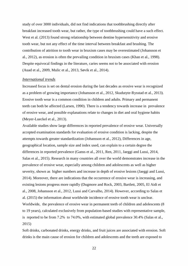
Dental health related perceptions and attitudes
tooth-brushing frequency twice or more daily once daily less than once daily
555 (63.9) 198 (22.8) 103 (11.9)
5.28 6.64 9.18
6.12 7.57 7.91
0.001
parental control of oral hygiene
yes no
698 (80.3) 164 (18.9)
5.70 7.65
6.61 7.51
0.001
dental fear no yes
801 (92.2) 56 (6.4)
5.71 11.02
6.63 7.73
0.001
self-rated dental health very good, good neither good nor bad bad, very bad
493 (56.7) 292 (33.6) 77 (8.9)
4.44 7.29 11.90
5.69 6.81 9.17
0.001
Dental health parameters
proximal enamel lesions <6 ≥6
482 (55.5) 387 (44.5)
3.81 8.92
5.14 7.68
0.001
number of teeth with gingival bleeding
<4 ≥4
148 (17.1) 718 (82.6)
5.05 6.28
5.30 7.15
0.017
General health status
BMI normal weight underweight overweight obese
620 (71.3) 50 (5.8) 139 (16.0) 59 (6.8)
5.63 5.02 7.55 8.44
6.56 5.21 7.94 7.88
0.001
chronic diseases/allergy no yes
621 (71.5) 245 (28.2)
5.92 6.51
6.84 7.00
0.255
self-rated general health very good, good neither good nor bad bad, very bad
635 (73.1) 180 (20.7) 41 (4.7)
5.51 7.43 9.56
6.39 7.47 9.40
0.001

33
21.8% for use of snuff). Regarding sugar intake, 12.5% of the adolescents reported frequent
consumption. This was significantly associated with higher caries prevalence (Table 1).
Frequent sugar consumption was more than twice as common in boys as in girls (17% vs 8%).
Intensity and frequency of physical activity and time spent daily in front of the TV/computer
screen during weekdays or weekends were not associated with differences in caries scores
(Table 1).
Dental health-related perceptions and attitudes
A majority of girls (80%) were brushing their teeth at least twice a day compared to 50% of
the boys. There was a considerable difference in mean DMFS score between the three
toothbrushing frequency groups (p< 0.001) (Table 1). Over 80% of the parents used to control
oral hygiene of their children. These adolescents had significantly lower DMFS scores than
adolescents without parental control of oral hygiene (p=0.001) (Table 1).
Dental fear was highly significantly associated with higher mean DMFS scores and
adolescents who rated their oral health as bad had almost 3 times higher mean DMFS scores
than those who rated their oral health as good (p< 0.001) (Table 1).
Dental health parameters
Only 5.6% (49) of the adolescents were recorded completely caries free (DMFS = 0, PEL=0),
and 11.6 % (101) had only PEL. About 23% (196) had more than 9 surfaces with PEL. The
prevalence of dentinal caries was statistically significantly associated with PEL scores (Table
1). Number of teeth with gingival bleeding showed an association with caries prevalence in
the bivariate analysis (p=0.017) (Table 1) that disappeared in the multivariate model. Only
2.3% (20) of the participants had periodontal pockets ≥4mm and only one presented with a
pocket >5mm.
General health
Over 70% (620) of the students had normal weight, while 6.8 % (59) where obese. There was
a statistically significant association between BMI and DMFS score (p=0.001), with higher
caries prevalence linked to overweight/obesity (Table 1).
Most of the recorded chronic diseases were allergy-related conditions. No association
between DMFS scores and chronic diseases was detected. Adolescents who rated their general
health as bad had almost 2 times higher mean DMFS scores than those who rated their general
health as good (p< 0.001).

34
Independent variables - Multivariate model
The final multivariate regression model comprised only the variables snuff use, dental fear,
self-rated dental health and PEL, while the impact of the other selected disappeared (Table 2)
indicating substantial co-variance among the parameters included. However, parents’
education level and sugar consumption were close to statistical significance. Having ≥6 dental
surfaces with PEL increased the chances to have high DMFS scores by O.R. 3.28.
Table 2. Multivariate logistic regression analysis including factors with p-values ≤ 0.05 from
the bivariate analysis (Table 1) included in the final model. DMFS-scores are dichotomized
with cut-off point DMFS = 6.
Independent variables Bivariate p value
Multivariate OR (95% CI)
Multivariate p value
parents attended college/university
both one none don’t know
P = 0.001
P = 0.092
parental family status
both parents one parent none of parents
P = 0.002
P = 0.133
smoking no yes
P = 0.009 P = 0.962
snuff use no yes
P = 0.001 1 1.57 (1.12-2.21)
P = 0.010
sugar consumption low high
P = 0.002 P = 0.084
tooth-brushing frequency
twice or more daily once daily less than once daily
P = 0.001
P = 0.798
parental control of oral hygiene
yes no
P = 0.001 P = 0.222
dental fear no yes
P = 0.001 1 3.26 (1.64-6.49)
P = 0.001
self-rated dental health
very good, good average bad, very bad
P = 0.001
1 1.99 (1.43-2.77) 4.51 (2.49-8.16)
P = 0.001
initial approximal caries
<6 ≥6
P = 0.001 1 3.25 (2.39-4.43)
P = 0.001
teeth with gingival bleeding
<4 ≥4
P = 0.017 P = 0.553
BMI normal weight underweight overweight obese
P = 0.001
P = 0.623
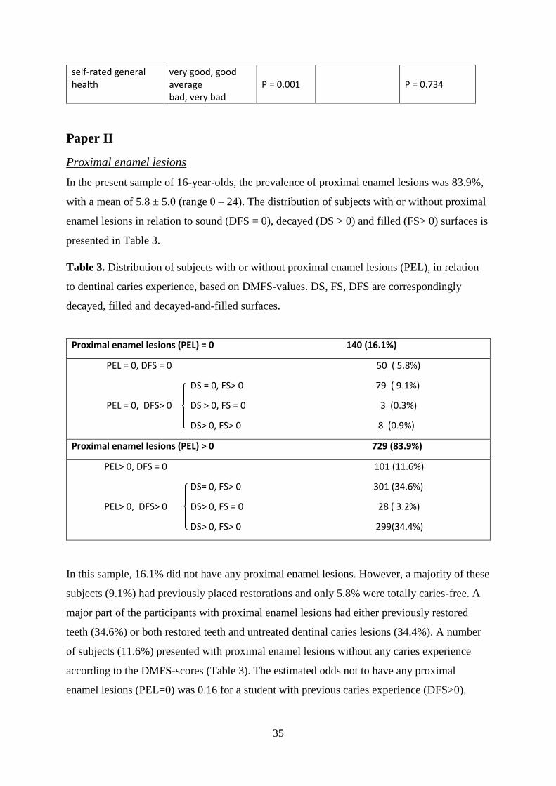
35
self-rated general health
very good, good average bad, very bad
P = 0.001
P = 0.734
Paper II
Proximal enamel lesions
In the present sample of 16-year-olds, the prevalence of proximal enamel lesions was 83.9%,
with a mean of 5.8 ± 5.0 (range 0 – 24). The distribution of subjects with or without proximal
enamel lesions in relation to sound (DFS = 0), decayed (DS > 0) and filled (FS> 0) surfaces is
presented in Table 3.
Table 3. Distribution of subjects with or without proximal enamel lesions (PEL), in relation
to dentinal caries experience, based on DMFS-values. DS, FS, DFS are correspondingly
decayed, filled and decayed-and-filled surfaces.
Proximal enamel lesions (PEL) = 0 140 (16.1%)
PEL = 0, DFS = 0 50 ( 5.8%)
DS = 0, FS> 0 79 ( 9.1%)
PEL = 0, DFS> 0 DS > 0, FS = 0 3 (0.3%)
DS> 0, FS> 0 8 (0.9%)
Proximal enamel lesions (PEL) > 0 729 (83.9%)
PEL> 0, DFS = 0 101 (11.6%)
DS= 0, FS> 0 301 (34.6%)
PEL> 0, DFS> 0 DS> 0, FS = 0 28 ( 3.2%)
DS> 0, FS> 0 299(34.4%)
In this sample, 16.1% did not have any proximal enamel lesions. However, a majority of these
subjects (9.1%) had previously placed restorations and only 5.8% were totally caries-free. A
major part of the participants with proximal enamel lesions had either previously restored
teeth (34.6%) or both restored teeth and untreated dentinal caries lesions (34.4%). A number
of subjects (11.6%) presented with proximal enamel lesions without any caries experience
according to the DMFS-scores (Table 3). The estimated odds not to have any proximal
enamel lesions (PEL=0) was 0.16 for a student with previous caries experience (DFS>0),

36
compared to 0.56 for a student without previous experience (DFS=0), (OR = 3.45; 95% CI
2.31 - 5.18).
Need for non-operative caries treatment
According to the distribution of proximal enamel lesions, 83.9% of the participants (729)
were in need of non-operative caries treatment, either as the only treatment modality (46.2%)
or in combination with restorative treatment (37.6%) (Table 3). When using the D-value of
the DMFS-index as diagnostic criterion, 38.8% (338) of the participants were in need of
operative dental treatment. When proximal enamel lesions were included in the diagnosis, the
number of participants in need of individual operative and/or non-operative caries treatment
was 85.1% (740) (Table 3).
Quality of dental restorations
In this material, 81.2% (706) of the 16-year-olds had experienced restorative care (Table 4).
The distribution of individuals in accordance with the score of their poorest restoration is
presented in Table 4. More than one-third of the participants with fillings (35.0%) had at least
one restoration below acceptable quality levels (poor/unacceptable).
Table 4. Distribution of subjects according to the quality of the poorest dental restoration.
¹The quality of restorations in two subjects was not possible to assess due to orthodontic braces.
Individuals with restorations 7061 (81.2%)
Quality of poorest filling
Good 37 (5.3%)
Acceptable 421 (59.8%)
Poor 159 (22.6%)
Unacceptable 87 (12.4%)

37
Paper III
Prevalence, distribution and severity of dental erosive wear
In the randomly selected sample of 392 adolescents, 38% (148) showed dental erosive wear.
Looking at the severity of the erosive wear, approximately equal numbers of participants with
erosive wear limited to enamel or extending into dentine were found. Figure 1 shows the
distribution of participants who had healthy surfaces (grade 0), surfaces with lesions limited
to enamel (grade 1-2) and surfaces with lesions extending into dentine (grade 3-5). In the
group of participants with erosive wear extending into dentine (20%), half of the participants
had both, lesions limited to enamel and lesions extending into dentine, and half showed only
lesions extending into dentine. Of the group with erosive wear localized to the enamel (18%),
76% had ≤ 2 affected surfaces while the rest (24%) had 3 or more. The gender distribution of
the participants studied was almost equal, 51% (199) were male and 49% (193) were female.
A significantly higher share of males (65%) exhibited erosive wear (p < 0.001), and lesions
extending into the dentine were also more common in male adolescents (p < 0.001) (Figure
2).
When assessing the severity of erosive wear based on the type of tooth, more erosive wear
was found on molars (18%) compared to both the central incisors (11%) and lateral incisors
(10%). On the upper central incisors, erosive wear limited to the enamel was most common,
while about the same amounts of enamel and dentine lesions were found on the molars. When
comparing the level of erosive wear on the first permanent molars, significantly more lesions
were found in the lower jaw in comparison to upper jaw, 8% in the upper and 29% in the
lower jaw, respectively (Table 5).
Figure 1. Prevalence of dental erosive wear in the examined population (n=392), divided into
individuals without erosive wear, with erosive wear limited to enamel, or erosive wear
extending into dentine.
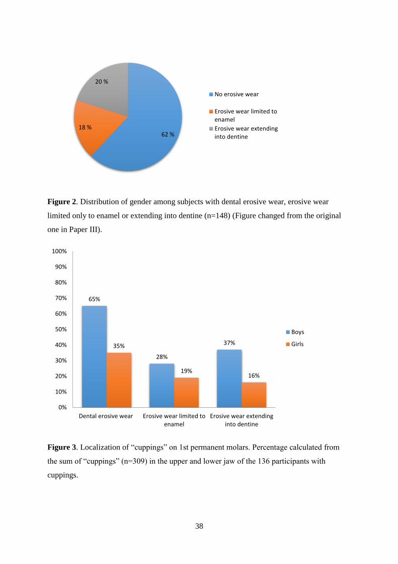
38
Figure 2. Distribution of gender among subjects with dental erosive wear, erosive wear
limited only to enamel or extending into dentine (n=148) (Figure changed from the original
one in Paper III).
Figure 3. Localization of “cuppings” on 1st permanent molars. Percentage calculated from
the sum of “cuppings” (n=309) in the upper and lower jaw of the 136 participants with
cuppings.
62 %18 %
20 %
No erosive wear
Erosive wear limited toenamel
Erosive wear extendinginto dentine
65%
28%
37%35%
19%16%
0%
10%
20%
30%
40%
50%
60%
70%
80%
90%
100%
Dental erosive wear Erosive wear limited toenamel
Erosive wear extendinginto dentine
Boys
Girls

39
Table 5. Prevalence and severity of erosive wear based on the type of tooth surface (Table
changed from the original one in Paper III).
DEW
% (n)
Enamel lesions
% (n)
Dentine lesions
% (n)
Total
% (n)
UJ central incisors (B and P) 5 (68) 4 (64) 0 (4) 100 (1462)
UJ lateral incisors (B and P) 3 (43) 3 (43) 0 (0) 100 (1466)
1st permanent molars in total (O) 17 (270) 9 (139) 8 (131) 100 (1556)
UJ 1st permanent molars (O) 7 (54) 5 (40) 2 (14) 100 (782)
LJ 1st permanent molars (O) 28 (216) 13 (99) 15 (117) 100 (774)
DEW: Dental erosive wear, UJ: upper jaw, LJ: lower jaw, B: buccal surfaces, P: palatal surfaces, O: occlusal surfaces
Table 6. Prevalence and number of cuppings.
Lesion extension
Individuals with DEW % (n)
Individuals with DEW and cuppings % (n)
1 cupping % (n)
2 cuppings % (n)
≥3 cuppings % (n)
Enamel 48 (71) 48 (65) 62 (40) 26 (17) 12 (8) Dentine 52 (77) 52 (71) 18 (13) 37 (26) 45 (32) Total 100 (148) 100 (136)
17%
36%10%
37%
0%
10%
20%
30%
40%
50%
60%
70%
80%
Maxilla Mandible
Cuppings limited to enamel Cuppings extending into dentine

40
“Cupping” on the molars was found in 92% (136 out of the 148) of the participants with
dental erosive wear. They were equally distributed between individuals with erosive wear
lesions limited to enamel (48%) and those with erosive wear extending into dentine (52%). Of
the participants with lesions limited to enamel, 62% had 1 “cupping” and 12% had 3 or more.
In the group of participants with lesions extending into dentine only 18% had 1 “cupping”,
while 45% had 3 or more (Table 6). In total, 309 “cuppings” on the first permanent molars
were registered, 27% (85) in the upper jaw and 73% (224) in the lower jaw (Figure 3). In the
upper jaw the “cuppings” were localized mostly in the enamel, while in the lower jaw they
were equally common in the enamel and dentine. The mesiobuccal cusp was most often
affected (75%, n = 231).
6. GENERAL DISCUSSION
The main focus of the present study was the dental health conditions among a group of
adolescents in northern Norway and specifically, dental caries and erosive lesions. Particular
emphasis is placed on proximal enamel lesions and non-operative treatment intervention. The
study has a cross-sectional design and, since the sequence of events (exposure to variables and
onset of disease) is uncertain, the detection of definite causal relations is not possible and only
indications for associations are disclosed (Levin, 2006).
Methodological considerations (Paper I)
Representativity
The present thesis was based on a cross-sectional oral and general health study, the Fit
Futures, which is a population-based youth study (Winther et al., 2014).
The attendance rate (90%) was high, compared with similar studies (Mulic et al., 2013,
Norderyd et al., 2015b), and male and female genders were equally represented (50.7% males,
49.3% females). Among the 1010 participants in the oral part of Fit Futures study, 95 % were
born in 1992 or later, and within this group, all subjects born in 1994 (869, 51.7% males
versus 48.3% females) were included in the present study.
Lower secondary education is compulsory in Norway, and in 2010-2011, more than 95% of
students in Troms county had moved on from lower secondary school to first year of upper
secondary school, usually as 16-year-olds (Statistics Norway, 2014).
In Troms County, 86% of the population lives in urban or densely populated, urban-like areas
(Statistics Norway, 2014). The number of participants from the two municipalities (92% from

41
Tromsø municipality (urban) against 8% from Balsfjord municipality (rural)) corresponds to
the demographic structure between urban / rural areas in Troms county.
On the basis of the above information, the results of the study can be considered
representative for the county and valid indicators for oral health conditions in the age group
examined in this part of Northern Norway. The combination of oral and general health part in
the study provided an opportunity to monitor oral health among adolescents in the region and
correlate dental caries and a multitude of determinants (sociodemographic characteristics, oral
health attitudes, general health, lifestyle). However, the possibilities for direct comparison
with national oral health statistics in Norway are limited, as the reference groups used
nationally are 12 and 18 year-olds (Statistics Norway, 2014).
Non-participation in a study may result in bias if the reasons for non-participation are
associated with either the exposure or the outcome studied affecting the representativity of the
sample (Galea and Tracy, 2007). Non-participants have often low socioeconomic status,
which is related to poor health conditions and, among adolescents, non-attendance to dental
examination is associated with poor oral health (Galea and Tracy, 2007, Fägerstad et al.,
2016). Analysis of non-participation in this study was not performed, but the overall high
attendance rate might have reduced the risk for non-response bias (Galea and Tracy, 2007,
Mindell et al., 2015).
The questionnaires
Questionnaires are prone to various forms of response bias in health research. Risk of bias
might be reduced in the present study, as the questionnaires used were previously validated,
pretested and used in studies of comparable age cohorts (Choi and Pak, 2005, Cook, 2010,
Furberg, 2010). The use of self-administered electronic questionnaire for information about
dietary habits and lifestyle might have facilitated reporting of some sensitive behaviors
compared to paper and pencil or face to face interview methods (Hallfors et al., 2000).
Ethnicity
Ethnicity in the present study was registered based on self-perceived ethnicity, since ethnicity
information for residents in Norway is not recorded. Presently, self-perceived ethnicity is
prevailing over geographic or language indicators of ethnicity in health research (Bhopal,
2004). Additional questions on student's country of birth, mother's country of birth and
father's country of birth provided supplementary, objective information on ethnic origin, and
could secure identification of second generation of immigrants (Stronks et al., 2009).

42
Concerning validity of self-reported information, Nordahl et al. (2011) showed that students
as young as 11 years can give valid answers about own and parents ethnicity background.
Sami participants
Among the 869 participants in the present study, 3.6% individuals considered themselves as
Sami (self-perceived ethnicity). The Sami Parliament electoral register is a register of Sami
people in Norway. For 2011, 1.5% of the total number of inhabitants in both participating
municipalities was registered in the Sami Parliament electoral register. However, the Sami
electoral register is not based on self-perceived ethnicity. It includes only people over 18
years who vote for the Sami Parliament and fulfill some language criteria. Consequently, it is
not possible to assess whether the percentage of Sami participants in the present study
corresponds to the percentage of Sami people in the total population of the included
municipalities, a challenge discussed by Pettersen (2015).
Moreover, the low number of identified Sami participants may have resulted in an unreliable
estimation of dental health in this group of adolescents.
Participants with immigrant background
The percentage of students with immigrant background in the sample of 869 participants
(13.1%) was higher than that reported from Statistics Norway in 2010 (8.4% in Tromsø and
3.3% in Balsfjord municipalities) (Statistics Norway, 2014). The official statistical data are
supplied from various sources (the local population registry offices, tax administration etc.),
since an ethnicity registry at an individual level is lacking in Norway, and may not be as
accurate as an ethnicity registry would be. The higher percentage of students with immigrant
background in the assessed sample may be explained by the younger average age of
individuals with immigrant background in Norway the last years (Statistics Norway, 2014). In
that way, immigrants may be overrepresented in school ages.
The estimate of number of participants with immigrant background identified may have
resulted in a reliable estimate of dental health in this group of adolescents.
Caries registration
The registration of proximal lesions was based on intraoral radiographs (bite-wings) which is
a common method for assessing prevalence of caries in proximal surfaces. The intraoral
radiographic examination has low sensitivity for the diagnosis of proximal initial lesions (The
Swedish Council on Technology Assessment in Health Care, 2007) and the quality of
evidence for radiographic detection of non-cavitated lesions is poor (Gomez et al., 2013,

43
Mejare et al., 2015). Although bitewings are not necessary for assessing epidemiological
trends (Poorterman et al., 1999, Bloemendal et al., 2004, Baelum et al., 2006, Agustsdottir et
al., 2010, Nørrisgaard et al., 2016), they are necessary for calculating the prevalence of
disease.
Calibration
The oral investigation was performed in a clinical setting and the principal investigator (IDJ)
was thoroughly calibrated with experienced clinicians reaching substantial intra- and inter-
observer agreement (Kappa=0.70 and 0.71 respectively) securing reliability. Calibration is
extensively dealt with in Materials and Methods.
Statistics
Parametric statistical tests (Students t-test and ANOVA) were applied for descriptive purposes
(Table 1) using DMFS scores as a continuous variable. Although the caries data were skewed,
these parametric tests are robust and acceptable considering the large number of observations
(Fagerland, 2012). In the present study, a number of variables in the initial bivariate analyses
showed a significant bivariate association, but this number of variables decreased
substantially in multivariate analysis, indicating substantial covariance. Simple bivariate
correlations can be severely biased and lead to false conclusions due to the ignorance of
possible correlations and covariance with other background characteristics, while the
multivariate analysis may disclose underlying relationships by accounting for the influence of
other observable variables (Fuchs and Woessmann, 2004).
Methodological considerations (Paper II)
Proximal enamel lesions
In Paper II only proximal enamel lesions were included, as proximal surfaces of the young
adolescents contribute mostly to the caries burden in later age (Mejàre et al., 2004, Crossner
and Unell, 2007, Sköld, 2016). Smooth and occlusal surfaces were not included, as buccal and
lingual decay is a minor problem among Nordic teenagers and in order to eliminate
uncertainties in visual discrimination between hypomineralizations and enamel lesions
(Crossner and Unell, 2007, Norderyd et al., 2015a).
Active – arrested lesions
Activity assessment of the lesions was not performed in the present study due to the cross-
sectional study design. Assessment of lesion activity at one single visit is shown to result in
poor accuracy and reliability (Ekstrand et al., 2009, Ismail et al., 2015). Activity assessment at

44
one examination is possible only based on a combination of clinical indicators including
visual appearance, tactile feeling, potential for biofilm accumulation and gingival status and
necessitates additional resources, such as trained/calibrated examiners and previously cleaned
teeth (Ismail et al., 2015).
The non-discrimination of active and arrested caries in this study might have resulted in some
over-estimation of the need for non-operative treatment. Agustsdottir et al. (2010), in the
Icelandic Oral Health Survey (data collected in 2004-2005), reported activity in the majority
of lesions in all age groups of young people examined. In their study, 70% of initial
precavitated lesions among the 15-year-olds were active.
A possible over-estimation of the need for non-operative treatment in the present study entails
the use of non-operative treatment in all non-cavitated initial lesions, also in those non-
actives. However, due to the nature of non-operative procedures, these measures are not
harmful.
Quality of fillings
The quality of restorations in this cross-sectional study was evaluated exclusively based on
the assessment of their current performance and considering tooth prognosis since information
about accurate dates, history and clinical parameters of placement of restorations were
missing. Moreover, since the unit for analysis was the individual and not the restoration, the
comparison with other studies is difficult.
The inclusion of radiographic criteria, in addition to the clinical ones, for the evaluation of
restorations, might have increased the number of participants identified with at least one
defective filling. This finding has been reported previously by Sonbul and Birkhed (2010).
The use of exclusively clinical criteria would have left a number of defect restorations
undetected (anatomical form, marginal adaptation etc.).
Methodological considerations (Paper III)
Erosive wear registration
Due to the extensive workload for three examiners to evaluate eight intraoral photos of 869
students, the assessment of dental erosive wear was not performed in the total group of 16-
year-olds but in a representative randomly selected subgroup of 392 students. The subgroup
had almost equal gender distribution (50.8% males, 49.2% females).
Furthermore, partial mouth scoring was decided for efficient use of available resources (time
and finances), in order not to increase the number of false positive lesions detected, and not to
reduce accuracy (Young et al., 2008). Erosive wear in the present study was scored on

45
selected teeth / surfaces, which were consistently more affected by dental erosion based on a
number of earlier relevant studies (Mulic et al., 2010). The first molars and the palatal and
buccal surfaces of maxillary incisors may be particularly exposed to erosive wear action, due
to earlier eruption of these teeth (Isaksson et al., 2013, Mulic et al., 2013). Mandibular front-
teeth were excluded from scoring due to difficulties in distinguishing wear due to erosion
from wear supposed to be caused by attrition/abrasion, although presence of the last
mentioned conditions is low in young age (Al-Dlaigan et al., 2001).
Identical study methods, i.e. same sample size, use of the same grading system, calibrated
clinicians, choice of the index teeth and the age of the participants allow for the comparison of
prevalence of dental erosion in this study with two other studies from Norway (Mulic et al.,
2013, Søvik et al., 2014).
The use of clinical photos
Erosive wear in the present study was recorded from clinical intraoral photographs of high
quality, eight for each participant, taken by a specially trained dental assistant following a
standardized procedure (see Materials and Methods). The observer could spend an unlimited
amount of time examining the photos and the scoring could easily be repeated.
Concerning the validity of this method, Al-Malik et al. (2001a) compared scoring of erosions
in children (primary maxillary incisors) by using clinical and photographic examination, and
found substantial agreement, although the extent of wear in dentin might be underestimated in
photographs (Al-Malik et al., 2001b). Furthermore, the findings of Mulic et al. (2010)
suggested that both methods, clinical examination and use of clinical photos could be
acceptable for measuring dental erosive wear. Hove et al. (2013) also used photographs and
clinical examination for scoring of dental erosions and found no significant difference
between these methods. This documentation indicates that the present erosion data are
reliable.
Calibration
In the present study, the average inter-observer agreement expressed by weighted Kappa
based on the photographs was calculated to be 0.84 for the three dentists, and the intra-
observer agreement was 0.71 (observer 1), 0.73 (observer 2) and 0.89 (observer 3), indicating
substantial agreement. This was advantageous, as, in a range of relevant studies, low inter-
examiner agreement was reported (Larsen et al., 2005, Mulic et al., 2010, Søvik et al., 2014).
The low inter-examiner agreement was attributed to the difficulty for examiners to detect
initial erosive lesions and the increased variability among the many examiners involved in the

46
studies. In the present study, the thorough calibration of the examiners, the lower number of
examiners than in the studies mentioned, and the relatively lower percentage of erosive wear
in enamel found may explain the higher agreement levels.
Ethical considerations
The project concerned research on personal and health data and was approved by the Regional
Committee for Medical Research Ethics (2012/1197 REK Nord) and the Norwegian Social
Science Data Services (NSD) (07/00886-11). The study complies with the Declaration of
Helsinki, International Ethical Guidelines for Biomedical Research Involving Human
Subjects and the International Guidelines for Ethical Review of Epidemiological Studies.
Participants gave a written informed consent prior to the study. Participation was voluntary
and the participants retained the right to withdraw their consent at any moment from the
study. As the study concerned students in first year of upper secondary school and some
among them were younger than 16 years of age by the day of the study, additional written
consent was obtained for these subjects from their parents/guardians. Participants were
informed in detail orally, electronically and a brochure was distributed to students and
parents/guardians. The information given to participants concerned the objectives of the
study, a description of examinations and measurements included, the type of data collected for
each participant, and the future use of data collected in research.
The oral part of Fit Futures included clinical procedures (dental examination, taking of intra-
oral radiographs) well known and used regularly during the annual clinical examination in the
PDS. The clinical examination was performed by experienced personnel and did replace the
annual clinical examination. In case that the participants already had completed annual oral
examination, they were excluded from radiographic examination. Clinical and radiographic
data were recorded in patient files for treatment or for follow up. Clinical intraoral pictures
did not display individual identification.
Upon completion of data collection, the data was stored de-identified in the Tromsø Study
database (EUTRO), a data storage and management system, designed and administrated by
the Department of Community Medicine, UiT The Arctic University of Norway, Tromsø (The
Department of Community Medicine, 2010).

47
Main findings
Caries prevalence (Paper I)
The prevalence of dental caries and the mean DMFT/S in the present sample correspond with
the figures reported at the Norwegian national level and with the figures for Troms county
(Statistics Norway, 2014). This indicates that the caries status among 16-year-olds in
Northern Norway has improved and that the previously documented regional difference
between the North and South of Norway is diminishing (Mulic et al., 2013). However, data
from epidemiological studies facilitating direct comparison with the age group studied are
lacking. Results from Scandinavian studies indicate, however, that the caries prevalence
among adolescents in Norway is higher than in Sweden, Denmark and Finland (Table 5).
Table 7. Mean caries score and prevalence of caries in the present study (Jacobsen et al,
2016) compared with results from similar Scandinavian studies.
(Di+mMFT/S is including both initial (i) and manifest (m) caries).
Study / country Age / year of
examination
Sample
(N)
Caries score Prevalence of
caries
Jacobsen et al. (2016)
Norway
16-year-olds
2010-11
869 6.1 (DMFS)
4.2 (DMFT)
82.7% (DMFT)
Mulic et al. (2013)
Oslo
18-year-olds
2008-2009
1426 4.0 (DMFT)
76.0% (DMFT)
Nørrisgaard et al.
(2016) Denmark
15-year-olds
18-year-olds
2012
1509
1168
2.06 (DMFS)
3.92 (DMFS)
45.3% (DMFT)
66.0% (DMFT)
Kämppi et al. (2013)
Finland
19-21-year-olds 13504 4.11 (DMFT) 78.7% (DMFT)
Norderyd et al.
(2015a) Sweden
15-year-olds
2013
101 2.9 (Di+mMFS) 57% (Di+mMFT)
The documented inferior caries status among adolescents in Norway is surprising, since the
Scandinavian societies and their dental care systems are quite similar. They have health

48
priorities based on a social welfare model and public dental health services for children and
adolescents are based on prevention and are free of charge. There are, however, minor
differences in the structure and provision of dental care in the various Scandinavian countries
(number of work force and their tasks, risk strategy including definition of risk individuals,
recall routines, prevention at group level and time allocated to prevention) (Wang and
Aspelund, 2010). Nadanovsky and Sheiham (1995) have argued that the contribution to the
caries decline from chair-side dentistry is of minor importance in comparison with the role of
larger scale measures on society level like fluoridated toothpaste, oral health education at
schools and availability of non-cariogenic snacks. Both clinical and chair-side activities and
more general factors might explain the observed difference in caries prevalence (Table 7).
However, our data do not offer any clue regarding reasons for the observed difference and it is
beyond the scope of the present investigation to analyze these aspects.
Ethnicity and caries (Paper I)
Sami ethnicity was not associated with higher caries prevalence in the present study.
However, the low number of identified Sami participants may have resulted in an unreliable
estimate of dental health in this group of adolescents.
According to Pettersen (2015), most of the Sami population are well integrated in the modern
Norwegian society and only a few are still pursuing traditional reindeer herding and a
nomadic lifestyle. Thus, the finding of no difference in oral health between ethnic Norwegians
and individuals with Sami background in the present sample may be explained by a high
degree of social integration of Sami people within the Norwegian society. The dual self-
perceived ethnicity (Norwegian and Sami) stated by a majority of the Sami participants
supports this argument, indicating a weak relationship with Sami ethnicity (Lund et al., 2007).
Immigrant ethnicity was not associated with inferior oral health among 16-year-olds in the
present study. This is contrary to the results from comparable Scandinavian studies
documenting higher levels of caries among children with immigrant background, stating that
the reported trends of caries reduction may concern only non-immigrants (Sundby and
Petersen, 2003, Wigen and Wang, 2010, Stecksén-Blicks et al., 2014, Östberg et al., 2016).
Julihn et al. (2010) and Jacobsson et al. (2011) found that immigrant adolescents had higher
caries prevalence, irrespective of whether or not they were born abroad and despite their
enrollment in regular dental public health care programs. In the present study, the adolescents
of immigrant background were included in regular public dental care and were well integrated
in the Norwegian society. This might explain the registration of no difference in their dental

49
health condition. Investigation of immigrants’ dental health according to country/region of
birth and to immigration status (immigrants or descendants) might have disclosed further
differences in dental status (Nordahl et al., 2011), Bast et al. (2015). However, such analyses
were not performed in the present study due to too few participants giving insufficient
statistical power.
Lifestyle factors and dental caries (Paper I)
Findings in the present study confirm the already shown association of lifestyle factors with
dental caries (Sakki et al., 1994, Caufield and Griffen, 2000, Christensen et al., 2015). Among
lifestyle factors tested in the present study, cigarette smoking, use of snuff and consumption
of sugar were found to be statistically significantly associated with dentinal caries in a
bivariate model. However, only use of snuff was still significant in the final multivariate
model. This indicates covariance among lifestyle factors and is in agreement with a number of
previous studies (Burt and Pai, 2001, Hugoson et al., 2012, Benedetti et al., 2013, Holmen et
al., 2013).
Proximal enamel lesions (Paper II)
The high prevalence of PEL recorded in the present study is in agreement with results from
other similar studies in low caries prevalence populations (Alm et al., 2007, Norderyd et al.,
2015a). This finding is important since early proximal lesions in adolescents contribute
substantially to the caries burden in later age (Mejàre et al., 2004, Crossner and Unell, 2007,
Sköld, 2016). Most lesions develop before the age of 20 years, as shown by Crossner and
Unell (2007) and Norderyd et al. (2015a).
Quality of restorations (Paper II)
The present study disclosed a high number of 16-year-olds (28%) with at least one filling of
poor / unacceptable quality. This high prevalence might in part be due to the radiographic
evaluation of quality of fillings which increases the number of participants identified with
defective fillings while only clinical evaluation may leave a number of defects undetected
(Sonbul and Birkhed (2010). It is surprising that over one third of the present young
participants were in need of repair / replacement of defective filling(s), since these fillings
must be rather new. Defective fillings contribute substantially to the high restorative burden in
everyday dental practice (Qvist, 2012). As restorative dental materials currently used are of
good quality, patient and dentist factors might explain the failure of restorations (Kopperud et
al., 2016). Concerning dentist factors, the quality of clinical restorative work in the PDS in

50
Norway is good and homogenous according to Dobloug and Grytten (2015). It was therefore
surprising to detect high prevalence of defective fillings in the present study.
Dental erosions (Paper III)
In the present study, 38% of the sample presented with erosive wear and almost equal
numbers had erosive enamel lesions or lesions extending into dentine (Figure 1). These
findings correspond well with the results of a study on 18-year-olds in Oslo using the same
index (Mulic et al., 2013). Studies from other Nordic countries (Arnadottir et al., 2010,
Hasselkvist et al., 2010, Isaksson et al., 2014) have reported varying prevalence of dental
erosive wear in adolescents. The difference in prevalence may be explained by several factors,
such as differences in number and group of teeth examined, index used, age group and
timespan of the examination. Furthermore, variation in dietary habits and lifestyle among
participants from different countries may also influence the results. In their systematic review,
Salas et al. (2015) found high variation in prevalence of erosive conditions among children
and adolescents worldwide and indicate the importance of choosing an index detecting
specifically the erosive condition and not tooth wear with combined etiology.
Concerning gender distribution, the prevalence and severity of dental erosions were
significantly higher in males than females in the present study. This corresponds well with
previous research (Hasselkvist et al., 2010, Mulic et al., 2012a, Mulic et al., 2013, Søvik et al.,
2014, Hasselkvist et al., 2016) although there are some studies indicating the opposite or no
gender difference (Bartlett et al., 1998, Isaksson et al., 2014).
In the present study, 93% of adolescents with erosive wear also presented with cuppings on
first molars, mostly in lower jaw. Cuppings on molars are considered as an initial sign of
erosive wear (El Aidi et al., 2010, Isaksson et al., 2014, Søvik et al., 2014) and their presence
is pointing to the need for accurate examination of the patient’s erosive status.
It is shown that erosive wear is increasing with age (El Aidi et al., 2010, Hasselkvist et al.,
2016). Hasselkvist et al. (2016) reported 35% progression of erosive wear from 13-14 to 17-
19 years of age. Øvrebø (2011)) found that drinking habits of young adolescents from
Northern Norway changed during adolescence and especially boys adopted poorer drinking
habits related to sugary drinks. Norwegian youth from 10 to 24 years of age represent the
highest proportion of consumers of this category of drinks (Statistics Norway, 2014). The
official statistics of Norway do not cover the consumption of the full range of drinks with
erosive action among adolescents. They provide information on the consumption of
traditional sugary soft drinks only. Despite a decline in the consumption of drinks belonging
51
to the last mentioned category (carbonated beverages, juices), other beverages with erosive
potential have recently been introduced on the market (energy drinks, zero drinks, drinks with
artificial sweeteners) and the market is constantly supplied with drinks with erosive potential.
The majority of 16-year-olds who present with erosive wear today will likely need treatment
in the future.
7. CONCLUDING REMARKS
According to the findings in the present study, dental caries and erosive wear are challenging
conditions among North Norwegian 16-year-olds. These North Norwegian 16-year-olds were
in need of restorative treatment related to untreated caries (39%) and compromised
restorations (28%), and 10% had DMFT>9. Their caries status was inferior to adolescents
from other Scandinavian countries, but comparable to the prevalence reported from the South
of Norway.
The prevalence of proximal enamel lesions was high, entailing a need for non-operative
treatment interventions. In spite of being enrolled in a public dental health care system free of
charge, of good quality and characterized by high financial and human resource allocations,
over 85% of the examined 16-year-olds were in need of individual operative and/or non-
operative treatment.
The prevalence and severity of erosive wear were high among the North Norwegian 16-year-
olds. Increasing consumption of erosive drinks among Norwegian youths and the absence of
strategies for preventing erosive conditions are matters of concern as they may result in
complicated future treatments.
The present study is, to our knowledge, the first study assessing the treatment need of
adolescents in Norway with focus on non-operative treatment and documents that the caries
disease and erosive conditions are widespread within this teenage population. These findings
further indicate reasons for considering a change in the PDS treatment strategy. Instead of
trying to identify risk-groups and to prolong recall intervals, a relevant and successful
treatment strategy for teenagers might be general prevention and shorter recall intervals in
order to diagnose any active disease (caries or erosion) in time for non-operative treatment.
This implies an individually adjusted treatment, cost effectually performed by auxiliary dental
personnel, during a period when many permanent tooth surfaces are newly erupted. The teen
years is a vulnerable time in life. Adolescents leave parental guidance, family routines and
food habits, and need support on the way to an adult life. The DMFS-score and the high
number of 16-year-olds with restorations in need of repair or replacement further indicate the

52
importance of a “non-operative” treatment strategy in order to reduce the need of traditional
restorative care aiming at minimizing the vicious operative re-treatment circle throughout life.

53
REFERENCES
Aas JA, Griffen AL, Dardis SR, Lee AM, Olsen I, Dewhirst FE, et al. (2008). Bacteria of dental caries in primary and permanent teeth in children and young adults. J Clin Microbiol, 46(4):1407-1417.
Addy M (2005). Tooth brushing, tooth wear and dentine hypersensitivity—are they associated? Int Dent J, 55(S4):261-267.
Adekoya SM, Brustad M (2012). Oral health of adults in northern Norway–A pilot study. Nor Epidemiol, 22(1):31-38.
Agustsdottir H, Gudmundsdottir H, Eggertsson H, Jonsson SH, Gudlaugsson JO, Saemundsson SR, et al. (2010). Caries prevalence of permanent teeth: a national survey of children in Iceland using ICDAS. Community Dent Oral Epidemiol, 38(4):299-309.
Al-Dlaigan YH, Shaw L, Smith A (2001). Dental erosion in a group of British 14-year-old school children. Part II: Influence of dietary intake. Br Dent J, 190(5):258-261.
Al-Malik MI, Holt RD, Bedi R (2001a). Clinical and photographic assessment of erosion in 2-5-year-old children in Saudi Arabia. Community Dent Health, 18(4):232-235.
Al-Malik MI, Holt RD, Bedi R, Speight PM (2001b). Investigation of an index to measure tooth wear in primary teeth. J Dent, 29(2):103-107.
Alm A, Wendt LK, Koch G, Birkhed D (2007). Prevalence of approximal caries in posterior teeth in 15-year-old Swedish teenagers in relation to their caries experience at 3 years of age. Caries Res, 41(5):392-398.
Amaechi BT, Higham SM, Edgar WM (2003). Influence of abrasion in clinical manifestation of human dental erosion. J Oral Rehabil, 30(4):407-413.
Amarante E, Raadal M, Espelid I (1998). Impact of diagnostic criteria on the prevalence of dental caries in Norwegian children aged 5, 12 and 18 years. Community Dent Oral Epidemiol, 26(2):87-94.
Araújo D, Alencar M, Freitas MC, Azevedo L, Atta M, Wang L (2014). Incipient caries lesions: from concept to actual treatment approaches. J Res Dent, 2(5):418-427.
Arnadottir IB, Holbrook WP, Eggertsson H, Gudmundsdottir H, Jonsson SH, Gudlaugsson JO, et al. (2010). Prevalence of dental erosion in children: a national survey. Community Dent Oral Epidemiol, 38(6):521-526.
Asmyhr O, Grytten J, Holst D (2012). Occurrence of risk factors for dental erosion in the population of young adults in Norway. Community Dent Oral Epidemiol, 40(5):425-431.
Ástvaldsdóttir Á, Dagerhamn J, van Dijken JWV, Naimi-Akbar A, Sandborgh-Englund G, Tranæus S, et al. (2015). Longevity of posterior resin composite restorations in adults–A systematic review. J Dent, 43(8):934-935.
Auad SM, Waterhouse PJ, Nunn JH, Moynihan PJ (2009). Dental caries and its association with sociodemographics, erosion, and diet in schoolchildren from southeast Brazil. Pediatr Dent, 31(3):229-235.
Baelum V, Heidmann J, Nyvad B (2006). Dental caries paradigms in diagnosis and diagnostic research. Eur J Oral Sci, 114(4):263-277.
Bartlett DW (2005). The role of erosion in tooth wear: aetiology, prevention and management. Int Dent J, 55(S4):277-284.
Bartlett DW (2016). A personal perspective and update on erosive tooth wear–10 years on: Part 1–Diagnosis and prevention. Br Dent J, 221(3):115-119.
Bartlett DW, Coward PY, Nikkah C, Wilson RF (1998). The prevalence of tooth wear in a cluster sample of adolescent schoolchildren and its relationship with potential explanatory factors. Br Dent J, 184(3):125-129.

54
Bartlett DW, Ganss C, Lussi A (2008). Basic Erosive Wear Examination (BEWE): a new scoring system for scientific and clinical needs. Clin Oral Investig, 12 (Suppl 1):S65-S68.
Bartlett DW, Lussi A, West NX, Bouchard P, Sanz M, Bourgeois D (2013). Prevalence of tooth wear on buccal and lingual surfaces and possible risk factors in young European adults. J Dent, 41(11):1007-1013.
Bast LS, Nordahl H, Christensen LB, Holstein BE (2015). Tooth brushing among 11-to 15-year-olds in Denmark: combined effect of social class and migration status. Community Dent Health, 32(1):51-55.
Bayne CS, Schmalz G (2005). Reprinting the classic article on USPHS evaluation methods for measuring the clinical research performance of restorative materials. Clin Oral Investig, 9(4):215-232.
Benedetti G, Campus G, Strohmenger L, Lingström P (2013). Tobacco and dental caries: a systematic review. Acta Odontol Scand, 71(3-4):363-371.
Bernabé E, Sheiham A (2014a). Age, Period and Cohort Trends in Caries of Permanent Teeth in Four Developed Countries. Am J Public Health, 104(7):e115-e121.
Bernabé E, Sheiham A (2014b). Extent of differences in dental caries in permanent teeth between childhood and adulthood in 26 countries. Int Dent J, 64(5):241-245.
Bhopal R (2004). Glossary of terms relating to ethnicity and race: for reflection and debate. J Epidemiol Community Health, 58(6):441-445.
Bloemendal E, de Vet HCW, Bouter LM (2004). The value of bitewing radiographs in epidemiological caries research: a systematic review of the literature. J Dent, 32(4):255-264.
Bowden GH (1997). Does assessment of microbial composition of plaque/saliva allow for diagnosis of disease activity of individuals? Community Dent Oral Epidemiol, 25(1):76-81.
Brantley CF, Bader JD, Shugars DA, Nesbit SP (1995). Does the cycle of rerestoration lead to larger restorations? J Am Dent Assoc, 126(10):1407-1413.
Bratthall D, Hänsel‐Petersson G, Sundberg H (1996). Reasons for the caries decline: what do the experts believe? Eur J Oral Sci, 104(4):416-422.
Broadbent JM, Page LF, Thomson WM, Poulton R (2013). Permanent dentition caries through the first half of life. Br Dent J, 215(7):E12.
Burt BA, Pai S (2001). Sugar consumption and caries risk: a systematic review. J Dent Educ, 65(10):1017-1023.
Burt BA, Szpunar SM (1994). The Michigan study: the relationship between sugars intake and dental caries over three years. Int Dent J, 44(3):230-240.
Carvalho TS, Colon P, Ganss C, Huysmans MC, Lussi A, Schlueter N, et al. (2015). Consensus report of the European Federation of Conservative Dentistry: erosive tooth wear—diagnosis and management. Clin Oral Investig, 19(7):1557-1561.
Caufield PW, Griffen AL (2000). Dental caries: an infectious and transmissible disease. Pediatr Clin North Am, 47(5):1001-1019.
Cavadini C, Siega-Riz A, Popkin BM (2000). US adolescent food intake trends from 1965 to 1996. Arch Dis Child, 83(1):18-24.
Choi BC, Pak AW (2005). A catalog of biases in questionnaires. Prev Chronic Dis, 2(1):A13. Christensen LB, Bardow A, Ekstrand K, Fiehn NE, Heitmann BL, Qvist V, et al. (2015). Root
caries, root surface restorations and lifestyle factors in adult Danes. Acta Odontol Scand, 73(6):467-473.
Christensen LB, Petersen PE, Hede B (2010). Oral health in children in Denmark under different public dental health care schemes. Community Dent Health, 27(2):94-101.
Cole TJ, Lobstein T (2012). Extended international (IOTF) body mass index cut‐offs for thinness, overweight and obesity. Pediatr Obes, 7(4):284-294.
Cook C (2010). Mode of administration bias. J Man Manip Ther, 18(2):61-63. Cooper T (2012). The value of longevity: Product quality and sustainable consumption.
Global and Regional Research on Sustainable Consumption & Production: Achievements, Challenges, and Dialogues, 13-15 June, Rio de Janeiro, Brazil.
55
Crossner CG, Unell L (2007). A longitudinal study of dental health from the age of 14 to 41. Swed Dent J, 31(2):65-74.
Cvar JF, Ryge G (1971). Criteria for the clinical evaluation of dental restorative materials. San Francisco pp. 1-42: US Department of Health, Education and Welfare Document no. 790244 US Public Health Service.
Cvar JF, Ryge G (2005). Reprint of Criteria for the clinical evaluation of dental restorative materials. Clin Oral Investig, 9(4):215-232.
da Rosa Rodolpho PA, Cenci MS, Donassollo TA, Loguércio AD, Demarco FF (2006). A clinical evaluation of posterior composite restorations: 17-year findings. J Dent, 34(7):427-435.
da Rosa Rodolpho PA, Donassollo TA, Cenci MS, Loguércio AD, Moraes RR, Bronkhorst EM, et al. (2011). 22-Year clinical evaluation of the performance of two posterior composites with different filler characteristics. Dent Mater, 27(10):955-963.
da Silva Bastos VdA, Freitas-Fernandes LB, da Silva Fidalgo TK, Martins C, Mattos CT, de Souza IPR, et al. (2015). Mother-to-child transmission of Streptococcus mutans: a systematic review and meta-analysis. J Dent, 43(2):181-191.
Davis WB, Winter PJ (1979). The effect of abrasion on enamel and dentine and exposure to dietary acid. Br Dent J, 148(11-12):253-256.
Demarco FF, Collares K, Coelho-de-Souza FH, Correa MB, Cenci MS, Moraes RR, et al. (2015). Anterior composite restorations: A systematic review on long-term survival and reasons for failure. Dent Mater, 31(10):1214-1224.
Demarco FF, Corrêa MB, Cenci MS, Moraes RR, Opdam NJM (2012). Longevity of posterior composite restorations: not only a matter of materials. Dent Mater, 28(1):87-101.
Dobloug A, Grytten J (2015). Dentist-specific effects on the longevity of dental restorations. Community Dent Oral Epidemiol, 43(1):68-74.
Dorri M, Dunne S, Walsh T, Schwendicke F (2015). Micro-invasive interventions for managing proximal dental decay in primary and permanent teeth. Cochrane Database Syst Rev, (11):CD010431.
Dugmore CR, Rock WP (2003). The progression of tooth erosion in a cohort of adolescents of mixed ethnicity. Int J Paediatr Dent, 13(5):295-303.
Eisenburger M, Shellis RP, Addy M (2003). Comparative study of wear of enamel induced by alternating and simultaneous combinations of abrasion and erosion in vitro. Caries Res, 37(6):450-455.
Ekstrand K, Qvist V (2014). The impact of a national caries strategy in Greenland after 4 years. Int J Paediatr Dent, 25(4):255-266.
Ekstrand KR, Christiansen ME (2005). Outcomes of a non-operative caries treatment programme for children and adolescents. Caries Res, 39(6):455-467.
Ekstrand KR, Zero DT, Martignon S, Pitts NB (2009). Lesion activity assessment. In: Pitts, N. (ed.) Detection, Assessment, Diagnosis and Monitoring of Caries. Basel, Switzerland: Karger, pp. 63-90.
El Aidi H, Bronkhorst EM, Huysmans M, Truin GJ (2011). Multifactorial Analysis of Factors Associated with the Incidence and Progression of Erosive Tooth Wear. Caries Res, 45(3):303-312.
El Aidi H, Bronkhorst EM, Huysmans MC, Truin G-J (2010). Dynamics of tooth erosion in adolescents: a 3-year longitudinal study. J Dent, 38(2):131-137.
El Aidi H, Bronkhorst EM, Truin GJ (2008). A longitudinal study of tooth erosion in adolescents. J Dent Res, 87(8):731-735.
Fagerland M (2012). t-tests, non-parametric tests, and large studies--a paradox of statistical practice? BMC Med Res Methodol, 12(1):78.
Featherstone J (2008). Dental caries: a dynamic disease process. Aust Dent J, 53(3):286-291.
Fejerskov O (1997). Concepts of dental caries and their consequences for understanding the disease. Community Dent Oral Epidemiol, 25(1):5-12.

56
Fejerskov O, Escobar G, Jøssing M, Baelum V (2013). A functional natural dentition for all–and for life? The oral healthcare system needs revision. J Oral Rehabil, 40(9):707-722.
Fejerskov O, Thylstrup A, Larsen MJ (1981). Rational use of fluorides in caries prevention: a concept based on possible cariostatic mechanisms. Acta Odontol Scand, 39(4):241-249.
Ferracane JL (2013). Resin-based composite performance: are there some things we can’t predict? Dent Mater, 29(1):51-58.
Fontana M, Young DA, Wolff MS, Pitts NB, Longbottom C (2010). Defining dental caries for 2010 and beyond. Dent Clin North Am, 54(3):423-440.
Fuchs T, Woessmann L (2004). Computers and student learning: Bivariate and multivariate evidence on the availability and use of computers at home and at school. Brussels Economic Review, 47(3-4):350-386.
Furberg AS (2010). Fit Futures En del av Tromsøundersøkelsen. Protokoll. Tromsø, Norway: The Department of Community Medicine, UiT The Arctic University of Norway.
Fägerstad A, Windahl J, Arnrup K (2016). Understanding avoidance and non-attendance among adoles-cents in dental care-an integrative review. Community Dent Health, 33:1-13.
Galea S, Tracy M (2007). Participation rates in epidemiologic studies. Ann Epidemiol, 17(9):643-653.
Ganss C (2006). Definition of erosion and links to tooth wear. In: Lussi, A. (ed.) Dental Erosion From Diagnosis to Therapy. Basel, Switzerland: Karger, pp. 9-16.
Ganss C, Young A, Lussi A (2011). Tooth wear and erosion: methodological issues in epidemiological and public health research and the future research agenda. Community Dent Health, 28(3):191-195.
Gimmestad AL, Holst D, Grytten J (2006). Tannhelse og etnisk bakgrunn. Barn og ungdom i Oslo fra 1999 til 2004. Nor Tannlegeforen Tid, 13(116):836-841.
Gleason P, Suitor C (2001). Children's diets in the mid-1990s: dietary intake and its relationship with school meal participation. Special Nutrition Programs. Alexandria VA, USA: U.S. Department of Agriculture, Food and Nutrition Service, Office of Analysis, Nutrition and Evaluation.
Gomez J, Tellez M, Pretty IA, Ellwood RP, Ismail AI (2013). Non‐cavitated carious lesions detection methods: a systematic review. Community Dent Oral Epidemiol, 41(1):55-66.
Groeneveld A (1985). Longitudinal study of prevalence of enamel lesions in a fluoridated and non-fluoridated area. Community Dent Oral Epidemiol, 13(3):159-163.
Groeneveld A, Van Eck AA, Backer Dirks O (1990). Fluoride in caries prevention: is the effect pre-or post-eruptive? J Dent Res, 69(Spec No):751-755.
Hallfors D, Khatapoush S, Kadushin C, Watson K, Saxe L (2000). A comparison of paper vs computer-assisted self interview for school alcohol, tobacco, and other drug surveys. Eval Program Plann, 23(2):149-155.
Hara AT, Ando M, Gonzalez-Cabezas C, Cury JA, Serra MC, Zero DT (2006). Protective effect of the dental pellicle against erosive challenges in situ. J Dent Res, 85(7):612-616.
Hasselkvist A, Johansson A, Johansson AK (2010). Dental erosion and soft drink consumption in Swedish children and adolescents and the development of a simplified erosion partial recording system. Swed Dent J, 34(4):187-195.
Hasselkvist A, Johansson A, Johansson AK (2016). A 4 year prospective longitudinal study of progression of dental erosion associated to lifestyle in 13–14 year-old Swedish adolescents. J Dent, 47:55-62.
Hausen H, Seppä L, Poutanen R, Niinimaa A, Lahti S, Kärkkäinen S, et al. (2007). Noninvasive control of dental caries in children with active initial lesions. Caries Res, 41(5):384-391.

57
Helse- og omsorgsdepartementet (2007). St. meld. nr. 35 (2006-2007) Tilgjengelighet, kompetanse og sosial utjevning. Framtidas tannhelsetjenester. Dagens situasjon. Generelt om tannhelsen hos barn og unge. Oslo, Norway: pp. 27-29.
Hickel R, Brüshaver K, Ilie N (2013). Repair of restorations–criteria for decision making and clinical recommendations. Dent Mater, 29(1):28-50.
Hickel R, Peschke A, Tyas M, Mjör I, Bayne S, Peters M, et al. (2010). FDI World Dental Federation: clinical criteria for the evaluation of direct and indirect restorations—update and clinical examples. Clin Oral Investig, 14(4):349-366.
Hickel R, Roulet JF, Bayne S, Heintze SD, Mjör IA, Peters M, et al. (2007). Recommendations for conducting controlled clinical studies of dental restorative materials. Clin Oral Investig, 11(1):5-33.
Hietasalo P, Seppä L, Lahti S, Niinimaa A, Kallio J, Aronen P, et al. (2009). Cost‐effectiveness of an experimental caries‐control regimen in a 3.4‐yr randomized
clinical trial among 11–12‐yr‐old Finnish schoolchildren. Eur J Oral Sci, 117(6):728-733.
Holmen A, Stromberg U, Magnusson K, Twetman S (2013). Tobacco use and caries risk among adolescents--a longitudinal study in Sweden. BMC Oral Health, 13:31.
Hooper S, West NX, Pickles MJ, Joiner A, Newcombe RG, Addy M (2003). Investigation of erosion and abrasion on enamel and dentine: a model in situ using toothpastes of different abrasivity. J Clin Periodontol, 30(9):802-808.
Hove LH, Mulic A, Tveit AB, Stenhagen KR, Skaare AB, Espelid I (2013). Registration of dental erosive wear on study models and intra-oral photographs. Eur Arch Paediatr Dent, 14(1):29-34.
Hugoson A, Hellqvist L, Rolandsson M, Birkhed D (2012). Dental caries in relation to smoking and the use of Swedish snus: epidemiological studies covering 20 years (1983-2003). Acta Odontol Scand, 70(4):289-296.
Hugoson A, Koch G (2008). Thirty year trends in the prevalence and distribution of dental caries in Swedish adults (1973-2003). Swed Dent J, 32(2):57-67.
Hugoson A, Koch G, Göthberg C, Helkimo AN, Lundin SA, Norderyd O, et al. (2005). Oral health of individuals aged 3-80 years in Jonkoping, Sweden during 30 years (1973-2003). II. Review of clinical and radiographic findings. Swed Dent J, 29(4):139-155.
Huysmans MC, Chew HP, Ellwood RP (2011). Clinical studies of dental erosion and erosive wear. Caries Res, 45 (Suppl 1):60-68.
Imfeld T (1996). Dental erosion. Definition, classification and links. Eur J Oral Sci, 104(2):151-155.
Isaksson H, Alm A, Koch G, Birkhed D, Wendt LK (2013). Caries Prevalence in Swedish 20-Year-Olds in Relation to Their Previous Caries Experience. Caries Res, 47(3):234-242.
Isaksson H, Birkhed D, Wendt LK, Alm A, Nilsson M, Koch G (2014). Prevalence of dental erosion and association with lifestyle factors in Swedish 20-year olds. Acta Odontol Scand, 72(6):448-457.
Ismail AI, Pitts NB, Tellez M (2015). The International Caries Classification and Management System (ICCMS™) An Example of a Caries Management Pathway. BMC Oral Health, 15(Suppl 1):S9.
Jacobsen BK, Eggen AE, Mathiesen EB, Wilsgaard T, Njølstad I (2012). Cohort profile: The Tromsø Study. Int J Epidemiol, 41(4):961-967.
Jacobsen I, Eriksen H, Espelid I, Schmalfuss A, Ullbro C, Crossner C (2016). Prevalence of dental caries among 16-year-olds in Troms County, Northern Norway. Swed Dent J, 40(2):191-201.
Jacobsson B, Koch G, Magnusson T, Hugoson A (2011). Oral health in young individuals with foreign and Swedish backgrounds—a ten-year perspective. Eur Arch Paediatr Dent, 12(3):151-158.
Jaeggi T, Lussi A (2014). Prevalence, incidence and distribution of erosion. In: Lussi, A. & Ganss, C. (eds.) Erosive Tooth Wear. Basel, Switzerland: Karger.
58
Johansson A, Johansson AK, Omar R, Carlsson GE (2008). Rehabilitation of the worn dentition. J Oral Rehabil, 35(7):548-566.
Johansson AK, Omar R, Carlsson GE, Johansson A (2012). Dental erosion and its growing importance in clinical practice: from past to present. International Journal of Dentistry, 2012:632907.
Jokstad A, Bayne S, Blunck U, Tyas M, Wilson N (2001). Quality of dental restorations FDI Commission Project 2–95. Int Dent J, 51(3):117-158.
Julihn A, Ekbom A, Modéer T (2010). Migration background: a risk factor for caries development during adolescence. Eur J Oral Sci, 118(6):618-625.
Kassebaum NJ, Bernabé E, Dahiya M, Bhandari B, Murray CJL, Marcenes W (2015). Global Burden of Untreated Caries A Systematic Review and Metaregression. J Dent Res, 94(5):650-658.
Kay E, Vascott D, Hocking A, Nield H, Dorr C, Barrett H (2016). A review of approaches for dental practice teams for promoting oral health. Community Dent Oral Epidemiol, 44(4):313-330.
Khan F, Young WG (2011). The multifactorial nature of toothwear. In: Khan, F. & Young, W. (eds.) Toothwear, the ABC of the Worn Dentition. Chichester, UK: Wiley-Blackwell, pp. 1-15.
Khan F, Young WG, Daley TJ (1998). Dental erosion and bruxism. A tooth wear analysis from south east Queensland. Aust Dent J, 43(2):117-127.
Kidd E (2011). The implications of the new paradigm of dental caries. J Dent, 39(Suppl 2):S3-S8.
Koch G, Poulsen S (2009). Pediatric dentistry: a clinical approach, 2nd ed. Chichester, U.K., Wiley-Blackwell, pp. 61-70.
Kopperud SE, Ástvaldsdóttir Á, Jacobsen T (2016). Hvordan går det med kompositte restaureringer? Nor Tannlegeforen Tid, 126(11):858-863.
Kopperud SE, Espelid I, Tveit AB, Skudutyte-Rysstad R (2015). Risk factors for caries development on tooth surfaces adjacent to newly placed class II composites–a pragmatic, practice based study. J Dent, 43(11):1323-1329.
Kopperud SE, Tveit AB, Gaarden T, Sandvik L, Espelid I (2012). Longevity of posterior dental restorations and reasons for failure. Eur J Oral Sci, 120(6):539-548.
Kubo S (2011). Longevity of resin composite restorations. Jpn Dent Sci Rev, 47(1):43-55. Kugel G, Arsenault P, Papas A (2009). Treatment modalities for caries management,
including a new resin infiltration system. Compend Contin Educ Dent, 30(Spec Issue 3):1-10.
Kuper NK, Opdam NJM, Bronkhorst EM, Huysmans MC (2012). The influence of approximal restoration extension on the development of secondary caries. J Dent, 40(3):241-247.
Kuzmina I, Ekstrand KR (2015). Outcomes 18 years after implementation of a nonoperative caries preventive program–the Nexö‐method–on children in Moscow, Russia. Community Dent Oral Epidemiol, 43(4):308-316.
Kämppi A, Tanner T, Päkkilä J, Patinen P, Järvelin M-R, Tjäderhane L, et al. (2013). Geographical distribution of dental caries prevalence and associated factors in young adults in Finland. Caries Res, 47(4):346-354.
Lagerweij MD, van Loveren C (2015). Declining Caries Trends: Are We Satisfied? Curr Oral Health Rep, 2(4):212-217.
Landis JR, Koch GG (1977). The measurement of observer agreement for categorical data. Biometrics, 33(1):159-174.
Larsen M (1990). Chemical events during tooth dissolution. J Dent Res, 69(2 suppl):575-580. Larsen MJ, Nyvad B (1999). Enamel erosion by some soft drinks and orange juices relative
to their pH, buffering effect and contents of calcium phosphate. Caries Res, 33(1):81-87.
Larsen MJ, Poulsen S, Hansen I (2005). Erosion of the teeth: prevalence and distribution in a group of Danish school children. Eur J Paediatr Dent, 6(1):44-47.

59
Laske M, Opdam NJM, Bronkhorst EM, Braspenning JC, Huysmans MC (2016a). Longevity of direct restorations in Dutch dental practices. Descriptive study out of a practice based research network. J Dent, 46:12-17.
Laske M, Opdam NJM, Bronkhorst EM, Braspenning JCC, Huysmans MC (2016b). Ten-Year Survival of Class II Restorations Placed by General Practitioners. JDR Clin Trans Res, 1(3):292-299.
Leong PM, Gussy MG, Barrow SYL, Silva‐Sanigorski A, Waters E (2013). A systematic review of risk factors during first year of life for early childhood caries. Int J Paediatr Dent, 23(4):235-250.
Levin KA (2006). Study design III: Cross-sectional studies. Evid Based Dent, 7(1):24-25. Loomans B, Özcan M (2016). Intraoral Repair of Direct and Indirect Restorations:
Procedures and Guidelines. Oper Dent, 41(S7):S68-S78. Lund E, Melhus M, Hansen KL, Nystad T, Broderstad AR, Selmer R, et al. (2007). Population
based study of health and living conditions in areas with both Sami and Norwegian populations-The SAMINOR study. Int J Circumpolar Health, 66(2):113-128.
Lussi A, Carvalho TS (2014). Erosive tooth wear: A multifactorial condition of growing concern and increasing knowledge. In: Lussi, A. & Ganss, C. (eds.) Erosive Tooth Wear: From Diagnosis to Therapy. Basel, Switzerland: Karger, pp. 1-15.
Lussi A, Carvalho TS (2015). The Future of Fluorides and Other Protective Agents in Erosion Prevention. Caries Res, 49(Suppl 1):18-29.
Lussi A, Lussi J, Carvalho TS, Cvikl B (2014). Toothbrushing after an erosive attack: will waiting avoid tooth wear? Eur J Oral Sci, 122(5):353-359.
Lussi A, Megert B, Shellis RP, Wang X (2012a). Analysis of the erosive effect of different dietary substances and medications. Br J Nutr, 107(2):252-262.
Lussi A, Schlueter N, Rakhmatullina E, Ganss C (2011). Dental erosion–an overview with emphasis on chemical and histopathological aspects. Caries Res, 45(1):2-12.
Lussi A, von Salis-Marincek M, Ganss C, Hellwig E, Cheaib Z, Jaeggi T (2012b). Clinical study monitoring the pH on tooth surfaces in patients with and without erosion. Caries Res, 46(6):507-512.
Lynch CD, Opdam NJ, Hickel R, Brunton PA, Gurgan S, Kakaboura A, et al. (2014). Guidance on posterior resin composites: Academy of operative dentistry-European section. J Dent, 42(4):377-383.
Lyshol H, Biehl A (2009). Tannhelsestatus i Norge. En oppsummering av eksisterende kunnskap. Report 2009:5. Nydalen, Norway: Norwegian Institute of Public Health.
Marcenes W, Kassebaum N, Bernabé E, Flaxman A, Naghavi M, Lopez A, et al. (2013). Global Burden of Oral Conditions in 1990-2010 A Systematic Analysis. J Dent Res, 92(7):592-597.
Marinho VC (2014). Cochrane fluoride reviews: an overview of the evidence on caries prevention with fluoride treatments. Fac Dental J, 5(2):78-83.
Marsh P (2003). Are dental diseases examples of ecological catastrophes? Microbiology, 149(2):279-294.
Marsh PD (2006). Dental plaque as a biofilm and a microbial community–implications for health and disease. BMC Oral Health, 6(Suppl 1):S14.
Marsh PD, Head DA, Devine DA (2015a). Dental plaque as a biofilm and a microbial community—Implications for treatment. J Oral Biosci, 57(4):185-191.
Marsh PD, Head DA, Devine DA (2015b). Ecological Approaches to Oral Biofilms: Control without Killing. Caries Res, 49(Suppl 1):46-54.
Marthaler TM (2004). Changes in dental caries 1953–2003. Caries Res, 38(3):173-181. Martignon S, Chavarría N, Ekstrand KR (2010). Caries status and proximal lesion behaviour
during a 6-year period in young adult Danes: an epidemiological investigation. Clin Oral Investig, 14(4):383-390.
Mejàre I, Axelsson S, Dahlén G, Espelid I, Norlund A, Tranæus S, et al. (2014). Caries risk assessment. A systematic review. Acta Odontol Scand, 72(2):81-91.

60
Mejàre I, Stenlund H, Zelezny-Holmlund C (2004). Caries incidence and lesion progression from adolescence to young adulthood: a prospective 15-year cohort study in Sweden. Caries Res, 38(2):130-141.
Mejare IA, Klingberg G, Mowafi FK, Stecksen-Blicks C, Twetman SH, Tranaeus SH (2015). A systematic map of systematic reviews in pediatric dentistry-what do we really know? PLoS One, 10(2):e0117537.
Meurman JH, ten Gate JM (1996). Pathogenesis and modifying factors of dental erosion. Eur J Oral Sci, 104(2):199-206.
Meyer-Lueckel H, Paris S (2016). When and How to Intervene in the Caries Process. Oper Dent, 41(S7):S35-S47.
Meyer-Lueckel H, Paris S, Ekstrand K (2013). Caries Management-Science and Clinical Practice, Stuttgart, Germany, Thieme, pp. 70-85, 119-145.
Mindell JS, Giampaoli S, Goesswald A, Kamtsiuris P, Mann C, Männistö S, et al. (2015). Sample selection, recruitment and participation rates in health examination surveys in Europe–experience from seven national surveys. BMC Med Res Methodol, 15(1):1.
Mjör IA, Moorhead JE, Dahl JE (2000). Reasons for replacement of restorations in permanent teeth in general dental practice. Int Dent J, 50(6):361-366.
Mulic A, Kopperud ES (2013). Dentale erosjoner: synspunkter og behandlingsstrategier blant norske tannleger. Nor Tannlegeforen Tid, 123(11):784-788.
Mulic A, Skudutyte‐Rysstad R, Tveit AB, Skaare AB (2012a). Risk indicators for dental
erosive wear among 18‐yr‐old subjects in Oslo, Norway. Eur J Oral Sci, 120(6):531-538.
Mulic A, Tveit AB, Hove LH, Skaare AB (2011). Dental erosive wear among Norwegian wine tasters. Acta Odontol Scand, 69(1):21-26.
Mulic A, Tveit AB, Skaare AB (2013). Prevalence and severity of dental erosive wear among a group of Norwegian 18-year-olds. Acta Odontol Scand, 71(3-4):475-481.
Mulic A, Tveit AB, Songe D, Sivertsen H, Skaare AB (2012b). Dental erosive wear and salivary flow rate in physically active young adults. BMC Oral Health, 12(1):8.
Mulic A, Tveit AB, Wang NJ, Hove LH, Espelid I, Skaare AB (2010). Reliability of two clinical scoring systems for dental erosive wear. Caries Res, 44(3):294-299.
Murdoch-Kinch C, McLean M (2003). Minimally invasive dentistry. J Am Dent Assoc, 134(1):87-95.
Muts EJ, van Pelt H, Edelhoff D, Krejci I, Cune M (2014). Tooth wear: a systematic review of treatment options. J Prosthet Dent, 112(4):752-759.
Nadanovsky P, Sheiham A (1995). Relative contribution of dental services to the changes in
caries levels of 12‐year‐old children in 18 industrialized countries in the 1970s and early 1980s. Community Dent Oral Epidemiol, 23(6):331-339.
Nordahl H, Krølner R, Páll G, Currie C, Andersen A (2011). Measurement of ethnic background in cross-national school surveys: agreement between students' and parents' responses. J Adolesc Health, 49(3):272-277.
Norderyd O, Koch G, Papias A, Anastassaki Köhler A, Nydell Helkimo A, Brahm CO, et al. (2015a). Oral health of individuals aged 3-80 years in Jönköping, Sweden during 40 years (1973-2013): II. Review of clinical and radiographic findings. Swed Dent J, 39(2):69-86.
Norderyd O, Koch G, Papias A, Anastassaki Köhler A, Nydell Helkimo A, Brahm CO, et al. (2015b). Oral health of individuals aged 3–80 years in Jönköping, Sweden, during 40 years (1973–2013): I. Review of findings on oral care habits and knowledge of oral health. Swed Dent J, 39(2):57-68.
Norwegian Institute of Public Health. (2010). Fact sheet - Dental Health in Norway. Available: https://www.fhi.no/en/mp/dental-health/dental-health-in-norway---fact-shee/ [Accessed 23.2.2017].
Nunn JH (1996). Prevalence of dental erosion and the implications for oral health. Eur J Oral Sci, 104(2):156-161.
Nyvad B, Machiulskiene V, Baelum V (1999). Reliability of a new caries diagnostic system differentiating between active and inactive caries lesions. Caries Res, 33(4):252-260.

61
Nørrisgaard PE, Qvist V, Ekstrand K (2016). Prevalence, risk surfaces and inter-municipality variations in caries experience in Danish children and adolescents in 2012. Acta Odontol Scand, 74(4):291-297.
Opdam NJ, Bronkhorst EM, Loomans BA, Huysmans M-C (2012). Longevity of repaired restorations: a practice based study. J Dent, 40(10):829-835.
Opdam NJ, van de Sande FH, Bronkhorst E, Cenci MS, Bottenberg P, Pallesen U, et al. (2014). Longevity of Posterior Composite Restorations A Systematic Review and Meta-analysis. J Dent Res, 93(10):943-949.
Pallesen U, van Dijken J, Halken J, Hallonsten A, Höigaard R (2013). Longevity of posterior resin composite restorations in permanent teeth in Public Dental Health Service: A prospective 8 years follow up. J Dent, 41(4):297-306.
Petersen PE, Bourgeois D, Ogawa H, Estupinan-Day S, Ndiaye C (2005). The global burden of oral diseases and risks to oral health. Bull World Health Organ, 83(9):661-669.
Pettersen T. (2015). Sámi ethnicity as a variable. Premises and implications for population-based studies on health and living conditions in Norway. Doctoral Thesis, UiT The arctic university of Norway, Tromsø, Norway.
Pitts NB (2004a). Are we ready to move from operative to non-operative/preventive treatment of dental caries in clinical practice? Caries Res, 38(3):294-304.
Pitts NB (2004b). Modern concepts of caries measurement. J Dent Res, 83(spec No C):C43-C47.
Poorterman JH, Aartman IH, Kalsbeek H (1999). Underestimation of the prevalence of approximal caries and inadequate restorations in a clinical epidemiological study. Community Dent Oral Epidemiol, 27(5):331-337.
Pretty IA, Ekstrand KR (2015). Detection and monitoring of early caries lesions: a review. Eur Arch Paediatr Dent, 17(1):13-25.
Qvist V (2012). Longevity of restorations: the ‘death spiral’. In: Fejerskov, O. & Kidd, E. (eds.) Dental Caries: The Disease and Its Clinical Management. 2nd ed. Oxford, UK Blackwell Munksgaard, pp. 443-455.
Ren YF (2011). Dental Erosion: Etiology, Diagnosis and Prevention. RDH Magazine, 33(9):87-96.
Ren YF, Liu X, Fadel N, Malmstrom H, Barnes V, Xu T (2011). Preventive effects of dentifrice containing 5000ppm fluoride against dental erosion in situ. J Dent, 39(10):672-678.
Sakki TK, Knuuttila MLE, Vimpari SS, Kivelä SL (1994). Lifestyle, dental caries and number of teeth. Community Dent Oral Epidemiol, 22(5 PT 1):298-302.
Salas MM, Nascimento GG, Huysmans MC, Demarco FF (2015). Estimated prevalence of erosive tooth wear in permanent teeth of children and adolescents: An epidemiological systematic review and meta-regression analysis. J Dent, 43(1):42-50.
Schwendicke F, Dörfer C, Schlattmann P, Page LF, Thomson W, Paris S (2015). Socioeconomic Inequality and Caries A Systematic Review and Meta-Analysis. J Dent Res, 94(1):10-18.
Schwendicke F, Meyer-Lueckel H, Stolpe M, Dörfer CE, Paris S (2014). Costs and effectiveness of treatment alternatives for proximal caries lesions. PLoS One, 9(1):e86992.
Selwitz RH, Ismail AI, Pitts NB (2007). Dental caries. Lancet, 369(9555):51-59. Shellis R, Addy M (2014). The interactions between attrition, abrasion and erosion in tooth
wear. In: Lussi, A. & Ganss, C. (eds.) Erosive Tooth Wear : From Diagnosis to Therapy. Basel, Switzerland: Karger, pp. 32-45.
Shellis RP (2015). The Dental Erosion Process. In: Amaechi, B. (ed.) Dental Erosion and Its Clinical Management. Cham, Switzerland: Springer International Publishing, pp. 13-33.
Shellis RP, Barbour ME, Jesani A, Lussi A (2013). Effects of buffering properties and undissociated acid concentration on dissolution of dental enamel in relation to pH and acid type. Caries Res, 47(6):601-611.

62
Singh KA, Spencer AJ (2004). Relative effects of pre‐and post‐eruption water fluoride on caries experience by surface type of permanent first molars. Community Dent Oral Epidemiol, 32(6):435-446.
Skeie MS, Espelid I, Skaare AB, Gimmestad A (2005). Caries patterns in an urban preschool population in Norway. Eur J Paediatr Dent, 6(1):16-22.
Skeie MS, Klock KS (2014). Scandinavian systems monitoring the oral health in children and adolescents; an evaluation of their quality and utility in the light of modern perspectives of caries management. BMC Oral Health, 14:43.
Skeie SM, Klock SK, Haugejorden O (2012). Nåværende norske rutiner for innsamling av data innen kariesepidemiologi blant barn/unge - holder rutinene fortsatt mål? Nor Tannlegeforen Tid, 122(7):522-530.
Skudutyte-Rysstad R, Tveit AB, Espelid I, Kopperud SE (2016). Posterior composites and new caries on adjacent surfaces - any association? Longitudinal study with a split-mouth design. BMC Oral Health, 16(1):1-6.
Skudutyte‐Rysstad R, Mulic A, Skeie MS, Skaare AB (2013). Awareness and attitudes
related to dental erosive wear among 18‐yr‐old adolescents in Oslo, Norway. Eur J Oral Sci, 121(5):471-476.
Sköld UM (2016). Approximal caries increment in relation to baseline approximal caries prevalence among adolescents in Sweden with and without a school-based fluoride varnish programme. Community Dent Health, 33(4):1-5.
Sonbul H, Birkhed D (2010). Risk profile and quality of dental restorations: a cross-sectional study. Acta Odontol Scand, 68(2):122-128.
Statistics Norway (2014). Statistisk sentralbyrå. Available: https://www.ssb.no/statistikkbanken [Accessed 15.1.2015].
Staxrud F, Tveit AB, Rukke HV, Kopperud SE (2016). Repair of defective composite restorations. A questionnaire study among dentists in the Public Dental Service in Norway. J Dent, 52:50-54.
Stecksén-Blicks C, Hasslöf P, Kieri C, Widman K (2014). Caries and background factors in Swedish 4-year-old children with special reference to immigrant status. Acta Odontol Scand, 72(8):852-858.
Stronks K, Kulu-Glasgow I, Agyemang C (2009). The utility of ‘country of birth’ for the classification of ethnic groups in health research: the Dutch experience. Ethn Health, 14(3):255-269.
Sundby A, Petersen PE (2003). Oral health status in relation to ethnicity of children in the Municipality of Copenhagen, Denmark. Int J Paediatr Dent, 13(3):150 -157.
Söderholm KJ, Tyas MJ, Jokstad A (1998). Determinants of quality in operative dentistry. Crit Rev Oral Biol Med, 9(4):464-479.
Søvik JB, Tveit AB, Storesund T, Mulic A (2014). Dental erosion: a widespread condition nowadays? A cross-sectional study among a group of adolescents in Norway. Acta Odontol Scand, 72(7):523-529.
Takahashi N, Nyvad B (2008). Caries ecology revisited: microbial dynamics and the caries process. Caries Res, 42(6):409-418.
Takahashi N, Nyvad B (2011). The Role of Bacteria in the Caries Process Ecological Perspectives. J Dent Res, 90(3):294-303.
Ten Cate JM (2013). Contemporary perspective on the use of fluoride products in caries prevention. Br Dent J, 214(4):161-167.
The Department of Community Medicine (2010). EUTRO. UiT The Arctic University of Norway. Available: https://uit.no/om/enhet/artikkel?p_document_id=329938&p_dimension_id=88111&p_menu=155466 [Accessed 15.05.2017 2017].
The Swedish Council on Technology Assessment in Health Care (2007). Karies—diagnostik, riskbedömning och icke-invasiv behandling. En systematisk litteraturöversikt. Rapportnr: 188. Stockholm, Sweden: SBU.

63
Topping GV, Pitts NB (2009). Clinical visual caries detection. In: Pitts, N. (ed.) Detection, Assessment, Diagnosis and Monitoring of Caries. Basel, Switzerland: Karger, pp. 15-41.
Twetman S (2015). The evidence base for professional and self-care prevention-caries, erosion and sensitivity. BMC Oral Health, 15(Suppl 1):S4.
Tyas MJ, Anusavice KJ, Frencken JE, Mount GJ (2000). Minimal intervention dentistry—a review*. Int Dent J, 50(1):1-12.
Wang NJ, Aspelund GØ (2010). Preventive care and recall intervals. Targeting of services in child dental care in Norway. Community Dent Health, 27(1):5-11.
Watt RG (2012). Social determinants of oral health inequalities: implications for action. Community Dent Oral Epidemiol, 40(Suppl 2):44-48.
West NX, Sanz M, Lussi A, Bartlett D, Bouchard P, Bourgeois D (2013). Prevalence of dentine hypersensitivity and study of associated factors: a European population-based cross-sectional study. J Dent, 41(10):841-851.
WHO (2012). Oral Health Fact Sheet No 318. Available: http://www.who.int/mediacentre/factsheets/fs318/en/# [Accessed 21.09.2016].
WHO (2016). Global DMFT for 12-year-olds: 2015. Available: http://www.mah.se/CAPP/Country-Oral-Health-Profiles/According-to-Alphabetical/Global-DMFT-for-12-year-olds-2011/ [Accessed 21.09.2016].
Widström E, Järvinen S, Feraru IV, Rãducanu AM, Feraru SE, Herteliu C, et al. (2011). Caries prevalence and use of dental services in Finnish children and adolescents in 2009. Oral health and dental management, 10(4):185-192.
Widström E, Koposova N, Nordengen R, Bergdahl M, Eriksen H, Fabrikant E (2010). Oral health care and dental treatment needs in the Barents region. Int J Circumpolar Health, 69(5):486-499.
Wigen TI, Espelid I, Skaare AB, Wang NJ (2011). Family characteristics and caries experience in preschool children. A longitudinal study from pregnancy to 5 years of age. Community Dent Oral Epidemiol, 39(4):311-317.
Wigen TI, Wang NJ (2010). Caries and background factors in Norwegian and immigrant 5‐year‐old children. Community Dent Oral Epidemiol, 38(1):19-28.
Wigen TI, Wang NJ (2012). Parental influences on dental caries development in preschool children. An overview with emphasis on recent Norwegian research. Nor Epidemiol, 22(1):13-19.
Wilberg MM (2012). Kvalitetsindikatorer innen tannhelse i Norge og Norden. Nor Epidemiol, 22(1):55-58.
Wilson MA, Cowan AJ, Randall RC, Crisp RJ, Wilson NH (2002). A practice-based, randomized, controlled clinical trial of a new resin composite restorative: one-year results. Oper Dent, 27(5):423-429.
Wilson NHF, Lynch CD, Brunton PA, Hickel R, Meyer-Lueckel H, Gurgan S, et al. (2016). Criteria for the Replacement of Restorations: Academy of Operative Dentistry European Section. Oper Dent, 41(S7):S48-S57.
Winther A, Dennison E, Ahmed LA, Furberg AS, Grimnes G, Jorde R, et al. (2014). The Tromso Study: Fit Futures: a study of Norwegian adolescents' lifestyle and bone health. Arch Osteoporos, 214(9):185.
Young A, Amaechi BT, Dugmore C, Holbrook P, Nunn J, Schiffner U, et al. (2008). Current erosion indices--flawed or valid? Summary. Clin Oral Investig, 12 (Suppl 1):S59-S63.
Young DA, Featherstone JD (2013). Caries management by risk assessment. Community Dent Oral Epidemiol, 41(1):e53-e63.
Zini A, Krivoroutski Y, Vered Y (2014). Primary prevention of dental erosion by calcium and fluoride: a systematic review. Int J Dent Hyg, 12(1):17-24.
Östberg AL, Skeie MS, Skaare AB, Espelid I (2016). Caries increment in young children in Skaraborg, Sweden: associations with parental sociodemography, health habits, and attitudes. Int J Paediatr Dent, 27(1):47-55.
Øvrebø EM (2011). Food habits of school pupils in Tromsø, Norway, in the transition from 13 to 15 years of age. Int J Consum Stud, 35(5):520-528.

64