demonstration of viral particles in lymph nodes from hiv-related and non-hiv-related...
TRANSCRIPT

The Ultrastructural and Immunohistochemical Demonstration of Viral Particles in Lymph Nodes From Human Immunodeficiency Virus-Related and Non-Human Immunodeficiency Virus-Related Lymphadenopathy Syndromes
CARL J. O'HARA, MD, JEROME E. GROOPMAN, MD, AND MICHELINE FEDERMAN, PHD
Viral particles have been demonstrated by electron microscopy in lymph nodes from patients with acquired immune deficiency syndrome AIDS-related persistent generalized lymphadenopathy (PGL) syndrome. Immunohistochemical and in situ hybridization studies have identified these viruses as the human immunodefi- ciency virus (HIV). In this study, we examined 20 PGL lymph nodes and found viral particles in 18 cases. Immunohistochem- ical studies on these cases revealed positive staining for the H IV core protein P24 within germinal centers of secondary follicles. In addition we found viral particles, morphologically indistin- guishable from those observed in PGL lymph nodes, in 13 of 15 non-HIV related reactive lymph nodes. Immunohistochemical staining of these lymph nodes for the P24 core protein was neg- ative. None of the patients in this group had risk factors for developing AIDS and none exhibited clinical evidence of im- mune deficiency. We conclude that the viral particles observed in PGL lymph nodes are most likely HIV, but similar particles can be seen in reactive lymph nodes not associated with HIV infec- tion. The discrete localization of these particles within germinal centers has been observed for other viruses and immune com- plexes and a possible mechanism of this antigen deposit ion is discussed. HuM PATHOL 19:545--549, 1988.
The syndrome of acquired immune deficiency resulting from infection with the retrovirus, human immunodeficiency virus (HIV) (HTLV-III, LAV) is now well-described both clinically and pathologically. The spectrum of HIV infection ranges from an asymptomatic seropositive stage to a syndrome char- acterized by persistent generalized lymphadenopathy (PGL) which, in a slowly progessive manner, evolves into the final stage of severe immune dysfunction characteristic of acquired immune deficiency syn-
From the Departments of Pathology and Medicine, New En- gland Deaconess Hospital, Harvard Medical School, Boston, MA Revision accepted for publication 30 July 1987.
Dr Groopman's research is supported in part by grants from the National Heart Lung Blood Institute HL33774 and the NEDH Cancer and Immune Deficiency Research Fund.
Address correspondence and reprint request to Dr O'Hara: Department of Pathology, New England Deaconess Hospital, 185 Pilgrim Road, Boston, MA 02215.
Keywords: HIV particles, persistent generalized lymphadenop- athy, reactive lymph node, ultrastructure, immunohistochemistry.
�9 1988 by W.B. Saunders Company.
0046-8177/88 $0.00 + .25
drome (AIDS). The mechanism by which HIV medi- ates its deleterious effects on the immune system is thought to involve primarily an interaction with the CD4 membrane antigen on the surface of the T- helper/inducer subset of T cells) This interaction leads to a gradual depletion of circulating and tissue- based T-helper/ inducer cells that ultimately has a crippling effect on the many varied components of the immune system. The morphologic correlate of these adverse viral effects in lymph nodes is mani- fested by florid hyperplasia of lymphoid cells with formation of dramatically enlarged and irregular sec- ondary follicles and expanded germinal centers noted especially in the PGL syndrome. Ultrastructur- al and immunohistochemical evaluation of such flor- idly hyperplastic lymph nodes has revealed a very dis- tinct localization of viral particles or corresponding viral antigens primarily within the germinal centers of secondary follicles. 2-9 Specifically, by electron mi- croscopy, round viral particles measuring 100 to 120 nm in diameter with outer limiting membranes and central electron dense cores have been identified, ex- tracellularly, along the labyrinthine cell processes of dendritic reticulum cells (DRCs) that in the normal lymph node form the interconnecting meshwork common to all germinal centers. 2-6 Nondistinct ho- mogeneous structures suggestive of viral particles measuring approximately 80 nm in diameter have also been observed along with the more characteristic cored particles; however, the precise nature of these structures is uncertain. 6 Immunohistochemical stud- ies using antibodies to a variety of HIV antigens but especially to the core protein P24 have revealed a similar distinct localization of viral antigens within .germinal centers of secondary follicles. 5--~ Similarly, m situ hybridization studies using a probe to viral RNA has demonstrated positive cells within germinal centers, v These studies support the identification of the particles noted by electron microscopy in PGL lymph nodes as being the HIV. In this report we confirm the presence of such viral particles both ul- trastructurally and by immunohistochemistry in PGL lymph nodes, but we also demonstrate morphologi- cally similar structures in the ultrastructural exami- nation of non-HIV related reactive lymph nodes.
545

HUMAN PATHOLOGY Volume 19, No. 5 [May 1988]
METHOD
A random series of lymph nodes removed surgi- cally from patients at risk for developing AIDS and from patients with no associated risk for developing AIDS were chosen for study. All lymph nodes exhib- ited follicular hyperplasia by light microscopy. Those nodes which failed to demonstrate significant second- ary follicle formation were excluded from the study. A total o f 20 PGL lymph nodes and 15 non- HIV-related reactive lymph nodes were chosen. The 20 patients with PGL fulfilled the criteria outlined by the Centers for Disease Control (CDC)---occurrence of lymphadenopathy involving two or more extrain- guinal sites, of at least 3 months' duration, in the ab- sence of any known intercurrent illness that causes l ymphadenopa thy - - and included 19 homosexual men and one woman with narcotic addiction. Sero- logic data regarding HIV exposure was available for 16 of these patients and, as indicated in Table 1, all had positive antibodies (enzyme-linked immunosor- bent assay [ELISA] test and Western blot). The 15 cases of non-HIV related lymphadenopathy included patients with various malignancies suspected of hav- ing nodal metastases, patients with collagen vascular diseases, and a small group of patients with lymph- adenopathy of unknown etiology. None of these pa- tients belonged in a high-risk group for developing AIDS nor had clinical evidence of immune defi- ciency. Since there was no indication for doing so, serologic testing for HIV was not performed.
A specimen was taken from each lymph node for electron microscopy (EM) and routine histology, and snap-frozen for immunohistochemical studies. Spec- imens taken for electron microscopy were routinely embedded and one micron sections were examined
TABLE 1. Persistent Generalized Lymphadenopathy
Electron Microscopy
Patient Age (yrs)/ Anti-P24 No. Sex Particles Cores of H I V HIV
1 36/M + + + + + Pos * 2 34/M + + + + + Neg Pos 3 36/M + + + + + Pos Pos 4 29/M + + + + + Pos Pos 5 34/M + + + + + Pos Pos 6 30/M + + + + + Pos * 7 34/M + + + + + Pos Pos 8 42/M + + + + + Pos Pos 9 32/M + + + + Pos Pos
10 24/M + + + + Pos * 11 40/M + + + Pos Pos 12 24/M + + 0 Pos Pos 13 33/M + + 0 Pos * 14 26/M + + 0 Pos Pos 15 32/M + + 0 Pos Pos 16 24/F + + 0 Pos Pos 17 38/M + + 0 Pos Pos 18 26/M + 0 Pos Pos 19 41/M 0 0 Pos Pos 20 27/M 0 0 Pos Pos
ABBREVIATIONS: Vos, positive; Neg, negative. * Data not available.
by light microscopy until a follicular center was iden- tified. In most cases germinal centers were easily found, but some cases required more extensive sam- pling of blocks (> 10 blocks) before an acceptable ger- minal center could be obtained. Thin sections from these acceptable follicular areas were then examined and a search was made for the dendritic reticulum cells of the germinal center. Locating these dendritic cells was a major prerequisite for the subsequent demonstration of viral particles and from 1 to 5 hours of EM time was required for their identification. For routine histology, tissue was fixed in formalin, pro- cessed, sectioned, and stained with hematoxylin and eosin. Immunohis tochemis t ry was pe r fo rmed on cryostat sections of frozen tissue blocks and, on aver- age, two to three germinal centers could be identified per section. Anti-P24 (HIV core protein) (a gift of L. Lasky, Genetech, Inc, South San Francisco, CA) was applied as the primary antibody (1:100 and 1:500 di- lution) and was followed sequentially, after appropri- ate washes, by peroxidase conjugated rabbit anti- mouse immunoglobulin (1:30 dilution) and peroxi- dase conjugated swine anti-rabbit immunoglobulin (1:60 dilution) (DAKO, Santa Barbara, CA). The an- tigen was then localized with d iaminobenzid ine (DAB, Sigma Chemical Co, St Louis, MO) and slides were counterstained with hematoxylin. Control slides included omitting the primary antibody (using buffer only) and use of an irrelevant primary antibody (an- tiepithelial membrane antigen, EMA). All specimens for EM and immunohistochemistry were evaluated without prior knowledge of HIV exposure.
RESULTS
Data regarding the age and sex distribution of both the HIV-related and non-HIV-related study groups are outlined in Tables 1 and 2.
Persistent Generalized Lymphadenopathy Lymph nodes from the PGL group were charac-
teristically enlarged and exhibited marked follicular hyperplasia by light microscopy. Electron microscopic examination of these lymph nodes revealed viral-like particles dispersed extracellularly along the labyrin- thine cell processes of germinal center DRCs in 18 of 20 lymph nodes. In 11 of these 18 lymph nodes, oc- casional particles were identified that measured ap- proximately 100 nm in diameter with well-delineated outer limiting membranes surrounding central to ec- centric predominantly round electron-dense cores (Fig 1). Accompanying these "cored" particles, smaller, more abundant homogeneous structures were also seen in the extracellular interstices of the DRCs. In 7 of 18 lymph nodes only these latter ho- mogeneous structures were present and no cored particles were identified despite an intensive search. In two of the 20 PGL lymph nodes neither type of particles were found; however, these two cases exhib- ited poor secondary follicle formation in the speci- men taken for EM.
546

HIV-RELATED AND NON-HIV-RELATED LYMPHADENOPATHY SYNDROMES [O'Hara et al]
TABLE 2. Reactive Lymph Nodes
Electron Microscopy
Pat ient Age (yrs)/ Ant i -P24 No. Sex Particles Cores of HIV
1 16/M + + + + + Neg 2 76/F + + + + + Neg 3 67/I? + + + + + Neg 4 56/M + + + + + Neg 5 59/F + + + + N e g 6 55/F + + + + Neg 7 50/F + + + + Neg 8 77/F + + + + Neg 9 42/M + + + 0 Neg
10 82/M + + 0 Neg 11 26/F + 0 Neg 12 18/F + + 0 Neg 13 22/M + + 0 Neg 14 57/M 0 0 N e g 15 26/M 0 0 Neg
NOTE: Serologic test ing for H I V was not pe r fo rmed .
With immunohistochemical stains, all but one PGL lymph node exhibited positive staining for anti- P24 of H1V within the germinal centers of secondary follicles. The positive staining was distinctly confined to the ge rmina l cen te rs and d e p o s i t e d in an "interstitial" distribution rather than delineating in- dividual cells (Fig 2). No staining was seen in mantle zones or interfollicular areas. Corresponding serolog- ic test results for HIV in these patients is also outlined in Table 1, and except for four patients on whom data was not available, all were positive, attesting to prior HIV exposure and most likely to ongoing HIV infection. With the immunohistochemical demonstra- tion of the HIV-associated viral antigen (P24) in the same location as the core particles observed by EM and, given the reported in situ hybridization results and the morphologic similarity of the cored struc- tures depicted in Figure 1 to those structures de- scribed in in vitro HIV cultures] 5 it can be safely concluded that the particles observed in the germinal centers o f PGL lymph nodes are indeed HIV. Whether the smaller (non-cored) homogeneous par- ticles are related to HIV, however, is speculative at the present time.
Non-HIV Related Reactive Lymph Nodes
Table 2 outlines the results obtained from study of non-HIV related reactive lymph nodes. Of major significance was the finding of viral-like particles, morphologically indistinguishable from those ob- served in PGL, located along the labyrinthine cell processes of germinal center DRCs. As shown in Fig- ure 3, these particles are approximately 100 nm in diameter and possess a central to eccentric electron- dense core. These cored particles were seen in 8 of the 15 lymph nodes. Less distinct, smaller, homoge- neous particles were also seen in 5 lymph nodes and in two cases neither core particles nor homogeneous particles were observed. Of major importance, how-
FIGURE 1. Electron micrograph from PGL lymph node. 'Viral" par- ticles are seen extracellularly, in between elaborate processes of dendritic reticulum cells. Note well-formed cell junctions. Original magnification x 24,000, bar = 1 t~m, Inset:, Higher magnification of particle with eccentric core. Original magnification • bar 100 nm.
ever, was the absence of staining for the P24 protein of HIV in all 15 cases.
DISCUSSION
The ultrastructural demonstration of viral parti- cles in lymph nodes from patients with HIV associ- ated PGL has been well documented. 2-6 These parti- cles, designated type C by some 15 and type D by others, 3'6 are characteristically found in the extracel- lular space between interconnecting cell processes of germinal center DRCs. The most characteristic and uniformly depicted particle measures 100 to 120 nm in diameter and consists of an outer limiting mem- brane surrounding a central round to cylindrical elec- tron-dense core. To date, these particles have been reported only in PGL lymph nodes and with the cor- roboration of immunohistochemical and in situ hy- bridization studies have been shown to be HIV. 5'7-~
In our study, we included a variety of non- HIV-associated lymphadenopathy cases in addition to PGL and found cored viral particles indistinguish-
547

HUMAN PATHOLOGY Volume 19, No. 5 [May 1988]
FIGURE 2. Cryostat section of PGL lymph node with immunohisto- chemical stain for the HIV core protein P24. This entire field repre- sents a germinal center of a hyperplastic secondary follicle, Note the dark staining deposited primarily in an "interstitial" pattern be- tween cells. Discrete s~aining of individual cells is not seen. Original magnification xl,000.
able from those observed in PGL. As in our PGL cases, detection of these particles by EM demanded thorough and fastidious examination of nodal tissue blocks until well-formed germinal centers were seen and the interwoven network of the dendritic reticular cells was recognized. Even when this was accom- plished, the cored particles were difficult to find in the m a j o r i t y o f cases (bo th PGL a n d non- HIV-reactive cases). That these non-HIV related lymphadenopathy cases had not been exposed to HIV is attested to, first, by the absence of pertinent history that would place these patients in a particular high-risk group and of clinical evidence of immune deficiency and, second, by the absence of immuno- histochemical staining for the HIV core protein, P24, within germinal centers of reactive lymph nodes.
In the process of making this basic observation about the presence of cored viral particles within ger- minal centers of both PGL and non-PGL-reactive lymph nodes, two questions arose, What is the mech- anism by which these viral particles in PGL lymph nodes deposit, and is this a localized distinct distribu- tion of particles within germinal centers? One hy- pothesis invokes the "antigen trapping" ability of ger- minal center DRCs that are believed to function in the presentation of antigens to B and T cells as part of the normal immune response. This may rely on an Fc receptor -media ted interaction between the DRC membrane and the viral particles which, when depos- ited along the DRC cell ~processes, are usually present as immune complexes. Whether the virus actually
FIGURE 3. Electron micrograph from reactive node, Note that dendritic reticuJum cell processes with junctions, and "Mral" particles are identical to those seen in Figure I. Original magnification x24,000, bar = 1 i~m. Inset: Higher magnification of particles, one with a core and one without. Origina~ magnification x84,000, bar = 100 rim.
infects the DRC network of cells is speculative, and, to date, convincing evidence of actual infection of these cells by HIV is lacking. The localization of a variety of viral particles and/or immune complexes along the DRC of germinal centers has been previously well- documented. Type C viral particles in the C58 leuke- mogenic strain of mice have been demonst ra ted within nodal germinal centers among the intercon- necting processes of follicular dendritic cells. I~ The Rauscher and Abelson leukemia viruses have similar- ly been detected between the dendritic cell processes of lymph node germinal centers. 1I'12 Follicular hy- perplasia and antigen localization similar to that de- scribed for these viruses have also been noted for injected I125-1abeled immune complexes formed by Salmonella adelaide flagella and corresponding anti- flagella antibodies. I~ Thus, although many facets of their function remain unclear, the DRCs of lymph node germinal centers appear pivotal in processing and responding to a variety of viral agents including, it would appear, the retrovirus HIV.
Given the distinct association between HIV and
548
HIV-RELATED AND NON-HIV-RELATED LYMPHADENOPATHY SYNDROMES [O'Hara et al]
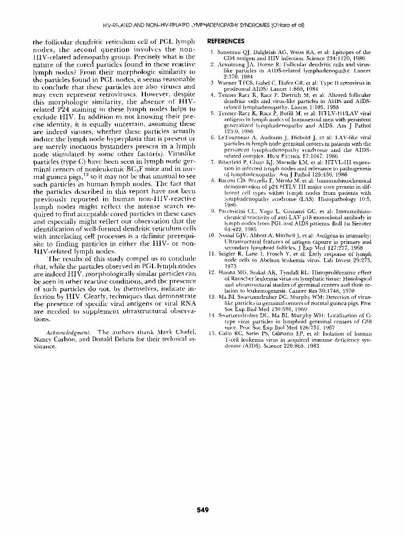
the follicular dendrit ic ret iculum cell o f PGL lymph n o d e s , the s e c o n d q u e s t i o n invo lves the n o n - HIV-re la ted adenopa thy group. Precisely what is the na ture of the cored particles found in these reactive lymph nodes? From their morphologic similarity to the particles found in PGL nodes, it seems reasonable to conclude that these particles are also viruses and may even represent retroviruses. However, despite this mor pho log i c similarity, the absence o f H I V - related P24 staining in these lymph nodes helps to exclude HIV. In addit ion to not knowing their pre- cise identity, it is equally uncertain, assuming these are indeed viruses, whether these particles actually induce the lymph node hyperplasia that is present or are merely inocuous bystanders present in a lymph node stimulated by some other factor(s). Viruslike particles (type C) have been seen in lymph node ger- minal centers of nonleukemic BC3F mice and in nor- mal guinea pigs, 13 so it may not be that unusual to see such particles in h u m a n lymph nodes. The fact that the particles described in this repor t have not been previous ly r e p o r t e d in h u m a n n o n - H I V - r e a c t i v e lymph nodes might reflect the intense search re- quired to find acceptable cored particles in these cases and especially might reflect our observation that the identification of well-formed dendrit ic ret iculum cells with interlacing cell processes is a definite prerequi- site to f inding particles in either the HIV- or non- HIV-re la ted lymph nodes.
The results o f this study compel us to conclude that, while the particles observed in PGL lymph nodes are indeed HIV, morphologically similar particles can be seen in o ther reactive conditions, and the presence o f such particles do not, by themselves, indicate in- fection by HIV. Clearly, techniques that demonst ra te the presence o f specific viral antigens or viral RNA are needed to supplement ultrastructural observa- tions.
Acknowledgment. The authors thank Mark Chafel, Nancy Carlson, and Donald Delutis for their technical as- sistance.
REFERENCES
1. Sattentau QJ, Dalgleish AG, Weiss RA, et al: Epitopes of the CD4 antigen and HIV infection. Science 234:1120, 1986
2. Armstrong JA, Horne R: Follicular dendritic cells and virus- like particles in AIDS-related lymphadenopathy. Lancet 2:370, 1984
3. Warner TFCS, Gabel C, Hafez GR, et ah Type D retrovirus in prodromal AIDS? Lancet 1:860, 1984
4. Tenner-Racz K, Racz P, Dietrich M, et al: Altered follicular dendritic cells and virus-like particles in AIDS and AIDS- related lymphadenopathy. Lancet 1:105, 1985
5. Tenner-Racz K, Racz P, Bofill M, et al: HTLV-III/LAV viral antigens in lymph nodes of homosexual men with persistent generalized lymphadenopathy and AIDS. Am J Pathol 123:9, 1986
6. LeTourneau A, Audouin J, Diebold J, et ah LAV-like viral particles in lymph node germinal centers in patients with the persistent lymphadenopathy syndrome and the AIDS- related complex. HUM PATHOL 17: 1047, 1986
7. Biberfeld P, Chayt KJ, Marselle LM, et al: HTVL-III expres- sion in infected iymph nodes and relevance to pathogenesis of lymphadenopathy. Am J Pathol 125:436, 1986
8. Baroni CD, Pezzella F, Mirolo M, et al: Immunohistochemical demonstration of p24 HTLV-III major core protein in dif- ferent cell types within lymph nodes from patients with lymphadenopathy syndrome (LAS). Histopathology 10:5, 1986
9. Parravicini CL, Vago L, Costanzi GC, et al: Immunohisto- chemical reactivity of anti-LAV p 18 monoclonal antibody in lymph nodes froin PGL and AIDS patients. Boll 1st Sieroter 64:422, 1985
10. Nossal GJV, Abbott A, Mitchell J, et al: Antigens in immunity: Uttrastructural features of antigen capture in primary and secondary lymphoid follicles. J Exp Med 127:277, 1968
11. Seigler R, Lane I, Frosch Y, et al: Early response of lymph node cells to Abelson leukemia virus. Lab Invest 29:273, 1973
12. Hanna MG, Szakal AK, Tyndall RL: Histoproliferative effect of Rauscher leukemia virus on lymphatic tissue: Histological and ultrastructural studies of germinal centers and their re- lation to leukemogenesis. Cancer Res 30:1748, 1970
13. Ma BI, Swartzendruber DC, Murphy WM: Detection of virus- like particles in germinal centers of normal guinea pigs. Proc Soc Exp Biol Med 130:586, 1969
14. Swartzendruber DC, Ma BI, Murphy WH: Localization of C- type virus particles in lymphoid germinal centers of C58 mice. Proc Soc Exp Biol Med 126:731, 1967
15. Gallo RC, Sarin PS, Gilmann EP, et al: Isolation of human T-cell leukemia virus in acquired immune deficiency syn- drome (AIDS). Science 220:865, 1983
549