delivery of optical contrast agents ... - kortum.rice.edu of optical...rice university department of...
TRANSCRIPT
Journal of Biomedical Optics 142 021013 MarchApril 2009
Dpd
ARD6H
KUD6H
RRD6HE
1
Arpfeopaihw
sdsie
ApHr
J
elivery of optical contrast agents using Triton-X100 art 2 enhanced mucosal permeation for the etection of cancer biomarkers
nne L van de Ven ice University epartment of Bioengineering MS 142 100 Main Street Keck Hall Suite 116 ouston Texas 77005
aren Adler-Storthz niversity of Texas Dental Branch epartment of Diagnostic Sciences 516 MD Anderson Blvd DBB 4133 ouston Texas 77030
ebecca Richards-Kortum ice University epartment of Bioengineering MS 142 100 Main Street Keck Hall Suite 116 ouston Texas 77005 -mail rkortumriceedu
Abstract Uniform delivery of optical contrast agents through mu-cosal tissue has proven a significant challenge Topical permeation enhancers that have proven useful for skin demonstrate limited suc-cess in mucosal tissue We sought to develop a topical permeation strategy capable of delivering tissue-impermeant molecular-specific contrast agents through mucosal epithelium in a uniform controlled manner We demonstrate that Triton-X100 can be utilized to deliver targeted and untargeted optical contrast agents through freshly ex-cised normal mucosal epithelium and epithelial cancer Macromol-ecules up to 150 kDa in size were successfully delivered via transcel-lular and paracellular routes The depth of Triton-mediated permeation was modulated by varying the treatment time and con-centration Uniform epithelial penetration to a depth of 500 m was achieved in 15 h for molecules of 40 kDa or less Larger optical probes required longer treatment times Coadministration of molecular-specific contrast agents with Triton-X100 treatment facili-tated simultaneous labeling of biomarkers on the cell membrane in the cytoplasm and in the nucleus with high specificity Together these data suggest that Triton-X100 is a promising topical permeation enhancer for mucosal delivery of tissue-impermeant molecular-specific optical contrast agents copy 2009 Society of Photo-Optical Instrumentation Engineers DOI 10111713090437
Keywords detergents mucosa epidermal growth factor receptor Cyclin D1 fluorescence Paper 08271SSR received Aug 4 2008 revised manuscript received Nov 11 2008 accepted for publication Dec 1 2008 published online Mar 10 2009
Introduction
dvances in molecular-specific optical contrast agents haveecently shown promise for the detection of cancer and itsrecursors Targeting agents that have been successfully usedor optical detection of cancer biomarkers in preclinical mod-ls include antibodies1ndash6 growth factors7ndash11 peptide analogsf extracellular ligands12ndash15 and enzymatically activatableolymers16ndash20 The coupling of optical tags to these targetinggents allows molecular events to be monitored noninvasivelyn vivo21 These molecular-specific optical contrast agentsave the potential to provide dynamic real-time informationithout the need for biopsy and associated patient discomfort
The use of optical contrast agents as topical probes for thecreening of epithelial precancer has been hindered by theifficulty of delivering macromolecules through mucosal tis-ue The molecular changes preceding cancer generally beginn the basal layers of epithelium22 thus early detection strat-gies require contrast agents be delivered at least several hun-
ddress all correspondence to Rebecca Richards-Kortum Rice University De-artment of Bioengineering MS 142 6100 Main Street Keck Hall Suite 116ouston Texas 77005 Tel 713-348-3823 Fax 713-348-5877 E-mail
kortumriceedu
ournal of Biomedical Optics 021013-
dred microns deep Studies of small molecule optical probes with tissue-permeant properties such as fluorescent sugar derivatives23ndash25 and acriflavine26ndash28 have highlighted the promise of using optical imaging to distinguish molecular changes in small populations of cells The penetration of larger molecules through mucosal tissue however is substan-tially reduced for molecular weights above 300 Da29 Chemi-cal modification strategies including increasing lipophilicity3031 conjugation with polymers3233 and encapsu-lation in liposomes34ndash36 have improved the penetration of molecules up to 6 kDa in size
Topical permeation enhancers substances that temporarily reduce the impermeability of tissues have been investigated for the delivery of larger molecules Dimethyl sulfoxide DMSO for example enhances the transdermal permeation of a variety of drugs by modifying the keratin structure and lipid composition of skin37 Combinations of topical perme-ation enhancers have been shown to facilitate the transdermal penetration of proteins as large as 140 kDa3839 Permeation enhancers shown to be effective in skin however have met little success in mucosal tissue40 Studies utilizing various sur-factant lipid and bile salt formulations have demonstrated
1083-3668200914202101313$2500 copy 2009 SPIE
1 MarchApril 2009 Vol 142
van de Ven Adler-Storthz and Richards-Kortum Delivery of optical contrast agents using Triton-X100 part 2hellip
e a
Tc ot d a te o p pto d
te rut a ot omfe d t c c wXet
22Ttm w f M sf
2T i P s a t
J
nhanced mucosal penetration of only small molecules suchs insulin 6 kDa and calcitonin 35 kDa41
In our companion paper Part 1 it was demonstrated thatriton-X100 can be used to permeabilize live cells in a suffi-iently reproducible manner to facilitate intracellular labelingf cancer biomarkers We hypothesized that the permeabiliza-ion properties of Triton-X100 would render it useful for theelivery of cell- and tissue-impermeant optical contrastsgents though mucosal tissue To evaluate Triton-X100 as aopical permeation enhancer for contrast agent delivery sev-ral key criteria were identified including i uniform deliveryf molecules up to 150 kDa in size ii a controlled depth ofermeation and iii the capability to wash out unboundrobes for high optical contrast Because of the need for con-rolled local delivery rather than systemic delivery new meth-dologies for the assessment of contrast agent delivery wereeveloped
In this paper we describe the use of Triton-X100 to deliverargeted and untargeted optical contrast agents in three differ-nt tissues To translate Triton-X100 for tissue use biopsies ofeproducible dimensions are topically treated with a fixed vol-me of 05ndash25 Triton-X100 The concentration and dura-ion of treatment adjusted relative to the epithelial thicknessnd cell density is selected to expose the cells to a final dosef 11 pmolcell or less Using this approach transverse sec-ions of bladder and oral mucosa specimens are probed withptically active contrast agents following Triton-X100 treat-ent to determine the extent of tissue permeation Using con-
ocal microscopy the depth and rate of macromolecule pen-tration are evaluated as a function of optical probe size toetermine under what conditions Triton-X100 treatment meetshe design criteria The delivery of molecular-specific opticalontrast agents is tested in xenograft tumor specimensotreated with Triton-X100 and compared to tissues treatedith DMSO or saline solution We demonstrate that Triton-100 can facilitate simultaneous labeling of clinically rel-
vant extracellular and intracellular biomarkers in a con-rolled uniform manner
Materials and Methods 1 Cell Lines
he targeting of EGFR- and CyclinD1-specific optical con-rast agents was evaluated using 1483 cells and xenograft tu-
ors This EGFR-positive cell line derived from a patientith head and neck squamous cell carcinoma42 was obtained
rom Dr Reuben Lotan at the MD Anderson Cancer CenterHouston Texas The 1483 cells were cultured in Dulbeccorsquos
odified Eagle Medium Nutrient Mix F-12reg mediumupplemented with L-glutamine Invitrogen Carlsbad Cali-ornia and 10 fetal bovine serum Hyclone Logan Utah
2 Tissue Models
hree different tissue models were evaluated for permeabilityn the presence and absence of topical permeation enhancersorcine oral mucosa was selected as a model of stratifiedquamous epithelium Heads of American Yorkshire pigsged 6 months were obtained from a local slaughterhouse athe time of sacrifice The buccal mucosa of the oral cavity and
1 mm of underlying tissue was separated from the sur-
ournal of Biomedical Optics 021013-
rounding muscle layers via dissection Guinea pig bladder mucosa was selected as a model of transitional epithelium Whole bladders were excised from 2ndash3 week old female Hartley guinea pigs Charles River Laboratories Wilmington Massachusetts directly following animal sacrifice Xenograft tumors generated subcutaneously in mice were selected as a model of squamous cell carcinoma for the study of cancer biomarker targeting Briefly 1483 cells 2106 viable cells in 100 L PBS were implanted subcutaneously in the left and right posterior mammary fat pads of 6ndash8 week-old fe-male NuNu mice Charles River Laboratories When tumors reached 4ndash5 mm diam the animals were anaesthetized and sacrificed via cervical dislocation All animals were cared for in accordance with institutional guidelines The protocols were reviewed and approved by the Institutional Animal Care and Use Committee at Rice University
23 Permeation of Fresh Tissues
To reproducibly permeate tissues for contrast-agent delivery tissue biopsies of uniform surface area were topically treated with permeation enhancers A 4-mm-diam dermal punch Miltex Inc York Pennsylvania was used to produce cylin-drical samples of oral and bladder mucosa Subcutaneously generated tumors grown as cylinders to the appropriate diam-eter were cut in half to expose the tumor surface The tissue cylinders were embedded vertically into 3 volvol ultra-pure agarose Invitrogen to prevent the influx of permeation enhancers and contrast agents at the tissue margins The Darcy permeability of 3 agarose has been reported as 450 nm243 and the diffusivity of macromolecules through 3 agarose is limited44 The apical surface of the epithelium was left free of agarose to facilitate topical labeling In the case of the bladder tissue the underlying muscle layers were sufficiently thick to allow the biopsies to be positioned hori-zontally in the agarose without stretching or bending Two topical permeation enhancers Triton-X100 and DMSO Sigma-Aldrich St Louis Missouri diluted in phosphate buffered saline PBS Sigma-Aldrich were evaluated for their ability to permeate fresh tissue PBS alone was used as a negative control One milliliter of permeation-enhancing so-lution was topically applied to the apical surface each tissue biopsy for 0ndash4 h at 4 degC
24 Permeabilization Detection Using Macromolecules
To determine whether Triton-X100ndashtreated tissues are selec-tively permeable to macromolecules of a specific size bladder biopsies were probed with fluorescent macromolecules of three sizes The apical surface of the epithelium was topically treated for 1 h with 05 Triton-X100 to yield a treatment dose of 08 pmolcell assuming full-thickness permeation or PBS washed once in PBS and covered with a 111 mix-ture of rhodamine-dextran 3 kDa Invitrogen fluorescein-dextran 40 kDa Invitrogen and AlexaFluor647 IgG 150 kDa Invitrogen each diluted to a concentration of 1 M in PBS Tissues were immersed with this solution for 15 min at 4 degC and then imaged using fluorescence confocal microscopy at three different excitation wavelengths de-scribed below Images were collected in 5-m steps from the surface into the tissue Following imaging the tissues were
2 MarchApril 2009 Vol 142
van de Ven Adler-Storthz and Richards-Kortum Delivery of optical contrast agents using Triton-X100 part 2hellip
w t 1
2T a t p f t p d 4 o w
2T f se w T 1 cs 1 C c e e di v ca tte
2
T b f a t Ds p f s d et
J
ashed three times in cold PBS 15 min total and reimagedo assess the removal of unbound macromolecules A total of0 bladders were evaluated in independent experiments
5 Tracking the Time Course of Tissue Permeation
o monitor Triton-mediated permeation of bladder epitheliums a function of time fresh biopsies were treated for differentime intervals The apical surface of the agarose-embeddedunch biopsies were topically treated with 05 Triton-X100or 0 5 10 15 30 or 60 min Following the permeationreatment the tissues were washed once in cold PBS androbed with the 111 mixture of fluorescent macromoleculesescribed above Confocal microscopy images were collected0 m below the tissue surface to allow for optical sectioningf both the epithelium and underlying tissues Each time pointas evaluated in five independent experiments
6 Determination of the Depth of Penetration
he depth of Triton-mediated macromolecule penetration as aunction of time and concentration was evaluated in crossections of fresh oral mucosa The apical surface of agarose-mbedded punch biopsies were topically treated for 0ndash4 hith Triton-X100 concentrations ranging from 0 to 25hese concentrations were selected to treat cells with up to1 pmolcell of Triton-X100 The permeabilized tissues wereross-sectioned using a Krumdieck tissue slicer Alabama Re-earch amp Development Munford Alabama and immersed for5 min in the 111 mixture of fluorescent macromoleculesonfocal fluorescence images of the transverse sections wereollected 40 m below the cut surface to avoid confoundingffects due to damage at cut surface The penetration depth forach size of macromolecule was evaluated by measuring theistance between the apical surface of the tissue and the lead-ng edge of the fluorescent macromolecules using ImageJ134 software httprsbwebnihgovij Measurements wereollected at 20-m intervals across four representative im-ges in four independent experiments 16 images total perreatment condition The rate of tissue permeation as a func-ion of Triton-X100 concentration was determined from a lin-ar least-squares fit to the data
7 Comparison of Macromolecule Penetration in Normal and Cancer Tissue
o determine whether the depth and rate of permeation variesetween tissues macromolecule penetration was assessed inresh normal oral mucosa and squamous cell carcinoma Thepical surface of agarose-embedded oral biopsies and 1483umors were treated for 1 h with only 1 Triton-X100 10MSO or PBS Following the permeation treatment the tis-
ues were cross-sectioned with a Krumdieck tissue slicer androbed with the 111 mixture of fluorescent macromoleculesor 15 min Confocal fluorescence images of the transverseections were collected 40 m below the cut surface Theepth of penetration from the apical surface was assessed forach macromolecule at 20-m intervals across four represen-ative images from four independent experiments 16 images
ournal of Biomedical Optics 021013-
total per treatment condition The rate of tissue permeation was determined from a linear least-squares fit to the data
28 Estimation of the Normalized Triton-X100 Concentration in Tissue
The average dose of Triton-X100 per cell was determined as a function of treatment time and depth Briefly the average per-meation depth was measured at different time intervals for tissues of known surface area treated with a fixed volume and concentration of Triton-X100 An average cell was approxi-mated as a sphere 833 m in diameter based on confocal microscopy images The measured volume of treated tissue was divided by the average cell volume to yield the number of treated cells The concentration of Triton-X100 in moles per milliliter was divided by the number of treated cells to deter-mine the normalized Triton-X100 concentration
29 Cell Viability Assessment in Tissue Phantoms
Cell viability following Triton-X100 treatment was assessed in three-dimensional tissue phantoms The optical properties of these phantoms have been validated in Ref 45 Briefly subconfluent monolayers of 1483 cells were treated with 005 trypsin-EDTA Invitrogen pelleted and washed once with cold PBS The cells were counted under light micros-copy using a hemocytometer pelleted in resuspended in a collagen type I gel 21 mg mL Roche Diagnostics Corp Indianapolis Indiana at concentration of 1ndash10107 cells ml Then 100 L aliquots were cured for 30 min at 37 degC and then placed in prewarmed complete me-dia and 24 h later the tissue phantoms were treated with 1 mL of 0ndash5 Triton-X100 for 1 h at 4 degC The Triton-X100 concentrations were selected to yield a normalized concentra-tion of 055 pmolcell and above to permeabilize all cells After treatment the phantoms were washed and placed in fresh prewarmed media Cell viability was assessed 4 h fol-lowing Triton-X100 treatment Prewarmed 3-45-dimethylthiazol-2-yl-25-diphenyl tetrazolium bromide MTT Sigma-Aldrich was added to the supernatant for 20 min at a concentration of 05 mg mL In viable cells re-duction of the MTT reagent led to the intracellular deposition of a blue formazan product visible to the eye The tissue phan-toms were imaged whole and in cross section using light microscopy
210 Synthesis and Validation of Molecular-Specific Contrast Agents
Mouse antihuman antibodies targeted to epidermal growth factor receptor EGFR clone 108 custom synthesized by the Baylor College of Medicine Houston Texas and Cyclin D1 clone 72-13G Santa Cruz Biotechnology Inc Santa Cruz California were reacted with AlexaFluorreg 647 and Al-exaFluorreg 488 carboxylic acid succinimidyl esters using commercially available labeling kits Invitrogen The purified conjugates were suspended in PBS at a concentration of 10 and 02 mg mL respectively Dye-labeled isotype controls were synthesized at the same concentrations Prior to tissue labeling the bioactivity and specificity of the conjugates were
3 MarchApril 2009 Vol 142
van de Ven Adler-Storthz and Richards-Kortum Delivery of optical contrast agents using Triton-X100 part 2hellip
c b
2
T mcb Da a w ts a o i t
2
T m 1 t c a a tfbq t
2
Af 4 lt w s e 6 at 1 n h s wt p a
J
onfirmed in live and fixed 1483 cells as described in Ref 46oth in the presence and absence of Triton-X100
11 Comparison of AntiEGFR Delivery Strategies in Tumors
o assess the ability of permeation enhancers to deliverolecular-specific optical contrast agents through tissue sub-
utaneously generated 1483 xenograft tumors were for la-eled for EGFR in the presence of 1 Triton-X100 10MSO or PBS AntiEGFR-647 150 was diluted in perme-
tion enhancers and topically applied to the cut surface ofgarose-embedded tumors for 1 h at 4 degC The labeled tissuesere washed three times in cold PBS sliced perpendicular to
he surface using a Krumdieck tissue slicer and counter-tained for 30 s with 005 acriflavine-HCL Sigma-Aldrich cell permeant nucleic acid dye The depth and localizationf EGFR labeling was assessed from confocal fluorescencemages acquired 40 m below the cross sectionrsquos surface Aotal of 10 tumors 5 mice were evaluated
12 Demonstration of Multitarget Labeling in Tumors
o demonstrate the feasibility of simultaneously targetingultiple cancer biomarkers in different cell compartments
483 xenograft tumors were simultaneously labeled withhree tissue andor cell impermeant probes AntiEGFR-647150 antiCyclinD1-488 110 and propidium iodide15 M Sigma Aldrich were used to label cell membraneytoplasmic and nuclear targets respectively The contrastgents and isotype controls were diluted in 1 Triton-X100nd topically applied for 1 h The labeled tissues were washedhree times in cold PBS and sliced perpendicular to the sur-ace using a Krumdieck tissue slicer The localization of la-eling was assessed from confocal fluorescence images ac-uired 40 m below the cross sectionrsquos surface A total of 10umors 5 mice were evaluated
13 Confocal Image Acquisition and Analysis
ll images were obtained using a Carl Zeiss LSM 510 con-ocal microscope Thornwood New York equipped with88 nm 30 mW 543 nm 1 mW and 633 nm 5 mWasers Images were collected using photomultiplier tube de-ectors and Zeiss LSM 5 image examiner software Samplesere sequentially excited with each laser line with power
ettings of 50 100 and 100 respectively Fluorescencemission was collected using 500ndash530 565ndash615 and50ndash710 nm bandpass filters respectively Tissue reflectancet 633 nm was collected using a 635-nm dichroic beam split-er Images were acquired at 05 fps using a 63X oil 20X or0X objectives with a pinhole of 256 Airy units For theonspecific macromolecule permeation studies the gain waseld constant just below saturation level of the extracellularolution In the molecular-specific targeting studies the gainas held constant at 440 for antiEGFR-647 633-nm excita-
ion 650 for antiCyclin D1-488 488-nm excitation 400 forropidium iodide 543-nm excitation and 535 forcriflavine-HCL 488-nm excitation
ournal of Biomedical Optics 021013-
3 Results 31 Permeabilization of Fresh Guinea Pig Bladder The ability of Triton-X100 to permeabilize guinea pig bladder mucosa was assessed using untargeted fluorescent macromol-ecules of three different sizes Figure 1 shows representative confocal images of tissue biopsies topically treated for 1 hwith 05 Triton-X100 or PBS and then probed with a 111 mixture 3 kDa rhodamine-dextran 40 kDa fluorescein-dextran and 150 kDa Alexa647-IgG The tissue reflectance images are shown on the left and the corresponding fluores-cence images are shown to the right The yellowwhite lines indicate the basal boundaries of the epithelium Because of the three-dimensional folding of the deflated bladder it was possible to image the transitional epithelium in cross section using confocal microscopy at a depth of 40 m In the reflec-tance images the epithelium was distinguished from the un-derlying more highly scattering tissues by its darker appear-ance Triton-X100 treatment facilitated trans-epithelial penetration of all three sizes of molecules The 3 and 40 kDa molecules successfully penetrated both the cytoplasm and nucleus of epithelial cells whereas the 150 kDa molecules only penetrated the cytoplasm of epithelial cells With several brief washes a significant portion of the macromolecules were removed In the PBS-treated controls the macromol-ecule penetration was limited to one to two layers of superfi-cial cells
The time course of tissue permeation with Triton-X100 treatment was tracked using confocal microscopy Figure 2 shows representative images of biopsies treated with 05 Triton-X100 for 5 10 and 15 min and then probed with fluo-rescent macromolecules of three sizes At 5 min the leading edge of permeabilized epithelium featured irregular borders At 10 min full-thickness epithelial permeation was observed in some epithelial regions Individual nonpermeabilized cells were easily distinguished by their dark appearance At 15 min trans-epithelial penetration was observed for all three sizes of molecules With time the overall fluorescence inten-sity of the epithelium increased to approach that of the exog-enous solution however the nuclei of cells probed with 150 kDa molecules remained dark Fluorescence in the un-derlying tissues was observed as early as 5 min and increased steadily with time
32 Depth of Mucosal Permeation
The depth of Triton-mediated permeation was assessed in a model of porcine oral mucosa ranging in thickness from 600 to 1000 m Epithelial punch biopsies were topically treated with different concentrations of Triton-X100 ranging from 0 to 25 for different time intervals ranging from 0 to 4 h sliced in cross section and probed with fluorescent macromolecules Representative confocal fluorescent images of macromolecule permeation as a function of time can be found in Fig 3 Figure 4 demonstrates the relationship of treat-ment time and Triton-X100 concentration to the depth of tis-sue permeation Triton-X100 treatment was found to produce very uniform reproducible tissue permeation with well-defined borders parallel to the treatment surface When the leading edge of macromolecule labeling was assessed at regu-lar time intervals the depth of penetration was found to in-
4 MarchApril 2009 Vol 142
van de Ven Adler-Storthz and Richards-Kortum Delivery of optical contrast agents using Triton-X100 part 2hellip
F0ybww
c m 1 5 l
a f c te c naa nm r w
J
ig 1 Macromolecule penetration of guinea pig bladder epithelium 5 Triton-X100 or PBS and then probed with a 111 mixture of 3 kellowwhite lines indicate the basal surface of the epithelium In refley its darker appearance Following treatment with Triton-X100 tranashing with PBS the fluorescence intensity of the labeled tissue waas limited to a few cells in the superficial layers The scale bar repr
rease at a steady linear rate Fig 4a The smaller probeolecules consistently showed deeper penetration than the50 kDa molecules Macromolecule penetration to a depth of00 m was achieved in 15 h for molecules of 40 kDa oress and in 4 h for molecules of 150 kDa
Using a linear least-squares fit the rate of tissue perme-tion was evaluated for a range of Triton-X100 concentrationsFig 4b Increasing the Triton-X100 concentration wasound to increase the rate of tissue permeation however atoncentrations of 1 Triton-X100 little improvement inhe rate of permeation was observed Size-dependent differ-nces in macromolecule penetration were observed for alloncentrations of Triton-X100 although there was generallyo significant difference between the penetration depth of 3-nd 40 kDa macromolecules Longer probe times were evalu-ted to confirm that the differences in penetration depth wereot due to inadequate labeling time data not shown Further-ore samples in which the keratin layer was mechanically
emoved showed no differences in the rate of permeationhen compared to intact specimens data not shown
ournal of Biomedical Optics 021013-
ng permeation treatment Tissues were topically treated for 1 h with damine-dextran 40 kDa fluorescein-dextran and Alexa647-IgG The images the epithelium was distinguished from the underlying tissues elial penetration was observed for all three sizes of molecules After cantly reduced In PBS-treated controls macromolecule penetration
100 m
33 Comparison of Macromolecule Penetration in Normal and Cancer Tissue
To determine whether the depth and rate of topical permeation varies between normal and abnormal tissue macromolecule penetration following of topical permeation was evaluated in normal oral mucosa and tumor models Figure 5 compares the depth and rate of macromolecule penetration in two tissue models using three different labeling solutions The maximal penetration depth following 1 h of treatment with 1 Triton-X100 was found to be 25 greater in squamous carcinoma tissue than normal epithelium Fig 5a Little permeation 100 m deep was observed in DMSO- and PBS-treated tissues When the rate of macromolecule penetration follow-ing Triton-X100 treatment of was evaluated as a function of size all sizes of macromolecules consistently penetrated more rapidly in carcinoma tissue than normal epithelium Fig 5b As previously observed the rate of macromolecule per-meation was highly reproducible and linear at all time points evaluated The largest difference in penetration rate was ob-
followiDa rhoctances-epiths signifiesents
5 MarchApril 2009 Vol 142
van de Ven Adler-Storthz and Richards-Kortum Delivery of optical contrast agents using Triton-X100 part 2hellip
Fwteps
see
3
T f n puaa cX wl TXc f
J
ig 2 The time course of tissue permeation following treatment withith 05 Triton-X100 for 0ndash60 min and then probed fluorescent mol
he tissue surface are shown for the 5- 10- and 15-min time popithelium The depth and uniformity of permeation increased witenetration of different size probe molecules Fluorescence signal in scale bar represents 100 m
erved for the 150 kDa molecules although this size of mol-cule consistently penetrated more slowly than smaller mol-cules
4 Relation of Triton-X100 Concentration to the Number of Treated Cells
he average dose of Triton-X100 per cell was estimated asunction of treatment time and depth Table 1 provides theormalized Triton-X100 concentration in terms of picomoleser cell The calculations were based on an average cell vol-me the known tissue surface area and the measured perme-tion depth In the case where only one cell layer was perme-bilized 83 m deep the Triton-X100 concentration perell was on the order of 62ndash123 pmolcell As the Triton-100 permeated deeper the average concentration per cellas reduced Full thickness permeation of the bladder epithe-
ium with a 05 solution yielded an average of 083 pmolriton-X100 per cell A 30ndash60 min treatment of 10 Triton-100 in thicker tissues further lowered the normalized con-
entration to 028ndash057 for oral mucosa tissue and 021ndash042or squamous cell carcinoma tissue
ournal of Biomedical Optics 021013-
Triton-X100 Fresh guinea pig bladder biopsies were topically treated f three sizes Representative confocal images acquired 40 m below e yellowwhite lines indicate the apical and basal surfaces of the ent time Differences were observed between the cell and tissue
was observed as early as 5 min following Triton-X100 treatment The
35 Viability Assessment of Tissue Phantoms
Tissue phantoms were assayed for cell viability following treatment with different concentrations of Triton-X100 Figure 6 shows representative images of phantoms containing five million cells each treated with 0ndash5 Triton-X100 for 1 h and then coincubated with MTT reagent The normalized Triton-X100 concentration per cell was determined from the number of cells incorporated into each phantom Phantoms treated with 1 Triton-X100 or less 33 pmolcell demonstrated an intense blue color indicative of MTT reduction by viable cells When imaged in cross section these phantoms showed intense staining throughout In contrast phantoms treated with concentrations above 1 33 pmolcell demonstrated an overall loss in color intensity as well as a lack of staining at the phantom periphery suggestive of cell death at these higher Triton-X100 concentrations
36 EGFR Targeting in Cancer Tissue
To determine whether Triton-X100 is useful for the delivery of molecular-specific optical contrast agents subcutaneous tu-
05 ecules oints Thh treatmtroma
6 MarchApril 2009 Vol 142
van de Ven Adler-Storthz and Richards-Kortum Delivery of optical contrast agents using Triton-X100 part 2hellip
FfRc
mm tX ca
FwtiT
J
ig 3 Permeation of the porcine oral mucosa following treatment wresh tissue biopsies for 0 ndash60 min then the tissue was sliced probeepresentative confocal images acquired 40 m below the cut surfacharacterized by a well-defined border that progressed deeper with ti
ors were labeled for EGFR in the presence of topical per-eation enhancers Freshly excised tumors were topically
reated for 2 h with antiEGFR-647 diluted in 1 Triton-100 10 DMSO or PBS The labeled tissues were sliced in
ross section counterstained with acriflavine-HCL and im-ged perpendicular to the labeling surface Figure 7 shows
ig 4 Permeation of normal oral mucosa as a function of time and Trith different concentrations of Triton-X100 for regular time intervals
40 kDa and 150 kDa The depth and rate of macromransverse tissue sections a The relationship between the depth of pncrease in permeation depth is observed with time b Rate of tisriton-X100 concentration increased the depth of tissue permeabilizat
ournal of Biomedical Optics 021013-
Triton-X100 Triton-X100 was topically applied to the top surface of fluorescent molecules of three sizes and imaged in cross section
hown for the 10- 30- and 60-min time points Tissue permeation was e scale bar represents 100 m
representative confocal fluorescence images of EGFR labeling in the presence of different topical permeation enhancers The surface of the tissue where the contrast agent was applied appears at the top of each panel EGFR labeling is denoted in red and acriflavine-HCL labeling in green Treatment with 1 Triton-X100 facilitated the uniform delivery of antiEGFR-647
00 concentration Normal porcine oral mucosa was topically treated and probed with fluorescent macromolecules of three sizes 3 kDa penetration was determined from confocal fluorescence images of tion and duration of treatment with 1 Triton-X100 A steady linear meation as a function of Triton-X100 concentration Increasing the wever the extent of macromolecule penetration was limited by size
ith 1d with
e are sme Th
iton-X1 slicedoleculeermea
sue perion ho
7 MarchApril 2009 Vol 142
van de Ven Adler-Storthz and Richards-Kortum Delivery of optical contrast agents using Triton-X100 part 2hellip
F1wpcpm
tt c r d set l
3T c t p e
J
ig 5 Comparison of macromolecule penetration in normal oral muco0 DMSO or PBS sliced and then probed with fluorescent macroas determined from confocal fluorescence images of transverse tissueermeation enhancers measured using 3 kDa rhodamine-dextran Tancer tissue than normal epithelium Little permeation was observenetration by size with 1 Triton-X100 treatment The rate of peacromolecules evaluated
o a depth of 500ndash600 m Washing of the tissue after con-rast agent delivery yielded a honeycomb pattern of labelingonsistent with that observed in vitro EGFR labeling waseproducibly observed around all squamous cells to the fullepth of contrast agent delivery The intensity of labeling wasimilar to that observed in cell monolayers labeled in the pres-nce and absence of Triton-X100 data not shown In con-rast EGFR labeling in DMSO- and PBS-treated tissues wasimited to the superficial layers of cells 20ndash60 m deep
7 Multitarget Labeling in Tumors
o demonstrate the feasibility of delivering multiple opticalontrast agents to different cell compartments three differentargeting moieties were topically applied to tumors in theresence of Triton-X100 The specificity and bioactivity ofach targeting moiety was established in vitro prior to tissue
Table 1 Triton-X100 concentration per cell as a
Tissue
Triton-X100 concentration
Averagepermeation d
m
Bladder deflated
05 83
60
Oral mucosa 10 83
175
350
Squamous cell 10 83 carcinoma
235
470
ournal of Biomedical Optics 021013-
squamous cell carcinoma Tissues were treated with 1 Triton-X100 les of three sizes The depth and rate of macromolecule penetration ns a The maximal penetration depth following a 1-h treatment with imal depth with Triton-X100 treatment was found to be greater for DMSO- and PBS-treated controls b The rate of macromolecule n was consistently more rapid in carcinoma tissue for all sizes of
labeling No differences in targeting were observed with Triton-X100 co-treatment of live and fixed cells data not shown Figure 8 shows representative confocal images of freshly excised tumor tissue simultaneously labeled for EGFR Cyclin D1 and nucleic acid content in the presence of 1 Triton-X100 The tissues were topically labeled washed sliced and imaged perpendicular to the labeling surface AntiEGFR-647 labeling red appeared in a characteristic honeycomb pattern AntiCyclin D1-488 labeling green was diffusely spread through the cytoplasm of cancer cells Pro-pidium iodide labeling orange appeared in the nuclei of per-meabilized cells EGFR Cyclin D1 and propidium iodide labeling always appeared together overlay suggesting that the labeling is limited to cells permeabilized by the Triton-X100 treatment Little to no antibody labeling was observed when isotype controls and propidium iodide were adminis-
on of treatment time and depth
Approx treatment time min
No of Treated cells 106
Normalized concentration pmolcell
5
15
134
100
617
083
5 134 123
30 291 057
60 5816 028
5 134 123
30 390 042
60 780 021
sa andmolecu sectiohe maxed withnetratio
functi
epth
8 MarchApril 2009 Vol 142
van de Ven Adler-Storthz and Richards-Kortum Delivery of optical contrast agents using Triton-X100 part 2hellip
Ff
c m B o at
t c s t
Fasuw
J
ig 6 Tissue phantoms assayed for viability after treatment with dif-erent concentrations of Triton-X100 Phantoms containing 5 106
ells each were treated with 1 mL Triton-X100 for 1 h returned toedia for 4 h and then coincubated with a MTT reagent for 20 minlue color deposition is indicative of viable cells Intense staining wasbserved in phantoms treated with 1 Triton-X100 or less33 pmolcell Phantoms treated with Triton-X100 concentrationsbove 1 33 pmolcell demonstrated an overall loss in color in-ensity and staining at the periphery
ered in the presence of Triton-X100 suggesting that unboundontrast agents were sufficiently removed with the washingteps No propidium iodide labeling was present in tissuesreated with PBS instead of Triton-X100 data not shown
ig 7 EGFR targeting in the presence of topical permeation enhancersntiEGFR-647 red diluted in 1 Triton-X100 10 DMSO or PBShown at the top Tissues were counterstained with acriflavine-HCLniform EGFR labeling was observed around all squamous cells to a das limited to the superficial cell layers The scale bar represents 100
ournal of Biomedical Optics 021013-
4 Discussion
Optical contrast agents developed for cancer biomarker detec-tion have demonstrated high specificity in cell and tissue models Generally ranging in size from 6 kDa peptides and affibodies to 150 kDa antibodies most molecular-specific contrast agents have a limited ability to penetrate mucosal tissue Chemical modification and encapsulation strategies to improve tissue penetration have only proven useful for mol-ecules at the small end of the size spectrum41 The topical delivery of tissue-impermeant contrast agents presents an in-teresting challenge because the success of tissue labeling is dependent on the efficiency and uniformity of contrast agent delivery Furthermore unbound contrast agents generally need to be washed out prior to imaging for high optical con-trast In this paper we evaluate ability of Triton-X100 to de-liver optical contrast agents that lack the appropriate physio-chemical properties for unaided tissue permeation
Confocal microscopy studies of macromolecule penetra-tion in the bladder mucosa were used to examine the unifor-mity of tissue permeabilization The bladder mucosa is a par-ticularly interesting model because the natural folding of the deflated bladder allows the epithelium to be imaged in cross section without the need for tissue slicing In this model we observed that a fixed volume of 05 Triton-X100 can deliver molecules up to 150 kDa in size across treated epithelium By
taneously generated 1483 tumors were topically labeled for 2 h with ssues were sliced and imaged perpendicular to the labeling surface to highlight the tissue morphology In Triton-X100 treated tissues 500ndash600 m EGFR labeling in the DMSO- and PBS-treated controls
Subcu The ti greenepth of m
9 MarchApril 2009 Vol 142
van de Ven Adler-Storthz and Richards-Kortum Delivery of optical contrast agents using Triton-X100 part 2hellip
Fmrwcwm
omXp s a a ml o p i dXg
st d o p ar p e c
J
ig 8 Cotargeting of clinically relevant cancer biomarkers Subcutaolecular-specific contrast agents diluted in 10 Triton-X100 The c
ed Cyclin D1 in the cytoplasm antiCyclin D1 green and nucleicere washed sliced and imaged perpendicular to the labeling suompartments EGFR Cyclin D1 and nucleic acid labeling always apas highly uniform and reproducible from sample to sample Little toent suggesting that the washing of the tissue was sufficient to remo
ur estimate the cells of the epithelium are receiving a treat-ent dose ranging from 617 to 083 pmolcell as the Triton-100 progresses through the tissue Tissue permeation ap-ears to primarily follow a transcellular route appearing as atrong fluorescent signal within cells In tissues probed with 3nd 40 kDa macromolecules the fluorescence is uniformcross the entire epithelium In tissues probed with 150 kDaolecules only the cell nuclei appear as dark spots Extracel-
ular fluorescence is evident between cells at the leading edgef the treatment suggesting that Triton-X100 also facilitatesaracellular delivery Washing of the epithelium removes bothntracellular and extracellular probes yielding a uniformlyark tissue Together these observations indicate that Triton-100 may be useful for the delivery of contrast agents tar-eted to both intracellular and extracellular biomarkers
Studies of Triton-X100 treatment in fresh oral mucosapecimens indicate that the depth of permeation can be con-rolled The leading edge of permeation treatment was wellefined at all concentrations examined allowing measurementf penetration depth as a function time and concentration Theenetration depth increased linearly with treatment time forll sizes of probe macromolecules over a 4-h observation pe-iod There was no apparent delay for the contrast agents toermeate through an intact keratin layer The maximal rate ofpithelial permeation increased with increasing Triton-X100oncentration These trends were highly reproducible varying10 from sample to sample This data demonstrate that it is
ournal of Biomedical Optics 021013-1
y generated 1483 tumors were topically labeled for 1 h with three agents were targeted to EGFR on the cell membrane antiEGFR-647 in the nucleus propidium iodide orange After labeling the tissues The contrast agents were found to localize to the appropriate cell together in treated tissue overlay The observed pattern of labeling eling was observed in IgG-treated controls with Triton-X100 cotreat-ound contrast agents The scale bar represents 20 m
possible to regulate the depth of Triton-X100 treatment by adjusting the Triton-X100 concentration and treatment dura-tion The depth of labeling following treatment was deter-mined by contrast agent size with larger probes demonstrat-ing shallower penetration at the same treatment concentration as smaller molecules Interestingly it does not appear advan-tageous to use Triton-X100 concentrations much above 1 because little improvement in the rate of permeation is ob-served between tissues treated with 1 and 25 Triton-X100
Differences were apparent in the permeation of tissues when compared to cell monolayers In our companion paper Part 1 we showed that cells can be permeabilized with 027 pmolcell Triton-X100 and that nuclear penetration of 40 and 150 kDa molecules can be achieved with 055ndash11 pmolcell Triton-X100 In tissue nuclear penetra-tion is limited to molecules of 40 kDa or less Occasional nuclear penetration of 150 kDa molecules is observed at the surface of treated tissue and is generally limited to one to two cell layers If we consider an oral biopsy treated with 1 Triton-X100 for 1 h the normalized Triton-X100 concentra-tion is in the range of 055ndash11 pmolcell for approximately half of the treatment time Enhanced nuclear permeation is not observed with higher Triton-X100 concentrations or changes in treatment time Thus it appears likely that Triton-X100 preferentially permeabilizes cell membranes in its movement
neouslontrast acids rface peared no labve unb
0 MarchApril 2009 Vol 142
van de Ven Adler-Storthz and Richards-Kortum Delivery of optical contrast agents using Triton-X100 part 2hellip
t t
c c ctm T t s l c sX o 3 w
ct cc a te p stc dm
a at ccs b w os t it m b p l de T cic s Di
J
hrough tissue and is not sufficiently concentrated locally forhe generation of large nuclear pores
In tissue the normalized Triton-X100 concentrationhanges with treatment time and depth such that superficialells are treated to higher doses of Triton-X100 than deeperells Nevertheless cell viability experiments in tissue phan-oms seem to indicate that in a three-dimensional environ-ent cells are more likely to remain viable at higher doses ofriton-X100 In phantoms the viability cutoff is33 pmolcell with cells at the periphery losing their ability
o metabolize MTT at higher concentrations These studiesuggest that at Triton-X100 concentrations of 33 pmolcell oress cell viability is determined the mean concentration perell rather than local concentration gradients This leads us topeculate that tissues can be safely permeated with Triton-100 as long as the treatment time and concentration areptimized to fall below a normalized concentration of3 pmolcell Further studies are needed to determinehether this will hold true in vivo
Depth control will be advantageous for the use of topicalontrast agents in humans By limiting the penetration of op-ical contrast agents to a finite depth the systemic delivery ofontrast agents with undefined toxicity can be avoided Be-ause the mucosal epithelium is a renewable tissue contrastgents adhering to epithelial cells will likely be shed withissue turnover For the optical detection of disease biomark-rs some compensation may be required for differences inermeation rate of normal and abnormal tissue In the tissuestudied here however a 25 difference was observed be-ween normal squamous epithelium and undifferentiated can-er tissue Depth-sensitive optical imaging or spectroscopyevices could potentially be used to monitor the depth of per-eation and determine when to halt the reaction by washingThe use of Triton-X100 as a topical permeation enhancer
llows the delivery of optical contrast agents that lack theppropriate size and chemical properties for tissue penetra-ion Molecular-specific contrast agents as large as antibodiesan be successfully delivered to both intracellular and extra-ellular targets using Triton-X100 In this paper we demon-trate that three clinically relevant biomarkers of cancer cane simultaneously labeled with the topical co-administrationith Triton-X100 The EGFR which is located on the surfacef cells is thought to be a promising target because its expres-ion is elevated in a variety of dysplastic and cancerousissues47ndash49 The overexpression of Cyclin D1 detected heren the cytoplasm is associated with a poor prognosis in pa-ients with head and neck tumors50ndash53 Measures of nuclearorphology delineated here with propidium iodide have
een proposed as an objective measure of the transition fromreneoplastic to neoplastic tissue54 In vitro studies of cellabeling in the presence and absence of Triton-X100 haveemonstrated that structure and availability of these biomark-rs in live cells is similar to that fixed cells data not shownhe targeting of these biomarkers with molecular-specificontrast agents in tissue yielded uniform reproducible label-ng in all transverse sections examined by confocal micros-opy The localization and intensity of tissue labeling wasimilar to that observed in vitro In contrast treatment withMSO or PBS failed to enhance the penetration tissue-
mpermeant optical probes The feasibility of translating Triton-X100 for in vivo use
ournal of Biomedical Optics 021013-1
remains to be determined In our companion paper we dem-onstrated that Triton-X100ndashmediated permeabilization of cells is reversible and that cells retain their metabolic and prolif-erative capacities while recovering membrane integrity Oth-ers have demonstrated that only concentrations of 10 pro-duce visible side effects in patch tests of human skin consisting of transdermal water loss and erythema55 It has been reported that two or more permeation enhancers applied together can act synergistically56 suggesting that it may be possible to further reduce the effective Triton-X100 concen-tration by combining it with other topical agents Tissue dam-age reported with the use of other surfactant-based permeation enhancers has generally been limited and reversible57ndash59 The next step will be to characterize the safety toxicity biological activity and reversibility of Triton-X100ndashmediated perme-ation at clinically useful doses in animal models
5 Conclusions This paper demonstrates that Triton-X100 is a promising topi-cal permeation enhancer for the mucosal delivery of tissue-impermeant optical contrast agents Compared to other topical permeation enhancers Triton-X100 can deliver a much broader size range of molecules No chemical modification of optical contrast agents is needed both targeted and untargeted molecules as large as 150 kDa can be delivered uniformly through normal and cancer tissue The depth of tissue perme-ation can be modulated allowing for epithelial containment of topical contrast agents The coadministration of Triton-X100 with molecular-specific contrast agents facilitates the simulta-neous labeling of cell membrane cytoplasmic and nuclear targets Further work is needed to establish the safety of this approach for in vivo use
Acknowledgments
We thank Vivian Mack for her assistance with cell culture and animal care This work was supported in part by an NIH BRP Grant No CA103830 and a training fellowship from the Keck Center Nanobiology Training Program of the Gulf Coast Consortia NIH Grant No 5 T90 DK071054-03
References 1 S Folli P Westermann D Braichotte A Pelegrin G Wagnieres H
van den Bergh and J P Mach ldquoAntibody-indocyanin conjugates for immunophotodetection of human squamous cell carcinoma in nude micerdquo Cancer Res 5410 2643ndash2649 1994
2 B Ballou G W Fisher J S Deng T R Hakala M Srivastava and D L Farkas ldquoCyanine fluorochrome-labeled antibodies in vivo as-sessment of tumor imaging using Cy3 Cy5 Cy55 and Cy7rdquo Can-cer Detect Prev 223 251ndash257 1998
3 N S Soukos M R Hamblin T F Deutsch and T Hasan ldquoMono-clonal antibody-tagged receptor-targeted contrast agents for detection of cancersrdquo Proc SPIE 4259 115ndash128 2001
4 N S Soukos M R Hamblin S Keel R L Fabian T F Deutsch and T Hasan ldquoEpidermal growth factor receptor-targeted immuno-photodiagnosis and photoimmunotherapy of oral precancer in vivordquo Cancer Res 6111 4490ndash4496 2001
5 Y Koyama T Barrett Y Hama G Ravizzini P L Choyke and H Kobayashi ldquoIn vivo molecular imaging to diagnose and subtype tu-mors through receptor-targeted optically labeled monoclonal antibod-iesrdquo Neoplasia 912 1021ndash1029 2007
6 Y Koyama Y Hama Y Urano D M Nguyen P L Choyke and H Kobayashi ldquoSpectral fluorescence molecular imaging of lung me-tastases targeting HER2neurdquo Clin Cancer Res 1310 2936ndash2945 2007
1 MarchApril 2009 Vol 142
van de Ven Adler-Storthz and Richards-Kortum Delivery of optical contrast agents using Triton-X100 part 2hellip
1
1
1
1
1
1
1
1
1
1
2
2
2
2
2
2
2
2
2
J
7 K E Adams S Ke S Kwon F Liang Z Fan Y Lu K Hirschi ME Mawad M A Barry and E M Sevick-Muraca ldquoComparison ofvisible and near-infrared wavelength-excitable fluorescent dyes formolecular imaging of cancerrdquo J Biomed Opt 122 024017 2007
8 J L Kovar W M Volcheck J Chen and M A Simpson ldquoPurifi-cation method directly influences effectiveness of an epidermalgrowth factor-coupled targeting agent for noninvasive tumor detec-tion in micerdquo Anal Biochem 3611 47ndash54 2007
9 J L Kovar M A Johnson W M Volcheck J Chen and M ASimpson ldquoHyaluronidase expression induces prostate tumor metasta-sis in an orthotopic mouse modelrdquo Am J Pathol 1694 1415ndash14262006
0 S Ke X Wen M Gurfinkel C Charnsangavej S Wallace E MSevick-Muraca and C Li ldquoNear-infrared optical imaging of epider-mal growth factor receptor in breast cancer xenograftsrdquo Cancer Res6322 7870ndash7875 2003
1 K L Carraway 3rd and R A Cerione ldquoFluorescent-labeled growthfactor molecules serve as probes for receptor binding and endocyto-sisrdquo Biochemistry 3245 12039ndash12045 1993
2 C H Tung Y Lin W K Moon and R Weissleder ldquoA receptor-targeted near-infrared fluorescence probe for in vivo tumor imagingrdquoChemBioChem 38 784ndash786 2002
3 S Achilefu R B Dorshow J E Bugaj and R Rajagopalan ldquoNovelreceptor-targeted fluorescent contrast agents for in vivo tumor imag-ingrdquo Invest Radiol 358 479ndash485 2000
4 A Becker C Hessenius K Licha B Ebert U Sukowski W Sem-mler B Wiedenmann and C Grotzinger ldquoReceptor-targeted opticalimaging of tumors with near-infrared fluorescent ligandsrdquo Nat Bio-technol 194 327ndash331 2001
5 D Citrin T Scott M Sproull C Menard P J Tofilon and KCamphausen ldquoIn vivo tumor imaging using a near-infrared-labeledendostatin moleculerdquo Int J Radiat Oncol Biol Phys 582 536ndash541 2004
6 R Weissleder C H Tung U Mahmood and A Bogdanov Jr ldquoInvivo imaging of tumors with protease-activated near-infrared fluores-cent probesrdquo Nat Biotechnol 174 375ndash378 1999
7 C H Tung U Mahmood S Bredow and R Weissleder ldquoIn vivoimaging of proteolytic enzyme activity using a novel molecular re-porterrdquo Cancer Res 6017 4953ndash4958 2000
8 K Marten C Bremer K Khazaie M Sameni B Sloane C HTung and R Weissleder ldquoDetection of dysplastic intestinal ad-enomas using enzyme-sensing molecular beacons in micerdquo Gastro-enterology 1222 406ndash414 2002
9 C Bremer C H Tung A Bogdanov Jr and R Weissleder ldquoImag-ing of differential protease expression in breast cancers for detectionof aggressive tumor phenotypesrdquo Radiology 2223 814ndash818 2002
0 M Kamiya H Kobayashi Y Hama Y Koyama M Bernardo TNagano P L Choyke and Y Urano ldquoAn enzymatically activatedfluorescence probe for targeted tumor imagingrdquo J Am Chem Soc12913 3918ndash3929 2007
1 J V Frangioni ldquoIn vivo near-infrared fluorescence imagingrdquo CurrOpin Chem Biol 75 626ndash634 2003
2 R Cotran V Kumar and T Collins Robbins Pathologic Basis ofDisease W B Saunders Philadelphia 1999
3 J Levi Z Cheng O Gheysens M Patel C T Chan Y Wang MNamavari and S S Gambhir ldquoFluorescent fructose derivatives forimaging breast cancer cellsrdquo Bioconjugate Chem 183 628ndash6342007
4 R OrsquoNeil L Wu and N Mullani ldquoUptake of a fluorescent deoxy-glucose analog 2-NBDG in tumor cellsrdquo Mol Imaging Biol 76388ndash392 2005
5 Z Cheng J Levi Z Xiong O Gheysens S Keren X Chen and SS Gambhir ldquoNear-infrared fluorescent deoxyglucose analogue fortumor optical imaging in cell culture and living micerdquo BioconjugateChem 173 662ndash669 2006
6 T J Muldoon M C Pierce D L Nida M D Williams A Gillen-water and R Richards-Kortum ldquoSubcellular-resolution molecularimaging within living tissue by fiber microendoscopyrdquo Opt Express1525 16413ndash16423 2007
7 Y Kakeji S Yamaguchi D Yoshida K Tanoue M Ueda A Ma-sunari T Utsunomiya M Imamura H Honda Y Maehara and MHashizume ldquoDevelopment and assessment of morphologic criteriafor diagnosing gastric cancer using confocal endomicroscopy an exvivo and in vivo studyrdquo Endoscopy 389 886ndash890 2006
8 A L Polglase W J McLaren S A Skinner R Kiesslich M F
ournal of Biomedical Optics 021013-1
Neurath and P M Delaney ldquoA fluorescence confocal endomicro-scope for in vivo microscopy of the upper- and the lower-GI tractrdquo Gastrointest Endosc 625 686ndash695 2005
29 I A Siegel K T Izutsu and E Watson ldquoMechanisms of non-electrolyte penetration across dog and rabbit oral mucosa in vitrordquo Arch Oral Biol 265 357ndash361 1981
30 V H Lee ldquoEnzymatic barriers to peptide and protein absorptionrdquo Crit Rev Ther Drug Carrier Syst 52 69ndash97 1988
31 R A Hughes I Toth P Ward S J Ireland and W A Gibbons ldquoLipidic peptides III lipidic amino acid and oligomer conjugates of morphinerdquo J Pharm Sci 8012 1103ndash1105 1991
32 H A Benson ldquoTransdermal drug delivery penetration enhancement techniquesrdquo Curr Drug Delivery 21 23ndash33 2005
33 M Thanou J C Verhoef and H E Junginger ldquoOral drug absorption enhancement by chitosan and its derivativesrdquo Adv Drug Delivery Rev 522 117ndash126 2001
34 V Erjavec Z Pavlica M Sentjurc and M Petelin ldquoIn vivo study of liposomes as drug carriers to oral mucosa using EPR oximetryrdquo Int J Pharm 3071 1ndash8 2006
35 H Li J H An J S Park and K Han ldquoMultivesicular liposomes for oral delivery of recombinant human epidermal growth factorrdquo Arch Pharmacal Res 288 988ndash994 2005
36 F S Farshi A Y Ozer M T Ercan and A A Hincal ldquoIn-vivo studies in the treatment of oral ulcers with liposomal dexamethasone sodium phosphaterdquo J Microencapsul 135 537ndash544 1996
37 A N C Anigbogu A C Williams B W Barry and H G M Edwards ldquoFourier transform raman spectroscopy of interactions be-tween the penetration enhancer dimethyl sulfoxide and human stra-tum corneumrdquo Int J Pharm 1252 265ndash282 1995
38 H A E Benson ldquoTransfersomes for transdermal drug deliveryrdquo Expert Opin Drug Delivery 36 727ndash737 2006
39 P N Gupta V Mishra P Singh A Rawat P Dubey S Mahor and S P Vyas ldquoTetanus toxoid-loaded transfersomes for topical immuni-zationrdquo J Pharm Pharmacol 573 295ndash301 2005
40 J Hao and P W Heng ldquoBuccal delivery systemsrdquo Drug Dev Ind Pharm 298 821ndash832 2003
41 F Veuillez Y N Kalia Y Jacques J Deshusses and P Buri ldquoFac-tors and strategies for improving buccal absorption of peptidesrdquo Eur J Pharm Biopharm 512 93ndash109 2001
42 P G Sacks S M Parnes G E Gallick Z Mansouri R Lichtner K L Satya-Prakash S Pathak and D F Parsons ldquoEstablishment and characterization of two new squamous cell carcinoma cell lines de-rived from tumors of the head and neckrdquo Cancer Res 4810 2858ndash2866 1988
43 K J Mattern C Nakornchai and W M Deen ldquoDarcy permeability of agarose-glycosaminoglycan gels analyzed using fiber-mixture and Donnan modelsrdquo Biophys J 952 648ndash656 2008
44 E M Johnson D A Berk R K Jain and W M Deen ldquoHindered diffusion in agarose gels test of effective medium modelrdquo Biophys J 702 1017ndash1023 1996
45 K Sokolov J Galvan A Myakov A Lacy R Lotan and R Richards-Kortum ldquoRealistic three-dimensional epithelial tissue phan-toms for biomedical opticsrdquo J Biomed Opt 71 148ndash156 2002
46 E R Hsu E V Anslyn S Dharmawardhane R Alizadeh-Naderi J S Aaron K V Sokolov A K El-Naggar A M Gillenwater and R R Richards-Kortum ldquoA far-red fluorescent contrast agent to image epidermal growth factor receptor expressionrdquo Photochem Photobiol 793 272ndash279 2004
47 H Modjtahedi and C J Dean ldquoThe receptor for EGF and its ligands expression prognostic value and target for therapy in cancerrdquo Int J Oncol 4 277ndash296 1994
48 W Yasui H Sumiyoshi J Hata T Kameda A Ochiai H Ito and E Tahara ldquoExpression of epidermal growth factor receptor in human gastric and colonic carcinomasrdquo Cancer Res 481 137ndash141 1988
49 J A Vet F M Debruyne and J A Schalken ldquoMolecular prognostic factors in bladder cancerrdquo World J Urol 122 84ndash88 1994
50 A Namazie S Alavi O I Olopade G Pauletti N Aghamoham-madi M Aghamohammadi J A Gornbein T C Calcaterra D J Slamon M B Wang and E S Srivatsan ldquoCyclin D1 amplification and p16MTS1CDK4I deletion correlate with poor prognosis in head and neck tumorsrdquo Laryngoscope 1123 472ndash481 2002
51 J A Akervall R J Michalides H Mineta A Balm A Borg M R Dictor Y Jin B Loftus F Mertens and J P Wennerberg ldquoAmpli-fication of cyclin D1 in squamous cell carcinoma of the head and neck and the prognostic value of chromosomal abnormalities and
2 MarchApril 2009 Vol 142
van de Ven Adler-Storthz and Richards-Kortum Delivery of optical contrast agents using Triton-X100 part 2hellip
5
5
5
5
J
cyclin D1 overexpressionrdquo Cancer 792 380ndash389 1997 2 R J Michalides N M van Veelen P M Kristel A A Hart B M
Loftus F J Hilgers and A J Balm ldquoOverexpression of cyclin D1indicates a poor prognosis in squamous cell carcinoma of the headand neckrdquo Arch Otolaryngol 1235 497ndash502 1997
3 R Michalides N van Veelen A Hart B Loftus E Wientjens andA Balm ldquoOverexpression of cyclin D1 correlates with recurrence ina group of forty-seven operable squamous cell carcinomas of thehead and neckrdquo Cancer Res 555 975ndash978 1995
4 M F Mitchell W N Hittelman W K Hong R Lotan and DSchottenfeld ldquoThe natural history of cervical intraepithelial neopla-sia an argument for intermediate endpoint biomarkersrdquo Cancer Epi-demiol Biomarkers Prev 37 619ndash626 1994
5 F van Ruissen M Le J M Carroll P G M van der Valk and JSchalkwijk ldquoDifferential effects of detergents on keratinocyte geneexpressionrdquo J Invest Dermatol 1104 358 1998
ournal of Biomedical Optics 021013-1
56 B Mollgaard ldquoSynergistic effects in percutaneous enhancementrdquo in Pharmaceutical Skin Penetration Enhancement K A Walters and J Hadgraft Eds pp 220ndash242 Marcel Dekker New York 1993
57 E S Swenson W B Milisen and W Curatolo ldquoIntestinal perme-ability enhancement efficacy acute local toxicity and reversibilityrdquo Pharmacol Res 118 1132ndash1142 1994
58 E K Anderberg and P Artursson ldquoEpithelial transport of drugs in cell culture VIII Effects of sodium dodecyl sulfate on cell mem-brane and tight junction permeability in human intestinal epithelial Caco-2 cellsrdquo J Pharm Sci 824 392ndash398 1993
59 E K Anderberg C Nystrom and P Artursson ldquoEpithelial transport of drugs in cell culture VII Effects of pharmaceutical surfactant excipients and bile acids on transepithelial permeability in monolay-ers of human intestinal epithelial Caco-2 cellsrdquo J Pharm Sci 819 879ndash887 1992
3 MarchApril 2009 Vol 142
van de Ven Adler-Storthz and Richards-Kortum Delivery of optical contrast agents using Triton-X100 part 2hellip
w t 1
2T a t p f t p d 4 o w
2T f se w T 1 cs 1 C c e e di v ca tte
2
T b f a t Ds p f s d et
J
ashed three times in cold PBS 15 min total and reimagedo assess the removal of unbound macromolecules A total of0 bladders were evaluated in independent experiments
5 Tracking the Time Course of Tissue Permeation
o monitor Triton-mediated permeation of bladder epitheliums a function of time fresh biopsies were treated for differentime intervals The apical surface of the agarose-embeddedunch biopsies were topically treated with 05 Triton-X100or 0 5 10 15 30 or 60 min Following the permeationreatment the tissues were washed once in cold PBS androbed with the 111 mixture of fluorescent macromoleculesescribed above Confocal microscopy images were collected0 m below the tissue surface to allow for optical sectioningf both the epithelium and underlying tissues Each time pointas evaluated in five independent experiments
6 Determination of the Depth of Penetration
he depth of Triton-mediated macromolecule penetration as aunction of time and concentration was evaluated in crossections of fresh oral mucosa The apical surface of agarose-mbedded punch biopsies were topically treated for 0ndash4 hith Triton-X100 concentrations ranging from 0 to 25hese concentrations were selected to treat cells with up to1 pmolcell of Triton-X100 The permeabilized tissues wereross-sectioned using a Krumdieck tissue slicer Alabama Re-earch amp Development Munford Alabama and immersed for5 min in the 111 mixture of fluorescent macromoleculesonfocal fluorescence images of the transverse sections wereollected 40 m below the cut surface to avoid confoundingffects due to damage at cut surface The penetration depth forach size of macromolecule was evaluated by measuring theistance between the apical surface of the tissue and the lead-ng edge of the fluorescent macromolecules using ImageJ134 software httprsbwebnihgovij Measurements wereollected at 20-m intervals across four representative im-ges in four independent experiments 16 images total perreatment condition The rate of tissue permeation as a func-ion of Triton-X100 concentration was determined from a lin-ar least-squares fit to the data
7 Comparison of Macromolecule Penetration in Normal and Cancer Tissue
o determine whether the depth and rate of permeation variesetween tissues macromolecule penetration was assessed inresh normal oral mucosa and squamous cell carcinoma Thepical surface of agarose-embedded oral biopsies and 1483umors were treated for 1 h with only 1 Triton-X100 10MSO or PBS Following the permeation treatment the tis-
ues were cross-sectioned with a Krumdieck tissue slicer androbed with the 111 mixture of fluorescent macromoleculesor 15 min Confocal fluorescence images of the transverseections were collected 40 m below the cut surface Theepth of penetration from the apical surface was assessed forach macromolecule at 20-m intervals across four represen-ative images from four independent experiments 16 images
ournal of Biomedical Optics 021013-
total per treatment condition The rate of tissue permeation was determined from a linear least-squares fit to the data
28 Estimation of the Normalized Triton-X100 Concentration in Tissue
The average dose of Triton-X100 per cell was determined as a function of treatment time and depth Briefly the average per-meation depth was measured at different time intervals for tissues of known surface area treated with a fixed volume and concentration of Triton-X100 An average cell was approxi-mated as a sphere 833 m in diameter based on confocal microscopy images The measured volume of treated tissue was divided by the average cell volume to yield the number of treated cells The concentration of Triton-X100 in moles per milliliter was divided by the number of treated cells to deter-mine the normalized Triton-X100 concentration
29 Cell Viability Assessment in Tissue Phantoms
Cell viability following Triton-X100 treatment was assessed in three-dimensional tissue phantoms The optical properties of these phantoms have been validated in Ref 45 Briefly subconfluent monolayers of 1483 cells were treated with 005 trypsin-EDTA Invitrogen pelleted and washed once with cold PBS The cells were counted under light micros-copy using a hemocytometer pelleted in resuspended in a collagen type I gel 21 mg mL Roche Diagnostics Corp Indianapolis Indiana at concentration of 1ndash10107 cells ml Then 100 L aliquots were cured for 30 min at 37 degC and then placed in prewarmed complete me-dia and 24 h later the tissue phantoms were treated with 1 mL of 0ndash5 Triton-X100 for 1 h at 4 degC The Triton-X100 concentrations were selected to yield a normalized concentra-tion of 055 pmolcell and above to permeabilize all cells After treatment the phantoms were washed and placed in fresh prewarmed media Cell viability was assessed 4 h fol-lowing Triton-X100 treatment Prewarmed 3-45-dimethylthiazol-2-yl-25-diphenyl tetrazolium bromide MTT Sigma-Aldrich was added to the supernatant for 20 min at a concentration of 05 mg mL In viable cells re-duction of the MTT reagent led to the intracellular deposition of a blue formazan product visible to the eye The tissue phan-toms were imaged whole and in cross section using light microscopy
210 Synthesis and Validation of Molecular-Specific Contrast Agents
Mouse antihuman antibodies targeted to epidermal growth factor receptor EGFR clone 108 custom synthesized by the Baylor College of Medicine Houston Texas and Cyclin D1 clone 72-13G Santa Cruz Biotechnology Inc Santa Cruz California were reacted with AlexaFluorreg 647 and Al-exaFluorreg 488 carboxylic acid succinimidyl esters using commercially available labeling kits Invitrogen The purified conjugates were suspended in PBS at a concentration of 10 and 02 mg mL respectively Dye-labeled isotype controls were synthesized at the same concentrations Prior to tissue labeling the bioactivity and specificity of the conjugates were
3 MarchApril 2009 Vol 142
van de Ven Adler-Storthz and Richards-Kortum Delivery of optical contrast agents using Triton-X100 part 2hellip
c b
2
T mcb Da a w ts a o i t
2
T m 1 t c a a tfbq t
2
Af 4 lt w s e 6 at 1 n h s wt p a
J
onfirmed in live and fixed 1483 cells as described in Ref 46oth in the presence and absence of Triton-X100
11 Comparison of AntiEGFR Delivery Strategies in Tumors
o assess the ability of permeation enhancers to deliverolecular-specific optical contrast agents through tissue sub-
utaneously generated 1483 xenograft tumors were for la-eled for EGFR in the presence of 1 Triton-X100 10MSO or PBS AntiEGFR-647 150 was diluted in perme-
tion enhancers and topically applied to the cut surface ofgarose-embedded tumors for 1 h at 4 degC The labeled tissuesere washed three times in cold PBS sliced perpendicular to
he surface using a Krumdieck tissue slicer and counter-tained for 30 s with 005 acriflavine-HCL Sigma-Aldrich cell permeant nucleic acid dye The depth and localizationf EGFR labeling was assessed from confocal fluorescencemages acquired 40 m below the cross sectionrsquos surface Aotal of 10 tumors 5 mice were evaluated
12 Demonstration of Multitarget Labeling in Tumors
o demonstrate the feasibility of simultaneously targetingultiple cancer biomarkers in different cell compartments
483 xenograft tumors were simultaneously labeled withhree tissue andor cell impermeant probes AntiEGFR-647150 antiCyclinD1-488 110 and propidium iodide15 M Sigma Aldrich were used to label cell membraneytoplasmic and nuclear targets respectively The contrastgents and isotype controls were diluted in 1 Triton-X100nd topically applied for 1 h The labeled tissues were washedhree times in cold PBS and sliced perpendicular to the sur-ace using a Krumdieck tissue slicer The localization of la-eling was assessed from confocal fluorescence images ac-uired 40 m below the cross sectionrsquos surface A total of 10umors 5 mice were evaluated
13 Confocal Image Acquisition and Analysis
ll images were obtained using a Carl Zeiss LSM 510 con-ocal microscope Thornwood New York equipped with88 nm 30 mW 543 nm 1 mW and 633 nm 5 mWasers Images were collected using photomultiplier tube de-ectors and Zeiss LSM 5 image examiner software Samplesere sequentially excited with each laser line with power
ettings of 50 100 and 100 respectively Fluorescencemission was collected using 500ndash530 565ndash615 and50ndash710 nm bandpass filters respectively Tissue reflectancet 633 nm was collected using a 635-nm dichroic beam split-er Images were acquired at 05 fps using a 63X oil 20X or0X objectives with a pinhole of 256 Airy units For theonspecific macromolecule permeation studies the gain waseld constant just below saturation level of the extracellularolution In the molecular-specific targeting studies the gainas held constant at 440 for antiEGFR-647 633-nm excita-
ion 650 for antiCyclin D1-488 488-nm excitation 400 forropidium iodide 543-nm excitation and 535 forcriflavine-HCL 488-nm excitation
ournal of Biomedical Optics 021013-
3 Results 31 Permeabilization of Fresh Guinea Pig Bladder The ability of Triton-X100 to permeabilize guinea pig bladder mucosa was assessed using untargeted fluorescent macromol-ecules of three different sizes Figure 1 shows representative confocal images of tissue biopsies topically treated for 1 hwith 05 Triton-X100 or PBS and then probed with a 111 mixture 3 kDa rhodamine-dextran 40 kDa fluorescein-dextran and 150 kDa Alexa647-IgG The tissue reflectance images are shown on the left and the corresponding fluores-cence images are shown to the right The yellowwhite lines indicate the basal boundaries of the epithelium Because of the three-dimensional folding of the deflated bladder it was possible to image the transitional epithelium in cross section using confocal microscopy at a depth of 40 m In the reflec-tance images the epithelium was distinguished from the un-derlying more highly scattering tissues by its darker appear-ance Triton-X100 treatment facilitated trans-epithelial penetration of all three sizes of molecules The 3 and 40 kDa molecules successfully penetrated both the cytoplasm and nucleus of epithelial cells whereas the 150 kDa molecules only penetrated the cytoplasm of epithelial cells With several brief washes a significant portion of the macromolecules were removed In the PBS-treated controls the macromol-ecule penetration was limited to one to two layers of superfi-cial cells
The time course of tissue permeation with Triton-X100 treatment was tracked using confocal microscopy Figure 2 shows representative images of biopsies treated with 05 Triton-X100 for 5 10 and 15 min and then probed with fluo-rescent macromolecules of three sizes At 5 min the leading edge of permeabilized epithelium featured irregular borders At 10 min full-thickness epithelial permeation was observed in some epithelial regions Individual nonpermeabilized cells were easily distinguished by their dark appearance At 15 min trans-epithelial penetration was observed for all three sizes of molecules With time the overall fluorescence inten-sity of the epithelium increased to approach that of the exog-enous solution however the nuclei of cells probed with 150 kDa molecules remained dark Fluorescence in the un-derlying tissues was observed as early as 5 min and increased steadily with time
32 Depth of Mucosal Permeation
The depth of Triton-mediated permeation was assessed in a model of porcine oral mucosa ranging in thickness from 600 to 1000 m Epithelial punch biopsies were topically treated with different concentrations of Triton-X100 ranging from 0 to 25 for different time intervals ranging from 0 to 4 h sliced in cross section and probed with fluorescent macromolecules Representative confocal fluorescent images of macromolecule permeation as a function of time can be found in Fig 3 Figure 4 demonstrates the relationship of treat-ment time and Triton-X100 concentration to the depth of tis-sue permeation Triton-X100 treatment was found to produce very uniform reproducible tissue permeation with well-defined borders parallel to the treatment surface When the leading edge of macromolecule labeling was assessed at regu-lar time intervals the depth of penetration was found to in-
4 MarchApril 2009 Vol 142
van de Ven Adler-Storthz and Richards-Kortum Delivery of optical contrast agents using Triton-X100 part 2hellip
F0ybww
c m 1 5 l
a f c te c naa nm r w
J
ig 1 Macromolecule penetration of guinea pig bladder epithelium 5 Triton-X100 or PBS and then probed with a 111 mixture of 3 kellowwhite lines indicate the basal surface of the epithelium In refley its darker appearance Following treatment with Triton-X100 tranashing with PBS the fluorescence intensity of the labeled tissue waas limited to a few cells in the superficial layers The scale bar repr
rease at a steady linear rate Fig 4a The smaller probeolecules consistently showed deeper penetration than the50 kDa molecules Macromolecule penetration to a depth of00 m was achieved in 15 h for molecules of 40 kDa oress and in 4 h for molecules of 150 kDa
Using a linear least-squares fit the rate of tissue perme-tion was evaluated for a range of Triton-X100 concentrationsFig 4b Increasing the Triton-X100 concentration wasound to increase the rate of tissue permeation however atoncentrations of 1 Triton-X100 little improvement inhe rate of permeation was observed Size-dependent differ-nces in macromolecule penetration were observed for alloncentrations of Triton-X100 although there was generallyo significant difference between the penetration depth of 3-nd 40 kDa macromolecules Longer probe times were evalu-ted to confirm that the differences in penetration depth wereot due to inadequate labeling time data not shown Further-ore samples in which the keratin layer was mechanically
emoved showed no differences in the rate of permeationhen compared to intact specimens data not shown
ournal of Biomedical Optics 021013-
ng permeation treatment Tissues were topically treated for 1 h with damine-dextran 40 kDa fluorescein-dextran and Alexa647-IgG The images the epithelium was distinguished from the underlying tissues elial penetration was observed for all three sizes of molecules After cantly reduced In PBS-treated controls macromolecule penetration
100 m
33 Comparison of Macromolecule Penetration in Normal and Cancer Tissue
To determine whether the depth and rate of topical permeation varies between normal and abnormal tissue macromolecule penetration following of topical permeation was evaluated in normal oral mucosa and tumor models Figure 5 compares the depth and rate of macromolecule penetration in two tissue models using three different labeling solutions The maximal penetration depth following 1 h of treatment with 1 Triton-X100 was found to be 25 greater in squamous carcinoma tissue than normal epithelium Fig 5a Little permeation 100 m deep was observed in DMSO- and PBS-treated tissues When the rate of macromolecule penetration follow-ing Triton-X100 treatment of was evaluated as a function of size all sizes of macromolecules consistently penetrated more rapidly in carcinoma tissue than normal epithelium Fig 5b As previously observed the rate of macromolecule per-meation was highly reproducible and linear at all time points evaluated The largest difference in penetration rate was ob-
followiDa rhoctances-epiths signifiesents
5 MarchApril 2009 Vol 142
van de Ven Adler-Storthz and Richards-Kortum Delivery of optical contrast agents using Triton-X100 part 2hellip
Fwteps
see
3
T f n puaa cX wl TXc f
J
ig 2 The time course of tissue permeation following treatment withith 05 Triton-X100 for 0ndash60 min and then probed fluorescent mol
he tissue surface are shown for the 5- 10- and 15-min time popithelium The depth and uniformity of permeation increased witenetration of different size probe molecules Fluorescence signal in scale bar represents 100 m
erved for the 150 kDa molecules although this size of mol-cule consistently penetrated more slowly than smaller mol-cules
4 Relation of Triton-X100 Concentration to the Number of Treated Cells
he average dose of Triton-X100 per cell was estimated asunction of treatment time and depth Table 1 provides theormalized Triton-X100 concentration in terms of picomoleser cell The calculations were based on an average cell vol-me the known tissue surface area and the measured perme-tion depth In the case where only one cell layer was perme-bilized 83 m deep the Triton-X100 concentration perell was on the order of 62ndash123 pmolcell As the Triton-100 permeated deeper the average concentration per cellas reduced Full thickness permeation of the bladder epithe-
ium with a 05 solution yielded an average of 083 pmolriton-X100 per cell A 30ndash60 min treatment of 10 Triton-100 in thicker tissues further lowered the normalized con-
entration to 028ndash057 for oral mucosa tissue and 021ndash042or squamous cell carcinoma tissue
ournal of Biomedical Optics 021013-
Triton-X100 Fresh guinea pig bladder biopsies were topically treated f three sizes Representative confocal images acquired 40 m below e yellowwhite lines indicate the apical and basal surfaces of the ent time Differences were observed between the cell and tissue
was observed as early as 5 min following Triton-X100 treatment The
35 Viability Assessment of Tissue Phantoms
Tissue phantoms were assayed for cell viability following treatment with different concentrations of Triton-X100 Figure 6 shows representative images of phantoms containing five million cells each treated with 0ndash5 Triton-X100 for 1 h and then coincubated with MTT reagent The normalized Triton-X100 concentration per cell was determined from the number of cells incorporated into each phantom Phantoms treated with 1 Triton-X100 or less 33 pmolcell demonstrated an intense blue color indicative of MTT reduction by viable cells When imaged in cross section these phantoms showed intense staining throughout In contrast phantoms treated with concentrations above 1 33 pmolcell demonstrated an overall loss in color intensity as well as a lack of staining at the phantom periphery suggestive of cell death at these higher Triton-X100 concentrations
36 EGFR Targeting in Cancer Tissue
To determine whether Triton-X100 is useful for the delivery of molecular-specific optical contrast agents subcutaneous tu-
05 ecules oints Thh treatmtroma
6 MarchApril 2009 Vol 142
van de Ven Adler-Storthz and Richards-Kortum Delivery of optical contrast agents using Triton-X100 part 2hellip
FfRc
mm tX ca
FwtiT
J
ig 3 Permeation of the porcine oral mucosa following treatment wresh tissue biopsies for 0 ndash60 min then the tissue was sliced probeepresentative confocal images acquired 40 m below the cut surfacharacterized by a well-defined border that progressed deeper with ti
ors were labeled for EGFR in the presence of topical per-eation enhancers Freshly excised tumors were topically
reated for 2 h with antiEGFR-647 diluted in 1 Triton-100 10 DMSO or PBS The labeled tissues were sliced in
ross section counterstained with acriflavine-HCL and im-ged perpendicular to the labeling surface Figure 7 shows
ig 4 Permeation of normal oral mucosa as a function of time and Trith different concentrations of Triton-X100 for regular time intervals
40 kDa and 150 kDa The depth and rate of macromransverse tissue sections a The relationship between the depth of pncrease in permeation depth is observed with time b Rate of tisriton-X100 concentration increased the depth of tissue permeabilizat
ournal of Biomedical Optics 021013-
Triton-X100 Triton-X100 was topically applied to the top surface of fluorescent molecules of three sizes and imaged in cross section
hown for the 10- 30- and 60-min time points Tissue permeation was e scale bar represents 100 m
representative confocal fluorescence images of EGFR labeling in the presence of different topical permeation enhancers The surface of the tissue where the contrast agent was applied appears at the top of each panel EGFR labeling is denoted in red and acriflavine-HCL labeling in green Treatment with 1 Triton-X100 facilitated the uniform delivery of antiEGFR-647
00 concentration Normal porcine oral mucosa was topically treated and probed with fluorescent macromolecules of three sizes 3 kDa penetration was determined from confocal fluorescence images of tion and duration of treatment with 1 Triton-X100 A steady linear meation as a function of Triton-X100 concentration Increasing the wever the extent of macromolecule penetration was limited by size
ith 1d with
e are sme Th
iton-X1 slicedoleculeermea
sue perion ho
7 MarchApril 2009 Vol 142
van de Ven Adler-Storthz and Richards-Kortum Delivery of optical contrast agents using Triton-X100 part 2hellip
F1wpcpm
tt c r d set l
3T c t p e
J
ig 5 Comparison of macromolecule penetration in normal oral muco0 DMSO or PBS sliced and then probed with fluorescent macroas determined from confocal fluorescence images of transverse tissueermeation enhancers measured using 3 kDa rhodamine-dextran Tancer tissue than normal epithelium Little permeation was observenetration by size with 1 Triton-X100 treatment The rate of peacromolecules evaluated
o a depth of 500ndash600 m Washing of the tissue after con-rast agent delivery yielded a honeycomb pattern of labelingonsistent with that observed in vitro EGFR labeling waseproducibly observed around all squamous cells to the fullepth of contrast agent delivery The intensity of labeling wasimilar to that observed in cell monolayers labeled in the pres-nce and absence of Triton-X100 data not shown In con-rast EGFR labeling in DMSO- and PBS-treated tissues wasimited to the superficial layers of cells 20ndash60 m deep
7 Multitarget Labeling in Tumors
o demonstrate the feasibility of delivering multiple opticalontrast agents to different cell compartments three differentargeting moieties were topically applied to tumors in theresence of Triton-X100 The specificity and bioactivity ofach targeting moiety was established in vitro prior to tissue
Table 1 Triton-X100 concentration per cell as a
Tissue
Triton-X100 concentration
Averagepermeation d
m
Bladder deflated
05 83
60
Oral mucosa 10 83
175
350
Squamous cell 10 83 carcinoma
235
470
ournal of Biomedical Optics 021013-
squamous cell carcinoma Tissues were treated with 1 Triton-X100 les of three sizes The depth and rate of macromolecule penetration ns a The maximal penetration depth following a 1-h treatment with imal depth with Triton-X100 treatment was found to be greater for DMSO- and PBS-treated controls b The rate of macromolecule n was consistently more rapid in carcinoma tissue for all sizes of
labeling No differences in targeting were observed with Triton-X100 co-treatment of live and fixed cells data not shown Figure 8 shows representative confocal images of freshly excised tumor tissue simultaneously labeled for EGFR Cyclin D1 and nucleic acid content in the presence of 1 Triton-X100 The tissues were topically labeled washed sliced and imaged perpendicular to the labeling surface AntiEGFR-647 labeling red appeared in a characteristic honeycomb pattern AntiCyclin D1-488 labeling green was diffusely spread through the cytoplasm of cancer cells Pro-pidium iodide labeling orange appeared in the nuclei of per-meabilized cells EGFR Cyclin D1 and propidium iodide labeling always appeared together overlay suggesting that the labeling is limited to cells permeabilized by the Triton-X100 treatment Little to no antibody labeling was observed when isotype controls and propidium iodide were adminis-
on of treatment time and depth
Approx treatment time min
No of Treated cells 106
Normalized concentration pmolcell
5
15
134
100
617
083
5 134 123
30 291 057
60 5816 028
5 134 123
30 390 042
60 780 021
sa andmolecu sectiohe maxed withnetratio
functi
epth
8 MarchApril 2009 Vol 142
van de Ven Adler-Storthz and Richards-Kortum Delivery of optical contrast agents using Triton-X100 part 2hellip
Ff
c m B o at
t c s t
Fasuw
J
ig 6 Tissue phantoms assayed for viability after treatment with dif-erent concentrations of Triton-X100 Phantoms containing 5 106
ells each were treated with 1 mL Triton-X100 for 1 h returned toedia for 4 h and then coincubated with a MTT reagent for 20 minlue color deposition is indicative of viable cells Intense staining wasbserved in phantoms treated with 1 Triton-X100 or less33 pmolcell Phantoms treated with Triton-X100 concentrationsbove 1 33 pmolcell demonstrated an overall loss in color in-ensity and staining at the periphery
ered in the presence of Triton-X100 suggesting that unboundontrast agents were sufficiently removed with the washingteps No propidium iodide labeling was present in tissuesreated with PBS instead of Triton-X100 data not shown
ig 7 EGFR targeting in the presence of topical permeation enhancersntiEGFR-647 red diluted in 1 Triton-X100 10 DMSO or PBShown at the top Tissues were counterstained with acriflavine-HCLniform EGFR labeling was observed around all squamous cells to a das limited to the superficial cell layers The scale bar represents 100
ournal of Biomedical Optics 021013-
4 Discussion
Optical contrast agents developed for cancer biomarker detec-tion have demonstrated high specificity in cell and tissue models Generally ranging in size from 6 kDa peptides and affibodies to 150 kDa antibodies most molecular-specific contrast agents have a limited ability to penetrate mucosal tissue Chemical modification and encapsulation strategies to improve tissue penetration have only proven useful for mol-ecules at the small end of the size spectrum41 The topical delivery of tissue-impermeant contrast agents presents an in-teresting challenge because the success of tissue labeling is dependent on the efficiency and uniformity of contrast agent delivery Furthermore unbound contrast agents generally need to be washed out prior to imaging for high optical con-trast In this paper we evaluate ability of Triton-X100 to de-liver optical contrast agents that lack the appropriate physio-chemical properties for unaided tissue permeation
Confocal microscopy studies of macromolecule penetra-tion in the bladder mucosa were used to examine the unifor-mity of tissue permeabilization The bladder mucosa is a par-ticularly interesting model because the natural folding of the deflated bladder allows the epithelium to be imaged in cross section without the need for tissue slicing In this model we observed that a fixed volume of 05 Triton-X100 can deliver molecules up to 150 kDa in size across treated epithelium By
taneously generated 1483 tumors were topically labeled for 2 h with ssues were sliced and imaged perpendicular to the labeling surface to highlight the tissue morphology In Triton-X100 treated tissues 500ndash600 m EGFR labeling in the DMSO- and PBS-treated controls
Subcu The ti greenepth of m
9 MarchApril 2009 Vol 142
van de Ven Adler-Storthz and Richards-Kortum Delivery of optical contrast agents using Triton-X100 part 2hellip
Fmrwcwm
omXp s a a ml o p i dXg
st d o p ar p e c
J
ig 8 Cotargeting of clinically relevant cancer biomarkers Subcutaolecular-specific contrast agents diluted in 10 Triton-X100 The c
ed Cyclin D1 in the cytoplasm antiCyclin D1 green and nucleicere washed sliced and imaged perpendicular to the labeling suompartments EGFR Cyclin D1 and nucleic acid labeling always apas highly uniform and reproducible from sample to sample Little toent suggesting that the washing of the tissue was sufficient to remo
ur estimate the cells of the epithelium are receiving a treat-ent dose ranging from 617 to 083 pmolcell as the Triton-100 progresses through the tissue Tissue permeation ap-ears to primarily follow a transcellular route appearing as atrong fluorescent signal within cells In tissues probed with 3nd 40 kDa macromolecules the fluorescence is uniformcross the entire epithelium In tissues probed with 150 kDaolecules only the cell nuclei appear as dark spots Extracel-
ular fluorescence is evident between cells at the leading edgef the treatment suggesting that Triton-X100 also facilitatesaracellular delivery Washing of the epithelium removes bothntracellular and extracellular probes yielding a uniformlyark tissue Together these observations indicate that Triton-100 may be useful for the delivery of contrast agents tar-eted to both intracellular and extracellular biomarkers
Studies of Triton-X100 treatment in fresh oral mucosapecimens indicate that the depth of permeation can be con-rolled The leading edge of permeation treatment was wellefined at all concentrations examined allowing measurementf penetration depth as a function time and concentration Theenetration depth increased linearly with treatment time forll sizes of probe macromolecules over a 4-h observation pe-iod There was no apparent delay for the contrast agents toermeate through an intact keratin layer The maximal rate ofpithelial permeation increased with increasing Triton-X100oncentration These trends were highly reproducible varying10 from sample to sample This data demonstrate that it is
ournal of Biomedical Optics 021013-1
y generated 1483 tumors were topically labeled for 1 h with three agents were targeted to EGFR on the cell membrane antiEGFR-647 in the nucleus propidium iodide orange After labeling the tissues The contrast agents were found to localize to the appropriate cell together in treated tissue overlay The observed pattern of labeling eling was observed in IgG-treated controls with Triton-X100 cotreat-ound contrast agents The scale bar represents 20 m
possible to regulate the depth of Triton-X100 treatment by adjusting the Triton-X100 concentration and treatment dura-tion The depth of labeling following treatment was deter-mined by contrast agent size with larger probes demonstrat-ing shallower penetration at the same treatment concentration as smaller molecules Interestingly it does not appear advan-tageous to use Triton-X100 concentrations much above 1 because little improvement in the rate of permeation is ob-served between tissues treated with 1 and 25 Triton-X100
Differences were apparent in the permeation of tissues when compared to cell monolayers In our companion paper Part 1 we showed that cells can be permeabilized with 027 pmolcell Triton-X100 and that nuclear penetration of 40 and 150 kDa molecules can be achieved with 055ndash11 pmolcell Triton-X100 In tissue nuclear penetra-tion is limited to molecules of 40 kDa or less Occasional nuclear penetration of 150 kDa molecules is observed at the surface of treated tissue and is generally limited to one to two cell layers If we consider an oral biopsy treated with 1 Triton-X100 for 1 h the normalized Triton-X100 concentra-tion is in the range of 055ndash11 pmolcell for approximately half of the treatment time Enhanced nuclear permeation is not observed with higher Triton-X100 concentrations or changes in treatment time Thus it appears likely that Triton-X100 preferentially permeabilizes cell membranes in its movement
neouslontrast acids rface peared no labve unb
0 MarchApril 2009 Vol 142
van de Ven Adler-Storthz and Richards-Kortum Delivery of optical contrast agents using Triton-X100 part 2hellip
t t
c c ctm T t s l c sX o 3 w
ct cc a te p stc dm
a at ccs b w os t it m b p l de T cic s Di
J
hrough tissue and is not sufficiently concentrated locally forhe generation of large nuclear pores
In tissue the normalized Triton-X100 concentrationhanges with treatment time and depth such that superficialells are treated to higher doses of Triton-X100 than deeperells Nevertheless cell viability experiments in tissue phan-oms seem to indicate that in a three-dimensional environ-ent cells are more likely to remain viable at higher doses ofriton-X100 In phantoms the viability cutoff is33 pmolcell with cells at the periphery losing their ability
o metabolize MTT at higher concentrations These studiesuggest that at Triton-X100 concentrations of 33 pmolcell oress cell viability is determined the mean concentration perell rather than local concentration gradients This leads us topeculate that tissues can be safely permeated with Triton-100 as long as the treatment time and concentration areptimized to fall below a normalized concentration of3 pmolcell Further studies are needed to determinehether this will hold true in vivo
Depth control will be advantageous for the use of topicalontrast agents in humans By limiting the penetration of op-ical contrast agents to a finite depth the systemic delivery ofontrast agents with undefined toxicity can be avoided Be-ause the mucosal epithelium is a renewable tissue contrastgents adhering to epithelial cells will likely be shed withissue turnover For the optical detection of disease biomark-rs some compensation may be required for differences inermeation rate of normal and abnormal tissue In the tissuestudied here however a 25 difference was observed be-ween normal squamous epithelium and undifferentiated can-er tissue Depth-sensitive optical imaging or spectroscopyevices could potentially be used to monitor the depth of per-eation and determine when to halt the reaction by washingThe use of Triton-X100 as a topical permeation enhancer
llows the delivery of optical contrast agents that lack theppropriate size and chemical properties for tissue penetra-ion Molecular-specific contrast agents as large as antibodiesan be successfully delivered to both intracellular and extra-ellular targets using Triton-X100 In this paper we demon-trate that three clinically relevant biomarkers of cancer cane simultaneously labeled with the topical co-administrationith Triton-X100 The EGFR which is located on the surfacef cells is thought to be a promising target because its expres-ion is elevated in a variety of dysplastic and cancerousissues47ndash49 The overexpression of Cyclin D1 detected heren the cytoplasm is associated with a poor prognosis in pa-ients with head and neck tumors50ndash53 Measures of nuclearorphology delineated here with propidium iodide have
een proposed as an objective measure of the transition fromreneoplastic to neoplastic tissue54 In vitro studies of cellabeling in the presence and absence of Triton-X100 haveemonstrated that structure and availability of these biomark-rs in live cells is similar to that fixed cells data not shownhe targeting of these biomarkers with molecular-specificontrast agents in tissue yielded uniform reproducible label-ng in all transverse sections examined by confocal micros-opy The localization and intensity of tissue labeling wasimilar to that observed in vitro In contrast treatment withMSO or PBS failed to enhance the penetration tissue-
mpermeant optical probes The feasibility of translating Triton-X100 for in vivo use
ournal of Biomedical Optics 021013-1
remains to be determined In our companion paper we dem-onstrated that Triton-X100ndashmediated permeabilization of cells is reversible and that cells retain their metabolic and prolif-erative capacities while recovering membrane integrity Oth-ers have demonstrated that only concentrations of 10 pro-duce visible side effects in patch tests of human skin consisting of transdermal water loss and erythema55 It has been reported that two or more permeation enhancers applied together can act synergistically56 suggesting that it may be possible to further reduce the effective Triton-X100 concen-tration by combining it with other topical agents Tissue dam-age reported with the use of other surfactant-based permeation enhancers has generally been limited and reversible57ndash59 The next step will be to characterize the safety toxicity biological activity and reversibility of Triton-X100ndashmediated perme-ation at clinically useful doses in animal models
5 Conclusions This paper demonstrates that Triton-X100 is a promising topi-cal permeation enhancer for the mucosal delivery of tissue-impermeant optical contrast agents Compared to other topical permeation enhancers Triton-X100 can deliver a much broader size range of molecules No chemical modification of optical contrast agents is needed both targeted and untargeted molecules as large as 150 kDa can be delivered uniformly through normal and cancer tissue The depth of tissue perme-ation can be modulated allowing for epithelial containment of topical contrast agents The coadministration of Triton-X100 with molecular-specific contrast agents facilitates the simulta-neous labeling of cell membrane cytoplasmic and nuclear targets Further work is needed to establish the safety of this approach for in vivo use
Acknowledgments
We thank Vivian Mack for her assistance with cell culture and animal care This work was supported in part by an NIH BRP Grant No CA103830 and a training fellowship from the Keck Center Nanobiology Training Program of the Gulf Coast Consortia NIH Grant No 5 T90 DK071054-03
References 1 S Folli P Westermann D Braichotte A Pelegrin G Wagnieres H
van den Bergh and J P Mach ldquoAntibody-indocyanin conjugates for immunophotodetection of human squamous cell carcinoma in nude micerdquo Cancer Res 5410 2643ndash2649 1994
2 B Ballou G W Fisher J S Deng T R Hakala M Srivastava and D L Farkas ldquoCyanine fluorochrome-labeled antibodies in vivo as-sessment of tumor imaging using Cy3 Cy5 Cy55 and Cy7rdquo Can-cer Detect Prev 223 251ndash257 1998
3 N S Soukos M R Hamblin T F Deutsch and T Hasan ldquoMono-clonal antibody-tagged receptor-targeted contrast agents for detection of cancersrdquo Proc SPIE 4259 115ndash128 2001
4 N S Soukos M R Hamblin S Keel R L Fabian T F Deutsch and T Hasan ldquoEpidermal growth factor receptor-targeted immuno-photodiagnosis and photoimmunotherapy of oral precancer in vivordquo Cancer Res 6111 4490ndash4496 2001
5 Y Koyama T Barrett Y Hama G Ravizzini P L Choyke and H Kobayashi ldquoIn vivo molecular imaging to diagnose and subtype tu-mors through receptor-targeted optically labeled monoclonal antibod-iesrdquo Neoplasia 912 1021ndash1029 2007
6 Y Koyama Y Hama Y Urano D M Nguyen P L Choyke and H Kobayashi ldquoSpectral fluorescence molecular imaging of lung me-tastases targeting HER2neurdquo Clin Cancer Res 1310 2936ndash2945 2007
1 MarchApril 2009 Vol 142
van de Ven Adler-Storthz and Richards-Kortum Delivery of optical contrast agents using Triton-X100 part 2hellip
1
1
1
1
1
1
1
1
1
1
2
2
2
2
2
2
2
2
2
J
7 K E Adams S Ke S Kwon F Liang Z Fan Y Lu K Hirschi ME Mawad M A Barry and E M Sevick-Muraca ldquoComparison ofvisible and near-infrared wavelength-excitable fluorescent dyes formolecular imaging of cancerrdquo J Biomed Opt 122 024017 2007
8 J L Kovar W M Volcheck J Chen and M A Simpson ldquoPurifi-cation method directly influences effectiveness of an epidermalgrowth factor-coupled targeting agent for noninvasive tumor detec-tion in micerdquo Anal Biochem 3611 47ndash54 2007
9 J L Kovar M A Johnson W M Volcheck J Chen and M ASimpson ldquoHyaluronidase expression induces prostate tumor metasta-sis in an orthotopic mouse modelrdquo Am J Pathol 1694 1415ndash14262006
0 S Ke X Wen M Gurfinkel C Charnsangavej S Wallace E MSevick-Muraca and C Li ldquoNear-infrared optical imaging of epider-mal growth factor receptor in breast cancer xenograftsrdquo Cancer Res6322 7870ndash7875 2003
1 K L Carraway 3rd and R A Cerione ldquoFluorescent-labeled growthfactor molecules serve as probes for receptor binding and endocyto-sisrdquo Biochemistry 3245 12039ndash12045 1993
2 C H Tung Y Lin W K Moon and R Weissleder ldquoA receptor-targeted near-infrared fluorescence probe for in vivo tumor imagingrdquoChemBioChem 38 784ndash786 2002
3 S Achilefu R B Dorshow J E Bugaj and R Rajagopalan ldquoNovelreceptor-targeted fluorescent contrast agents for in vivo tumor imag-ingrdquo Invest Radiol 358 479ndash485 2000
4 A Becker C Hessenius K Licha B Ebert U Sukowski W Sem-mler B Wiedenmann and C Grotzinger ldquoReceptor-targeted opticalimaging of tumors with near-infrared fluorescent ligandsrdquo Nat Bio-technol 194 327ndash331 2001
5 D Citrin T Scott M Sproull C Menard P J Tofilon and KCamphausen ldquoIn vivo tumor imaging using a near-infrared-labeledendostatin moleculerdquo Int J Radiat Oncol Biol Phys 582 536ndash541 2004
6 R Weissleder C H Tung U Mahmood and A Bogdanov Jr ldquoInvivo imaging of tumors with protease-activated near-infrared fluores-cent probesrdquo Nat Biotechnol 174 375ndash378 1999
7 C H Tung U Mahmood S Bredow and R Weissleder ldquoIn vivoimaging of proteolytic enzyme activity using a novel molecular re-porterrdquo Cancer Res 6017 4953ndash4958 2000
8 K Marten C Bremer K Khazaie M Sameni B Sloane C HTung and R Weissleder ldquoDetection of dysplastic intestinal ad-enomas using enzyme-sensing molecular beacons in micerdquo Gastro-enterology 1222 406ndash414 2002
9 C Bremer C H Tung A Bogdanov Jr and R Weissleder ldquoImag-ing of differential protease expression in breast cancers for detectionof aggressive tumor phenotypesrdquo Radiology 2223 814ndash818 2002
0 M Kamiya H Kobayashi Y Hama Y Koyama M Bernardo TNagano P L Choyke and Y Urano ldquoAn enzymatically activatedfluorescence probe for targeted tumor imagingrdquo J Am Chem Soc12913 3918ndash3929 2007
1 J V Frangioni ldquoIn vivo near-infrared fluorescence imagingrdquo CurrOpin Chem Biol 75 626ndash634 2003
2 R Cotran V Kumar and T Collins Robbins Pathologic Basis ofDisease W B Saunders Philadelphia 1999
3 J Levi Z Cheng O Gheysens M Patel C T Chan Y Wang MNamavari and S S Gambhir ldquoFluorescent fructose derivatives forimaging breast cancer cellsrdquo Bioconjugate Chem 183 628ndash6342007
4 R OrsquoNeil L Wu and N Mullani ldquoUptake of a fluorescent deoxy-glucose analog 2-NBDG in tumor cellsrdquo Mol Imaging Biol 76388ndash392 2005
5 Z Cheng J Levi Z Xiong O Gheysens S Keren X Chen and SS Gambhir ldquoNear-infrared fluorescent deoxyglucose analogue fortumor optical imaging in cell culture and living micerdquo BioconjugateChem 173 662ndash669 2006
6 T J Muldoon M C Pierce D L Nida M D Williams A Gillen-water and R Richards-Kortum ldquoSubcellular-resolution molecularimaging within living tissue by fiber microendoscopyrdquo Opt Express1525 16413ndash16423 2007
7 Y Kakeji S Yamaguchi D Yoshida K Tanoue M Ueda A Ma-sunari T Utsunomiya M Imamura H Honda Y Maehara and MHashizume ldquoDevelopment and assessment of morphologic criteriafor diagnosing gastric cancer using confocal endomicroscopy an exvivo and in vivo studyrdquo Endoscopy 389 886ndash890 2006
8 A L Polglase W J McLaren S A Skinner R Kiesslich M F
ournal of Biomedical Optics 021013-1
Neurath and P M Delaney ldquoA fluorescence confocal endomicro-scope for in vivo microscopy of the upper- and the lower-GI tractrdquo Gastrointest Endosc 625 686ndash695 2005
29 I A Siegel K T Izutsu and E Watson ldquoMechanisms of non-electrolyte penetration across dog and rabbit oral mucosa in vitrordquo Arch Oral Biol 265 357ndash361 1981
30 V H Lee ldquoEnzymatic barriers to peptide and protein absorptionrdquo Crit Rev Ther Drug Carrier Syst 52 69ndash97 1988
31 R A Hughes I Toth P Ward S J Ireland and W A Gibbons ldquoLipidic peptides III lipidic amino acid and oligomer conjugates of morphinerdquo J Pharm Sci 8012 1103ndash1105 1991
32 H A Benson ldquoTransdermal drug delivery penetration enhancement techniquesrdquo Curr Drug Delivery 21 23ndash33 2005
33 M Thanou J C Verhoef and H E Junginger ldquoOral drug absorption enhancement by chitosan and its derivativesrdquo Adv Drug Delivery Rev 522 117ndash126 2001
34 V Erjavec Z Pavlica M Sentjurc and M Petelin ldquoIn vivo study of liposomes as drug carriers to oral mucosa using EPR oximetryrdquo Int J Pharm 3071 1ndash8 2006
35 H Li J H An J S Park and K Han ldquoMultivesicular liposomes for oral delivery of recombinant human epidermal growth factorrdquo Arch Pharmacal Res 288 988ndash994 2005
36 F S Farshi A Y Ozer M T Ercan and A A Hincal ldquoIn-vivo studies in the treatment of oral ulcers with liposomal dexamethasone sodium phosphaterdquo J Microencapsul 135 537ndash544 1996
37 A N C Anigbogu A C Williams B W Barry and H G M Edwards ldquoFourier transform raman spectroscopy of interactions be-tween the penetration enhancer dimethyl sulfoxide and human stra-tum corneumrdquo Int J Pharm 1252 265ndash282 1995
38 H A E Benson ldquoTransfersomes for transdermal drug deliveryrdquo Expert Opin Drug Delivery 36 727ndash737 2006
39 P N Gupta V Mishra P Singh A Rawat P Dubey S Mahor and S P Vyas ldquoTetanus toxoid-loaded transfersomes for topical immuni-zationrdquo J Pharm Pharmacol 573 295ndash301 2005
40 J Hao and P W Heng ldquoBuccal delivery systemsrdquo Drug Dev Ind Pharm 298 821ndash832 2003
41 F Veuillez Y N Kalia Y Jacques J Deshusses and P Buri ldquoFac-tors and strategies for improving buccal absorption of peptidesrdquo Eur J Pharm Biopharm 512 93ndash109 2001
42 P G Sacks S M Parnes G E Gallick Z Mansouri R Lichtner K L Satya-Prakash S Pathak and D F Parsons ldquoEstablishment and characterization of two new squamous cell carcinoma cell lines de-rived from tumors of the head and neckrdquo Cancer Res 4810 2858ndash2866 1988
43 K J Mattern C Nakornchai and W M Deen ldquoDarcy permeability of agarose-glycosaminoglycan gels analyzed using fiber-mixture and Donnan modelsrdquo Biophys J 952 648ndash656 2008
44 E M Johnson D A Berk R K Jain and W M Deen ldquoHindered diffusion in agarose gels test of effective medium modelrdquo Biophys J 702 1017ndash1023 1996
45 K Sokolov J Galvan A Myakov A Lacy R Lotan and R Richards-Kortum ldquoRealistic three-dimensional epithelial tissue phan-toms for biomedical opticsrdquo J Biomed Opt 71 148ndash156 2002
46 E R Hsu E V Anslyn S Dharmawardhane R Alizadeh-Naderi J S Aaron K V Sokolov A K El-Naggar A M Gillenwater and R R Richards-Kortum ldquoA far-red fluorescent contrast agent to image epidermal growth factor receptor expressionrdquo Photochem Photobiol 793 272ndash279 2004
47 H Modjtahedi and C J Dean ldquoThe receptor for EGF and its ligands expression prognostic value and target for therapy in cancerrdquo Int J Oncol 4 277ndash296 1994
48 W Yasui H Sumiyoshi J Hata T Kameda A Ochiai H Ito and E Tahara ldquoExpression of epidermal growth factor receptor in human gastric and colonic carcinomasrdquo Cancer Res 481 137ndash141 1988
49 J A Vet F M Debruyne and J A Schalken ldquoMolecular prognostic factors in bladder cancerrdquo World J Urol 122 84ndash88 1994
50 A Namazie S Alavi O I Olopade G Pauletti N Aghamoham-madi M Aghamohammadi J A Gornbein T C Calcaterra D J Slamon M B Wang and E S Srivatsan ldquoCyclin D1 amplification and p16MTS1CDK4I deletion correlate with poor prognosis in head and neck tumorsrdquo Laryngoscope 1123 472ndash481 2002
51 J A Akervall R J Michalides H Mineta A Balm A Borg M R Dictor Y Jin B Loftus F Mertens and J P Wennerberg ldquoAmpli-fication of cyclin D1 in squamous cell carcinoma of the head and neck and the prognostic value of chromosomal abnormalities and
2 MarchApril 2009 Vol 142
van de Ven Adler-Storthz and Richards-Kortum Delivery of optical contrast agents using Triton-X100 part 2hellip
5
5
5
5
J
cyclin D1 overexpressionrdquo Cancer 792 380ndash389 1997 2 R J Michalides N M van Veelen P M Kristel A A Hart B M
Loftus F J Hilgers and A J Balm ldquoOverexpression of cyclin D1indicates a poor prognosis in squamous cell carcinoma of the headand neckrdquo Arch Otolaryngol 1235 497ndash502 1997
3 R Michalides N van Veelen A Hart B Loftus E Wientjens andA Balm ldquoOverexpression of cyclin D1 correlates with recurrence ina group of forty-seven operable squamous cell carcinomas of thehead and neckrdquo Cancer Res 555 975ndash978 1995
4 M F Mitchell W N Hittelman W K Hong R Lotan and DSchottenfeld ldquoThe natural history of cervical intraepithelial neopla-sia an argument for intermediate endpoint biomarkersrdquo Cancer Epi-demiol Biomarkers Prev 37 619ndash626 1994
5 F van Ruissen M Le J M Carroll P G M van der Valk and JSchalkwijk ldquoDifferential effects of detergents on keratinocyte geneexpressionrdquo J Invest Dermatol 1104 358 1998
ournal of Biomedical Optics 021013-1
56 B Mollgaard ldquoSynergistic effects in percutaneous enhancementrdquo in Pharmaceutical Skin Penetration Enhancement K A Walters and J Hadgraft Eds pp 220ndash242 Marcel Dekker New York 1993
57 E S Swenson W B Milisen and W Curatolo ldquoIntestinal perme-ability enhancement efficacy acute local toxicity and reversibilityrdquo Pharmacol Res 118 1132ndash1142 1994
58 E K Anderberg and P Artursson ldquoEpithelial transport of drugs in cell culture VIII Effects of sodium dodecyl sulfate on cell mem-brane and tight junction permeability in human intestinal epithelial Caco-2 cellsrdquo J Pharm Sci 824 392ndash398 1993
59 E K Anderberg C Nystrom and P Artursson ldquoEpithelial transport of drugs in cell culture VII Effects of pharmaceutical surfactant excipients and bile acids on transepithelial permeability in monolay-ers of human intestinal epithelial Caco-2 cellsrdquo J Pharm Sci 819 879ndash887 1992
3 MarchApril 2009 Vol 142

van de Ven Adler-Storthz and Richards-Kortum Delivery of optical contrast agents using Triton-X100 part 2hellip
c b
2
T mcb Da a w ts a o i t
2
T m 1 t c a a tfbq t
2
Af 4 lt w s e 6 at 1 n h s wt p a
J
onfirmed in live and fixed 1483 cells as described in Ref 46oth in the presence and absence of Triton-X100
11 Comparison of AntiEGFR Delivery Strategies in Tumors
o assess the ability of permeation enhancers to deliverolecular-specific optical contrast agents through tissue sub-
utaneously generated 1483 xenograft tumors were for la-eled for EGFR in the presence of 1 Triton-X100 10MSO or PBS AntiEGFR-647 150 was diluted in perme-
tion enhancers and topically applied to the cut surface ofgarose-embedded tumors for 1 h at 4 degC The labeled tissuesere washed three times in cold PBS sliced perpendicular to
he surface using a Krumdieck tissue slicer and counter-tained for 30 s with 005 acriflavine-HCL Sigma-Aldrich cell permeant nucleic acid dye The depth and localizationf EGFR labeling was assessed from confocal fluorescencemages acquired 40 m below the cross sectionrsquos surface Aotal of 10 tumors 5 mice were evaluated
12 Demonstration of Multitarget Labeling in Tumors
o demonstrate the feasibility of simultaneously targetingultiple cancer biomarkers in different cell compartments
483 xenograft tumors were simultaneously labeled withhree tissue andor cell impermeant probes AntiEGFR-647150 antiCyclinD1-488 110 and propidium iodide15 M Sigma Aldrich were used to label cell membraneytoplasmic and nuclear targets respectively The contrastgents and isotype controls were diluted in 1 Triton-X100nd topically applied for 1 h The labeled tissues were washedhree times in cold PBS and sliced perpendicular to the sur-ace using a Krumdieck tissue slicer The localization of la-eling was assessed from confocal fluorescence images ac-uired 40 m below the cross sectionrsquos surface A total of 10umors 5 mice were evaluated
13 Confocal Image Acquisition and Analysis
ll images were obtained using a Carl Zeiss LSM 510 con-ocal microscope Thornwood New York equipped with88 nm 30 mW 543 nm 1 mW and 633 nm 5 mWasers Images were collected using photomultiplier tube de-ectors and Zeiss LSM 5 image examiner software Samplesere sequentially excited with each laser line with power
ettings of 50 100 and 100 respectively Fluorescencemission was collected using 500ndash530 565ndash615 and50ndash710 nm bandpass filters respectively Tissue reflectancet 633 nm was collected using a 635-nm dichroic beam split-er Images were acquired at 05 fps using a 63X oil 20X or0X objectives with a pinhole of 256 Airy units For theonspecific macromolecule permeation studies the gain waseld constant just below saturation level of the extracellularolution In the molecular-specific targeting studies the gainas held constant at 440 for antiEGFR-647 633-nm excita-
ion 650 for antiCyclin D1-488 488-nm excitation 400 forropidium iodide 543-nm excitation and 535 forcriflavine-HCL 488-nm excitation
ournal of Biomedical Optics 021013-
3 Results 31 Permeabilization of Fresh Guinea Pig Bladder The ability of Triton-X100 to permeabilize guinea pig bladder mucosa was assessed using untargeted fluorescent macromol-ecules of three different sizes Figure 1 shows representative confocal images of tissue biopsies topically treated for 1 hwith 05 Triton-X100 or PBS and then probed with a 111 mixture 3 kDa rhodamine-dextran 40 kDa fluorescein-dextran and 150 kDa Alexa647-IgG The tissue reflectance images are shown on the left and the corresponding fluores-cence images are shown to the right The yellowwhite lines indicate the basal boundaries of the epithelium Because of the three-dimensional folding of the deflated bladder it was possible to image the transitional epithelium in cross section using confocal microscopy at a depth of 40 m In the reflec-tance images the epithelium was distinguished from the un-derlying more highly scattering tissues by its darker appear-ance Triton-X100 treatment facilitated trans-epithelial penetration of all three sizes of molecules The 3 and 40 kDa molecules successfully penetrated both the cytoplasm and nucleus of epithelial cells whereas the 150 kDa molecules only penetrated the cytoplasm of epithelial cells With several brief washes a significant portion of the macromolecules were removed In the PBS-treated controls the macromol-ecule penetration was limited to one to two layers of superfi-cial cells
The time course of tissue permeation with Triton-X100 treatment was tracked using confocal microscopy Figure 2 shows representative images of biopsies treated with 05 Triton-X100 for 5 10 and 15 min and then probed with fluo-rescent macromolecules of three sizes At 5 min the leading edge of permeabilized epithelium featured irregular borders At 10 min full-thickness epithelial permeation was observed in some epithelial regions Individual nonpermeabilized cells were easily distinguished by their dark appearance At 15 min trans-epithelial penetration was observed for all three sizes of molecules With time the overall fluorescence inten-sity of the epithelium increased to approach that of the exog-enous solution however the nuclei of cells probed with 150 kDa molecules remained dark Fluorescence in the un-derlying tissues was observed as early as 5 min and increased steadily with time
32 Depth of Mucosal Permeation
The depth of Triton-mediated permeation was assessed in a model of porcine oral mucosa ranging in thickness from 600 to 1000 m Epithelial punch biopsies were topically treated with different concentrations of Triton-X100 ranging from 0 to 25 for different time intervals ranging from 0 to 4 h sliced in cross section and probed with fluorescent macromolecules Representative confocal fluorescent images of macromolecule permeation as a function of time can be found in Fig 3 Figure 4 demonstrates the relationship of treat-ment time and Triton-X100 concentration to the depth of tis-sue permeation Triton-X100 treatment was found to produce very uniform reproducible tissue permeation with well-defined borders parallel to the treatment surface When the leading edge of macromolecule labeling was assessed at regu-lar time intervals the depth of penetration was found to in-
4 MarchApril 2009 Vol 142
van de Ven Adler-Storthz and Richards-Kortum Delivery of optical contrast agents using Triton-X100 part 2hellip
F0ybww
c m 1 5 l
a f c te c naa nm r w
J
ig 1 Macromolecule penetration of guinea pig bladder epithelium 5 Triton-X100 or PBS and then probed with a 111 mixture of 3 kellowwhite lines indicate the basal surface of the epithelium In refley its darker appearance Following treatment with Triton-X100 tranashing with PBS the fluorescence intensity of the labeled tissue waas limited to a few cells in the superficial layers The scale bar repr
rease at a steady linear rate Fig 4a The smaller probeolecules consistently showed deeper penetration than the50 kDa molecules Macromolecule penetration to a depth of00 m was achieved in 15 h for molecules of 40 kDa oress and in 4 h for molecules of 150 kDa
Using a linear least-squares fit the rate of tissue perme-tion was evaluated for a range of Triton-X100 concentrationsFig 4b Increasing the Triton-X100 concentration wasound to increase the rate of tissue permeation however atoncentrations of 1 Triton-X100 little improvement inhe rate of permeation was observed Size-dependent differ-nces in macromolecule penetration were observed for alloncentrations of Triton-X100 although there was generallyo significant difference between the penetration depth of 3-nd 40 kDa macromolecules Longer probe times were evalu-ted to confirm that the differences in penetration depth wereot due to inadequate labeling time data not shown Further-ore samples in which the keratin layer was mechanically
emoved showed no differences in the rate of permeationhen compared to intact specimens data not shown
ournal of Biomedical Optics 021013-
ng permeation treatment Tissues were topically treated for 1 h with damine-dextran 40 kDa fluorescein-dextran and Alexa647-IgG The images the epithelium was distinguished from the underlying tissues elial penetration was observed for all three sizes of molecules After cantly reduced In PBS-treated controls macromolecule penetration
100 m
33 Comparison of Macromolecule Penetration in Normal and Cancer Tissue
To determine whether the depth and rate of topical permeation varies between normal and abnormal tissue macromolecule penetration following of topical permeation was evaluated in normal oral mucosa and tumor models Figure 5 compares the depth and rate of macromolecule penetration in two tissue models using three different labeling solutions The maximal penetration depth following 1 h of treatment with 1 Triton-X100 was found to be 25 greater in squamous carcinoma tissue than normal epithelium Fig 5a Little permeation 100 m deep was observed in DMSO- and PBS-treated tissues When the rate of macromolecule penetration follow-ing Triton-X100 treatment of was evaluated as a function of size all sizes of macromolecules consistently penetrated more rapidly in carcinoma tissue than normal epithelium Fig 5b As previously observed the rate of macromolecule per-meation was highly reproducible and linear at all time points evaluated The largest difference in penetration rate was ob-
followiDa rhoctances-epiths signifiesents
5 MarchApril 2009 Vol 142
van de Ven Adler-Storthz and Richards-Kortum Delivery of optical contrast agents using Triton-X100 part 2hellip
Fwteps
see
3
T f n puaa cX wl TXc f
J
ig 2 The time course of tissue permeation following treatment withith 05 Triton-X100 for 0ndash60 min and then probed fluorescent mol
he tissue surface are shown for the 5- 10- and 15-min time popithelium The depth and uniformity of permeation increased witenetration of different size probe molecules Fluorescence signal in scale bar represents 100 m
erved for the 150 kDa molecules although this size of mol-cule consistently penetrated more slowly than smaller mol-cules
4 Relation of Triton-X100 Concentration to the Number of Treated Cells
he average dose of Triton-X100 per cell was estimated asunction of treatment time and depth Table 1 provides theormalized Triton-X100 concentration in terms of picomoleser cell The calculations were based on an average cell vol-me the known tissue surface area and the measured perme-tion depth In the case where only one cell layer was perme-bilized 83 m deep the Triton-X100 concentration perell was on the order of 62ndash123 pmolcell As the Triton-100 permeated deeper the average concentration per cellas reduced Full thickness permeation of the bladder epithe-
ium with a 05 solution yielded an average of 083 pmolriton-X100 per cell A 30ndash60 min treatment of 10 Triton-100 in thicker tissues further lowered the normalized con-
entration to 028ndash057 for oral mucosa tissue and 021ndash042or squamous cell carcinoma tissue
ournal of Biomedical Optics 021013-
Triton-X100 Fresh guinea pig bladder biopsies were topically treated f three sizes Representative confocal images acquired 40 m below e yellowwhite lines indicate the apical and basal surfaces of the ent time Differences were observed between the cell and tissue
was observed as early as 5 min following Triton-X100 treatment The
35 Viability Assessment of Tissue Phantoms
Tissue phantoms were assayed for cell viability following treatment with different concentrations of Triton-X100 Figure 6 shows representative images of phantoms containing five million cells each treated with 0ndash5 Triton-X100 for 1 h and then coincubated with MTT reagent The normalized Triton-X100 concentration per cell was determined from the number of cells incorporated into each phantom Phantoms treated with 1 Triton-X100 or less 33 pmolcell demonstrated an intense blue color indicative of MTT reduction by viable cells When imaged in cross section these phantoms showed intense staining throughout In contrast phantoms treated with concentrations above 1 33 pmolcell demonstrated an overall loss in color intensity as well as a lack of staining at the phantom periphery suggestive of cell death at these higher Triton-X100 concentrations
36 EGFR Targeting in Cancer Tissue
To determine whether Triton-X100 is useful for the delivery of molecular-specific optical contrast agents subcutaneous tu-
05 ecules oints Thh treatmtroma
6 MarchApril 2009 Vol 142
van de Ven Adler-Storthz and Richards-Kortum Delivery of optical contrast agents using Triton-X100 part 2hellip
FfRc
mm tX ca
FwtiT
J
ig 3 Permeation of the porcine oral mucosa following treatment wresh tissue biopsies for 0 ndash60 min then the tissue was sliced probeepresentative confocal images acquired 40 m below the cut surfacharacterized by a well-defined border that progressed deeper with ti
ors were labeled for EGFR in the presence of topical per-eation enhancers Freshly excised tumors were topically
reated for 2 h with antiEGFR-647 diluted in 1 Triton-100 10 DMSO or PBS The labeled tissues were sliced in
ross section counterstained with acriflavine-HCL and im-ged perpendicular to the labeling surface Figure 7 shows
ig 4 Permeation of normal oral mucosa as a function of time and Trith different concentrations of Triton-X100 for regular time intervals
40 kDa and 150 kDa The depth and rate of macromransverse tissue sections a The relationship between the depth of pncrease in permeation depth is observed with time b Rate of tisriton-X100 concentration increased the depth of tissue permeabilizat
ournal of Biomedical Optics 021013-
Triton-X100 Triton-X100 was topically applied to the top surface of fluorescent molecules of three sizes and imaged in cross section
hown for the 10- 30- and 60-min time points Tissue permeation was e scale bar represents 100 m
representative confocal fluorescence images of EGFR labeling in the presence of different topical permeation enhancers The surface of the tissue where the contrast agent was applied appears at the top of each panel EGFR labeling is denoted in red and acriflavine-HCL labeling in green Treatment with 1 Triton-X100 facilitated the uniform delivery of antiEGFR-647
00 concentration Normal porcine oral mucosa was topically treated and probed with fluorescent macromolecules of three sizes 3 kDa penetration was determined from confocal fluorescence images of tion and duration of treatment with 1 Triton-X100 A steady linear meation as a function of Triton-X100 concentration Increasing the wever the extent of macromolecule penetration was limited by size
ith 1d with
e are sme Th
iton-X1 slicedoleculeermea
sue perion ho
7 MarchApril 2009 Vol 142
van de Ven Adler-Storthz and Richards-Kortum Delivery of optical contrast agents using Triton-X100 part 2hellip
F1wpcpm
tt c r d set l
3T c t p e
J
ig 5 Comparison of macromolecule penetration in normal oral muco0 DMSO or PBS sliced and then probed with fluorescent macroas determined from confocal fluorescence images of transverse tissueermeation enhancers measured using 3 kDa rhodamine-dextran Tancer tissue than normal epithelium Little permeation was observenetration by size with 1 Triton-X100 treatment The rate of peacromolecules evaluated
o a depth of 500ndash600 m Washing of the tissue after con-rast agent delivery yielded a honeycomb pattern of labelingonsistent with that observed in vitro EGFR labeling waseproducibly observed around all squamous cells to the fullepth of contrast agent delivery The intensity of labeling wasimilar to that observed in cell monolayers labeled in the pres-nce and absence of Triton-X100 data not shown In con-rast EGFR labeling in DMSO- and PBS-treated tissues wasimited to the superficial layers of cells 20ndash60 m deep
7 Multitarget Labeling in Tumors
o demonstrate the feasibility of delivering multiple opticalontrast agents to different cell compartments three differentargeting moieties were topically applied to tumors in theresence of Triton-X100 The specificity and bioactivity ofach targeting moiety was established in vitro prior to tissue
Table 1 Triton-X100 concentration per cell as a
Tissue
Triton-X100 concentration
Averagepermeation d
m
Bladder deflated
05 83
60
Oral mucosa 10 83
175
350
Squamous cell 10 83 carcinoma
235
470
ournal of Biomedical Optics 021013-
squamous cell carcinoma Tissues were treated with 1 Triton-X100 les of three sizes The depth and rate of macromolecule penetration ns a The maximal penetration depth following a 1-h treatment with imal depth with Triton-X100 treatment was found to be greater for DMSO- and PBS-treated controls b The rate of macromolecule n was consistently more rapid in carcinoma tissue for all sizes of
labeling No differences in targeting were observed with Triton-X100 co-treatment of live and fixed cells data not shown Figure 8 shows representative confocal images of freshly excised tumor tissue simultaneously labeled for EGFR Cyclin D1 and nucleic acid content in the presence of 1 Triton-X100 The tissues were topically labeled washed sliced and imaged perpendicular to the labeling surface AntiEGFR-647 labeling red appeared in a characteristic honeycomb pattern AntiCyclin D1-488 labeling green was diffusely spread through the cytoplasm of cancer cells Pro-pidium iodide labeling orange appeared in the nuclei of per-meabilized cells EGFR Cyclin D1 and propidium iodide labeling always appeared together overlay suggesting that the labeling is limited to cells permeabilized by the Triton-X100 treatment Little to no antibody labeling was observed when isotype controls and propidium iodide were adminis-
on of treatment time and depth
Approx treatment time min
No of Treated cells 106
Normalized concentration pmolcell
5
15
134
100
617
083
5 134 123
30 291 057
60 5816 028
5 134 123
30 390 042
60 780 021
sa andmolecu sectiohe maxed withnetratio
functi
epth
8 MarchApril 2009 Vol 142
van de Ven Adler-Storthz and Richards-Kortum Delivery of optical contrast agents using Triton-X100 part 2hellip
Ff
c m B o at
t c s t
Fasuw
J
ig 6 Tissue phantoms assayed for viability after treatment with dif-erent concentrations of Triton-X100 Phantoms containing 5 106
ells each were treated with 1 mL Triton-X100 for 1 h returned toedia for 4 h and then coincubated with a MTT reagent for 20 minlue color deposition is indicative of viable cells Intense staining wasbserved in phantoms treated with 1 Triton-X100 or less33 pmolcell Phantoms treated with Triton-X100 concentrationsbove 1 33 pmolcell demonstrated an overall loss in color in-ensity and staining at the periphery
ered in the presence of Triton-X100 suggesting that unboundontrast agents were sufficiently removed with the washingteps No propidium iodide labeling was present in tissuesreated with PBS instead of Triton-X100 data not shown
ig 7 EGFR targeting in the presence of topical permeation enhancersntiEGFR-647 red diluted in 1 Triton-X100 10 DMSO or PBShown at the top Tissues were counterstained with acriflavine-HCLniform EGFR labeling was observed around all squamous cells to a das limited to the superficial cell layers The scale bar represents 100
ournal of Biomedical Optics 021013-
4 Discussion
Optical contrast agents developed for cancer biomarker detec-tion have demonstrated high specificity in cell and tissue models Generally ranging in size from 6 kDa peptides and affibodies to 150 kDa antibodies most molecular-specific contrast agents have a limited ability to penetrate mucosal tissue Chemical modification and encapsulation strategies to improve tissue penetration have only proven useful for mol-ecules at the small end of the size spectrum41 The topical delivery of tissue-impermeant contrast agents presents an in-teresting challenge because the success of tissue labeling is dependent on the efficiency and uniformity of contrast agent delivery Furthermore unbound contrast agents generally need to be washed out prior to imaging for high optical con-trast In this paper we evaluate ability of Triton-X100 to de-liver optical contrast agents that lack the appropriate physio-chemical properties for unaided tissue permeation
Confocal microscopy studies of macromolecule penetra-tion in the bladder mucosa were used to examine the unifor-mity of tissue permeabilization The bladder mucosa is a par-ticularly interesting model because the natural folding of the deflated bladder allows the epithelium to be imaged in cross section without the need for tissue slicing In this model we observed that a fixed volume of 05 Triton-X100 can deliver molecules up to 150 kDa in size across treated epithelium By
taneously generated 1483 tumors were topically labeled for 2 h with ssues were sliced and imaged perpendicular to the labeling surface to highlight the tissue morphology In Triton-X100 treated tissues 500ndash600 m EGFR labeling in the DMSO- and PBS-treated controls
Subcu The ti greenepth of m
9 MarchApril 2009 Vol 142
van de Ven Adler-Storthz and Richards-Kortum Delivery of optical contrast agents using Triton-X100 part 2hellip
Fmrwcwm
omXp s a a ml o p i dXg
st d o p ar p e c
J
ig 8 Cotargeting of clinically relevant cancer biomarkers Subcutaolecular-specific contrast agents diluted in 10 Triton-X100 The c
ed Cyclin D1 in the cytoplasm antiCyclin D1 green and nucleicere washed sliced and imaged perpendicular to the labeling suompartments EGFR Cyclin D1 and nucleic acid labeling always apas highly uniform and reproducible from sample to sample Little toent suggesting that the washing of the tissue was sufficient to remo
ur estimate the cells of the epithelium are receiving a treat-ent dose ranging from 617 to 083 pmolcell as the Triton-100 progresses through the tissue Tissue permeation ap-ears to primarily follow a transcellular route appearing as atrong fluorescent signal within cells In tissues probed with 3nd 40 kDa macromolecules the fluorescence is uniformcross the entire epithelium In tissues probed with 150 kDaolecules only the cell nuclei appear as dark spots Extracel-
ular fluorescence is evident between cells at the leading edgef the treatment suggesting that Triton-X100 also facilitatesaracellular delivery Washing of the epithelium removes bothntracellular and extracellular probes yielding a uniformlyark tissue Together these observations indicate that Triton-100 may be useful for the delivery of contrast agents tar-eted to both intracellular and extracellular biomarkers
Studies of Triton-X100 treatment in fresh oral mucosapecimens indicate that the depth of permeation can be con-rolled The leading edge of permeation treatment was wellefined at all concentrations examined allowing measurementf penetration depth as a function time and concentration Theenetration depth increased linearly with treatment time forll sizes of probe macromolecules over a 4-h observation pe-iod There was no apparent delay for the contrast agents toermeate through an intact keratin layer The maximal rate ofpithelial permeation increased with increasing Triton-X100oncentration These trends were highly reproducible varying10 from sample to sample This data demonstrate that it is
ournal of Biomedical Optics 021013-1
y generated 1483 tumors were topically labeled for 1 h with three agents were targeted to EGFR on the cell membrane antiEGFR-647 in the nucleus propidium iodide orange After labeling the tissues The contrast agents were found to localize to the appropriate cell together in treated tissue overlay The observed pattern of labeling eling was observed in IgG-treated controls with Triton-X100 cotreat-ound contrast agents The scale bar represents 20 m
possible to regulate the depth of Triton-X100 treatment by adjusting the Triton-X100 concentration and treatment dura-tion The depth of labeling following treatment was deter-mined by contrast agent size with larger probes demonstrat-ing shallower penetration at the same treatment concentration as smaller molecules Interestingly it does not appear advan-tageous to use Triton-X100 concentrations much above 1 because little improvement in the rate of permeation is ob-served between tissues treated with 1 and 25 Triton-X100
Differences were apparent in the permeation of tissues when compared to cell monolayers In our companion paper Part 1 we showed that cells can be permeabilized with 027 pmolcell Triton-X100 and that nuclear penetration of 40 and 150 kDa molecules can be achieved with 055ndash11 pmolcell Triton-X100 In tissue nuclear penetra-tion is limited to molecules of 40 kDa or less Occasional nuclear penetration of 150 kDa molecules is observed at the surface of treated tissue and is generally limited to one to two cell layers If we consider an oral biopsy treated with 1 Triton-X100 for 1 h the normalized Triton-X100 concentra-tion is in the range of 055ndash11 pmolcell for approximately half of the treatment time Enhanced nuclear permeation is not observed with higher Triton-X100 concentrations or changes in treatment time Thus it appears likely that Triton-X100 preferentially permeabilizes cell membranes in its movement
neouslontrast acids rface peared no labve unb
0 MarchApril 2009 Vol 142
van de Ven Adler-Storthz and Richards-Kortum Delivery of optical contrast agents using Triton-X100 part 2hellip
t t
c c ctm T t s l c sX o 3 w
ct cc a te p stc dm
a at ccs b w os t it m b p l de T cic s Di
J
hrough tissue and is not sufficiently concentrated locally forhe generation of large nuclear pores
In tissue the normalized Triton-X100 concentrationhanges with treatment time and depth such that superficialells are treated to higher doses of Triton-X100 than deeperells Nevertheless cell viability experiments in tissue phan-oms seem to indicate that in a three-dimensional environ-ent cells are more likely to remain viable at higher doses ofriton-X100 In phantoms the viability cutoff is33 pmolcell with cells at the periphery losing their ability
o metabolize MTT at higher concentrations These studiesuggest that at Triton-X100 concentrations of 33 pmolcell oress cell viability is determined the mean concentration perell rather than local concentration gradients This leads us topeculate that tissues can be safely permeated with Triton-100 as long as the treatment time and concentration areptimized to fall below a normalized concentration of3 pmolcell Further studies are needed to determinehether this will hold true in vivo
Depth control will be advantageous for the use of topicalontrast agents in humans By limiting the penetration of op-ical contrast agents to a finite depth the systemic delivery ofontrast agents with undefined toxicity can be avoided Be-ause the mucosal epithelium is a renewable tissue contrastgents adhering to epithelial cells will likely be shed withissue turnover For the optical detection of disease biomark-rs some compensation may be required for differences inermeation rate of normal and abnormal tissue In the tissuestudied here however a 25 difference was observed be-ween normal squamous epithelium and undifferentiated can-er tissue Depth-sensitive optical imaging or spectroscopyevices could potentially be used to monitor the depth of per-eation and determine when to halt the reaction by washingThe use of Triton-X100 as a topical permeation enhancer
llows the delivery of optical contrast agents that lack theppropriate size and chemical properties for tissue penetra-ion Molecular-specific contrast agents as large as antibodiesan be successfully delivered to both intracellular and extra-ellular targets using Triton-X100 In this paper we demon-trate that three clinically relevant biomarkers of cancer cane simultaneously labeled with the topical co-administrationith Triton-X100 The EGFR which is located on the surfacef cells is thought to be a promising target because its expres-ion is elevated in a variety of dysplastic and cancerousissues47ndash49 The overexpression of Cyclin D1 detected heren the cytoplasm is associated with a poor prognosis in pa-ients with head and neck tumors50ndash53 Measures of nuclearorphology delineated here with propidium iodide have
een proposed as an objective measure of the transition fromreneoplastic to neoplastic tissue54 In vitro studies of cellabeling in the presence and absence of Triton-X100 haveemonstrated that structure and availability of these biomark-rs in live cells is similar to that fixed cells data not shownhe targeting of these biomarkers with molecular-specificontrast agents in tissue yielded uniform reproducible label-ng in all transverse sections examined by confocal micros-opy The localization and intensity of tissue labeling wasimilar to that observed in vitro In contrast treatment withMSO or PBS failed to enhance the penetration tissue-
mpermeant optical probes The feasibility of translating Triton-X100 for in vivo use
ournal of Biomedical Optics 021013-1
remains to be determined In our companion paper we dem-onstrated that Triton-X100ndashmediated permeabilization of cells is reversible and that cells retain their metabolic and prolif-erative capacities while recovering membrane integrity Oth-ers have demonstrated that only concentrations of 10 pro-duce visible side effects in patch tests of human skin consisting of transdermal water loss and erythema55 It has been reported that two or more permeation enhancers applied together can act synergistically56 suggesting that it may be possible to further reduce the effective Triton-X100 concen-tration by combining it with other topical agents Tissue dam-age reported with the use of other surfactant-based permeation enhancers has generally been limited and reversible57ndash59 The next step will be to characterize the safety toxicity biological activity and reversibility of Triton-X100ndashmediated perme-ation at clinically useful doses in animal models
5 Conclusions This paper demonstrates that Triton-X100 is a promising topi-cal permeation enhancer for the mucosal delivery of tissue-impermeant optical contrast agents Compared to other topical permeation enhancers Triton-X100 can deliver a much broader size range of molecules No chemical modification of optical contrast agents is needed both targeted and untargeted molecules as large as 150 kDa can be delivered uniformly through normal and cancer tissue The depth of tissue perme-ation can be modulated allowing for epithelial containment of topical contrast agents The coadministration of Triton-X100 with molecular-specific contrast agents facilitates the simulta-neous labeling of cell membrane cytoplasmic and nuclear targets Further work is needed to establish the safety of this approach for in vivo use
Acknowledgments
We thank Vivian Mack for her assistance with cell culture and animal care This work was supported in part by an NIH BRP Grant No CA103830 and a training fellowship from the Keck Center Nanobiology Training Program of the Gulf Coast Consortia NIH Grant No 5 T90 DK071054-03
References 1 S Folli P Westermann D Braichotte A Pelegrin G Wagnieres H
van den Bergh and J P Mach ldquoAntibody-indocyanin conjugates for immunophotodetection of human squamous cell carcinoma in nude micerdquo Cancer Res 5410 2643ndash2649 1994
2 B Ballou G W Fisher J S Deng T R Hakala M Srivastava and D L Farkas ldquoCyanine fluorochrome-labeled antibodies in vivo as-sessment of tumor imaging using Cy3 Cy5 Cy55 and Cy7rdquo Can-cer Detect Prev 223 251ndash257 1998
3 N S Soukos M R Hamblin T F Deutsch and T Hasan ldquoMono-clonal antibody-tagged receptor-targeted contrast agents for detection of cancersrdquo Proc SPIE 4259 115ndash128 2001
4 N S Soukos M R Hamblin S Keel R L Fabian T F Deutsch and T Hasan ldquoEpidermal growth factor receptor-targeted immuno-photodiagnosis and photoimmunotherapy of oral precancer in vivordquo Cancer Res 6111 4490ndash4496 2001
5 Y Koyama T Barrett Y Hama G Ravizzini P L Choyke and H Kobayashi ldquoIn vivo molecular imaging to diagnose and subtype tu-mors through receptor-targeted optically labeled monoclonal antibod-iesrdquo Neoplasia 912 1021ndash1029 2007
6 Y Koyama Y Hama Y Urano D M Nguyen P L Choyke and H Kobayashi ldquoSpectral fluorescence molecular imaging of lung me-tastases targeting HER2neurdquo Clin Cancer Res 1310 2936ndash2945 2007
1 MarchApril 2009 Vol 142
van de Ven Adler-Storthz and Richards-Kortum Delivery of optical contrast agents using Triton-X100 part 2hellip
1
1
1
1
1
1
1
1
1
1
2
2
2
2
2
2
2
2
2
J
7 K E Adams S Ke S Kwon F Liang Z Fan Y Lu K Hirschi ME Mawad M A Barry and E M Sevick-Muraca ldquoComparison ofvisible and near-infrared wavelength-excitable fluorescent dyes formolecular imaging of cancerrdquo J Biomed Opt 122 024017 2007
8 J L Kovar W M Volcheck J Chen and M A Simpson ldquoPurifi-cation method directly influences effectiveness of an epidermalgrowth factor-coupled targeting agent for noninvasive tumor detec-tion in micerdquo Anal Biochem 3611 47ndash54 2007
9 J L Kovar M A Johnson W M Volcheck J Chen and M ASimpson ldquoHyaluronidase expression induces prostate tumor metasta-sis in an orthotopic mouse modelrdquo Am J Pathol 1694 1415ndash14262006
0 S Ke X Wen M Gurfinkel C Charnsangavej S Wallace E MSevick-Muraca and C Li ldquoNear-infrared optical imaging of epider-mal growth factor receptor in breast cancer xenograftsrdquo Cancer Res6322 7870ndash7875 2003
1 K L Carraway 3rd and R A Cerione ldquoFluorescent-labeled growthfactor molecules serve as probes for receptor binding and endocyto-sisrdquo Biochemistry 3245 12039ndash12045 1993
2 C H Tung Y Lin W K Moon and R Weissleder ldquoA receptor-targeted near-infrared fluorescence probe for in vivo tumor imagingrdquoChemBioChem 38 784ndash786 2002
3 S Achilefu R B Dorshow J E Bugaj and R Rajagopalan ldquoNovelreceptor-targeted fluorescent contrast agents for in vivo tumor imag-ingrdquo Invest Radiol 358 479ndash485 2000
4 A Becker C Hessenius K Licha B Ebert U Sukowski W Sem-mler B Wiedenmann and C Grotzinger ldquoReceptor-targeted opticalimaging of tumors with near-infrared fluorescent ligandsrdquo Nat Bio-technol 194 327ndash331 2001
5 D Citrin T Scott M Sproull C Menard P J Tofilon and KCamphausen ldquoIn vivo tumor imaging using a near-infrared-labeledendostatin moleculerdquo Int J Radiat Oncol Biol Phys 582 536ndash541 2004
6 R Weissleder C H Tung U Mahmood and A Bogdanov Jr ldquoInvivo imaging of tumors with protease-activated near-infrared fluores-cent probesrdquo Nat Biotechnol 174 375ndash378 1999
7 C H Tung U Mahmood S Bredow and R Weissleder ldquoIn vivoimaging of proteolytic enzyme activity using a novel molecular re-porterrdquo Cancer Res 6017 4953ndash4958 2000
8 K Marten C Bremer K Khazaie M Sameni B Sloane C HTung and R Weissleder ldquoDetection of dysplastic intestinal ad-enomas using enzyme-sensing molecular beacons in micerdquo Gastro-enterology 1222 406ndash414 2002
9 C Bremer C H Tung A Bogdanov Jr and R Weissleder ldquoImag-ing of differential protease expression in breast cancers for detectionof aggressive tumor phenotypesrdquo Radiology 2223 814ndash818 2002
0 M Kamiya H Kobayashi Y Hama Y Koyama M Bernardo TNagano P L Choyke and Y Urano ldquoAn enzymatically activatedfluorescence probe for targeted tumor imagingrdquo J Am Chem Soc12913 3918ndash3929 2007
1 J V Frangioni ldquoIn vivo near-infrared fluorescence imagingrdquo CurrOpin Chem Biol 75 626ndash634 2003
2 R Cotran V Kumar and T Collins Robbins Pathologic Basis ofDisease W B Saunders Philadelphia 1999
3 J Levi Z Cheng O Gheysens M Patel C T Chan Y Wang MNamavari and S S Gambhir ldquoFluorescent fructose derivatives forimaging breast cancer cellsrdquo Bioconjugate Chem 183 628ndash6342007
4 R OrsquoNeil L Wu and N Mullani ldquoUptake of a fluorescent deoxy-glucose analog 2-NBDG in tumor cellsrdquo Mol Imaging Biol 76388ndash392 2005
5 Z Cheng J Levi Z Xiong O Gheysens S Keren X Chen and SS Gambhir ldquoNear-infrared fluorescent deoxyglucose analogue fortumor optical imaging in cell culture and living micerdquo BioconjugateChem 173 662ndash669 2006
6 T J Muldoon M C Pierce D L Nida M D Williams A Gillen-water and R Richards-Kortum ldquoSubcellular-resolution molecularimaging within living tissue by fiber microendoscopyrdquo Opt Express1525 16413ndash16423 2007
7 Y Kakeji S Yamaguchi D Yoshida K Tanoue M Ueda A Ma-sunari T Utsunomiya M Imamura H Honda Y Maehara and MHashizume ldquoDevelopment and assessment of morphologic criteriafor diagnosing gastric cancer using confocal endomicroscopy an exvivo and in vivo studyrdquo Endoscopy 389 886ndash890 2006
8 A L Polglase W J McLaren S A Skinner R Kiesslich M F
ournal of Biomedical Optics 021013-1
Neurath and P M Delaney ldquoA fluorescence confocal endomicro-scope for in vivo microscopy of the upper- and the lower-GI tractrdquo Gastrointest Endosc 625 686ndash695 2005
29 I A Siegel K T Izutsu and E Watson ldquoMechanisms of non-electrolyte penetration across dog and rabbit oral mucosa in vitrordquo Arch Oral Biol 265 357ndash361 1981
30 V H Lee ldquoEnzymatic barriers to peptide and protein absorptionrdquo Crit Rev Ther Drug Carrier Syst 52 69ndash97 1988
31 R A Hughes I Toth P Ward S J Ireland and W A Gibbons ldquoLipidic peptides III lipidic amino acid and oligomer conjugates of morphinerdquo J Pharm Sci 8012 1103ndash1105 1991
32 H A Benson ldquoTransdermal drug delivery penetration enhancement techniquesrdquo Curr Drug Delivery 21 23ndash33 2005
33 M Thanou J C Verhoef and H E Junginger ldquoOral drug absorption enhancement by chitosan and its derivativesrdquo Adv Drug Delivery Rev 522 117ndash126 2001
34 V Erjavec Z Pavlica M Sentjurc and M Petelin ldquoIn vivo study of liposomes as drug carriers to oral mucosa using EPR oximetryrdquo Int J Pharm 3071 1ndash8 2006
35 H Li J H An J S Park and K Han ldquoMultivesicular liposomes for oral delivery of recombinant human epidermal growth factorrdquo Arch Pharmacal Res 288 988ndash994 2005
36 F S Farshi A Y Ozer M T Ercan and A A Hincal ldquoIn-vivo studies in the treatment of oral ulcers with liposomal dexamethasone sodium phosphaterdquo J Microencapsul 135 537ndash544 1996
37 A N C Anigbogu A C Williams B W Barry and H G M Edwards ldquoFourier transform raman spectroscopy of interactions be-tween the penetration enhancer dimethyl sulfoxide and human stra-tum corneumrdquo Int J Pharm 1252 265ndash282 1995
38 H A E Benson ldquoTransfersomes for transdermal drug deliveryrdquo Expert Opin Drug Delivery 36 727ndash737 2006
39 P N Gupta V Mishra P Singh A Rawat P Dubey S Mahor and S P Vyas ldquoTetanus toxoid-loaded transfersomes for topical immuni-zationrdquo J Pharm Pharmacol 573 295ndash301 2005
40 J Hao and P W Heng ldquoBuccal delivery systemsrdquo Drug Dev Ind Pharm 298 821ndash832 2003
41 F Veuillez Y N Kalia Y Jacques J Deshusses and P Buri ldquoFac-tors and strategies for improving buccal absorption of peptidesrdquo Eur J Pharm Biopharm 512 93ndash109 2001
42 P G Sacks S M Parnes G E Gallick Z Mansouri R Lichtner K L Satya-Prakash S Pathak and D F Parsons ldquoEstablishment and characterization of two new squamous cell carcinoma cell lines de-rived from tumors of the head and neckrdquo Cancer Res 4810 2858ndash2866 1988
43 K J Mattern C Nakornchai and W M Deen ldquoDarcy permeability of agarose-glycosaminoglycan gels analyzed using fiber-mixture and Donnan modelsrdquo Biophys J 952 648ndash656 2008
44 E M Johnson D A Berk R K Jain and W M Deen ldquoHindered diffusion in agarose gels test of effective medium modelrdquo Biophys J 702 1017ndash1023 1996
45 K Sokolov J Galvan A Myakov A Lacy R Lotan and R Richards-Kortum ldquoRealistic three-dimensional epithelial tissue phan-toms for biomedical opticsrdquo J Biomed Opt 71 148ndash156 2002
46 E R Hsu E V Anslyn S Dharmawardhane R Alizadeh-Naderi J S Aaron K V Sokolov A K El-Naggar A M Gillenwater and R R Richards-Kortum ldquoA far-red fluorescent contrast agent to image epidermal growth factor receptor expressionrdquo Photochem Photobiol 793 272ndash279 2004
47 H Modjtahedi and C J Dean ldquoThe receptor for EGF and its ligands expression prognostic value and target for therapy in cancerrdquo Int J Oncol 4 277ndash296 1994
48 W Yasui H Sumiyoshi J Hata T Kameda A Ochiai H Ito and E Tahara ldquoExpression of epidermal growth factor receptor in human gastric and colonic carcinomasrdquo Cancer Res 481 137ndash141 1988
49 J A Vet F M Debruyne and J A Schalken ldquoMolecular prognostic factors in bladder cancerrdquo World J Urol 122 84ndash88 1994
50 A Namazie S Alavi O I Olopade G Pauletti N Aghamoham-madi M Aghamohammadi J A Gornbein T C Calcaterra D J Slamon M B Wang and E S Srivatsan ldquoCyclin D1 amplification and p16MTS1CDK4I deletion correlate with poor prognosis in head and neck tumorsrdquo Laryngoscope 1123 472ndash481 2002
51 J A Akervall R J Michalides H Mineta A Balm A Borg M R Dictor Y Jin B Loftus F Mertens and J P Wennerberg ldquoAmpli-fication of cyclin D1 in squamous cell carcinoma of the head and neck and the prognostic value of chromosomal abnormalities and
2 MarchApril 2009 Vol 142
van de Ven Adler-Storthz and Richards-Kortum Delivery of optical contrast agents using Triton-X100 part 2hellip
5
5
5
5
J
cyclin D1 overexpressionrdquo Cancer 792 380ndash389 1997 2 R J Michalides N M van Veelen P M Kristel A A Hart B M
Loftus F J Hilgers and A J Balm ldquoOverexpression of cyclin D1indicates a poor prognosis in squamous cell carcinoma of the headand neckrdquo Arch Otolaryngol 1235 497ndash502 1997
3 R Michalides N van Veelen A Hart B Loftus E Wientjens andA Balm ldquoOverexpression of cyclin D1 correlates with recurrence ina group of forty-seven operable squamous cell carcinomas of thehead and neckrdquo Cancer Res 555 975ndash978 1995
4 M F Mitchell W N Hittelman W K Hong R Lotan and DSchottenfeld ldquoThe natural history of cervical intraepithelial neopla-sia an argument for intermediate endpoint biomarkersrdquo Cancer Epi-demiol Biomarkers Prev 37 619ndash626 1994
5 F van Ruissen M Le J M Carroll P G M van der Valk and JSchalkwijk ldquoDifferential effects of detergents on keratinocyte geneexpressionrdquo J Invest Dermatol 1104 358 1998
ournal of Biomedical Optics 021013-1
56 B Mollgaard ldquoSynergistic effects in percutaneous enhancementrdquo in Pharmaceutical Skin Penetration Enhancement K A Walters and J Hadgraft Eds pp 220ndash242 Marcel Dekker New York 1993
57 E S Swenson W B Milisen and W Curatolo ldquoIntestinal perme-ability enhancement efficacy acute local toxicity and reversibilityrdquo Pharmacol Res 118 1132ndash1142 1994
58 E K Anderberg and P Artursson ldquoEpithelial transport of drugs in cell culture VIII Effects of sodium dodecyl sulfate on cell mem-brane and tight junction permeability in human intestinal epithelial Caco-2 cellsrdquo J Pharm Sci 824 392ndash398 1993
59 E K Anderberg C Nystrom and P Artursson ldquoEpithelial transport of drugs in cell culture VII Effects of pharmaceutical surfactant excipients and bile acids on transepithelial permeability in monolay-ers of human intestinal epithelial Caco-2 cellsrdquo J Pharm Sci 819 879ndash887 1992
3 MarchApril 2009 Vol 142
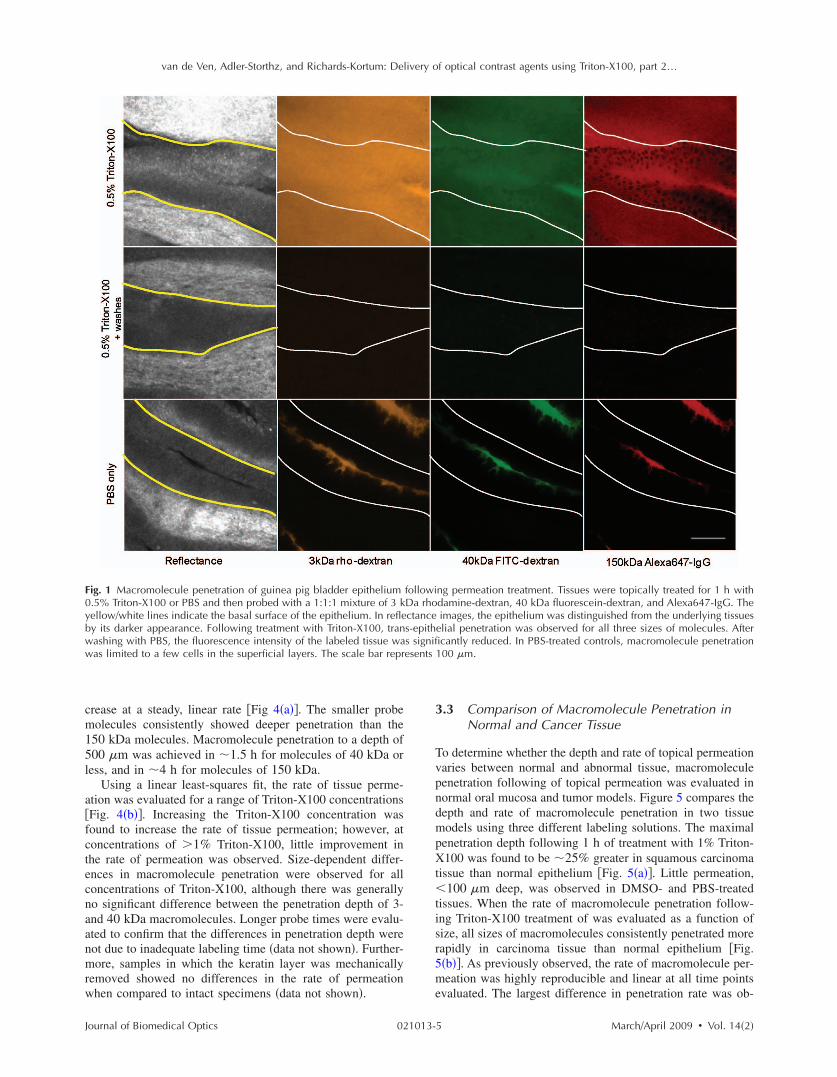
van de Ven Adler-Storthz and Richards-Kortum Delivery of optical contrast agents using Triton-X100 part 2hellip
F0ybww
c m 1 5 l
a f c te c naa nm r w
J
ig 1 Macromolecule penetration of guinea pig bladder epithelium 5 Triton-X100 or PBS and then probed with a 111 mixture of 3 kellowwhite lines indicate the basal surface of the epithelium In refley its darker appearance Following treatment with Triton-X100 tranashing with PBS the fluorescence intensity of the labeled tissue waas limited to a few cells in the superficial layers The scale bar repr
rease at a steady linear rate Fig 4a The smaller probeolecules consistently showed deeper penetration than the50 kDa molecules Macromolecule penetration to a depth of00 m was achieved in 15 h for molecules of 40 kDa oress and in 4 h for molecules of 150 kDa
Using a linear least-squares fit the rate of tissue perme-tion was evaluated for a range of Triton-X100 concentrationsFig 4b Increasing the Triton-X100 concentration wasound to increase the rate of tissue permeation however atoncentrations of 1 Triton-X100 little improvement inhe rate of permeation was observed Size-dependent differ-nces in macromolecule penetration were observed for alloncentrations of Triton-X100 although there was generallyo significant difference between the penetration depth of 3-nd 40 kDa macromolecules Longer probe times were evalu-ted to confirm that the differences in penetration depth wereot due to inadequate labeling time data not shown Further-ore samples in which the keratin layer was mechanically
emoved showed no differences in the rate of permeationhen compared to intact specimens data not shown
ournal of Biomedical Optics 021013-
ng permeation treatment Tissues were topically treated for 1 h with damine-dextran 40 kDa fluorescein-dextran and Alexa647-IgG The images the epithelium was distinguished from the underlying tissues elial penetration was observed for all three sizes of molecules After cantly reduced In PBS-treated controls macromolecule penetration
100 m
33 Comparison of Macromolecule Penetration in Normal and Cancer Tissue
To determine whether the depth and rate of topical permeation varies between normal and abnormal tissue macromolecule penetration following of topical permeation was evaluated in normal oral mucosa and tumor models Figure 5 compares the depth and rate of macromolecule penetration in two tissue models using three different labeling solutions The maximal penetration depth following 1 h of treatment with 1 Triton-X100 was found to be 25 greater in squamous carcinoma tissue than normal epithelium Fig 5a Little permeation 100 m deep was observed in DMSO- and PBS-treated tissues When the rate of macromolecule penetration follow-ing Triton-X100 treatment of was evaluated as a function of size all sizes of macromolecules consistently penetrated more rapidly in carcinoma tissue than normal epithelium Fig 5b As previously observed the rate of macromolecule per-meation was highly reproducible and linear at all time points evaluated The largest difference in penetration rate was ob-
followiDa rhoctances-epiths signifiesents
5 MarchApril 2009 Vol 142
van de Ven Adler-Storthz and Richards-Kortum Delivery of optical contrast agents using Triton-X100 part 2hellip
Fwteps
see
3
T f n puaa cX wl TXc f
J
ig 2 The time course of tissue permeation following treatment withith 05 Triton-X100 for 0ndash60 min and then probed fluorescent mol
he tissue surface are shown for the 5- 10- and 15-min time popithelium The depth and uniformity of permeation increased witenetration of different size probe molecules Fluorescence signal in scale bar represents 100 m
erved for the 150 kDa molecules although this size of mol-cule consistently penetrated more slowly than smaller mol-cules
4 Relation of Triton-X100 Concentration to the Number of Treated Cells
he average dose of Triton-X100 per cell was estimated asunction of treatment time and depth Table 1 provides theormalized Triton-X100 concentration in terms of picomoleser cell The calculations were based on an average cell vol-me the known tissue surface area and the measured perme-tion depth In the case where only one cell layer was perme-bilized 83 m deep the Triton-X100 concentration perell was on the order of 62ndash123 pmolcell As the Triton-100 permeated deeper the average concentration per cellas reduced Full thickness permeation of the bladder epithe-
ium with a 05 solution yielded an average of 083 pmolriton-X100 per cell A 30ndash60 min treatment of 10 Triton-100 in thicker tissues further lowered the normalized con-
entration to 028ndash057 for oral mucosa tissue and 021ndash042or squamous cell carcinoma tissue
ournal of Biomedical Optics 021013-
Triton-X100 Fresh guinea pig bladder biopsies were topically treated f three sizes Representative confocal images acquired 40 m below e yellowwhite lines indicate the apical and basal surfaces of the ent time Differences were observed between the cell and tissue
was observed as early as 5 min following Triton-X100 treatment The
35 Viability Assessment of Tissue Phantoms
Tissue phantoms were assayed for cell viability following treatment with different concentrations of Triton-X100 Figure 6 shows representative images of phantoms containing five million cells each treated with 0ndash5 Triton-X100 for 1 h and then coincubated with MTT reagent The normalized Triton-X100 concentration per cell was determined from the number of cells incorporated into each phantom Phantoms treated with 1 Triton-X100 or less 33 pmolcell demonstrated an intense blue color indicative of MTT reduction by viable cells When imaged in cross section these phantoms showed intense staining throughout In contrast phantoms treated with concentrations above 1 33 pmolcell demonstrated an overall loss in color intensity as well as a lack of staining at the phantom periphery suggestive of cell death at these higher Triton-X100 concentrations
36 EGFR Targeting in Cancer Tissue
To determine whether Triton-X100 is useful for the delivery of molecular-specific optical contrast agents subcutaneous tu-
05 ecules oints Thh treatmtroma
6 MarchApril 2009 Vol 142
van de Ven Adler-Storthz and Richards-Kortum Delivery of optical contrast agents using Triton-X100 part 2hellip
FfRc
mm tX ca
FwtiT
J
ig 3 Permeation of the porcine oral mucosa following treatment wresh tissue biopsies for 0 ndash60 min then the tissue was sliced probeepresentative confocal images acquired 40 m below the cut surfacharacterized by a well-defined border that progressed deeper with ti
ors were labeled for EGFR in the presence of topical per-eation enhancers Freshly excised tumors were topically
reated for 2 h with antiEGFR-647 diluted in 1 Triton-100 10 DMSO or PBS The labeled tissues were sliced in
ross section counterstained with acriflavine-HCL and im-ged perpendicular to the labeling surface Figure 7 shows
ig 4 Permeation of normal oral mucosa as a function of time and Trith different concentrations of Triton-X100 for regular time intervals
40 kDa and 150 kDa The depth and rate of macromransverse tissue sections a The relationship between the depth of pncrease in permeation depth is observed with time b Rate of tisriton-X100 concentration increased the depth of tissue permeabilizat
ournal of Biomedical Optics 021013-
Triton-X100 Triton-X100 was topically applied to the top surface of fluorescent molecules of three sizes and imaged in cross section
hown for the 10- 30- and 60-min time points Tissue permeation was e scale bar represents 100 m
representative confocal fluorescence images of EGFR labeling in the presence of different topical permeation enhancers The surface of the tissue where the contrast agent was applied appears at the top of each panel EGFR labeling is denoted in red and acriflavine-HCL labeling in green Treatment with 1 Triton-X100 facilitated the uniform delivery of antiEGFR-647
00 concentration Normal porcine oral mucosa was topically treated and probed with fluorescent macromolecules of three sizes 3 kDa penetration was determined from confocal fluorescence images of tion and duration of treatment with 1 Triton-X100 A steady linear meation as a function of Triton-X100 concentration Increasing the wever the extent of macromolecule penetration was limited by size
ith 1d with
e are sme Th
iton-X1 slicedoleculeermea
sue perion ho
7 MarchApril 2009 Vol 142
van de Ven Adler-Storthz and Richards-Kortum Delivery of optical contrast agents using Triton-X100 part 2hellip
F1wpcpm
tt c r d set l
3T c t p e
J
ig 5 Comparison of macromolecule penetration in normal oral muco0 DMSO or PBS sliced and then probed with fluorescent macroas determined from confocal fluorescence images of transverse tissueermeation enhancers measured using 3 kDa rhodamine-dextran Tancer tissue than normal epithelium Little permeation was observenetration by size with 1 Triton-X100 treatment The rate of peacromolecules evaluated
o a depth of 500ndash600 m Washing of the tissue after con-rast agent delivery yielded a honeycomb pattern of labelingonsistent with that observed in vitro EGFR labeling waseproducibly observed around all squamous cells to the fullepth of contrast agent delivery The intensity of labeling wasimilar to that observed in cell monolayers labeled in the pres-nce and absence of Triton-X100 data not shown In con-rast EGFR labeling in DMSO- and PBS-treated tissues wasimited to the superficial layers of cells 20ndash60 m deep
7 Multitarget Labeling in Tumors
o demonstrate the feasibility of delivering multiple opticalontrast agents to different cell compartments three differentargeting moieties were topically applied to tumors in theresence of Triton-X100 The specificity and bioactivity ofach targeting moiety was established in vitro prior to tissue
Table 1 Triton-X100 concentration per cell as a
Tissue
Triton-X100 concentration
Averagepermeation d
m
Bladder deflated
05 83
60
Oral mucosa 10 83
175
350
Squamous cell 10 83 carcinoma
235
470
ournal of Biomedical Optics 021013-
squamous cell carcinoma Tissues were treated with 1 Triton-X100 les of three sizes The depth and rate of macromolecule penetration ns a The maximal penetration depth following a 1-h treatment with imal depth with Triton-X100 treatment was found to be greater for DMSO- and PBS-treated controls b The rate of macromolecule n was consistently more rapid in carcinoma tissue for all sizes of
labeling No differences in targeting were observed with Triton-X100 co-treatment of live and fixed cells data not shown Figure 8 shows representative confocal images of freshly excised tumor tissue simultaneously labeled for EGFR Cyclin D1 and nucleic acid content in the presence of 1 Triton-X100 The tissues were topically labeled washed sliced and imaged perpendicular to the labeling surface AntiEGFR-647 labeling red appeared in a characteristic honeycomb pattern AntiCyclin D1-488 labeling green was diffusely spread through the cytoplasm of cancer cells Pro-pidium iodide labeling orange appeared in the nuclei of per-meabilized cells EGFR Cyclin D1 and propidium iodide labeling always appeared together overlay suggesting that the labeling is limited to cells permeabilized by the Triton-X100 treatment Little to no antibody labeling was observed when isotype controls and propidium iodide were adminis-
on of treatment time and depth
Approx treatment time min
No of Treated cells 106
Normalized concentration pmolcell
5
15
134
100
617
083
5 134 123
30 291 057
60 5816 028
5 134 123
30 390 042
60 780 021
sa andmolecu sectiohe maxed withnetratio
functi
epth
8 MarchApril 2009 Vol 142
van de Ven Adler-Storthz and Richards-Kortum Delivery of optical contrast agents using Triton-X100 part 2hellip
Ff
c m B o at
t c s t
Fasuw
J
ig 6 Tissue phantoms assayed for viability after treatment with dif-erent concentrations of Triton-X100 Phantoms containing 5 106
ells each were treated with 1 mL Triton-X100 for 1 h returned toedia for 4 h and then coincubated with a MTT reagent for 20 minlue color deposition is indicative of viable cells Intense staining wasbserved in phantoms treated with 1 Triton-X100 or less33 pmolcell Phantoms treated with Triton-X100 concentrationsbove 1 33 pmolcell demonstrated an overall loss in color in-ensity and staining at the periphery
ered in the presence of Triton-X100 suggesting that unboundontrast agents were sufficiently removed with the washingteps No propidium iodide labeling was present in tissuesreated with PBS instead of Triton-X100 data not shown
ig 7 EGFR targeting in the presence of topical permeation enhancersntiEGFR-647 red diluted in 1 Triton-X100 10 DMSO or PBShown at the top Tissues were counterstained with acriflavine-HCLniform EGFR labeling was observed around all squamous cells to a das limited to the superficial cell layers The scale bar represents 100
ournal of Biomedical Optics 021013-
4 Discussion
Optical contrast agents developed for cancer biomarker detec-tion have demonstrated high specificity in cell and tissue models Generally ranging in size from 6 kDa peptides and affibodies to 150 kDa antibodies most molecular-specific contrast agents have a limited ability to penetrate mucosal tissue Chemical modification and encapsulation strategies to improve tissue penetration have only proven useful for mol-ecules at the small end of the size spectrum41 The topical delivery of tissue-impermeant contrast agents presents an in-teresting challenge because the success of tissue labeling is dependent on the efficiency and uniformity of contrast agent delivery Furthermore unbound contrast agents generally need to be washed out prior to imaging for high optical con-trast In this paper we evaluate ability of Triton-X100 to de-liver optical contrast agents that lack the appropriate physio-chemical properties for unaided tissue permeation
Confocal microscopy studies of macromolecule penetra-tion in the bladder mucosa were used to examine the unifor-mity of tissue permeabilization The bladder mucosa is a par-ticularly interesting model because the natural folding of the deflated bladder allows the epithelium to be imaged in cross section without the need for tissue slicing In this model we observed that a fixed volume of 05 Triton-X100 can deliver molecules up to 150 kDa in size across treated epithelium By
taneously generated 1483 tumors were topically labeled for 2 h with ssues were sliced and imaged perpendicular to the labeling surface to highlight the tissue morphology In Triton-X100 treated tissues 500ndash600 m EGFR labeling in the DMSO- and PBS-treated controls
Subcu The ti greenepth of m
9 MarchApril 2009 Vol 142
van de Ven Adler-Storthz and Richards-Kortum Delivery of optical contrast agents using Triton-X100 part 2hellip
Fmrwcwm
omXp s a a ml o p i dXg
st d o p ar p e c
J
ig 8 Cotargeting of clinically relevant cancer biomarkers Subcutaolecular-specific contrast agents diluted in 10 Triton-X100 The c
ed Cyclin D1 in the cytoplasm antiCyclin D1 green and nucleicere washed sliced and imaged perpendicular to the labeling suompartments EGFR Cyclin D1 and nucleic acid labeling always apas highly uniform and reproducible from sample to sample Little toent suggesting that the washing of the tissue was sufficient to remo
ur estimate the cells of the epithelium are receiving a treat-ent dose ranging from 617 to 083 pmolcell as the Triton-100 progresses through the tissue Tissue permeation ap-ears to primarily follow a transcellular route appearing as atrong fluorescent signal within cells In tissues probed with 3nd 40 kDa macromolecules the fluorescence is uniformcross the entire epithelium In tissues probed with 150 kDaolecules only the cell nuclei appear as dark spots Extracel-
ular fluorescence is evident between cells at the leading edgef the treatment suggesting that Triton-X100 also facilitatesaracellular delivery Washing of the epithelium removes bothntracellular and extracellular probes yielding a uniformlyark tissue Together these observations indicate that Triton-100 may be useful for the delivery of contrast agents tar-eted to both intracellular and extracellular biomarkers
Studies of Triton-X100 treatment in fresh oral mucosapecimens indicate that the depth of permeation can be con-rolled The leading edge of permeation treatment was wellefined at all concentrations examined allowing measurementf penetration depth as a function time and concentration Theenetration depth increased linearly with treatment time forll sizes of probe macromolecules over a 4-h observation pe-iod There was no apparent delay for the contrast agents toermeate through an intact keratin layer The maximal rate ofpithelial permeation increased with increasing Triton-X100oncentration These trends were highly reproducible varying10 from sample to sample This data demonstrate that it is
ournal of Biomedical Optics 021013-1
y generated 1483 tumors were topically labeled for 1 h with three agents were targeted to EGFR on the cell membrane antiEGFR-647 in the nucleus propidium iodide orange After labeling the tissues The contrast agents were found to localize to the appropriate cell together in treated tissue overlay The observed pattern of labeling eling was observed in IgG-treated controls with Triton-X100 cotreat-ound contrast agents The scale bar represents 20 m
possible to regulate the depth of Triton-X100 treatment by adjusting the Triton-X100 concentration and treatment dura-tion The depth of labeling following treatment was deter-mined by contrast agent size with larger probes demonstrat-ing shallower penetration at the same treatment concentration as smaller molecules Interestingly it does not appear advan-tageous to use Triton-X100 concentrations much above 1 because little improvement in the rate of permeation is ob-served between tissues treated with 1 and 25 Triton-X100
Differences were apparent in the permeation of tissues when compared to cell monolayers In our companion paper Part 1 we showed that cells can be permeabilized with 027 pmolcell Triton-X100 and that nuclear penetration of 40 and 150 kDa molecules can be achieved with 055ndash11 pmolcell Triton-X100 In tissue nuclear penetra-tion is limited to molecules of 40 kDa or less Occasional nuclear penetration of 150 kDa molecules is observed at the surface of treated tissue and is generally limited to one to two cell layers If we consider an oral biopsy treated with 1 Triton-X100 for 1 h the normalized Triton-X100 concentra-tion is in the range of 055ndash11 pmolcell for approximately half of the treatment time Enhanced nuclear permeation is not observed with higher Triton-X100 concentrations or changes in treatment time Thus it appears likely that Triton-X100 preferentially permeabilizes cell membranes in its movement
neouslontrast acids rface peared no labve unb
0 MarchApril 2009 Vol 142
van de Ven Adler-Storthz and Richards-Kortum Delivery of optical contrast agents using Triton-X100 part 2hellip
t t
c c ctm T t s l c sX o 3 w
ct cc a te p stc dm
a at ccs b w os t it m b p l de T cic s Di
J
hrough tissue and is not sufficiently concentrated locally forhe generation of large nuclear pores
In tissue the normalized Triton-X100 concentrationhanges with treatment time and depth such that superficialells are treated to higher doses of Triton-X100 than deeperells Nevertheless cell viability experiments in tissue phan-oms seem to indicate that in a three-dimensional environ-ent cells are more likely to remain viable at higher doses ofriton-X100 In phantoms the viability cutoff is33 pmolcell with cells at the periphery losing their ability
o metabolize MTT at higher concentrations These studiesuggest that at Triton-X100 concentrations of 33 pmolcell oress cell viability is determined the mean concentration perell rather than local concentration gradients This leads us topeculate that tissues can be safely permeated with Triton-100 as long as the treatment time and concentration areptimized to fall below a normalized concentration of3 pmolcell Further studies are needed to determinehether this will hold true in vivo
Depth control will be advantageous for the use of topicalontrast agents in humans By limiting the penetration of op-ical contrast agents to a finite depth the systemic delivery ofontrast agents with undefined toxicity can be avoided Be-ause the mucosal epithelium is a renewable tissue contrastgents adhering to epithelial cells will likely be shed withissue turnover For the optical detection of disease biomark-rs some compensation may be required for differences inermeation rate of normal and abnormal tissue In the tissuestudied here however a 25 difference was observed be-ween normal squamous epithelium and undifferentiated can-er tissue Depth-sensitive optical imaging or spectroscopyevices could potentially be used to monitor the depth of per-eation and determine when to halt the reaction by washingThe use of Triton-X100 as a topical permeation enhancer
llows the delivery of optical contrast agents that lack theppropriate size and chemical properties for tissue penetra-ion Molecular-specific contrast agents as large as antibodiesan be successfully delivered to both intracellular and extra-ellular targets using Triton-X100 In this paper we demon-trate that three clinically relevant biomarkers of cancer cane simultaneously labeled with the topical co-administrationith Triton-X100 The EGFR which is located on the surfacef cells is thought to be a promising target because its expres-ion is elevated in a variety of dysplastic and cancerousissues47ndash49 The overexpression of Cyclin D1 detected heren the cytoplasm is associated with a poor prognosis in pa-ients with head and neck tumors50ndash53 Measures of nuclearorphology delineated here with propidium iodide have
een proposed as an objective measure of the transition fromreneoplastic to neoplastic tissue54 In vitro studies of cellabeling in the presence and absence of Triton-X100 haveemonstrated that structure and availability of these biomark-rs in live cells is similar to that fixed cells data not shownhe targeting of these biomarkers with molecular-specificontrast agents in tissue yielded uniform reproducible label-ng in all transverse sections examined by confocal micros-opy The localization and intensity of tissue labeling wasimilar to that observed in vitro In contrast treatment withMSO or PBS failed to enhance the penetration tissue-
mpermeant optical probes The feasibility of translating Triton-X100 for in vivo use
ournal of Biomedical Optics 021013-1
remains to be determined In our companion paper we dem-onstrated that Triton-X100ndashmediated permeabilization of cells is reversible and that cells retain their metabolic and prolif-erative capacities while recovering membrane integrity Oth-ers have demonstrated that only concentrations of 10 pro-duce visible side effects in patch tests of human skin consisting of transdermal water loss and erythema55 It has been reported that two or more permeation enhancers applied together can act synergistically56 suggesting that it may be possible to further reduce the effective Triton-X100 concen-tration by combining it with other topical agents Tissue dam-age reported with the use of other surfactant-based permeation enhancers has generally been limited and reversible57ndash59 The next step will be to characterize the safety toxicity biological activity and reversibility of Triton-X100ndashmediated perme-ation at clinically useful doses in animal models
5 Conclusions This paper demonstrates that Triton-X100 is a promising topi-cal permeation enhancer for the mucosal delivery of tissue-impermeant optical contrast agents Compared to other topical permeation enhancers Triton-X100 can deliver a much broader size range of molecules No chemical modification of optical contrast agents is needed both targeted and untargeted molecules as large as 150 kDa can be delivered uniformly through normal and cancer tissue The depth of tissue perme-ation can be modulated allowing for epithelial containment of topical contrast agents The coadministration of Triton-X100 with molecular-specific contrast agents facilitates the simulta-neous labeling of cell membrane cytoplasmic and nuclear targets Further work is needed to establish the safety of this approach for in vivo use
Acknowledgments
We thank Vivian Mack for her assistance with cell culture and animal care This work was supported in part by an NIH BRP Grant No CA103830 and a training fellowship from the Keck Center Nanobiology Training Program of the Gulf Coast Consortia NIH Grant No 5 T90 DK071054-03
References 1 S Folli P Westermann D Braichotte A Pelegrin G Wagnieres H
van den Bergh and J P Mach ldquoAntibody-indocyanin conjugates for immunophotodetection of human squamous cell carcinoma in nude micerdquo Cancer Res 5410 2643ndash2649 1994
2 B Ballou G W Fisher J S Deng T R Hakala M Srivastava and D L Farkas ldquoCyanine fluorochrome-labeled antibodies in vivo as-sessment of tumor imaging using Cy3 Cy5 Cy55 and Cy7rdquo Can-cer Detect Prev 223 251ndash257 1998
3 N S Soukos M R Hamblin T F Deutsch and T Hasan ldquoMono-clonal antibody-tagged receptor-targeted contrast agents for detection of cancersrdquo Proc SPIE 4259 115ndash128 2001
4 N S Soukos M R Hamblin S Keel R L Fabian T F Deutsch and T Hasan ldquoEpidermal growth factor receptor-targeted immuno-photodiagnosis and photoimmunotherapy of oral precancer in vivordquo Cancer Res 6111 4490ndash4496 2001
5 Y Koyama T Barrett Y Hama G Ravizzini P L Choyke and H Kobayashi ldquoIn vivo molecular imaging to diagnose and subtype tu-mors through receptor-targeted optically labeled monoclonal antibod-iesrdquo Neoplasia 912 1021ndash1029 2007
6 Y Koyama Y Hama Y Urano D M Nguyen P L Choyke and H Kobayashi ldquoSpectral fluorescence molecular imaging of lung me-tastases targeting HER2neurdquo Clin Cancer Res 1310 2936ndash2945 2007
1 MarchApril 2009 Vol 142
van de Ven Adler-Storthz and Richards-Kortum Delivery of optical contrast agents using Triton-X100 part 2hellip
1
1
1
1
1
1
1
1
1
1
2
2
2
2
2
2
2
2
2
J
7 K E Adams S Ke S Kwon F Liang Z Fan Y Lu K Hirschi ME Mawad M A Barry and E M Sevick-Muraca ldquoComparison ofvisible and near-infrared wavelength-excitable fluorescent dyes formolecular imaging of cancerrdquo J Biomed Opt 122 024017 2007
8 J L Kovar W M Volcheck J Chen and M A Simpson ldquoPurifi-cation method directly influences effectiveness of an epidermalgrowth factor-coupled targeting agent for noninvasive tumor detec-tion in micerdquo Anal Biochem 3611 47ndash54 2007
9 J L Kovar M A Johnson W M Volcheck J Chen and M ASimpson ldquoHyaluronidase expression induces prostate tumor metasta-sis in an orthotopic mouse modelrdquo Am J Pathol 1694 1415ndash14262006
0 S Ke X Wen M Gurfinkel C Charnsangavej S Wallace E MSevick-Muraca and C Li ldquoNear-infrared optical imaging of epider-mal growth factor receptor in breast cancer xenograftsrdquo Cancer Res6322 7870ndash7875 2003
1 K L Carraway 3rd and R A Cerione ldquoFluorescent-labeled growthfactor molecules serve as probes for receptor binding and endocyto-sisrdquo Biochemistry 3245 12039ndash12045 1993
2 C H Tung Y Lin W K Moon and R Weissleder ldquoA receptor-targeted near-infrared fluorescence probe for in vivo tumor imagingrdquoChemBioChem 38 784ndash786 2002
3 S Achilefu R B Dorshow J E Bugaj and R Rajagopalan ldquoNovelreceptor-targeted fluorescent contrast agents for in vivo tumor imag-ingrdquo Invest Radiol 358 479ndash485 2000
4 A Becker C Hessenius K Licha B Ebert U Sukowski W Sem-mler B Wiedenmann and C Grotzinger ldquoReceptor-targeted opticalimaging of tumors with near-infrared fluorescent ligandsrdquo Nat Bio-technol 194 327ndash331 2001
5 D Citrin T Scott M Sproull C Menard P J Tofilon and KCamphausen ldquoIn vivo tumor imaging using a near-infrared-labeledendostatin moleculerdquo Int J Radiat Oncol Biol Phys 582 536ndash541 2004
6 R Weissleder C H Tung U Mahmood and A Bogdanov Jr ldquoInvivo imaging of tumors with protease-activated near-infrared fluores-cent probesrdquo Nat Biotechnol 174 375ndash378 1999
7 C H Tung U Mahmood S Bredow and R Weissleder ldquoIn vivoimaging of proteolytic enzyme activity using a novel molecular re-porterrdquo Cancer Res 6017 4953ndash4958 2000
8 K Marten C Bremer K Khazaie M Sameni B Sloane C HTung and R Weissleder ldquoDetection of dysplastic intestinal ad-enomas using enzyme-sensing molecular beacons in micerdquo Gastro-enterology 1222 406ndash414 2002
9 C Bremer C H Tung A Bogdanov Jr and R Weissleder ldquoImag-ing of differential protease expression in breast cancers for detectionof aggressive tumor phenotypesrdquo Radiology 2223 814ndash818 2002
0 M Kamiya H Kobayashi Y Hama Y Koyama M Bernardo TNagano P L Choyke and Y Urano ldquoAn enzymatically activatedfluorescence probe for targeted tumor imagingrdquo J Am Chem Soc12913 3918ndash3929 2007
1 J V Frangioni ldquoIn vivo near-infrared fluorescence imagingrdquo CurrOpin Chem Biol 75 626ndash634 2003
2 R Cotran V Kumar and T Collins Robbins Pathologic Basis ofDisease W B Saunders Philadelphia 1999
3 J Levi Z Cheng O Gheysens M Patel C T Chan Y Wang MNamavari and S S Gambhir ldquoFluorescent fructose derivatives forimaging breast cancer cellsrdquo Bioconjugate Chem 183 628ndash6342007
4 R OrsquoNeil L Wu and N Mullani ldquoUptake of a fluorescent deoxy-glucose analog 2-NBDG in tumor cellsrdquo Mol Imaging Biol 76388ndash392 2005
5 Z Cheng J Levi Z Xiong O Gheysens S Keren X Chen and SS Gambhir ldquoNear-infrared fluorescent deoxyglucose analogue fortumor optical imaging in cell culture and living micerdquo BioconjugateChem 173 662ndash669 2006
6 T J Muldoon M C Pierce D L Nida M D Williams A Gillen-water and R Richards-Kortum ldquoSubcellular-resolution molecularimaging within living tissue by fiber microendoscopyrdquo Opt Express1525 16413ndash16423 2007
7 Y Kakeji S Yamaguchi D Yoshida K Tanoue M Ueda A Ma-sunari T Utsunomiya M Imamura H Honda Y Maehara and MHashizume ldquoDevelopment and assessment of morphologic criteriafor diagnosing gastric cancer using confocal endomicroscopy an exvivo and in vivo studyrdquo Endoscopy 389 886ndash890 2006
8 A L Polglase W J McLaren S A Skinner R Kiesslich M F
ournal of Biomedical Optics 021013-1
Neurath and P M Delaney ldquoA fluorescence confocal endomicro-scope for in vivo microscopy of the upper- and the lower-GI tractrdquo Gastrointest Endosc 625 686ndash695 2005
29 I A Siegel K T Izutsu and E Watson ldquoMechanisms of non-electrolyte penetration across dog and rabbit oral mucosa in vitrordquo Arch Oral Biol 265 357ndash361 1981
30 V H Lee ldquoEnzymatic barriers to peptide and protein absorptionrdquo Crit Rev Ther Drug Carrier Syst 52 69ndash97 1988
31 R A Hughes I Toth P Ward S J Ireland and W A Gibbons ldquoLipidic peptides III lipidic amino acid and oligomer conjugates of morphinerdquo J Pharm Sci 8012 1103ndash1105 1991
32 H A Benson ldquoTransdermal drug delivery penetration enhancement techniquesrdquo Curr Drug Delivery 21 23ndash33 2005
33 M Thanou J C Verhoef and H E Junginger ldquoOral drug absorption enhancement by chitosan and its derivativesrdquo Adv Drug Delivery Rev 522 117ndash126 2001
34 V Erjavec Z Pavlica M Sentjurc and M Petelin ldquoIn vivo study of liposomes as drug carriers to oral mucosa using EPR oximetryrdquo Int J Pharm 3071 1ndash8 2006
35 H Li J H An J S Park and K Han ldquoMultivesicular liposomes for oral delivery of recombinant human epidermal growth factorrdquo Arch Pharmacal Res 288 988ndash994 2005
36 F S Farshi A Y Ozer M T Ercan and A A Hincal ldquoIn-vivo studies in the treatment of oral ulcers with liposomal dexamethasone sodium phosphaterdquo J Microencapsul 135 537ndash544 1996
37 A N C Anigbogu A C Williams B W Barry and H G M Edwards ldquoFourier transform raman spectroscopy of interactions be-tween the penetration enhancer dimethyl sulfoxide and human stra-tum corneumrdquo Int J Pharm 1252 265ndash282 1995
38 H A E Benson ldquoTransfersomes for transdermal drug deliveryrdquo Expert Opin Drug Delivery 36 727ndash737 2006
39 P N Gupta V Mishra P Singh A Rawat P Dubey S Mahor and S P Vyas ldquoTetanus toxoid-loaded transfersomes for topical immuni-zationrdquo J Pharm Pharmacol 573 295ndash301 2005
40 J Hao and P W Heng ldquoBuccal delivery systemsrdquo Drug Dev Ind Pharm 298 821ndash832 2003
41 F Veuillez Y N Kalia Y Jacques J Deshusses and P Buri ldquoFac-tors and strategies for improving buccal absorption of peptidesrdquo Eur J Pharm Biopharm 512 93ndash109 2001
42 P G Sacks S M Parnes G E Gallick Z Mansouri R Lichtner K L Satya-Prakash S Pathak and D F Parsons ldquoEstablishment and characterization of two new squamous cell carcinoma cell lines de-rived from tumors of the head and neckrdquo Cancer Res 4810 2858ndash2866 1988
43 K J Mattern C Nakornchai and W M Deen ldquoDarcy permeability of agarose-glycosaminoglycan gels analyzed using fiber-mixture and Donnan modelsrdquo Biophys J 952 648ndash656 2008
44 E M Johnson D A Berk R K Jain and W M Deen ldquoHindered diffusion in agarose gels test of effective medium modelrdquo Biophys J 702 1017ndash1023 1996
45 K Sokolov J Galvan A Myakov A Lacy R Lotan and R Richards-Kortum ldquoRealistic three-dimensional epithelial tissue phan-toms for biomedical opticsrdquo J Biomed Opt 71 148ndash156 2002
46 E R Hsu E V Anslyn S Dharmawardhane R Alizadeh-Naderi J S Aaron K V Sokolov A K El-Naggar A M Gillenwater and R R Richards-Kortum ldquoA far-red fluorescent contrast agent to image epidermal growth factor receptor expressionrdquo Photochem Photobiol 793 272ndash279 2004
47 H Modjtahedi and C J Dean ldquoThe receptor for EGF and its ligands expression prognostic value and target for therapy in cancerrdquo Int J Oncol 4 277ndash296 1994
48 W Yasui H Sumiyoshi J Hata T Kameda A Ochiai H Ito and E Tahara ldquoExpression of epidermal growth factor receptor in human gastric and colonic carcinomasrdquo Cancer Res 481 137ndash141 1988
49 J A Vet F M Debruyne and J A Schalken ldquoMolecular prognostic factors in bladder cancerrdquo World J Urol 122 84ndash88 1994
50 A Namazie S Alavi O I Olopade G Pauletti N Aghamoham-madi M Aghamohammadi J A Gornbein T C Calcaterra D J Slamon M B Wang and E S Srivatsan ldquoCyclin D1 amplification and p16MTS1CDK4I deletion correlate with poor prognosis in head and neck tumorsrdquo Laryngoscope 1123 472ndash481 2002
51 J A Akervall R J Michalides H Mineta A Balm A Borg M R Dictor Y Jin B Loftus F Mertens and J P Wennerberg ldquoAmpli-fication of cyclin D1 in squamous cell carcinoma of the head and neck and the prognostic value of chromosomal abnormalities and
2 MarchApril 2009 Vol 142
van de Ven Adler-Storthz and Richards-Kortum Delivery of optical contrast agents using Triton-X100 part 2hellip
5
5
5
5
J
cyclin D1 overexpressionrdquo Cancer 792 380ndash389 1997 2 R J Michalides N M van Veelen P M Kristel A A Hart B M
Loftus F J Hilgers and A J Balm ldquoOverexpression of cyclin D1indicates a poor prognosis in squamous cell carcinoma of the headand neckrdquo Arch Otolaryngol 1235 497ndash502 1997
3 R Michalides N van Veelen A Hart B Loftus E Wientjens andA Balm ldquoOverexpression of cyclin D1 correlates with recurrence ina group of forty-seven operable squamous cell carcinomas of thehead and neckrdquo Cancer Res 555 975ndash978 1995
4 M F Mitchell W N Hittelman W K Hong R Lotan and DSchottenfeld ldquoThe natural history of cervical intraepithelial neopla-sia an argument for intermediate endpoint biomarkersrdquo Cancer Epi-demiol Biomarkers Prev 37 619ndash626 1994
5 F van Ruissen M Le J M Carroll P G M van der Valk and JSchalkwijk ldquoDifferential effects of detergents on keratinocyte geneexpressionrdquo J Invest Dermatol 1104 358 1998
ournal of Biomedical Optics 021013-1
56 B Mollgaard ldquoSynergistic effects in percutaneous enhancementrdquo in Pharmaceutical Skin Penetration Enhancement K A Walters and J Hadgraft Eds pp 220ndash242 Marcel Dekker New York 1993
57 E S Swenson W B Milisen and W Curatolo ldquoIntestinal perme-ability enhancement efficacy acute local toxicity and reversibilityrdquo Pharmacol Res 118 1132ndash1142 1994
58 E K Anderberg and P Artursson ldquoEpithelial transport of drugs in cell culture VIII Effects of sodium dodecyl sulfate on cell mem-brane and tight junction permeability in human intestinal epithelial Caco-2 cellsrdquo J Pharm Sci 824 392ndash398 1993
59 E K Anderberg C Nystrom and P Artursson ldquoEpithelial transport of drugs in cell culture VII Effects of pharmaceutical surfactant excipients and bile acids on transepithelial permeability in monolay-ers of human intestinal epithelial Caco-2 cellsrdquo J Pharm Sci 819 879ndash887 1992
3 MarchApril 2009 Vol 142
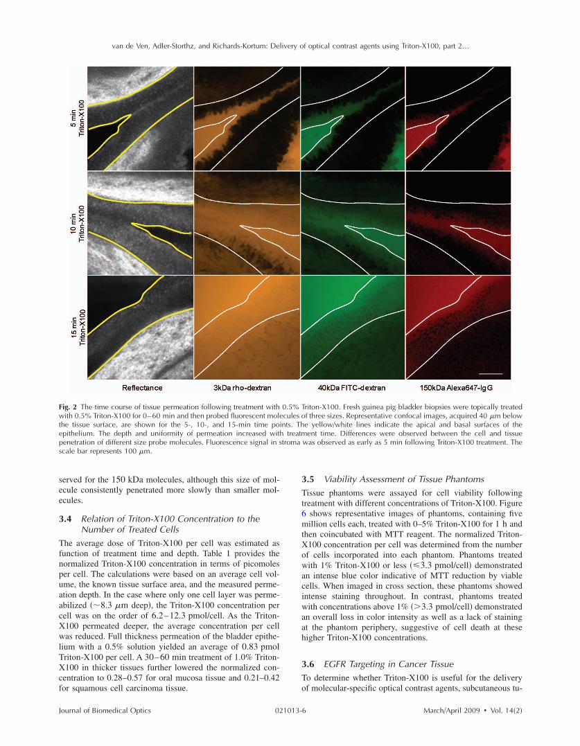
van de Ven Adler-Storthz and Richards-Kortum Delivery of optical contrast agents using Triton-X100 part 2hellip
Fwteps
see
3
T f n puaa cX wl TXc f
J
ig 2 The time course of tissue permeation following treatment withith 05 Triton-X100 for 0ndash60 min and then probed fluorescent mol
he tissue surface are shown for the 5- 10- and 15-min time popithelium The depth and uniformity of permeation increased witenetration of different size probe molecules Fluorescence signal in scale bar represents 100 m
erved for the 150 kDa molecules although this size of mol-cule consistently penetrated more slowly than smaller mol-cules
4 Relation of Triton-X100 Concentration to the Number of Treated Cells
he average dose of Triton-X100 per cell was estimated asunction of treatment time and depth Table 1 provides theormalized Triton-X100 concentration in terms of picomoleser cell The calculations were based on an average cell vol-me the known tissue surface area and the measured perme-tion depth In the case where only one cell layer was perme-bilized 83 m deep the Triton-X100 concentration perell was on the order of 62ndash123 pmolcell As the Triton-100 permeated deeper the average concentration per cellas reduced Full thickness permeation of the bladder epithe-
ium with a 05 solution yielded an average of 083 pmolriton-X100 per cell A 30ndash60 min treatment of 10 Triton-100 in thicker tissues further lowered the normalized con-
entration to 028ndash057 for oral mucosa tissue and 021ndash042or squamous cell carcinoma tissue
ournal of Biomedical Optics 021013-
Triton-X100 Fresh guinea pig bladder biopsies were topically treated f three sizes Representative confocal images acquired 40 m below e yellowwhite lines indicate the apical and basal surfaces of the ent time Differences were observed between the cell and tissue
was observed as early as 5 min following Triton-X100 treatment The
35 Viability Assessment of Tissue Phantoms
Tissue phantoms were assayed for cell viability following treatment with different concentrations of Triton-X100 Figure 6 shows representative images of phantoms containing five million cells each treated with 0ndash5 Triton-X100 for 1 h and then coincubated with MTT reagent The normalized Triton-X100 concentration per cell was determined from the number of cells incorporated into each phantom Phantoms treated with 1 Triton-X100 or less 33 pmolcell demonstrated an intense blue color indicative of MTT reduction by viable cells When imaged in cross section these phantoms showed intense staining throughout In contrast phantoms treated with concentrations above 1 33 pmolcell demonstrated an overall loss in color intensity as well as a lack of staining at the phantom periphery suggestive of cell death at these higher Triton-X100 concentrations
36 EGFR Targeting in Cancer Tissue
To determine whether Triton-X100 is useful for the delivery of molecular-specific optical contrast agents subcutaneous tu-
05 ecules oints Thh treatmtroma
6 MarchApril 2009 Vol 142
van de Ven Adler-Storthz and Richards-Kortum Delivery of optical contrast agents using Triton-X100 part 2hellip
FfRc
mm tX ca
FwtiT
J
ig 3 Permeation of the porcine oral mucosa following treatment wresh tissue biopsies for 0 ndash60 min then the tissue was sliced probeepresentative confocal images acquired 40 m below the cut surfacharacterized by a well-defined border that progressed deeper with ti
ors were labeled for EGFR in the presence of topical per-eation enhancers Freshly excised tumors were topically
reated for 2 h with antiEGFR-647 diluted in 1 Triton-100 10 DMSO or PBS The labeled tissues were sliced in
ross section counterstained with acriflavine-HCL and im-ged perpendicular to the labeling surface Figure 7 shows
ig 4 Permeation of normal oral mucosa as a function of time and Trith different concentrations of Triton-X100 for regular time intervals
40 kDa and 150 kDa The depth and rate of macromransverse tissue sections a The relationship between the depth of pncrease in permeation depth is observed with time b Rate of tisriton-X100 concentration increased the depth of tissue permeabilizat
ournal of Biomedical Optics 021013-
Triton-X100 Triton-X100 was topically applied to the top surface of fluorescent molecules of three sizes and imaged in cross section
hown for the 10- 30- and 60-min time points Tissue permeation was e scale bar represents 100 m
representative confocal fluorescence images of EGFR labeling in the presence of different topical permeation enhancers The surface of the tissue where the contrast agent was applied appears at the top of each panel EGFR labeling is denoted in red and acriflavine-HCL labeling in green Treatment with 1 Triton-X100 facilitated the uniform delivery of antiEGFR-647
00 concentration Normal porcine oral mucosa was topically treated and probed with fluorescent macromolecules of three sizes 3 kDa penetration was determined from confocal fluorescence images of tion and duration of treatment with 1 Triton-X100 A steady linear meation as a function of Triton-X100 concentration Increasing the wever the extent of macromolecule penetration was limited by size
ith 1d with
e are sme Th
iton-X1 slicedoleculeermea
sue perion ho
7 MarchApril 2009 Vol 142
van de Ven Adler-Storthz and Richards-Kortum Delivery of optical contrast agents using Triton-X100 part 2hellip
F1wpcpm
tt c r d set l
3T c t p e
J
ig 5 Comparison of macromolecule penetration in normal oral muco0 DMSO or PBS sliced and then probed with fluorescent macroas determined from confocal fluorescence images of transverse tissueermeation enhancers measured using 3 kDa rhodamine-dextran Tancer tissue than normal epithelium Little permeation was observenetration by size with 1 Triton-X100 treatment The rate of peacromolecules evaluated
o a depth of 500ndash600 m Washing of the tissue after con-rast agent delivery yielded a honeycomb pattern of labelingonsistent with that observed in vitro EGFR labeling waseproducibly observed around all squamous cells to the fullepth of contrast agent delivery The intensity of labeling wasimilar to that observed in cell monolayers labeled in the pres-nce and absence of Triton-X100 data not shown In con-rast EGFR labeling in DMSO- and PBS-treated tissues wasimited to the superficial layers of cells 20ndash60 m deep
7 Multitarget Labeling in Tumors
o demonstrate the feasibility of delivering multiple opticalontrast agents to different cell compartments three differentargeting moieties were topically applied to tumors in theresence of Triton-X100 The specificity and bioactivity ofach targeting moiety was established in vitro prior to tissue
Table 1 Triton-X100 concentration per cell as a
Tissue
Triton-X100 concentration
Averagepermeation d
m
Bladder deflated
05 83
60
Oral mucosa 10 83
175
350
Squamous cell 10 83 carcinoma
235
470
ournal of Biomedical Optics 021013-
squamous cell carcinoma Tissues were treated with 1 Triton-X100 les of three sizes The depth and rate of macromolecule penetration ns a The maximal penetration depth following a 1-h treatment with imal depth with Triton-X100 treatment was found to be greater for DMSO- and PBS-treated controls b The rate of macromolecule n was consistently more rapid in carcinoma tissue for all sizes of
labeling No differences in targeting were observed with Triton-X100 co-treatment of live and fixed cells data not shown Figure 8 shows representative confocal images of freshly excised tumor tissue simultaneously labeled for EGFR Cyclin D1 and nucleic acid content in the presence of 1 Triton-X100 The tissues were topically labeled washed sliced and imaged perpendicular to the labeling surface AntiEGFR-647 labeling red appeared in a characteristic honeycomb pattern AntiCyclin D1-488 labeling green was diffusely spread through the cytoplasm of cancer cells Pro-pidium iodide labeling orange appeared in the nuclei of per-meabilized cells EGFR Cyclin D1 and propidium iodide labeling always appeared together overlay suggesting that the labeling is limited to cells permeabilized by the Triton-X100 treatment Little to no antibody labeling was observed when isotype controls and propidium iodide were adminis-
on of treatment time and depth
Approx treatment time min
No of Treated cells 106
Normalized concentration pmolcell
5
15
134
100
617
083
5 134 123
30 291 057
60 5816 028
5 134 123
30 390 042
60 780 021
sa andmolecu sectiohe maxed withnetratio
functi
epth
8 MarchApril 2009 Vol 142
van de Ven Adler-Storthz and Richards-Kortum Delivery of optical contrast agents using Triton-X100 part 2hellip
Ff
c m B o at
t c s t
Fasuw
J
ig 6 Tissue phantoms assayed for viability after treatment with dif-erent concentrations of Triton-X100 Phantoms containing 5 106
ells each were treated with 1 mL Triton-X100 for 1 h returned toedia for 4 h and then coincubated with a MTT reagent for 20 minlue color deposition is indicative of viable cells Intense staining wasbserved in phantoms treated with 1 Triton-X100 or less33 pmolcell Phantoms treated with Triton-X100 concentrationsbove 1 33 pmolcell demonstrated an overall loss in color in-ensity and staining at the periphery
ered in the presence of Triton-X100 suggesting that unboundontrast agents were sufficiently removed with the washingteps No propidium iodide labeling was present in tissuesreated with PBS instead of Triton-X100 data not shown
ig 7 EGFR targeting in the presence of topical permeation enhancersntiEGFR-647 red diluted in 1 Triton-X100 10 DMSO or PBShown at the top Tissues were counterstained with acriflavine-HCLniform EGFR labeling was observed around all squamous cells to a das limited to the superficial cell layers The scale bar represents 100
ournal of Biomedical Optics 021013-
4 Discussion
Optical contrast agents developed for cancer biomarker detec-tion have demonstrated high specificity in cell and tissue models Generally ranging in size from 6 kDa peptides and affibodies to 150 kDa antibodies most molecular-specific contrast agents have a limited ability to penetrate mucosal tissue Chemical modification and encapsulation strategies to improve tissue penetration have only proven useful for mol-ecules at the small end of the size spectrum41 The topical delivery of tissue-impermeant contrast agents presents an in-teresting challenge because the success of tissue labeling is dependent on the efficiency and uniformity of contrast agent delivery Furthermore unbound contrast agents generally need to be washed out prior to imaging for high optical con-trast In this paper we evaluate ability of Triton-X100 to de-liver optical contrast agents that lack the appropriate physio-chemical properties for unaided tissue permeation
Confocal microscopy studies of macromolecule penetra-tion in the bladder mucosa were used to examine the unifor-mity of tissue permeabilization The bladder mucosa is a par-ticularly interesting model because the natural folding of the deflated bladder allows the epithelium to be imaged in cross section without the need for tissue slicing In this model we observed that a fixed volume of 05 Triton-X100 can deliver molecules up to 150 kDa in size across treated epithelium By
taneously generated 1483 tumors were topically labeled for 2 h with ssues were sliced and imaged perpendicular to the labeling surface to highlight the tissue morphology In Triton-X100 treated tissues 500ndash600 m EGFR labeling in the DMSO- and PBS-treated controls
Subcu The ti greenepth of m
9 MarchApril 2009 Vol 142
van de Ven Adler-Storthz and Richards-Kortum Delivery of optical contrast agents using Triton-X100 part 2hellip
Fmrwcwm
omXp s a a ml o p i dXg
st d o p ar p e c
J
ig 8 Cotargeting of clinically relevant cancer biomarkers Subcutaolecular-specific contrast agents diluted in 10 Triton-X100 The c
ed Cyclin D1 in the cytoplasm antiCyclin D1 green and nucleicere washed sliced and imaged perpendicular to the labeling suompartments EGFR Cyclin D1 and nucleic acid labeling always apas highly uniform and reproducible from sample to sample Little toent suggesting that the washing of the tissue was sufficient to remo
ur estimate the cells of the epithelium are receiving a treat-ent dose ranging from 617 to 083 pmolcell as the Triton-100 progresses through the tissue Tissue permeation ap-ears to primarily follow a transcellular route appearing as atrong fluorescent signal within cells In tissues probed with 3nd 40 kDa macromolecules the fluorescence is uniformcross the entire epithelium In tissues probed with 150 kDaolecules only the cell nuclei appear as dark spots Extracel-
ular fluorescence is evident between cells at the leading edgef the treatment suggesting that Triton-X100 also facilitatesaracellular delivery Washing of the epithelium removes bothntracellular and extracellular probes yielding a uniformlyark tissue Together these observations indicate that Triton-100 may be useful for the delivery of contrast agents tar-eted to both intracellular and extracellular biomarkers
Studies of Triton-X100 treatment in fresh oral mucosapecimens indicate that the depth of permeation can be con-rolled The leading edge of permeation treatment was wellefined at all concentrations examined allowing measurementf penetration depth as a function time and concentration Theenetration depth increased linearly with treatment time forll sizes of probe macromolecules over a 4-h observation pe-iod There was no apparent delay for the contrast agents toermeate through an intact keratin layer The maximal rate ofpithelial permeation increased with increasing Triton-X100oncentration These trends were highly reproducible varying10 from sample to sample This data demonstrate that it is
ournal of Biomedical Optics 021013-1
y generated 1483 tumors were topically labeled for 1 h with three agents were targeted to EGFR on the cell membrane antiEGFR-647 in the nucleus propidium iodide orange After labeling the tissues The contrast agents were found to localize to the appropriate cell together in treated tissue overlay The observed pattern of labeling eling was observed in IgG-treated controls with Triton-X100 cotreat-ound contrast agents The scale bar represents 20 m
possible to regulate the depth of Triton-X100 treatment by adjusting the Triton-X100 concentration and treatment dura-tion The depth of labeling following treatment was deter-mined by contrast agent size with larger probes demonstrat-ing shallower penetration at the same treatment concentration as smaller molecules Interestingly it does not appear advan-tageous to use Triton-X100 concentrations much above 1 because little improvement in the rate of permeation is ob-served between tissues treated with 1 and 25 Triton-X100
Differences were apparent in the permeation of tissues when compared to cell monolayers In our companion paper Part 1 we showed that cells can be permeabilized with 027 pmolcell Triton-X100 and that nuclear penetration of 40 and 150 kDa molecules can be achieved with 055ndash11 pmolcell Triton-X100 In tissue nuclear penetra-tion is limited to molecules of 40 kDa or less Occasional nuclear penetration of 150 kDa molecules is observed at the surface of treated tissue and is generally limited to one to two cell layers If we consider an oral biopsy treated with 1 Triton-X100 for 1 h the normalized Triton-X100 concentra-tion is in the range of 055ndash11 pmolcell for approximately half of the treatment time Enhanced nuclear permeation is not observed with higher Triton-X100 concentrations or changes in treatment time Thus it appears likely that Triton-X100 preferentially permeabilizes cell membranes in its movement
neouslontrast acids rface peared no labve unb
0 MarchApril 2009 Vol 142
van de Ven Adler-Storthz and Richards-Kortum Delivery of optical contrast agents using Triton-X100 part 2hellip
t t
c c ctm T t s l c sX o 3 w
ct cc a te p stc dm
a at ccs b w os t it m b p l de T cic s Di
J
hrough tissue and is not sufficiently concentrated locally forhe generation of large nuclear pores
In tissue the normalized Triton-X100 concentrationhanges with treatment time and depth such that superficialells are treated to higher doses of Triton-X100 than deeperells Nevertheless cell viability experiments in tissue phan-oms seem to indicate that in a three-dimensional environ-ent cells are more likely to remain viable at higher doses ofriton-X100 In phantoms the viability cutoff is33 pmolcell with cells at the periphery losing their ability
o metabolize MTT at higher concentrations These studiesuggest that at Triton-X100 concentrations of 33 pmolcell oress cell viability is determined the mean concentration perell rather than local concentration gradients This leads us topeculate that tissues can be safely permeated with Triton-100 as long as the treatment time and concentration areptimized to fall below a normalized concentration of3 pmolcell Further studies are needed to determinehether this will hold true in vivo
Depth control will be advantageous for the use of topicalontrast agents in humans By limiting the penetration of op-ical contrast agents to a finite depth the systemic delivery ofontrast agents with undefined toxicity can be avoided Be-ause the mucosal epithelium is a renewable tissue contrastgents adhering to epithelial cells will likely be shed withissue turnover For the optical detection of disease biomark-rs some compensation may be required for differences inermeation rate of normal and abnormal tissue In the tissuestudied here however a 25 difference was observed be-ween normal squamous epithelium and undifferentiated can-er tissue Depth-sensitive optical imaging or spectroscopyevices could potentially be used to monitor the depth of per-eation and determine when to halt the reaction by washingThe use of Triton-X100 as a topical permeation enhancer
llows the delivery of optical contrast agents that lack theppropriate size and chemical properties for tissue penetra-ion Molecular-specific contrast agents as large as antibodiesan be successfully delivered to both intracellular and extra-ellular targets using Triton-X100 In this paper we demon-trate that three clinically relevant biomarkers of cancer cane simultaneously labeled with the topical co-administrationith Triton-X100 The EGFR which is located on the surfacef cells is thought to be a promising target because its expres-ion is elevated in a variety of dysplastic and cancerousissues47ndash49 The overexpression of Cyclin D1 detected heren the cytoplasm is associated with a poor prognosis in pa-ients with head and neck tumors50ndash53 Measures of nuclearorphology delineated here with propidium iodide have
een proposed as an objective measure of the transition fromreneoplastic to neoplastic tissue54 In vitro studies of cellabeling in the presence and absence of Triton-X100 haveemonstrated that structure and availability of these biomark-rs in live cells is similar to that fixed cells data not shownhe targeting of these biomarkers with molecular-specificontrast agents in tissue yielded uniform reproducible label-ng in all transverse sections examined by confocal micros-opy The localization and intensity of tissue labeling wasimilar to that observed in vitro In contrast treatment withMSO or PBS failed to enhance the penetration tissue-
mpermeant optical probes The feasibility of translating Triton-X100 for in vivo use
ournal of Biomedical Optics 021013-1
remains to be determined In our companion paper we dem-onstrated that Triton-X100ndashmediated permeabilization of cells is reversible and that cells retain their metabolic and prolif-erative capacities while recovering membrane integrity Oth-ers have demonstrated that only concentrations of 10 pro-duce visible side effects in patch tests of human skin consisting of transdermal water loss and erythema55 It has been reported that two or more permeation enhancers applied together can act synergistically56 suggesting that it may be possible to further reduce the effective Triton-X100 concen-tration by combining it with other topical agents Tissue dam-age reported with the use of other surfactant-based permeation enhancers has generally been limited and reversible57ndash59 The next step will be to characterize the safety toxicity biological activity and reversibility of Triton-X100ndashmediated perme-ation at clinically useful doses in animal models
5 Conclusions This paper demonstrates that Triton-X100 is a promising topi-cal permeation enhancer for the mucosal delivery of tissue-impermeant optical contrast agents Compared to other topical permeation enhancers Triton-X100 can deliver a much broader size range of molecules No chemical modification of optical contrast agents is needed both targeted and untargeted molecules as large as 150 kDa can be delivered uniformly through normal and cancer tissue The depth of tissue perme-ation can be modulated allowing for epithelial containment of topical contrast agents The coadministration of Triton-X100 with molecular-specific contrast agents facilitates the simulta-neous labeling of cell membrane cytoplasmic and nuclear targets Further work is needed to establish the safety of this approach for in vivo use
Acknowledgments
We thank Vivian Mack for her assistance with cell culture and animal care This work was supported in part by an NIH BRP Grant No CA103830 and a training fellowship from the Keck Center Nanobiology Training Program of the Gulf Coast Consortia NIH Grant No 5 T90 DK071054-03
References 1 S Folli P Westermann D Braichotte A Pelegrin G Wagnieres H
van den Bergh and J P Mach ldquoAntibody-indocyanin conjugates for immunophotodetection of human squamous cell carcinoma in nude micerdquo Cancer Res 5410 2643ndash2649 1994
2 B Ballou G W Fisher J S Deng T R Hakala M Srivastava and D L Farkas ldquoCyanine fluorochrome-labeled antibodies in vivo as-sessment of tumor imaging using Cy3 Cy5 Cy55 and Cy7rdquo Can-cer Detect Prev 223 251ndash257 1998
3 N S Soukos M R Hamblin T F Deutsch and T Hasan ldquoMono-clonal antibody-tagged receptor-targeted contrast agents for detection of cancersrdquo Proc SPIE 4259 115ndash128 2001
4 N S Soukos M R Hamblin S Keel R L Fabian T F Deutsch and T Hasan ldquoEpidermal growth factor receptor-targeted immuno-photodiagnosis and photoimmunotherapy of oral precancer in vivordquo Cancer Res 6111 4490ndash4496 2001
5 Y Koyama T Barrett Y Hama G Ravizzini P L Choyke and H Kobayashi ldquoIn vivo molecular imaging to diagnose and subtype tu-mors through receptor-targeted optically labeled monoclonal antibod-iesrdquo Neoplasia 912 1021ndash1029 2007
6 Y Koyama Y Hama Y Urano D M Nguyen P L Choyke and H Kobayashi ldquoSpectral fluorescence molecular imaging of lung me-tastases targeting HER2neurdquo Clin Cancer Res 1310 2936ndash2945 2007
1 MarchApril 2009 Vol 142
van de Ven Adler-Storthz and Richards-Kortum Delivery of optical contrast agents using Triton-X100 part 2hellip
1
1
1
1
1
1
1
1
1
1
2
2
2
2
2
2
2
2
2
J
7 K E Adams S Ke S Kwon F Liang Z Fan Y Lu K Hirschi ME Mawad M A Barry and E M Sevick-Muraca ldquoComparison ofvisible and near-infrared wavelength-excitable fluorescent dyes formolecular imaging of cancerrdquo J Biomed Opt 122 024017 2007
8 J L Kovar W M Volcheck J Chen and M A Simpson ldquoPurifi-cation method directly influences effectiveness of an epidermalgrowth factor-coupled targeting agent for noninvasive tumor detec-tion in micerdquo Anal Biochem 3611 47ndash54 2007
9 J L Kovar M A Johnson W M Volcheck J Chen and M ASimpson ldquoHyaluronidase expression induces prostate tumor metasta-sis in an orthotopic mouse modelrdquo Am J Pathol 1694 1415ndash14262006
0 S Ke X Wen M Gurfinkel C Charnsangavej S Wallace E MSevick-Muraca and C Li ldquoNear-infrared optical imaging of epider-mal growth factor receptor in breast cancer xenograftsrdquo Cancer Res6322 7870ndash7875 2003
1 K L Carraway 3rd and R A Cerione ldquoFluorescent-labeled growthfactor molecules serve as probes for receptor binding and endocyto-sisrdquo Biochemistry 3245 12039ndash12045 1993
2 C H Tung Y Lin W K Moon and R Weissleder ldquoA receptor-targeted near-infrared fluorescence probe for in vivo tumor imagingrdquoChemBioChem 38 784ndash786 2002
3 S Achilefu R B Dorshow J E Bugaj and R Rajagopalan ldquoNovelreceptor-targeted fluorescent contrast agents for in vivo tumor imag-ingrdquo Invest Radiol 358 479ndash485 2000
4 A Becker C Hessenius K Licha B Ebert U Sukowski W Sem-mler B Wiedenmann and C Grotzinger ldquoReceptor-targeted opticalimaging of tumors with near-infrared fluorescent ligandsrdquo Nat Bio-technol 194 327ndash331 2001
5 D Citrin T Scott M Sproull C Menard P J Tofilon and KCamphausen ldquoIn vivo tumor imaging using a near-infrared-labeledendostatin moleculerdquo Int J Radiat Oncol Biol Phys 582 536ndash541 2004
6 R Weissleder C H Tung U Mahmood and A Bogdanov Jr ldquoInvivo imaging of tumors with protease-activated near-infrared fluores-cent probesrdquo Nat Biotechnol 174 375ndash378 1999
7 C H Tung U Mahmood S Bredow and R Weissleder ldquoIn vivoimaging of proteolytic enzyme activity using a novel molecular re-porterrdquo Cancer Res 6017 4953ndash4958 2000
8 K Marten C Bremer K Khazaie M Sameni B Sloane C HTung and R Weissleder ldquoDetection of dysplastic intestinal ad-enomas using enzyme-sensing molecular beacons in micerdquo Gastro-enterology 1222 406ndash414 2002
9 C Bremer C H Tung A Bogdanov Jr and R Weissleder ldquoImag-ing of differential protease expression in breast cancers for detectionof aggressive tumor phenotypesrdquo Radiology 2223 814ndash818 2002
0 M Kamiya H Kobayashi Y Hama Y Koyama M Bernardo TNagano P L Choyke and Y Urano ldquoAn enzymatically activatedfluorescence probe for targeted tumor imagingrdquo J Am Chem Soc12913 3918ndash3929 2007
1 J V Frangioni ldquoIn vivo near-infrared fluorescence imagingrdquo CurrOpin Chem Biol 75 626ndash634 2003
2 R Cotran V Kumar and T Collins Robbins Pathologic Basis ofDisease W B Saunders Philadelphia 1999
3 J Levi Z Cheng O Gheysens M Patel C T Chan Y Wang MNamavari and S S Gambhir ldquoFluorescent fructose derivatives forimaging breast cancer cellsrdquo Bioconjugate Chem 183 628ndash6342007
4 R OrsquoNeil L Wu and N Mullani ldquoUptake of a fluorescent deoxy-glucose analog 2-NBDG in tumor cellsrdquo Mol Imaging Biol 76388ndash392 2005
5 Z Cheng J Levi Z Xiong O Gheysens S Keren X Chen and SS Gambhir ldquoNear-infrared fluorescent deoxyglucose analogue fortumor optical imaging in cell culture and living micerdquo BioconjugateChem 173 662ndash669 2006
6 T J Muldoon M C Pierce D L Nida M D Williams A Gillen-water and R Richards-Kortum ldquoSubcellular-resolution molecularimaging within living tissue by fiber microendoscopyrdquo Opt Express1525 16413ndash16423 2007
7 Y Kakeji S Yamaguchi D Yoshida K Tanoue M Ueda A Ma-sunari T Utsunomiya M Imamura H Honda Y Maehara and MHashizume ldquoDevelopment and assessment of morphologic criteriafor diagnosing gastric cancer using confocal endomicroscopy an exvivo and in vivo studyrdquo Endoscopy 389 886ndash890 2006
8 A L Polglase W J McLaren S A Skinner R Kiesslich M F
ournal of Biomedical Optics 021013-1
Neurath and P M Delaney ldquoA fluorescence confocal endomicro-scope for in vivo microscopy of the upper- and the lower-GI tractrdquo Gastrointest Endosc 625 686ndash695 2005
29 I A Siegel K T Izutsu and E Watson ldquoMechanisms of non-electrolyte penetration across dog and rabbit oral mucosa in vitrordquo Arch Oral Biol 265 357ndash361 1981
30 V H Lee ldquoEnzymatic barriers to peptide and protein absorptionrdquo Crit Rev Ther Drug Carrier Syst 52 69ndash97 1988
31 R A Hughes I Toth P Ward S J Ireland and W A Gibbons ldquoLipidic peptides III lipidic amino acid and oligomer conjugates of morphinerdquo J Pharm Sci 8012 1103ndash1105 1991
32 H A Benson ldquoTransdermal drug delivery penetration enhancement techniquesrdquo Curr Drug Delivery 21 23ndash33 2005
33 M Thanou J C Verhoef and H E Junginger ldquoOral drug absorption enhancement by chitosan and its derivativesrdquo Adv Drug Delivery Rev 522 117ndash126 2001
34 V Erjavec Z Pavlica M Sentjurc and M Petelin ldquoIn vivo study of liposomes as drug carriers to oral mucosa using EPR oximetryrdquo Int J Pharm 3071 1ndash8 2006
35 H Li J H An J S Park and K Han ldquoMultivesicular liposomes for oral delivery of recombinant human epidermal growth factorrdquo Arch Pharmacal Res 288 988ndash994 2005
36 F S Farshi A Y Ozer M T Ercan and A A Hincal ldquoIn-vivo studies in the treatment of oral ulcers with liposomal dexamethasone sodium phosphaterdquo J Microencapsul 135 537ndash544 1996
37 A N C Anigbogu A C Williams B W Barry and H G M Edwards ldquoFourier transform raman spectroscopy of interactions be-tween the penetration enhancer dimethyl sulfoxide and human stra-tum corneumrdquo Int J Pharm 1252 265ndash282 1995
38 H A E Benson ldquoTransfersomes for transdermal drug deliveryrdquo Expert Opin Drug Delivery 36 727ndash737 2006
39 P N Gupta V Mishra P Singh A Rawat P Dubey S Mahor and S P Vyas ldquoTetanus toxoid-loaded transfersomes for topical immuni-zationrdquo J Pharm Pharmacol 573 295ndash301 2005
40 J Hao and P W Heng ldquoBuccal delivery systemsrdquo Drug Dev Ind Pharm 298 821ndash832 2003
41 F Veuillez Y N Kalia Y Jacques J Deshusses and P Buri ldquoFac-tors and strategies for improving buccal absorption of peptidesrdquo Eur J Pharm Biopharm 512 93ndash109 2001
42 P G Sacks S M Parnes G E Gallick Z Mansouri R Lichtner K L Satya-Prakash S Pathak and D F Parsons ldquoEstablishment and characterization of two new squamous cell carcinoma cell lines de-rived from tumors of the head and neckrdquo Cancer Res 4810 2858ndash2866 1988
43 K J Mattern C Nakornchai and W M Deen ldquoDarcy permeability of agarose-glycosaminoglycan gels analyzed using fiber-mixture and Donnan modelsrdquo Biophys J 952 648ndash656 2008
44 E M Johnson D A Berk R K Jain and W M Deen ldquoHindered diffusion in agarose gels test of effective medium modelrdquo Biophys J 702 1017ndash1023 1996
45 K Sokolov J Galvan A Myakov A Lacy R Lotan and R Richards-Kortum ldquoRealistic three-dimensional epithelial tissue phan-toms for biomedical opticsrdquo J Biomed Opt 71 148ndash156 2002
46 E R Hsu E V Anslyn S Dharmawardhane R Alizadeh-Naderi J S Aaron K V Sokolov A K El-Naggar A M Gillenwater and R R Richards-Kortum ldquoA far-red fluorescent contrast agent to image epidermal growth factor receptor expressionrdquo Photochem Photobiol 793 272ndash279 2004
47 H Modjtahedi and C J Dean ldquoThe receptor for EGF and its ligands expression prognostic value and target for therapy in cancerrdquo Int J Oncol 4 277ndash296 1994
48 W Yasui H Sumiyoshi J Hata T Kameda A Ochiai H Ito and E Tahara ldquoExpression of epidermal growth factor receptor in human gastric and colonic carcinomasrdquo Cancer Res 481 137ndash141 1988
49 J A Vet F M Debruyne and J A Schalken ldquoMolecular prognostic factors in bladder cancerrdquo World J Urol 122 84ndash88 1994
50 A Namazie S Alavi O I Olopade G Pauletti N Aghamoham-madi M Aghamohammadi J A Gornbein T C Calcaterra D J Slamon M B Wang and E S Srivatsan ldquoCyclin D1 amplification and p16MTS1CDK4I deletion correlate with poor prognosis in head and neck tumorsrdquo Laryngoscope 1123 472ndash481 2002
51 J A Akervall R J Michalides H Mineta A Balm A Borg M R Dictor Y Jin B Loftus F Mertens and J P Wennerberg ldquoAmpli-fication of cyclin D1 in squamous cell carcinoma of the head and neck and the prognostic value of chromosomal abnormalities and
2 MarchApril 2009 Vol 142
van de Ven Adler-Storthz and Richards-Kortum Delivery of optical contrast agents using Triton-X100 part 2hellip
5
5
5
5
J
cyclin D1 overexpressionrdquo Cancer 792 380ndash389 1997 2 R J Michalides N M van Veelen P M Kristel A A Hart B M
Loftus F J Hilgers and A J Balm ldquoOverexpression of cyclin D1indicates a poor prognosis in squamous cell carcinoma of the headand neckrdquo Arch Otolaryngol 1235 497ndash502 1997
3 R Michalides N van Veelen A Hart B Loftus E Wientjens andA Balm ldquoOverexpression of cyclin D1 correlates with recurrence ina group of forty-seven operable squamous cell carcinomas of thehead and neckrdquo Cancer Res 555 975ndash978 1995
4 M F Mitchell W N Hittelman W K Hong R Lotan and DSchottenfeld ldquoThe natural history of cervical intraepithelial neopla-sia an argument for intermediate endpoint biomarkersrdquo Cancer Epi-demiol Biomarkers Prev 37 619ndash626 1994
5 F van Ruissen M Le J M Carroll P G M van der Valk and JSchalkwijk ldquoDifferential effects of detergents on keratinocyte geneexpressionrdquo J Invest Dermatol 1104 358 1998
ournal of Biomedical Optics 021013-1
56 B Mollgaard ldquoSynergistic effects in percutaneous enhancementrdquo in Pharmaceutical Skin Penetration Enhancement K A Walters and J Hadgraft Eds pp 220ndash242 Marcel Dekker New York 1993
57 E S Swenson W B Milisen and W Curatolo ldquoIntestinal perme-ability enhancement efficacy acute local toxicity and reversibilityrdquo Pharmacol Res 118 1132ndash1142 1994
58 E K Anderberg and P Artursson ldquoEpithelial transport of drugs in cell culture VIII Effects of sodium dodecyl sulfate on cell mem-brane and tight junction permeability in human intestinal epithelial Caco-2 cellsrdquo J Pharm Sci 824 392ndash398 1993
59 E K Anderberg C Nystrom and P Artursson ldquoEpithelial transport of drugs in cell culture VII Effects of pharmaceutical surfactant excipients and bile acids on transepithelial permeability in monolay-ers of human intestinal epithelial Caco-2 cellsrdquo J Pharm Sci 819 879ndash887 1992
3 MarchApril 2009 Vol 142
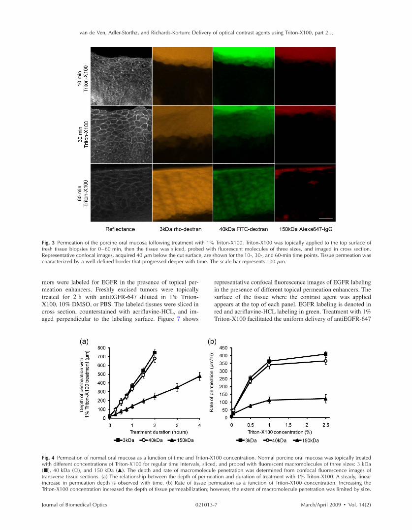
van de Ven Adler-Storthz and Richards-Kortum Delivery of optical contrast agents using Triton-X100 part 2hellip
FfRc
mm tX ca
FwtiT
J
ig 3 Permeation of the porcine oral mucosa following treatment wresh tissue biopsies for 0 ndash60 min then the tissue was sliced probeepresentative confocal images acquired 40 m below the cut surfacharacterized by a well-defined border that progressed deeper with ti
ors were labeled for EGFR in the presence of topical per-eation enhancers Freshly excised tumors were topically
reated for 2 h with antiEGFR-647 diluted in 1 Triton-100 10 DMSO or PBS The labeled tissues were sliced in
ross section counterstained with acriflavine-HCL and im-ged perpendicular to the labeling surface Figure 7 shows
ig 4 Permeation of normal oral mucosa as a function of time and Trith different concentrations of Triton-X100 for regular time intervals
40 kDa and 150 kDa The depth and rate of macromransverse tissue sections a The relationship between the depth of pncrease in permeation depth is observed with time b Rate of tisriton-X100 concentration increased the depth of tissue permeabilizat
ournal of Biomedical Optics 021013-
Triton-X100 Triton-X100 was topically applied to the top surface of fluorescent molecules of three sizes and imaged in cross section
hown for the 10- 30- and 60-min time points Tissue permeation was e scale bar represents 100 m
representative confocal fluorescence images of EGFR labeling in the presence of different topical permeation enhancers The surface of the tissue where the contrast agent was applied appears at the top of each panel EGFR labeling is denoted in red and acriflavine-HCL labeling in green Treatment with 1 Triton-X100 facilitated the uniform delivery of antiEGFR-647
00 concentration Normal porcine oral mucosa was topically treated and probed with fluorescent macromolecules of three sizes 3 kDa penetration was determined from confocal fluorescence images of tion and duration of treatment with 1 Triton-X100 A steady linear meation as a function of Triton-X100 concentration Increasing the wever the extent of macromolecule penetration was limited by size
ith 1d with
e are sme Th
iton-X1 slicedoleculeermea
sue perion ho
7 MarchApril 2009 Vol 142
van de Ven Adler-Storthz and Richards-Kortum Delivery of optical contrast agents using Triton-X100 part 2hellip
F1wpcpm
tt c r d set l
3T c t p e
J
ig 5 Comparison of macromolecule penetration in normal oral muco0 DMSO or PBS sliced and then probed with fluorescent macroas determined from confocal fluorescence images of transverse tissueermeation enhancers measured using 3 kDa rhodamine-dextran Tancer tissue than normal epithelium Little permeation was observenetration by size with 1 Triton-X100 treatment The rate of peacromolecules evaluated
o a depth of 500ndash600 m Washing of the tissue after con-rast agent delivery yielded a honeycomb pattern of labelingonsistent with that observed in vitro EGFR labeling waseproducibly observed around all squamous cells to the fullepth of contrast agent delivery The intensity of labeling wasimilar to that observed in cell monolayers labeled in the pres-nce and absence of Triton-X100 data not shown In con-rast EGFR labeling in DMSO- and PBS-treated tissues wasimited to the superficial layers of cells 20ndash60 m deep
7 Multitarget Labeling in Tumors
o demonstrate the feasibility of delivering multiple opticalontrast agents to different cell compartments three differentargeting moieties were topically applied to tumors in theresence of Triton-X100 The specificity and bioactivity ofach targeting moiety was established in vitro prior to tissue
Table 1 Triton-X100 concentration per cell as a
Tissue
Triton-X100 concentration
Averagepermeation d
m
Bladder deflated
05 83
60
Oral mucosa 10 83
175
350
Squamous cell 10 83 carcinoma
235
470
ournal of Biomedical Optics 021013-
squamous cell carcinoma Tissues were treated with 1 Triton-X100 les of three sizes The depth and rate of macromolecule penetration ns a The maximal penetration depth following a 1-h treatment with imal depth with Triton-X100 treatment was found to be greater for DMSO- and PBS-treated controls b The rate of macromolecule n was consistently more rapid in carcinoma tissue for all sizes of
labeling No differences in targeting were observed with Triton-X100 co-treatment of live and fixed cells data not shown Figure 8 shows representative confocal images of freshly excised tumor tissue simultaneously labeled for EGFR Cyclin D1 and nucleic acid content in the presence of 1 Triton-X100 The tissues were topically labeled washed sliced and imaged perpendicular to the labeling surface AntiEGFR-647 labeling red appeared in a characteristic honeycomb pattern AntiCyclin D1-488 labeling green was diffusely spread through the cytoplasm of cancer cells Pro-pidium iodide labeling orange appeared in the nuclei of per-meabilized cells EGFR Cyclin D1 and propidium iodide labeling always appeared together overlay suggesting that the labeling is limited to cells permeabilized by the Triton-X100 treatment Little to no antibody labeling was observed when isotype controls and propidium iodide were adminis-
on of treatment time and depth
Approx treatment time min
No of Treated cells 106
Normalized concentration pmolcell
5
15
134
100
617
083
5 134 123
30 291 057
60 5816 028
5 134 123
30 390 042
60 780 021
sa andmolecu sectiohe maxed withnetratio
functi
epth
8 MarchApril 2009 Vol 142
van de Ven Adler-Storthz and Richards-Kortum Delivery of optical contrast agents using Triton-X100 part 2hellip
Ff
c m B o at
t c s t
Fasuw
J
ig 6 Tissue phantoms assayed for viability after treatment with dif-erent concentrations of Triton-X100 Phantoms containing 5 106
ells each were treated with 1 mL Triton-X100 for 1 h returned toedia for 4 h and then coincubated with a MTT reagent for 20 minlue color deposition is indicative of viable cells Intense staining wasbserved in phantoms treated with 1 Triton-X100 or less33 pmolcell Phantoms treated with Triton-X100 concentrationsbove 1 33 pmolcell demonstrated an overall loss in color in-ensity and staining at the periphery
ered in the presence of Triton-X100 suggesting that unboundontrast agents were sufficiently removed with the washingteps No propidium iodide labeling was present in tissuesreated with PBS instead of Triton-X100 data not shown
ig 7 EGFR targeting in the presence of topical permeation enhancersntiEGFR-647 red diluted in 1 Triton-X100 10 DMSO or PBShown at the top Tissues were counterstained with acriflavine-HCLniform EGFR labeling was observed around all squamous cells to a das limited to the superficial cell layers The scale bar represents 100
ournal of Biomedical Optics 021013-
4 Discussion
Optical contrast agents developed for cancer biomarker detec-tion have demonstrated high specificity in cell and tissue models Generally ranging in size from 6 kDa peptides and affibodies to 150 kDa antibodies most molecular-specific contrast agents have a limited ability to penetrate mucosal tissue Chemical modification and encapsulation strategies to improve tissue penetration have only proven useful for mol-ecules at the small end of the size spectrum41 The topical delivery of tissue-impermeant contrast agents presents an in-teresting challenge because the success of tissue labeling is dependent on the efficiency and uniformity of contrast agent delivery Furthermore unbound contrast agents generally need to be washed out prior to imaging for high optical con-trast In this paper we evaluate ability of Triton-X100 to de-liver optical contrast agents that lack the appropriate physio-chemical properties for unaided tissue permeation
Confocal microscopy studies of macromolecule penetra-tion in the bladder mucosa were used to examine the unifor-mity of tissue permeabilization The bladder mucosa is a par-ticularly interesting model because the natural folding of the deflated bladder allows the epithelium to be imaged in cross section without the need for tissue slicing In this model we observed that a fixed volume of 05 Triton-X100 can deliver molecules up to 150 kDa in size across treated epithelium By
taneously generated 1483 tumors were topically labeled for 2 h with ssues were sliced and imaged perpendicular to the labeling surface to highlight the tissue morphology In Triton-X100 treated tissues 500ndash600 m EGFR labeling in the DMSO- and PBS-treated controls
Subcu The ti greenepth of m
9 MarchApril 2009 Vol 142
van de Ven Adler-Storthz and Richards-Kortum Delivery of optical contrast agents using Triton-X100 part 2hellip
Fmrwcwm
omXp s a a ml o p i dXg
st d o p ar p e c
J
ig 8 Cotargeting of clinically relevant cancer biomarkers Subcutaolecular-specific contrast agents diluted in 10 Triton-X100 The c
ed Cyclin D1 in the cytoplasm antiCyclin D1 green and nucleicere washed sliced and imaged perpendicular to the labeling suompartments EGFR Cyclin D1 and nucleic acid labeling always apas highly uniform and reproducible from sample to sample Little toent suggesting that the washing of the tissue was sufficient to remo
ur estimate the cells of the epithelium are receiving a treat-ent dose ranging from 617 to 083 pmolcell as the Triton-100 progresses through the tissue Tissue permeation ap-ears to primarily follow a transcellular route appearing as atrong fluorescent signal within cells In tissues probed with 3nd 40 kDa macromolecules the fluorescence is uniformcross the entire epithelium In tissues probed with 150 kDaolecules only the cell nuclei appear as dark spots Extracel-
ular fluorescence is evident between cells at the leading edgef the treatment suggesting that Triton-X100 also facilitatesaracellular delivery Washing of the epithelium removes bothntracellular and extracellular probes yielding a uniformlyark tissue Together these observations indicate that Triton-100 may be useful for the delivery of contrast agents tar-eted to both intracellular and extracellular biomarkers
Studies of Triton-X100 treatment in fresh oral mucosapecimens indicate that the depth of permeation can be con-rolled The leading edge of permeation treatment was wellefined at all concentrations examined allowing measurementf penetration depth as a function time and concentration Theenetration depth increased linearly with treatment time forll sizes of probe macromolecules over a 4-h observation pe-iod There was no apparent delay for the contrast agents toermeate through an intact keratin layer The maximal rate ofpithelial permeation increased with increasing Triton-X100oncentration These trends were highly reproducible varying10 from sample to sample This data demonstrate that it is
ournal of Biomedical Optics 021013-1
y generated 1483 tumors were topically labeled for 1 h with three agents were targeted to EGFR on the cell membrane antiEGFR-647 in the nucleus propidium iodide orange After labeling the tissues The contrast agents were found to localize to the appropriate cell together in treated tissue overlay The observed pattern of labeling eling was observed in IgG-treated controls with Triton-X100 cotreat-ound contrast agents The scale bar represents 20 m
possible to regulate the depth of Triton-X100 treatment by adjusting the Triton-X100 concentration and treatment dura-tion The depth of labeling following treatment was deter-mined by contrast agent size with larger probes demonstrat-ing shallower penetration at the same treatment concentration as smaller molecules Interestingly it does not appear advan-tageous to use Triton-X100 concentrations much above 1 because little improvement in the rate of permeation is ob-served between tissues treated with 1 and 25 Triton-X100
Differences were apparent in the permeation of tissues when compared to cell monolayers In our companion paper Part 1 we showed that cells can be permeabilized with 027 pmolcell Triton-X100 and that nuclear penetration of 40 and 150 kDa molecules can be achieved with 055ndash11 pmolcell Triton-X100 In tissue nuclear penetra-tion is limited to molecules of 40 kDa or less Occasional nuclear penetration of 150 kDa molecules is observed at the surface of treated tissue and is generally limited to one to two cell layers If we consider an oral biopsy treated with 1 Triton-X100 for 1 h the normalized Triton-X100 concentra-tion is in the range of 055ndash11 pmolcell for approximately half of the treatment time Enhanced nuclear permeation is not observed with higher Triton-X100 concentrations or changes in treatment time Thus it appears likely that Triton-X100 preferentially permeabilizes cell membranes in its movement
neouslontrast acids rface peared no labve unb
0 MarchApril 2009 Vol 142
van de Ven Adler-Storthz and Richards-Kortum Delivery of optical contrast agents using Triton-X100 part 2hellip
t t
c c ctm T t s l c sX o 3 w
ct cc a te p stc dm
a at ccs b w os t it m b p l de T cic s Di
J
hrough tissue and is not sufficiently concentrated locally forhe generation of large nuclear pores
In tissue the normalized Triton-X100 concentrationhanges with treatment time and depth such that superficialells are treated to higher doses of Triton-X100 than deeperells Nevertheless cell viability experiments in tissue phan-oms seem to indicate that in a three-dimensional environ-ent cells are more likely to remain viable at higher doses ofriton-X100 In phantoms the viability cutoff is33 pmolcell with cells at the periphery losing their ability
o metabolize MTT at higher concentrations These studiesuggest that at Triton-X100 concentrations of 33 pmolcell oress cell viability is determined the mean concentration perell rather than local concentration gradients This leads us topeculate that tissues can be safely permeated with Triton-100 as long as the treatment time and concentration areptimized to fall below a normalized concentration of3 pmolcell Further studies are needed to determinehether this will hold true in vivo
Depth control will be advantageous for the use of topicalontrast agents in humans By limiting the penetration of op-ical contrast agents to a finite depth the systemic delivery ofontrast agents with undefined toxicity can be avoided Be-ause the mucosal epithelium is a renewable tissue contrastgents adhering to epithelial cells will likely be shed withissue turnover For the optical detection of disease biomark-rs some compensation may be required for differences inermeation rate of normal and abnormal tissue In the tissuestudied here however a 25 difference was observed be-ween normal squamous epithelium and undifferentiated can-er tissue Depth-sensitive optical imaging or spectroscopyevices could potentially be used to monitor the depth of per-eation and determine when to halt the reaction by washingThe use of Triton-X100 as a topical permeation enhancer
llows the delivery of optical contrast agents that lack theppropriate size and chemical properties for tissue penetra-ion Molecular-specific contrast agents as large as antibodiesan be successfully delivered to both intracellular and extra-ellular targets using Triton-X100 In this paper we demon-trate that three clinically relevant biomarkers of cancer cane simultaneously labeled with the topical co-administrationith Triton-X100 The EGFR which is located on the surfacef cells is thought to be a promising target because its expres-ion is elevated in a variety of dysplastic and cancerousissues47ndash49 The overexpression of Cyclin D1 detected heren the cytoplasm is associated with a poor prognosis in pa-ients with head and neck tumors50ndash53 Measures of nuclearorphology delineated here with propidium iodide have
een proposed as an objective measure of the transition fromreneoplastic to neoplastic tissue54 In vitro studies of cellabeling in the presence and absence of Triton-X100 haveemonstrated that structure and availability of these biomark-rs in live cells is similar to that fixed cells data not shownhe targeting of these biomarkers with molecular-specificontrast agents in tissue yielded uniform reproducible label-ng in all transverse sections examined by confocal micros-opy The localization and intensity of tissue labeling wasimilar to that observed in vitro In contrast treatment withMSO or PBS failed to enhance the penetration tissue-
mpermeant optical probes The feasibility of translating Triton-X100 for in vivo use
ournal of Biomedical Optics 021013-1
remains to be determined In our companion paper we dem-onstrated that Triton-X100ndashmediated permeabilization of cells is reversible and that cells retain their metabolic and prolif-erative capacities while recovering membrane integrity Oth-ers have demonstrated that only concentrations of 10 pro-duce visible side effects in patch tests of human skin consisting of transdermal water loss and erythema55 It has been reported that two or more permeation enhancers applied together can act synergistically56 suggesting that it may be possible to further reduce the effective Triton-X100 concen-tration by combining it with other topical agents Tissue dam-age reported with the use of other surfactant-based permeation enhancers has generally been limited and reversible57ndash59 The next step will be to characterize the safety toxicity biological activity and reversibility of Triton-X100ndashmediated perme-ation at clinically useful doses in animal models
5 Conclusions This paper demonstrates that Triton-X100 is a promising topi-cal permeation enhancer for the mucosal delivery of tissue-impermeant optical contrast agents Compared to other topical permeation enhancers Triton-X100 can deliver a much broader size range of molecules No chemical modification of optical contrast agents is needed both targeted and untargeted molecules as large as 150 kDa can be delivered uniformly through normal and cancer tissue The depth of tissue perme-ation can be modulated allowing for epithelial containment of topical contrast agents The coadministration of Triton-X100 with molecular-specific contrast agents facilitates the simulta-neous labeling of cell membrane cytoplasmic and nuclear targets Further work is needed to establish the safety of this approach for in vivo use
Acknowledgments
We thank Vivian Mack for her assistance with cell culture and animal care This work was supported in part by an NIH BRP Grant No CA103830 and a training fellowship from the Keck Center Nanobiology Training Program of the Gulf Coast Consortia NIH Grant No 5 T90 DK071054-03
References 1 S Folli P Westermann D Braichotte A Pelegrin G Wagnieres H
van den Bergh and J P Mach ldquoAntibody-indocyanin conjugates for immunophotodetection of human squamous cell carcinoma in nude micerdquo Cancer Res 5410 2643ndash2649 1994
2 B Ballou G W Fisher J S Deng T R Hakala M Srivastava and D L Farkas ldquoCyanine fluorochrome-labeled antibodies in vivo as-sessment of tumor imaging using Cy3 Cy5 Cy55 and Cy7rdquo Can-cer Detect Prev 223 251ndash257 1998
3 N S Soukos M R Hamblin T F Deutsch and T Hasan ldquoMono-clonal antibody-tagged receptor-targeted contrast agents for detection of cancersrdquo Proc SPIE 4259 115ndash128 2001
4 N S Soukos M R Hamblin S Keel R L Fabian T F Deutsch and T Hasan ldquoEpidermal growth factor receptor-targeted immuno-photodiagnosis and photoimmunotherapy of oral precancer in vivordquo Cancer Res 6111 4490ndash4496 2001
5 Y Koyama T Barrett Y Hama G Ravizzini P L Choyke and H Kobayashi ldquoIn vivo molecular imaging to diagnose and subtype tu-mors through receptor-targeted optically labeled monoclonal antibod-iesrdquo Neoplasia 912 1021ndash1029 2007
6 Y Koyama Y Hama Y Urano D M Nguyen P L Choyke and H Kobayashi ldquoSpectral fluorescence molecular imaging of lung me-tastases targeting HER2neurdquo Clin Cancer Res 1310 2936ndash2945 2007
1 MarchApril 2009 Vol 142
van de Ven Adler-Storthz and Richards-Kortum Delivery of optical contrast agents using Triton-X100 part 2hellip
1
1
1
1
1
1
1
1
1
1
2
2
2
2
2
2
2
2
2
J
7 K E Adams S Ke S Kwon F Liang Z Fan Y Lu K Hirschi ME Mawad M A Barry and E M Sevick-Muraca ldquoComparison ofvisible and near-infrared wavelength-excitable fluorescent dyes formolecular imaging of cancerrdquo J Biomed Opt 122 024017 2007
8 J L Kovar W M Volcheck J Chen and M A Simpson ldquoPurifi-cation method directly influences effectiveness of an epidermalgrowth factor-coupled targeting agent for noninvasive tumor detec-tion in micerdquo Anal Biochem 3611 47ndash54 2007
9 J L Kovar M A Johnson W M Volcheck J Chen and M ASimpson ldquoHyaluronidase expression induces prostate tumor metasta-sis in an orthotopic mouse modelrdquo Am J Pathol 1694 1415ndash14262006
0 S Ke X Wen M Gurfinkel C Charnsangavej S Wallace E MSevick-Muraca and C Li ldquoNear-infrared optical imaging of epider-mal growth factor receptor in breast cancer xenograftsrdquo Cancer Res6322 7870ndash7875 2003
1 K L Carraway 3rd and R A Cerione ldquoFluorescent-labeled growthfactor molecules serve as probes for receptor binding and endocyto-sisrdquo Biochemistry 3245 12039ndash12045 1993
2 C H Tung Y Lin W K Moon and R Weissleder ldquoA receptor-targeted near-infrared fluorescence probe for in vivo tumor imagingrdquoChemBioChem 38 784ndash786 2002
3 S Achilefu R B Dorshow J E Bugaj and R Rajagopalan ldquoNovelreceptor-targeted fluorescent contrast agents for in vivo tumor imag-ingrdquo Invest Radiol 358 479ndash485 2000
4 A Becker C Hessenius K Licha B Ebert U Sukowski W Sem-mler B Wiedenmann and C Grotzinger ldquoReceptor-targeted opticalimaging of tumors with near-infrared fluorescent ligandsrdquo Nat Bio-technol 194 327ndash331 2001
5 D Citrin T Scott M Sproull C Menard P J Tofilon and KCamphausen ldquoIn vivo tumor imaging using a near-infrared-labeledendostatin moleculerdquo Int J Radiat Oncol Biol Phys 582 536ndash541 2004
6 R Weissleder C H Tung U Mahmood and A Bogdanov Jr ldquoInvivo imaging of tumors with protease-activated near-infrared fluores-cent probesrdquo Nat Biotechnol 174 375ndash378 1999
7 C H Tung U Mahmood S Bredow and R Weissleder ldquoIn vivoimaging of proteolytic enzyme activity using a novel molecular re-porterrdquo Cancer Res 6017 4953ndash4958 2000
8 K Marten C Bremer K Khazaie M Sameni B Sloane C HTung and R Weissleder ldquoDetection of dysplastic intestinal ad-enomas using enzyme-sensing molecular beacons in micerdquo Gastro-enterology 1222 406ndash414 2002
9 C Bremer C H Tung A Bogdanov Jr and R Weissleder ldquoImag-ing of differential protease expression in breast cancers for detectionof aggressive tumor phenotypesrdquo Radiology 2223 814ndash818 2002
0 M Kamiya H Kobayashi Y Hama Y Koyama M Bernardo TNagano P L Choyke and Y Urano ldquoAn enzymatically activatedfluorescence probe for targeted tumor imagingrdquo J Am Chem Soc12913 3918ndash3929 2007
1 J V Frangioni ldquoIn vivo near-infrared fluorescence imagingrdquo CurrOpin Chem Biol 75 626ndash634 2003
2 R Cotran V Kumar and T Collins Robbins Pathologic Basis ofDisease W B Saunders Philadelphia 1999
3 J Levi Z Cheng O Gheysens M Patel C T Chan Y Wang MNamavari and S S Gambhir ldquoFluorescent fructose derivatives forimaging breast cancer cellsrdquo Bioconjugate Chem 183 628ndash6342007
4 R OrsquoNeil L Wu and N Mullani ldquoUptake of a fluorescent deoxy-glucose analog 2-NBDG in tumor cellsrdquo Mol Imaging Biol 76388ndash392 2005
5 Z Cheng J Levi Z Xiong O Gheysens S Keren X Chen and SS Gambhir ldquoNear-infrared fluorescent deoxyglucose analogue fortumor optical imaging in cell culture and living micerdquo BioconjugateChem 173 662ndash669 2006
6 T J Muldoon M C Pierce D L Nida M D Williams A Gillen-water and R Richards-Kortum ldquoSubcellular-resolution molecularimaging within living tissue by fiber microendoscopyrdquo Opt Express1525 16413ndash16423 2007
7 Y Kakeji S Yamaguchi D Yoshida K Tanoue M Ueda A Ma-sunari T Utsunomiya M Imamura H Honda Y Maehara and MHashizume ldquoDevelopment and assessment of morphologic criteriafor diagnosing gastric cancer using confocal endomicroscopy an exvivo and in vivo studyrdquo Endoscopy 389 886ndash890 2006
8 A L Polglase W J McLaren S A Skinner R Kiesslich M F
ournal of Biomedical Optics 021013-1
Neurath and P M Delaney ldquoA fluorescence confocal endomicro-scope for in vivo microscopy of the upper- and the lower-GI tractrdquo Gastrointest Endosc 625 686ndash695 2005
29 I A Siegel K T Izutsu and E Watson ldquoMechanisms of non-electrolyte penetration across dog and rabbit oral mucosa in vitrordquo Arch Oral Biol 265 357ndash361 1981
30 V H Lee ldquoEnzymatic barriers to peptide and protein absorptionrdquo Crit Rev Ther Drug Carrier Syst 52 69ndash97 1988
31 R A Hughes I Toth P Ward S J Ireland and W A Gibbons ldquoLipidic peptides III lipidic amino acid and oligomer conjugates of morphinerdquo J Pharm Sci 8012 1103ndash1105 1991
32 H A Benson ldquoTransdermal drug delivery penetration enhancement techniquesrdquo Curr Drug Delivery 21 23ndash33 2005
33 M Thanou J C Verhoef and H E Junginger ldquoOral drug absorption enhancement by chitosan and its derivativesrdquo Adv Drug Delivery Rev 522 117ndash126 2001
34 V Erjavec Z Pavlica M Sentjurc and M Petelin ldquoIn vivo study of liposomes as drug carriers to oral mucosa using EPR oximetryrdquo Int J Pharm 3071 1ndash8 2006
35 H Li J H An J S Park and K Han ldquoMultivesicular liposomes for oral delivery of recombinant human epidermal growth factorrdquo Arch Pharmacal Res 288 988ndash994 2005
36 F S Farshi A Y Ozer M T Ercan and A A Hincal ldquoIn-vivo studies in the treatment of oral ulcers with liposomal dexamethasone sodium phosphaterdquo J Microencapsul 135 537ndash544 1996
37 A N C Anigbogu A C Williams B W Barry and H G M Edwards ldquoFourier transform raman spectroscopy of interactions be-tween the penetration enhancer dimethyl sulfoxide and human stra-tum corneumrdquo Int J Pharm 1252 265ndash282 1995
38 H A E Benson ldquoTransfersomes for transdermal drug deliveryrdquo Expert Opin Drug Delivery 36 727ndash737 2006
39 P N Gupta V Mishra P Singh A Rawat P Dubey S Mahor and S P Vyas ldquoTetanus toxoid-loaded transfersomes for topical immuni-zationrdquo J Pharm Pharmacol 573 295ndash301 2005
40 J Hao and P W Heng ldquoBuccal delivery systemsrdquo Drug Dev Ind Pharm 298 821ndash832 2003
41 F Veuillez Y N Kalia Y Jacques J Deshusses and P Buri ldquoFac-tors and strategies for improving buccal absorption of peptidesrdquo Eur J Pharm Biopharm 512 93ndash109 2001
42 P G Sacks S M Parnes G E Gallick Z Mansouri R Lichtner K L Satya-Prakash S Pathak and D F Parsons ldquoEstablishment and characterization of two new squamous cell carcinoma cell lines de-rived from tumors of the head and neckrdquo Cancer Res 4810 2858ndash2866 1988
43 K J Mattern C Nakornchai and W M Deen ldquoDarcy permeability of agarose-glycosaminoglycan gels analyzed using fiber-mixture and Donnan modelsrdquo Biophys J 952 648ndash656 2008
44 E M Johnson D A Berk R K Jain and W M Deen ldquoHindered diffusion in agarose gels test of effective medium modelrdquo Biophys J 702 1017ndash1023 1996
45 K Sokolov J Galvan A Myakov A Lacy R Lotan and R Richards-Kortum ldquoRealistic three-dimensional epithelial tissue phan-toms for biomedical opticsrdquo J Biomed Opt 71 148ndash156 2002
46 E R Hsu E V Anslyn S Dharmawardhane R Alizadeh-Naderi J S Aaron K V Sokolov A K El-Naggar A M Gillenwater and R R Richards-Kortum ldquoA far-red fluorescent contrast agent to image epidermal growth factor receptor expressionrdquo Photochem Photobiol 793 272ndash279 2004
47 H Modjtahedi and C J Dean ldquoThe receptor for EGF and its ligands expression prognostic value and target for therapy in cancerrdquo Int J Oncol 4 277ndash296 1994
48 W Yasui H Sumiyoshi J Hata T Kameda A Ochiai H Ito and E Tahara ldquoExpression of epidermal growth factor receptor in human gastric and colonic carcinomasrdquo Cancer Res 481 137ndash141 1988
49 J A Vet F M Debruyne and J A Schalken ldquoMolecular prognostic factors in bladder cancerrdquo World J Urol 122 84ndash88 1994
50 A Namazie S Alavi O I Olopade G Pauletti N Aghamoham-madi M Aghamohammadi J A Gornbein T C Calcaterra D J Slamon M B Wang and E S Srivatsan ldquoCyclin D1 amplification and p16MTS1CDK4I deletion correlate with poor prognosis in head and neck tumorsrdquo Laryngoscope 1123 472ndash481 2002
51 J A Akervall R J Michalides H Mineta A Balm A Borg M R Dictor Y Jin B Loftus F Mertens and J P Wennerberg ldquoAmpli-fication of cyclin D1 in squamous cell carcinoma of the head and neck and the prognostic value of chromosomal abnormalities and
2 MarchApril 2009 Vol 142
van de Ven Adler-Storthz and Richards-Kortum Delivery of optical contrast agents using Triton-X100 part 2hellip
5
5
5
5
J
cyclin D1 overexpressionrdquo Cancer 792 380ndash389 1997 2 R J Michalides N M van Veelen P M Kristel A A Hart B M
Loftus F J Hilgers and A J Balm ldquoOverexpression of cyclin D1indicates a poor prognosis in squamous cell carcinoma of the headand neckrdquo Arch Otolaryngol 1235 497ndash502 1997
3 R Michalides N van Veelen A Hart B Loftus E Wientjens andA Balm ldquoOverexpression of cyclin D1 correlates with recurrence ina group of forty-seven operable squamous cell carcinomas of thehead and neckrdquo Cancer Res 555 975ndash978 1995
4 M F Mitchell W N Hittelman W K Hong R Lotan and DSchottenfeld ldquoThe natural history of cervical intraepithelial neopla-sia an argument for intermediate endpoint biomarkersrdquo Cancer Epi-demiol Biomarkers Prev 37 619ndash626 1994
5 F van Ruissen M Le J M Carroll P G M van der Valk and JSchalkwijk ldquoDifferential effects of detergents on keratinocyte geneexpressionrdquo J Invest Dermatol 1104 358 1998
ournal of Biomedical Optics 021013-1
56 B Mollgaard ldquoSynergistic effects in percutaneous enhancementrdquo in Pharmaceutical Skin Penetration Enhancement K A Walters and J Hadgraft Eds pp 220ndash242 Marcel Dekker New York 1993
57 E S Swenson W B Milisen and W Curatolo ldquoIntestinal perme-ability enhancement efficacy acute local toxicity and reversibilityrdquo Pharmacol Res 118 1132ndash1142 1994
58 E K Anderberg and P Artursson ldquoEpithelial transport of drugs in cell culture VIII Effects of sodium dodecyl sulfate on cell mem-brane and tight junction permeability in human intestinal epithelial Caco-2 cellsrdquo J Pharm Sci 824 392ndash398 1993
59 E K Anderberg C Nystrom and P Artursson ldquoEpithelial transport of drugs in cell culture VII Effects of pharmaceutical surfactant excipients and bile acids on transepithelial permeability in monolay-ers of human intestinal epithelial Caco-2 cellsrdquo J Pharm Sci 819 879ndash887 1992
3 MarchApril 2009 Vol 142
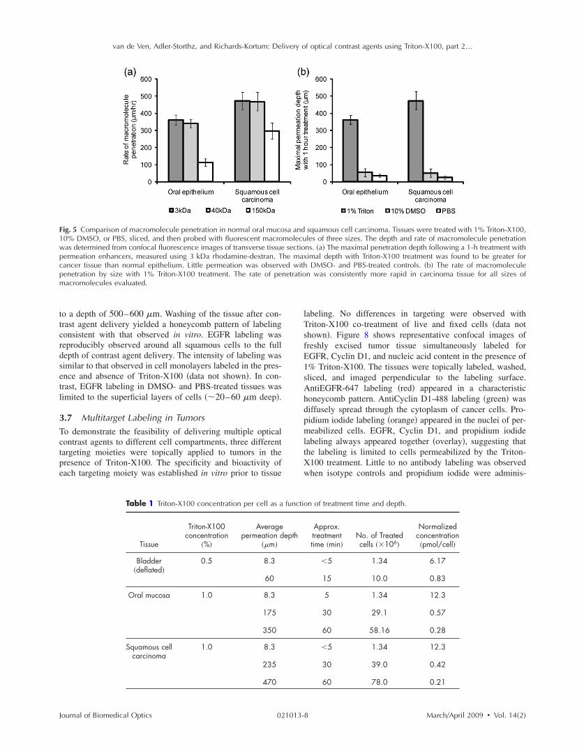
van de Ven Adler-Storthz and Richards-Kortum Delivery of optical contrast agents using Triton-X100 part 2hellip
F1wpcpm
tt c r d set l
3T c t p e
J
ig 5 Comparison of macromolecule penetration in normal oral muco0 DMSO or PBS sliced and then probed with fluorescent macroas determined from confocal fluorescence images of transverse tissueermeation enhancers measured using 3 kDa rhodamine-dextran Tancer tissue than normal epithelium Little permeation was observenetration by size with 1 Triton-X100 treatment The rate of peacromolecules evaluated
o a depth of 500ndash600 m Washing of the tissue after con-rast agent delivery yielded a honeycomb pattern of labelingonsistent with that observed in vitro EGFR labeling waseproducibly observed around all squamous cells to the fullepth of contrast agent delivery The intensity of labeling wasimilar to that observed in cell monolayers labeled in the pres-nce and absence of Triton-X100 data not shown In con-rast EGFR labeling in DMSO- and PBS-treated tissues wasimited to the superficial layers of cells 20ndash60 m deep
7 Multitarget Labeling in Tumors
o demonstrate the feasibility of delivering multiple opticalontrast agents to different cell compartments three differentargeting moieties were topically applied to tumors in theresence of Triton-X100 The specificity and bioactivity ofach targeting moiety was established in vitro prior to tissue
Table 1 Triton-X100 concentration per cell as a
Tissue
Triton-X100 concentration
Averagepermeation d
m
Bladder deflated
05 83
60
Oral mucosa 10 83
175
350
Squamous cell 10 83 carcinoma
235
470
ournal of Biomedical Optics 021013-
squamous cell carcinoma Tissues were treated with 1 Triton-X100 les of three sizes The depth and rate of macromolecule penetration ns a The maximal penetration depth following a 1-h treatment with imal depth with Triton-X100 treatment was found to be greater for DMSO- and PBS-treated controls b The rate of macromolecule n was consistently more rapid in carcinoma tissue for all sizes of
labeling No differences in targeting were observed with Triton-X100 co-treatment of live and fixed cells data not shown Figure 8 shows representative confocal images of freshly excised tumor tissue simultaneously labeled for EGFR Cyclin D1 and nucleic acid content in the presence of 1 Triton-X100 The tissues were topically labeled washed sliced and imaged perpendicular to the labeling surface AntiEGFR-647 labeling red appeared in a characteristic honeycomb pattern AntiCyclin D1-488 labeling green was diffusely spread through the cytoplasm of cancer cells Pro-pidium iodide labeling orange appeared in the nuclei of per-meabilized cells EGFR Cyclin D1 and propidium iodide labeling always appeared together overlay suggesting that the labeling is limited to cells permeabilized by the Triton-X100 treatment Little to no antibody labeling was observed when isotype controls and propidium iodide were adminis-
on of treatment time and depth
Approx treatment time min
No of Treated cells 106
Normalized concentration pmolcell
5
15
134
100
617
083
5 134 123
30 291 057
60 5816 028
5 134 123
30 390 042
60 780 021
sa andmolecu sectiohe maxed withnetratio
functi
epth
8 MarchApril 2009 Vol 142
van de Ven Adler-Storthz and Richards-Kortum Delivery of optical contrast agents using Triton-X100 part 2hellip
Ff
c m B o at
t c s t
Fasuw
J
ig 6 Tissue phantoms assayed for viability after treatment with dif-erent concentrations of Triton-X100 Phantoms containing 5 106
ells each were treated with 1 mL Triton-X100 for 1 h returned toedia for 4 h and then coincubated with a MTT reagent for 20 minlue color deposition is indicative of viable cells Intense staining wasbserved in phantoms treated with 1 Triton-X100 or less33 pmolcell Phantoms treated with Triton-X100 concentrationsbove 1 33 pmolcell demonstrated an overall loss in color in-ensity and staining at the periphery
ered in the presence of Triton-X100 suggesting that unboundontrast agents were sufficiently removed with the washingteps No propidium iodide labeling was present in tissuesreated with PBS instead of Triton-X100 data not shown
ig 7 EGFR targeting in the presence of topical permeation enhancersntiEGFR-647 red diluted in 1 Triton-X100 10 DMSO or PBShown at the top Tissues were counterstained with acriflavine-HCLniform EGFR labeling was observed around all squamous cells to a das limited to the superficial cell layers The scale bar represents 100
ournal of Biomedical Optics 021013-
4 Discussion
Optical contrast agents developed for cancer biomarker detec-tion have demonstrated high specificity in cell and tissue models Generally ranging in size from 6 kDa peptides and affibodies to 150 kDa antibodies most molecular-specific contrast agents have a limited ability to penetrate mucosal tissue Chemical modification and encapsulation strategies to improve tissue penetration have only proven useful for mol-ecules at the small end of the size spectrum41 The topical delivery of tissue-impermeant contrast agents presents an in-teresting challenge because the success of tissue labeling is dependent on the efficiency and uniformity of contrast agent delivery Furthermore unbound contrast agents generally need to be washed out prior to imaging for high optical con-trast In this paper we evaluate ability of Triton-X100 to de-liver optical contrast agents that lack the appropriate physio-chemical properties for unaided tissue permeation
Confocal microscopy studies of macromolecule penetra-tion in the bladder mucosa were used to examine the unifor-mity of tissue permeabilization The bladder mucosa is a par-ticularly interesting model because the natural folding of the deflated bladder allows the epithelium to be imaged in cross section without the need for tissue slicing In this model we observed that a fixed volume of 05 Triton-X100 can deliver molecules up to 150 kDa in size across treated epithelium By
taneously generated 1483 tumors were topically labeled for 2 h with ssues were sliced and imaged perpendicular to the labeling surface to highlight the tissue morphology In Triton-X100 treated tissues 500ndash600 m EGFR labeling in the DMSO- and PBS-treated controls
Subcu The ti greenepth of m
9 MarchApril 2009 Vol 142
van de Ven Adler-Storthz and Richards-Kortum Delivery of optical contrast agents using Triton-X100 part 2hellip
Fmrwcwm
omXp s a a ml o p i dXg
st d o p ar p e c
J
ig 8 Cotargeting of clinically relevant cancer biomarkers Subcutaolecular-specific contrast agents diluted in 10 Triton-X100 The c
ed Cyclin D1 in the cytoplasm antiCyclin D1 green and nucleicere washed sliced and imaged perpendicular to the labeling suompartments EGFR Cyclin D1 and nucleic acid labeling always apas highly uniform and reproducible from sample to sample Little toent suggesting that the washing of the tissue was sufficient to remo
ur estimate the cells of the epithelium are receiving a treat-ent dose ranging from 617 to 083 pmolcell as the Triton-100 progresses through the tissue Tissue permeation ap-ears to primarily follow a transcellular route appearing as atrong fluorescent signal within cells In tissues probed with 3nd 40 kDa macromolecules the fluorescence is uniformcross the entire epithelium In tissues probed with 150 kDaolecules only the cell nuclei appear as dark spots Extracel-
ular fluorescence is evident between cells at the leading edgef the treatment suggesting that Triton-X100 also facilitatesaracellular delivery Washing of the epithelium removes bothntracellular and extracellular probes yielding a uniformlyark tissue Together these observations indicate that Triton-100 may be useful for the delivery of contrast agents tar-eted to both intracellular and extracellular biomarkers
Studies of Triton-X100 treatment in fresh oral mucosapecimens indicate that the depth of permeation can be con-rolled The leading edge of permeation treatment was wellefined at all concentrations examined allowing measurementf penetration depth as a function time and concentration Theenetration depth increased linearly with treatment time forll sizes of probe macromolecules over a 4-h observation pe-iod There was no apparent delay for the contrast agents toermeate through an intact keratin layer The maximal rate ofpithelial permeation increased with increasing Triton-X100oncentration These trends were highly reproducible varying10 from sample to sample This data demonstrate that it is
ournal of Biomedical Optics 021013-1
y generated 1483 tumors were topically labeled for 1 h with three agents were targeted to EGFR on the cell membrane antiEGFR-647 in the nucleus propidium iodide orange After labeling the tissues The contrast agents were found to localize to the appropriate cell together in treated tissue overlay The observed pattern of labeling eling was observed in IgG-treated controls with Triton-X100 cotreat-ound contrast agents The scale bar represents 20 m
possible to regulate the depth of Triton-X100 treatment by adjusting the Triton-X100 concentration and treatment dura-tion The depth of labeling following treatment was deter-mined by contrast agent size with larger probes demonstrat-ing shallower penetration at the same treatment concentration as smaller molecules Interestingly it does not appear advan-tageous to use Triton-X100 concentrations much above 1 because little improvement in the rate of permeation is ob-served between tissues treated with 1 and 25 Triton-X100
Differences were apparent in the permeation of tissues when compared to cell monolayers In our companion paper Part 1 we showed that cells can be permeabilized with 027 pmolcell Triton-X100 and that nuclear penetration of 40 and 150 kDa molecules can be achieved with 055ndash11 pmolcell Triton-X100 In tissue nuclear penetra-tion is limited to molecules of 40 kDa or less Occasional nuclear penetration of 150 kDa molecules is observed at the surface of treated tissue and is generally limited to one to two cell layers If we consider an oral biopsy treated with 1 Triton-X100 for 1 h the normalized Triton-X100 concentra-tion is in the range of 055ndash11 pmolcell for approximately half of the treatment time Enhanced nuclear permeation is not observed with higher Triton-X100 concentrations or changes in treatment time Thus it appears likely that Triton-X100 preferentially permeabilizes cell membranes in its movement
neouslontrast acids rface peared no labve unb
0 MarchApril 2009 Vol 142
van de Ven Adler-Storthz and Richards-Kortum Delivery of optical contrast agents using Triton-X100 part 2hellip
t t
c c ctm T t s l c sX o 3 w
ct cc a te p stc dm
a at ccs b w os t it m b p l de T cic s Di
J
hrough tissue and is not sufficiently concentrated locally forhe generation of large nuclear pores
In tissue the normalized Triton-X100 concentrationhanges with treatment time and depth such that superficialells are treated to higher doses of Triton-X100 than deeperells Nevertheless cell viability experiments in tissue phan-oms seem to indicate that in a three-dimensional environ-ent cells are more likely to remain viable at higher doses ofriton-X100 In phantoms the viability cutoff is33 pmolcell with cells at the periphery losing their ability
o metabolize MTT at higher concentrations These studiesuggest that at Triton-X100 concentrations of 33 pmolcell oress cell viability is determined the mean concentration perell rather than local concentration gradients This leads us topeculate that tissues can be safely permeated with Triton-100 as long as the treatment time and concentration areptimized to fall below a normalized concentration of3 pmolcell Further studies are needed to determinehether this will hold true in vivo
Depth control will be advantageous for the use of topicalontrast agents in humans By limiting the penetration of op-ical contrast agents to a finite depth the systemic delivery ofontrast agents with undefined toxicity can be avoided Be-ause the mucosal epithelium is a renewable tissue contrastgents adhering to epithelial cells will likely be shed withissue turnover For the optical detection of disease biomark-rs some compensation may be required for differences inermeation rate of normal and abnormal tissue In the tissuestudied here however a 25 difference was observed be-ween normal squamous epithelium and undifferentiated can-er tissue Depth-sensitive optical imaging or spectroscopyevices could potentially be used to monitor the depth of per-eation and determine when to halt the reaction by washingThe use of Triton-X100 as a topical permeation enhancer
llows the delivery of optical contrast agents that lack theppropriate size and chemical properties for tissue penetra-ion Molecular-specific contrast agents as large as antibodiesan be successfully delivered to both intracellular and extra-ellular targets using Triton-X100 In this paper we demon-trate that three clinically relevant biomarkers of cancer cane simultaneously labeled with the topical co-administrationith Triton-X100 The EGFR which is located on the surfacef cells is thought to be a promising target because its expres-ion is elevated in a variety of dysplastic and cancerousissues47ndash49 The overexpression of Cyclin D1 detected heren the cytoplasm is associated with a poor prognosis in pa-ients with head and neck tumors50ndash53 Measures of nuclearorphology delineated here with propidium iodide have
een proposed as an objective measure of the transition fromreneoplastic to neoplastic tissue54 In vitro studies of cellabeling in the presence and absence of Triton-X100 haveemonstrated that structure and availability of these biomark-rs in live cells is similar to that fixed cells data not shownhe targeting of these biomarkers with molecular-specificontrast agents in tissue yielded uniform reproducible label-ng in all transverse sections examined by confocal micros-opy The localization and intensity of tissue labeling wasimilar to that observed in vitro In contrast treatment withMSO or PBS failed to enhance the penetration tissue-
mpermeant optical probes The feasibility of translating Triton-X100 for in vivo use
ournal of Biomedical Optics 021013-1
remains to be determined In our companion paper we dem-onstrated that Triton-X100ndashmediated permeabilization of cells is reversible and that cells retain their metabolic and prolif-erative capacities while recovering membrane integrity Oth-ers have demonstrated that only concentrations of 10 pro-duce visible side effects in patch tests of human skin consisting of transdermal water loss and erythema55 It has been reported that two or more permeation enhancers applied together can act synergistically56 suggesting that it may be possible to further reduce the effective Triton-X100 concen-tration by combining it with other topical agents Tissue dam-age reported with the use of other surfactant-based permeation enhancers has generally been limited and reversible57ndash59 The next step will be to characterize the safety toxicity biological activity and reversibility of Triton-X100ndashmediated perme-ation at clinically useful doses in animal models
5 Conclusions This paper demonstrates that Triton-X100 is a promising topi-cal permeation enhancer for the mucosal delivery of tissue-impermeant optical contrast agents Compared to other topical permeation enhancers Triton-X100 can deliver a much broader size range of molecules No chemical modification of optical contrast agents is needed both targeted and untargeted molecules as large as 150 kDa can be delivered uniformly through normal and cancer tissue The depth of tissue perme-ation can be modulated allowing for epithelial containment of topical contrast agents The coadministration of Triton-X100 with molecular-specific contrast agents facilitates the simulta-neous labeling of cell membrane cytoplasmic and nuclear targets Further work is needed to establish the safety of this approach for in vivo use
Acknowledgments
We thank Vivian Mack for her assistance with cell culture and animal care This work was supported in part by an NIH BRP Grant No CA103830 and a training fellowship from the Keck Center Nanobiology Training Program of the Gulf Coast Consortia NIH Grant No 5 T90 DK071054-03
References 1 S Folli P Westermann D Braichotte A Pelegrin G Wagnieres H
van den Bergh and J P Mach ldquoAntibody-indocyanin conjugates for immunophotodetection of human squamous cell carcinoma in nude micerdquo Cancer Res 5410 2643ndash2649 1994
2 B Ballou G W Fisher J S Deng T R Hakala M Srivastava and D L Farkas ldquoCyanine fluorochrome-labeled antibodies in vivo as-sessment of tumor imaging using Cy3 Cy5 Cy55 and Cy7rdquo Can-cer Detect Prev 223 251ndash257 1998
3 N S Soukos M R Hamblin T F Deutsch and T Hasan ldquoMono-clonal antibody-tagged receptor-targeted contrast agents for detection of cancersrdquo Proc SPIE 4259 115ndash128 2001
4 N S Soukos M R Hamblin S Keel R L Fabian T F Deutsch and T Hasan ldquoEpidermal growth factor receptor-targeted immuno-photodiagnosis and photoimmunotherapy of oral precancer in vivordquo Cancer Res 6111 4490ndash4496 2001
5 Y Koyama T Barrett Y Hama G Ravizzini P L Choyke and H Kobayashi ldquoIn vivo molecular imaging to diagnose and subtype tu-mors through receptor-targeted optically labeled monoclonal antibod-iesrdquo Neoplasia 912 1021ndash1029 2007
6 Y Koyama Y Hama Y Urano D M Nguyen P L Choyke and H Kobayashi ldquoSpectral fluorescence molecular imaging of lung me-tastases targeting HER2neurdquo Clin Cancer Res 1310 2936ndash2945 2007
1 MarchApril 2009 Vol 142
van de Ven Adler-Storthz and Richards-Kortum Delivery of optical contrast agents using Triton-X100 part 2hellip
1
1
1
1
1
1
1
1
1
1
2
2
2
2
2
2
2
2
2
J
7 K E Adams S Ke S Kwon F Liang Z Fan Y Lu K Hirschi ME Mawad M A Barry and E M Sevick-Muraca ldquoComparison ofvisible and near-infrared wavelength-excitable fluorescent dyes formolecular imaging of cancerrdquo J Biomed Opt 122 024017 2007
8 J L Kovar W M Volcheck J Chen and M A Simpson ldquoPurifi-cation method directly influences effectiveness of an epidermalgrowth factor-coupled targeting agent for noninvasive tumor detec-tion in micerdquo Anal Biochem 3611 47ndash54 2007
9 J L Kovar M A Johnson W M Volcheck J Chen and M ASimpson ldquoHyaluronidase expression induces prostate tumor metasta-sis in an orthotopic mouse modelrdquo Am J Pathol 1694 1415ndash14262006
0 S Ke X Wen M Gurfinkel C Charnsangavej S Wallace E MSevick-Muraca and C Li ldquoNear-infrared optical imaging of epider-mal growth factor receptor in breast cancer xenograftsrdquo Cancer Res6322 7870ndash7875 2003
1 K L Carraway 3rd and R A Cerione ldquoFluorescent-labeled growthfactor molecules serve as probes for receptor binding and endocyto-sisrdquo Biochemistry 3245 12039ndash12045 1993
2 C H Tung Y Lin W K Moon and R Weissleder ldquoA receptor-targeted near-infrared fluorescence probe for in vivo tumor imagingrdquoChemBioChem 38 784ndash786 2002
3 S Achilefu R B Dorshow J E Bugaj and R Rajagopalan ldquoNovelreceptor-targeted fluorescent contrast agents for in vivo tumor imag-ingrdquo Invest Radiol 358 479ndash485 2000
4 A Becker C Hessenius K Licha B Ebert U Sukowski W Sem-mler B Wiedenmann and C Grotzinger ldquoReceptor-targeted opticalimaging of tumors with near-infrared fluorescent ligandsrdquo Nat Bio-technol 194 327ndash331 2001
5 D Citrin T Scott M Sproull C Menard P J Tofilon and KCamphausen ldquoIn vivo tumor imaging using a near-infrared-labeledendostatin moleculerdquo Int J Radiat Oncol Biol Phys 582 536ndash541 2004
6 R Weissleder C H Tung U Mahmood and A Bogdanov Jr ldquoInvivo imaging of tumors with protease-activated near-infrared fluores-cent probesrdquo Nat Biotechnol 174 375ndash378 1999
7 C H Tung U Mahmood S Bredow and R Weissleder ldquoIn vivoimaging of proteolytic enzyme activity using a novel molecular re-porterrdquo Cancer Res 6017 4953ndash4958 2000
8 K Marten C Bremer K Khazaie M Sameni B Sloane C HTung and R Weissleder ldquoDetection of dysplastic intestinal ad-enomas using enzyme-sensing molecular beacons in micerdquo Gastro-enterology 1222 406ndash414 2002
9 C Bremer C H Tung A Bogdanov Jr and R Weissleder ldquoImag-ing of differential protease expression in breast cancers for detectionof aggressive tumor phenotypesrdquo Radiology 2223 814ndash818 2002
0 M Kamiya H Kobayashi Y Hama Y Koyama M Bernardo TNagano P L Choyke and Y Urano ldquoAn enzymatically activatedfluorescence probe for targeted tumor imagingrdquo J Am Chem Soc12913 3918ndash3929 2007
1 J V Frangioni ldquoIn vivo near-infrared fluorescence imagingrdquo CurrOpin Chem Biol 75 626ndash634 2003
2 R Cotran V Kumar and T Collins Robbins Pathologic Basis ofDisease W B Saunders Philadelphia 1999
3 J Levi Z Cheng O Gheysens M Patel C T Chan Y Wang MNamavari and S S Gambhir ldquoFluorescent fructose derivatives forimaging breast cancer cellsrdquo Bioconjugate Chem 183 628ndash6342007
4 R OrsquoNeil L Wu and N Mullani ldquoUptake of a fluorescent deoxy-glucose analog 2-NBDG in tumor cellsrdquo Mol Imaging Biol 76388ndash392 2005
5 Z Cheng J Levi Z Xiong O Gheysens S Keren X Chen and SS Gambhir ldquoNear-infrared fluorescent deoxyglucose analogue fortumor optical imaging in cell culture and living micerdquo BioconjugateChem 173 662ndash669 2006
6 T J Muldoon M C Pierce D L Nida M D Williams A Gillen-water and R Richards-Kortum ldquoSubcellular-resolution molecularimaging within living tissue by fiber microendoscopyrdquo Opt Express1525 16413ndash16423 2007
7 Y Kakeji S Yamaguchi D Yoshida K Tanoue M Ueda A Ma-sunari T Utsunomiya M Imamura H Honda Y Maehara and MHashizume ldquoDevelopment and assessment of morphologic criteriafor diagnosing gastric cancer using confocal endomicroscopy an exvivo and in vivo studyrdquo Endoscopy 389 886ndash890 2006
8 A L Polglase W J McLaren S A Skinner R Kiesslich M F
ournal of Biomedical Optics 021013-1
Neurath and P M Delaney ldquoA fluorescence confocal endomicro-scope for in vivo microscopy of the upper- and the lower-GI tractrdquo Gastrointest Endosc 625 686ndash695 2005
29 I A Siegel K T Izutsu and E Watson ldquoMechanisms of non-electrolyte penetration across dog and rabbit oral mucosa in vitrordquo Arch Oral Biol 265 357ndash361 1981
30 V H Lee ldquoEnzymatic barriers to peptide and protein absorptionrdquo Crit Rev Ther Drug Carrier Syst 52 69ndash97 1988
31 R A Hughes I Toth P Ward S J Ireland and W A Gibbons ldquoLipidic peptides III lipidic amino acid and oligomer conjugates of morphinerdquo J Pharm Sci 8012 1103ndash1105 1991
32 H A Benson ldquoTransdermal drug delivery penetration enhancement techniquesrdquo Curr Drug Delivery 21 23ndash33 2005
33 M Thanou J C Verhoef and H E Junginger ldquoOral drug absorption enhancement by chitosan and its derivativesrdquo Adv Drug Delivery Rev 522 117ndash126 2001
34 V Erjavec Z Pavlica M Sentjurc and M Petelin ldquoIn vivo study of liposomes as drug carriers to oral mucosa using EPR oximetryrdquo Int J Pharm 3071 1ndash8 2006
35 H Li J H An J S Park and K Han ldquoMultivesicular liposomes for oral delivery of recombinant human epidermal growth factorrdquo Arch Pharmacal Res 288 988ndash994 2005
36 F S Farshi A Y Ozer M T Ercan and A A Hincal ldquoIn-vivo studies in the treatment of oral ulcers with liposomal dexamethasone sodium phosphaterdquo J Microencapsul 135 537ndash544 1996
37 A N C Anigbogu A C Williams B W Barry and H G M Edwards ldquoFourier transform raman spectroscopy of interactions be-tween the penetration enhancer dimethyl sulfoxide and human stra-tum corneumrdquo Int J Pharm 1252 265ndash282 1995
38 H A E Benson ldquoTransfersomes for transdermal drug deliveryrdquo Expert Opin Drug Delivery 36 727ndash737 2006
39 P N Gupta V Mishra P Singh A Rawat P Dubey S Mahor and S P Vyas ldquoTetanus toxoid-loaded transfersomes for topical immuni-zationrdquo J Pharm Pharmacol 573 295ndash301 2005
40 J Hao and P W Heng ldquoBuccal delivery systemsrdquo Drug Dev Ind Pharm 298 821ndash832 2003
41 F Veuillez Y N Kalia Y Jacques J Deshusses and P Buri ldquoFac-tors and strategies for improving buccal absorption of peptidesrdquo Eur J Pharm Biopharm 512 93ndash109 2001
42 P G Sacks S M Parnes G E Gallick Z Mansouri R Lichtner K L Satya-Prakash S Pathak and D F Parsons ldquoEstablishment and characterization of two new squamous cell carcinoma cell lines de-rived from tumors of the head and neckrdquo Cancer Res 4810 2858ndash2866 1988
43 K J Mattern C Nakornchai and W M Deen ldquoDarcy permeability of agarose-glycosaminoglycan gels analyzed using fiber-mixture and Donnan modelsrdquo Biophys J 952 648ndash656 2008
44 E M Johnson D A Berk R K Jain and W M Deen ldquoHindered diffusion in agarose gels test of effective medium modelrdquo Biophys J 702 1017ndash1023 1996
45 K Sokolov J Galvan A Myakov A Lacy R Lotan and R Richards-Kortum ldquoRealistic three-dimensional epithelial tissue phan-toms for biomedical opticsrdquo J Biomed Opt 71 148ndash156 2002
46 E R Hsu E V Anslyn S Dharmawardhane R Alizadeh-Naderi J S Aaron K V Sokolov A K El-Naggar A M Gillenwater and R R Richards-Kortum ldquoA far-red fluorescent contrast agent to image epidermal growth factor receptor expressionrdquo Photochem Photobiol 793 272ndash279 2004
47 H Modjtahedi and C J Dean ldquoThe receptor for EGF and its ligands expression prognostic value and target for therapy in cancerrdquo Int J Oncol 4 277ndash296 1994
48 W Yasui H Sumiyoshi J Hata T Kameda A Ochiai H Ito and E Tahara ldquoExpression of epidermal growth factor receptor in human gastric and colonic carcinomasrdquo Cancer Res 481 137ndash141 1988
49 J A Vet F M Debruyne and J A Schalken ldquoMolecular prognostic factors in bladder cancerrdquo World J Urol 122 84ndash88 1994
50 A Namazie S Alavi O I Olopade G Pauletti N Aghamoham-madi M Aghamohammadi J A Gornbein T C Calcaterra D J Slamon M B Wang and E S Srivatsan ldquoCyclin D1 amplification and p16MTS1CDK4I deletion correlate with poor prognosis in head and neck tumorsrdquo Laryngoscope 1123 472ndash481 2002
51 J A Akervall R J Michalides H Mineta A Balm A Borg M R Dictor Y Jin B Loftus F Mertens and J P Wennerberg ldquoAmpli-fication of cyclin D1 in squamous cell carcinoma of the head and neck and the prognostic value of chromosomal abnormalities and
2 MarchApril 2009 Vol 142
van de Ven Adler-Storthz and Richards-Kortum Delivery of optical contrast agents using Triton-X100 part 2hellip
5
5
5
5
J
cyclin D1 overexpressionrdquo Cancer 792 380ndash389 1997 2 R J Michalides N M van Veelen P M Kristel A A Hart B M
Loftus F J Hilgers and A J Balm ldquoOverexpression of cyclin D1indicates a poor prognosis in squamous cell carcinoma of the headand neckrdquo Arch Otolaryngol 1235 497ndash502 1997
3 R Michalides N van Veelen A Hart B Loftus E Wientjens andA Balm ldquoOverexpression of cyclin D1 correlates with recurrence ina group of forty-seven operable squamous cell carcinomas of thehead and neckrdquo Cancer Res 555 975ndash978 1995
4 M F Mitchell W N Hittelman W K Hong R Lotan and DSchottenfeld ldquoThe natural history of cervical intraepithelial neopla-sia an argument for intermediate endpoint biomarkersrdquo Cancer Epi-demiol Biomarkers Prev 37 619ndash626 1994
5 F van Ruissen M Le J M Carroll P G M van der Valk and JSchalkwijk ldquoDifferential effects of detergents on keratinocyte geneexpressionrdquo J Invest Dermatol 1104 358 1998
ournal of Biomedical Optics 021013-1
56 B Mollgaard ldquoSynergistic effects in percutaneous enhancementrdquo in Pharmaceutical Skin Penetration Enhancement K A Walters and J Hadgraft Eds pp 220ndash242 Marcel Dekker New York 1993
57 E S Swenson W B Milisen and W Curatolo ldquoIntestinal perme-ability enhancement efficacy acute local toxicity and reversibilityrdquo Pharmacol Res 118 1132ndash1142 1994
58 E K Anderberg and P Artursson ldquoEpithelial transport of drugs in cell culture VIII Effects of sodium dodecyl sulfate on cell mem-brane and tight junction permeability in human intestinal epithelial Caco-2 cellsrdquo J Pharm Sci 824 392ndash398 1993
59 E K Anderberg C Nystrom and P Artursson ldquoEpithelial transport of drugs in cell culture VII Effects of pharmaceutical surfactant excipients and bile acids on transepithelial permeability in monolay-ers of human intestinal epithelial Caco-2 cellsrdquo J Pharm Sci 819 879ndash887 1992
3 MarchApril 2009 Vol 142
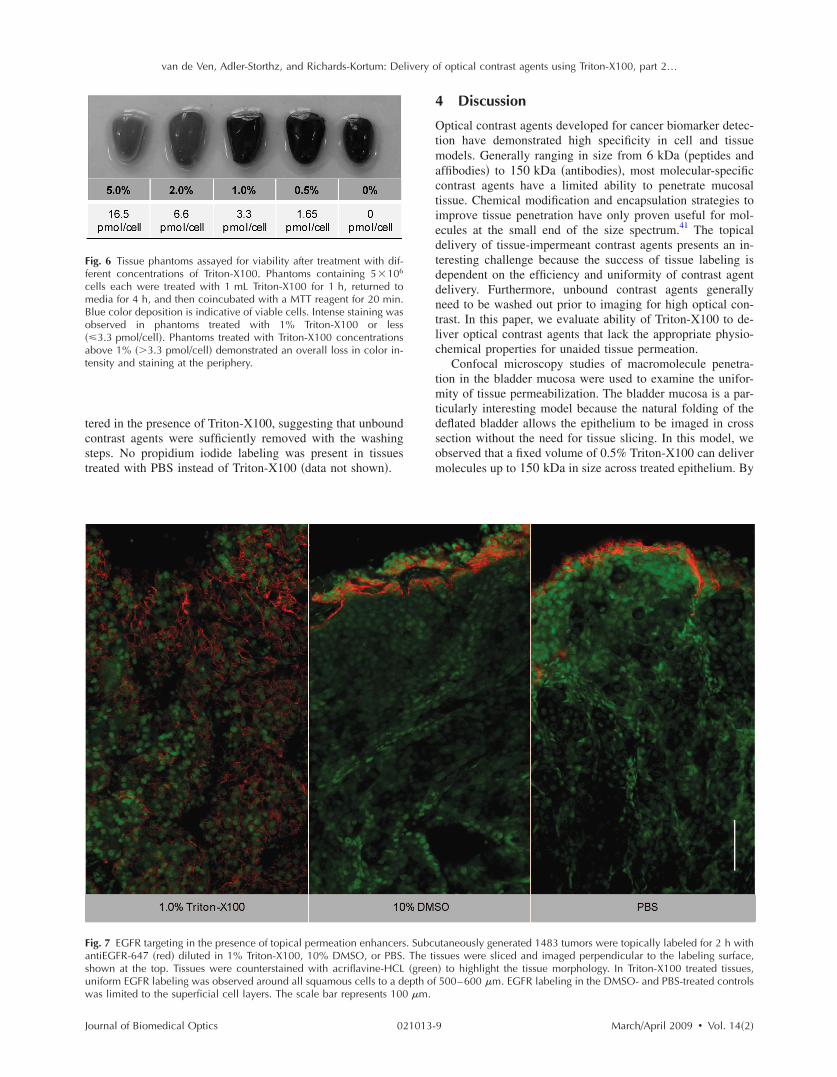
van de Ven Adler-Storthz and Richards-Kortum Delivery of optical contrast agents using Triton-X100 part 2hellip
t t
c c ctm T t s l c sX o 3 w
ct cc a te p stc dm
a at ccs b w os t it m b p l de T cic s Di
J
hrough tissue and is not sufficiently concentrated locally forhe generation of large nuclear pores
In tissue the normalized Triton-X100 concentrationhanges with treatment time and depth such that superficialells are treated to higher doses of Triton-X100 than deeperells Nevertheless cell viability experiments in tissue phan-oms seem to indicate that in a three-dimensional environ-ent cells are more likely to remain viable at higher doses ofriton-X100 In phantoms the viability cutoff is33 pmolcell with cells at the periphery losing their ability
o metabolize MTT at higher concentrations These studiesuggest that at Triton-X100 concentrations of 33 pmolcell oress cell viability is determined the mean concentration perell rather than local concentration gradients This leads us topeculate that tissues can be safely permeated with Triton-100 as long as the treatment time and concentration areptimized to fall below a normalized concentration of3 pmolcell Further studies are needed to determinehether this will hold true in vivo
Depth control will be advantageous for the use of topicalontrast agents in humans By limiting the penetration of op-ical contrast agents to a finite depth the systemic delivery ofontrast agents with undefined toxicity can be avoided Be-ause the mucosal epithelium is a renewable tissue contrastgents adhering to epithelial cells will likely be shed withissue turnover For the optical detection of disease biomark-rs some compensation may be required for differences inermeation rate of normal and abnormal tissue In the tissuestudied here however a 25 difference was observed be-ween normal squamous epithelium and undifferentiated can-er tissue Depth-sensitive optical imaging or spectroscopyevices could potentially be used to monitor the depth of per-eation and determine when to halt the reaction by washingThe use of Triton-X100 as a topical permeation enhancer
llows the delivery of optical contrast agents that lack theppropriate size and chemical properties for tissue penetra-ion Molecular-specific contrast agents as large as antibodiesan be successfully delivered to both intracellular and extra-ellular targets using Triton-X100 In this paper we demon-trate that three clinically relevant biomarkers of cancer cane simultaneously labeled with the topical co-administrationith Triton-X100 The EGFR which is located on the surfacef cells is thought to be a promising target because its expres-ion is elevated in a variety of dysplastic and cancerousissues47ndash49 The overexpression of Cyclin D1 detected heren the cytoplasm is associated with a poor prognosis in pa-ients with head and neck tumors50ndash53 Measures of nuclearorphology delineated here with propidium iodide have
een proposed as an objective measure of the transition fromreneoplastic to neoplastic tissue54 In vitro studies of cellabeling in the presence and absence of Triton-X100 haveemonstrated that structure and availability of these biomark-rs in live cells is similar to that fixed cells data not shownhe targeting of these biomarkers with molecular-specificontrast agents in tissue yielded uniform reproducible label-ng in all transverse sections examined by confocal micros-opy The localization and intensity of tissue labeling wasimilar to that observed in vitro In contrast treatment withMSO or PBS failed to enhance the penetration tissue-
mpermeant optical probes The feasibility of translating Triton-X100 for in vivo use
ournal of Biomedical Optics 021013-1
remains to be determined In our companion paper we dem-onstrated that Triton-X100ndashmediated permeabilization of cells is reversible and that cells retain their metabolic and prolif-erative capacities while recovering membrane integrity Oth-ers have demonstrated that only concentrations of 10 pro-duce visible side effects in patch tests of human skin consisting of transdermal water loss and erythema55 It has been reported that two or more permeation enhancers applied together can act synergistically56 suggesting that it may be possible to further reduce the effective Triton-X100 concen-tration by combining it with other topical agents Tissue dam-age reported with the use of other surfactant-based permeation enhancers has generally been limited and reversible57ndash59 The next step will be to characterize the safety toxicity biological activity and reversibility of Triton-X100ndashmediated perme-ation at clinically useful doses in animal models
5 Conclusions This paper demonstrates that Triton-X100 is a promising topi-cal permeation enhancer for the mucosal delivery of tissue-impermeant optical contrast agents Compared to other topical permeation enhancers Triton-X100 can deliver a much broader size range of molecules No chemical modification of optical contrast agents is needed both targeted and untargeted molecules as large as 150 kDa can be delivered uniformly through normal and cancer tissue The depth of tissue perme-ation can be modulated allowing for epithelial containment of topical contrast agents The coadministration of Triton-X100 with molecular-specific contrast agents facilitates the simulta-neous labeling of cell membrane cytoplasmic and nuclear targets Further work is needed to establish the safety of this approach for in vivo use
Acknowledgments
We thank Vivian Mack for her assistance with cell culture and animal care This work was supported in part by an NIH BRP Grant No CA103830 and a training fellowship from the Keck Center Nanobiology Training Program of the Gulf Coast Consortia NIH Grant No 5 T90 DK071054-03
References 1 S Folli P Westermann D Braichotte A Pelegrin G Wagnieres H
van den Bergh and J P Mach ldquoAntibody-indocyanin conjugates for immunophotodetection of human squamous cell carcinoma in nude micerdquo Cancer Res 5410 2643ndash2649 1994
2 B Ballou G W Fisher J S Deng T R Hakala M Srivastava and D L Farkas ldquoCyanine fluorochrome-labeled antibodies in vivo as-sessment of tumor imaging using Cy3 Cy5 Cy55 and Cy7rdquo Can-cer Detect Prev 223 251ndash257 1998
3 N S Soukos M R Hamblin T F Deutsch and T Hasan ldquoMono-clonal antibody-tagged receptor-targeted contrast agents for detection of cancersrdquo Proc SPIE 4259 115ndash128 2001
4 N S Soukos M R Hamblin S Keel R L Fabian T F Deutsch and T Hasan ldquoEpidermal growth factor receptor-targeted immuno-photodiagnosis and photoimmunotherapy of oral precancer in vivordquo Cancer Res 6111 4490ndash4496 2001
5 Y Koyama T Barrett Y Hama G Ravizzini P L Choyke and H Kobayashi ldquoIn vivo molecular imaging to diagnose and subtype tu-mors through receptor-targeted optically labeled monoclonal antibod-iesrdquo Neoplasia 912 1021ndash1029 2007
6 Y Koyama Y Hama Y Urano D M Nguyen P L Choyke and H Kobayashi ldquoSpectral fluorescence molecular imaging of lung me-tastases targeting HER2neurdquo Clin Cancer Res 1310 2936ndash2945 2007
1 MarchApril 2009 Vol 142
van de Ven Adler-Storthz and Richards-Kortum Delivery of optical contrast agents using Triton-X100 part 2hellip
1
1
1
1
1
1
1
1
1
1
2
2
2
2
2
2
2
2
2
J
7 K E Adams S Ke S Kwon F Liang Z Fan Y Lu K Hirschi ME Mawad M A Barry and E M Sevick-Muraca ldquoComparison ofvisible and near-infrared wavelength-excitable fluorescent dyes formolecular imaging of cancerrdquo J Biomed Opt 122 024017 2007
8 J L Kovar W M Volcheck J Chen and M A Simpson ldquoPurifi-cation method directly influences effectiveness of an epidermalgrowth factor-coupled targeting agent for noninvasive tumor detec-tion in micerdquo Anal Biochem 3611 47ndash54 2007
9 J L Kovar M A Johnson W M Volcheck J Chen and M ASimpson ldquoHyaluronidase expression induces prostate tumor metasta-sis in an orthotopic mouse modelrdquo Am J Pathol 1694 1415ndash14262006
0 S Ke X Wen M Gurfinkel C Charnsangavej S Wallace E MSevick-Muraca and C Li ldquoNear-infrared optical imaging of epider-mal growth factor receptor in breast cancer xenograftsrdquo Cancer Res6322 7870ndash7875 2003
1 K L Carraway 3rd and R A Cerione ldquoFluorescent-labeled growthfactor molecules serve as probes for receptor binding and endocyto-sisrdquo Biochemistry 3245 12039ndash12045 1993
2 C H Tung Y Lin W K Moon and R Weissleder ldquoA receptor-targeted near-infrared fluorescence probe for in vivo tumor imagingrdquoChemBioChem 38 784ndash786 2002
3 S Achilefu R B Dorshow J E Bugaj and R Rajagopalan ldquoNovelreceptor-targeted fluorescent contrast agents for in vivo tumor imag-ingrdquo Invest Radiol 358 479ndash485 2000
4 A Becker C Hessenius K Licha B Ebert U Sukowski W Sem-mler B Wiedenmann and C Grotzinger ldquoReceptor-targeted opticalimaging of tumors with near-infrared fluorescent ligandsrdquo Nat Bio-technol 194 327ndash331 2001
5 D Citrin T Scott M Sproull C Menard P J Tofilon and KCamphausen ldquoIn vivo tumor imaging using a near-infrared-labeledendostatin moleculerdquo Int J Radiat Oncol Biol Phys 582 536ndash541 2004
6 R Weissleder C H Tung U Mahmood and A Bogdanov Jr ldquoInvivo imaging of tumors with protease-activated near-infrared fluores-cent probesrdquo Nat Biotechnol 174 375ndash378 1999
7 C H Tung U Mahmood S Bredow and R Weissleder ldquoIn vivoimaging of proteolytic enzyme activity using a novel molecular re-porterrdquo Cancer Res 6017 4953ndash4958 2000
8 K Marten C Bremer K Khazaie M Sameni B Sloane C HTung and R Weissleder ldquoDetection of dysplastic intestinal ad-enomas using enzyme-sensing molecular beacons in micerdquo Gastro-enterology 1222 406ndash414 2002
9 C Bremer C H Tung A Bogdanov Jr and R Weissleder ldquoImag-ing of differential protease expression in breast cancers for detectionof aggressive tumor phenotypesrdquo Radiology 2223 814ndash818 2002
0 M Kamiya H Kobayashi Y Hama Y Koyama M Bernardo TNagano P L Choyke and Y Urano ldquoAn enzymatically activatedfluorescence probe for targeted tumor imagingrdquo J Am Chem Soc12913 3918ndash3929 2007
1 J V Frangioni ldquoIn vivo near-infrared fluorescence imagingrdquo CurrOpin Chem Biol 75 626ndash634 2003
2 R Cotran V Kumar and T Collins Robbins Pathologic Basis ofDisease W B Saunders Philadelphia 1999
3 J Levi Z Cheng O Gheysens M Patel C T Chan Y Wang MNamavari and S S Gambhir ldquoFluorescent fructose derivatives forimaging breast cancer cellsrdquo Bioconjugate Chem 183 628ndash6342007
4 R OrsquoNeil L Wu and N Mullani ldquoUptake of a fluorescent deoxy-glucose analog 2-NBDG in tumor cellsrdquo Mol Imaging Biol 76388ndash392 2005
5 Z Cheng J Levi Z Xiong O Gheysens S Keren X Chen and SS Gambhir ldquoNear-infrared fluorescent deoxyglucose analogue fortumor optical imaging in cell culture and living micerdquo BioconjugateChem 173 662ndash669 2006
6 T J Muldoon M C Pierce D L Nida M D Williams A Gillen-water and R Richards-Kortum ldquoSubcellular-resolution molecularimaging within living tissue by fiber microendoscopyrdquo Opt Express1525 16413ndash16423 2007
7 Y Kakeji S Yamaguchi D Yoshida K Tanoue M Ueda A Ma-sunari T Utsunomiya M Imamura H Honda Y Maehara and MHashizume ldquoDevelopment and assessment of morphologic criteriafor diagnosing gastric cancer using confocal endomicroscopy an exvivo and in vivo studyrdquo Endoscopy 389 886ndash890 2006
8 A L Polglase W J McLaren S A Skinner R Kiesslich M F
ournal of Biomedical Optics 021013-1
Neurath and P M Delaney ldquoA fluorescence confocal endomicro-scope for in vivo microscopy of the upper- and the lower-GI tractrdquo Gastrointest Endosc 625 686ndash695 2005
29 I A Siegel K T Izutsu and E Watson ldquoMechanisms of non-electrolyte penetration across dog and rabbit oral mucosa in vitrordquo Arch Oral Biol 265 357ndash361 1981
30 V H Lee ldquoEnzymatic barriers to peptide and protein absorptionrdquo Crit Rev Ther Drug Carrier Syst 52 69ndash97 1988
31 R A Hughes I Toth P Ward S J Ireland and W A Gibbons ldquoLipidic peptides III lipidic amino acid and oligomer conjugates of morphinerdquo J Pharm Sci 8012 1103ndash1105 1991
32 H A Benson ldquoTransdermal drug delivery penetration enhancement techniquesrdquo Curr Drug Delivery 21 23ndash33 2005
33 M Thanou J C Verhoef and H E Junginger ldquoOral drug absorption enhancement by chitosan and its derivativesrdquo Adv Drug Delivery Rev 522 117ndash126 2001
34 V Erjavec Z Pavlica M Sentjurc and M Petelin ldquoIn vivo study of liposomes as drug carriers to oral mucosa using EPR oximetryrdquo Int J Pharm 3071 1ndash8 2006
35 H Li J H An J S Park and K Han ldquoMultivesicular liposomes for oral delivery of recombinant human epidermal growth factorrdquo Arch Pharmacal Res 288 988ndash994 2005
36 F S Farshi A Y Ozer M T Ercan and A A Hincal ldquoIn-vivo studies in the treatment of oral ulcers with liposomal dexamethasone sodium phosphaterdquo J Microencapsul 135 537ndash544 1996
37 A N C Anigbogu A C Williams B W Barry and H G M Edwards ldquoFourier transform raman spectroscopy of interactions be-tween the penetration enhancer dimethyl sulfoxide and human stra-tum corneumrdquo Int J Pharm 1252 265ndash282 1995
38 H A E Benson ldquoTransfersomes for transdermal drug deliveryrdquo Expert Opin Drug Delivery 36 727ndash737 2006
39 P N Gupta V Mishra P Singh A Rawat P Dubey S Mahor and S P Vyas ldquoTetanus toxoid-loaded transfersomes for topical immuni-zationrdquo J Pharm Pharmacol 573 295ndash301 2005
40 J Hao and P W Heng ldquoBuccal delivery systemsrdquo Drug Dev Ind Pharm 298 821ndash832 2003
41 F Veuillez Y N Kalia Y Jacques J Deshusses and P Buri ldquoFac-tors and strategies for improving buccal absorption of peptidesrdquo Eur J Pharm Biopharm 512 93ndash109 2001
42 P G Sacks S M Parnes G E Gallick Z Mansouri R Lichtner K L Satya-Prakash S Pathak and D F Parsons ldquoEstablishment and characterization of two new squamous cell carcinoma cell lines de-rived from tumors of the head and neckrdquo Cancer Res 4810 2858ndash2866 1988
43 K J Mattern C Nakornchai and W M Deen ldquoDarcy permeability of agarose-glycosaminoglycan gels analyzed using fiber-mixture and Donnan modelsrdquo Biophys J 952 648ndash656 2008
44 E M Johnson D A Berk R K Jain and W M Deen ldquoHindered diffusion in agarose gels test of effective medium modelrdquo Biophys J 702 1017ndash1023 1996
45 K Sokolov J Galvan A Myakov A Lacy R Lotan and R Richards-Kortum ldquoRealistic three-dimensional epithelial tissue phan-toms for biomedical opticsrdquo J Biomed Opt 71 148ndash156 2002
46 E R Hsu E V Anslyn S Dharmawardhane R Alizadeh-Naderi J S Aaron K V Sokolov A K El-Naggar A M Gillenwater and R R Richards-Kortum ldquoA far-red fluorescent contrast agent to image epidermal growth factor receptor expressionrdquo Photochem Photobiol 793 272ndash279 2004
47 H Modjtahedi and C J Dean ldquoThe receptor for EGF and its ligands expression prognostic value and target for therapy in cancerrdquo Int J Oncol 4 277ndash296 1994
48 W Yasui H Sumiyoshi J Hata T Kameda A Ochiai H Ito and E Tahara ldquoExpression of epidermal growth factor receptor in human gastric and colonic carcinomasrdquo Cancer Res 481 137ndash141 1988
49 J A Vet F M Debruyne and J A Schalken ldquoMolecular prognostic factors in bladder cancerrdquo World J Urol 122 84ndash88 1994
50 A Namazie S Alavi O I Olopade G Pauletti N Aghamoham-madi M Aghamohammadi J A Gornbein T C Calcaterra D J Slamon M B Wang and E S Srivatsan ldquoCyclin D1 amplification and p16MTS1CDK4I deletion correlate with poor prognosis in head and neck tumorsrdquo Laryngoscope 1123 472ndash481 2002
51 J A Akervall R J Michalides H Mineta A Balm A Borg M R Dictor Y Jin B Loftus F Mertens and J P Wennerberg ldquoAmpli-fication of cyclin D1 in squamous cell carcinoma of the head and neck and the prognostic value of chromosomal abnormalities and
2 MarchApril 2009 Vol 142
van de Ven Adler-Storthz and Richards-Kortum Delivery of optical contrast agents using Triton-X100 part 2hellip
5
5
5
5
J
cyclin D1 overexpressionrdquo Cancer 792 380ndash389 1997 2 R J Michalides N M van Veelen P M Kristel A A Hart B M
Loftus F J Hilgers and A J Balm ldquoOverexpression of cyclin D1indicates a poor prognosis in squamous cell carcinoma of the headand neckrdquo Arch Otolaryngol 1235 497ndash502 1997
3 R Michalides N van Veelen A Hart B Loftus E Wientjens andA Balm ldquoOverexpression of cyclin D1 correlates with recurrence ina group of forty-seven operable squamous cell carcinomas of thehead and neckrdquo Cancer Res 555 975ndash978 1995
4 M F Mitchell W N Hittelman W K Hong R Lotan and DSchottenfeld ldquoThe natural history of cervical intraepithelial neopla-sia an argument for intermediate endpoint biomarkersrdquo Cancer Epi-demiol Biomarkers Prev 37 619ndash626 1994
5 F van Ruissen M Le J M Carroll P G M van der Valk and JSchalkwijk ldquoDifferential effects of detergents on keratinocyte geneexpressionrdquo J Invest Dermatol 1104 358 1998
ournal of Biomedical Optics 021013-1
56 B Mollgaard ldquoSynergistic effects in percutaneous enhancementrdquo in Pharmaceutical Skin Penetration Enhancement K A Walters and J Hadgraft Eds pp 220ndash242 Marcel Dekker New York 1993
57 E S Swenson W B Milisen and W Curatolo ldquoIntestinal perme-ability enhancement efficacy acute local toxicity and reversibilityrdquo Pharmacol Res 118 1132ndash1142 1994
58 E K Anderberg and P Artursson ldquoEpithelial transport of drugs in cell culture VIII Effects of sodium dodecyl sulfate on cell mem-brane and tight junction permeability in human intestinal epithelial Caco-2 cellsrdquo J Pharm Sci 824 392ndash398 1993
59 E K Anderberg C Nystrom and P Artursson ldquoEpithelial transport of drugs in cell culture VII Effects of pharmaceutical surfactant excipients and bile acids on transepithelial permeability in monolay-ers of human intestinal epithelial Caco-2 cellsrdquo J Pharm Sci 819 879ndash887 1992
3 MarchApril 2009 Vol 142
van de Ven Adler-Storthz and Richards-Kortum Delivery of optical contrast agents using Triton-X100 part 2hellip
1
1
1
1
1
1
1
1
1
1
2
2
2
2
2
2
2
2
2
J
7 K E Adams S Ke S Kwon F Liang Z Fan Y Lu K Hirschi ME Mawad M A Barry and E M Sevick-Muraca ldquoComparison ofvisible and near-infrared wavelength-excitable fluorescent dyes formolecular imaging of cancerrdquo J Biomed Opt 122 024017 2007
8 J L Kovar W M Volcheck J Chen and M A Simpson ldquoPurifi-cation method directly influences effectiveness of an epidermalgrowth factor-coupled targeting agent for noninvasive tumor detec-tion in micerdquo Anal Biochem 3611 47ndash54 2007
9 J L Kovar M A Johnson W M Volcheck J Chen and M ASimpson ldquoHyaluronidase expression induces prostate tumor metasta-sis in an orthotopic mouse modelrdquo Am J Pathol 1694 1415ndash14262006
0 S Ke X Wen M Gurfinkel C Charnsangavej S Wallace E MSevick-Muraca and C Li ldquoNear-infrared optical imaging of epider-mal growth factor receptor in breast cancer xenograftsrdquo Cancer Res6322 7870ndash7875 2003
1 K L Carraway 3rd and R A Cerione ldquoFluorescent-labeled growthfactor molecules serve as probes for receptor binding and endocyto-sisrdquo Biochemistry 3245 12039ndash12045 1993
2 C H Tung Y Lin W K Moon and R Weissleder ldquoA receptor-targeted near-infrared fluorescence probe for in vivo tumor imagingrdquoChemBioChem 38 784ndash786 2002
3 S Achilefu R B Dorshow J E Bugaj and R Rajagopalan ldquoNovelreceptor-targeted fluorescent contrast agents for in vivo tumor imag-ingrdquo Invest Radiol 358 479ndash485 2000
4 A Becker C Hessenius K Licha B Ebert U Sukowski W Sem-mler B Wiedenmann and C Grotzinger ldquoReceptor-targeted opticalimaging of tumors with near-infrared fluorescent ligandsrdquo Nat Bio-technol 194 327ndash331 2001
5 D Citrin T Scott M Sproull C Menard P J Tofilon and KCamphausen ldquoIn vivo tumor imaging using a near-infrared-labeledendostatin moleculerdquo Int J Radiat Oncol Biol Phys 582 536ndash541 2004
6 R Weissleder C H Tung U Mahmood and A Bogdanov Jr ldquoInvivo imaging of tumors with protease-activated near-infrared fluores-cent probesrdquo Nat Biotechnol 174 375ndash378 1999
7 C H Tung U Mahmood S Bredow and R Weissleder ldquoIn vivoimaging of proteolytic enzyme activity using a novel molecular re-porterrdquo Cancer Res 6017 4953ndash4958 2000
8 K Marten C Bremer K Khazaie M Sameni B Sloane C HTung and R Weissleder ldquoDetection of dysplastic intestinal ad-enomas using enzyme-sensing molecular beacons in micerdquo Gastro-enterology 1222 406ndash414 2002
9 C Bremer C H Tung A Bogdanov Jr and R Weissleder ldquoImag-ing of differential protease expression in breast cancers for detectionof aggressive tumor phenotypesrdquo Radiology 2223 814ndash818 2002
0 M Kamiya H Kobayashi Y Hama Y Koyama M Bernardo TNagano P L Choyke and Y Urano ldquoAn enzymatically activatedfluorescence probe for targeted tumor imagingrdquo J Am Chem Soc12913 3918ndash3929 2007
1 J V Frangioni ldquoIn vivo near-infrared fluorescence imagingrdquo CurrOpin Chem Biol 75 626ndash634 2003
2 R Cotran V Kumar and T Collins Robbins Pathologic Basis ofDisease W B Saunders Philadelphia 1999
3 J Levi Z Cheng O Gheysens M Patel C T Chan Y Wang MNamavari and S S Gambhir ldquoFluorescent fructose derivatives forimaging breast cancer cellsrdquo Bioconjugate Chem 183 628ndash6342007
4 R OrsquoNeil L Wu and N Mullani ldquoUptake of a fluorescent deoxy-glucose analog 2-NBDG in tumor cellsrdquo Mol Imaging Biol 76388ndash392 2005
5 Z Cheng J Levi Z Xiong O Gheysens S Keren X Chen and SS Gambhir ldquoNear-infrared fluorescent deoxyglucose analogue fortumor optical imaging in cell culture and living micerdquo BioconjugateChem 173 662ndash669 2006
6 T J Muldoon M C Pierce D L Nida M D Williams A Gillen-water and R Richards-Kortum ldquoSubcellular-resolution molecularimaging within living tissue by fiber microendoscopyrdquo Opt Express1525 16413ndash16423 2007
7 Y Kakeji S Yamaguchi D Yoshida K Tanoue M Ueda A Ma-sunari T Utsunomiya M Imamura H Honda Y Maehara and MHashizume ldquoDevelopment and assessment of morphologic criteriafor diagnosing gastric cancer using confocal endomicroscopy an exvivo and in vivo studyrdquo Endoscopy 389 886ndash890 2006
8 A L Polglase W J McLaren S A Skinner R Kiesslich M F
ournal of Biomedical Optics 021013-1
Neurath and P M Delaney ldquoA fluorescence confocal endomicro-scope for in vivo microscopy of the upper- and the lower-GI tractrdquo Gastrointest Endosc 625 686ndash695 2005
29 I A Siegel K T Izutsu and E Watson ldquoMechanisms of non-electrolyte penetration across dog and rabbit oral mucosa in vitrordquo Arch Oral Biol 265 357ndash361 1981
30 V H Lee ldquoEnzymatic barriers to peptide and protein absorptionrdquo Crit Rev Ther Drug Carrier Syst 52 69ndash97 1988
31 R A Hughes I Toth P Ward S J Ireland and W A Gibbons ldquoLipidic peptides III lipidic amino acid and oligomer conjugates of morphinerdquo J Pharm Sci 8012 1103ndash1105 1991
32 H A Benson ldquoTransdermal drug delivery penetration enhancement techniquesrdquo Curr Drug Delivery 21 23ndash33 2005
33 M Thanou J C Verhoef and H E Junginger ldquoOral drug absorption enhancement by chitosan and its derivativesrdquo Adv Drug Delivery Rev 522 117ndash126 2001
34 V Erjavec Z Pavlica M Sentjurc and M Petelin ldquoIn vivo study of liposomes as drug carriers to oral mucosa using EPR oximetryrdquo Int J Pharm 3071 1ndash8 2006
35 H Li J H An J S Park and K Han ldquoMultivesicular liposomes for oral delivery of recombinant human epidermal growth factorrdquo Arch Pharmacal Res 288 988ndash994 2005
36 F S Farshi A Y Ozer M T Ercan and A A Hincal ldquoIn-vivo studies in the treatment of oral ulcers with liposomal dexamethasone sodium phosphaterdquo J Microencapsul 135 537ndash544 1996
37 A N C Anigbogu A C Williams B W Barry and H G M Edwards ldquoFourier transform raman spectroscopy of interactions be-tween the penetration enhancer dimethyl sulfoxide and human stra-tum corneumrdquo Int J Pharm 1252 265ndash282 1995
38 H A E Benson ldquoTransfersomes for transdermal drug deliveryrdquo Expert Opin Drug Delivery 36 727ndash737 2006
39 P N Gupta V Mishra P Singh A Rawat P Dubey S Mahor and S P Vyas ldquoTetanus toxoid-loaded transfersomes for topical immuni-zationrdquo J Pharm Pharmacol 573 295ndash301 2005
40 J Hao and P W Heng ldquoBuccal delivery systemsrdquo Drug Dev Ind Pharm 298 821ndash832 2003
41 F Veuillez Y N Kalia Y Jacques J Deshusses and P Buri ldquoFac-tors and strategies for improving buccal absorption of peptidesrdquo Eur J Pharm Biopharm 512 93ndash109 2001
42 P G Sacks S M Parnes G E Gallick Z Mansouri R Lichtner K L Satya-Prakash S Pathak and D F Parsons ldquoEstablishment and characterization of two new squamous cell carcinoma cell lines de-rived from tumors of the head and neckrdquo Cancer Res 4810 2858ndash2866 1988
43 K J Mattern C Nakornchai and W M Deen ldquoDarcy permeability of agarose-glycosaminoglycan gels analyzed using fiber-mixture and Donnan modelsrdquo Biophys J 952 648ndash656 2008
44 E M Johnson D A Berk R K Jain and W M Deen ldquoHindered diffusion in agarose gels test of effective medium modelrdquo Biophys J 702 1017ndash1023 1996
45 K Sokolov J Galvan A Myakov A Lacy R Lotan and R Richards-Kortum ldquoRealistic three-dimensional epithelial tissue phan-toms for biomedical opticsrdquo J Biomed Opt 71 148ndash156 2002
46 E R Hsu E V Anslyn S Dharmawardhane R Alizadeh-Naderi J S Aaron K V Sokolov A K El-Naggar A M Gillenwater and R R Richards-Kortum ldquoA far-red fluorescent contrast agent to image epidermal growth factor receptor expressionrdquo Photochem Photobiol 793 272ndash279 2004
47 H Modjtahedi and C J Dean ldquoThe receptor for EGF and its ligands expression prognostic value and target for therapy in cancerrdquo Int J Oncol 4 277ndash296 1994
48 W Yasui H Sumiyoshi J Hata T Kameda A Ochiai H Ito and E Tahara ldquoExpression of epidermal growth factor receptor in human gastric and colonic carcinomasrdquo Cancer Res 481 137ndash141 1988
49 J A Vet F M Debruyne and J A Schalken ldquoMolecular prognostic factors in bladder cancerrdquo World J Urol 122 84ndash88 1994
50 A Namazie S Alavi O I Olopade G Pauletti N Aghamoham-madi M Aghamohammadi J A Gornbein T C Calcaterra D J Slamon M B Wang and E S Srivatsan ldquoCyclin D1 amplification and p16MTS1CDK4I deletion correlate with poor prognosis in head and neck tumorsrdquo Laryngoscope 1123 472ndash481 2002
51 J A Akervall R J Michalides H Mineta A Balm A Borg M R Dictor Y Jin B Loftus F Mertens and J P Wennerberg ldquoAmpli-fication of cyclin D1 in squamous cell carcinoma of the head and neck and the prognostic value of chromosomal abnormalities and
2 MarchApril 2009 Vol 142
van de Ven Adler-Storthz and Richards-Kortum Delivery of optical contrast agents using Triton-X100 part 2hellip
5
5
5
5
J
cyclin D1 overexpressionrdquo Cancer 792 380ndash389 1997 2 R J Michalides N M van Veelen P M Kristel A A Hart B M
Loftus F J Hilgers and A J Balm ldquoOverexpression of cyclin D1indicates a poor prognosis in squamous cell carcinoma of the headand neckrdquo Arch Otolaryngol 1235 497ndash502 1997
3 R Michalides N van Veelen A Hart B Loftus E Wientjens andA Balm ldquoOverexpression of cyclin D1 correlates with recurrence ina group of forty-seven operable squamous cell carcinomas of thehead and neckrdquo Cancer Res 555 975ndash978 1995
4 M F Mitchell W N Hittelman W K Hong R Lotan and DSchottenfeld ldquoThe natural history of cervical intraepithelial neopla-sia an argument for intermediate endpoint biomarkersrdquo Cancer Epi-demiol Biomarkers Prev 37 619ndash626 1994
5 F van Ruissen M Le J M Carroll P G M van der Valk and JSchalkwijk ldquoDifferential effects of detergents on keratinocyte geneexpressionrdquo J Invest Dermatol 1104 358 1998
ournal of Biomedical Optics 021013-1
56 B Mollgaard ldquoSynergistic effects in percutaneous enhancementrdquo in Pharmaceutical Skin Penetration Enhancement K A Walters and J Hadgraft Eds pp 220ndash242 Marcel Dekker New York 1993
57 E S Swenson W B Milisen and W Curatolo ldquoIntestinal perme-ability enhancement efficacy acute local toxicity and reversibilityrdquo Pharmacol Res 118 1132ndash1142 1994
58 E K Anderberg and P Artursson ldquoEpithelial transport of drugs in cell culture VIII Effects of sodium dodecyl sulfate on cell mem-brane and tight junction permeability in human intestinal epithelial Caco-2 cellsrdquo J Pharm Sci 824 392ndash398 1993
59 E K Anderberg C Nystrom and P Artursson ldquoEpithelial transport of drugs in cell culture VII Effects of pharmaceutical surfactant excipients and bile acids on transepithelial permeability in monolay-ers of human intestinal epithelial Caco-2 cellsrdquo J Pharm Sci 819 879ndash887 1992
3 MarchApril 2009 Vol 142