delhi journal of · delhi journal of ophthalmology (djo), once called visiscan, is a quarterly...
TRANSCRIPT

1
DJO Vol. 20, No.3, January-March, 2010
General InformationDelhi Journal of Ophthalmology (DJO), once called Visiscan, is a quarterly journal brought out by the Delhi Opthalmological Society. The journal aims at providing a platform to its readers for free exchange of ideas and information in accordance with the rules laid out for such publication. The DJO aims to become an easily readable referenced journal which will provide the specialists with up to date data and the residents with articles providing expert opinions supported with references.
Contribution Methodology
contribution in carrying out the work and it should be original. It should be accompanied by a letter of transmittal. The article can be sent by email to the Editor or the hard copy posted. Articles received will be sent to reviewers whose comments will be emailed to the author(s) within 4-6 weeks. The identity of the authors and the reviewers will not be revealed to each other by the editorial team. The contributors shall be responsible for statements in his/her/their work including the changes made during editing. Detailed instructions to the contributors and for advetisements are included at the end of journal. Request for reprints or any queries should be addressed to the
Editorial ProcessThe DJO has Dr Rohit Saxena as its Editor who is assisted by a team of renowned ophthalmologists and an
journal by setting standards for the published work.
Editorial OfficeDr Rohit Saxena, Room No. 479, Dr R.P. Centre for Ophthalmic Sciences, AIIMS, New Delhi-110029Ph +91-011-26593182, Email : [email protected]
Published by : Dr Rohit Saxena, Editor DJO, on behalf of Delhi Ophthalmological Society, Delhi
Editorial Assistant : Varun Kumar
Delhi Journal ofOphthalmology
Parijat Chandra
Tushar Agarwal
Chandrashekhar Kumar
Shibal Bhartiya
Munish Dhawan
Harinder Sethi
Raghav Gupta
Ashish Kakkar
M.Vanathi
Prakash Chand Agarwal
Swati Phuljhele
Reena Sharma
Shraddha Puranik
Varun Gogia
Sashwat Ray
Saptrishi Majumdar
Editorial Committee
Rajvardhan Azad
Vimla Menon
Atul Kumar
Ashok K Grover
Mahipal S Sachdev
Lalit Verma
Sharad Lakhotia
P V Chaddha
Dinesh Talwar
K.P.S Malik
Pradeep Sharma
V P Gupta
S. BhartiAshok GargP K Pandey
Rasik B. Vajpayee
Ramanjit Sihota
Divender Sood
Rishi Mohan
Namrata Sharma
Tanuj Dada
Rajinder Khanna
Harbans Lal
Amit Khosla
B Ghosh
Kirti Singh
B P Guliani
S P Garg
Arun Baweja
Sanjay Mishra
Editorial BoardEditorRohit Saxena
Managing EditorRajesh Sinha
3
DJO Vol. 20, No.3, January-March, 2010
Editorial
5. Impossible is nothing ................................... Rohit Saxena
Major Review
7. BlepharophimosisSaurabh Kamal, Goel Ruchi, Kumar Sushil, Tiwari Bhawna, Dulgach Pratima
15. Accomodative EsotropiaDeepali Garg Mathur
19. Role of Radiotherapy in ManagementShraddha Puranik, Prashant Nathani, Sanjay Kumar Mishra
25. Eyelid traumaPrashant Yadav, Neelam Pushker, M.S. Bajaj, M. Chandra, B. Chawla, D. Shrey, P. Bhimseria, K. Gupta , K. Rishi, R. Meel
34. High Risk Corneal GraftingM Vanathi, Kanchan Chawhan, A Panda, Tushar Agarwal
Preferred Pratice Patterns
40. Preparation Of Instruments And O.T.Uday Gajiwala, Rajesh Patel, Parin Shah, Rohan Chariwala, Viren Patel
Cases Report
47. Lenticonus PosteriorOjaswita Singh, Rashi Shyam, A.K.Gupta
48. Cholesterol Granuloma of the OrbitMandeep S. Bajaj, Seema Kashyap, Neelam Pushker, Vidushi Sharma, R. Balasubramanya, Rachna Meel, Anoop Kishore Gupta
50. Cancer associated retinopathyShahana Mazumdar, Navneet Mehrotra
54. Mesectodermal Leiomyoma Of Ciliary BodySabia Rashid, M. Q. Keng, M. Farooq, A. R. Khan
History of Ophthalmology
56. Doctor, Your Eponyms Are ShowingSashwat Ray
Instruments Scan
59. Role of Radio Imaging in OphthalmologySatya Karna, Varun Gogia
Instruction to Authors65. Instruction to Authors
Contents

5
DJO Vol. 20, No.3, January-March, 2010
Editorial
IMPOSSIBLE IS NOTHING
Dear Friends,
We meet again through the pages of our third issue and hope that we continue to
while making reading the journal fun and insightful for all. The entire editorial board is working overtime to bring forth this journal for you and we will welcome all comments and observations that are aimed at enhancing the quality of the journal.
In the beginning of the 1900’s a crisis had engulfed physicists, who felt that all laws that needed to be discovered had already been done and all we needed to know to understand the world around us was there and only minor issues needed to be cleared. Then came Albert Einstein with Quantum Mechanics, and the rest is history.
A similar crisis perhaps had hit ophthalmologists and cataract surgeon who at the peak of ICCE had breached the 2 minute barrier for cataract surgery and thought nothing more is there to cataract surgery. But it took just an idea to change our world. Today ICCE and aphakia is an option only for the incompetent. With CTRs, Cionni Rings and glued IOLs, aphakic glasses are obsolete. Perhaps we will soon have a “quantum Phaco”, something that will make current pheco incisions seem too large.
Each day in our life is an achievement and we must cherish it as such. Every little thought, every little
ideas have to germinate and what was unheard of; has to become law. Every time the “abstract or new” becomes “conventional or gold standard”, we have but a few minutes to admire the new before we start another journey towards a new summit, a new peak, to follow a new ideas, a new dream.
law to follow their dreams, to give us what we take as granted today.
Those who thought big and worked to make it possible because….
Impossible is nothing
Dr Rohit Saxena
This is the third issue of Delhi Journal Of Ophthalmology in this term July 2009 - June 2010.
Any DOS member who has not received the previous two issue (July - Sept. 2009 and Oct. - Dec. 2009) please contact DOS Secretariat [email protected], [email protected] or Editor, DJO [email protected] copies have come back due to incorrect addresses, so members are requested to please provide correct adderesses and contact details to DOS Secretariat.

7
DJO Vol. 20, No.3, January-March, 2010
Major Review
Mustarde1 has categorized congenital eyelid anomalies into three groups: First restricted to soft tissue such as blepharophimosis, epicanthal folds, second includes soft tissue abnormalities associated with bone deformities such as mandibulofacial dysostosis and the third group comprises deformities in which soft tissues may or may not be abnormal but the primary deformity involves the bony orbit such as seen in Apert and Crouzon syndromes. For ophthalmologists, it is important to appreciate that many of these abnormalities are associated with systemic problems.
2
in 1921. The cases in that family were reviewed by Owens etal3 in 1960. Many authors have documented the primary features of syndrome, namely, blepharophimosis, blepharoptosis, epicanthus inversus, and telecanthus. Kohn and Romano4 stressed the importance of telecanthus and other associated features. Therefore this syndrome had also been called as Kohn-Romano syndrome.
GENETICSThe frequency of BPES is estimated to be 1 in
50,000.13 A number of authors believe the AD
transmission.3,5-7 Townes and Muechler8 described
a family with blepharophimosis, and their proband,
a 29-year-old girl, had primary amenorrhea. It is
possible that preferential male transmission occurs
because of reduced fertility in females.
Three different types are recognized. Type 1 and 2
are inherited in AD pattern.20 Type 1 is characterized
by 100% penetrance, male to male transmission, and
infertility of females. Type 2 is equally inherited
through both sexes, and the penetrance is estimated to
be 96.5%. Type 3 resembles type 2 except for having
associated hypertelorism. Advanced maternal age,
increases the risk for sporadic BPES.21
BPES is caused by mutations in the FOXL2 gene9
(chromosome 3q23). FOXL2 expression is restricted
to developing eyelids of fetal and adult ovarian
granulosa cells.10 In previous mutation studies11
intra-genic mutations were found in 70% of patients.
Genomic rearrangements encompassing or outside the
FOXL2 account for 16% of all molecular defects.12
Large submicroscopic deletions result in mental
retardation.11
MORPHOLOGICAL LANDMARKS OF THE ORBITSThe distance between the orbits varies with age. In
clinics, evaluation of the interocular distances is based
on the measurement of the following landmarks: Inter
pupillary distance (IPD), Inner canthal distance (ICD),
Outer inter canthal distances (OCD), and Horizontal
estimation of normality is to consider that the ICD is
equivalent to PFL (Figure 1).
BLEPHAROPHIMOSIS – PTOSIS EPICANTHUS INVERSUS SYNDROME (BPES)
Saurabh Kamal, Goel Ruchi, Kumar Sushil, Tiwari Bhawna, Dulgach PratimaGuru Nanak Eye Center, Maulana Azad Medical College, New Delhi
Figure 1 Morphological Landmarks Of The Orbits

DJO Vol. 20, No.3 , January-March, 2010
8
14,15
ICD and OCD are 16 and 59 mm, respectively, in
16
mm at the age of 14 years.22 IPD can also be calculated
as (OCD – ICD)/2 + ICD. The eyelid pathology in
dystopia canthorum of Waardenburg produces a short
displacement of inner canthi, so the formula would
serve as a mean to diagnose hypertelorism. Calculated
and measured IPD are the same in the normal eyes and
in hypertelorism, but not in Waardenburg’s syndrome
(Figure 2).
Also ICD is almost half of the IPD.
Alternative indices have been described, such as
Farkas canthal index17,18
It is higher than 42 in hypertelorism and lower than
38 in hypotelorism. It is useful for off-clinic analysis
of photographs.
According to the normal range in the Chinese
population, the ratio between the ICD and HPFL was
approximately 1 – 1.2.32 Depending on this ratio, the
BPES can be graded into:
Mild – 1.3 – 1.5
Moderate – 1.5 – 1.8
Severe - > 1.8
The surgical results can be regarded as good if, the
ratio can be reduced to less than 1.3
CLINICAL FEATURESBPES is recognized as tetrad of blepharoptosis,
blepharophimosis, epicanthus inversus and
telecanthus.4 (Figure 3)
Type 1 (classic BPES) have ptosis, telecanthus and
epicanthus inversus as prominent features. Patients
with type 2 have ptosis, telecanthus and lateral
ectropion. Type 3 resembles type 2 except for having
associated hypertelorism.
Blepaharoptosis is usually bilateral, symmetric and
severe, demonstrating poor levator action. These
patients have hypolplasia of tarsal plate with absence
of the eyelid fold. Vertical brow width and its convex
arch, is increased from constant utilization of the
frontalis.
Blepharophimosis denotes diminution of horizontal
Epicanthus inversus fold originate in the lower lid
and sweep superiorly and medially over the canthus.
This may diminish the normal canthal depression.
The caruncle and plica semilunaris are hypoplastic.
the inner canthi. This is subdivided into primary and
as an increased ICD with normal OCD and IPD.
associated with hypertelorism. In BPES, length of the
MCT is increased from 8-9 to 13 mm.
and OCD. Clinically, the best parameter is IPD. The
Blepharophimosis – (BPES)
Figure 2 Schematic representation of abnormal distances between the eyes
Figure 3 A Case of BPES

9
DJO Vol. 20, No.3, January-March, 2010
scan is useful. In most cases, the angle between the
orbits is increased. False hypertelorism can be due to
dystopia canthorum.
Additional lid features includes upper eyelid margin’s
characteristics S-shape and the lower eyelid margin’s
downward concavity, particularly laterally producing
ectropion. Trichiasis also occurs.
Lacrimal system may be affected. The lower punctum is
displaced laterally, while the upper punctum medially.
Other variations includes: posterior ectopia of lower
punctum, aplasia of upper punctum, stenosis and
elongation of canaliculi, and punctual reduplication.
18% incidence of nasolacrimal drainage problems
have also been noticed.26
Additional features include nystagmus,
microphthalmos, microcornea, strabismus,
underaction of superior and inferior rectus and disc
colobomas.
addition to epicanthus may give the young patient
a Down syndrome-like appearance. However, the
intellectual development is usually normal, although
familial mental subnormality is described. Mental
retardation also occurs, especially in sporadic cases,
in which extraocular anomalies also may be found. 19
The palate may be high arched and the ears low set
and cupped with overhanging helix.
Another key feature of BPES is gonadal dysgenesis
or premature ovarian failure in females. There
are two main possibilities22 - Germ cells may be
depleted at an unusually rapid rate from the postnatal
ovary or primary error may occur during ovarian
morphogenesis.
The incidence of strabismus and refractive error has
also been studied.24 In this series 20% had manifest
strabismus, out of which 70% had esotropia, 25%
exotropia and 5% had hypertropia. 34% patients
patients had anisometropic hypermetropia and 34%
had anisometropic myopia. Bilateral amblyopia is
found in ~ 56% of patients.25
OTHER SYNDROMES WITH SHORTENING OF PALPEBRAL FISSURE23•Campomelic dysplasia – bowed tibia, hypoplastic
•DiGeorge sequence – defects involving fourth
brachial arch and derivatives of third and fourth
pharyngeal pouch.
•Dubowitz Syndrome – peculiar facies, infantile
eczema, small stature, microcephaly
•Fetal alcohol Syndrome – prenatal onset growth
•Oculodentodigital Syndrome – microphthalmos,
•Toriello-Carey Syndrome – agenesis/hypoplasia of
corpus callosum
•William Syndrome – prominent lips, hoarse voice,
cardiac anomalies.
•Waardenburg’s Syndrome (Dystopia canthorum)
– medial ankyloblepharon, telecanthus, lateralization
of puncti, heterochromia iridis, deafness.
•Trisomy 18 – clenched hand, short sternum, low
•Young-Simpson syndrome - heart defect,
hypothyroidism, mental retardation
MANAGEMENTSurgical correction has traditionally been deferred
until the preschool years to allow for growth of nasal
bridge, which helps to reduce the epicanthal folds and
allows the tissues to enlarge enough to make surgical
correction easier.
In a study to determine the optimal age for surgery
and incidence of amblyopia, patients with coexistent
strabismus had a 64% incidence of amblyopia compared
to 24% for those without strabismus.26 Patients with
severe ptosis had lower rates of amblyopia than those
with moderate ptosis but had their ptosis corrected
at a median age of 2 years compared to 5 years for
later. The authors concluded that BPES has high rate
of amblyopia and co-existent strabismus doubles its
risk. Ptosis, alone causes mild to moderate amblyopia.
Patients with severe ptosis should be corrected before
3 years and all other patients before 5 years of age.
Classically Mustarde31 had staged the correction of
the various deformities in the following manner:
1. Correction of epicanthus and telecanthus.
2. Correction of ptosis.
3. Correction, if necessary, of vertical skin shortage
So far, most of the surgical techniques are two-
staged procedures, consisting of Mustarde’s medial
canthoplasty followed by fascia lata sling after 1 year.
Only a few reports recommend one-stage treatment.27-30
In one-stage procedure, the vertical and horizontal
lengths pull against each other. This results in the
Blepharophimosis – (BPES)

DJO Vol. 20, No.3 , January-March, 2010
10
development of excessively strong traction in different
in developing loosening of the medial canthopexy
and poor elevation of upper lid. About 1 week to 10
swelling decreases.28,32
STAGEWISE CORRECTION OF BLEPHAROPHIMOSIS SYNDROMEStage 1 Correction of Epicanthus and telecanthusThe basic problem to be addressed is the lateral
displacement of the soft tissue at medial canthus,
with lengthening of MCT. The surgery consists of the
reorganization of the three soft tissue elements – skin
and the underlying fascia, subcutaneous tissues and
muscle, and MCT (Table 1).
Medial canthal skin is thicker and contains more
glandular elements; consequently more scarring. Small
dog ears and excessive skin tension, also promote
scarring. During soft tissue excision, canalicular
damage is prevented by inserting lacrimal probes.
In some there may be lateral displacement of the
anterior lacrimal crest and clinically no hypertelorism.
The solution to the problem is to perform transnasal
wiring.
Earlier lateral canthoplasty was done to extent the
a procedure, and there is tendency for conjunctiva to
creep over the lid margin. The lateral support for lid
may be lost causing ectropion.32
Operative techniques:
1. Spaeth’s Double Z-plasty35
This procedure is used to correct only the Epicanthus
(Figure 4). “Z” is marked on each lid with two apices
joining over the MCT. Flaps are raised, transposed
and sutured.
2. Verwey’s Y-V medial canthoplasty34
Horizontally oriented “Y” over the medial canthus
with the base of the stem at the desired new position is
drawn (Figure 5). Pinching the skin together over the
nasal dorsum will help this determination. The arms of
“Y” are extended to the supraorbital and infraorbital
folds. Skin incision is made and subcutaneous tissue
then trimmed and suture into the shape of “V”. It
corrects both the Epicanthus and Telecanthus.
Disadvantages include prominent V shaped scar
which doesn’t conform to Langer’s line and that only
the leading edge of the V is maximally advanced.
3. Roveda technique34
An incision is given parallel to and about 5-6 mm
medial to the crest of the epicanthal fold (Figure 6).
At the midpoint of the curve, “Y” shape incision is
given. The arms of the “Y” are extended to a distance
is formed which should lie 1-2 mm medial to the
excised and incision closed in W shape manner.
31
Soft Tissue abnormality Surgical procedureSkin Spaeth’s Double Z-plasty
Verwey’s Y-V medial canthoplasty
Roveda technique
Mustarde’s Double Z-plasty
Kohn’s C-U plasty
Soft tissue Excision
Medial Canthal Tendon Plication (Bunnell’s technique)
Resection
Trans-nasal wiring
Blepharophimosis – (BPES)
Figure 4 Spaeth’s Double Z-plasty
Figure 5 Verwey’s Y-V medial canthoplasty
Figure 6 Roveda technique
Table1. Surgical correction of soft tissue elements responsible for Telecanthus

11
DJO Vol. 20, No.3, January-March, 2010
The intended site of the new canthus is marked,
making the proposed ICD one-half of the IPD. By
drawing the skin towards the nose and obliterating
fold, second point is made and two points are then
joined. From the centre of this, two lines are drawn
outward at 60 degree, each equal in length to the
original line less than 2 mm (Figure 7, 8 ). From
these lines, back-cuts towards the nose of the same
length are made at 45 degree. Finally, paramarginal
extensions along the lids are drawn. The incisions are
The site of new canthus is now cleared of all tissues
down to periosteum by blunt dissection. The MCT is
then divided and the mattress suture is passed from
and sutured.
The considerable suture material placed in small area
may accentuate scarring.
5. Kohn’s C-U plasty34
with approximately 7 mm width. (Figure 9). The size
depending on the case. The C should be placed
approximately 5 mm medial to the medial canthus.
Skin and muscle is then excised. MCT canthoplasty or
trans-nasal wiring can be simultaneously performed.
The advantage of this procedure is that it results in
less visible scar, owing to its conformity to Langer’s
lines.
33 (combined double “Z” and
Incision lines are mapped out as follows (Figure 10).
A point is made on the epicanthal folds medial to the
actual site of the present canthus. Another point is
made at the desired position of the canthus. A straight
line is drawn between these two points on each side.
Paramarginal lines are marked on the upper and
lower eyelids and connected to the initial point on the
curved line is drawn on the epicanthal fold through
the angle of the Y and is equal in length on each
side. This line forms the central member of the two
constructed roughly parallel to the stem of the Y.
depending on the amount of effect necessary. The
the stem of the Y. The MCT shortening is performed.
apex of the V with a bite to the bony insertion of MCT
to hold the skin as medial and posterior as possible.
aesthetically pleasing results. Construction is easily
remembered; meticulous measuring, angle plotting,
Blepharophimosis – (BPES)
Figure 7 Mustarde’s Double Z-plasty
Figure 9 Kohn’s C-U plasty
Figure 8 Post op case of BPES
DJO Vol. 20, No.3 , January-March, 2010
12
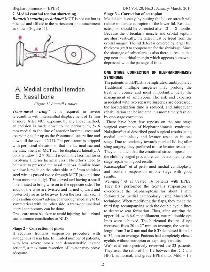
7. Medial canthal tendon shorteningBunnell’s suturing technique28 MCT is not cut but is
as shown (Figure 11).
Trans-nasal wiring36 It is required in severe
telecanthus with intercanthal displacement of 12 mm
or more. After MCT exposure by any above method,
an incision is made down to the periosteum, 5- 6
mm medial to the line of anterior lacrimal crest and
extending as far up as the frontonasal suture line and
down till the level of NLD. The periosteum is stripped
with periosteal elevator, so that the lacrimal sac and
the attachment of MCT can be displaced laterally. A
involving anterior lacrimal crest. No efforts need to
be made to preserve the nasal mucosa. Similar bony
window is made on the other side. A 0.3mm stainless
steel wire is passed twice through MCT (second time
3mm more medially). The curved awl having a small
hole is used to bring wire on to the opposite side. The
ends of the wire are twisted and turned upward and
posteriorly so as to be away from the lacrimal sac. If
one canthus doesn’t advance far enough medially to be
symmetrical with the other side, a trans-conjunctival
lateral canthotomy can be done.
Great care must be taken to avoid injuring the lacrimal
sac, common canaliculus or NLD.
Stage 2 – Correction of ptosisIt requires frontalis suspension procedure with
autogenous fascia lata. In limited number of patients,
with less severe ptosis and demonstrable levator
action37, a maximum resection of levator may prove
adequate.
Stage 3 – Correction of ectropionMedial canthopexy, by putting the lids on stretch will
reduce moderate ectropion of the lower lid. Residual
ectropion should be corrected after 12 – 18 months.
Because the orbicularis muscle and orbital septum
are short vertically, the latter must be freed from the
orbital margin. The lid defect is covered by larger full
thickness graft to compensate for the shrinkage. Since
the shortage of orbicularis is also there, it results in a
gap near the orbital margin which appears somewhat
depressed with the passage of time
ONE STAGE CORRECTION OF BLEPHAROPHIMSIS SYNDROMEThe patients with BPES have high rate of amblyopia.25
Traditional multiple surgeries may prolong the
treatment course and most importantly, delay the
management of amblyopia. The risk and expenses
associated with two separate surgeries are decreased,
the hospitalization time is reduced, and subsequent
rehabilitation can be initiated in a more timely fashion
by one-stage correction.
There have been few reposts on the one stage
surgical correction of blepharophimosis syndrome.
Nakajima28 et al described good surgical results using
medial canthoplasty and levator resection in one
stage. Due to tendency towards marked lid lag after
sling surgery, they preferred to use levator resection.
They concluded that the emotional stress imposed on
the child by staged procedure, can be avoided by one
stage repair with good results.
Karacaoglan30 et al performed medial canthoplasty
and frontalis suspension in one stage with good
results.
Wei-qing38 et al treated 16 patients with BPES.
overcorrect the blepharoptosis for about 1 mm
followed by medial canthoplasty using Mustarde’s
to decrease scar formation. Thus, after suturing the
increased from 20 to 27 mm on average, the vertical
length from 3 to 8 mm and the ICD decreased from 40
to 34 mm on average. Patients had completely closed
eyelids without ectropion or exposing keratitis.
Wu32 et al retrospectively reviewed the 23 patients.
They used the ratio of 1 – 1.2 between the ICD and
HPFL as normal, and grade BPES into: Mild – 1.3
Blepharophimosis – (BPES)
Figure 11 Bunnell’s suture

13
DJO Vol. 20, No.3, January-March, 2010
– 1.5, Moderate – 1.5 – 1.8, Severe - > 1.8. The
surgery consisted of lateral canthotomy, medial
cantholpasty, transnasal wiring and fascia lata sling
or levator resection. In this study, 70% patients had
good outcomes. Only 2 patients had the transnasal
wiring loosened, attributed to the cheese wiring.
11% patients with HPFL more than 2mm underwent
reoperation for ptosis as compared to 100% patients
with HPFL less than 2mm. Therefore, regarding the
stage operation provides acceptable results both in
functional and cosmetic improvements.
ConclusionBlepharophimosis - ptosis - epicanthus inversus
syndrome (BPES) is a complex of multiple eyelid
deformities. It is also associated with ocular, lacrimal,
nasal and auricular anomalies. It has a great impact
on patient’s functional status and visual development.
Patients with severe ptosis especially with co-existent
strabismus should have their ptosis corrected before
3 years of age, and all other patients should undergo
surgery before 5 years of age. Depending on the
levator function, one can choose medial canthoplasty
with frontalis suspension using autogenous fascia lata
or levator resection. The one-stage surgical correction
can be performed when a child is less than 3 years of
multiple stages of reconstruction are advised as early
as possible.
REFERENCES1. Mustarde JC: Congenital deformities in the orbital
region. Proc R Soc Med 64:1121,1971
2. Dimitry TJ. Hereditary ptosis. AmJ Ophthalmol 1921;
4: 655-8.
3. Owens N, Hadley R, Kloepper R. Hereditary
blepharophimosis, ptosis epicanthus inversus. JlInt Coll
Surg 1960; 33: 558-74.
4. Kohn R, Romano PE. Blepharoptosis,
blepharophimosis, epicanthus inversus and telecanthus-a
syndrome with no name. Am J Ophthalmol 1971; 72: 625-
31.
5. Usher C. Epicanthus with ptosis. Trans Ophthalmol
Soc UK 1935; 55: 194-232.
6. Usher C. A pedigree of epicanthus and ptosis. Ann
Eugenics 1925; 1:126-38.
7. Waardenburg PJ, Franceschetti A, Klein D. Genetics
8. Townes PL, Muechler EK. Blepharophimosis, ptosis,
epicanthus inversus and primary amenorrhea. Arch
Ophthalmol 1979; 97: 1664-6.
9. Crisponi L., Deiana M., Loi A., Chiappe F., Uda M.,
Amati P., Bisceglia L. et al. 2001 The putative forkhead
transcription factor FOXL2 is mutated in blepharophimosis/
ptosis/epicanthus inversus syndrome. Nat. Genet. 27, 159–
166.
10. Cocquet J., De Baere E., Gareil M., Pannetier M., Xia
X., Fellous M. and Veitia R. A. 2003 Structure, evolution
and expression of the FOXL2 transcription unit. Cytogenet.
Genome Res. 101, 206–211.
11. De Baere E., Beysen D., Oley C., Lorenz B., Cocquet
J., De Sutter P., Devriendt K. et al. 2003 FOXL2 and BPES:
mutational hotspots, phenotypic variability, and revision
of the genotype phenotype correlation. Am.J.Hum.Genet.
72, 478–487.
12. Beysen D., Raes J., Leroy B. P., Lucassen A., Yates
J. R., Clayton Smith J., Ilyina H. et al. 2005 Deletions
Involving Long-Range Conserved Nongenic Sequences
Upstream and Downstream of FOXL2 as a Novel Disease-
Causing Mechanism in Blepharophimosis syndrome.
Am.J.Hum.Genet. 77, 205–218.
13. Oley C, Baraitser M. Blepharophimosis, ptosis,
epicanthus inversus syndrome (BPES syndrome). J Med
Genet 1988; 25:47-51.
size in newborn infants. J Pediatr 1978; 92: 787.
length from 29 weeks gestation to 14 years. J Pediatr 1987;
111: 267-8.
16. Laestadius ND, Aase JM, Smith DW. Normal inner
canthal and outer orbital dimensions. J Pediatr 1969; 74:
465-8.
17. Farkas LG, Ross RB, Posnick JC, et al: Orbital
measurements in 63 hyperteloric patients. Differences
Surg, 1989; 17:245–9.
18. Ward RE, Jamison PL, Farkas LG: Craniofacial
variability index: a simple measure of normal and abnormal
variation in the head and face. Am J Med Genet, 1998;
80:232–40.
19. Fryns J P, Kleczkowska A, Dereymaeker A, Hoefnagels
M, Heremans G, Marien J &
van der Berghe H: A genetic-diagnostic survey in an
institutionalized population of 173 severely mentally
retarded patients. Clin Genet 1986; 30: 315-323.
20. Zlotogora J, SagiM, Cohen T: The blepharophimosis,
ptosis, and epicanthus inversus syndrome: delineation of
two types. Am J Hum Genet; 1983: 35:1020–7.
21. Finley W H, Callahan A & Thompson J N : Parental
age in the blepharophimosis, ptosis, epicanthus inversus,
telecanthus complex. Am J Med Genet; 1990: 36: 414-
417.
of Ovarian Failure in Blepharophimosis Ptosis Epicanthus
Inversus Syndrome: FOXL2 Is a Conserved, Early-Acting
Blepharophimosis – (BPES)

DJO Vol. 20, No.3 , January-March, 2010
14
Gene in Vertebrate Ovarian Development Endocrinology;
144(7):3237–3243.
syndrome: Am J Ophthalmol 1996; 122:2.
24. Dawson EL, Hardy TG, Collin JR, Lee JP; The
incidence of strabismus and refractive error in patients
with blepharophimosis, ptosis and epicanthus inversus
syndrome(BPES): Strabismus. 2003 Sep; 11(3):173-7.
development in the blepharophimosis
syndrome :BJO; 1991 (75), 746-748.
26. Beckingsale PS, Sullivan TJ, Wong VA, Oley C.
Blepharophimosis: a recommendation for early surgery in
patients with severe ptosis: Clin Experiment Ophthalmol.;
2003 Apr;31(2):138-42.
27. Dang H, Zhao GC. One-stage surgical reconstruction
of congenital blepharophimosis syndrome. Chin J Plast
Surg Burn (Chin) 1993; 9: 353-354.
28. Nakajima T, Yoshimura Y, Onishi K, Sakakibara A.
One stage repair of blepharophimosis. Plast Reconstr Surg
1991; 87: 24-31.
29. Wang TL, Zhang HM, Wang JQ. Surgical treatment
of congenital blepharophimosis syndrome and long-term
follow-up. Chin J Med Aesthetic Cosmetol (Chin) 2003;
9: 328-330.
30. Karacaolan N, Sahin U, Ercan U, Bozdogan N. One-
stage repair of blepharophimosis: a new method. Plast
Reconstr Surg 1994; 93: 1406-1409.
31. John Clark Mustarde. Repair and reconstruction in
orbital region, Churchill livingstone. Third edition, 467
– 96.
32. S-Y Wyu, L Ma, Y-J Tsai, JZ-C Kuo. One stage
correction for blepharophimisis syndrome. Eye 2008; 22:
380 -88.
33. Richard L., Thaddeus S. Nowinski, The Five-Flap
Technique for Blepharophimosis; Arch Ophthalmol
1989;107:448-452.
34. Hornblass A, Hanig CJ, Kohn R, Callahan MA,
Oculoplastic, Orbital, and Reconstructive surgery, William
and Wilkins, First edition, Volume 1. Eyelids, 103-18, 378-
83.
35. Collin JRO. A manual of systematic eyelid surgery,
Butterworth Heinemann, Third edition, 155
36. Nesi FA, Lisman RD, Levine MR Smith’s Ophthalmic
plastic and reconstructive surgery, Mosby, Second edition,
990-95.
37. Kobus K, Wójcicki P, Rychlik D. Analysis of treatment
results 389 patients with congenital blepharoptosis. Klin
Oczna. 2008;110(4-6):159-65.
38. Wei-qing H, Qun Q, Ru Z, Xiao-jun W, Xue-quan
F. Surgical strategy for congenital blepharophimosis
syndrome. Chinese medical journal 2007; 120(16):1413-
1415.
Blepharophimosis – (BPES)

15
DJO Vol. 20, No.3, January-March, 2010
Major Review
INTRODUCTION:Accomodative esotropia (AE) is a common entity, accounting for approximately half of all childhood esodeviations1. Described by Donders way back
the eyes associated with the accommodative effort necessary to overcome the blurred image caused by hypermmetropia. It classically presents in the preschool years, 1.5 – 3 years, (though an early onset type has been seen under 1 year too2) , the deviation being typically eliminated by controlling the accommodative effort with optical correction of the hypermmetropia3.
and one would expect good results. However, our evidence indicates that AE treatment outcomes are inconsistent and often less than ideal. The results are largely governed by the initial management of hypermmetropia while the deviation is still intermittent (How early and how much?), the recognition and treatment of associated abnormal distance-near relationship and amblyopia and patient compliance. Variations in treatment measures in hypermmetropia management (full v/s undercorrection) as well as duration of treatment may account for differences in stereoacuity levels and precipitation of deterioration of control whereby strabismus surgery may be indicated5.
ACCOMODATIVE ESOTROPIAWHEN TO REMOVE GLASSES
ABSTRACT: Accomodative Esotropia (AE) is an acquired convergent deviation of the eyes
hypermmetropia. Raab has summarized a number of well known characteristic features of this entity including post infancy onset, initial intermittency of deviation, above normal hypermmetropia, elimination or reduction of deviation with glasses and frequent association of anisometropia and amblyopia.The mainstay of treatment is full correction determined by cycloplegic refraction. Details as to how cycloplegia is attained is important, with cyclopentolate being far less effective in causing cycloplegia than atropine. Weaning children with AE off spectacle correction has been known to encourage fusional divergence and emmetropisation. Whether undercorrection of hypermmetropia stimulates increased reduction of hypermmetropia is controversial. On the other hand, studies have shown that a mild reduction of hypermmetropic reduction in AE may lead to larger angles of deviation and precipitate deterioration. Thus the
Deepali Garg Mathur Consultant , Max Eye Care, New Delhi
To add to our consternation many patients of accommodative esotropia do not have bifoveal fusion. Emmetropisation and spontaneous resolution of AE occurs rarely if at all and may take many years.5
Whether a child with accommodative aeotropia will ever “Grow out of glasses” remains a pertinent question, the main outcome measure being resolution or non- resolution of esotropia following weaning and eventual discontinuation of glasses.
CASE PROFILES:To exemplify the course of AE, the following cases are cited:
CASE I : 4 YRS MALE CHILD FULLY ACCOMODATIVE ESOTROPIA WITH LEFT AMBLYOPIA
DJO Vol. 20, No.3 , January-March, 2010
16
A 4 year old male child presented with left convergent squint at the age of 2.5 years. There was an associated left amblyopia. Cycloplegic refraction done under atropine revealed a hypermmetropic error of + 3.50 D in both eyes. AC/A ratio was normal by gradient method. Child was prescribed the full cycloplegic correction ( corrected only for distance) with occlusion therapy. Within 6 months, the child was orthotropic for near and distance with glasses. Visual acuity with Lea Symbols was 3/ 4.5 in both eyes with good alternation. Stereoacuity was 240 sec/arc.
CASE II - 11 YRS MALE CHILD WITH FULLY ACCOMODATIVE ESOTROPIA WITH LEFT AMBLYOPIA
The child presented at 3.5 yrs with a left convergent squint. He was refracted under tropicacyl and Homatropine variously and groosly undercorrected with +1.50 D in both eyes till 5.5 yrs. Occlusion therapy was however started for left amblyopia. He was advised surgery at the age of 5 years. Refused
surgery and presented to us at 5.5 yrs. Presenting BCVA was 6/9 in the right and 6/18 in the left eye. Refraction under atropine revealed + 4.0 D in the right eye and + 5.0 D in the left eye. In 6 months, he was orthotropic for near and distance with glasses. BCVA improved to 6/6 in the right and 6/6p in the left eye. Stereoacuity remained poor ( 480 sec/arc).
CASE III: 6 YRS FEMALE CHILD : ACCOMODATIVE ESOTROPIA WITH HIGH AC/A RATIO
Diagnosed as accommodative esotropia at the age of 4 yrs. Refraction done under atropine and full refractive correction prescribed . BCVA was 6/6p in both eyes. The child is orthotropic for distance with glasses. Residual near deviation wth high AC/A ratio overlooked. Repeat refraction and prescription of bifocals planned. Stereoacuity – 240 sec/arc.
Accomodative Esotropia
17
DJO Vol. 20, No.3, January-March, 2010
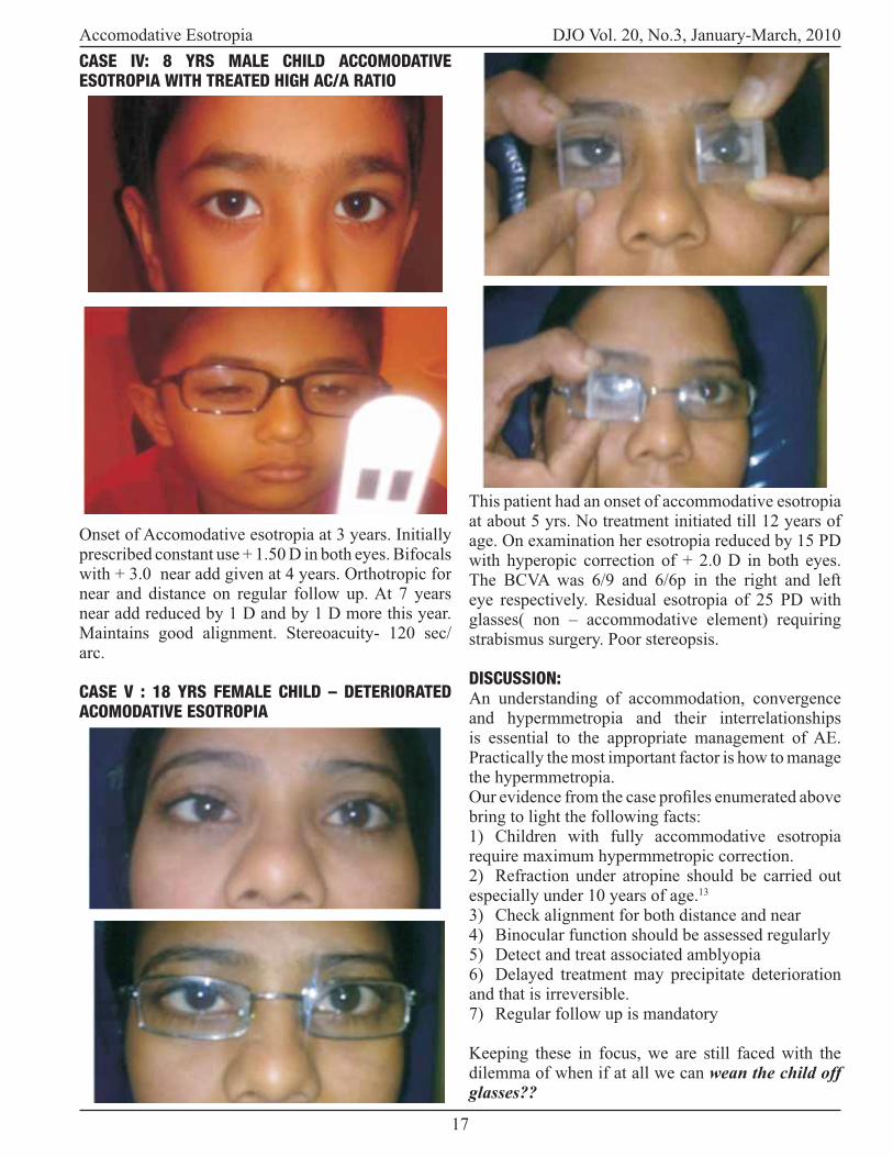
CASE IV: 8 YRS MALE CHILD ACCOMODATIVE ESOTROPIA WITH TREATED HIGH AC/A RATIO
Onset of Accomodative esotropia at 3 years. Initially prescribed constant use + 1.50 D in both eyes. Bifocals with + 3.0 near add given at 4 years. Orthotropic for near and distance on regular follow up. At 7 years near add reduced by 1 D and by 1 D more this year. Maintains good alignment. Stereoacuity- 120 sec/arc.
CASE V : 18 YRS FEMALE CHILD – DETERIORATED ACOMODATIVE ESOTROPIA
This patient had an onset of accommodative esotropia at about 5 yrs. No treatment initiated till 12 years of age. On examination her esotropia reduced by 15 PD with hyperopic correction of + 2.0 D in both eyes. The BCVA was 6/9 and 6/6p in the right and left eye respectively. Residual esotropia of 25 PD with glasses( non – accommodative element) requiring strabismus surgery. Poor stereopsis.
DISCUSSION:An understanding of accommodation, convergence and hypermmetropia and their interrelationships is essential to the appropriate management of AE. Practically the most important factor is how to manage the hypermmetropia.
bring to light the following facts:1) Children with fully accommodative esotropia require maximum hypermmetropic correction.2) Refraction under atropine should be carried out especially under 10 years of age.13
3) Check alignment for both distance and near 4) Binocular function should be assessed regularly5) Detect and treat associated amblyopia6) Delayed treatment may precipitate deterioration and that is irreversible.7) Regular follow up is mandatory
Keeping these in focus, we are still faced with the dilemma of when if at all we can wean the child off glasses??
Accomodative Esotropia

DJO Vol. 20, No.3 , January-March, 2010
18
Perusal of literature in this regard has varying
plays a causative role in at least 50 % of all cases of accommodative esotropia. Studies have revealed that hyperopia increases before the onset of esotropia and then decreases after 7-8 yrs. Raab observed a reduction of 0.18 D per year in accommodative esotropes compared with 0.22 D per year in normal hyperopic children.2 Other studies have indicated that esotropes behave differently from normal hyperopes and are destined to remain more hyperopic than their counterparts.The need for bifocals can however be eliminated by early teens. The impact of full correction of hypermmetropia versus undercorrection has also been under close scrutiny.The stimulus for emmetropisation is blur. So it has been postulated that prescribing the full spectacle correction may prevent emmetropisation.12
Also speculated is that esotropes may be able to discontinue spectacle use as a result of increased fusional divergence amplitudes, the loss of hyperopia or a reduction in the synkinesis between accommodation and convergence. 10
However other studies state that accommodative esotropes behave differently from the normal population because of an intrinsic defect in emmetropisation behavior leading to slower loss of hyperopia and are destined to remain that way.9
How long does the patient have to wear glasses? Most reviews of AE are concerned primarily with its management in childhood .One of the few long range follow ups done by swan on 39 adults( 23-46 yrs ), found that 38/39 adults diagnosed and treated for AE were still wearing glasses or contact lenses full time.6 Shipmann et al reported a recurrence of accommodative esotropia in 11 adults between 20-65 yrs of age.11 In synchronization, Mc Ewen states that even a small reduction of only 1 D in the full spectacle correction of a fully controlled esotrope
8
Also while undercorrection may improve fusional divergence, it may not cause a more rapid decrease in hypermmetropia.
CONCLUSION1) Full spectacle correction in accommodative esotropia restores alignment, averts amblyopia and maintains stereopsis.
early adulthood3) Accomodative esotropes have to wear optical correction till adulthood.The prospects of discontinuing glasses remain poor while the need for
bifocals can be eliminated by 10-12 years4) Weaning of glasses if attempted should be only in cases with good alignment and binocularity but under close scrutiny and follow up for deterioration.Present efforts should be directed to a more cautious and realistic though unfortunately not more encouraging advice to affected individuals PARKS- 1986
References1. Greenberg AE, Mohney BG, Diehl NN, Burke
JP. Incidence and types of childhood esotropia.
Ophthalmology.2007; 114(1):170-4
2. Bakers John D, Park MM. Early onset accommodative
esotropia. American Journal of ophthalmology. 1980;90:11-
8.
3. Raab Edward L. Etiological factors in accommodative
esodeviation .Trans Am Ophthalmol Society.1982; 80:657-
94.
4. Ludwig IH, Imberman SP, Thompson HW, Parks MM.
Long term study of accommodative esotropia. Trans Am
Ophthalmol Soc.2003;101;158-62.
management on the natural history and treatment outcome
of accommodative esotropia. Transaction of the American
Ophthalmological Society.2006;104:303-21.
6. Swan KC. Accommodative esotropia long range
follow up. Ophthalmology.1983;90: 1141-5 .
7. Mulvihill A, McCann A, Flitcroft I. Outcome in
refractive accommodative esotropia.Br J Ophthalmol
2000;84:746-9.
8. Mac Ewen CJ , Lymburn EG. Is the maximum
hypermetropic correction necessary in children with
fully accommodative esotropia? Br J Ophthalmology.
2008;92,1329-32.
9. Ingram RM, Gill LE and Lambert TW. Effect of
spectacles on changes of spherical hypermetropia in
infants who did, and did not, have strabismus. Br J
Ophthalmol.2000;84:324-6.
10. Hutcheson KA et al. Weaning children with
accommodative esotropia out of spectacles: a pilot study.
Br J Ophthalmol.2003;87:4-7
11. Shippman S,Weseley CR, Cohen KR. Accomodative
esotropia in adults. J Paediatr Ophthalmol
Strabismus.1993;30;368-371
12. Repka MX, Wellish K.et al. Changes in the refractive
error of 94 spectacle-treated patients with acquired
accommodative esotropia. Binocular Vision.1989;4:15-21
13. Rosenbaum AL.Bateman JB et al. Cycloplegic
refraction in esotropic refraction . Ophthalmology 1981;
88:1031-1034
Accomodative Esotropia
19
DJO Vol. 20, No.3, January-March, 2010
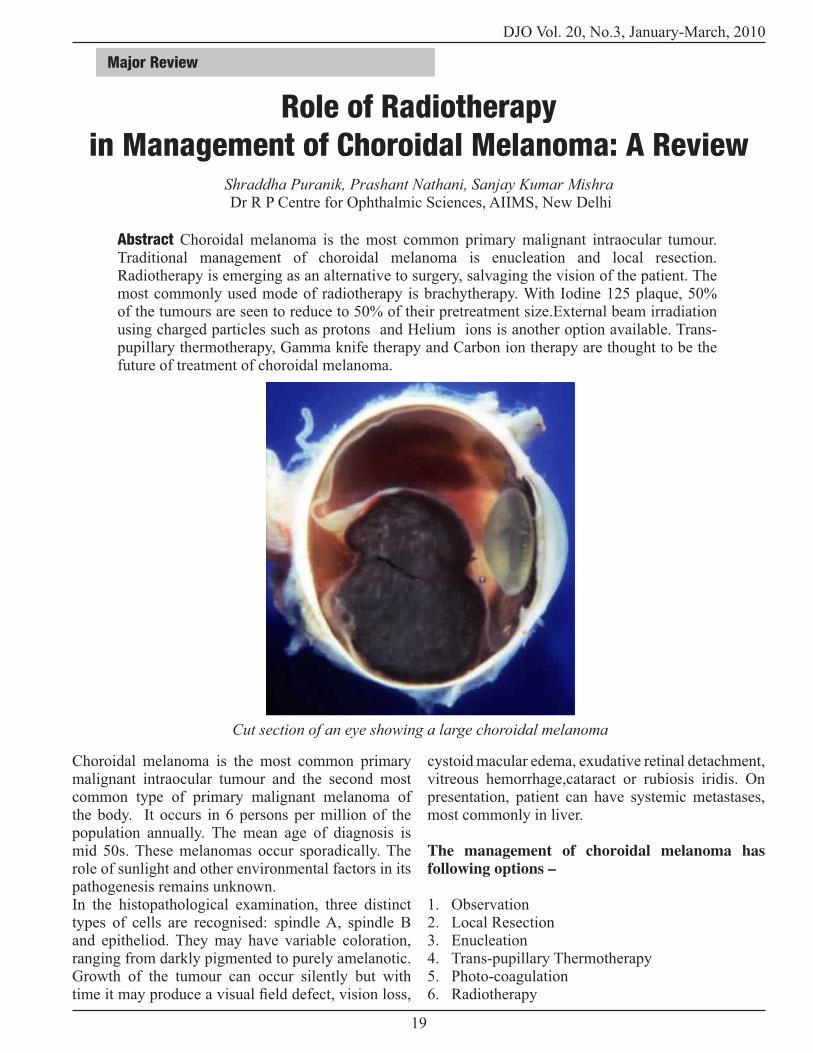
Choroidal melanoma is the most common primary malignant intraocular tumour and the second most common type of primary malignant melanoma of the body. It occurs in 6 persons per million of the population annually. The mean age of diagnosis is mid 50s. These melanomas occur sporadically. The role of sunlight and other environmental factors in its pathogenesis remains unknown.In the histopathological examination, three distinct types of cells are recognised: spindle A, spindle B and epitheliod. They may have variable coloration, ranging from darkly pigmented to purely amelanotic. Growth of the tumour can occur silently but with
cystoid macular edema, exudative retinal detachment, vitreous hemorrhage,cataract or rubiosis iridis. On presentation, patient can have systemic metastases, most commonly in liver.
The management of choroidal melanoma has following options –
1. Observation2. Local Resection3. Enucleation4. Trans-pupillary Thermotherapy5. Photo-coagulation6. Radiotherapy
Major Review
Abstract Choroidal melanoma is the most common primary malignant intraocular tumour. Traditional management of choroidal melanoma is enucleation and local resection. Radiotherapy is emerging as an alternative to surgery, salvaging the vision of the patient. The most commonly used mode of radiotherapy is brachytherapy. With Iodine 125 plaque, 50% of the tumours are seen to reduce to 50% of their pretreatment size.External beam irradiation using charged particles such as protons and Helium ions is another option available. Trans-pupillary thermotherapy, Gamma knife therapy and Carbon ion therapy are thought to be the future of treatment of choroidal melanoma.
Role of Radiotherapy in Management of Choroidal Melanoma: A Review
Shraddha Puranik, Prashant Nathani, Sanjay Kumar Mishra Dr R P Centre for Ophthalmic Sciences, AIIMS, New Delhi
Cut section of an eye showing a large choroidal melanoma

DJO Vol. 20, No.3 , January-March, 2010
20
Periodic observation for the growth of the tumour is done with serial fundus photography and USG. Local resection is done in selected cases of small melanomas, in the form of Sclerouveoretinectomy (full thickness eye wall resection)1 or partial lamellar sclerouveactomy. Enucleation and exenteration are other surgical options for advanced tumours. Photo-coagulation2, done in past with Xenon laser, gives better tumour control. Nowadays, it is done with Argon Laser which offers fewer complications. Trans-pupillary thermotherapy (TTT) is the modality of treatment that delivers heat to the tumour in infra-
system. It is preferred over Photo-coagulation for tumours less than 3mm in thickness and are located more than 3mm away from the fovea. TTT is used as a supplement to Plaque therapy. Radiotherapy is most commonly used in the form of Plaque therapy. In the past cobalt-60 plaques were used. These days, Iodine-125 and Ruthenium-106 are used. The other form of radiotherapy is charged particle application.Metastases in liver are treated with hepatic arterial chemo-embolisation with Cis- platin and Polyvinyl sponge.
Brachytherapy for melanoma
Brachytherapy is the application of radiation from isotopes over very short distances, in contact with
1930 with Radon seeds. Now we use Iodine 125 and Ruthenium 106. In Collaborative Ocular Melanoma Study (COMS), the medium sized tumour patients were randomised between enucleation and I- 125brachytherapy3. COMS detected equal mortality rates
in both the groups up to 12 years of follow up.Indications of brachytherapy in choroidal melanoma are-1. Small melanoma with evidence of growth on follow up2. Medium sized melanomas of choroid and ciliary body in eyes with useful vision3. Large melanoma ( Up to 16 mm in diameter and 8-10mm in thickness)4. Unilateral large melanoma5. Melanoma overhanging the optic disc
Total dose of radiation given is 50- 100 Gy . They gave 100 Gy radiation to the tumour apex at the rate of 50-125cGy per hour. Iodine 125 is convenient with respect to half life, shielding, tissue penetration and physical form.Gold plaque is used which has 0.4mm thickness with lip around its perimeter. Within the gold plaque, there is sialistic seed carrier insert with evenly spaced troughs that accept I-125 seeds. The carrier is designed so that seeds are adjacent to gold. The prepared loops facilitate anchoring the plaque to the sclera with sutures.Before placement of plaque, it is very important to measure dimensions of tumour (esp the base) properly either comparing it with the disc diameter or with Indirect ophthalmoscopy and 20D lens with grid. Extra ocular extension less than or equal to 2mm is acceptable for brachytherapy 4. Usual implant duration is 4-5 days. On follow up, I/O, fundus photography and USG should be done.Half of the tumours shrink to
up visit, the tumour actually increases in height due
increases by 15% or boundary expands by 250 mm, tumour expansion is suspected. In such a case, patient should be followed up every 3 monthly. If additional expansion by 250 mm or increase in thickness by additional 15% is detected, brachytherapy is declared to be a failure.Plaque tilt after initial accurate placement occurs frequently during brachytherapy and may represent an important cause of local treatment failure.In COMS, 10% of eyes were enucleated because of suspected or documented recurrence5 after brachytherapy . Factors affecting recurrence are--intra operative USG to see placement of plaque-Size of tumour at the time of surgery-proximity of posterior tumour margin with the disc.Local edge recurrences can be controlled with photocoagulation or TTT. Brachytherapy can cause changes in the surrounding retina like atrophy,
Role of Radiotherapy in Management
Gold plaque for brachytherapy
Placement of plaquePlaque for
brachytherapy
Plaque for brachytherapy

21
DJO Vol. 20, No.3, January-March, 2010
gliosis or blood vessel abnormalities. Tumour and radiation effects lead to poor visual acuity in 77% of eyes. The amount and severity of retinopathy and optic neuropathy after iodine-125 brachytherapy increased through 8 years of follow-up. Assessment of photographs and angiograms has provided reliable estimates that local tumour control was achieved in 90% of the cases. Tumour and radiation effects led to poor visual acuity in 77% of eyes. The metastatic rate was 13% and the mortality rate was 3%.The 5-year cumulative incidences of cataract, iris neovascularization, and glaucoma are found to be (20) 69%, 62% , and 60% respectively. The 5-year incidences of maculopathy and optic neuropathy are 52% and 46%, and those of vitreous hemorrhage and persistent RD were 36% and 25%, respectively. Cataract is the earliest complication to appear.
Charged Particle Irradiation of uveal melanoma
This is done with the help of external beam irradiation using charged particles such as protons and Helium ions. These have minimal scatter and
are up to 24 mm in diameter and 14 mm in height. Tumours involving macula, disc or both or with small extrascleral extensions are not contraindications for this therapy. Any systemic metastasis or primary malignancy in the other tissues must be ruled out.The conjunctiva is incised, tumour is localised with transillumination and indirect ophthalmoscopy. Extrascleral extension is to be looked for. Edges of the tumour are marked with pencil and four metal rings, 2.5 mm in diameter, are sutured to the sclera to outline the tumour. If the tumour is in contact with optic nerve, rings are placed only anteriorly and laterally while the distance of rings from the posterior margin is estimated from fundus photographs. For tumours extending to ciliary body and iris, rings are placed at the choroidal edges of the tumour and the distance of anterior margins from the rings is measured. A light beam coaxial with the central axis of proton beam is used to position the tumour relative to the beam.Three dimensional treatment planning computer programmes is used. A total dose of 70 cobalt Gy
7 to 10 days. Positioning of the patient is achieved with a head holder which allows controlled rotation of the head around two mutually perpendicular axes intersecting at a point that is positioned accurately on the axis of collimator. Orientation of patient’s eye is
or the other eye. Lid speculum is used to keep the eye
around the base of the tumour. Each treatment takes about 1min.First follow up is to be done 6 weeks after treatment and then 6 monthly. Most of the tumours
6.
considered complete regression and is observed in 15% of cases. Serous retinal detachment due to the tumours can transiently increase in size after therapy. But it resolves after few months. Post-treatment visual outcome depends on height of the tumour, its proximity in relation to fovea and disc7, pre-treatment vision, dose of radiation received at macula, degree of retinal detachment and presence of diabetes8,9. Risk
after treatment is 16% in low risk group and 99% in high risk group. The complications of external beam radiation therapy include intratumour hemorrhage, transient diplopia, lid epithelitis, punctual occlusion,
Role of Radiotherapy in Management
External beam radiotherapy
External beam radiotherapy

DJO Vol. 20, No.3 , January-March, 2010
22
epithelial keratopathy, radiation vasculopathy, maculopathy and papillopathy10. Rubeiosis iridis, Neovascular glaucoma are observed in 16% eyes and are associated with large tumour size. Cataract development chances increase with the amount of lens doses and larger tumour height. Local recurrence after therapy is observed in 2 to 5% of the tumours 8.It can be retreated successfully with repeat proton irradiation or laser photocoagulation. Survival is poor after local recurrences; 10 year survival rates were 72.6%for patients who experienced tumour control and 47.5%for those with re-growth. The 5 year cumulative probability of developing metastasis after charged particle irradiation and enucleation is 20%. The probability of metastatic death varies between 5% for patients with lower risk characteristics and 63% for those with high risk characteristics ( large tumour, advanced age , moderate to heavy tumour pigmentation, presence of symptoms, ciliary body tumour origin and green or blue iris colour)8.
Laser TreatmentArgon blue –green laser (wavelengths 488 and 514.5 nm) are used. For TTT, Nd:YAG laser with a wavelength of 1064.5nm is considered these days. But the risk of unwanted tissue disruption and haemorrhage is higher with it11. Krypton red (647.1nm) offers deeper penetration in the
amelanotic melanomas, but the power generated by
haemorrhage are higher. Lasers treat the intra-ocular tumours at a temperature level of more than 75 degree C. Photocoagulation should be restricted as a treatment modality for small choroidal melanomas,
media. Laser can be used in treating the tumours close to macula or disc where radiation therapy can cause severe vision loss, optic neuropathy, radiation retinopathy or macular oedema. During therapy,
separating the mass from its blood supply followed by direct tumour coagulation. Because of the presence of pigmentation, the tumour absorbs more light and doses are accordingly adjusted to avoid explosive burns. Firstly tumour is surrounded by 1 or 2 rows of laser with a spot size of 800 to 1000mm and an exposure time of 1 to 1.5 seconds. After 4 to 5 weeks a second circulating coagulation is performed with treatment of surface of the tumour. Third coagulation is performed after additional 4 to 5 weeks when entire surface of the tumour is photocoagulated again. Treatment is
suspended when choroidal mass is converted to an
be required to achieve this. Argon laser is used in the 13.
Neovascularisation of retina, developing after vein occlusions is the most severe complication of lasers15.Vitreus hemorrhage, tractional retinal detachment, pre-retinal membrane formation, cystoid macular oedema or burns in anterior segment can also occur. Recurrent tumour growth either at the margin of tumour or at the centre are reported. Tumour regression is achieved in more than 80% of cases. Shields et al reported an initial response rate of 100% but re-growth was reported in 14% of cases on follow up16. COMS has shown that enucleation has no prognostic advantage compared to eye salvaging radiotherapies and other modalities of treatment17.
Transpupillary ThermotherapyTTT gives long exposure sub-threshold photocoagulation with a long wavelength. The maximum temperature achieved is 42 to 44 degree C which is used to enhance the cytotoxic effect of radiotherapy. A temperature of 65 degree C with TTT can be considered as directly cytotoxic and no additional radiotherapy is required18.Diode laser (810nm) with large spot size up to 3mm for exposure time of 60 seconds is used. TTT is initiated at the tumour centre and no circumvallating treatment is performed. 3 to 4 sessions are required to destroy the tumour. Tumour regression is seen in 6 to 9 months leaving an atrophic scar with central pigment and visible sclera.
located behind the equator, regression achieved was more than 90% of the cases.9% of the tumours showed re-growth in tumours abutting or overhanging the disc or those requiring more than 3 sessions for regression18. Complications are comparable to photocoagulation treatment.Laser treatment as ancillary therapy• Either TTT or photocoagulation is helpful in reducing the risk of continuous or recurrent tumour growth after surgical excision. Routine photocoagulation at the edges of pseudocoloboma is recommended after the trans-scleral local resection.• The combination of radiotherapy and TTT (Sandwich technique) has shown a tumour control rate of 97% at 5 years24. Radiation induced complications are an important indication of supplementary photo-coagulation. OCT is useful in the early detection of radiation-induced macular oedema in this situation, before clinical signs of radiation maculopathy develop and before substantial visual loss occurs.
Role of Radiotherapy in Management

23
DJO Vol. 20, No.3, January-March, 2010
associated with larger tumour size.• Exudative retinal detachment either primary or secondary to brachytherapy can be treated with scatter photocoagulation combined with vitreoretinal surgery19.
Recent Advances in RadiotherapyGamma knife Radiosurgery As an alternative for enucleation, gamma knife radiotherapy is proposed using a radiation dose of 30 to 50 cGy. After giving retro-bulbar anaesthesia and
is localised with the help of gadolinium enhanced MRI. Maximum dose is delivered at 100% isodose line while periphery of the tumour receives 50% of the isodose.78 patients with a mean age of 64 years, and tumour more than 3mm thickness showed a 5 year survival rate of 81.9% with local tumour control of 91%20. These rates are comparable to enucleation. Though the eye retention rate was 89.7%, there was
major ocular complications seen were exudative retinopathy (in 33.3%), neovascular glaucoma (in 18.7%), radiogenic retinopathy (in 13.5%), optic neuropathy (in 15.5%), vitreus hemorrhage (in 10.4%).
Carbon Ion TherapyThe vertical (140 MeV/u) and horizontal (170 MeV/u) carbon ion beams from the synchrotron are shaped, using the passive beam delivery system, such as to irradiate the target volume of the tumour. The range
spread-out Bragg peaks (SOBPs) with a region of uniform cell killing. The apertures and range compensators are designed for individual patients. Dose distributions are calculated with either a broad beam or a pencil beam algorithm using parameters determined by measurements and calculations. In a study by Koyama Ito H27, the system was used for 12 patients during one year. For nine patients two-port treatment was assessed to be more effective than mono-port therapy and these patients were treated with two fractions of vertical beams and three fractions of horizontal beams.
ConclusionIn future, radiotherapy is likely to become an alternative for enucleation in the management of choroidal melanoma. The COMS gives us the data suggesting that survival following more recently developed brachytherapy is similar to enucleation. In situations
when the primary outcomes of differing treatments are essentially the same, any preference of one therapy over another would have to be based on other criteria, such as severity of adverse effects, quality of life issues, or treatment costs. Knowledge about the impact of enucleation for choroidal melanoma on the performance of vision-dependent activities might
References1. Meyer-Schwickerath G Excision of malignant
melanoma of choroid Mod Probl ophthalmol1974;12:562-
566
2. Shields JA Glazer LC Mieler WF et al Comparision of
xenon arc and argon laser photocoagulation in the treatment
of choroidal melanoma Am J Ophthalmol 1990;109:647-
655
3. The COMS group. The COMS randomised trial
of iodine-125 brachytherapy for choroidal melanoma,
Ophthal 2001 ;119:969-82
4. Pach JM, Robertson DM, Taney BS et al, Prognostic
factorsin choroidal and ciliary body melanomaswith
extrascleral extension. Am J Ophthalmol 1986;101:325-
331.
5. Jampol Lm, Moy CS, Murray TGet al. The COMS
randomised trial of I-125 brachytherapy for choroidal
melanoma. 4 Local treatment failure and enucleation in
Ophthalmology 2002;109:2197-2206.
6. Wilkes SR, Gragoudas ES, Regression patterns of uveal
melanomas after proton beam irradiation. Ophthalmology
1982;89:840-43
7. Seddon JM, Gragoudas ES, PolivogianisL et al. Visual
outcome after Proton beam irradiation of uveal melanoma.
Ophthalmology 1986;93:666-674
8. Seddon JM, Gragoudas ES, Egan Km et al. Uveal
melanomas near the optic disc or fovea: visual results after
proton beam irradiation. Ophthalmology 1987;94:354-
361
9. Gragoudas ES, Li W , Goitein M et al Evidence based
estimates of out come in patients irradiated for intraocular
melanoma Arch Ophthalmol 2002;120:1665-1671.
10. Gragoudas ES, Li W, Lane AM et al .Risk factors for
radiation maculopathy and papillopathy after intraocular
irradiation. Ophthalmology 1999;106:1571-1577.
11. Egger E , Schalenbourg A, ZografosL et al.
Maximising local tumour control and survival after proton
beam radiotherapy for uveal melanoma. Int J Radiat Oncol
Biol Phys2001;51:138-147
12. Courdi A, CaujolleJP, Grange JD et al. Result of
proton beam therapy of uveal melanomas treated in Nice.
Int J Radiat Oncol Biol Phys 1999;45:5-11
Role of Radiotherapy in Management

DJO Vol. 20, No.3 , January-March, 2010
24
13. Rol P, Fankhauser F, Giger H et al.Transpupillary
laser phototherapyfor retinal and choroidal tumours: a
rational approach. Graefes Arch Clin Exp Ophthalmol
2000;238:249-272.
14. Jalkh AE, Trempe CL, Nasrallah FPet al. Treatment
of small choroidal melanomas with photocoagulation.
Ophthalmic surg 1988;19:738-742.
15. Currie ZI, Rennie IG, Talbort JF. Iatrogenic choroidal
neovasularisationfollowing argon laser photocoagulation
for choroidal malignant melanoma Graefes Arch Clin Exp
Ophthalmol 1996;234:221-226
16. Shields JA, Glazer LC, Mieler WF et al. Comparision
of xenon arc and argon laser in treatment of choroidal
melanomas. Am J Ophthalmol 1990;109:647-655.
17. Robertson DM. Changing concepts in management
of choroidal melanoma. Am J Ophthalmol 2003;136:161-
170
18. Oosterhuis JA, Journee-de Korver HG, Keunen JE.
Transpupillary thermotherapy: results in 50 patients with
choroidal melanoma. Arch Ophthalmol 1998;116:157-
162.
19. Bartelema YM, Oosterhuis, Journee-de korver JG et al.
Combined plaque therapy and transpullary thermotherapy
in choroidal melanoma 5 years experience Br J Ophthalmol
2003;87:1370-1373
20. .5.5.Radtke ND, Augsberger JJ, Schmitt T. Management
of exudative retinal detachment after plaque therapy for
intraocular melanoma Am J Ophthalmol 1991;112:92-94
21. .5.5.Gamma Knife Radiosurgery in Ocular melanoma.
Br J Ophthalmol, Jan 2009
22. .5.5. Phys Med Biol. 2007 Sep 7;52(17):5341-52.
Carbon ion therapy for ocular melanoma: planning
orthogonal two-port treatment
23. .5.5.Boldt HC, Melia BM, Liu JC, Reynolds SM;
Collaborative Ocular Melanoma Study Group. I-125
brachytherapy for choroidal melanoma photographic and
angiographic abnormalities: the Collaborative Ocular
Melanoma Study: COMS Report No. 30. Ophthalmology.
2009 Jan;116(1):106-115
24. .5.5.Puusaari I, Heikkonen J, Kivelä T Ocular
complications after iodine brachytherapy for large uveal
melanomas.Ophthalmology 2004 Sep;111(9):1768-77
25. .5.5. Sagoo MS, Shields ES et al. Plaque radiotherapy
for juxtrachoroidal melanoma overhanging the optic disc.
Arch Ophthalm 2008 nov;126(11):1515-22
26. .5.5.Almony A, Breit S et al. Tilting of radio-active
plaques after initial accurate placement for treatment of
uveal melanoma. Arch Ophalm 2008 Jan 2008 (1):65-70.
27. .5.5.PHYS Med Biol.2007 Sep 7;52(17):5341-52
Role of Radiotherapy in Management

25
DJO Vol. 20, No.3, January-March, 2010
Eyelid trauma still seems to be a taboo among general ophthalmologists. We present a general overview such that these cases may be taken up in most settings.
Work up of a patient with eyelid trauma :History Taking :It is of note that periocular trauma can occur as an isolated injury or as a small part of multisystem trauma. It is imperative to obtain adequate details as to how the injury occurred. If it appears that there was deep penentration in to the orbit , it is to be determined as to what instrument caused the injury as there could be fragments or foreign bodies lodged within. Other aspects worth mention are history of alcohol intake as this may lead to distortion of facts presented and children may not give adequate history. As almost all cases of trauma are taken up under general anaesthesia the importance of nil per oral status and neurological and cardiopulmonary status is not to be undermined.
Recording of visual acuity :Preoperative vision is extremely important for medical and medicolegal purposes. Both distance and near vision must be recorded. Importantly the vision should be assessed for both the eyes as you maybe dealing with optic nerve/ chiasmal traction.The pupils as repeatedly quoted are a window to the brain. Direct, consensual and evidence of any RAPD are to be documented.
Evaluation of the globe and orbit :Examination of the eye is important in periocular trauma. The eye itself should be examined for movements and and any sign of perforation. Patients are most likely to be uncooperative so thorough evaluation in General Anesthesia is mandatory, as a globe perforation may be missed in the surgeons zeal to repair the periorbita.
Radilogical studies : The Periocular trauma needs adequate imaging to asses the underlying anatomical derangements. A CT Scan is the most appropriate study. Be sure the quality is good as many a time the CT may only be assessed when the patient is already under General Anesthesia. An MRI is to be performed when optic nerve injury is of suspect.
Investigations :A routine Hemogram and a Bleeding Time, Clotting Time may allow to assess the blood loss as the facial area is highly vascular.
Types of Eyelid Trauma and its management :Injury of the eyelid may be divided into blunt and penentrating trauma.
Blunt Trauma :Ecchymosis and edema are commonly the presenting signs of blunt trauma. A complete evaluation may be advised as described above.
Penetrating Injury : A thorough knowledge of eyelid anatomy is essential while repairing a penentrating eyelid injury. The management depends on the depth and location of injury.
We discuss management of four most common types of eyelid trauma :
A Eyelid Margin RepairB Simple Laceration Repair C Complex Laceration RepairD Canalicular Laceration Repair
Eyelid trauma : A ReviewPrashant Yadav, Neelam Pushker, M.S. Bajaj, M. Chandra, B. Chawla,
D. Shrey, P. Bhimseria, K. Gupta , K. Rishi, R. Meel.Dr R P Centre for Ophthalmic Sciences, AIIMS, New Delhi
Major Review

DJO Vol. 20, No.3 , January-March, 2010
26
Figure. 1 : Eyelid Margin Repair : A. The eyelid margin is aligned with 6 ‘0’ silk grey line sutures,anterior lamella sutures and posterior lamella sutures.The tarsus to tarsus closure is done with 6 ‘0’ vicryl sutures. B. The tarsal sutures are tied and cut, the lid margin sutures are tied and left long. C. The skin isclosed with 6 ‘0’ silk and the long lid margin suturesare tied into them to avoid any abrasion.
The eyelid margin repair requires dexterity and in allpossibilities should be done under the microscope and if not under adequate light. The repair begins with identifying the adequate landmarks of the eyelid.
The following surgical steps maybe followed1,2,3
1. GA or local anaesthesia ( xylocaine + adrenaline+ topical anaesthesia)2. Assess and align the lid margin with two forceps,be careful not to pull as traumatic tissue is lackingstrength.3. If the lid margin is closing with strain a lateralcanthotomy may be done.4. Hold one edge of the cut end with a forceps
grey line with a 6 ‘0’ silk suture. Watch the needle as it comes out through the centre of the lacerationvertically. Now in the same plane enter the other end of the laceration and come out long through the grey line on the other side (try to keep the distance of entryand exit at the same distance on both sides ). Oncethis is done you have to go back the same way but with shorter bites. Practice this on two cut ends of thermocol. Once this vertical mattress suture is in
place give one temporary tie to see the alignment. If you are not happy replace it. Once the alignment isadequate tie the 6 ‘0’ silk and leave the cut end long.Once tied the margins should have a pout upwards as
notch later on.5. Once this has been achieved two interrupted sutures maybe passed through the anterior lamellaand the posterior lamella in a vertical fashion. All these sutures should be left long as there is the risk of corneal abrasion / defect.6. The remaining defect of the eyelid if deep maybe closed in layers , deeper tissues with vicryl and
you have left long may be now pulled and included inthe knot of one your skin sutures. You can leave thesesutures for seven - ten days. A bit longer is better astissues under traction may tend to gape on removal.7. Supportive treatment in the form of The Penicillin
days, steroids may be included to reduce edema and
B. Simple Laceration Repair4,5
Facial lacerations should be repaired within 24 hoursfor best results or else infection and necrosis mayencroach. Before repairing any trauma be sure tothoroughly irrigate the wound with betadine and saline,This will help in washing off any contamination. Inour experience it is best to repair complex lacerations in GA. Two reasons may be cited for this. A) Patient cooperation. B) Injecting local anaesthesia makes the cut margins boggy and such margins may not attain
involving just the skin and orbicularis require onlyskin sutures. Basic principles of plastic repair may be followed which include conservative debridement ( extensive debridement is not necessary as facial wounds heal magically due a generous vasculature),use of small caliber sutures, eversion of wound edgesand early suture removal. It is also important to assess that the laceration has not involved deeper structuresof the orbit and the brain ( communication throughthe orbital roof or cribriform plate ). Any orbital fat visible indicates the violation of the orbital septumand requires the need to do a levator exploration.A lacerated levator aponeurosis/ Muscle must berepaired to achieve adequate pre – trauma function.Upper eyelid lagophthalmos and tethering to the superior orbital rim are common if the orbital septum is incorporated into the laceration repair. Orbital septum lacerations need not be sutured as they may
Eyelid trauma
27
DJO Vol. 20, No.3, January-March, 2010
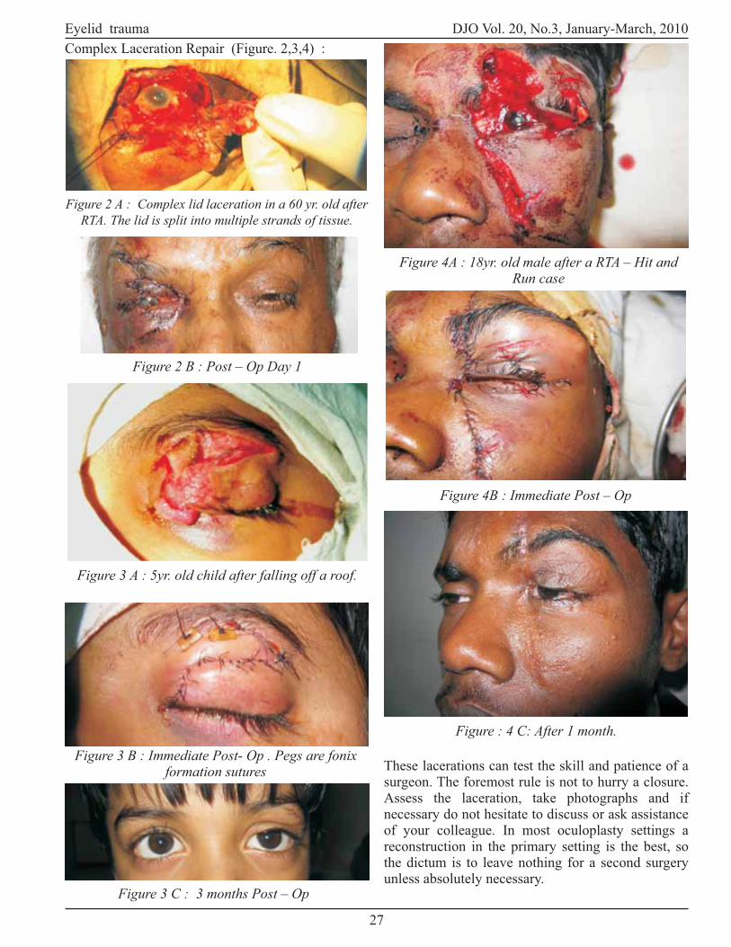
Complex Laceration Repair (Figure. 2,3,4) :
These lacerations can test the skill and patience of a surgeon. The foremost rule is not to hurry a closure. Assess the laceration, take photographs and if necessary do not hesitate to discuss or ask assistance of your colleague. In most oculoplasty settings a reconstruction in the primary setting is the best, so the dictum is to leave nothing for a second surgery unless absolutely necessary.
Figure 2 A : Complex lid laceration in a 60 yr. old after RTA. The lid is split into multiple strands of tissue.
Figure 2 B : Post – Op Day 1
Figure 3 A : 5yr. old child after falling off a roof.
Figure 3 B : Immediate Post- Op . Pegs are fonix formation sutures
Figure 3 C : 3 months Post – Op
Figure 4A : 18yr. old male after a RTA – Hit and Run case
Figure 4B : Immediate Post – Op
Figure : 4 C: After 1 month.
Eyelid trauma

DJO Vol. 20, No.3 , January-March, 2010
28
C. Complex Laceration RepairThe following priorities may be cited when dealing with complex reconstructions.6,7
• Development of a stable eyelid margin• Provision of adequate vertical eyelid height• Adequate eyelid closure• Smooth epithelialized internal surface• Maximum cosmesis ans symmetry.
The following steps maybe followed in complex lacerations.A. Clean and inspect the wound1. Rule out deep injury2. Remove any foreign material3. Do not debride any tissue unless it is infected as this may unnecessarily enlarge the defect.
B. Steps of laceration Repair :8,9
1. To begin the laceration repair identify anatomic landmarks such as the eyebrow, the lid margin and the canthus.
taken which will be discussed later. It is to be noted that during all lid reconstructions it is vital to use the Jaeger type lid guard. The under surface of the guard should be well lubricated with Xylocaine jelly or ointment. Inadvertent perforation during lid repair may occur if this precaution is not taken, as you
3. To reduce the strain you may place temporary ‘tacking’ sutures on the anterior lamella which may ease your suturing of the posterior lamella.
suturing the posterior lamella, the conjunctiva, the tarsus , the canthal tendons and levator aponeurosis.5. The conjunctiva is apposed with 8 ‘0’ vicryl .6. The tarsus is closed with 6 ‘0’ vicryl.7. Once the tarsus is sutured it is important to identify orbital septum and the levator aponeurosis . Many a time the septum has been breached and the fat prolapsed. This actually can make it easier to identify the levator which lies under the fat. The levator may be found intact, lacerated or disinserted. The levator maybe carefully explored and repaired. Levator should be repaired with non absorbable sutures such as 6 ‘0’ Nylon. Again it is important to assess whether the
the muscle a secondary repair may be attempted at a later stage.8. The lid margin is closed as described above. 9. The subcutaneous tissue is approximated with
6 ‘0’ vicryl and the overlying skin with 6 ‘0’ silk. Adequate closure of deep structures allows accurate approximation of skin and subcutaneous tissues.10. The Canthal tendons are important structures which are commonly damaged in lid trauma. The Lateral canthal tendon is inserted onto the whitnall’s tubercle. When the tendon can be isolated it can be attached directy to the lateral orbital rim with 4 ‘0’ nylon or ethibond. The effect of the tendon may be recreated by reattaching a portion of the tarsus ( The upper and lower crus of the lateral canthal tendon extend from each tarsal plate to form the stem of the tendon ) to the lateral orbital room.This is done by splitting the lamella towards the lateral edge of the lid. After splitting the lamella a tarsal strip is created. This tarsal strip is hooked with 4 ‘0’ Nylon. Now the lateral orbital rim is exposed and the tarsal strip is attached to periosteum of the orbital rim. It is important to take careful bites near the rim as the globe lies snug out there with the lateral rectus.10
11. The medial canthal tendon is a more complicated structure than the lateral canthal tendon. There are two limbs, the anterior limb is attached to the anterior lacrimal crest and the posterior limb to the posterior lacrimal crest. Avulsion of the anterior limb does not distort the position of the eyelid while the posterior limb does. Suturing the posterior limb of the medial canthal tendon can easily damage the nasolacrimal system. This becomes easier if the Naso Laerimal Duet system is already damaged. The suturing may be done with 4 ‘0’ Nylon. (Figure 5) 11
Figure 5A : Medial Canthal Avulsion : A punch drunk case of a 40 yr. old male
Figure 5B : Immediate Post – Op
Eyelid trauma

29
DJO Vol. 20, No.3, January-March, 2010
D. Canalicular Laceration Repair A canalicular laceration should be assumed when there is any injury close to the lid marg-in and medial to the puncta. These lacerations may occur by 1. Direct trauma 2. An avulsion due to a lateral pull on the eyelid. Direct trauma lacerations are easier to repair as the laceration lies in the centre of the canaliculi while avulsion type injuries tear the canaliculus from the sac itself making visualization of the proximal cut
Methods :1. Repair using Crawford stents.2. Repair using the pigtail probe.
Identifying cut ends of the canaliculus2. Passing the stent through the torn canaliculus3. Passing the stent into the nasolacrimal duct4. Suturing the pericanalicular tissue5. Passing thte other stent through the nose
1. Identifying the cut ends of the canaliculus :• The microscope should be used preferably in all cases.• Pouring normal saline to pool in the medial canthal area and then pushing air through the intact canaliculus allows bubbles to be visible in the area of the medial end of the cut canaliculus.• Diluted Fluorescein is taken and pushed throught the intact canaliculus to become visible through the medial end of the cut canaliculus. Dyes such as methylene blue and gential violet should be avoided
• The cut end should be explored under direct microscopic visualization.
2. Passing the stent through the canaliculus :• Thread the stent through the punctum and out
Figure 5C : After 2 months Post – Op.
Figure 6A : Canalicular laceration after fall from bicycle.
Figure 6B : Immediate Post – Op with silicon stent in situ.
Figure 7 : Canalicular Laceration repair with Crawford stents : A. Pass the stent through the cut end of the canaliculus and into the lacrimal sac. B. Intubate the opposite canaliculus and nasolacrimal duct. C. Reinforce the medial canthal tendon with a 4’0’ Vicryl suture D. Approximate the canalicular mucosa or adjacent tissues. E. Use additional sutures in the orbicularis muscle as required to reinforce the wound. Suture the skin. Tie the stents in the nose.
Eyelid trauma
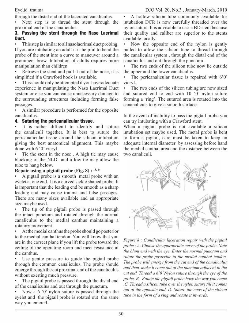
DJO Vol. 20, No.3 , January-March, 2010
30
through the distal end of the lacerated canaliculus.• Next step is to thread the stent through the proximal end of the canaliculus3. Passing the stent through the Naso Lacrimal Duct.• This step is similar to all nasolacrimal duct probing. If you are intubating an adult it is helpful to bend the probe of the stent into a curve to maneuver around a prominent brow. Intubation of adults requires more manipulation than children.• Retrieve the stent and pull it out of the nose, it is
• This should only be attempted if you have adequate experience in manipulating the Naso Laerimal Duet system or else you can cause unnecessary damage to the surrounding structures including forming false passages.• A similar procedure is performed for the opposite canaliculus.4. Suturing the pericanalicular tissue.
the canaliculi together. It is best to suture the pericanalicular tissue around the silicon intubation giving the best anatomical alignment. This maybe done with 6 ‘0’ vicryl.• Tie the stent in the nose . A high tie may cause blocking of the NLD and a low tie may allow the tube to hang below.
13, 14
• A pigtail probe is a smooth metal probe with an eyelet at one end. It is a curved sickle shaped probe. It is important that the leading end be smooth as a sharp leading end may cause trauma and false passages. There are many sizes available and an appropriate size maybe used.• The tip of the pigtail probe is passed through the intact punctum and rotated through the normal canaliculus to the medial canthus maintaining a rotatory movement.• At the medial canthus the probe should go posterior to the medial canthal tendon. You will know that you are in the correct plane if you lift the probe toward the ceiling of the operating room and meet resistance at the canthus.• Use gentle pressure to guide the pigtail probe through the common canaliculus. The probe should emerge through the cut proximal end of the canaliculus without exerting much pressure.• The pigtail probe is passed through the distal end of the canaliculus and out through the punctum.• Now a 6 ‘0’ nylon suture is passed through the eyelet and the pigtail probe is rotated out the same way you entered.
• A hollow silicon tube commonly available for intubation DCR is now carefully threaded over the nylon suture. It is advisable to use a BD stent because their quality and caliber are superior to the stents available locally.• Now the opposite end of the nylon is gently pulled to allow the silicon tube to thread through the canalicular system , through the distal end of the canaliculus and out through the punctum.• The two ends of the silicon tube now lie outside the upper and the lower canaliculus.• The pericanalicular tissue is repaired with 6’0’ vicryl.• The two ends of the silicon tubing are now sized and sutured end to end with 10 ‘0’ nylon suture forming a ‘ring’. The sutured area is rotated into the cananaliculs to give a smooth surface.
In the event of inability to pass the pigtail probe you can try intubating with a Crawford stent.When a pigtail probe is not available a silicon intubation set maybe used. The metal probe is bent to form a pigtail, care must be taken to keep an adequate internal diameter by assessing before hand the medial canthal area and the distance between the two canaliculi.
Figure 8 : Canalicular laceration repair with the pigtail probe : A. Choose the appropriate curve of the probe. Note the blunt end with the eye. Enter the normal punctum and rotate the probe posterior to the medial canthal tendon. The probe will emerge from the cut end of the canaliculus and then make it come out of the punctum adjacent to the cut end. Thread a 6’0’ Nylon suture through the eye of the probe. B. Rotate the pigtail probe back the way you came .C. Thread a silicon tube over the nylon suture till it comes out of the opposite end. D. Suture the ends of the silicon tube in the form of a ring and rotate it inwards.
Eyelid trauma
31
DJO Vol. 20, No.3, January-March, 2010
Post operative carePost operative care is of utmost importance, as you may spend hours closing a complex lid laceration but infection can ruin your efforts. • Don’t be in a hurry to close. Make sure sutured edges are properly cleaned with betadine and ointment applied.• A good antibiotic preferably Intravenous for the 1st 2 – 3 days (e.g. Monocef + Vancomycin) followed by oral Amoxyciilin + Clavulanic acid.• Oral Steroids maybe given in clean cases to
• Sutures are to be removed in 7 – 10 days and sutures under traction can be removed after 2 weeks.• Silicon intubation maybe removed after 4 months.• Fornix formation sutures are removed in 3 weeks.
References :1. J R O Collin , Manual of systematic eyelid surgery :
Churchill Livingstone, 1984.
2. Mark R. Levine, Manual of Oculoplastic surgery :
Butterworth Heinemann , 1996.
repair. Arch Ophthalmol. 1997; 115:566-567.
4. Mustarde J C : Repair and Reconstruction in the orbital
region, Churchill Livingstone, 1971.
5. Mustarde J C : Major Reconstruction of the eyelids :
functional and aesthetic considerations. Clin Plast Surg
1981; 8 : 227 – 231
6. American Academy of Ophthalmology : Orbit, Eyelids
and Lacrimal System, Section 7, Basic and Clinical Science
Course, 2007 – 2008.
reconstruction. Surv Ophthal, 1987; 31: 249-261.
8. Spinelli et al :Periocular Reconstruction , Plast.
Reconstr. Surg. 1993, 91: 1017 – 1024.
9. Smith et al : Techniques available in reconstruction
surgery of the eyelid, BJO, 1970, 54: 450 – 455.
10. Mc Cord et al : Reconstruction of lower eyelid and
outer canthus, Oculoplastic Surgery, ed. 3, chap. 5, Pg. 119
– 144, Raven Press, 1995.
11. Howard GR et al : Medial canthoplasty. Arch
Ophthalmol, 1992;110: 1793-1797.
12. Hawes et al : Effectiveness of bicanalicular silicone
intubation in the repair of canalicular lacerations.
Ophthalmic plast. And Reconstr Surg. 1985; 1 : 185 –
190
Surv Ophthal, 1991; 36; 113 – 132
14. Baylis et al , Repair of the lacerated canaliculus,
Ophthalmology, 1978, 85, 1271 – 1276.
Eyelid trauma
DJO Vol. 20, No.3 , January-March, 2010
34
Corneal grafting in high risk corneas remains a challenge. The management of high risk corneal grafts mandates a thorough knowledge and experience in handling systemic immunosuppressive therapy besides optimal surgical expertise. The immune privilege afforded by the absence of antigen presenting cells in the donor tissue, avascularity of the recipient tissue and, anterior chamber associated immunodeviation help to maintain clear corneal allografts without supplemental immunosuppressive regimen in “normal” risk cases of keratoplasty.1 It is to be observed that primary low-risk corneal transplants have the highest success rate (90%) and lowest rejection rate (11–18%). High risk corneal grafts are those in conditions in which the cornea loses its immune-privileged status, resulting in its susceptibility to immunological rejection induced failure. Eyes under
quadrants of stromal vascularisation and/or a history of previous graft rejection.2 Herpes simplex virus keratitis (HSVK),chemical injury, large sized grafts, HLA incompatibility, previous failed corneal graft/s, decreasing age of recipient, glaucoma, and peripheral anterior synechiae3,4 may be considered as other risk factors. The risk of graft failure has been found to vary substantially even within a high-risk population. 2 In the Collaborative Corneal Transplantation Study5
or more quadrants of vascularisation, or one in which a graft had previously been rejected. Two quadrant deep vascularisation is less at risk than in which, all four quadrants are heavily vascularised. It has been observed that the incidence of rejection increases with both the number of quadrants vascularised and with the total number of vessels crossing the proposed graft/donor junction.6 Graft rejection in a previously grafted eye relates more to the number of blood vessels in the cornea than to the number of previous grafts.7 Recipient corneas can be divided into low, medium, and high risk depending on the number of quadrants of vascularisation (avascular, 1–2 quadrants, and 3+ quadrants respectively). 8 Reported failure rates vary between 60% and 90% in high-risk corneal grafts.9-11
occurs in lymphoid tissues. With topical steroids not reaching these secondary lymphoid organs, and the effect of systemic steroids not seeming
activated T cells, it is essential to administer systemic immunosuppressives to achieve clear graft survival in high risk keratoplasty cases. Considering that corneal transplantation is not a life-saving procedure,
when choosing an immunosuppressive medication. Various immunosuppressive agents (table 1) have
prolonging graft survival in patients with high-risk keratoplasty especially in those patients who are bilaterally blind. When immunosuppressive therapy is to be considered in high risk corneal grafting cases, appropriate caution should be exercised in its use, with particular attention to side effects, proper patient selection and information before instituting treatment. Obtaining an informed consent is mandatory in all cases. Treatment plan protocols call for exclusion of patients with a history of malignant disorders, serological evidence of HIV or HbsAg positivity, systemic infections that require therapy at the time of entry, active peptic ulcer disease, inadequate contraceptive measures, pregnancy, or patients aged 18 years or below. Corneal surgeons dealing with high risk cases need devise appropriate treatment regimens to enhance graft survival. Intensive topical
systemic corticosteroid has not been shown to add
and other lesser toxic alternatives, immunosuppresion has now become more acceptable. The adoption of appropriate treatment regimens a few days before surgery to optimize the local corneal environment before grafting is advisable. The use of tissue typing in high risk vascularised corneas is still controversial but can be considered. No consensus exists on one single therapy for all these grafts and devising therapeutic regimens, sometimes with multiple medications can help to improve graft survival especially in the bilaterally blind patients. Studies report improvement with both short and long term systemic CsA in high
Major Review
High risk corneal grafting: an overviewM Vanathi, Kanchan Chawhan, A Panda, Tushar Agarwal
Cornea Services, Dr R P Centre for Ophthalmic Sciences, AIIMS, New Delhi
35
DJO Vol. 20, No.3, January-March, 2010
risk keratoplasty.12 Some degree of immunological privilege has been considered to be re-established, indicating that CsA can eventually be safely withdrawn.10 Few authors have found no statistical
to variations in criteria for high risk and inclusion 13
Intensive topical corticosteroid therapy decreases
of immunologically active cells and also reducing the expression of class 2 antigens in the recipient cornea. Long-term results of the use of systemic mycophenolate mofetil (MMF) and tacrolimus for the prevention of rejection has been seen to be effective in patients with high-risk corneal grafts.
Cyclosporine:Cyclosporin A is a powerful immunosuppressive agent
It acts at the early stages of antigenic sensitisation. It is an immunomodulator and works on T cells by binding to an intracellular peptide cyclophilin. It inhibits antigen presentation and thus lymphokine production. By virtue of its anticyclophilin activity, cyclosporin reduces production of interleukin-2 (IL-2), thus limiting the activation of CD4+ and CD8+ T cells. CsA, is a very potent prophylactic agent for preventing corneal allograft rejection and is used in specialised centres after high risk keratoplasty. Although therapy with CsA allows superior graft survival, its use is limited because of a wide range of side effects such as diabetogenicity, arterial hypertension, hyperlipidaemia, nephrotoxicity. Apart from this for CsA to be effective, the daily dose needs to be adjusted to maintain optimal trough levels of CsA which makes its usage expensive and labour intensive with regular laboratory drug monitoring.
Treatment Schedule:After baseline evaluation including clinical history, blood pressure, total cell count, serum urea, creatinine, electrolytes, and liver function tests, oral CsA is given as daily dosage starting immediately after surgery in a loading dose of 10 -15 mg/kg/day. Thereafter, CsA blood levels are to be monitored and measured biweekly. Blood pressure, complete blood cell count, serum creatinine and liver functions are to be evaluated every 2–4 weeks. Patients are to be
then monthly. CsA is given for 12 months unless
be adjusted to the whole blood trough levels with
a target range of 120–150 ng/ml. With stabilisation of the CsA levels the frequency of the tests may be reduced. The advantages of systemic CsA include
complications.
Topical CsA is prepared either in olive/ castor oil
It is prescribed 5 times a day along with the topical corticosteroid drop in high risk patients both pre and post operatively. As blood cyclosporin A level after topical therapy is negligible, monitoring of blood cyclosporine A levels is not required. However, monitoring of renal and liver function is required.Side effects: Elevated serum urea and creatinine, hypertension, gum hyperplasia, increased sweating, backache, nausea, feeling unwell, oral candidiasis, cramps, bone marrow toxicity, hirsutism and paraesthesia of the extremities are the commonly occurring side effects.
These side effects are rare when the drug blood level is well monitored and kept at low therapeutic levels of approximately 200 ng/ml. The use of CsA in solid organ transplantation has been associated with an increased risk of lymphoproliferative disorders that has been attributed to the long duration of therapy and the use in conjunction with other immunosuppressive drugs. This calls for a stringent reconsideration on our indications for its use in ocular conditions.
Mycophenolate mofetil:
combination with CsA following renal transplantation. Unlike CsA, MMA does not interfere with IL-2 pathways. It reversibly inhibits the de novo formation of guanosine nucleosides by inhibiting the enzyme inosine monophosphate dehydrogenase. As T and B cells are predominantly dependent on the de novo synthesis of guanosine nucleosides, the purine biosynthesis of these cells is selectively inhibited. MMF is to be given in a dosage of 2 g (1 g twice daily). Routine blood samples need to be taken every 2–4 weeks to check for drug toxicity. Patient follow ups
month, bi-weekly for 2 months and monthly for 3 months and every three months thereafter during which laboratory assessments are performed. The need for blood level adapted dosing for MMF remains controversial. Lesser postoperative visits, reduced cost and logistics of postoperative immunosuppresion as a result of the omission of monitoring drug titres are advantages with MMF usage. MMF in
High risk corneal grafting

DJO Vol. 20, No.3 , January-March, 2010
36
combination with a short postoperative course of oral steroids is just as effective as CsA and oral steroids inpreventing acute rejection following high risk cornealtransplantation.14
Tacrolimus:Tacrolimus (FK 506) is a macrolide antibiotic isolated from the soil fungus Streptomyces tsukubaensis, withpotent immunosuppressive activity, 10 to 100 timesmore potent than CsA. The mechanism of action of tacrolimus is similar to cyclosporin. While CsA binds to the immunophilin, cyclophilin, tacrolimus binds to
(FKBP) and inhibits the T cell receptor-mediated signal transduction required for transcription of interleukin 2 and other lymphokines.
Treatment schedule:Baseline evaluation includes clinical history, blood pressure, total blood count, blood urea, creatinine and electrolytes, and liver function tests. Oral tacrolimus is to be initiated at a dose of 2 mg/day (1 mg twicedaily) on the day of surgery, and is to be further adjusted (2 – 8 mg/day) aiming for whole blood tacrolimus trough level between 1 and 12 mg/l alongwith monitoring clinical signs. The recommended levels are the lower end of the range between 1and 12 mg/l for grafts that do not show any signsof vascularisation or rejection, at the middle of therange for grafts that have vascularisation or rejection features. Trough levels are to be measured 12 h after the last dose of tacrolimus and are not to be allowed to rise above 12 mg/l in order to preventtoxicity.
Side effects:The common side effect of tacrolimus includehypertension or exacerbation of pre-existinghypertension, headaches, malaise and gastrointestinal disturbances, paraesthesia, pancreatitis, folliculitis, reversible increase in serum creatinine, insomnia,diabetes, increased frequency of epileptic episodesand lymphopaenia.
Recommendations:We recommend systemic immunosuppressant therapy for 12 - 18 months postoperatively or until suture removal, whichever is later. Patients with clear graftsin their only seeing eye are usually given the option
with rejection episodes while on immunosuppressive, the drug is to be continued for a period of one year after the last rejection episode. Those with graft
rejection episodes after discontinuing systemicimmunosuppression, may be counseled for further treatment for one year. Patients who are unable totolerate the required dose may be given the option of changing over to an immunosuppressive agent with
treatment. Patients who do not opt for commencing systemic immunosuppressive therapy again or changein the nature of immunosuppressive agent maybe managed with steroids or topical cyclosporine.However, the use of topical cyclosporin is limited bythe local discomfort and epithelial toxicity associated with its use.15
Repeat grafts usually have extensive vascularised corneal bed preoperatively and a history of previousfailure, sometimes immune mediated. (Figure 1 and 2)Repeat corneal transplantation is a subset of high risk condition for immune graft rejection. Rumelt et al 16followed up 28 re-grafts treated with oral cyclosporinefor an average of 26.6 months and reported rejection-related failure in 32% and non-rejection related failurein 36%. They also found that six (21.4%) of 28 graftson CsA rejected while on treatment and another three(10.7%) failed after treatment was stopped compared
High risk corneal grafting
Figure 2: Vascularised recipient bed
Figure 1: Failing graft secondary to immunerejection

37
DJO Vol. 20, No.3, January-March, 2010
CsA. Their results showed that oral cyclosporine
of immune graft rejection in heavily vascularised, repeated keratoplasties in humans. Only 25% of the
in visual acuity; 32% of the regrafts had immune graft rejection, and ultimately all of them failed despite the continuation of oral cyclosporine A and the addition of topical and systemic corticosteroids. The Kaplan-Meier analysis showed a delayed failure rate of the treated group compared with the untreated group
also the average time of cyclosporine A treatment, suggesting that cyclosporin A might have had effect in prevention of immune regraft rejection and failure while being used. However, despite cyclosporine A treatment, when the regrafts that failed due to causes other than immune rejection were excluded, the survival was comparable during the entire follow up period for both the treated and untreated groups. Hill17,18 treated 18 patients with high risk keratoplasties with oral cyclosporin A in conjunction with topical and systemic corticosteroids and found that a higher percentage of 88.9% remained clear. As the degree of corneal vascularisation and indications for the primary transplantations and their preoperative and postoperative status were not reported in this, a comparison of results is of limited value. Prevention of immune graft rejection is limited in heavily vascularised, repeated keratoplasties and the conjunctive use of systemic and topical corticosteroid
16 found that once the immune rejection occurs the regraft fails despite continuation of systemic cyclosporin A and extensive topical and systemic corticosteroids, in contrast to Hill’s study.18 It has been postulated that vascularised corneas behave like other vascularised organs, but due to their smaller size, they may be more affected by immune rejection limiting the effect of cyclosporin A. It is to be noted that repeated corneal grafts are more prone to develop postoperative complications compared with primary grafts and the visual and graft outcome of eyes with regrafts may be poor even in eyes with a clear graft due to other intraocular
and inadequately matched patients have also been attributed to CsA not being effective in some cases. The effect of topical cyclosporin A on high risk corneal transplants is yet to be established. As allosensitisation following a corneal graft occurs outside the eye, at the local lymph nodes, topical therapy with CsA may be ineffective. Various groups
have reported use of topical CsA.19,20 A randomised controlled study did not demonstrate improved graft survival with topical CsA.20 Topical cyclosporin A is effective in prevention of immune rejection of corneal graft in humans only when combined with topical corticosteroids.21 Topical cyclosporin A treatment has
eyes with no or minimal corneal vascularisation.22
The treatment of corneal graft rejection with 0.5% topical cyclosporine is effective in eyes with satisfactory preoperative corneal transplantation beds,
effects in eyes with poor preoperative corneal transplantation beds.22 The recurrence of rejection was found to resolve by resumption of the cyclosporine eyedrops. Topical CsA in shields or in fragments have
systemic CsA alone, and CsA fragments also seem to be as effective as shields in preventing corneal allograft rejection.23 Cyclosporine-treated grafts have
T lymphocytes indicating that the topical application of cyclosporine actively inhibits the entry of T cells into the grafts .24
Preliminary results of a randomized multicentre trial estimated the ratio of high risk corneal grafts without immune reactions to be about 89% 1 year postoperatively in patients on MMF, in contrast to only 67% in the control group.25 Adverse effects of the drug such as gastroenterotoxicity, tachycardia, arthralgia or systemic infections were also found to be reversible. Hence systemic MMF may be an effective and safe immune modulating drug in the prophylaxis of immune reactions after penetrating high-risk keratoplasty. Birnbaum et al26 report statistically
CsA in preventing immune reactions after high-risk keratoplasty with both being comparable in potency regarding clear graft survival. Tacrolimus, systemic and topical, is also evolving as a relatively safe and effective option in reducing rejection and prolonging graft survival in patients with high-risk keratoplasty.4
Other Immunospuppressives: Rapamycin is more potent than CsA and tacrolimus. It binds to FK – 506 binding protein (FKBP) and inhibits the immunophilin activity. It also interferes with the IL-2 induced signals. It suppresses T cell activation at the level of lymphokine production. It is lipophilic in nature and has better corneal penetration. A singly
keratoplasty in comparison to MMF.27 Rapamycin was administered orally once daily (blood trough level 4-10 ng/ml) for 6 months and then tapered over 2 weeks.
High risk corneal grafting

DJO Vol. 20, No.3 , January-March, 2010
38
References:1. Streilein JW. Peripheral tolerance induction: lessons
from immune privileged sites and tissues. Transplant Proc
1996; 28:2066–70.
2. Maguire MG, Stark WJ, Gottsch JD, Stulting RD,
Sugar A, Fink NE, et al. Risk factors for corneal graft failure
and rejection in the collaborative corneal transplantation
Rapamycin and MMF were seen to show similar
risk keratoplasty, until the duration of administration. However rapamycin has been observed to have a broad spectrum of side effects. In conclusion, immunosuppressive therapy has a
grafts but is to be used with extreme caution and good judgment.Summary: Corneal grafting in high risk corneas remains a challenge. High risk corneal grafts are those conditions in which the cornea loses its immune-privileged status, resulting in its susceptibility to
immunological rejection induced failure. High risk
quadrants of vascularisation, or one in which a graft had previously been rejected. Various immunosuppressive
for prolonging graft survival in patients with high-risk keratoplasty especially in those patients who are bilaterally blind. When immunosuppressive therapy is to be considered in high risk corneal grafting cases, appropriate caution should be exercised in its use, with particular attention to side effects, proper patient selection and information before instituting treatment.
studies. Collaborative Corneal Transplantation Studies
Research Group. Ophthalmology 1994; 101:1536–47.
3. Price MO, Thompson RW Jr, Price FW Jr. Risk factors
for various causes of failure in initial corneal grafts. Arch
Ophthalmol 2003; 121:1087–92.
4. Joseph A, Raj D, Shanmuganathan V, Powell RJ, Dua
HS. Tacrolimus immunosuppression in high-risk corneal
High risk corneal grafting

39
DJO Vol. 20, No.3, January-March, 2010
grafts. Br J Ophthalmol. 2007; 91:51-5. Epub 2006 Sep
6.
5. The Collaborative Corneal Transplantation Studies
Research Group. The Collaborative Corneal Transplantation
Studies (CCTS). Effectiveness of histocompatibility
matching in high-risk corneal transplantation. Arch
Ophthalmol 1992; 110:1392–403.
6. Khodadoust AA. The allograft rejection reaction:
the leading cause of late graft failure of clinical corneal
grafts. In: Porter R, Knight J, eds. Corneal graft failure.
Ciba Foundation Symposium 15. Amsterdam: Elsevier,
1973:151–64.
7. D’Amaro J, Volker-Dieben HJ, Kruit PJ, de Lange
the survival of corneal allografts. Transplant Proc 1991;
23:368–72.
8. Hill JC. Immunosuppression in corneal transplantation.
Eye 1995; 9:247–53.
9. Bersudsky V, Blum-Hareuveni T, Rehany U, Rumelt
Ophthalmology 2001; 108:461–9.
10. Hill JC. Systemic cyclosporine in high-risk keratoplasty.
Short- versus long-term therapy. Ophthalmology 1994;
101:128–33.
11. Khodadoust AA. The allograft rejection reaction: the
leading cause of late failure of clinical corneal grafts. In:
Porter R, Knight J, eds. Corneal graft failure. New York:
Elsevier, 1973.
12. J C Hill. High risk corneal grafting. Br. J. Ophthalmol.
2002;86;945-947
13. Poon AC, Forbes JE, Dart JK, Subramaniam S, Bunce
C, Madison P, et al. Systemic cyclosporine A in high
risk penetrating keratoplasties: a case-control study. Br J
Ophthalmol 2001; 85:1464–9.
14. Godehardt, Helga Spelsberg, Christoph Althaus,
Rainer Sundmacher Alexander Reis, Thomas Reinhard,
et al. Mycophenolate mofetil versus cyclosporin A in risk
keratoplasty patients: a prospectively Br. J. Ophthalmol.
1999;83;1268-1271
15. Field AJ, Gottsch JD. Persisting epithelial herpes
simplex keratitis while on cyclosporin-A ointment. Aust N
Z J Ophthalmol 1995; 23:333–4.
16. Rumelt S, Bersudsky V, Blum-Hareuveni T, Rehany
Ul. Systemic cyclosporin A in high failure risk, repeated
corneal transplantation. Br J Ophthalmol 2002;86:988–92
17. Hill JC. The use of systemic cyclosporine A in
human corneal transplantation. A preliminary report. Doc
Ophthalmol 1986; 62:337–40.
18. Hill JC. The use of cyclosporine in high-risk
keratoplasty. Am J Ophthalmol 1989;107:506–10
19. Belin MW, Bouchard CS, Frantz S, et al. Topical
cyclosporine in high-risk corneal transplants.
Ophthalmology 1989; 96:1144–50.
20. Bouchard CS, Cavanagh HD. The high-risk
keratoplasty patient—quo vadis? Cornea 1994;13:1–4.
21. Miller K, Huber C, Niederwieser D, Gottinger W.
Successful engraftment of high-risk corneal allografts
with short-term immunosuppression with cyclosporine.
Transplantation 1988; 45:651–3.
22. Zhao JC, Jin XY. Local therapy of corneal allograft
rejection with cyclosporine. Am J Ophthalmol 1995;
119:189–94.
23. Mahlberg K, Uusitalo RJ, Oksala O. Prevention of
high risk corneal graft rejection using cyclosporine A
(CsA) incorporated into a collagen matrix. Ocul Immunol
24. Newton C, Gebhardt BM, Kaufman HE. Topically
applied cyclosporine in azone prolongs corneal allograft
survival. Invest Ophthalmol Vis Sci 1988;29: 208-15.
25. Reinhard T, Mayweg S, Sokolovska Y, Seitz
B, Mittelviefhaus H, Engelmann K, et al. Systemic
mycophenolate mofetil avoids immune reactions in
penetrating high-risk keratoplasty: preliminary results of
an ongoing prospectively randomized multicentre study.
Transpl Int. 2005; 18:703-8.
26. Birnbaum F, Böhringer D, Sokolovska Y, Sundmacher
R, Reinhard T. Immunosuppression with cyclosporine A
and mycophenolate mofetil after penetrating high-risk
keratoplasty: a retrospective study. Transplantation. 2005;
79:964-8.
27. Birnbaum F, Reis A, Böhringer D, Sokolowska Y,
Mayer K, Voiculescu A, et al. An open prospective pilot
study on the use of rapamycin after penetrating high-risk
keratoplasty. Transplantation. 2006; 81:767-72.
28. Bachmann BO, Bock F, Wiegand SJ, Maruyama K,
Dana MR, Kruse FE, et al. Promotion of graft survival by
vascular endothelial growth factor a neutralization after
high-risk corneal transplantation. Arch Ophthalmol. 2008;
126:71-7.
29. Shi W, Gao H, Xie L, Wang S. Sustained intraocular
rapamycin delivery effectively prevents high-risk corneal
allograft rejection and neovascularization in rabbits. Invest
Ophthalmol Vis Sci. 2006; 47:3339-44.
30. Shi W, Liu T, Xie L, Wang S. FK506 in a biodegradable
glycolide-co-clatide-co-caprolactone polymer for
prolongation of corneal allograft survival. Curr Eye Res.
2005; 30:969-76.
31. Alexander Reis, Thomas Reinhard, Adina Voiculescu,
Bernd Kutkuhn et al. Mycophenolate mofetil versus
cyclosporin A in high risk keratoplasty patients: a
prospectively randomized clinical trial. Br F Ophthalmol
1993; 83: 1268-1271.
32. Yamadam J, RezaDana M, Su-Ning, Alard P, Streilein
W. IL-Ira suppression allosensitization in corneal
transplant. Archives of Ophthlamology 1998; 116(10):
1351-1357.
High risk corneal grafting

DJO Vol. 20, No.3 , January-March, 2010
40
Preferred Prctice Pattern
Preparation of Instruments and O.T.Environment for Ophthalmic surgeries and its Monitoring
Uday Gajiwala, Rajesh Patel, Parin Shah, Rohan Chariwala, Viren Patel SEWA Rural, Jhagadia-393110, Dist.-Bharuch, Gujarat
Introduction:As we all know that a post operative Endoophthalmitis is dreaded complication of cataract surgery. Three most important reasons for developing post operative infection in the order of importance are;• Inadvertant Touch• Sterilization failure• Environment
Failure of sterilization may be due to Faulty technique, inadequate cleaning of the instruments, and tight packing of bin or Contamination after sterilization. Failure to adequately clean instrument results in higher
cannulated instruments and salt concentration. These
of sterilant with micro-organisms. So, the instrument cleaning is very important to prevent post operative infection and to increase life of the instruments.Environment cleaning is the third important reason. So, an attempt should be made to keep it as clean as possible and all activities should be monitored using various methods and check lists.
(A) Preparation of Instruments:Care of instruments:(i) Cleaning: Special attention should be given to clean the surgical instruments before sterilization.
surfaces, which respond to most types of cleaning to complicated devices that contain box locks, blind holes and interstices.• Instruments should be cleaned as soon as possible after their use; this will help avoid drying of debris or blood on the surface of the instruments. • Sharp and blunt instruments should be separated. • An ultrasonic cleaner can be used for cleaning the instruments. It is ideal for cleaning instruments. It thoroughly cleans every part of the instrument, including the depths of the cannula, tubes and other unreachable parts, with high frequency sound waves
generating bubbles and vacuum zones.• However, ultrasonic cleaners are not essential. One can use four bowl techniques for the cleaning of the instruments described below.
Technique of using Ultrasonic Cleaner:• Instrument should be thoroughly cleaned by washing in sterile distilled water or mineral water.
by the manufacturer is added.• The temperature of water should be 80 to 110degrees Fahrenheit for effectiveness of detergent.• Enzyme solution or detergent can be added for effective cleaning in the ultrasonic cleaner.• They should be kept in ultrasonic cleaner for at least 30 minutes.• After removing the instruments from ultrasonic
tooth brush.• Then washed in four basins containing mineral
contains mineral water with disinfectant. This should be done even if ultrasonic cleaner is used.
• They are then dried with clean towel; tipped with plastic sleeve and are segregated into separate sets. They are then packed in individual perforated stainless steel trays, which are placed in the bins with indicator and put in the autoclave. (Three indicator tapes should be placed, one in the bottom, one in the middle and one at the top of the bin), of course one strip on the external surface of the bin is required to tell us whether the bin is sterile or not, even without opening the lid.
with distilled water three times and then with air three times before autoclaving. N.B.: Chrome plated instruments should not be cleaned in an ultrasonic cleaner.
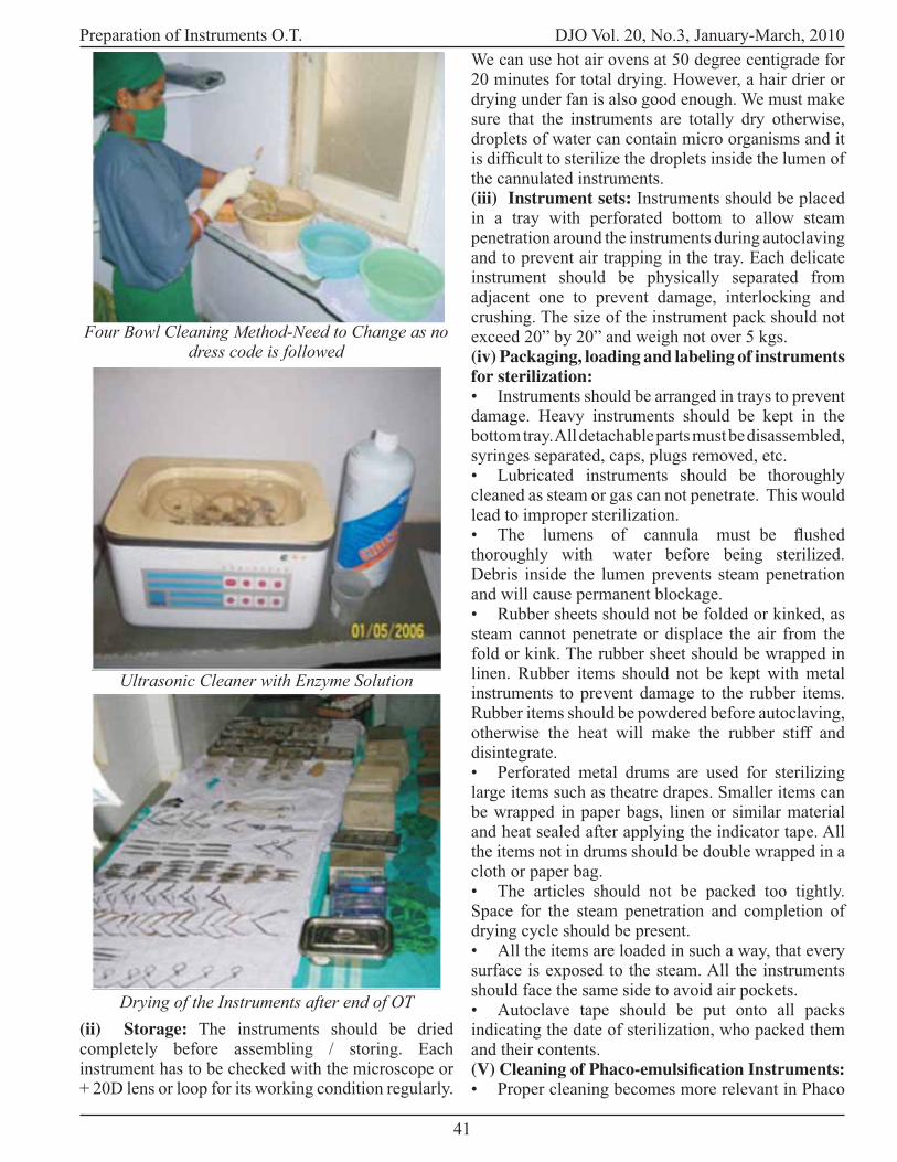
41
DJO Vol. 20, No.3, January-March, 2010
We can use hot air ovens at 50 degree centigrade for 20 minutes for total drying. However, a hair drier or drying under fan is also good enough. We must make sure that the instruments are totally dry otherwise, droplets of water can contain micro organisms and it
the cannulated instruments. Instruments should be placed
in a tray with perforated bottom to allow steam penetration around the instruments during autoclaving and to prevent air trapping in the tray. Each delicate instrument should be physically separated from adjacent one to prevent damage, interlocking and crushing. The size of the instrument pack should not exceed 20” by 20” and weigh not over 5 kgs.
for sterilization:• Instruments should be arranged in trays to prevent damage. Heavy instruments should be kept in the bottom tray. All detachable parts must be disassembled, syringes separated, caps, plugs removed, etc. • Lubricated instruments should be thoroughly cleaned as steam or gas can not penetrate. This would lead to improper sterilization.
thoroughly with water before being sterilized. Debris inside the lumen prevents steam penetration and will cause permanent blockage.• Rubber sheets should not be folded or kinked, as steam cannot penetrate or displace the air from the fold or kink. The rubber sheet should be wrapped in linen. Rubber items should not be kept with metal instruments to prevent damage to the rubber items. Rubber items should be powdered before autoclaving, otherwise the heat will make the rubber stiff and disintegrate.• Perforated metal drums are used for sterilizing large items such as theatre drapes. Smaller items can be wrapped in paper bags, linen or similar material and heat sealed after applying the indicator tape. All the items not in drums should be double wrapped in a cloth or paper bag.• The articles should not be packed too tightly. Space for the steam penetration and completion of drying cycle should be present.• All the items are loaded in such a way, that every surface is exposed to the steam. All the instruments should face the same side to avoid air pockets.• Autoclave tape should be put onto all packs indicating the date of sterilization, who packed them and their contents.
• Proper cleaning becomes more relevant in Phaco
Preparation of Instruments O.T.
The instruments should be dried completely before assembling / storing. Each instrument has to be checked with the microscope or + 20D lens or loop for its working condition regularly.
Four Bowl Cleaning Method-Need to Change as no dress code is followed
Ultrasonic Cleaner with Enzyme Solution
Drying of the Instruments after end of OT

DJO Vol. 20, No.3 , January-March, 2010
42
machine for the surgery.• The IV set is removed from the bottle. The irrigation tubings are disconnected. The irrigation aspiration tube is disconnected from the hand piece. The hand pieces are unplugged from the console.
switching off the machine and the saline collected in a bin. • Cleaning of the components:• All cleaning procedures must be done immediately after each surgical procedure; otherwise, tissue debris and salts from the saline irrigating solution may collect and cause permanent damage.
Ultrasonic hand piece:• The hand piece is wiped with soft non abrasive linen and distilled or sterile water to remove residual tissue. Both the irrigation and aspiration ports are
distilled or sterile water. It is repeated thrice with air.
Irrigation and aspiration hand piece:• Clean the hand piece, tip and sleeve with gauze piece dipped in isopropyl alcohol or antiseptic.
and tips with distilled or sterile water. • Tips and sleeve are usually disposable ones. But they can be re-used if properly sterilized. The
water.• All are then packed into trays for steam sterilization. Care should be taken to wrap the tubing and hand pieces separately in a cloth i.e. the metal components should not come in contact with the wire.
• When the autoclave cycle is completed and the contents are ready to be removed, place them on wire
they cool without developing condensation. If shelves
are not available, place the items in a cool place, but do not pile them on top of each other. If drums are used, they should be sealed immediately.• Once items have cooled they can be wrapped in a polythene bag to prevent dust and external damage. The articles should be used within 48 hours of being sterilized. The sterile packs must be stored above waist level, kept dry, protected from dust, handled only when necessary and used within sterilization date
(B) Preparation of OT Environment:
Theatre Cleaning is scheduled as Daily, Weekly & Monthly CleaningDaily Cleaning: After Surgery:• Enough time must be given to the operation theatre staff for the end of the day cleaning activities. This will ensure that the operation theatre is ready for the next day in a desirable manner.• All the extra items must be removed from the operation theatre.• The walls are cleaned with 1% sodium hypochlorite solution up to six ft. height daily.• Equipments such as electrical surgical units
• Cabinets & doors are cleaned, especially around handles or push plates where contamination is more likely.• Walls around the scrub sink need special attention.• Transportation carts and their wheels are cleaned with special attention after use; equipment is disassembled, cleaned and covered properly.• Microscope lenses should be cleaned with lens cleaning solutions.• Microscope head should be cleaned with bacillol spray and covered properly.• Operating table covers are changed and patient head support also be cleaned.• Trolleys, Revolving stools, Operating table, Foot stool, I. V. Stand are cleaned with 1% sodium hypochlorite solution.
water and another bucked with antiseptic lotion ( for e.g. : Dettol 1:40 dilution in water)
it. (two bucket technique)• Again dip in the Dettol solution, wring out and continue the mop in the same manner. • Slippers should be washed with detergent and
Flushing of both ports of Phaco probe with Water & Air
Preparation of Instruments O.T.

43
DJO Vol. 20, No.3, January-March, 2010
dried completely.• The washing of slippers should be done away from the instruments washing area or the scrub area.• At the end of each session, all the waste must be disposed off as per the waste disposal guidelines in colour coded bags. Over the last decade, the disposal of operating room and hospital waste has received much attention. Incineration has been advocated as a viable method of hospital waste disposal. Recently attention has been directed at preventing air pollution
waste treatment technologies. These options include
Weekly Cleaning:• All the articles removed from shelves and remove the equipments, furniture from the theatre.• Fans and Air conditioners (A.C. Filter) are cleaned,
detergent solution and dry under sunlight before replacing.
• Regular cleaning of all types of sterilizers as recommended by the manufactures.
and mopping with antiseptic solution (The dilution of the antiseptic according to the manufactures’ advice).• Bowls and buckets using inside the Theatre cleaned properly.
then the scrub area, sterilizing area and preoperative patient waiting area.
Monthly Cleaning:• Remove all the equipments from the theatre.
accumulated deposits are cleaned thoroughly. • Washing all the furniture and walls of the O.T. Cupboards are removed and areas behind the cupboards are cleaned.• Trolleys, Stools, Microscopes are cleaned and oiled where necessary, and then routine cleaning procedures are done as described above.
Fumigation:• Aerosol machine/Fogger is used for fumigation (electrically operated) as it can make micro particles. Equal amount of Formalin and distilled water is added depending upon the size of the Theater. After Fumigation, close the theatre for minimum 24 hours. Hydrogen peroxide 1% solution or Glutaraldehyde+ Formaldehyde combination can be used as a change fortnightly to prevent resistance developing against one agent.• Fumigation needs to be done once in a week.• Sterile quality of air in the OT is better achieved and maintained by employing air cleaner and ultraviolet lights and through improving the overall cleanliness in and around the OT. • Air cleaners are turned on while the surgical session is going on. While the ultraviolet lights are turned on over night after the surgical session
Daily Cleaning OT Equipments
Cleaning of OT walls with Sodium Hypochlorite
Air Cleaner
Ultraviolet Light
Preparation of Instruments O.T.

DJO Vol. 20, No.3 , January-March, 2010
44
when the last person leaves the OT. “Off when
This will continuously clean the inside air while we are working.• Humidity of the operation theatre can be
Indications:• If septic case has been operated, fumigation is mandatory after the procedure.• If any new construction or reconstruction of any theatre is done, fumigation is mandatory before the functioning of the same.• When routine surveillance reveals any pathogenic spore former, fumigation is mandatory.Details of fumigation of theatre:• Formalin 30ml of 40% Formalin dissolved in 90 ml of clean water for 1000 cft by aerosol spray.
• Seal the room air tight. Use adhesive to close gaps• Pour total amount into the Fogger/Aerosol and switch on. Timings can be set with timer. • Fogger is the best as the droplet size generated is smaller which can be spread all around effectively. • Leave it for 24 hours.• Aldekol-Formaldehyde – 6%, Glutaraldehyde 6% and Benzalkonium chloride 5%• For 4000 cft 325 aldekol in 350 ml of water sprayed for 30 minutes – close for 2 hrs – Switch on AC – OT ready in 3 hrs• Open the room wearing a mask. Put small amount of liquid ammonia solution inside the room for half an hour and also turn the exhaust fan of air conditioner on.
Fogger in Use for Fumigation
Preparation of Instruments O.T.

45
DJO Vol. 20, No.3, January-March, 2010
(C) Monitoring of Cleaning Activities:1. Monitoring of Environment: • The environment cleanliness is monitored biologically.• Open dish sedimentation method is generally used. A nutrient agar plate is left open in one corner of the operation theatre at the beginning of the activities in the morning. After half an hour, it is taken to the laboratory and incubated. Growth of less then 10colonies is considered good enough to continue the activities. Otherwise, the cleaning and fumigation need to be repeated and after cross checking biologically only the theatre is used for performing surgeries.• Bacterial counters are available but they are very costly and not used commonly.
2. Monitoring sterility:• Weekly samples should be taken from sterile items and scrubbed personnel at the end of the surgery and should be sent for biological testing.• Weekly samples should be taken from the
microscopes, and scrub area, area where the drums are stored or kept while using them.• Nasal and nail bed swabs also should be taken at regular intervals and sent for testing.
3. Monitoring of hygiene:• All the staff members working in the theatre must under go a regular health check up. Nails should be regularly checked and trimmed.• Any skin infection should be treated immediately and the person with skin lesion should not be allowed to work inside the operating room.• Nasal carrier state must be checked and all the staff members in carrier state must be treated.
4. Monitoring of activities:• All the activities must be monitored carefully using various check lists.
Check lists:Daily Cleaning Check List Eye O.T.:Date:1. Who checked Pre operative check list?2. Who put 5% Povidone iodine eye drop before giving block?3. Who checked Autoclave strip register?
it?5. Who checked clarity of Inj. RL?6. Who did preparation before arrival of surgeon? 7. Who did Fumigation? With what? (Formalin, 1%
Hydrogen Peroxide, Formaldehyde & Glutaraldehyde combination)8. Who did cleaning before leaving in evening? (Doors should be cleaned every day)9. Who checked Operation & emergency medicines stock?10. Who put on the U.V. light at night? Who put it off in the morning?11. Was the chlorination of water tank done yesterday? Who did it?12. Who checked anaesthesia trolley?13. Who replaced Bed sheet of O.T. Table in the evening?14. Who cleaned Equipments / Instruments (Cautery, Suction machine & O.T. Table) with 1 % Sodium hypochlorite??15. Notes:16. Signature of O.T. In-charge: Signature of HOD:
Weekly Cleaning Check List Eye O.T.:Date:1. List of medicines checked? Who did it? (Daily use + Emergency medicines)2. Who checked Eye O.T. Check list? (List except medicines)3. Did in charge prepare the list of O.T. staff posting?4. Who submitted Auto clave Report? Who checked it?5. In-charge checked the list of Sunday work done?6. Cleaning done on Saturday by shifting things? (Microscope, O.T. Table)7. Who did Sodium hypochlorite cleaning of sink?
Hypochlorite?9. Who fumigated Autoclave room on Saturday after cleaning?10. Who cleaned A/C Filters?11. Who cleaned Instruments? (Check blade and change it if necessary) (Check two way cannula)12. Who checked Staff nail?13. Who checked Chlorination? 14. Who changed Water in autoclave machine? (Change every fortnight)15. Who cleaned & autoclaved Bottle of surgical scrub and bottle of liquid soap? 16. Who checked Expiry dates of medicines? 17. Who cleaned the Operating Microscope lenses?
Monthly Cleaning Check List Eye O.T.:Date:1. Over book of change of O.T. Boy posting
Preparation of Instruments O.T.

DJO Vol. 20, No.3 , January-March, 2010
46
checked?2. Swab sample culture done on second Saturday?3. A/C cleaned by Air Blower on last Saturday?4. Did in-charge check the washing of O.T. on last Saturday?5. IOL Report prepared6. CME Lecture delivered and exam conducted for O.T. staff?7. Who cleaned the water tank? On which day?8. Who cleaned Drums? Holes checked?9. Webs outside windows removed?10. Note:11. Signature of O.T. Incharge: Signature of verifying person:
References1. Consensus guideline for the Prevention of Infections in
the Operating Room – Hospital Infection Control Society.
2. Uday Kelkar, Shreekant Kelkar, Abhijit M. Bal,
Sandhya Kulkarni, Sadanand Kulkarni: Microbiological
evaluation of various parameters in ophthalmic operating
Rooms. The need to establish Guidelines; IJO, 2002;
51:171-76.
3. General Precautions to safeguard against Post
Operative Infections following Ophthalmic Surgery –
NPCB, India
4. Hinduja Hospital, Mumbai – Infection Control
Manual
5. IAPB Guidelines for Eye Care
6. Infection Control Manual – CMC, Vellore
7. MJ Lights – Operative Operations Theater
8. Standardized Protocol for Cataract Surgery Sightsavers
International.
9. Sterilization and Aseptic Practice in an Ophthalmic
Operation Theatre & CD of Sterilization Protocol- Aravind
Eye Care Systems, Mudurai
10. Textbook of Hospital Infection Control – Shaheen
Mehtar
11. Ophthalmic operating theatre practice – A manual for
developing Countries, Ingrid Cox and sue Stevens
12. The sterile supply department: Guidelines for planning
and quality Management edited by Geetha Mehta.
13. WHO Guideline for Disinfection and Sterilization in
Healthcare Facilities, 2008
Preparation of Instruments O.T.

47
DJO Vol. 20, No.3, January-March, 2010
Case reportA 16 years old male attended our out patient department with gradual, painless, diminution of vision in the left eye for three years. His best corrected visual acuity was OD 6/6 with -0.50DS, OS 6/18 with -0.25DS.Examination of the anterior segment by slit lamp examination revealed posterior lenticonus in the left eye (Figure 1). Posterior lenticonus showed a cataractous opacity which produced a central dark shadow on retro- illumination (Figure 2). The extent of posterior lenticonus as measured on pentacam was 1920 microns (Figure 3). Intraocular pressure (applanation) was 19mm of Hg OU. Fundus examination was withthin normal limits in both eyes. The eyes were orthophoric. Ocular movements were normal in both the eyes.
DiscussionCases of posterior lenticonus can clinically present with reduced visual acuity, amblyopia (1, 4) and strabismus (1). Rarely lenticonus can present with leukocoria (5).
The pathogenesis of lenticonus is not well understood. Collins (2) found a tear in the posterior capsule in a case of posterior lenticonus. Histological investigations
this observation (3). Aberrant growth of sub capsular epithelium was found to overlie the lenticonus by Franceschetti and Rickly (6), Makley (3). The case in our study shows a protrusion of 1920 microns which is more than one half the thickness of the lens (3400 microns). Such a large extent of posterior protrusion is more likely due to abnormal development of the
In early cases refractive error should be corrected. Amblyopia should be treated by occlusion therapy, if present. Surgery on lens is indicated if visual acuity is less than 20/100 and can not be corrected with glasses.
References1. Chengkp, Hiles Da, Biglan Aw, Pettapiece Mc-
Management of posterior lenticonus. J.Pediatr.Ophthalmol.
Strabismus 1991; 28:143-149
2. Collins. Researches, London (1896)
3. Mackley . Amer .J .Ophthal. , 39,308 (1955)
4.Crough Er jr.,Parks Mm – Management of posterior
lenticonus complicated by unilateral cataract.
Am.J.Ophthalmol.1978; 85:503-508.
5.Fett D, Paez Jh, Isenberg S – Infantile leukocoria caused
by posterior lenticonus.Ann.Ophthalmol.1984; 16:679-
680, 684
6. Franceschetti and Ricklei. Arch Ophthal. 51,499(1954)
Lenticonus Posterior Ojaswita Singh, Rashi Shyam, A.K.Gupta
Shroff Eye Centre, New Delhi.
Case Report
Lenticonus is bulging of the lens capsule and the underlying cortex. It is a relatively uncommon clinical condition. The prevalence is estimated at 1 to 4 of every 100,000 children (1). We are reporting a case of posterior lenticonus because of it rarity and its unusually large size.
Figure 1:Slit lamp examination of posterior
lenticonus
Figure 2: Posterior lenticonus on retro-
illumination
Figure 3: Pentacam image Pentacam image shows the extent of posterior lenticonus. In this case it is 1920 microns (5320 – 3400 microns) beyond the
posterior lenticular surface

DJO Vol. 20, No.3 , January-March, 2010
48
Cholesterol Granuloma of the Orbit Mandeep S. Bajaj1, Seema Kashyap2, Neelam Pushker1, Vidushi Sharma1, R. Balasubramanya1,
Rachna Meel1, Anoop Kishore Gupta1
Case Report
1 Oculoplastic and Paediatric Ophthalmology Services, 2 Ocular Pathology ServicesDr. Rajendra Prasad Centre for Ophthalmic Sciences, AIIMS, New Delhi
Abstract
of the orbit, commencing mostly in the diploe of bone. We report a case of cholesterol granuloma in the orbital part of the frontal bone in a 47-year-old male patient. The patient presented with progressive and painless proptosis and downward displacement of the globe. CT scan showed a superotemporal, homogenous orbital mass involving the frontal bone with expansion and fossa formation with irregular margins. The outer table of the bone was partially absent, with extraperiosteal extension of the lesion. The mass was removed by an antero-lateral
In conclusion, cholesterol granuloma, though rare, is a readily recognizable lesion with characteristic clinical, radiological and histopathological features.
Introduction
lesion of the orbit, commencing mostly in the diploe of the frontal bone.1 It has also been reported in other regions of the head including the zygomatic bone, maxillary and ethmoidal sinuses, and the petrous bone.1-3 In the last decade only three cases have been reported.4-6 We report a case of a cholesterol granuloma in the orbital part of the frontal bone in a 47-year-old male patient.
Case ReportA 47-year-old man presented with right sided
painless, progressive proptosis for one year. No history of trauma was elicited. On examination 4mm of proptosis with inferior displacement of the right globe was present (Figure 1). The ocular movements
consistency was felt just beneath the irregular bony orbital margins in the superotemporal quadrant. Visual acuity was 20/20 in both the eyes. CT scan showed a superotemporal homogenous, orbital mass imvolving the frontal bone with its expansion and fossa formation with irregular margins. The outer table of the bone was partially absent with extraperiosteal extension of the lesion (Figure 2).
The mass was approached by an antero-lateral orbitotomy under general anesthesia. The periosteum
bone to the orbital margin, which was irregular in places. The outer table of the bone was thin, eroded and brittle near the orbital rim. On elevating the orbital periosteum from the orbital roof, a yellowish brown mass that was adherent to the bone was visualized subperiosteally. The mass was completely excised along with curettage of granulomatous material from
Figure 1. Preoperative clinical photograph of the patient (Anterior view).
Figure 2. Computed tomographic scan (coronal.view showing a superotemporal orbital mass imvolving the frontal bone. The outer table of the bone was partially absent, with extraperiosteal extension of the lesion

49
DJO Vol. 20, No.3, January-March, 2010
the frontal bone. The inner table of the bone was found to be intact.
Gross examination showed two irregularly shaped fragments of yellowish granular tissue each measuring 10x8x6mm and 9x5x4mm respectively. Microscopic examination showed numerous slit like spaces representing cholesterol clefts surrounded by multinucleated giant cells (Figure 3). Deposits of hemosiderin pigment and lipid laden macrophages
sections failed to reveal any epithelial structures
postoperative period was uneventful. No recurrence was observed at a follow up of 6 months.
DiscussionCholesterol granolomas are usually seen in adult
males. The age reported in the largest series of 27 patients ranged from 25 to 68 years (median, 43 years).1 The superotemporal quadrant of the orbit is involved most often as the predominant site of occurrence of the tumour is the frontal bone.1
Computed tomographic (CT) scans usually show a homogenous lesion, isodense with the brain, with expansion of the surrounding bone. The edges of the eroded bone may occasionally be irregular. In a reported series, all the cases had partial or complete erosion of the outer table of the bone allowing the lesion to expand extraperiosteally into the orbit, as was seen in our case. Rarely erosion of the inner table of the skull may be seen with extension into the extradural space or into the sinuses.1
The differential diagnosis of this case included orbital lesions ocurring at this location, associated with bone remodelling. It includes dermoid cyst, which has an identical clinical presentation but shows a low density lesion on CT scan, in comparision to Cholesterol granoloma which is isodense with the brain.1
edges in a dermoid are regular and scalloped with adjacent sclerosis and no destructive changes. An
history. Aneurysmal bone cyst has clinical similarity and evidence of bone destruction, histopathology shows evidence of old and recent hemorrhages and
are notably absent. The closest histopathological differential diagnosis of Cholesterol granuloma is the epidermoid cholesteatoma, which is fairly uncommon in the orbit and is encountered more often in the middle ear and mastoid antrum.1 The only differentiating point is that epidermiod cholesteatomas have a squamous lining which is absent in Cholesterol granulomas.
The exact origin of this lesion is uncertain.
which may occur either due to trauma or an anomaly in the dilpoe of the bone.1 An extraperiosteal approach for its removal allows the surgeon to aspirate its contents, strip or currette the lining, and remain safely extradural, even if a defect in the inner table of the bone is present. Incomplete removals have been attempted with no recurrence.1 Only one possible recurrence of Cholesterol granuloma is reported in the literature, which was seen in a epidermoid cholesteatoma.7
In conclusion, Cholesterol granuloma, though rare, should be considered in the differential diagnosis of lesions involving the superotemporal orbit in adults. It is a readily recognizable lesion with characteristic clinical, radiological and histopathological features. A timely and appropriate surgical intervention carries a relatively favourable prognosis.
References1. McNab AA, Wright JE. Orbitofrontal Cholesterol
Granuloma. Ophthalmology 1990; 97: 28-32.
2. Cingi E, Cingi C. Imaging case study of the month:
Ethmoidal cholesteatoma. Ann Otol Rhino Laryngol 199;
100: 424-426.
3. Sadoff RS, Pliskin A. Cholesteatoma (keratoma) of
the maxillary sinus: Report of a case. J Oral Maxillofac
Surg 1989; 47: 873-876.
4. Heaton RB, Ross JJ, Jochum JM, Henry MR. Cytologic
diagnosis of cholesterol granuloma. A case report. Acta
Cytol 1993; 37: 713-716.
5. Herrmann B, Rozot P, Baylac F, Civit T, Reynal C,
Ketema MA. Lipogranuloma of the orbit. Apropos of a
case. J Fr Ophtalmol 1996; 19: 780-784.
6. Karim MM, Inoue M, Hayashi Y, et al. Orbital
cholesterol granuloma with destruction of the lateral
orbital roof. Jpn J Ophthalmol 2000; 44: 179-182.
7. Pfeiffer RL, Nicholl RJ. Dermoids and epidermoids of
the orbit. Trans Am Ophthalmol Soc 1948; 46: 218-240.
Figure 3. Photomicrograph showing numerous cholesterol clefts along with lipid laden histiocytes
(H&E X40).
Cholesterol Granuloma of the Orbit

DJO Vol. 20, No.3 , January-March, 2010
50
Cancer associated retinopathyin a patient with non-Hodgkin’s lymphoma and congenital achondroplasia llowed
by drug induced thrombocytopenia and bilateral central retinal vein occlusion
Shahana Mazumdar, Navneet MehrotraICARE eye hospital, NOIDA U.P
Case Report
Introduction:Cancer associated retinopathy is a rare paraneoplastic syndrome in which autoantibodies directed at various retinal components cause progressive vision loss1.
syndrome depending on the primary malignancy such as cancer associated retinopathy (CAR), melanoma associated retinopathy (MAR), lymphoma associated retinopathy (LAR). Many patients develop the retinopathy before the primary malignancy has been found. Cases, where no malignancy can be found though antibodies can be detected, are referred to as autoimmune retinopathies (AR). The clinical features are similar in all types and result from rod or cone involvement. Rod involvement results in night blindness, delayed dark adaptation, photopsia, mid-peripheral scotomas. Cone involvement results in reduced visual acuity, photosensitivity, decreased color vision and central scotoma. CAR has been reported with a variety of carcinomas most commonly small cell lung cancer, endometrial, cervical and breast cancers. Several other cancers have been reported with less frequency. We report a patient with congenital achondroplasia who developed CAR following Non Hodgkins Lymphoma (CNS). He developed it during a quiescent phase but suffered a recurrence of the lymphoma a month later. On being treated for the recurrence the patient developed drug induced thrombocytopenia resulting in bilateral central retinal vein occlusions.
Case reportA 28 years old male with congenital achondroplasia
of light since one month in his only seeing eye, the right eye. He was a patient of Non Hodgkins Lymphoma (CNS) B cell type diagnosed three years earlier which had presented as a left orbital mass. The disease had been diagnosed following an orbital biopsy. Subsequently he underwent orbitotomy to remove the tumor but the vision in the left eye was lost due to compressive optic atrophy. There was no evidence of
right eye involvement. Bone marrow biopsy and CSF were negative for malignant cells. Patient received CHOP based chemotherapy (Cyclophosphamide, Doxorubicin, Vincristine and Prednisolone) and radiotherapy. A few months later he developed meningeal involvement and received intrathecal methotrexate and high dose methotrexate. He had suffered several recurrences since then receiving many cycles of chemotherapy. On presentation the disease had been quiescent for a few months and he was not on chemotherapy.On examination his best corrected visual acuity was 6/6 and no PL in the right and left eyes respectively. The anterior segments in both eyes were normal. The left eye showed an afferent pupillary defect. The posterior pole of the right eye revealed a normal optic
left eye showed a pale optic disc (Figure 3). Both eyes showed generalized arteriolar attenuation and widespread mottling of the retinal pigment epithelium (Figure 4). There was an area of lattice degeneration in the superotemporal retina of the right eye. He
revealed an incomplete ring scotoma (Figure 5). His
normal limits (Figure 6). Flash ERG showed mild delay in the implicit time with reduced amplitude in the scotopic response in the right eye. The left eye showed no response.He did not report for 4 months as he had suffered a recurrence of the malignancy (meningeal) with severe pain in the spine and had been under treatment. By now he had developed debilitating night blindness though his visual acuity was still 6/9.On examination both eyes revealed a marked increase in the retinal mottling (Figure7). He was scheduled for further sessions of chemotherapy. Based on the history of malignancy, rapidly progressing positive visual symptoms and night blindness, advancing retinal pigment epithelial mottling, arteriolar attenuation, concentric narrowing of visual
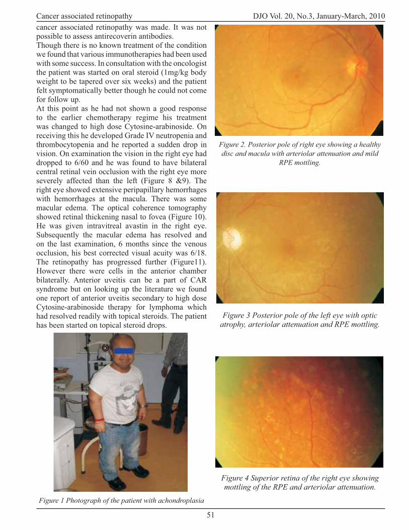
51
DJO Vol. 20, No.3, January-March, 2010
cancer associated retinopathy was made. It was not possible to assess antirecoverin antibodies. Though there is no known treatment of the condition we found that various immunotherapies had been used with some success. In consultation with the oncologist the patient was started on oral steroid (1mg/kg body weight to be tapered over six weeks) and the patient felt symptomatically better though he could not come for follow up. At this point as he had not shown a good response to the earlier chemotherapy regime his treatment was changed to high dose Cytosine-arabinoside. On receiving this he developed Grade IV neutropenia and thrombocytopenia and he reported a sudden drop in vision. On examination the vision in the right eye had dropped to 6/60 and he was found to have bilateral central retinal vein occlusion with the right eye more severely affected than the left (Figure 8 &9). The right eye showed extensive peripapillary hemorrhages with hemorrhages at the macula. There was some macular edema. The optical coherence tomography showed retinal thickening nasal to fovea (Figure 10).He was given intravitreal avastin in the right eye. Subsequently the macular edema has resolved and on the last examination, 6 months since the venous occlusion, his best corrected visual acuity was 6/18.The retinopathy has progressed further (Figure11).However there were cells in the anterior chamber bilaterally. Anterior uveitis can be a part of CAR syndrome but on looking up the literature we found one report of anterior uveitis secondary to high dose Cytosine-arabinoside therapy for lymphoma which had resolved readily with topical steroids. The patienthas been started on topical steroid drops.
Figure 1 Photograph of the patient with achondroplasia
Figure 2. Posterior pole of right eye showing a healthy disc and macula with arteriolar attenuation and mild
RPE mottling.
Figure 3 Posterior pole of the left eye with optic atrophy, arteriolar attenuation and RPE mottling.
Figure 4 Superior retina of the right eye showing mottling of the RPE and arteriolar attenuation.
Cancer associated retinopathy

DJO Vol. 20, No.3 , January-March, 2010
52
Figure 7 Fundus photograph after 4 months with marked increase in the RPE mottling
Figure 8 Acute central retinal vein occlusion in the right eye following thrombocytopenia secondary to
chemotherapy
Figure 9 Other eye had multiple retinal hemorrhages with milder central retinal vein occlusion
Figure 10 OCT of the macula with intraretinal thickening
Cancer associated retinopathy
Figure 11 Progression of retinopathy with pigment clumping and degranulation

53
DJO Vol. 20, No.3, January-March, 2010
DiscussionWe have discussed this case with the following in mind: to report the occurrence of CAR syndrome in a patient with achondroplasia and to report the occurrence of CAR in association with Non-Hodgkins lymphoma. We also wanted to report the ocular complications of cytosine-arabinoside therapy in our patient namely bilateral central retinal vein occlusion and possibly anterior uveitis.Cancer Associated Retinopathy (CAR) is a remote manifestation of cancer and is a paraneoplastic disease. The retinopathy can precede detection of the cancer and a high index of suspicion must be maintained in patients presenting with unexplained visual loss and symptoms. Cancers most commonly associated with CAR are small cell lung cancers, breast cancer1-7, endometrial, cervical and ovarian cancers. Less commonly it has been found with pancreatic cancers, other lung cancers, lymphomas, prostatic, bladder and laryngeal cancers, colon cancers and metastases of unknown origin. Similar paraneoplastic retinopathy has been described in patients with
associated retinopathy (MAR). Lymphoma associated retinopathy (LAR) is also used to describe the retinopathy when associated with lymphomas8.Symptoms are usually bilateral and can be due to affection of rods, cones or both. Cone related symptoms include photosensitivity, reduced vision, decreased color sensitivity and central scotoma. Rod related symptoms include prolonged dark adaptation, night blindness, positive visual phenomena such as
ways the retinopathy is progressive and ultimately the patient loses vision.Signs commonly seen are mottling of the pigment epithelium, arteriolar attenuation and mild optic disc pallor. Rarer signs include periphlebitis, arteriolar sheathing and cells in the vitreous and anterior
depression, ring scotoma or central scotoma. ERG shows a diminished rod or cone response9. CSF
Histopathlogically there is diffuse photoreceptor degeneration of both rods and cones with or without
6. The photoreceptor degeneration is believed to be immune mediated. Antibodies against retinal proteins have been found and there appears to be cross reactivity between cancer cells and retinal proteins. Several antibodies
is antirecoverin antibody10directed against retinal recoverin – a calcium binding protein located within
retinal photoreceptors. Detection of this antibody has been used for diagnosis though it can also be found in some non-paraneoplastic autoimmune retinopathies. Treatment with systemic steroids (oral and high dose intravenous)11 and other immunomodulators have met with mixed results. Treatment of the primary malignancy does not alter the visual prognosis.
References1.Sobottka B, Schlote T, Besch D, Dielebova T, Wilhelm
H, Zrenner E: Carcinoma-associated retinopathy: a review
with clinical examples. Klin Monatsbl Augenheilkd. 2000
Jan; 216(1): 17-24
2. Kornguth SE, Klein R, Appen R, Choate J: Occurrence of
anti-retinal ganglion cell antibodies in patients with small
cell carcinoma of the lung. Cancer 50:1289-93,1982
3. Thirkill CE, Roth AM, Keltner JL: Cancer-associated
retinopathy.Arch Ophthalmol 105:372-5, 1987
4. Klingele TG, Burde RM, Rappazzo JA, et al:
Paraneoplastic retinopathy. J Clin Neuroophthalmol 4:
239-45, 1984
5.Eltabbakh GH, Hoogerland DL, Kay MC: Paraneoplastic
retinopathy associated with uterine sarcoma. Gynecol
Oncol 58: 120-3, 1995
6. Sawyer RA, Selhorst JB, Zimmerman LE, Hoyt WF:
Blindness caused by photoreceptor degeneration as
a remote effect of cancer.Am J Ophthalmol 119:369-
70,1995
7. Guy J, Aptsiauri N: Treatment of paraneoplastic visual
loss with intravenous immunoglobulin: report of 3 cases.
Arch Ophthalmol 117:471-7, 1999
8. To KW, Thirkill CE, Jakobiee FA, Lessell S, Berson EL:
Lymphoma- associated Retinopathy. Ophthalmology 2002
Nov; 109(11): 2149-53.
9. Matsui Y , Mehta MC, Katsumi O, et al:
Graefes Arch Clin Exp Ophthalmol 230:324-8, 1992
10. Thirkill CE, Keltner JL: Commonalities and diversity
in the immune response of Cancer-associated retinopathy
(CAR) patients. Invest Ophthalmol Vis Sci 37 (Suppl):
S112, 1997
11. Nippon Ganka Gakkai Zasshi: Case of cancer-associated
retinopathy in which immediate treatment succeeded 2008
Sep; 112(9): 806-11
Cancer associated retinopathy
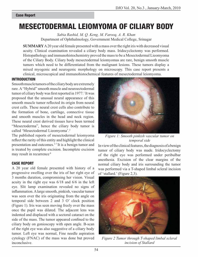
DJO Vol. 20, No.3 , January-March, 2010
54
INTRODUCTIONSmooth muscle tumors of the ciliary body are extremely
rare. A “Hybrid” smooth muscle and neuroectodermal 1 It was
proposed that the unusual neural appearance of this
crest cells. These neural crest cells also contribute to
the formation of bone, cartilage, connective tissue
and smooth muscles in the head and neck region.
These neural crest derived tissues have been termed
“Mesectoderma”, hence the ciliary body tumor is
called ‘Mesectodermal Lieomyoma’.2
The published reports of mesectodermal leiomyoma
presentation and outcomes.3-7 It is a benign tumor and
is treated by complete excision. Incomplete excision
may result in recurrence.8
CASE REPORTA 20 year old female presented with history of a
progressive swelling over the iris of her right eye of
3 months duration, compromising her vision. Visual
acuity in the right eye was 6/18 and 6/6 in the left
eye. Slit lamp examination revealed no signs of
was seen over the iris originating from the angle on
temporal side between 2 and 3 O’ clock position
(Figure 1). Iris was seen moving freely over the mass
once the pupil was dilated. The adjacent lens was
indented and displaced with a sectoral cataract on the
ciliary body on gonioscopy with open angle. B-scan
of the right eye was also suggestive of a ciliary body
tumor. Left eye was normal, Fine needle aspiration
cytology (FNAC) of the mass was done but proved
inconclusive.
MESECTODERMAL LEIOMYOMA OF CILIARY BODYSabia Rashid, M. Q. Keng, M. Farooq, A. R. Khan
Department of Ophthalmology, Government Medical College, Srinagar
SUMMARY A 20 year old female presented with a mass over the right iris with decreased visual
acuity. Clinical examination revealed a ciliary body mass. Iridocyclectomy was performed.
Histopathology and immunohistochemistry proved the mass to be a Mesectodermal Lieomyoma
of the Ciliary Body. Ciliary body mesectodermal leiomyomas are rare, benign smooth muscle
tumors which need to be differentiated from the malignant lesions. These tumors display a
mixed myogenic and neurogenic morphology on microscopy. This case report presents a
clinical, microscopical and immunohistochemical features of mesectodermal leiomyoma.
In view of the clinical features, the diagnosis of a benign
tumor of ciliary body was made. Iridocyclectomy
of the right eye was performed under peribulbar
anesthesia. Excision of the clear margins of the
normal ciliary body and iris surrounding the tumor
was performed via a T-shaped limbal scleral incision
of ‘stallard.’ (Figure 2,3).
Figure 1: Smooth pinkish vascular tumor on temporal side
Figure 2 Tumor through T-shaped limbal scleral incision of Stallard
Case Report

55
DJO Vol. 20, No.3, January-March, 2010
Macroscopically it was an ovoid tumor 8mm in
diameter with smooth, dark external surface. Cut
Microscopic examination revealed well circumscribed
tumour enclosed by ciliary epithelium. The tumor
cells were loosely packed and having round to
ovoid vesicular nuclei with 1-2 small nucleoli and
Nuclear atypia, mitosis and tumor necrosis were
absent. The stroma contained widely spaced thin
ectatic capillaries lined by single layer of cells. The
diagnosis of mesectodermal lieomyoma was made
strong immunoreactivity for smooth muscle actin
and negative reaction for S-100, epithelial membrane
DISCUSSIONThe ciliary body tumors include Naevi, Melanomas
and benign smooth muscle neoplasms. Smooth muscle
neoplasms are either leiomyomas or mesectodermal
lieomyoma. Mesectodermal lieomyomas are
extremely rare and only 15 cases have been reported
in literature so far.3
The differential diagnosis of mesectodermal
leiomyoma includes melanomas, gliomas, peripheral
nerve tumors or paraganglioma because of its
resemblance to ganglionic, astrocytic and peripheral
nerve tumors.
Published literature reported on leiomyomas has
shown that most of the cases are treated by resection
but some may need enucleation. Recurrences have
also been reported in a few cases and are attributed to
incomplete removal of the tumor.8
Maximum follow up and reports of recurrence has
been six years8. In our case the follow up is of 2 years
and there is no evidence of recurrence.
REFERENCES1. Jakobiec FA, Font RL, TSO Mo, Zimmerman LE.
Mesectodermal lieomyoma of the ciliary body: a tumor of
presumed neural crest origin. Cancer 1977; 39: 2102-13.
2. White V, Stevenson K, Gamer A, Hungerford J.
Mesectodermal lieomyoma of the ciliary body case report.
BJO 1989; 73: 12-18.
3. Alexander N, Odashiro MD. Report of two cases of
ciliary body mesectodermal lieomyoma unique expression
of neural markers. Ophthalmology Jan. 2007; 114: 157-
161.
4. Lowe RF, Greer CH. Lieomyoma of the ciliary body.
A clinicopathological case report. BJO 1970; 54: 383-7.
5. Croxatto JO, Malbran ES. Unusual ciliary body tumor,
mesectodermal lieomyoma. Ophthalmology 1982; 89:
1208-12.
6. Clinical and Pathological Status of mesectodermal
lieomyoma of the ciliary body. A case report. Graef’s
Archive for Clinical and Experimental Ophthalmology
March 1989; 227: 101-105.
7. Lai CT, Tai MC, Liang CM, Lee HS. Unusual uveal
tract tumor: mesectodermal lieomyoma of ciliary body.
Pathol Int 2004 May; 54(5): 337-42.
8. Seong Hwan Park, Ji-Hye Lee, Yang Scokdae.
Recurrent mesectodermal lieomyoma of ciliary body. J
Korean Med Sci 2003; 18: 614-7.
Figure 3 Complete Excision of the tumor through T-shaped limbal scleral incision of Stallard
Figure 4: Section of tumor under high power
Mesectodermal Leiomyoma Of Ciliary Body

DJO Vol. 20, No.3 , January-March, 2010
56
History of Ophthalmology
One of the greatest tributes a physician can have is to have a medical condition named after him. Eponymy is “the most enduring and perhaps most prestigious kind of recognition institutionalized in science”. Scientists are not known to be modest about themselves. They would not appreciate Thomas Jefferson’s famous remark after being named America’s second ambassador to France after Benjamin Franklin.Arriving in Paris in 1785, Jefferson was asked, ‘So you are to replace Dr. Franklin?’ he replied,‘ I succeed Dr Franklin. No one can replace him.’ Scientists love eponyms and they have the maximum eponyms after them and painters and musicians have the least number.
The vocabulary of medicine is replete with eponymous descriptions. “There are, for instance more than 15 eponymous designations credited to Fanconi, and there are at least 13 Smiths who independently described different and completely unrelated syndromes, thus rendering the eponyms Fanconi’s Syndrome and Smith’s Syndrome practically useless”. Ophthalmology is hardly less culpable: at least 15eponyms hang around Ernst Fuchs’ neck, (Fuchs’ adenoma, angle, coloboma, dellen, corneal dystrophy, gyrate atrophy, heterochromic iridocyclitis, lines of clearing, nodules, phenomenon, spot, spur, stomas,
is almost strangulated by the weight of 8 eponyms, (Elschnig’s pearls, conjunctivitis, spots, operation,
syndrome, theory, intracapsular forceps, scleral ring) David Cogan limps along with 5, (Cogan’s syndrome, Cogan Reese Syndrome, lid twitch sign, epithelial dystrophy and congenital oculomotor apraxia) Goldmann has the unbearable lightness of being with 7, (Goldmann perimetry, applanation tonometry, Goldmann-Favre syndrome, fundus contact lens, 3 mirror, gonioscope, adaptometer) and so on. The Eponyms Dictionaries Index edited by James A. Ruffner, lists 20,000 eponyms overall!In 1974, US National Institutes of Health held a conference on the naming of diseases and in 1975,JAMA published an article and decided to drop the possessive “s” in all its publications (Down Syndrome not Down’s Syndrome) but has not been able to convince journal editors. Eponyms can be of various kinds:• Named after male physicians (2,880 on whonamedit.com)• Named after female physicians (101)• First and last name
Austin FlintFoster Kennedy Syndrome
• Named after more than one personCharcot-Marie-Tooth-Hoffman disease
• Named after famous people with the diseaseLou Gehrig Syndrome (ALS – Amyotrophic
Lateral Sclerosis)
Ulysses (side effects of extensive diagnostic tests
DOCTOR, YOUR EPONYMS ARE SHOWING!Sashwat Ray
Shroff Eye Centre, New Delhi
Hans Goldmann Anton Elschnig David Cogan
History - “An account, mostly false, of events mostly unimportant, brought about by rulers, mostly knaves, and soldiers, mostly fools”
57
DJO Vol. 20, No.3, January-March, 2010
because of false positive results in course of routine investigation, like Ulysses, patient ends up weary after much fruitless exploration)o Lazarus (symptoms that occur due to prolonged cerebral anoxia mostly of temporal lobes, seen in post resuscitational states after cardiac arrest, “moving through a tunnel, seeing a light, feeling outside one’s body”, named after the Biblical Lazarus whom Jesus raised from the dead after 4 days)
Christmas Disease (after Stephen Christmas)
diagnosed in England in 1956 of this metabolic disorder)
disgruntled about it. He believes that eponyms do not reward the achievement of an original discoverer, because they are usually wrongly attributed. In fact Stigler’s law of eponymy states that eponyms are never named after the original discoverer (exception; Pott’s fracture has to be a legitimate eponym because it describes - not a patient’s fracture - but his own. Pott suffered this fracture of the lower leg bones when he was thrown from his horse). Mark M. Ravitch goes so far as to say that one of the following is usually true of eponyms:
the eponymized person didn’t understand the discovery; the eponym’s current meaning is different from that which was described; the eponym has no historical basis”
Story of the two HansAnd now we will talk about two scientists, one a German bacteriologist-Hans Reiter, the other, a Swiss ophthalmologist-Hans Goldmann.
The Double Life of Hans ReiterIn 1916, Dr. Hans Reiter examined a German military
pains, high fever and red eyes following a sexual liaison. The term Reiter Syndrome comprising of features of conjunctivitis, urethritis and arthritis, particularly when associated with HLA B- 27, was adopted in English language journals in 1940. Reiter’s
English language, in an arthritis textbook in 1941 and a journal article in 1942 by Dr. Bauer and Engleman. But behind the eponym lay the dark side of the moon. Dr. Hans Conrad Julius Reiter was one of
to Adolf Hitler in 1932, and was one of the most
prominent members of the eugenics movement that advocated killing all people who had any physical or mental disability. He declared that it is medicine’s responsibility to stop further transmission of inferior genes. He sanctioned medical experiments that killed thousands of people at Buchenwald Concentration Camp. One documented experiment was to infect 250 prisoners at Buchenwald with rickettsia and then massacre them. He wrote a book on racial hygiene and was a strong supporter of Hitler’s anti smoking campaign, medically progressive at that time, and was a talented teacher who was popular with his students and was even included in a book of the top 5000 Nazis. After the Nazis were defeated, he was arrested by the Red Army in Soviet Union-occupied Germany, tried at Nuremberg and was interned in an American prisoner of war camp. He was not convicted of any crimes in the Nuremberg Trials but that may
proof; the two top-ranking Nazi doctors with whom Dr. Reiter had planned experiments at Buchenwald committed suicide in 1945. After the war, Reiter continued publishing research papers and practiced medicine in Kassel. He died in 1969 at the age of 88. In 2000, Dr. Daniel J. Wallace and Dr Michael Weisman, writing in the Journal of Clinical Rheumatology, unveiled this stunning revelation. Acting on this information, the Spondylitis Association of America, a patient advocacy group, that represents people with Reiter’s Syndrome, voted to call Reiter’s Syndrome, Reactive Arthritis. Another eponym deriving from this period is that of Wegener’s Granulomatosis. Friedrich Wegener was a member of the Nazi brownshirts and his mentor Martin Staemmler an enthusiastic supporter of the Nazi Racial Hygiene Programme. At present there is no direct evidence that Wegener participated in any way in war crimes although he appears to be on a wanted list by the Polish Ministry for the Interior. Interestingly, Wegener himself did not refer to the disease as Wegener’s Granulomatosis, but at most referred to it as the “so called Wegener’s Granulomatosis”.
The Astonishing Universality of Hans Goldmann“Hans Goldmann took the torch and illuminated vast areas of ophthalmology. Every patient and every
Born in 1899 near Prague, this Renaissance man of ophthalmology wanted to become an astronomer but was persuaded by his parents “to do something practical” so he became an ophthalmologist. His universality was overwhelming. A list of his
Doctor, Your Eponyms Are Showing

DJO Vol. 20, No.3 , January-March, 2010
58
achievements reveals the vast scope and range of his interests.1. Pathogenesis of cataract in general, but glassblower’s cataract in particular, he clashed with Alfred Vogt in a 300-page long academic battle which was dangerous for him because he was a foreigner and Jewish in Switzerland. Vogt thought that the cataract was a result of infrared energy being absorbed directly by the lens, while Goldmann held that it was caused by heat transferred to the lens from the energy absorbed by the iris. Later experiments proved they were both right.
μL per minute.3. Designed the Goldmann cupola perimeter in 1945which remained the gold standard for perimetry for almost 50 years before his own student and chief guinea pig Franz Fankhauser ushered in the era of automated static perimetry with the invention of the Octopus.
activity was to reassemble a Hering colorimeter that had been left in a highly disordered state by the former chairman of the Institute. He had observed earlier that his own color sense was somewhat defective as he was unable to see the tuberculosis bacilli present in a stained microscopic slide. He proved to be an anomalous trichromat, his name was mentioned in a handbook of physiology as the “protoanomalous Goldmann”.5. Experiments related to the Stiles-Crawford effect. The Stiles-Crawford effect is one of those discoveries that came by chance. In1933, Stiles and Crawford were working in the National Physical Laboratory in Middlesex, England. They were building an instrument to measure the area of the pupil based on the photometric principle, but it did not work and after eliminating all possible sources of error they decided to study the cause of their failure and discovered the effect!6. Worked on the dark adaptometer which is the standard for dark adaptation (Goldmann - Weekers adaptometer)
8. The construction of a new slit-lamp, the Haag Streit 360, in 1933 and later, the Haag Streit 900 which is the world standard. The Gullstrand slit lamp was manufactured by Carl Zeiss. (Typically a classic Haag
from 10x to 6x, where as a Zeiss has parallel optics
9. In 1954, he invented the applanation tonometer, the most accurate measurement of intraocular pressure till date.10. Stereochronoscopy of the optic nerve head, which involves rapid detection of changes in the optic disc in glaucoma.11. Clinical research on Retinopathy of Prematurity and Diabetic Retinopathy12. Philosophical writings relating to the philosophy, spirit, power, limits and reasoning in science.Such was Hans Goldmann, the greatest of scientists in the branch of ophthalmology and such was the despicable double life of Hans Reiter. Both were honored with eponyms but what a world of difference!Sometimes eponyms can be frankly nationalistic. A case in point is Takayasu’s arteritis. Makito Takayasu presented the case of a young woman with retinal artery changes to the Japanese Society of Ophthalmology in 1908. It seems that Takayasu did not even recognize the peripheral artery changes of the disease. Nevertheless, by 1941 Yasuvo Niimi was proposing to honor Takayasu by calling the disease after him, something which must be seen in context of the political situation of that time, at the dawn of World War Two and the height of Japanese imperialist designs.So what’s in a name, you ask? Plenty of heartburn,
friction between two great English Prime Ministers William Gladstone and Benjamin Disraeli .In one such encounter Gladstone said derisively, “You will die either on the gallows or of a vile disease”, Disraeli came back with one of history’s greatest retort; “That will depend upon whether I embrace your principles or your mistress”. So it’s a refreshing change for us when a perfectly delightful gem of a syndrome emerges from the dreary, desert sand of the world of eponyms. It is a new syndrome, which has never been previously reported in the medical literature. It is called Tashima’s syndrome (C. K. Tashima, JAMA 194:208; 1965) and in its full-blown state (forme fruste manifestations may be described
physician earnestly searches for a new sign, disease, or syndrome to which his name can be attached!
Doctor, Your Eponyms Are Showing
59
DJO Vol. 20, No.3, January-March, 2010
Instrument Scan
Role of Radio Imaging in OphthalmologySatya Karna1, Varun Gogia2
1. Max Hospital and Shroff Eye Centre2. Dr. Rajendra Prasad Centre for Ophthalmic Sciences,
Common Indications for Neuroimaging in Ophthalmology1. Unilateral or bilateral visual loss (transient visual loss, unilateral or bilateral optic neuropathy, junctional scotoma, bitemporal hemianopia, homonymous hemianopia or cortical blindness)2. Efferent pupillary defects (anisocoria due to Horner syndrome or third nerve palsy) or afferent pupillary defects (e.g. RAPD or light-near dissociation of the pupils)3. Proptosis (thyroid eye disease, orbital tumours,
CCF)4. Diplopia or external ophthalmoplegia5. Lid abnormalities (lid retraction, lid lag, ptosis or orbital-lid lesion)6. Oscillopsia (nystagmus)7. Fundus abnormalities (papilloedema, optic atrophy, optic nerve hypoplasia, optic disc head drusen or choroidal folds)8. Ocular or orbital trauma (e.g. intraocular/ intraorbital foreign body or suspected fracture)9. Isolated headache or facial pain
Pre requisites for scanning1. Decide whether CT or MRI is needed – MRI is used in most neuro-ophthalmic conditions except in
situations and if MRI is contraindicated.2. Contrast is ordered for most cases in CT & MRI except in cases of renal failure and allergy to contrast3. The contrast is not required in cases of acute intracranial or intraorbital haemorrhage as a
non-contrast study is superior for assessing the hyperdensity of acute blood. It is also not required in cases of trauma (e.g. orbital, facial or skull base fracture, orbital or intracranial FB or traumatic optic neuropathy), birth hypoxia, hydrocephalus, sinus disease (sinusitis) and thyroid eye disease (iodinated contrast may even worsen systemic thyroid disease.)4. When ordering a CT scan it is important for the ophthalmologist to specify whether the study should include the orbit, the head or both after topographically localizing the lesion.5. Although the most commonly obtained orbital CT planes are axial and coronal, sagittal views can also be obtained with computerized reconstruction - the
their neck for direct imaging (e.g. trauma, cervical collar or cervical disc disease)
suppression for orbital post contrast study especially for optic nerve sheath meningiomas,optic neuritis and thyroid ophthalmopathy, FLAIR sequence for white matter lesions and GRE for stroke.7. If the clinical picture suggests a particular lesion but the imaging is normal – repeat the imaging with
interest.8. An ophthalmologist should communicate to the
lesion location, differential diagnosis and urgency of the imaging request to optimize the interpretation of imaging studies. When in doubt get another radiologist to give an independent report9. Better neuroimaging techniques should be used if
With rapid development in imaging techniques their role in diagnosis, management and follow up of cases in Ophthalmology has increased multifold. Need for imaging can be diverse more so when there is a diagnostic dilemma. Neuroimaging however can only supplement a good clinical examination and never replace it. In all cases of suspected intracranial lesion, it is important to clinically localise it so that the appropriate investigation with correct site is imaged. A wrong investigation or one that does not image the site of likely lesion is more dangerous than no investigation as it leads us into a sense of complacency and may delay diagnosis with fatal outcomes. The current article gives a set of guidelines for imaging in cases where good clinical examination may not provide the answer.

DJO Vol. 20, No.3 , January-March, 2010
60
Role of Radio Imaging in Ophthalmology
Indications of CT scan1.Acute orbital trauma (e.g. suspected orbital fracture, orbital hematoma, metallic or wooden foreign bodies, or traumatic optic neuropathy)2.Acute proptosis (e.g. orbital cellulitis, idiopathic
compressive optic neuropathy, post-surgical or spontaneous retrobulbar hemorrhage)3.Acute bitemporal hemianopia (e.g. pituitary apoplexy)4.Acute homonymous hemianopia or cortical blindness (e.g. acute stroke, acute hemorrhage from tumour or arteriovenous lesion)5. Acute, severe headache (‘worst headache of my life’) with or without acute third nerve palsy (subarachnoid hemorrhage due to ruptured intracranial aneurysm)6.Acute papilledema (e.g. to rule out intracranial tumor or bleed in the emergent setting)7.Acute visual loss8.Acute diplopia
conditions (retinoblastoma, optic nerve head drusen., meningioma , craniopharyngioma)
Right homonymous hemianopia (Figure 1) with left PCA infarct and occipital tuberculoma
Figure 1: Right homonymous hemianopia
Figure 2: Non contrast Axial CT scan Spontaneous intracranial haemorrhage involving thalamus and
extending to optic radiation
Figure 3: Acute spontaneous haemorrhage Left occipital lobe
Figure 4A
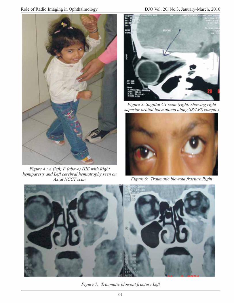
61
DJO Vol. 20, No.3, January-March, 2010Role of Radio Imaging in Ophthalmology
Figure 4 : A (left) B (above) HIE with Right hemiparesis and Left cerebral hemiatrophy seen on
Axial NCCT scan
Figure 7: Traumatic blowout fracture Left
Figure 6: Traumatic blowout fracture Right
Figure 5: Sagittal CT scan (right) showing right superior orbital haematoma along SR/LPS complex

DJO Vol. 20, No.3 , January-March, 2010
62
Role of Radio Imaging in Ophthalmology
Indications of MRIFor almost all neuro-ophthalmic indications, MRI provides superior soft-tissue discrimination (e.g. meninges, cavernous sinus, posterior fossa, dural venous sinuses and optic nerve) Normal anatomy is best demonstrated on T1WI. In distinction, T2WI are typically better for demonstrating intracranial or other pathology.
CT is often used in conjunction with MRICT & MRI are often complimentary & can offer a complete picture of nature of lesion. It is used in conjunction in disorders of bone or sinuses which includes the following -
1. Orbital, maxillofacial, calvarial or skull base fracture,2. Hyperostosis of bone associated with meningiomas,3. Fibrous dysplasia,
5. Craniosynostosis syndromes, 6. Sinusitis or sinus tumours, 7. Clivus or other skull base pathology, 8. Primary bone tumors, bone destruction or erosion
10. Craniopharyngioma, meningioma or retinoblastoma11. Hyperdensity of acute haemorrhage 12. Orbital, subdural, subarachnoid intraventricular or intraparenchymal haemorrhage associated with tumor, stroke or ruptured aneurysm.
MRI is contraindicated in case of severe claustrophobia, marked obesity exceeding table weight limit, uncooperative patient showing excessive mobility, cochlear implant, ferromagnetic aneurysm clip, pacemaker and in cases of metallic foreign body.
Gadolinium contrast material in MRIAs with CT, MR scanning with intravenous contrast material improves detection of pathology by demonstrating areas of blood–brain barrier breakdown. Unlike the iodinated contrast used for CT, the contrast material used for MR is a paramagnetic material called gadolinium. The gadolinium paramagnetic metal ion
intensity. Gadolinium contrast agents are chelated to form a larger, more stable complex around the more toxic, free gadolinium. This stable complex is excreted via the kidneys. Fortunately, unlike iodinated contrast material, serious side-effects from gadolinium are uncommon. The most common allergic reactions
are typically mild (e.g. skin rash, sweating, itching, hives and facial swelling), but there are reports of rare reactions that can be severe or even fatal. Until now MR with gadolinium was considered the ‘procedure
required a contrast study. Unfortunately, a relatively new disorder related to gadolinium contrast has
NSF is a disorder seen weeks to months following gadolinium contrast administration in patients with kidney failure. Clinically, NSF is characterized by thickening and hardening of the skin, typically over the extremities and trunk.
Specific imaging sequences 1. Fat suppression for orbital post-contrast study2. Fluid attenuation inversion recovery (FLAIR) for white matter lesions, 3.Gradient recall echo imaging (GRE) for haemorrhage and blood products4. Diffusion-weighted imaging (DWI) for stroke or PRES5. Perfusion weighted imaging (PWI) 6. Dynamic perfusion CT (PCT)
is used for all post-contrast orbital T1WI – in cases of optic neuritis, optic nerve sheath meningiomas.
FLAIR sequences in cases of suspected demyelinating disease as it may not be routinely performed as part of a standard MR protocol. Axial and Sagittal T1 weighted Gadolinium enhanced MRI brain – asymmetric periventricular plaques suggestive of demyelination in a young female patient with optic neuritis
Figure 8: Fat suppression for orbital post-contrast study

63
DJO Vol. 20, No.3, January-March, 2010Role of Radio Imaging in Ophthalmology
can
show blood products effectively, the approximate age
of a haematoma may be determined, haemorrhage
associated with AVM or cavernomas, intra- or extra-
axial intracranial haemorrhage
is based
on the microscopic random brownian motion of
water. These changes in water molecular diffusion
can be measured as signal intensity in vivo with DWI.
In certain pathological states, diffusion becomes
restricted (i.e. hyper intense or bright on DWI).
DWI can differentiate the various phases of cerebral
infarction namely hyperacute, acute, subacute and
chronic and therefore can detect hyperacute ischemic
stroke even before abnormalities are detected on
conventional T1W, T2W and FLAIR sequences
(Figure 8). It can also be used in cases of acute
ischemic homonymous hemianopia, cortical visual
impairment, acute cerebral emboli in patients with
embolic retinal arterial occlusion and in the basilar
syndrome with acute brainstem ischemia producing a
progressive ophthalmoplegia.
The
volume difference of DWI and PWI gives an
approximate measure of hypoperfused, but not yet
infarcted and potentially salvageable tissue (the
ischemic penumbra). It represents an indication for
thrombolysis based on potential reversibility of DWI
lesions.
MR or CT AngiographyIndications - 1. Hemispheric transient ischemic attacks
2. Homonymous hemianopia
3. Transient monocular visual loss
4. Completed cortical strokes
5. Isolated third nerve palsy
7. AV malformations
MRA over CTA: Advantages are lack of ionizing
radiation exposure, less nephrotoxic contrast material
(gadolinium vs. iodinated contrast), increased signal-
to-noise ratio and easier post-processing techniques.
CTA over MRA: Advantages are increased spatial
resolution, technically easier and faster study to
acquire, less motion artefacts
are probably equivalent
Figure 10: Diffusion Weighted Imaging
Figure 9: Gradient recall echo imaging (GRE)Figure 11 A: Cerebral infarct and B: Diffusion
Perfusion Mismatch

DJO Vol. 20, No.3 , January-March, 2010
64
Role of Radio Imaging in Ophthalmology
MRV preferred over CTV due to iodinated contrast material, ionizing radiation exposure, problems subtracting bone adjacent to the venous sinuses in CTVThus with the advent of newer sequences and techniques in a widely available array of existing imaging modalities, it is important to not to over-investigate the patients. For that an apt knowledge about the utility of each sequence and technique is a must for those dealing especially in neuro-opthalmology to get to the root cause of symptoms and make correct diagnosis and management. This article attempts a brief overview which may be useful in routine clinical practice to decide investigation of choice for a particular disease and to get correct imaging done.
Figure 12: Sagittal and oblique MRV: Dural venous sinus thrombosis in a patient presenting with
papilledema from increased intracranial pressure

65
DJO Vol. 20, No.3, January-March, 2010
Aims of the JournalDelhi Journal of Ophthalmology (DJO) is the quarterly journal published by Delhi Ophthalmological Society. The DJO aims to become an easily readable fully referenced journal which will provide the specialists with up to date data and the residents with articles that give expert opinions that are backed with references. We aim to help the reader by providing in a systematic manner:1. The views of experts on current advances in the
2. Case reports and clinical enigmas 3. Original articles preferably of clinical relevance4. Articles on diagnostic and surgical techniques 5. Selections, annotated by experts, of the most interesting papers from the great wealth of original publication.6. New ideas and innovations, new devices and instruments.7. Book review and letters to Editor. All correspondence shall be acknowledged within six weeks of receipt by the editorial board. Authors shall be intimated about the acceptance of their articles for publication within six weeks of the acknowledgment. The journal expects each contributor to have made a
The authors must take full responsibility to ensure that the manuscript contains no matter that is, to the best of contributors’ knowledge, libelous or unlawful, or infringes upon any Indian copyright laws. All contributors are requested to sign a ‘letter of transmittal’ at the time of submitting their manuscript for consideration for publication.
Submission of the ManuscriptAuthors are requested to read these instructions carefully before submitting manuscripts.All Manuscripts/contribution should be sent by post to Dr. Rohit Saxena, Editor, Delhi Journal for Ophthalmology, Room No. 479, Dr. RP Centre for Ophthalmic Sciences, AIIMS, New Delhi-110029.All manuscript and illustrations must be submitted in triplicate along with a CD. The author(s) should retain a copy for their future reference. To hasten the process of review, Manuscripts may be submitted electronically by email to Dr. Rohit Saxena at [email protected]. These should be in MS-Word format & the image should be in jpeg format (email) and Tiff format (Disc). Graphs and line drawing/diagrams must be sent in graphic format that is EPS,
Instruction to Authors
Instruction to Authors
diagrams in freehand. The disk should be labeled
name, and software used including version. The disk should be sent in proper packaging to avoid damage and corruption of the information during transit. Unreadable disks will be returned to the author for substitution.
Manuscript PreparationThe article should be prepared according to the “Uniform requirements for manuscripts submitted to biomedical journal” published in the New England Journal of Medicine 1997/ 336:309-15.1. Type using font size of 11 or 12 with black ink. 2. Use double spacing, throughout the manuscript including references, tables, and legends. 3. Do not use vertical lines or underlining, anywhere in the text or the table. 4. In the upper right-hand corner, identify each page with a number and a running title. 5. Number pages consecutively in Arabic numerals beginning with title page. 6. Other than the title page, do not identify authors else- where in the manuscript.
necessary, with their initials in parentheses. 8. Numeric equivalents must precede all percentages,
9. For a listing of standard abbreviations consult:
Cam- bridge University Press: 1994). Abbreviations should be used sparingly and must be preceded by the
intraocular pressure (IOP). However, common abbreviations must be used without full forms, for example, mm, mm Hg. Please use right eye and left eye, rather than OD and OS. 10. All hematological and clinical chemistry measurements should be reported in the International Systems of Units (SI). Temperature should be given in degrees Celsius. Length, height, weight and volume should be given in metric units.
Manuscript LayoutThe manuscript (including reference, legend, and tables) must be typed in double spacing on a 21.6x 27.6cm (8.5”x11”) paper with at least 2.5 cm
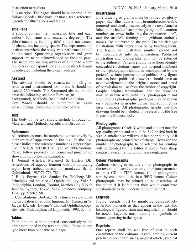
DJO Vol. 20, No.3 , January-March, 2010
66
(1”) margins. The pages should be numbered in the following order, title page, abstract, text, reference, legends for illustrations and tables
Title PageIt should contain the manuscript title and each author’s full name with academic degree(s). The abbreviated title (running title) should not exceed 40 characters, including spaces. The department and institutions where the study was performed should be indicated. Sponsoring organization and grant support are to be acknowledged on the title page. The name and mailing address of author to whom requests or correspondence should be directed must be indicated including the e-mail address
AbstractThe abstract should be structured for Original Articles and unstructured for others. It should not exceed 250 words. The Structured abstract should have the following sections: Purpose (orbackground), Methods, Result and Conclusion.Key Words: should be submitted to assist
TextThe body of the text should include Introduction, Materials and Methods, Results and Discussion
ReferencesAll references must be numbered consecutively by their order of appearance in the text. In the text, please indicate the reference number as superscripts. Use “INDEX MEDICUS” type of abbreviations. Please follow precisely the format and punctuation shown in the following examples. 1. Journal Articles: Melamed S, Epsteir DL:
argon laser trabeculoplasty in monkeys. Br. J. Ophthalmol. 1987:71:776-781.2. Book: Peyman GA, Sanders Dr, Goldberg MF: Principles and practice of Ophthalmology, Vol.: 3, Philadelphia, London, Toronot, Mexico City, Rio de Janiero, Sydney, Tokyo, W.B. Saunders company, 1980, pp 2150-2158.3. Contributions: Brubaker RF: clinical evaluation of the circulation of aquous humour. In: Transman W, Jacger EA, eds. Dauane’s Clinical Ophthalmology. Rev. eds. Philadelphia, JB Lippincott, 1993: 3: 1-11
TablesEach table must be numbered consecutively in the order mentioned in the text and titled. Please do not type more than one table on a page.
IllustrationsLine drawing or graphs must be printed on glossy paper. Each illustration should be numbered in Arabic numerals and cited consecutively in the text. Attach a label on the back of each print giving the illustration number, an arrow indicating the orientation “top”, and the article’s running title (without author’s name). Do not write on the print. Do not damage illustrations with paper clips or by bending them. The legend or illustration number should not be incorporated into the illustration. Published illustration and photographs will not be returned to the author(s). Patients should have there identity concealed (including names and hospital numbers) or their photographs should be accompanied by the
that has been published elsewhere should have an acknowledgment to the original source and proof of permission to use from the holder of copyright. Graphs, original illustrations, and line drawings may be drawn with India ink, photographed and submitted as photographic prints, or may be drawn on a computer in graphic format and submitted as laser printouts. All photographs, graphs and line
Electronic Manuscript) .
PhotographsAll photographs (black & white and colour) must be top quality prints and should be 5x7 or 4x6 inch in size. A smaller size will result in a poor quality. All photographs are printed free of charge. However, the number of photographs to be selected for printing will be decided by the Editorial board. Very sharp contrast is essential for colour representations.
Colour PhotographsAuthors wishing to include colour photographs in the text should send either on colour transparencies or on a CD in TIFF format. Color photographs sent by email should be in a JPEG format. Colour photographs may be printed at the discretion of the editor if it is felt that they would contribute substantially to the understanding of the text.
LegendsFigure legends must be numbered consecutively in Arabic numerals as they appear in the text. For
be noted. Legends must identify all symbols or
ReprintsOne reprint shall be sent free of cost to each contributor of the columns: review articles, current practice e, recent advances, original article, surgical
Instruction to Authors
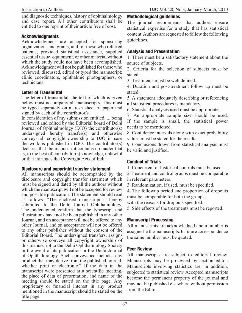
67
DJO Vol. 20, No.3, January-March, 2010
and diagnostic techniques, history of ophthalmology and case report. All other contributors shall be entitled to one reprint of their article free of cost.
AcknowledgmentsAcknowledgment are accepted for sponsoring organizations and grants, and for those who referred patients, provided statistical assistance, supplied essential tissue, equipment, or other material without which the study could not have been accomplished. Acknowledgment will not be published for those who reviewed, discussed, edited or typed the manuscript; clinic coordinators, ophthalmic photographers, or technicians.
Letter of TransmittalThe letter of transmittal, the text of which is given below must accompany all manuscripts. This must be typed separately on a fresh sheet of paper and signed by each of the contributor/s. In consideration of my submission entitled..... being reviewed and edited by the Editorial board of Delhi Journal of Ophthalmology (DJO) the contributor(s) undersigned hereby transfer(s) and otherwise conveys all copyright ownership to DJO in case the work is published in DJO. The contributor(s) declares that the manuscript contains no matter that is, to the best of contributor(s) knowledge, unlawful or that infringes the Copyright Acts of India.
Disclosure and copyright transfer statementAll manuscripts should be accompanied by the disclosure and copyright transfer statement which must be signed and dated by all the authors without which the manuscript will not be accepted for review and possible publication. The statement should read as follows: “The enclosed manuscript is hereby submitted to the Delhi Journal Ophthalmology.
illustrations have not be been published in any other Journal, and on acceptance will not be offered to any other Journal, and on acceptance will not be offered to any other publisher without the consent of the Editorial Board. The undersigned transfers, assigns or otherwise conveys all copyright ownership of this manuscript to the Delhi Ophthalmology Society in the event of its publication in the Delhi Journal of Ophthalmology. Such conveyance includes any product that may derive from the published journal, whether print or electronic.” If the data in the
the place of data of presentation, and name of the meeting should be stated on the title page. Any
mentioned in the manuscript should be stated on the title page.
Methodological guidelinesThe journal recommends that authors ensure
statistical expertise for a study that has statistical
content. Authors are requested to follow the following
guidelines.
Analysis and Presentation 1. There must be a satisfactory statement about the
source of subjects.
2. Criteria for the selection of subjects must be
stated.
4. Duration and post-treatment follow up must be
stated.
5. A statement adequately describing or referencing
all statistical procedures is mandatory.
6. Statistical analyses used must be appropriate.
7. An appropriate sample size should be used.
If the sample is small, the statistical power
needs to be mentioned.
values must be stated for the results.
9. Conclusions drawn from statistical analysis must
Conduct of Trials1. Concurrent or historical controls must be used.
2 Treatment and control groups must be comparable
in relevant parameters.
4. The followup period and proportion of dropouts
must be comparable for both the groups,
5. Side effects of the treatments must be reported.
Manuscript ProcessingAll manuscripts are acknowledged and a number is
assigned to the manuscripts. In future correspondence
the same number must be quoted.
Peer ReviewAll manuscripts are subject to editorial review.
Manuscripts may be processed by section editor.
Manuscripts involving statistics are, in addition,
subjected to statistical review. Accepted manuscripts
become the permanent property of the journal and
may not be published elsewhere without permission
from the Editor.
Instruction to Authors
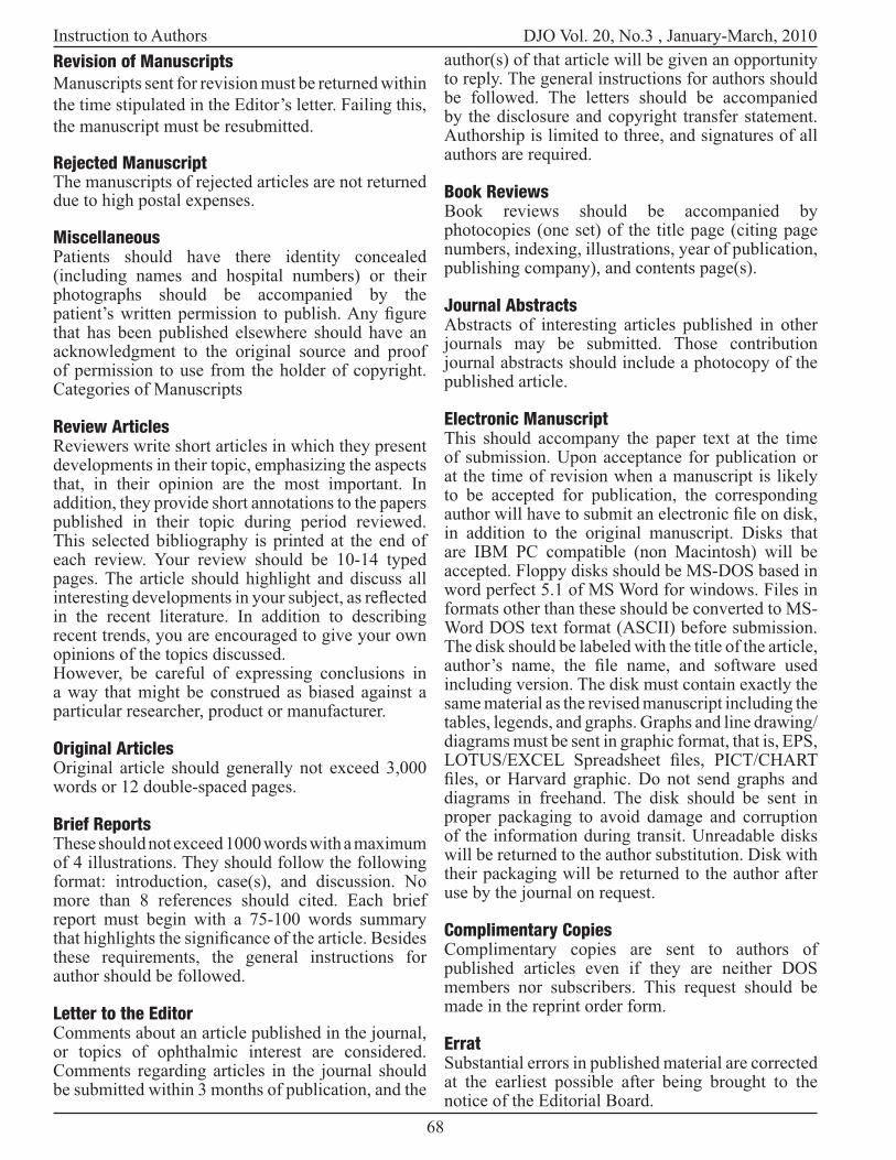
DJO Vol. 20, No.3 , January-March, 2010
68
Revision of ManuscriptsManuscripts sent for revision must be returned within
the time stipulated in the Editor’s letter. Failing this,
the manuscript must be resubmitted.
Rejected ManuscriptThe manuscripts of rejected articles are not returned due to high postal expenses.
MiscellaneousPatients should have there identity concealed (including names and hospital numbers) or their photographs should be accompanied by the
that has been published elsewhere should have an acknowledgment to the original source and proof of permission to use from the holder of copyright. Categories of Manuscripts
Review ArticlesReviewers write short articles in which they present developments in their topic, emphasizing the aspects that, in their opinion are the most important. In addition, they provide short annotations to the papers published in their topic during period reviewed. This selected bibliography is printed at the end of each review. Your review should be 10-14 typed pages. The article should highlight and discuss all
in the recent literature. In addition to describing recent trends, you are encouraged to give your own opinions of the topics discussed.However, be careful of expressing conclusions in a way that might be construed as biased against a particular researcher, product or manufacturer.
Original ArticlesOriginal article should generally not exceed 3,000 words or 12 double-spaced pages.
Brief ReportsThese should not exceed 1000 words with a maximum of 4 illustrations. They should follow the following format: introduction, case(s), and discussion. No more than 8 references should cited. Each brief report must begin with a 75-100 words summary
these requirements, the general instructions for author should be followed.
Letter to the EditorComments about an article published in the journal, or topics of ophthalmic interest are considered. Comments regarding articles in the journal should be submitted within 3 months of publication, and the
author(s) of that article will be given an opportunity to reply. The general instructions for authors should be followed. The letters should be accompanied by the disclosure and copyright transfer statement. Authorship is limited to three, and signatures of all authors are required.
Book ReviewsBook reviews should be accompanied by photocopies (one set) of the title page (citing page numbers, indexing, illustrations, year of publication, publishing company), and contents page(s).
Journal AbstractsAbstracts of interesting articles published in other journals may be submitted. Those contribution journal abstracts should include a photocopy of the published article.
Electronic ManuscriptThis should accompany the paper text at the time of submission. Upon acceptance for publication or at the time of revision when a manuscript is likely to be accepted for publication, the corresponding
in addition to the original manuscript. Disks that are IBM PC compatible (non Macintosh) will be accepted. Floppy disks should be MS-DOS based in word perfect 5.1 of MS Word for windows. Files in formats other than these should be converted to MS-Word DOS text format (ASCII) before submission. The disk should be labeled with the title of the article,
including version. The disk must contain exactly the same material as the revised manuscript including the tables, legends, and graphs. Graphs and line drawing/diagrams must be sent in graphic format, that is, EPS,
diagrams in freehand. The disk should be sent in proper packaging to avoid damage and corruption of the information during transit. Unreadable disks will be returned to the author substitution. Disk with their packaging will be returned to the author after use by the journal on request.
Complimentary CopiesComplimentary copies are sent to authors of published articles even if they are neither DOS members nor subscribers. This request should be made in the reprint order form.
ErratSubstantial errors in published material are corrected at the earliest possible after being brought to the notice of the Editorial Board.
Instruction to Authors