deformation corrected compressed sensing (dc-cs): a … · 1 deformation corrected compressed...
TRANSCRIPT

1
Deformation corrected compressed sensing(DC-CS): a novel framework for accelerated
dynamic MRISajan Goud Lingala, Student Member, IEEE, Edward DiBella, Member, IEEE and Mathews Jacob, Senior
Member, IEEE
Abstract—We propose a novel deformation corrected com-pressed sensing (DC-CS) framework to recover contrast en-hanced dynamic magnetic resonance images from undersampledmeasurements. We introduce a formulation that is capable ofhandling a wide class of sparsity/compactness priors on thedeformation corrected dynamic signal. In this work, we con-sider example compactness priors such as sparsity in temporalFourier domain, sparsity in temporal finite difference domain,and nuclear norm penalty to exploit low rank structure. Usingvariable splitting, we decouple the complex optimization problemto simpler and well understood sub problems; the resultingalgorithm alternates between simple steps of shrinkage baseddenoising, deformable registration, and a quadratic optimizationstep. Additionally, we employ efficient continuation strategies toreduce the risk of convergence to local minima. The decouplingenabled by the proposed scheme enables us to apply this schemeto contrast enhanced MRI applications. Through experimentson numerical phantom and in vivo myocardial perfusion MRIdatasets, we observe superior image quality of the proposed DC-CS scheme in comparison to the classical k-t FOCUSS withmotion estimation/correction scheme, and demonstrate reducedmotion artifacts over classical compressed sensing schemes thatutilize the compact priors on the original deformation un-corrected signal.
I. INTRODUCTION
Dynamic magnetic resonance imaging (DMRI) involvesimaging objects that are evolving in time, and is central toseveral clinical exams including cardiovascular, pulmonary,abdominal, brain, and vocal tract imaging. DMRI often suffersfrom compromises in image quality due to the slow acquisitionnature of MRI. For instance, good spatio-temporal resolution,extended slice coverage, and high signal to noise ratio arerequired for accurate quantification of myocardial perfusionMRI data. However, imaging at Nyquist k-space samplingrate often results in severe compromises in spatio-temporalresolution and slice coverage [1]. Classical approaches to over-come these challenges include parallel imaging [2], [3], andtheir combination with k− t spatio-temporal models [4]–[11].
Copyright (c) 2010 IEEE. Personal use of this material is permitted.However, permission to use this material for any other purposes must beobtained from the IEEE by sending a request to [email protected] Goud Lingala is with the Department of Biomedical Engineering, TheUniversity of Iowa, IA, USA (email: [email protected]). EdwardDiBella is with the Department of Radiology, University of Utah, Utah, USA.Mathews Jacob is with the Department of Electrical and Computer Engineer-ing, The University of Iowa, IA, USA.This work is supported by grants NSF CCF-0844812, NSF CCF-1116067,NIH 1R21HL109710-01A1, and AHA 12 PRE11920052.
Recently, several authors have proposed compressed sensing(CS) schemes that capitalize on the compactness/sparsity ofthe signal representation in appropriate transform domains.For example, sparsity of the temporal Fourier transform [12]and temporal finite differences [13] have been exploited inthe context of myocardial perfusion MRI. More recently,matrix recovery schemes utilizing the linear dependancies ofpixel time profiles using low rank image priors have beenproposed [14]–[16]. While all of these methods demonstratesuccessful recovery when the inter frame motion is minimal,the main challenge is the sensitivity of these methods tolarge inter frame motion. Specifically, the compactness of thesignal representation decreases with inter frame motion, thusrestricting the maximum possible acceleration (see Fig.1 for ademonstration); the reconstructions often suffer from temporalblurring and motion related artifacts at high accelerationfactors.
In this work, we introduce a general framework to minimizethe sensitivity of compressed sensing and low rank matrixrecovery schemes to inter frame motion. We jointly estimatethe dynamic images and inter frame motion, which is modeledas an elastic deformation, from the undersampled data. Ratherthan assuming compactness of the original signal, we assumethe deformation corrected signal to have a compact representa-tion. The proposed approach enables us to use arbitrary signalpriors (e.g. sparsity in specified transform domain, low-rankproperty, patch based low-rank priors) in the reconstruction;the appropriate method could be chosen depending on thespecified application. We introduce an efficient variable split-ting framework with continuation to decouple the problem intothree simpler and well-understood sub-problems. We alternatebetween (a) a shrinkage based denoising step (b) a deformableregistration step, and (c) a quadratic optimization step. Thedeformable registration scheme aims to register each frame inthe dataset to a corresponding frame with similar contrast inthe motion-compensated dataset. Hence, simpler least squaresdifference metrics are sufficient for the registration algorithm,even when the image contrast changes with time, such as indynamic contrast enhanced MRI. The presence of the globalenergy function enables us to design appropriate continuationstrategies to reduce the risk of convergence of the algorithmto local minima. A preliminary version of this work waspublished as a conference proceeding in [17].
In this paper, we demonstrate the utility of the proposedformulation in the context of myocardial perfusion MRI.
arX
iv:1
405.
7718
v2 [
cs.C
V]
2 S
ep 2
014
2
0 20 40 600
1
2
3x 10
!6
time frame number
pix
el i
nte
nsi
ty
OriginalWith motion compensation
0 20 40 60
!12
!10
!8
!6
i
log(!
i), !
i!si
ngula
r va
lues
OriginalWith motion compensation
orig
inal
defo
rmat
ion
corr
ecte
d
few image time frames Image time profile (x-t)
(d) Temporal Fourier (x-f): original (e) example myocardial pixel time profile
(f) Image time profile (x-t): deformation corrected (g) Temporal Fourier (x-f): deformation corrected (h) Singular values
OriginalWith deformation corrected
OriginalWith deformation corrected
Temporal Fourier(x-f)
(f) example myocardial pixeltime profile
(l) Singular values
(a) (b) (c) (d) (e)
(g) (h) (i) (j) (k)
OriginalWith deformation corrected
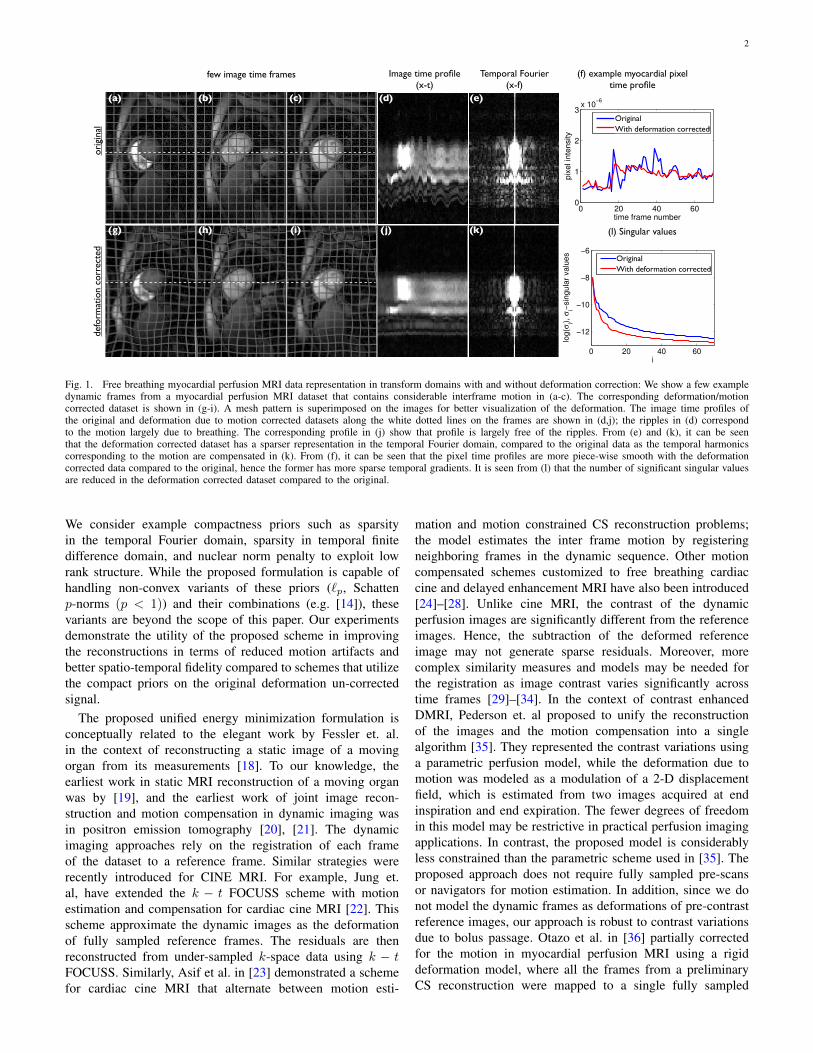
Fig. 1. Free breathing myocardial perfusion MRI data representation in transform domains with and without deformation correction: We show a few exampledynamic frames from a myocardial perfusion MRI dataset that contains considerable interframe motion in (a-c). The corresponding deformation/motioncorrected dataset is shown in (g-i). A mesh pattern is superimposed on the images for better visualization of the deformation. The image time profiles ofthe original and deformation due to motion corrected datasets along the white dotted lines on the frames are shown in (d,j); the ripples in (d) correspondto the motion largely due to breathing. The corresponding profile in (j) show that profile is largely free of the ripples. From (e) and (k), it can be seenthat the deformation corrected dataset has a sparser representation in the temporal Fourier domain, compared to the original data as the temporal harmonicscorresponding to the motion are compensated in (k). From (f), it can be seen that the pixel time profiles are more piece-wise smooth with the deformationcorrected data compared to the original, hence the former has more sparse temporal gradients. It is seen from (l) that the number of significant singular valuesare reduced in the deformation corrected dataset compared to the original.
We consider example compactness priors such as sparsityin the temporal Fourier domain, sparsity in temporal finitedifference domain, and nuclear norm penalty to exploit lowrank structure. While the proposed formulation is capable ofhandling non-convex variants of these priors (`p, Schattenp-norms (p < 1)) and their combinations (e.g. [14]), thesevariants are beyond the scope of this paper. Our experimentsdemonstrate the utility of the proposed scheme in improvingthe reconstructions in terms of reduced motion artifacts andbetter spatio-temporal fidelity compared to schemes that utilizethe compact priors on the original deformation un-correctedsignal.
The proposed unified energy minimization formulation isconceptually related to the elegant work by Fessler et. al.in the context of reconstructing a static image of a movingorgan from its measurements [18]. To our knowledge, theearliest work in static MRI reconstruction of a moving organwas by [19], and the earliest work of joint image recon-struction and motion compensation in dynamic imaging wasin positron emission tomography [20], [21]. The dynamicimaging approaches rely on the registration of each frameof the dataset to a reference frame. Similar strategies wererecently introduced for CINE MRI. For example, Jung et.al, have extended the k − t FOCUSS scheme with motionestimation and compensation for cardiac cine MRI [22]. Thisscheme approximate the dynamic images as the deformationof fully sampled reference frames. The residuals are thenreconstructed from under-sampled k-space data using k − tFOCUSS. Similarly, Asif et al. in [23] demonstrated a schemefor cardiac cine MRI that alternate between motion esti-
mation and motion constrained CS reconstruction problems;the model estimates the inter frame motion by registeringneighboring frames in the dynamic sequence. Other motioncompensated schemes customized to free breathing cardiaccine and delayed enhancement MRI have also been introduced[24]–[28]. Unlike cine MRI, the contrast of the dynamicperfusion images are significantly different from the referenceimages. Hence, the subtraction of the deformed referenceimage may not generate sparse residuals. Moreover, morecomplex similarity measures and models may be needed forthe registration as image contrast varies significantly acrosstime frames [29]–[34]. In the context of contrast enhancedDMRI, Pederson et. al proposed to unify the reconstructionof the images and the motion compensation into a singlealgorithm [35]. They represented the contrast variations usinga parametric perfusion model, while the deformation due tomotion was modeled as a modulation of a 2-D displacementfield, which is estimated from two images acquired at endinspiration and end expiration. The fewer degrees of freedomin this model may be restrictive in practical perfusion imagingapplications. In contrast, the proposed model is considerablyless constrained than the parametric scheme used in [35]. Theproposed approach does not require fully sampled pre-scansor navigators for motion estimation. In addition, since we donot model the dynamic frames as deformations of pre-contrastreference images, our approach is robust to contrast variationsdue to bolus passage. Otazo et al. in [36] partially correctedfor the motion in myocardial perfusion MRI using a rigiddeformation model, where all the frames from a preliminaryCS reconstruction were mapped to a single fully sampled
3
reference image to estimate the motion. The proposed schemecan be viewed as a systematic alternative for patch based low-rank methods that were recently introduced [37], [38]. Thesemethods cluster similar patches from adjacent frames andenforce low-rank penalties on them. The main challenge withthese schemes is the extensive book-keeping to ensure that allpatches are in some cluster. We have also investigated the useof a model-based registration method to handle simultaneousmotion and changes in contrast [39].
The rest of the paper is organized as described. In sectionsII, III, we describe the formulation of the DC-CS problem,the proposed variable splitting framework, and the resultingoptimization algorithm. In sections IV, V, we present ourexperimental evaluation and discussion on the feasibility ofthe DC-CS algorithm to improve reconstruction quality of freebreathing myocardial perfusion MRI data based on retrospec-tive resampling experiments on fully sampled numerical phan-tom and in-vivo datasets. We also demonstrate the feasibilityin improving free breathing reconstructions of radial data froma subject imaged during adenosine stress perfusion.
II. DEFORMATION CORRECTED COMPRESSED SENSING(DC-CS)
A. Dynamic image acquisition
The main objective of this paper is to recover the dy-namic dataset f(x, t) from its undersampled Fourier noisymeasurements b(ki, ti). Here, x is the spatial variable and tdenotes time. The measurement process in dynamic MRI canbe modeled as:
bi =
∫x
f(x, ti) exp(−jkTi x
)dx + ni; i = 0, ., s− 1; (1)
Here, (ki, ti) indicates the ith sampling location in k−t space.We denote the set of sampling locations as Ξ = {(ki, ti), i =0, .., s−1}. The above expression can be rewritten in the vectorform as
b = A(f) + n, (2)
where A is an operator that evaluates the Fourier Transformon the sampling locations specified in Ξ.
B. DC-CS model
The pixel time profiles in myocardial perfusion MRI arehighly structured in the presence of perfect gating and breath-holding. As demonstrated in Fig. 1, penalties such as temporalFourier sparsity (to exploit low temporal bandwidth), temporaltotal variation penalty (to exploit smooth pixel time profiles)or low-rank penalties (to exploit the redundancy between thepixel time profiles) can be used to make the recovery fromunder sampled data well posed. However, the compactness ofthe signal representations will be considerably disturbed inthe presence of inter frame motion, which can arise due tobreathing or inconsistent gating (see Fig. 1); due to which,the performance of the above schemes will be heavily com-promised.
We propose to overcome the above limitation by assumingthe deformation corrected dataset Tθ · f to be compact/sparse.
Note from Fig. 1.(f),(j),(k),(l) that the temporal variations ofTθ · f are considerably more structured than that of f 1. Wesimultaneously recover the deformation parameters θ(x, t) andthe dynamic images f(x, t) from under sampled data b(k, t)using the following minimization scheme:
{f∗, θ∗} = minf,θ‖A(f)− b‖22 + λ Φ (Tθ · f) ;
such that θ ∈ Θ (3)
Here A is the Fourier sampling operator as defined in (2) andTθ is the non-rigid image warping operator; θ(x, t) are thedeformation parameters that describe pixel wise displacementsdue to motion, which are estimated from the under sampleddata. The proposed scheme also yield Tθ(x,t) · f , which isthe deformation corrected version of f , as a by-product. Notethat (3) reduces to the classical CS setting when Tθ = I: theidentity operator. We use bilinear interpolation to discretizethe term Tθ · f .
The regularization term in (3) promotes the compact-ness/sparsity of the deformation corrected dataset Tθ ·f , ratherthan f . Here, Φ(u) denotes an arbitrary prior to exploit theredundancy in the data; λ is the corresponding regularizationparameter. The main advantage of the proposed scheme isthat it can be used with any spatio-temporal priors on thedeformation corrected dataset. The specific priors can bechosen depending on the specific application. For example,we can choose Φ(u) = ‖Ft‖`1 or Φ(u) = ‖∇t‖`1 , whereFt and ∇t are the temporal Fourier transform and temporalgradient operator, respectively. Another alternative is to chooseΦ(u) = ‖U‖∗, the nuclear norm of the Casorati matrix Uassociated with u(x, t) [9], [14]. This approach exploits thelow-rank property of the deformation/motion corrected dataset,resulting from the similarity between the temporal profilesof the pixels [14]–[16]. The ability of the scheme to handlearbitrary image priors makes this approach drastically differentfrom classical motion compensation schemes that register eachframe of f to a specific fully sampled frame.
The deformation field in (3) is assumed to be parametricallyrepresented in terms of the parameters θ. For example, Θ isthe set of B-spline coefficients if a B-spline model is used torepresent the deformation field as in [41], [42]. In this case,the spatial smoothness of the deformation map is controlled bythe grid spacing of the B-spline map. The spatial smoothnessconstraints can also be explicitly imposed using regularizationconstraints on the deformation field as in [43]. Our approach isclosely related to [43] as explained in detail in Section III-C.
III. DC-CS: OPTIMIZATION ALGORITHM
We propose to use a variable splitting approach [44], [45] todecouple the original problem in (3) to simpler sub-problems.We start by splitting the deformation term from the priorby introducing an auxiliary variable g. This enables us toreformulate the problem in (3) to the following constrained
1 In figure 1, registration was performed on the free breathing dataset itself.Starting from the second frame, the deformations were obtained by matchingthe nth frame in the moving sequence to the (n + 1) th frame of the deformedscene by using the demons registration algorithm [40]
4
optimization scheme:
{f, θ}∗ = arg minf,θ,g‖A(f)− b‖22 + λ Φ (g);
such that Tθ · f = g, θ ∈ Θ (4)
We solve (4) using the penalty based method, where the firstconstraint in (4) is relaxed and the corresponding quadraticviolation is penalized as:
{f, θ}∗ = arg minf,θ,g‖A(f)−b‖22+λ
(Φ (g) +
β
2‖Tθ · f − g‖22
);
such that θ ∈ Θ (5)
Here, β is the penalty parameter that enforces the constraint(Tθ ·f = g). When β approaches ∞, the solution of (5) tendsto that of (4), and equivalently (3). However, the convergenceof the algorithm will be slow when β is very high and thealgorithm may also be vulnerable to local minima. Hence, wewill use a continuation strategy where β is initialized withvery low values and is gradually increased, as discussed indetail below.
We use an iterative minimization scheme to minimize (5)with respect to three variables f, θ, g. Specifically, the iterativealgorithm alternates between the minimization of each vari-able, assuming the rest to be known. This approach results inthe following sub problems.
A. g sub-problem (Spatio-temporal denoising/dealiasing tominimize residual motion)
With f and θ fixed, the minimization of (5) with respect tog at each step is a denoising problem; it involves the proximalmapping [46] of the deformation corrected dataset Tθ · f︸ ︷︷ ︸
q
:
g∗ = ming
2
βΦ(g) + ‖ Tθ · f︸ ︷︷ ︸
q
−g‖22; (6)
Note that the above implies q is close enough to g, whilehaving a small cost φ(g). In many cases, we can find the g∗
analytically, as seen in the below section.The amount of regularization is specified by the parameter
β. Specifically, when β is small, the optimal g∗ is a highlysmoothed version of q. At each step, the proximal mappingsmooths out the residual motion induced rapid temporal vari-ations in the deformation corrected dataset (Tθ · f), yieldingthe next iterate of the motion compensated dataset (g∗).The alternation of the subproblems (6),(13) and (14) resultsin the joint estimation of the dynamic images (f) and thedeformation map (θ).
1) Sparsity penalty in the temporal Fourier transform do-main: We denote g = Ftg as the temporal Fourier Transformof g. Using Parseval’s theorem, we rewrite (6) as
g = ming
2
β‖g‖`1 + ‖q − g‖22; (7)
Note that this is a standard `1 proximal mapping [46]. Theoptimal g is obtained by the shrinkage of q [47]:
g =Ft(q)|Ft(q)|
(|Ft(q)| −
2
γβ
)+
, (8)
where + is the shrinkage operator defined as:
x+ =
{x if x ≥ 00 else . (9)
2) Nuclear norm penalty: When Φ(g) = ‖G‖∗, we rewrite(6) as
G∗ = minG
2
β‖G‖∗ + ‖Q−G‖2F , (10)
where G,Q are the Casorati matrices respectively associatedwith g(x, t) and q(x, t). The solution for the above proximalmapping is obtained analytically as [48]
G∗ = U
(S− 2
γβI
)+
V∗, (11)
where USV∗ = Q is the singular value decomposition of Q,and + is the shrinkage operator as defined in (9).
3) Temporal total variation (TV) penalty: When the penaltyis chosen as the temporal total variation criterion, (6) reducesto
g∗ = ming‖q − g‖22 +
2
β‖∇tg‖`1 (12)
This TV denoising problem does not have an analyticalsolution unlike the above cases. Hence, we solve them usingthe fast TV denoising algorithm (FTVd) [49]. Briefly, thisapproach uses a splitting of (12), followed by an alternatingalgorithm to solve for g∗.
B. f sub-problem (reconstruction update)
Assuming θ and g fixed in (5), the minimization withrespect to f reduces to:
minf‖A(f)− b‖22 +
λβ
2‖Tθ · f − g‖22; (13)
We solve this quadratic problem using the conjugate gradient(CG) algorithm. Note that when β approaches infinity, g tendsto Tθ · f , and (4) tends to (3), and the solution to the fsubproblem in (13) tends to the solution of (3), which is theoriginal problem we seek to solve.
C. θ sub-problem (Motion estimation)
Assuming the variables f and g in (5) to be known, wesolve for the motion parameters as:
minθ‖Tθ · f − g‖22, , such that θ ∈ Θ (14)
This is a registration problem, where the dynamic scene f(x, t)is registered frame by frame with a reference scene g(x, t).Since the reference series g is derived from the measurementsitself (obtained from (6)), we do not need to acquire additionalhigh resolution reference frames. In addition, the least squaressimilarity metric in (14) sufficient, even when the contrast ofthe images are changing as a function of time, because eachframe of g(x, t) has the same contrast as f(x, t). The temporalprofiles of the reference dataset g is significantly more smoothcompared to f . This approach enables us to decouple theeffects of smooth perfusion induced contrast changes and themore rapid changes resulting from respiratory motion.

5
The ideal deformable registration algorithm will directlyminimize (14) with a smoothness constraint on the deforma-tion field. In addition, the continuous deformable parametersshould be represented using a differentiable signal model suchas cubic B-spline representation [41], [42]. In this paper, weuse the demons algorithm [50], [51] to approximate (14).This is largely motivated by available open source imple-mentation of the demons algorithm [40]. We observe thatupdating the deformation map using the demons force field isa good approximation to the steepest descent minimization of(14), subject to elasticity regularization (see [52] for details).In addition, the comparison of the demons algorithm andthe direct steepest descent minimization of (14) subject toelasticity regularization is reported to provide qualitativelysimilar results [53]. We consider the demons two-dimensionalregistration algorithm that corrects for in-plane breathingmotion, although in theory a model to correct for out ofplane motion could be considered, when dealing with three-dimensional datasets. The demons algorithm is an iterativescheme, where the displacement field θ(x, ti) is updated asθn+1 = θn + un, where un is the force field evaluated atthe nth iteration. We refer the reader to [50], [51] for theexpression of the force field; the force field is derived fromintensity differences in the reference and target images . Asdiscussed previously, the smoothness of the deformation fieldis implicitly enforced by spatial smoothing the force fieldun+1 using an isotropic Gaussian filter Gσ(x), where σ isthe standard deviation of the Gaussian kernel. Note that σis the spatial smoothness parameter, and allows for spatiallyregularizing the deformation maps.
D. Continuation strategies to alternate between the sub-problems
The simultaneous estimation of f and θ according to (3) is anon-convex optimization problem. The variable splitting strat-egy described above enabled the decomposition of the originalproblem into three simpler subproblems in (6), (14), (13).However, the algorithm is not guaranteed to converge to theglobal minimum of the optimization problem. We now intro-duce continuation strategies to reduce the risk of convergenceto local minima and to ensure fast convergence. This approachis analogous to coarse to fine refinement strategies that aretypically used in many image processing applications.
1) Continuation over the penalty parameter (β): Theoptimization problem specified by (5) has to be solved witha large value of β to ensure that the constraint in (4) issatisfied. However, it is known that the resulting algorithmwill have poor convergence properties when β is set to behigh, even when T = I [54]. Specifically, the shrinkage stepin (6) essentially yields g ≈ Tθ ·f when β →∞. By contrast,if a low value of β is used, g computed using (6) is a heavilydenoised version of Tθ · f .
We adapt the continuation scheme from [49], [54], [55] toobtain faster convergence. The use of continuation strategycan be thought of as a coarse to fine refinement scheme.Specifically, we start with a low value of β, when the the costfunction is considerably more smooth than the original one.
Once the algorithm has converged to the global minimum ofthis cost, we increase β; the algorithm then converges to a localminimum of the new cost function, which is close enough tothe global minimum of the simpler cost function (with smallervalue of β). Thus, this continuation approach will encouragethe convergence of the algorithm to the global minimum,analogous to multi-resolution strategies used in non-convexproblems such as image registration. We emphasize that thereference scene is a result of continuation over β. We do notadapt approaches such as augmented Lagrangian, and split-Bregman that use a fixed value of β, which may lose theproperty of gradual convergence, while solving non-convexproblems.
2) Continuation over a deformation force strength pa-rameter: The motion estimation problem in (14) is itself anon-convex problem. Additionally when iterated along withthe reconstruction (13) and denoising (6) problems, there isa possibility that the estimated motion parameters could getstuck in undesirable local minima. Registration schemes oftenrely on coarse to fine continuation strategies to overcome localminima problems and to improve convergence [28], [51], [56].
The demons implementation [51] also recommends a con-tinuation on a force strength parameter α to speed up theconvergence of the algorithm and to minimize local minimaeffects. The parameter α manipulates the force field such thatit is sensitive to large deformations when α is small, whilelarger values of α makes the force field sensitive to finerdeformations [51]. Based on this, we adapt a continuationstrategy of correcting for bulk motion during the initial itera-tions by using a small value of α. As the iterations proceed,we gradually increase α, and correct for finer motion changes.Specifically, with an initial guess of θ = 0, the registrationsub-problem in (14) is solved in an outer loop starting witha small value of α, and the solution in θ is refined bygradually incrementing α towards high values; After the firstouter loop, the sub-problem in (14) is initialized with themotion estimates obtained from the previous iteration. We haveobserved that this continuation accelerates the convergenceof the DC-CS scheme considerably. Each time the demonsalgorithm is called, we run it for a maximum of 100 iterationsbefore termination.
The following pseudo code summarizes the continuationstrategies that we adapt to solve the cost in (5):
Initialization: f = finit, θ = 0, α > 0, β > 0;for out = 1 to Maxouter iterations
for in = 1 to Maxinner iterations
gn ←(6); spatio-temporal denoising/de-aliasing of the deformed scene;
fn ←(13); CG reconstruction update withdeformation correction;
if (costn − costn−1/costn < 10−3);cost as defined in (3);
break the inner loop;end
endwhile (‖θn − θn−1‖22/‖θn‖22 > 10−2); Check if the
6
deformation parameter update has converged;θn ←(14); Deformation estimation;
endα = α × 3; continuation over the deformation force
strength parameter;β = β × 10; continuation over the penalty parameter;
end
Note that the deformation compensated images Tθ · f isthe denoised version of f only in the first iteration becausethe deformation estimate in the first iteration is zero. Thedeformation field θ1 estimated at the first iteration is usedto derive the deformation compensated image in the seconditeration; it is obtained as the denoised version of Tθ1 · f . Theiterations, along with the continuation of the β parameters,will reduce the risk of the algorithm to converge to undesirablelocal minima. As the iteration number n increases, we haveTθn−1
· fn ≈ gn.
IV. EXPERIMENTAL EVALUATION
To validate the proposed DC-CS scheme, we perform retro-spective resampling experiments based on ground truth datafrom (i) the Physiologically improved non-uniform cardiactorso (PINCAT) numerical phantom, [57], [14], (ii) an invivo fully sampled myocardial perfusion MRI dataset withbreathing motion. We compare the proposed DC-CS schemewith different choices of compactness priors (Φ in (3)) against:(a) CS schemes that use the same priors, and (b) the k-tFOCUSS with ME/MC scheme. We show example myocardialperfusion MRI reconstructions using radial data from twosubjects imaged during shallow breathing at stress.We finallydemonstrate a scenario of accelerating an un-gated myocardialperfusion MRI dataset that contains both cardiac and respira-tory dynamics in addition to the contrast dynamics.
A. Datasets
1) PINCAT data: We numerically simulate the acquisitionof myocardial perfusion MRI data from a single short axisslice of the PINCAT phantom [14], [57]. We set the phantomparameters to obtain realistic cardiac perfusion dynamics andcontrast variations due to bolus passage, while accounting forrespiration with variability in breathing motion. The contrastvariations due to bolus passage are realistically modeled inregions of the right ventricle (RV), left ventricle (LV) andthe left ventricle myocardium. A temporal resolution of oneframe per heart-beat is assumed. The time series data consistsof 35 time frames capturing the first pass passage of bolusthrough the different regions of the heart. The spatial matrixsize is 64 x 64, which corresponds to a spatial resolution of3 x 3 mm2. For retrospective undersampling, we simulatethe acquisitions assuming golden angle pseudo radial k − tsampling pattern; (the angle between successive rays was111.25 degrees). The pseudo radial sampling involved griddingof the radially sampled data to the nearest point on a Cartesiangrid. Subsampling was performed by considering (30 to 8)rays/frame respectively.
2) In vivo fully sampled ECG gated myocardial perfusionMRI data: We consider a single slice from a fully sampledin vivo myocardial perfusion MRI scan. A healthy subjectwas scanned on a Siemens 3T Trio scanner at the Universityof Utah in accordance with the institutes review board. Datawas acquired using a saturation recovery FLASH sequence(TR/TE=2.5/1ms, Saturation recovery time =100ms, 3slices).A Cartesian grid (phase encodes × frequency encodes: 90 ×190, temporal resolution: 1 beat, spatial resolution: 2.5 mm ×2.5 mm × 8 mm) and a Gadolinium bolus of 0.04mmol/kg wasused under rest conditions. The data contains motion primarilydue to breathing and inconsistent gating. We added additionalinteger shifts to amplify motion (see fig 6). Similar to thenumerical phantom, we used the golden angle pseudo radialsampling at different subsampling levels (30 to 12 rays/frame)for retrospective undersampling.
3) Radial myocardial perfusion data sets: Data was ac-quired using a perfusion radial FLASH saturation recoverysequence (TR/TE ≈ 2.6/1.2 ms, 3 slices per beat, flip angleof 14 degrees, 2.3 × 2.3 × 8 mm pixel size, FOV: 280 mm2,bandwidth 1002 Hz/pixel ) on a Siemens 3T Trio scanner [58]using the Siemens cardiac coil array. 72 radial rays equallyspaced over π radians and with 256 samples per ray wereacquired for a given time frame and a given slice. These rayswere acquired in an interleaved manner in subsets of 6 rayseach. The rays in successive frames were rotated by a uniformangle of π/288 radians, which correspond to a period of 4across time. We considered two stress data sets that wereacquired on a free breathing normal subject, and a patient withsuspected ischemia where 0.03 mmol/kg of Gd contrast agentwas injected after 3 minutes of adenosine infusion. A PCAbased coil compression strategy [59] was used to compressthe four coil data set to a single coil principal component dataset. With this data, we performed single coil reconstructioncomparisons using 24 rays that were chosen to approximatelyfollow the golden angle distribution.
4) In-vivo fully sampled ungated myocardial perfusion MRIdata: We consider a single slice from a fully sampled un-gatedmyocardial perfusion MRI scan. We considered running thesame saturation recovery sequence (TR/TE=2.5/1ms, satura-tion recovery time = 100ms, 1 slice) on a different subject butwith no gating (phase encodes × frequency encodes: 108x190,temporal resolution: ≈ 4frames/beat,spatial resolution: 2.5 mm× 2.5 mm × 8 mm). The subject breathed heavily duringthis scan. The data set was subsampled using 30 rays /frameusing the pseudo-golden angle radial sampling patterns. Notethat the data contains pseudo cardiac motion on the top ofbreathing motion. Note from the x-t profile in Fig. 10 (fifthcolumn) and its temporal Fourier transform (sixth column)that the energy is distributed at almost all frequencies, mainlydue to the modulation of pseudo-periodic cardiac motion byrespiratory motion; this limits the utility of CS schemes with x-f sparsity. It is also challenging for the registration algorithm toregister frames in different cardiac phases to obtain smooth x-tprofiles. Specifically, the smoothness constraints on the motionfield have to considerably relaxed, which will result in a lessstable algorithm. We capitalize on the ability of the DC-CSscheme to use Fourier sparsity penalty since pseudo-periodic
7
(a)
end
of fi
rst
oute
r lo
op
(c)
end
of t
hird
ou
ter
loop
(d)
at c
onve
rgen
ce:
end
of t
enth
ou
ter
loop
f(x, t) g(x, t) T✓ · [f(x, t)]
g = T✓ · f
(b)
end
of s
econ
d ou
ter
loop
Image time profiles of
�⇡
106
�⇡
107
�⇡
108
�⇡
1016 0 20 40 60
!30
!25
!20
!15
iteration numberlo
g (
cost
)
(e) Ideal
(a)(b) (c)
(d)
spatio-temporal denoising of
deformed scene
reconstruction deformation estimation
f sub-problem
g sub-problem
sub-problem✓
Tuesday, July 1, 14
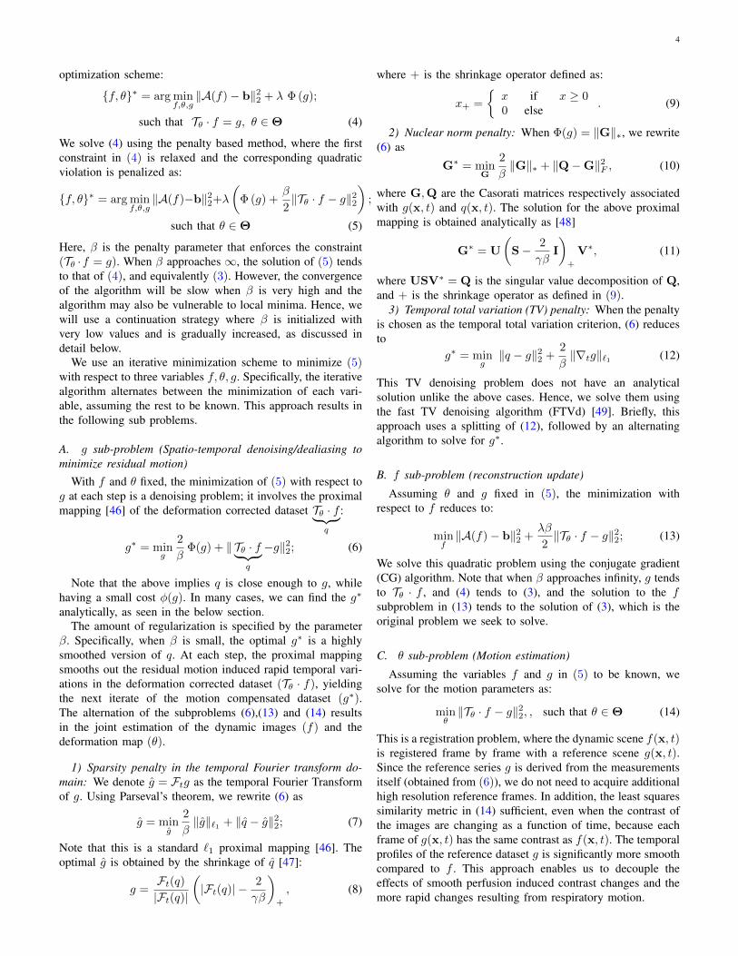
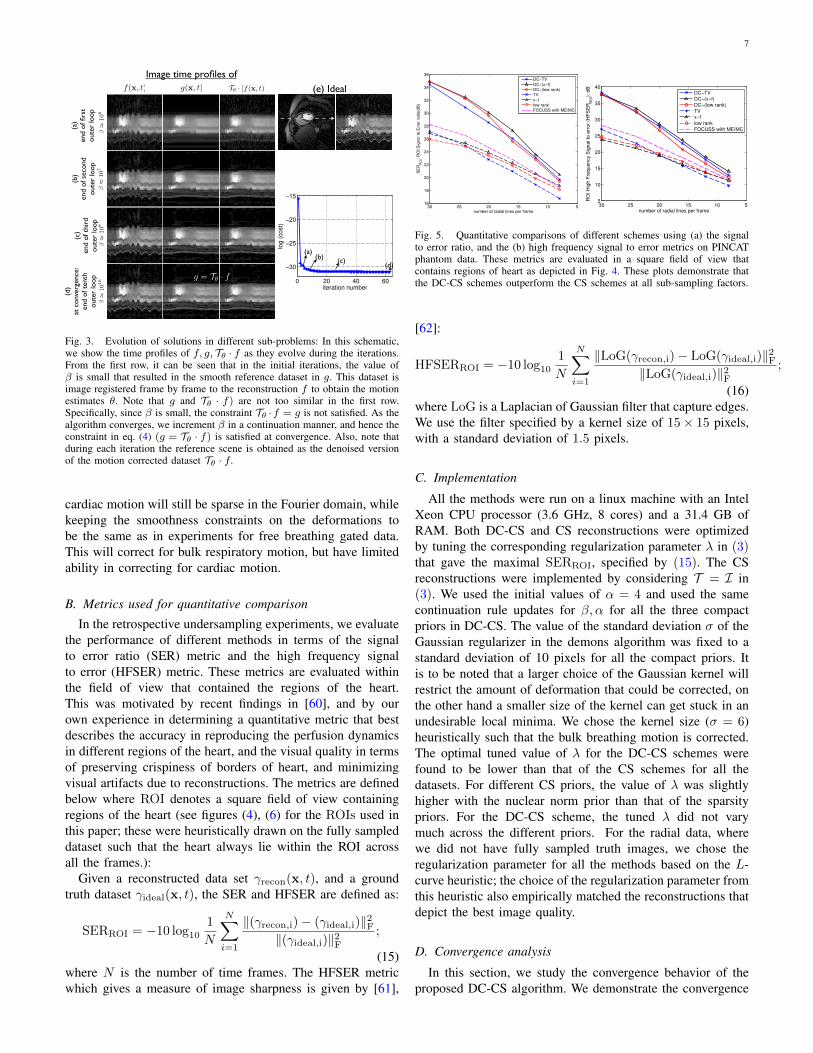
Fig. 3. Evolution of solutions in different sub-problems: In this schematic,we show the time profiles of f, g, Tθ · f as they evolve during the iterations.From the first row, it can be seen that in the initial iterations, the value ofβ is small that resulted in the smooth reference dataset in g. This dataset isimage registered frame by frame to the reconstruction f to obtain the motionestimates θ. Note that g and Tθ · f) are not too similar in the first row.Specifically, since β is small, the constraint Tθ ·f = g is not satisfied. As thealgorithm converges, we increment β in a continuation manner, and hence theconstraint in eq. (4) (g = Tθ · f) is satisfied at convergence. Also, note thatduring each iteration the reference scene is obtained as the denoised versionof the motion corrected dataset Tθ · f.
cardiac motion will still be sparse in the Fourier domain, whilekeeping the smoothness constraints on the deformations tobe the same as in experiments for free breathing gated data.This will correct for bulk respiratory motion, but have limitedability in correcting for cardiac motion.
B. Metrics used for quantitative comparison
In the retrospective undersampling experiments, we evaluatethe performance of different methods in terms of the signalto error ratio (SER) metric and the high frequency signalto error (HFSER) metric. These metrics are evaluated withinthe field of view that contained the regions of the heart.This was motivated by recent findings in [60], and by ourown experience in determining a quantitative metric that bestdescribes the accuracy in reproducing the perfusion dynamicsin different regions of the heart, and the visual quality in termsof preserving crispiness of borders of heart, and minimizingvisual artifacts due to reconstructions. The metrics are definedbelow where ROI denotes a square field of view containingregions of the heart (see figures (4), (6) for the ROIs used inthis paper; these were heuristically drawn on the fully sampleddataset such that the heart always lie within the ROI acrossall the frames.):
Given a reconstructed data set γrecon(x, t), and a groundtruth dataset γideal(x, t), the SER and HFSER are defined as:
SERROI = −10 log10
1
N
N∑i=1
‖(γrecon,i)− (γideal,i)‖2F‖(γideal,i)‖2F
;
(15)where N is the number of time frames. The HFSER metricwhich gives a measure of image sharpness is given by [61],
5101520253016
18
20
22
24
26
28
30
32
34
36
number of radial lines per frame
SE
RR
OI:
RO
I S
ign
al to
Err
or
ratio
(dB
)
DC−TV
DC−(x−f)
DC−(low rank)
TV
x−f
low rank
FOCUSS with ME/MC
510152025305
10
15
20
25
30
35
40
number of radial lines per frame
RO
I H
igh
Fre
qu
en
cy S
ign
al to
err
or
(HF
SE
RR
OI):
dB
DC−TV
DC−(x−f)
DC−(low rank)
TV
x−f
low rank
FOCUSS with ME/MC
Fig. 5. Quantitative comparisons of different schemes using (a) the signalto error ratio, and the (b) high frequency signal to error metrics on PINCATphantom data. These metrics are evaluated in a square field of view thatcontains regions of heart as depicted in Fig. 4. These plots demonstrate thatthe DC-CS schemes outperform the CS schemes at all sub-sampling factors.
[62]:
HFSERROI = −10 log10
1
N
N∑i=1
‖LoG(γrecon,i)− LoG(γideal,i)‖2F‖LoG(γideal,i)‖2F
;
(16)where LoG is a Laplacian of Gaussian filter that capture edges.We use the filter specified by a kernel size of 15× 15 pixels,with a standard deviation of 1.5 pixels.
C. Implementation
All the methods were run on a linux machine with an IntelXeon CPU processor (3.6 GHz, 8 cores) and a 31.4 GB ofRAM. Both DC-CS and CS reconstructions were optimizedby tuning the corresponding regularization parameter λ in (3)that gave the maximal SERROI, specified by (15). The CSreconstructions were implemented by considering T = I in(3). We used the initial values of α = 4 and used the samecontinuation rule updates for β, α for all the three compactpriors in DC-CS. The value of the standard deviation σ of theGaussian regularizer in the demons algorithm was fixed to astandard deviation of 10 pixels for all the compact priors. Itis to be noted that a larger choice of the Gaussian kernel willrestrict the amount of deformation that could be corrected, onthe other hand a smaller size of the kernel can get stuck in anundesirable local minima. We chose the kernel size (σ = 6)heuristically such that the bulk breathing motion is corrected.The optimal tuned value of λ for the DC-CS schemes werefound to be lower than that of the CS schemes for all thedatasets. For different CS priors, the value of λ was slightlyhigher with the nuclear norm prior than that of the sparsitypriors. For the DC-CS scheme, the tuned λ did not varymuch across the different priors. For the radial data, wherewe did not have fully sampled truth images, we chose theregularization parameter for all the methods based on the L-curve heuristic; the choice of the regularization parameter fromthis heuristic also empirically matched the reconstructions thatdepict the best image quality.
D. Convergence analysis
In this section, we study the convergence behavior of theproposed DC-CS algorithm. We demonstrate the convergence

8
0 10 20 3010
!14
10!13
10!12
10!11
CPU Time (minutes)
Cost
Spatial TV
0 5 10 15 20 25 30 3510
!14
10!13
10!12
10!11
CPU Time (minutes)
Cost
Continuation on !, "
Continuation on !, Fixed " # 106
Continuation on !, Fixed " # 1018
Fixed alpha = 4, Continuation on "
0 5 10 15 20 25 30 3510
15
20
25
CPU Time (minutes)
RO
I S
ignal t
o e
rror
ratio
, S
ER
RO
I
Continuation on !, "
Continuation on !, Fixed " # 106
Continuation on !, Fixed " # 1018
Fixed alpha = 4, Continuation on "
(a)
(b)
0 5 10 15 20 25 30 3510
!14
10!13
10!12
10!11
CPU Time (minutes)
Co
st
Continuation on !, ": Direct IFFT Initialization
Continuation on !, ": IDEAL Initialization
Continuation on !, ": Spatial TV Initialization
0 5 10 15 20 25 30 3510
15
20
25
CPU Time (minutes)
RO
I S
ign
al t
o e
rro
r ra
tio,
SE
RR
OI
Continuation on !, ": Direct IFFT Initialization
Continuation on !, ": IDEAL Initialization
Continuation on !, ": Spatial TV Initialization
(f)(d)
(e)
1018
106
1018
106
(c)
(c)
SERROI=18.7 dB
Continuation on ↵,�
(e)
Continuation on ↵,
Fixed � ⇡ 106
SERROI=16.1 dB
Fixed ↵ = 4,
Continuation on �
(f)
SERROI=12.2 dB
Continuation on ↵,
Fixed � ⇡ 1018
(d)
SERROI=12.2 dB
ROI
(i) Different continuation strategies with Direct IFFT initialization (ii) Proposed continuation with different initializations
(a)
(b)
Initi
aliz
atio
n
Cos
t (a
.u.)
CPU time (minutes)
0 10 20 30
Direct IFFT Ideal
At
conv
erge
nce
Spatial TV
Initi
aliz
atio
n
Direct IFFT Ideal
At
conv
erge
nce
SERROI=12.2 dB
SERROI=18.7 dBSERROI=18.7 dB SERROI=18.7 dB
SERROI=16.1 dB
Tuesday, July 1, 14
Fig. 2. Role of continuation: In (i), we study the evolution of the cost in (3) and the SERROI as a function of the CPU run time using different continuationstrategies with the zero filled direct IFFT reconstruction as the initialization. Here, we compare the proposed continuation strategy of updating α, β againstother strategies of not performing continuation on either one of the two parameters α, β. It can be seen that the scenarios without continuation depict poorconvergence and result in undesirable solutions (see i.(d,e,f)). In contrast, it can be seen from i.(a,b,c) that the continuation over both α, β depicts improvedconvergence and obtains a solution in i.(c) that is free of artifacts and blur. Further in (ii), we show the cost in (3) and SERROI versus CPU run time withthe proposed continuation scheme for different initializations. It can be seen that the algorithm converged to approximately the same solution irrespective ofthe choice of the initialization; the robustness to the initialization is attributed to the continuation rules which ensures a gradual update of the complexity ofthe problem.
(b)TV
SER ROI = 22.79 dBHFSER ROI = 18.9 dB
(c)DC-TV
SER ROI = 28.53 dBHFSER ROI = 27.77 dB
(d)low rank
SER ROI = 23.35 dBHFSER ROI = 19.5 dB
(e)DC-low rankSER ROI = 29.62 dB
HFSER ROI = 26.76 dB
(f)x-f
SER ROI = 23.9 dBHFSER ROI = 18.92 dB
(g)DC-(x-f)
SER ROI = 30.03 dBHFSER ROI = 28.47 dB
Estimated motion maps
applied on the reconstruction
Erro
r im
ages
(a)Ideal
Display scale
Reconstruction Error
Sampling for one frame
5101520253016
18
20
22
24
26
28
30
32
34
36
number of radial lines per frame
SE
RR
OI:
RO
I S
ign
al t
o E
rro
r ra
tio(d
B)
DC!TVDC!(x!f)DC!(low rank)TVx!flow rank
ROI
exam
ple
fram
etim
e pr
ofile
(h)FOCUSS-ME/MC
SER ROI = 24.68 dBHFSER ROI = 21.48 dB
Wednesday, July 2, 14
Fig. 4. Qualitative comparison on the PINCAT phantom data using 20 rays/frame: We show for each of the schemes a spatial frame, its image time profile,and the corresponding difference images and difference time profiles. The difference images are scaled up by a factor of 10 for better visualization. The timeprofile in the second row corresponds to the profile along the dotted line in the first image frame of the top row. The location of the image frame is depictedby the arrow in the time profile of (a). As depicted from these figures, DC-CS provides superior reconstructions compared to CS with regards to reducedmotion artifacts. The motion compensated time profiles shown in the bottom row depicts that the proposed algorithm was capable to estimate and correctmost of the inter-frame motion.

9
properties by considering the in vivo myocardial perfusionMRI dataset. We consider the recovery of this data fromFourier golden angle undersampled radial data using 16rays/frame; which approximately correspond to an accelerationfactor of 5 . We employ Φ to be the temporal finite difference(temporal TV) operator. We now demonstrate the role of thecontinuation, and discuss the algorithm’s dependence on theinitial guess of the reconstruction.
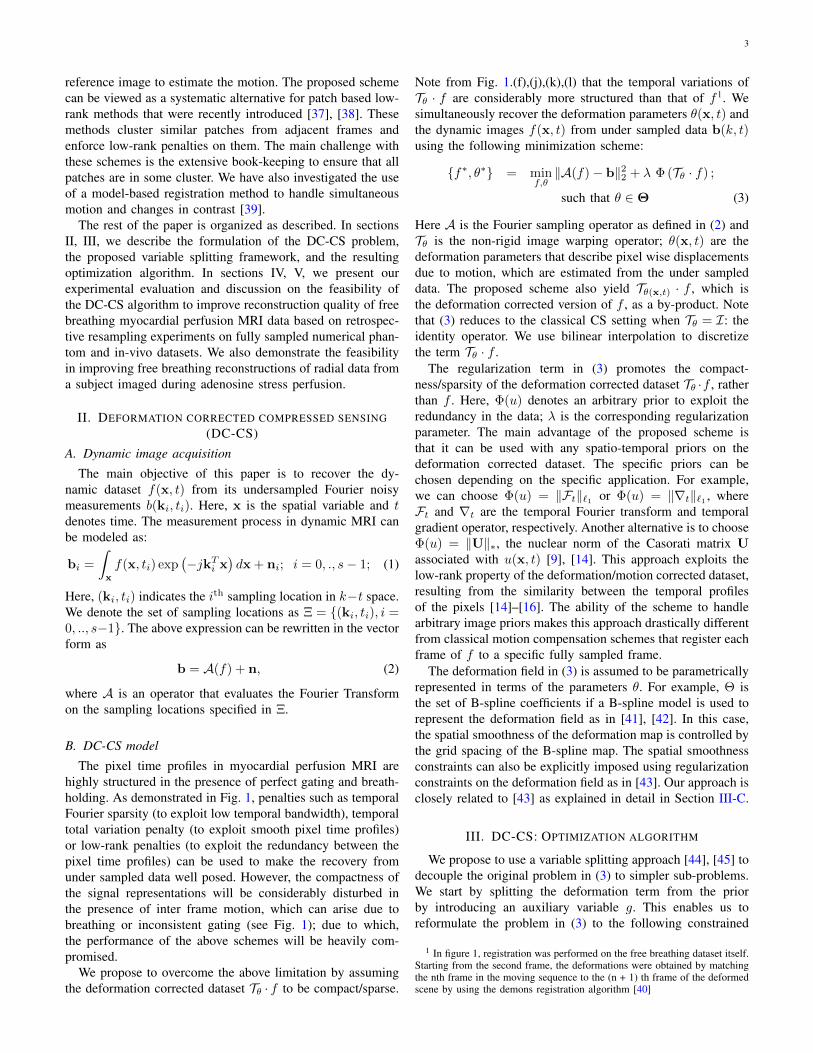
1) Role of continuation: As detailed in the pseudo codeabove, we define continuation over the deformation forcestrength parameter (α) and the penalty parameter (β) as astrategy of starting with small values of α, β and incrementingthem by small factors in an outer loop. In order to evaluatethe role of continuation, we study the below scenarios inFig. 2(i) with an initial guess determined by the zero filleddirect Inverse Fourier Transform (DIFFT) reconstruction; here,we study the decrement of the cost function in (3) and theevolution of the SERROI (15):
• Proposed strategy of continuation over both α, β startingwith α = 4, β ≈ 106;
• Continuation over α starting with α = 4 with a fixed lowvalue of β ≈ 106;
• Continuation over α starting with α = 4 with a fixed highvalue of β ≈ 1018;
• Fixed low value of α = 4 with a continuation over β;
A fixed high value of α was not considered, as this means thatthe strength of deformation forces are almost close to zero,resulting in minimal to no motion correction. Note the initialvalues of β above are derived as β0 = 1
max(Φ(Tθinit ·finit)), and
an empirical choice of α0 = 4. From Fig. 2 (i), we observethat the scenarios without the use of continuation had poorconvergence properties. Specifically, the scenario of using ahigh value of β showed slow convergence irrespective of thecontinuation over α (due to many CG steps while solving the fsub-problem), while a low value of β showed fast convergencebut resulted in an in-accurate solution. We also observedslow convergence without continuation over α. In contrast,we observe that the proposed strategy of continuation overβ, α to show both improved convergence speed and achievethe desired solution. We refer the reader to Fig. 3 to get asense of the outputs of f, g, Tθ · f during the iterations of theDC-CS scheme with continuation.
2) Choice of initial guess: : In Fig. 2 (ii), we study thebehavior of the algorithm with the proposed continuationscheme to different initial guesses of f . We consider differentinitial guesses obtained from the fully-sampled ground truthdata, zero filled direct IFFT reconstructed data, and a spatiallyregularized total variation (TV) reconstructed data. From Fig.2 (ii), we observed that that the algorithm was robust to thechoice of the initialization. The continuation ensures a gradualprogression in the complexity of the problem, thereby avoidingthe chance of getting stuck in undesirable local minima. Basedon these observations, we utilized the choice of the spatialtotal variation (TV) reconstruction as the initial guess to theproposed DC-CS scheme for the rest of the experiments in thepaper.
E. Comparisons on the PINCAT numerical phantom
In Fig. 4, we show qualitative comparisons of the pro-posed DC-CS scheme with different choices of potentialfunctions/compactness priors against CS schemes that usethe same priors. The qualitative comparisons are shown byconsidering undersampling using 20 rays/frame. We observethat all the three priors benefit from deformation/motioncorrection. Specifically, classical CS methods result in tem-poral stair casing (with temporal TV), motion blurring (withtemporal Fourier), loss in spatio-temporal fidelity and blurringof myocardial borders (with the nuclear norm based low rankprior). In contrast, the proposed DC-CS methods are found tobe more robust to these artifacts. We observe similar trendsover a range of subsampling factors as depicted in the SERROI
and HFSERROI plots in Fig. 5.
F. Comparisons on the fully sampled invivo myocardial per-fusion MRI dataset
In Fig. 6, we show the comparisons involving retrospectivesampling on the fully sampled in vivo Cartesian data. Thecomparisons are shown using 16 rays per frame. Similar tothe PINCAT phantom observations, we notice superior spatio-temporal fidelity and less motion artifacts with the proposedDC-CS scheme compared to its CS variants. The SERROI andHFENROI plots in Fig. 7 also depict the same trend over arange of subsampling factors.
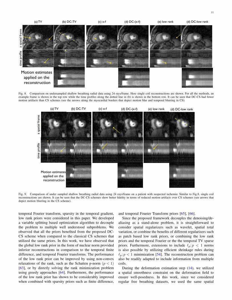
G. Comparisons on radial shallow breathing stress data
In Figs. 8, and 9, we show the comparisons using undersam-pled radial stress shallow breathing data from two subjects.We observe DC-CS to give better reconstructions in termsof minimizing motion blur and artifacts compared to CS.These preliminary results are demonstrated using a single coilwith 24 rays, however the performance could be improvedby extending to multiple coils and including spatial priors.Without the motion compensated, the low rank prior wasgenerally robust to motion artifacts but sensitive to temporalblurring during the peak contrast frames while the CS priorsbased on temporal TV and temporal FFT were sensitive tomotion artifacts. With the DC-CS scheme, we observe thereconstructions with temporal TV and temporal Fourier tobe slightly superior to that of the low rank prior in termsof image sharpness and reduced blurring. We however realizethat the performance of the low rank priors can be improved byconsidering non-convex Schatten p-norm priors and/or patchbased low rank priors, as shown in [14], [16].
H. Comparisons on the ungated free breathing myocardialperfusion dataset
We observe that the proposed algorithm provides less spatialand temporal blurring than the CS with x-f scheme withoutmotion compensation, which can also be appreciated from thex-t profiles and x-f profiles. Note from the x-f profile of thedeformation corrected dataset that the breathing motion cor-rected dataset is more sparse in the temporal Fourier domain.

10
Estimated motion maps
applied on the reconstruction
Display scale
Reconstruction Error
Sampling for one frame
(f)DC-(x-f)
SERROI=17.8 dBHFSERROI= 11.5 dB
(e)x-f
SERROI=16.1 dBHFSERROI=9.0 dB
(d)DC-TV
SERROI=18.7 dBHFSERROI=12.9 dB
(c)TV
SERROI=16.8 dBHFSERROI=9.9 dB
(h)DC-low rank
SERROI=17.4 dBHFSERROI=10.8 dB
(g)low rank
SERROI=16.4 dBHFSERROI= 9.2 dB
(a)Ideal
0
0.2
0.1
0
1
0.5
ex
am
ple
im
age
fra
me
sti
me
pro
file
err
or
image
se
rro
r p
rofi
le
ROI
(b)FOCUSS: ME/MC
SERROI= 17.0 dBHFSERROI=10.25 dB
Saturday, June 28, 14
Fig. 6. Performance evaluation using retrospective sampling on Cartesian data using 16 radial rays/frame: We show two example frames, the image timeprofile along the dotted line in (a), and the corresponding difference images for all the methods. The difference images are scaled up by a factor of five forbetter visualization. The image time frame locations in the first two rows are depicted by the dotted lines in the time profile of (a). The compressed sensing(CS) reconstructions exhibit considerable motion artifacts and temporal blurring (see arrows), while the proposed deformation corrected CS images (DC-CS)are robust to these compromises. The last row depicts that the proposed algorithm was capable of estimating and correcting most of the deformation due tointer frame motion.
Specifically, the energy at the cardiac harmonics are increased,while the energy at the other frequencies are decreased. Wealso observe that the motion compensation attenuates theharmonics at the respiratory motion frequency, except for theout of plane motion that our motion compensation algorithmcannot compensate. The superiority of image quality in DC-CS over CS can also be seen in the SERROI and HFSERROI
numbers.
V. DISCUSSION
In this paper, we proposed a generalized deformation cor-rected compressed sensing framework for dynamic MRI. Theproposed formulation is general enough to handle a wide classof compactness priors. Example priors based on sparsity in the
1214161820222426283015
16
17
18
19
20
21
number of radial lines per frame
SE
RR
OI:
RO
I S
ign
al to
Err
or
ratio
(dB
)
DC−TV
DC−(x−f)
DC−(low rank)
TV
x−f
low rank
FOCUS: ME/MC
121416182022242628306
8
10
12
14
16
18
number of radial lines per frame
RO
I H
igh
Fre
qu
en
cy S
ign
al to
err
or
(HF
SE
RR
OI):
dB
DC−TV
DC−(x−f)
DC−(low rank)
TV
x−f
low rank
FOCUSS with ME/MC
Fig. 7. Quantitative comparisons of different schemes using the signal toerror ratio (a), and high frequency Signal to error metrics (b) on the in vivomyocardial perfusion data. The metrics are evaluated in the regions of interestcontaining the heart as depicted in Fig. 6. These plots demonstrate that theDC-CS schemes outperform the CS schemes at all sub-sampling factors.
11(a) temporal FFT (b) temporal TV (c) low rank
a sp
atia
l fra
me
time
profi
le(a) TV (b) DC-TV (c) x-f (d) DC-(x-f) (e) low rank (d) DC-low rank
Motion estimates applied on the reconstruction
Wednesday, July 2, 14
Fig. 8. Comparison on undersampled shallow breathing radial data using 24 rays/frame. Here single coil reconstructions are shown. For all the methods, anexample frame is shown in the top row while the time profiles along the dotted line in (b) is shown in the bottom row. It can be seen that DC-CS had fewermotion artifacts than CS schemes (see the arrows along the myocardial borders that depict motion blur and temporal blurring in CS).
(a) temporal FFT (b) temporal TV (c) low rank
a sp
atia
l fra
me
time
profi
le
(a) TV (b) DC-TV (c) x-f (d) DC-(x-f) (e) low rank (d) DC-low rank
Motion estimates applied on the reconstruction
Wednesday, July 2, 14
Fig. 9. Comparison of under sampled shallow breathing radial data using 24 rays/frame on a patient with suspected ischemia: Similar to Fig.8, single coilreconstructions are shown. It can be seen that the DC-CS schemes show better fidelity in terms of reduced motion artifacts over CS schemes (see arrows thatdepict motion blurring in the CS scheme).
temporal Fourier transform, sparsity in the temporal gradient,low rank priors were considered in this paper. We developeda variable splitting based optimization algorithm to decouplethe problem to multiple well understood subproblems. Weobserved that all the priors benefited from the proposed DC-CS scheme when compared to the classical CS schemes thatutilized the same priors. In this work, we have observed thatthe global low rank prior in the form of nuclear norm providedinferior reconstructions in comparison to the temporal finitedifference, and temporal Fourier transforms. The performanceof the low rank prior can be improved by using non-convexrelaxations of the rank, such as the Schatten p-norm (p < 1)[63], or by directly solving the rank minimization problemusing greedy approaches [64]. Furthermore, the performanceof the low rank prior has shown to be considerably improvedwhen combined with sparsity priors such as finite difference,
and temporal Fourier Transform priors [65], [66].Since the proposed framework decouples the denoising/de-
aliasing as a stand-alone problem, it is straightforward toconsider spatial regularizers such as wavelet, spatial totalvariation, or combine the benefits of different regularizers suchas patch based low rank priors, or combining the low rankpriors and the temporal Fourier or the the temporal TV sparsepriors. Furthermore, extensions to include `p; p < 1 normsis also possible by utilizing efficient shrinkage rules during`p; p < 1 minimization [54]. The reconstruction problem canalso be readily adapted to include information from multiplecoils.
During the deformation estimation step (14), we utilizeda spatial smoothness constraint on the deformation field toensure well-posedness. In this work, since we consideredregular free breathing datasets, we used the same spatial

12
breathing harmonics
(a)
Ide
al(b
)x
-f(c
)D
C-C
S (
x-f
)
few spatial frames x-t profile x-f profile
Deformation corrected profiles
x-t profile x-f profile
breathing
cardiac harmonicscardiac motion
more prominent cardiac harmonics
breathing
un-corrected out of plane breathing
SER ROI = 19.67 dBHFSER ROI = 12.12 dB
SER ROI = 22.01 dBHFSER ROI = 15.89 dB
Thursday, July 3, 14
Fig. 10. Performance evaluation using retrospective undersampling of ungated perfusion MRI dataset: The ungated dataset was acquired without ECG gatingand breath-holding as described in Section IV.A; A few frames of the original data, data recovered using CS with x-f sparsity penalty, and the DC-CS schemeare shown in the first four columns. The ungated acquisition enables us to acquire diastolic and systolic frames. However, the acceleration of this dataset ischallenging due to the rapid cardiac motion (see high frequency ripples in x-t profile) and respiratory motion (see low frequency oscillations in x-t profile).The x-f space representation of the ground truth data depicts the modulation of the cardiac and contrast dynamics by the breathing motion at almost allthe frequencies. The proposed DC-CS scheme corrects for the breathing motion, and exploits the sparsity of quasi-periodic dataset with cardiac and contrastdynamics (see the x-f profile of the motion corrected dataset in the last row). The residual ripples in the x-t profile of the deformation corrected datasetcorrespond to out of plane breathing motion. From the x-f, and x-t plots, it can be seen that DC-CS provides superior reconstructions over CS reconstructions(also see arrows in (b)).
smoothness parameter of σ = 10 while registering all theframes. The algorithm in theory can capture sudden jumpsin motion such as patient gasps by controlling the spatialsmoothness parameter at the corresponding frames. Further-more, in regular breathing patterns, the framework can beimproved by constraining the deformation field to be spatially,and temporally smooth.
The algorithm was observed to be robust to the choice ofinitial guess. This is attributed to the continuation strategieswe adapt to update our reconstructions and deformations, andthe undersampling behavior of the radial pattern. We observedthat the spatial TV initialization provided a slight advantage interms of convergence speed over direct IFFT and ground truthinitializations, which motivated our choice of using spatial TVas an initialization in all our experiments . In this work, wedid not evaluate the proposed method with different choices ofsampling patterns. For example, a 1D Cartesian pattern couldresult in overlap artifacts, which may not serve as a goodinitialization. A natural way to get the algorithm working withsuch sampling pattern is to initialize it with a basic regularizedreconstruction that is free of artifacts such as the spatial TVreconstruction used in this work.
In this work, we do not consider to account for out of planemotion due to simplicity. However, during free breathing, outof plane motion is inevitable. Most of the current acquisitionschemes in myocardial perfusion MRI are 2-D in nature; it isdifficult to compensate for out of plane motion in these cases.Conceptually, the proposed scheme can be readily extendedto 3-D, which when used with 3-D acquisition schemes
can provide improved reconstructions. In addition, we alsoobserve that the DC-CS algorithm does not require perfectcompensation of motion. Any residual motion will only resultin a more complex image, which will be captured by theappropriate sparse/low-rank prior.
The run time of the entire algorithm was about 25 minsfor datasets of size 190 × 90 × 70. However, the currentimplementation was not optimized for speed. The main bottleneck of the reconstruction time was the deformation algo-rithm, which was implemented in MATLAB. For instance,the computational time spent for registering a single frameis of the order of 5-6 seconds for a 190x90 matrix. Perfusiondatasets typically contain about 50-60 time frames. The imageregistration algorithm was called for three times during theiterations. Overall, the computational time for the registrationalgorithm was about 16 minutes for the 190x90x70 matrixdepicted in figure 2. This clearly formed the crux of thealgorithm. The algorithm could be further optimized by otherstate of the art optimized for speed deformable registrationalgorithms, and also by the usage of graphical processing units(GPUs). In this work, we employed the demons algorithmto approximately solve (14). This approximation is a slightlimitation of our current DC-CS implementation. We willinvestigate the use of registration algorithms that directlyminimize (14) and study the impact of the assumptions of theregistration algorithm on the DC-CS framework in our futurework. In our work, we did not constrain the deformationto be invertible. Future work also include posing invertibilityconstraints on the deformation; this can benefit the stability of

13
the registration process as shown by [67].The current radial invivo results reported in this work were
evaluated only from two subjects. Future work of consideringdatasets from multiple patients is required to fully evaluate theclinical utility of the proposed method.
VI. CONCLUSION
We introduced a novel deformation corrected compressedsensing algorithm for accelerated dynamic MRI. The pro-posed framework has a generalized formulation capable ofhandling a wide class of compactness/sparsity priors on thedeformation corrected dynamic signal. We developed an ef-ficient variable splitting based optimization framework todecouple the complex joint reconstruction and deformationestimation problem to simpler problems of shrinkage baseddenoising, deformable registration, and quadratic optimiza-tion. The efficient decoupling of the subproblems makes theproposed scheme applicable to a wide range of dynamicMRI applications including dynamic contrast enhanced MRIapplications. Efficient continuation strategies were devised tocycle between the subproblems. We demonstrated that theproposed algorithm with continuation was robust to choice ofinitialization. Our experiments on a numerical phantom and,in vivo myocardial perfusion MRI datasets demonstrated thatthe proposed scheme was able to reduce motion artifacts andtemporal blurring that were present in compressed sensingreconstructions.
REFERENCES
[1] P. Kellman and A. E. Arai, “Imaging sequences for first pass perfusion-areview,” Journal of Cardiovascular Magnetic Resonance, vol. 9, no. 3,pp. 525–537, 2007.
[2] M. A. Griswold, P. M. Jakob, R. M. Heidemann, M. Nittka, V. Jellus,J. Wang, B. Kiefer, and A. Haase, “Generalized autocalibrating partiallyparallel acquisitions (GRAPPA),” Magnetic Resonance in Medicine,vol. 47, no. 6, pp. 1202–1210, 2002.
[3] S. Plein, A. Radjenovic, J. P. Ridgway, D. Barmby, J. P. Green-wood, S. G. Ball, and M. U. Sivananthan, “Coronary Artery Disease:Myocardial Perfusion MR Imaging with Sensitivity Encoding versusConventional Angiography,” Radiology, vol. 235, no. 2, pp. 423–430,2005.
[4] J. Tsao, P. Boesiger, and K. Pruessmann, “k-t BLAST and k-t SENSE:Dynamic MRI with high frame rate exploiting spatiotemporal correla-tions,” Magnetic Resonance in Medicine, vol. 50, no. 5, pp. 1031–1042,2003.
[5] S. Plein, S. Ryf, J. Schwitter, A. Radjenovic, P. Boesiger, and S. Kozerke,“Dynamic contrast-enhanced myocardial perfusion MRI accelerated withk-t sense,” Magnetic Resonance in Medicine, vol. 58, no. 4, pp. 777–785,2007.
[6] B. Sharif, J. A. Derbyshire, A. Z. Faranesh, and Y. Bresler, “Patient-adaptive reconstruction and acquisition in dynamic imaging with sen-sitivity encoding (PARADISE),” Magnetic Resonance in Medicine,vol. 64, no. 2, pp. 501–513, 2010.
[7] E. Di Bella, Y. Wu, A. Alexander, D. Parker, D. Green, and C. McGann,“Comparison of temporal filtering methods for dynamic contrast MRImyocardial perfusion studies,” Magnetic resonance in medicine, vol. 49,no. 5, pp. 895–902, 2003.
[8] A. S. Gupta and Z. Liang, “Dynamic imaging by temporal modelingwith principal component analysis,” in Proc. 9th Ann. Meeting Intl. Soc.Magn. Reson. Med, 2001, p. 10.
[9] Z.-P. Liang, “Spatiotemporal imaging with partially separable functions,”in 4th IEEE International Symposium on Biomedical Imaging: FromNano to Macro, 2007. ISBI 2007. IEEE, 2007, pp. 988–991.
[10] H. Pedersen, S. Kozerke, S. Ringgaard, K. Nehrke, and W. Kim, “k-tPCA: Temporally constrained k-t BLAST reconstruction using principalcomponent analysis,” Magnetic Resonance in Medicine, vol. 62, no. 3,pp. 706–716, 2009.
[11] J. Tsao and S. Kozerke, “MRI temporal acceleration techniques,” Journalof Magnetic Resonance Imaging, vol. 36, no. 3, pp. 543–560, 2012.
[12] R. Otazo, D. Kim, L. Axel, and D. Sodickson, “Combination ofcompressed sensing and parallel imaging for highly accelerated first-pass cardiac perfusion MRI,” Magnetic Resonance in Medicine, vol. 64,no. 3, pp. 767–776, 2010.
[13] G. Adluru, S. Awate, T. Tasdizen, R. Whitaker, and E. DiBella, “Tem-porally constrained reconstruction of dynamic cardiac perfusion MRI,”Magnetic Resonance in Medicine, vol. 57, no. 6, pp. 1027–1036, 2007.
[14] S. G. Lingala, Y. Hu, E. DiBella, and M. Jacob, “Accelerated dynamicMRI exploiting sparsity and low-rank structure: kt SLR,” MedicalImaging, IEEE Transactions on, vol. 30, no. 5, pp. 1042–1054, 2011.
[15] J. P. Haldar and Z.-P. Liang, “Spatiotemporal imaging with partially sep-arable functions: a matrix recovery approach,” in Biomedical Imaging:From Nano to Macro, 2010 IEEE International Symposium on. IEEE,2010, pp. 716–719.
[16] J. Trzasko, A. Manduca, and E. Borisch, “Local versus global low-rankpromotion in dynamic MRI series reconstruction,” in Proc. Int. Symp.Magn. Reson. Med, 2011, p. 4371.
[17] S. G. Lingala, M. Nadar, C. Chefd’Hotel, L. Zhang, and M. Jacob,“Unified reconstruction and motion estimation in cardiac perfusionMRI,” in IEEE International Symposium on Biomedical Imaging: FromNano to Macro, 2011. IEEE, 2011, pp. 65–68.
[18] J. Fessler, “Optimization transfer approach to joint registra-tion/reconstruction for motion-compensated image reconstruction,”in IEEE International Symposium on Biomedical Imaging: From Nanoto Macro, 2010. IEEE, 2010, pp. 596–599.
[19] F. Odille, P.-A. Vuissoz, P.-Y. Marie, and J. Felblinger, “GeneralizedReconstruction by Inversion of Coupled Systems (GRICS) applied tofree-breathing MRI,” Magnetic Resonance in Medicine, vol. 60, no. 1,pp. 146–157, 2008.
[20] M. Jacobson and J. A. Fessler, “Joint estimation of image and de-formation parameters in motion-corrected PET,” in Nuclear ScienceSymposium Conference Record, 2003 IEEE, vol. 5. IEEE, 2003, pp.3290–3294.
[21] M. Reyes, G. Malandain, P. M. Koulibaly, M. Gonzalez-Ballester, andJ. Darcourt, “Model-based respiratory motion compensation for emissiontomography image reconstruction,” Physics in medicine and biology,vol. 52, no. 12, p. 3579, 2007.
[22] H. Jung and J. C. Ye, “Motion estimated and compensated compressedsensing dynamic magnetic resonance imaging: What we can learnfrom video compression techniques,” International Journal of ImagingSystems and Technology, vol. 20, no. 2, pp. 81–98, 2010.
[23] M. S. Asif, L. Hamilton, M. Brummer, and J. Romberg, “Motion-adaptive spatio-temporal regularization for accelerated dynamic MRI,”Magnetic Resonance in Medicine, 2012.
[24] P. Kellman, C. Chefd’hotel, C. H. Lorenz, C. Mancini, A. E. Arai, andE. R. McVeigh, “High spatial and temporal resolution cardiac cine MRIfrom retrospective reconstruction of data acquired in real time usingmotion correction and resorting,” Magnetic Resonance in Medicine,vol. 62, no. 6, pp. 1557–1564, 2009.
[25] ——, “Fully automatic, retrospective enhancement of real-time acquiredcardiac cine MR images using image-based navigators and respiratorymotion-corrected averaging,” Magnetic Resonance in Medicine, vol. 59,no. 4, pp. 771–778, 2008.
[26] M. Usman, D. Atkinson, F. Odille, C. Kolbitsch, G. Vaillant, T. Scha-effter, P. G. Batchelor, and C. Prieto, “Motion corrected compressedsensing for free-breathing dynamic cardiac MRI,” Magnetic Resonancein Medicine, 2012.
[27] M. S. Hansen, T. S. Sørensen, A. E. Arai, and P. Kellman, “Retrospectivereconstruction of high temporal resolution cine images from real-time MRI using iterative motion correction,” Magnetic Resonance inMedicine, vol. 68, no. 3, pp. 741–750, 2012.
[28] M. J. Ledesma-Carbayo, P. Kellman, A. E. Arai, and E. R. McVeigh,“Motion corrected free-breathing delayed-enhancement imaging of my-ocardial infarction using nonrigid registration,” Journal of MagneticResonance Imaging, vol. 26, no. 1, pp. 184–190, 2007.
[29] G. Wollny, M. J. Ledesma-Carbayo, P. Kellman, and A. Santos, “Anew similarity measure for non-rigid breathing motion compensation ofmyocardial perfusion MRI,” in Engineering in Medicine and BiologySociety, 2008. EMBS 2008. 30th Annual International Conference ofthe IEEE. IEEE, 2008, pp. 3389–3392.
[30] G. Wollny, P. Kellman, A. Santos, and M. J. Ledesma-Carbayo, “Au-tomatic motion compensation of free breathing acquired myocardialperfusion data by using independent component analysis,” Medicalimage analysis, vol. 16, no. 5, pp. 1015–1028, 2012.

14
[31] M. B. Stegmann, H. Olafsdottir, and H. B. Larsson, “Unsupervisedmotion-compensation of multi-slice cardiac perfusion MRI,” MedicalImage Analysis, vol. 9, no. 4, pp. 394–410, 2005.
[32] G. Adluru, E. V. DiBella, and M. C. Schabel, “Model-based registrationfor dynamic cardiac perfusion MRI,” Journal of Magnetic ResonanceImaging, vol. 24, no. 5, pp. 1062–1070, 2006.
[33] G. Wollny, P. Kellman, A. Santos, and M.-J. Ledesma, “Nonrigid motioncompensation of free breathing acquired myocardial perfusion data,” inBildverarbeitung fur die Medizin 2011. Springer, 2011, pp. 84–88.
[34] F. Khalifa, G. M. Beache, A. Firjani, K. Welch, G. Gimel’farb, andA. El-Baz, “A new nonrigid registration approach for motion correctionof cardiac first-pass perfusion MRI,” in Image Processing (ICIP), 201219th IEEE International Conference on. IEEE, 2012, pp. 1665–1668.
[35] H. Pedersen, H. Olafsdottir, R. Larsen, and H. B. Larsson, “A Unifyingmodel of perfusion and motion applied to reconstruction of sparselysampled free-breathing myocardial perfusion MRI,” in IEEE Interna-tional Symposium on Biomedical Imaging: From Nano to Macro, 2010.IEEE, 2010, pp. 752–755.
[36] R. Otazo, D. Kim, L. Axel, and D. Sodickson, “Combination of com-pressed sensing and parallel imaging with respiratory motion correctionfor highly-accelerated cardiac perfusion MRI,” Journal of Cardiovascu-lar Magnetic Resonance, vol. 13, no. Suppl 1, p. O98, 2011.
[37] X. Chen, M. Salerno, Y. Yang, and F. H. Epstein, “Motion-compensatedcompressed sensing for dynamic contrast-enhanced MRI using regionalspatiotemporal sparsity and region tracking: Block low-rank sparsitywith motion-guidance (BLOSM),” Magnetic Resonance in Medicine,2013.
[38] H. Yoon, K. Kim, D. Kim, Y. Bresler, and J. Ye, “Motion AdaptivePatch-Based Low-Rank Approach for Compressed Sensing Cardiac CineMRI,” IEEE Transactions on Medical Imaging, vol. Early view, June2014.
[39] G. Adluru and E. V. DiBella, “Reconstruction with diffeomorphic motioncompensation for undersampled dynamic MRI,” in SPIE Optical Engi-neering+ Applications. International Society for Optics and Photonics,2013, pp. 88 581X–88 581X.
[40] “Demons deformable image registration,” Sep 2008. [On-line]. Available: http://www.mathworks.com/matlabcentral/fileexchange/21451-multimodality-non-rigid-demon-algorithm-image-registration
[41] J. Kybic and M. Unser, “Fast parametric elastic image registration,”IEEE Transactions on Image Processing,, vol. 12, no. 11, pp. 1427–1442, 2003.
[42] S. Y. Chun and J. A. Fessler, “A simple regularizer for B-spline nonrigidimage registration that encourages local invertibility,” IEEE Journal ofSelected Topics in Signal Processing,, vol. 3, no. 1, pp. 159–169, 2009.
[43] C. Chefd’Hotel, G. Hermosillo, and O. Faugeras, “Flows of diffeo-morphisms for multimodal image registration,” in IEEE InternationalSymposium on Biomedical Imaging, 2002. Proceedings. 2002. IEEE,2002, pp. 753–756.
[44] M. Afonso, J. Bioucas-Dias, and M. Figueiredo, “Fast image recoveryusing variable splitting and constrained optimization,” IEEE Transac-tions on Image Processing,, vol. 19, no. 9, pp. 2345–2356, 2010.
[45] S. Ramani and J. A. Fessler, “A splitting-based iterative algorithm foraccelerated statistical X-ray CT reconstruction,” IEEE Transactions onMedical Imaging,, vol. 31, no. 3, pp. 677–688, 2012.
[46] R. T. Rockafellar, Convex analysis. Princeton university press, 1997,no. 28.
[47] D. L. Donoho, “De-noising by soft-thresholding,” IEEE Transactions onInformation Theory,, vol. 41, no. 3, pp. 613–627, 1995.
[48] J.-F. Cai, E. J. Candes, and Z. Shen, “A singular value thresholding al-gorithm for matrix completion,” SIAM Journal on Optimization, vol. 20,no. 4, pp. 1956–1982, 2010.
[49] J. Yang, Y. Zhang, and W. Yin, “A fast alternating direction method forTVL1-L2 signal reconstruction from partial Fourier data,” IEEE Journalof Selected Topics in Signal Processing,, vol. 4, no. 2, pp. 288–297, 2010.
[50] J.-P. Thirion, “Image matching as a diffusion process: an analogy withMaxwell’s demons,” Medical image analysis, vol. 2, no. 3, pp. 243–260,1998.
[51] H. Wang, L. Dong, J. O’Daniel, R. Mohan, A. S. Garden, K. K. Ang,D. A. Kuban, M. Bonnen, J. Y. Chang, and R. Cheung, “Validation ofan accelerated ’demons’ algorithm for deformable image registration inradiation therapy,” Physics in Medicine and Biology, vol. 50, no. 12, p.2887, 2005.
[52] X. Pennec, P. Cachier, and N. Ayache, “Understanding the “demon’salgorithm”: 3D non-rigid registration by gradient descent,” in Medi-cal Image Computing and Computer-Assisted Intervention–MICCAI’99.Springer, 1999, pp. 597–605.
[53] M. Bro-Nielsen, “MEDICAL IMAGE REGISTRATION ANDSURGERY SIMULATION IMM-PHD-1996-25,” 1997.
[54] Y. Hu, S. Lingala, and M. Jacob, “A Fast Majorize–Minimize Algorithmfor the Recovery of Sparse and Low-Rank Matrices,” Image Processing,IEEE Transactions on, vol. 21, no. 2, pp. 742–753, 2012.
[55] J. Yang and Y. Zhang, “Alternating direction algorithms for `1-problemsin compressive sensing,” SIAM journal on scientific computing, vol. 33,no. 1, pp. 250–278, 2011.
[56] J. Kybic, P. Thevenaz, and M. Unser, “Multiresolution spline warpingfor EPI registration,” Proceedings of the SPIE: Mathematical Imaging-Wavelet Applications in Signal and Image Processing, Denver, Colorado,pp. 571–579, 1999.
[57] B. Sharif and Y. Bresler, “Physiologically improved NCAT phantom(PINCAT) enables in-silico study of the effects of beat-to-beat variabilityon cardiac MR,” in Proceedings of the Annual Meeting of ISMRM,Berlin, 2007, p. 3418.
[58] E. DiBella, J. Fluckiger, L. Chen, T. Kim, N. Pack, B. Matthews,G. Adluru, T. Priester, S. Kuppahally, R. Jiji et al., “The effect of obesityon regadenoson-induced myocardial hyperemia: a quantitative magneticresonance imaging study,” The International Journal of CardiovascularImaging (formerly Cardiac Imaging), pp. 1–10, 2011.
[59] M. Buehrer, K. Pruessmann, P. Boesiger, and S. Kozerke, “Arraycompression for MRI with large coil arrays,” Magnetic Resonance inMedicine, vol. 57, no. 6, pp. 1131–1139, 2007.
[60] C. Bilen, I. Selesnick, Y. Wang, R. Otazo, D. Kim, L. Axel, andD. Sodickson, “On compressed sensing in parallel MRI of cardiacperfusion using temporal wavelet and TV regularization,” IEEE. IEEEInternational Conference on Acoustics Speech and Signal Processing(ICASSP), 2010, 2010, pp. 630–633.
[61] S. Ravishankar and Y. Bresler, “MR image reconstruction from highlyundersampled k-space data by dictionary learning,” IEEE Transactionson Medical Imaging,, vol. 30, no. 5, pp. 1028–1041, 2011.
[62] S. G. Lingala and M. Jacob, “Blind compressive sensing dynamic MRI,”IEEE Transactions on Medical Imaging, vol. 32, no. 6, pp. 1132–1145,June 2013.
[63] S. Lingala, Y. Hu, E. Di Bella, and M. Jacob, “Accelerated dynamic MRIexploiting sparsity and low-rank structure: kt SLR,” IEEE Transactionson Medical Imaging,, vol. 30, no. 5, pp. 1042–1054, May 2011.
[64] J. P. Haldar and D. Hernando, “Rank-constrained solutions to linearmatrix equations using powerfactorization,” Signal Processing Letters,IEEE, vol. 16, no. 7, pp. 584–587, 2009.
[65] S. G. Lingala, E. DiBella, G. Adluru, C. McGann, and M. Jacob,“Accelerating free breathing myocardial perfusion MRI using multi coilradial k- t SLR,” Physics in medicine and biology, vol. 58, no. 20, p.7309, 2013.
[66] B. Zhao, J. P. Haldar, A. G. Christodoulou, and Z.-P. Liang, “Imagereconstruction from highly undersampled-space data with joint partialseparability and sparsity constraints,” Medical Imaging, IEEE Transac-tions on, vol. 31, no. 9, pp. 1809–1820, 2012.
[67] S. Y. Chun, C. Schretter, and J. A. Fessler, “Sufficient condition for localinvertibility of spatio-temporal 4d b-spline deformations,” in BiomedicalImaging: From Nano to Macro, 2010 IEEE International Symposium on.IEEE, 2010, pp. 1221–1224.