definition prolaps
TRANSCRIPT
8/3/2019 Definition Prolaps
http://slidepdf.com/reader/full/definition-prolaps 1/10
Definition/Description
Uterine prolapse is the condition of the uterus collapsing, falling down, or downward displacement
of the uterus with relation to the vagina.[1] It is also defined as the bulging of the uterus into the
vagina.[2] [3]
[4]
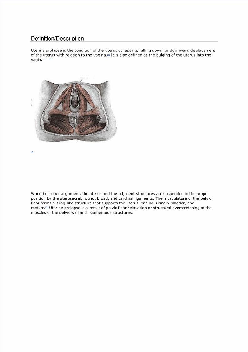
When in proper alignment, the uterus and the adjacent structures are suspended in the proper
position by the uterosacral, round, broad, and cardinal ligaments. The musculature of the pelvic
floor forms a sling-like structure that supports the uterus, vagina, urinary bladder, and
rectum.[2] Uterine prolapse is a result of pelvic floor relaxation or structural overstretching of the
muscles of the pelvic wall and ligamentous structures.

8/3/2019 Definition Prolaps
http://slidepdf.com/reader/full/definition-prolaps 2/10
Uterine prolapse is characterized under a more general classification called pelvic organ
prolapse which encompasses descent of the anterior, middle and posterior structures
into the vagina.[5]
Those organs that bulge anterior into the vagina are the urinary bladder which is called a
cystocele, the urethra, which is called a urethrocele or a combination, which is a
cystourethrocele.[2],[5]
The uterus and the vaginal vault, which is the apex of the vagina, make up the organs that
constitute the middle portion descent into the vagina. The vaginal vault often prolapses as
a result of a hysterectomy.[5]
The rectal bulge is called a rectocele and a bulge of part of the intestine and peritoneum is
called an enterocele, these make up the posterior portion of pelvic organ prolapse.[2],[5]
The information from this point forward will focus on uterine prolapse.
Uterine prolapse is classified using a four part grading system:
Grade 1: Descent of the uterus to above the hymen
Grade 2: Descent of the uterus to the hymen

8/3/2019 Definition Prolaps
http://slidepdf.com/reader/full/definition-prolaps 3/10
Grade 3: Descent of the uterus beyond the hymen
Grade 4: Total prolapse.[5]
[6]
Prevalence
Each source presents with a different prevalence depending on the researcher and the population
used. One study stated that the prevalence of pelvic organ prolapse, a clinical classification for all
of the pelvic structures prolapse into the vagina, was 50% for women who have give birth, though
most women are asymptomatic.[5]Another article cited that 50% of the female population in the
United States are affected by pelvic order prolapse with a prevalence rate that can vary from 30%
to 93%, varying among different populations.[7] A questionnaire based study stated that 46.8% of
the responses were positive for symptoms of pelvic organ prolapse and of the response group,
46.9% were vaginally examined with 21% having clinically relevant pelvic organ prolapse.[8]
Characteristics/Clinical PresentationThe primary symptoms of a uterine prolapse are backache, perineal pain, and a sense of
"heaviness" in the vaginal area.[2] Pain associated with uterine prolapse can be located centrally
or suprapubic, and can be described as "dragging" in the groin. This pain is due to stretching of
the ligamentous supports and secondarily to abrasion of the prolapsed tissues.[1] If the prolapse
has progressed into a grade three or third degree prolapse, the person may feel as though they
have a lump at the vaginal opening and have irritation and abrasion of the exposed mucous
membrane of the cervix and vagina. This is possible both during sexual intercourse and from
wiping with toileting procedures. The person may report that the symptoms are relieved by lying
down and exacerbated with prolonged standing, walking, coughing or straining. An associated and
often common complication of uterine prolapse is urinary incontinence.[2] Other descriptions used
are the feeling of sitting on a small ball and a report of repeated bladder infections.[9]
Summary from Differential Diagnosis for Physical Therapists:

8/3/2019 Definition Prolaps
http://slidepdf.com/reader/full/definition-prolaps 4/10
8/3/2019 Definition Prolaps
http://slidepdf.com/reader/full/definition-prolaps 5/10
Observation is often the first means of diagnosis.[2] Physical examination is the primary means for
diagnosis. A bimanual test is performed with a speculum while the person is at rest and when the
person is straining. If prolapse is not apparent with the first method, the person repeats the test
while standing with one foot on a chair. The person is then graded using a first through third
degree categorization. A first degree prolapse is characterized by descent of the uterus to above
the hymen. A second degree prolapse is to the level of the hymen and a third degree prolapse is
below the level of the hymen and protrudes through the vaginal opening.[2]
Urine culture is orderedif needed. If still unsure about the diagnosis, a pelvic ultrasonography or cystography can be
ordered.[5]
Causes
Women most at risk for this condition are those who have had multiple pregnancies and deliveries
in combination with obesity. Associated risk factors are trauma to the pudendal or sacral nerves
when giving birth. The disorder has been attributed to prolonged labor, bearing down before full
dilation, and forceful delivery of the placenta. Decreased muscle tone due to aging, excessive
strain during bowel movement and complications of pelvic surgery have also been associated with
prolapse of the uterus and adjacent organs.[2] Associated risk also exists with pelvic tumors and
neurologic condition like spina bifida and diabetic neuropathy which interferes with innervation of
pelvic musculature.[2] Genetics are suspected in this condition due to multiple familial relations and
generations with this and related conditions.[13] A recent article has found that Cesarean section
may lower the risk for pelvic organ prolapse.[14]
Summary of Causes:
Multiple pregnancies and deliveries
Obesity
Trauma to pudendal or sacral nerves
Aging related muscle changes
Excessive strain during bowel movements
Pelvic tumors
Genetic predisposition
Systemic Involvement
The digestive and urinary system can be impacted by uterine prolapse if the uterus obstructs the
bladder/urethra and the rectum, decreasing the ability to void.[15] The reproductive system can
also be impacted by painful intercourse, decreasing the ability for reproduction.[1]
Medical Management (current best evidence)Corrective Surgery
Corrective surgery was a once popular first step for uterine prolapse but has fallen second choice
to rehabilitation. When surgery is indicated, it is a management tool for second, third, and fourth-
degree uterine prolapse.[2] Pelvic organ prolapse surgery has a success rate of 65% to 90% and
has a repeated rate of operation at 30%. Patients who have more than one compartment involved
may need a combination of surgeries and surgery can often predispose patients to prolapse in
another compartment. Surgery can be either open or laparoscopic of the abdomen or can be in the
vagina using fasciae, mesh, tape or sutures to suspend the organs. Another surgical procedure
that is used in attempt to conserve the uterus is a sacrohysteropexy which is a Y-shaped graft that
attaches the uterus to the sacrum.[5] One case study that examined the effectiveness in
laparoscopic sacrohysteropexy, stated that this procedure “maintains durable anatomicrestoration, normal vaginal axis and sexual function.” It also requires less time and less adhesion

8/3/2019 Definition Prolaps
http://slidepdf.com/reader/full/definition-prolaps 6/10
formation due to the laparoscopic approach versus an abdominal route.[16] Vaginal hysterectomy,
vesicourethral suspension, and abdominal hysterectomy are other possible
approaches.[2] Important components for consideration of a surgical approach are:
Degree of prolapse
Desire for future pregnancies
Other medical conditions
The woman's desire to retain vaginal function
The woman's age and general health[9]
[17]
Pessary
A pessary is a shaped device made to support the uterus in the vagina. This is often a non-surgical
approach used for both uterine prolapse and urinary incontinence. There is a supportive type for
milder prolapse and a space-occupying type for more serious prolapse. The goal of the pessary is
to find the largest fit that is comfortable. They are to be removed regularly for cleaning by the
individual with correct education or by a health care professional.[5]
There are rings, rings with arubber support, cubes, donut shapes and inflatable balls. Depending on the degree of prolapse will
determine the type that is chosen. [13]Patients who are not eligible for use of a pessary are those
who cannot perform maintenance care of the pessary, those with vaginal ulcerations or lesions,
severe atrophy of the vagina and women who develop recurrent vaginitis.[18] Signs of improper fit
are: those who have pain when wearing the pessary, vaginal ulceration and infection, and the
inability to have a bowel movement or urinate.[13]
[19]
No Treatment
Surgical treatment is not appropriate when the woman has recently had a baby. Tissue damaged
during childbirth that has caused an associated prolapse, often begins to improve when
undergoing tissue healing. A symptomatic prolapse in the first few weeks after delivery,
especially in breastfeeding mothers have lower estrogen levels, does not necessarily predispose
the mother to long term issues. Improvement tends to occur after discontinuation of nursing and
the return of normal hormone levels. Other women and/or physicians do not elect for medical
treatment for Stage 1 and Stage 2 prolapse and take a wait and see approach.[13]

8/3/2019 Definition Prolaps
http://slidepdf.com/reader/full/definition-prolaps 7/10
Physical Therapy Management (current best evidence)
Pelvic floor strengthening exercise is currently the front line treatment before surgery and also
following surgery, these include but are not limited to Kegel exercises.[2][5] Other methods currently
used are pelvic floor musculature re-education, postural education, biofeedback and electrical
stimulation.[2]
Pelvic Floor Training
Pelvic floor muscles are seventy percent slow-twitch muscle fibers, which assist in muscle
endurance with generation of slow and sustained contractions. These muscles are designed to
have a less intense contraction, whereas the other thirty percent, which are fast twitch, are
designed for quick and forceful contraction. An example of fast twitch muscles are the muscles
that close the urethra during increased intra-abdominal pressure.[20] Pelvic floor training is
progressive resistive exercises for the pelvic floor that are often titled Kegel exercises. These
exercises improve urethral resistance and pelvic visceral support by increasing the voluntary
periurethral muscles. Pelvic floor exercises enhance the voluntary closing mechanisms. A thorough
assessment of pelvic floor function is necessary to determine the muscular strength and endurance
by manual muscle test. [21]
Kegel exercises are often explained as contracting the muscles that stopthe flow of urine. A sustained pelvic contraction for a minimum of two seconds is likely to ensure a
better response to physical therapy. A five point rating scale is used to describe the contractile
strength during pelvic musculature examination.
Five-Point Rating Scale[22]
Grade Description
0 No contraction
1 Flicker, only with muscles stretched
2 Weak squeeze, 2 second hold
3Fair squeeze with definite "lift" (in which thecontraction can be felt to move in an upwarddirection)
4Good squeeze, good hold with lift (thecontraction must be able to be repeated a fewtimes)
5 Strong squeeze, good lift, repeatable
One important observation with success of Kegel exercise is the identification of the correct
musculature contraction by a specialized Physical Therapist. Approximately 19% to 31% of women
who believe they perform Kegels actually perform them correctly. The woman is instructed tocontract her muscles around the examiners fingers while the examiner determines if the patient is
using auxiliary muscles like the abdomen, gluteals, or thighs. Bearing down is a common mistake
when asked to perform a pelvic muscle contraction. Once the women has achieved holding the
outer layer of the pelvic floor (bulbocavernosus and ischiocavernosis) in conjunction with
higher level muscles like the levator ani, she should attempt to hold both for ten seconds.[22] An
article by Lianne Herbruck listed a chart of instructions that summate the correct Kegel exercise
procedure for pelvic floor muscle training.
Proper Performance of Kegel Exercises for Pelvic Floor
Muscle Training[20]
Kegel exercises are performed to strengthen the
muscles of the pelvic floor to help increase support

8/3/2019 Definition Prolaps
http://slidepdf.com/reader/full/definition-prolaps 8/10
of the bladder and the urethra. They also can beused postpartum to facilitate circulation to theperineum, which promotes faster healing andincreases pelvic floor muscle tone.
Have the woman contract the muscles in theperineum/pelvic floor as if she is trying to preventpassage of intestinal gas. (The old adage of "stopping the flow of urine" can actually encourageretention and cause dysfunction of the micturationreflex).
She should feel the muscles draw upward andinward.
She should avoid straining or bearing-down motionswhile performing the contractions. (This can be
avoided by exhaling gently with an open mouth asshe contracts the muscles.)
Contractions should be intense, but should notinvolve abdomen, thighs, or buttocks.
The woman should be able to hold this contractionfor 5 to 10 seconds, but may need to work up tothat.
The woman should rest for 10 seconds betweencontractions.
Kegels should be performed at least 10 times, 3times a day, or from 30 to 80 times a day.
Current research prescribes a frequency of 30 contraction per day with an emphasis on increasing
the strength and intensity of the contraction. A greater emphasis is placed on devoting a particular
time to exercise and gradually increasing the amount and intensity of the exercise.[22] Though these
exercises can improve function, they cannot reverse a Grade 3 or 4 uterine prolapse.[23] These
exercises are often indicated as treatment for stress urinary incontinence, pelvic organ prolapse,
pelvic pain and defecatory dysfunction.[22]
Vaginal Cones
This exercise is used as a adjunct to contraction exercises of the pelvic floor. The patient insertsweighted cones into the vagina and is instructed to maintain the position of the weighted cone.
This method provides proprioceptive feedback to desired pelvic sustained contraction.[21] This is
thought to help improve the tone through active and sustained muscle contraction.[20]
Colpexin Sphere
The Colpexin Sphere is an intravaginal device that provides support to the pelvic floor musculature
and assists in elevation for more effective pelvic floor musculature exercises. "The Colpexin Sphere
is a smooth, round sphere made of medical grade polycarbonate plastic with an attached braided
nylon string for easy removal. It provides dual benefits for the management of pelvic organ
prolapse and improvement of pelvic floor muscle weakness. The Colpexin Sphere is available only
by prescription."[24] This is especially helpful for those who have urinary incontinence in association
with uterine prolapse. This device is appropriate for candidates who prefer a conservative
approach to pelvic floor prolapse management and urinary incontinence.[20] [24]

8/3/2019 Definition Prolaps
http://slidepdf.com/reader/full/definition-prolaps 9/10
Biofeedback
Biofeedback is used to detect and amplify internal physiological events and conditions using a
monitoring instrument. This training helps to develop conscious control over these body processes.
The objectives are to assist patients in gaining greater awareness and voluntary control over
muscular control and contraction. This allows for a refined control of pelvic floor musculature for
functional training. This technique uses a color video screen connected to a computerized unit
which monitors different channels using intravaginal probe or surface electrodes depending on the
muscles being selected.[21] The identification of the levator ani is important with contraction during
the Kegel exercises. If they are weak or absent, physical therapy is indicated.[20]
Behavioral modification
This technique is used to bring attention to the possible interactions between the patient'ssymptoms and their environment and provide techniques for behavioral modification. Such
techniques consist of conditioning, fluid intake regulation, and use of devices. Bladder training is
used for patients with associated incontinence for bladder prompted training, bladder drills,
bladder habit training, and bladder retraining. In bladder retraining the patient is to keep a record
of voiding activity over seven days and gradually increase increments between urination toward a
normal three hour interval. The patient attempts to resist the urge to urinate by squeezing the
pelvic floor and sphincter muscles until the urge resolves.[21]
Electrical Stimulation
Electrical stimulation is used to inhibit the micturition reflex and contract pelvic floor muscles.
Using a vaginal or anal probe, the electrical stimulation produces a contraction of the levator ani
muscle. Electrical stimulation is also used based on the theory that low-level electrical currentsmight re-innervate the pelvic floor and change the ratio of slow-to-fast-twitch muscle
fibers.[20] Electrostimulation is used in treatment of stress incontinence, enhancing the periurethral
sphincter and urge incontinence, inhibiting the overactive detruser muscle. There are no side-
effects except some discomfort but it is contraindicated for pregnancy, vaginal infection, retention
and demand pacemaker.[21]
Education
Education is an important aspect of treatment, especially education on positions of irritation and
management of pain. Education plays an important role during exercise and discussion of sexual
intercourse with gravity assisted positions. Supine with a pillow or wedge support under the pelvis
can be useful position for rest, pelvic floor exercise performance and during
intercourse.[1] Education is also important to help the patient understand why maintaining an ideal
body weight limits the pressure the abdominal content places on the pelvic floor. Patients also be

8/3/2019 Definition Prolaps
http://slidepdf.com/reader/full/definition-prolaps 10/10