defining the therapeutic window in spinal muscular …
TRANSCRIPT

DEFINING THE THERAPEUTIC WINDOW IN SPINAL MUSCULAR ATROPHY:
TIME POINTS STUDY
_______________________________________
A Thesis
presented to
the Faculty of the Graduate School
at the University of Missouri
_______________________________________________________
In Partial Fulfillment
of the Requirements for the Degree
Master of Science
_____________________________________________________
by
KATE LYNN ROBBINS
Dr. Christian L. Lorson, Thesis Supervisor
DECEMBER 2013

© Copyright by Kate Lynn Robbins 2013
All Rights Reserved
The undersigned, appointed by the dean of the Graduate School, have examined the thesis entitled
DEFINING THE THERAPEUTIC WINDOW IN SPINAL MUSCULAR ATROPHY:
TIME POINTS STUDY
presented by Kate Lynn Robbins,
a candidate for the degree of Master of Science in Veterinary Pathobiology,
and hereby certify that, in their opinion, it is worthy of acceptance.
Professor Christian L. Lorson
Professor Elizabeth C. Bryda
Professor Kevin D. Wells

DEDICATION PAGE
I dedicate this work to my Mother and Father, Debbie and George Robbins, whose wisdom,
support and perseverance helped shape my character allowing me to succeed in the pursuit of my
dreams.

ii
ACKNOWLEDGEMENTS
I would like to acknowledge my professors, colleagues, family and friends for their
dedication and guidance that allowed me this accomplishment of earning my Master’s degree.
Dr. Chris Lorson, thank you for this wonderful opportunity to work with an impressive group of
scientists and for the exposure to many techniques that have advanced my professional skills. I
appreciate your time, dedication, expertise and wisdom. Thank you for your commitment and for
providing everything necessary for me to perform this exciting project. My experience in your
laboratory has positively influenced my life and I thank you most greatly!
Dr. Elizabeth Bryda, you are an awe-inspiring role model. Your vitality inspires me and
reminds me that women can have it all, with dedication and perseverance. You are truly
amazing; as an educator, mother, friend and leader within the community. I have always
appreciated your perspective and wisdom throughout all my research endeavors.
Dr. Kevin Wells, you completely amaze me! Your knowledge and wisdom on –
everything! – is inspiring. I always appreciate learning from you because your detailed
explanations are how I prefer to learn! You teach me to think more critically, and to incorporate
knowledge of many perspectives upon scientific investigations.
Dr. John Critser, may you rest in peace. Thank you for your extensive coaching and
mentoring to help me improve my writing. I greatly appreciate the experience and apply what
you have taught me in my everyday experiences and scientific writing; and this has allowed me
to pay it forward as you once suggested. The scientific community will really miss you, indeed.

iii
Dr. Beth Critser, thank you for taking me under your wing at an important time in my
education. I would not have accomplished all that I have, had it not been for you! I really
appreciate all the knowledge I have learned from you.
To all of my past professors, thank you for your excellent instruction and aiding in my
fundamental understanding of science.
To all of my lab members, past and present, thank you for your help along the way and
for sharing your knowledge with me. Erkan Osman, you are an awesome friend and colleague!
Thank you for making me feel at home in the Lorson lab! Since day one, you have consistently
aided in my understanding of the SMA field, and helped me improve my technical skills, as well
as, taught me how to prepare professional posters and seminar presentations. You taught me
most of what I learned these past few years, including all my experiments, data managing and
organization, statistics, but most importantly, how to manage the tiny dancers! I cannot thank
you enough; my work in this lab was my final step before moving on and making a career for
myself in science, and I feel much more prepared after these past couple of years. You have been
a great friend and I truly thank you for how you have changed my life. Madeline, your smile is
contagious and your kindness never-ending. You and I are going to change the world! Monir, if
you ever miss your daughter and cannot get ahold of her, just call me- I’ll fill in. Pei-Fen, thank
you for always taking time to help me with my experiments, and for being super sweet. Hans,
your scientific knowledge and challenges have taught me to think more critically and become a
better scientist. Chrisite and Jolill, you always brightened the lab with your upbeat spirit and
dance moves. Jackie, thank you for all the productive discussions regarding this project, your
input was invaluable and it showed me the benefits of various approaches to tackle any scientific

iv
problems. Arleigh and Abby, you are the hardest working students I have ever met! Keep up the
good work, I know you will be successful! John Marston- I’m really not that gullible! Although,
your teasing always made me think twice. You are a good friend, and I appreciate you looking
out for me. I cannot begin to thank you enough for your help with the mice! It has been difficult
since you retired!
Mom and Dad, thank you for all your guidance and patience along the way. You are my
true role models and I thank you for helping mold me into a person I can be proud of. I definitely
would not have made it without your dedication throughout all these years.
Thank you to all my friends that have been there with me through the good and bad
times. Dr. Lydia Cook, DVM, PhD, there are not enough words to express how you have
transformed my life. You are an awesome role model, and a great friend and colleague that I
strive to be like, more and more, every day! I would truly be lost without you! Anne, Rose,
Susan, Denise, Carolina, Ayushi, Judy and Tony, thank you for your friendship. You have
always been there for me, guiding me spiritually and scientifically and I am very grateful for
having you in my life.
v
TABLE OF CONTENTS
ACKNOWLEDGEMENTS……………………………………………………………………….ii
LIST OF FIGURES……………………………………………………………………………...vii
LIST OF ABBREVIATIONS………………………………………………………………...... .ix
ABSTRACT………………………………………………………………………………..…… xi
Chapter
1. INTRODUCTION TO SPINAL MUSCULAR ATROPHY...........................................1-19
Spinal Muscular Atrophy Overview......................................................................................1
Spinal Muscular Atrophy Genetics…………………………………………………………2
SMN Protein Function ………………………………………………………………….….5
Clinical Assessment of Spinal Muscular Atrophy.................................................................8
Mouse Models of Spinal Muscular Atrophy..........................................................................9
Therapeutic intervention in SMA.........................................................................................11
Gene Therapy for Spinal Muscular Atrophy........................................................................13
Therapeutic Window for SMA Intervention........................................................................15

vi
2. SPINAL MUSCULAR ATROPHY - DISEASE DEVELOPMENT AND THERAPEUTIC WINDOW USING SCAAV9-SMN IN THE DELTA7 MOUSE MODEL...............20-55
Introduction..........................................................................................................................20
Materials and Methods.........................................................................................................22
ICV Injection of scAAV9-SMN Increases Survival of SMNΔ7 Mice.................................27
Weight Increase Observed in All Treated Groups...............................................................33
Early Treatment Improves Motor Function in Mice Injected with scAAV9-SMN.............39
SMN Protein Induction........................................................................................................44
Peripheral Distribution of scAAV9-SMN and Transduction Analysis................................48
Discussion............................................................................................................................50
3. CONCLUSIONS.............................................................................................................55-56
BIBLIOGRAPHY.....................................................................................................................57-66

vii
LIST OF FIGURES
Figure: Page:
Figure 1.1: Schematic representation of SMN1 and SMN2 at the DNA, RNA and protein level............................................................................................................................................3
Figure 1.2: Circle graphs depicting SMA Type I, Type II and Type III patients and their corresponding SMN2 copy number...........................................................................................4
Figure 1.3: Diagram of SMN’s role in snRNP biogenesis assembly...............................................7
Figure 2.1: ICV injection of scAAV9-SMN increases the survival of SMNΔ7 mice when administered at early time points.............................................................................................29
Figure 2.2: SMNΔ7 mice injected at early time points live longer on average compared to mice injected at later time points.............................................................................................30
Figure 2.3: P-table demonstrating statistically significant differences in average survival for mice in the treatment and non-treated groups..........................................................................31
Figure 2.4: Higher degree of variability in survival is observed within each group of mice injected at the median time points.............................................................................................32
Figure 2.5: SMNΔ7 mice treated with scAAV9-SMN gain weight throughout their lifespan......35
Figure 2.6: Percent weight gained from birth to peak....................................................................36
Figure 2.7: P-table demonstrating statistically significant differences in average weight gained from birth to peak all mice in the treatment and non-treated groups.......................................37
Figure 2.8: Representative images of SMNΔ7 mice......................................................................38
Figure 2.9: Percentage of animals able to right themselves...........................................................41
Figure 2.10: Average time to right for SMNΔ7 mice treated with scAAV9-SMN.......................42
Figure 2.11: Individual time to right on Day 14............................................................................43

viii
Figure 2.12: SMN protein induction is evident in SMNΔ7 mice injected at early and late time points........................................................................................................................................45
Figure 2.13: Robust SMN expression is observed four days post therapeutic administration.......47
Figure 2.14: Animals injected with scAAV9-GFP demonstrate substantial viral vector expression in the periphery.........................................................................................................................49
Figure 2.15: Overt phenotype of P2 treated animal with scAAV9-SMN compared to unaffected and non-injected littermates.....................................................................................................54

ix
LIST OF ABBREVIATIONS
AAV Adeno-associated virus
AchR Acetylcholine receptor
ALS Amyotrophic lateral sclerosis
ASO Antisense oligonucleotide
BBB Blood-brain barrier
C Cytosine
cDNA Complementary DNA
CNS Central nervous system
CSF Cerebrospinal fluid
DNA Deoxyribonucleic acid
E Embryonic day
GFP Green fluorescent protein
HEK293T Human embryonic kidney-293T cell line
HDACi Histone deacetylase inhibitors
hnRNP Heterogeneous ribonucleoprotein particle
ICV Intracerebroventricular
IP Intraperitoneal
ISS-N1 Intronic splicing silencer N-1
IV Intravenous
mRNA Messenger RNA
mSmn Murine survival motor neuron gene
x
NINDS National Institute of Neurological Disorders and Stroke
P Postnatal day
PBA Phenylbutyrate
PEI Polyethyleneimine
qPCR Quantitative polymerase chain reaction
RNA Ribonucleic acid
SACHDNC Secretary’s Advisory Committee on Heritable Diseases in Newborns and Children
SAHA Suberoylanilide hydroxamic acid
scAAV-SMN Self-complementary adeno-associated virus expressing full-length SMN
Sm Smith core protein
SMA Spinal Muscular Atrophy
SMN Survival Motor Neuron
SMN2 Severe mouse model with the genotype Smn-/-;SMN2
SMN∆7 SMN delta 7; SMN lacking exon 7
+/+
SMNRT
snRNP Small nuclear ribonucleoprotein
SMN Read-Through mouse model
STAT5 Signal transducer and activator of transcription 5
T Thymine
TSA Trichostatin A
v.g. Viral genomes
VPA Valproic acid

xi
ABSTRACT
Spinal muscular atrophy (SMA) is caused by the loss of a single gene, survival motor
neuron-1 (SMN1), which results in the rapid deterioration of motor neuron integrity and function,
most often leading to infantile death. Administration of self complementary adeno-associated
virus expressing full-length SMN cDNA (scAAV-SMN) has proven an effective means to rescue
the SMA phenotype in SMA mice, either by intravenous (IV) or intracerebroventricular (ICV)
administration at very early time points. We have recently shown that ICV delivery of scAAV9-
SMN is more effective than a similar dose of vector administered via an IV injection, thereby
providing an important mechanism to examine a timeline for ameliorating the disease and
determining the optimal therapeutic window. SMNΔ7 mice were injected with scAAV9-SMN
vector via ICV injection on a single day, from P2 through P8. At each delivery point from P2
through P7, scAAV9-SMN decreased disease severity, ranging from a near complete rescue (P2)
to a significant, albeit lesser degree (P7) in which animals lived ~130% longer. Our study
demonstrates that a maximal benefit is obtained when treatment is delivered during a specified
therapeutic window of the pre-symptomatic stages of SMA in the SMNΔ7 mouse model.
Although disease severity can be significantly decreased when SMN levels are increased at later
stages of the disease, there is a time (after postnatal day 8) at which therapy is no longer
effective.
1
CHAPTER ONE: INTRODUCTION TO SPINAL MUSCULAR ATROPHY
Spinal Muscular Atrophy Overview
The National Institute of Neurological Disorders and Stroke (NINDS) has classified over
600 neurological disorders affecting millions of people worldwide (50 million in the USA alone),
with an annual cost in hundreds of billions of dollars (National Institutes of Health website).
Some of the more prevalent neurodegenerative diseases include Amyotrophic Lateral Sclerosis
(ALS), Friedreich's Ataxia, Alzheimer's, Huntington's, Parkinson's, Lewy Body Disease, and
Spinal Muscular Atrophy (SMA) (NINDS website).
SMA is an autosomal recessive neurodegenerative disorder, and it is caused by the
mutation of a single gene, survival motor neuron-1 (SMN1) [1], which codes for the ubiquitously
expressed protein, SMN. Humans possess another nearly identical gene, SMN2, which can
produce fully functioning SMN protein, but only at low levels. This is due to a C to T conversion
within the 5′ end of exon 7 resulting in alternatively spliced SMN2-derived transcripts. SMA is a
motor neuron disease and is characterized by the loss of α-motor neurons in the ventral horn of
the spinal cord, which leads to muscle wasting, atrophy, paralysis and death in the more severe
cases [1]. SMA is a common genetic cause of infantile death with an incidence of 1:6,000 –
1:10,000 live births and a carrier frequency of 1 in 40 [2-5]. It is still unclear what critical
function is disrupted by the severe reduction of SMN protein levels, but this leads to the
pathogenesis of SMA.
Strategies to circumvent SMA progression focus on SMN1 gene replacement or the
alteration of SMN2 splicing [6]. Currently, there is no cure for SMA; and although clinical
studies have shown that early intervention and nutritional support have positive outcomes for

2
SMA patients [7], SMA still remains absent from the required newborn screening panel. A pilot
study is underway to assess the feasibility of prenatal screening for SMA in all newborns
(Newborn Screening Translational Research Network at https://www.nbstrn.org).
Spinal Muscular Atrophy Genetics
SMA was first described by Werdnig and Hoffmann in the 1890s and became known as
Werdnig-Hoffmann disease at that time, but today is synonymous with the most severe form of
SMA. In their autopsy reports, Werdnig and Hoffmann described loss of α-motor neurons in the
ventral horn of the spinal cord as well as atrophy of skeletal muscle [8]. It was not until 1995 that
the SMA-determining gene was identified by Lefebvre et al. (Figure 1.1A) [1, 9]. Telomeric
SMN1 and centromeric SMN2 are nearly identical and are positioned inversely from each other
due to an early intrachromosomal duplication event, at the genomic region of 5q13 [1]. SMN1
produces full-length transcript and functional SMN protein, but the alternatively spliced SMN2
transcript produces approximately 90% truncated protein (Figure 1.1B and 1.1C) [10]. Although
SMN1 and SMN2 are completely identical in amino acid sequence, there is a crucial
translationally silent "C" to "T" transition located in exon 7 at position +6, which results in
severe disruption of SMN2 splicing [11]. Importantly, SMN2 is a disease modifier because there
is a strong inverse correlation between SMN2 copy number and disease severity, where higher
SMN2 copy numbers result in a milder phenotype (Figure 1.2) [12, 13].

3
Figure 1.1. Schematic representation of SMN1 and SMN2 at the DNA, RNA and protein level.
A) SMN1 and SMN2 are located on Chromosome 5 in reverse order and differ by a single
nucleotide in exon 7. B) SMN2 is alternatively spliced resulting in a greater abundance of
transcripts lacking exon 7 (SMNΔ7). C) SMNΔ7 transcripts produce truncated, non-functional
protein.

4
Figure 1.2. Circle graphs depicting SMA Type I, Type II and Type III patients and their
corresponding SMN2 copy number. SMN2 is an important disease-modifying gene because each
copy provides ~10% basal SMN levels which collectively, increases overall SMN protein levels
with the addition of each SMN2 copy. Data adapted from [14].

5
There are a few nucleotide differences between SMN1 and SMN2 but these do not alter
protein structure [1, 11]. However, the C to T transition in exon 7 results in a functional
difference between the two genes due to aberrant splicing of the SMN2 transcript [11]. Positive
splicing regulators, such as SF2/ASF, are prevented from binding the region of exon 7, possibly
due to the disruption of an exonic splice enhancer region [15] or from the creation of an exonic
splice silencer region [16] following the single nucleotide change. Additionally, factors such as
hnRNP-A1, which promote the exclusion of exon 7 [16], recruit negative regulators such as
Element 1 [17] and ISS-N1 [18], which act as splicing repressors. This highly dynamic region in
SMN2 leads to aberrant splicing ~90% of the time, generating a truncated transcript lacking exon
7 (SMNΔ7) [1, 11, 19]. The SMNΔ7 protein cannot self-associate [20] or form complexes
efficiently with RNAs and proteins [21], making it unstable, and thus it is rapidly degraded [22].
SMN Protein Function
SMN is a multifunctional protein known to bind many cellular proteins [23-26] and a few
viral proteins [27-29]; and it is found in both the nucleus and cytoplasm of all cell types [30-32].
In the nucleus, SMN binds Gemin proteins forming aggregates called gems [12, 30, 31], which
have been used as a cell biomarker because gem numbers correspond to overall SMN levels [23].
There are a multitude of cellular activities in which SMN is involved, however, the best
understood function is the role SMN plays in snRNP biogenesis. SMN is crucial in
transcriptional activation [23, 27, 33], translational regulation [23, 33, 34] stress response [23,
33, 35, 36] and cell apoptosis [23, 37]. Furthermore, SMN functions in axonal RNA trafficking
[23, 33, 38] and RNA splicing [23, 33, 39]. In the context of snRNP biogenesis and assembly,

6
SMN is essential [23, 33, 40] (Figure 1.3). Pre-mRNA splicing is mediated by the spliceosome, a
complex of RNAs and RNA-binding proteins. In eukaryotic cells, these spliceosomal small
nuclear ribonucleoproteins (snRNPs) are generated by a master assemblyosome [41], the SMN
complex [23, 33, 40], before they organize into the final spliceosome machinery. snRNAs are
transcribed in the nucleus and exported to the cytoplasm where the SMN complex (consisting of
SMN and Gemins 2-8) [33] transfers Smith core proteins (Sm) onto the appropriate site of the
snRNA [42]. The binding of Sm proteins onto the snRNAs is highly specific, and the SMN
complex ensures this process is performed correctly [33, 42, 43]. Once the Sm cores have been
assembled, the 3’ end of the snRNA processed, and the 5’ cap hypermethylated, the mature
snRNP is imported into the nucleus by the SMN complex [44, 45] where it will associate with
snRNP-specific proteins to generate the complete spliceosomal complex. When SMN levels are
low, a decrease in Sm core assembly is observed which is correlated with SMA severity [33, 42].

7
Figure 1.3. Diagram of SMN’s role in snRNP biogenesis. In the cytoplasm, the SMN complex
assembles Sm proteins into a heptameric ring structure [23, 46]. Following transcription in the
nucleus, snRNAs are transported to the cytoplasm where the SMN complex assembles the
heptameric ring of Sm core proteins onto the specific binding site of the snRNA [33, 42, 43].
Following maturation, the SMN complex shuttles the snRNP into the nucleus where it will be
assembled into the final spliceosomal complex [44, 45].

8
Clinical Assessment of Spinal Muscular Atrophy
SMA is classified into five subtypes according to disease severity and age of onset. 95%
of SMA patients have a deletion in both copies of SMN1, while the remainder of the cases occur
from frameshift, nonsense and missense mutations [47]. Type 0 SMA is embryonic lethality, and
this occurs only if the fetus carries mutations in both the SMN1 and SMN2 genes. SMA Type I
(Werdnig-Hoffmann disease) is the most severe clinical presentation of the disease and the most
prevalent type among patients; 65% of all new cases and 80% of all current cases are Type I [7].
These patients present with symptoms before 6 months of age [48]; they have trouble eating and
breathing and they never gain the strength to sit upright or crawl. Children with SMA Type I die
by the age of two, usually from respiratory complications [7, 47, 48]. SMA Type II patients
become symptomatic within 18 months [48], and are diagnosed once parents notice an extensive
delay in motor control; for example the child may have trouble controlling head movements and
may not move much or cannot sit upright. These patients are wheelchair bound and their life
expectancy is variable, with 70% of patients living into adulthood [6]. Patients with Type III
SMA, also known as Kugelberg-Welander disease, exhibit symptoms in adolescence and may
eventually become wheelchair-bound [47] although they have a normal life expectancy [48].
Adult onset SMA, Type IV, is the mildest form of the disease and is characterized by mild
muscle weakness usually after the age of 25 [47]. The clinical spectrum of SMA is broad and it
may be difficult to classify an individual as having one type of SMA or another because often
symptoms and disease characteristics overlap [48]. However, appropriate diagnosis and
assessment of disease severity and progression are important in order to provide the patient with
therapeutic intervention specifically tailored to their needs [7, 49].

9
In 2006, Pyatt and Prior described a multiplex real-time PCR assay that identifies
affected individuals and carriers with 100% sensitivity and specificity [50]. Then in 2008, SMA
was nominated to be added to the federally mandated newborn screening panel [7]. The
Secretary’s Advisory Committee on Heritable Diseases in Newborns and Children (SACHDNC)
declined the proposal until further evidence could be collected through population-based
screening to determine the efficacy and feasibility of screening all newborns for SMA. This
population-based study is underway, involving several medical centers. Current studies are
ongoing at the Department of Pediatrics at the University of Utah, School of Medicine and
Colorado School of Public Health at the University of Colorado, coordinated under the Newborn
Screening Translational Research Network (NBSTRN at https://www.nbstrn.org).
Mouse Models of Spinal Muscular Atrophy
Multiple mouse models have been generated to dissect the biological processes and
pathogenesis of SMA and importantly, they are a powerful tool used to investigate preclinical
therapeutics in a mammalian context. Models exhibiting disease phenotypes ranging from severe
to intermediate have been created to mimic the pathology observed in Type I, and Type II and
Type III patients, respectively.
As with all species, except humans, mice have only a single Smn gene (equivalent of
human SMN1) [51, 52] which results in embryonic lethality when lost or mutated [53]. In 2000,
the first SMA mouse model (Smn-/-;SMN2+/+) was generated independently by two laboratories:
FVB.Cg-Smn1tm1Hung Tg(SMN2)2HungSmn1tm1Hung 54/J (Jax 005058; [ ]) and FVB.Cg-
Tg(SMN2)89AhmbSmn1tm1Msd 55/J (Jax 005024; [ ]). Known as the severe “SMN2” model, this
10
transgenic mouse line was developed by inserting 2 copies of the human SMN2 transgene into
the mouse Smn-null background (Smn-/- 53; [ ]). The low levels of SMN provided by the transgene
were sufficient to rescue embryonic lethality however; this model is born symptomatic, with an
average survival of 5 days and never reaches the same weight as unaffected littermates.
A slightly less severe model (Smn-/-;SMN2+/+;SMNΔ7+/+) was later developed in 2005 by
Le et al., with the addition of an SMN cDNA lacking exon7 (SMNΔ7) onto the background of
the severe model (FVB.Cg-Tg(SMN2*delta7)4299AhmbTg(SMN2)89AhmbSmn1tm1Msd
56
/J; Jax
005025) [ ]. As this work originated to investigate possible adverse effects incurred by the
SMNΔ7 product, this group instead observed that this protein product associates with and
stabilizes full-length SMN, as well as extends the survival to an average of 13 days. This model
is termed the SMNΔ7 mouse model and it is born presymptomatic. This model has been the
workhorse for many investigations in recent years because it becomes symptomatic during the
early postnatal stage and lives relatively long enough to evaluate therapeutic intervention.
As severe models are useful for evaluating the accelerated progression of SMA Type I, it
is imperative to have intermediate models with a milder disease phenotype available to study the
less severe pathogenesis of Types II and III SMA [6]. Therefore, intermediate models have been
generated that exhibit a milder disease progression for which subtle differences, once masked by
severe pathology, can now be brought to light; as well as, new therapeutics formulated that are
more appropriate for Type II and III patients.
Our lab recently generated an intermediate model for SMA called the Read through
model SMNRT (SMN2+/+;SMNRT;Smn-/- 57) [ ]. This mouse line has the same genetics as the
SMNΔ7 model, however, the Δ7 transgene is manipulated to produce a more stable isoform than

11
SMNΔ7 [58], called SMN read through. This model demonstrates a moderate disease severity
and these mice live an average lifespan of 30 days.
Another intermediate SMA mouse model, called the Smn2B/-
59
model, has also been used
for SMA research [ ]. These mice possess 15% of normal SMN protein levels and they become
symptomatic around postnatal day 10. Smn2B/-
mice live an average lifespan of 28 days.
Intermediate mouse models are advantageous for extracting subtle biological information that
may be overlooked by the overt disease phenotype of the severe models. Furthermore, use of
these intermediate models will aid in the development of SMN2-independent therapies.
Therapeutic Intervention in SMA
Therapeutics aimed to attenuate SMA disease progression have encompassed various
strategies ranging from those that focus on gene therapy or boosting SMN2 productivity, to those
that use small molecular compounds to augment pathology through secondary pathways [6].
Many treatments have evolved that target SMN2 either by activating the promoter or
increasing exon 7 inclusion during splicing [6]. Histone deacetylase inhibitors (HDACi) are
pharmacologic compounds that aid in the decondensation of chromatin, which promotes gene
expression, although non-specifically and at a global level. One such HDACi, sodium butyrate,
increased exon 7 inclusion in a cell-based model and increased SMN levels in motor neurons of
the spinal cord in SMA mice [60]. Valproic acid (VPA) and phenylbutyrate (PBA) HDAC
inhibitors only modestly increased SMN in patient fibroblasts in vitro, and the extent of this
increase was dependent on SMN2 copy number [61, 62]. Unfortunately, both of these compounds
provided minimal effectiveness in clinical trials [6, 63-65]. HDAC inhibitors, Trichostatin A

12
(TSA) and suberoylanilide hydroxamic acid (SAHA) both significantly extended survival in
SMA mice [66, 67]; and TSA treatment supplemented with nutritional support increased survival
even further [68]. In addition, administration of sodium butyrate or SAHA in the drinking water
of gestating female mice beginning on embryonic day 15, led to a decrease in disease severity of
SMA progeny or amelioration of embryonic lethality, respectively [60, 67]. Interestingly, HDAC
inhibitors seem to provide general neuronal protection in a manner independent of SMN,
although it is not clear as to how this occurs. However, it has been proposed that this
neuroprotection may be due to the inhibition of atrogene pathways which normally mediate the
breakdown of muscle proteins in a myogenin-dependent manner [69].
SMN expression has been shown to be in part, regulated via the STAT5 pathway [70,
71]. Prolactin, a known activator of this pathway, is a hormone and canonically penetrates the
blood-brain barrier. Farooq et al. (2011) demonstrated that increased SMN expression and
protein levels in vitro and in vivo could be attained, along with a significant extension of survival
in SMA mice following prolactin administration. Although prolactin did not provide a robust
rescue, it may be possible to use this treatment as a supplemental therapy to maximize effects of
other regimens.
Another strategy implemented to modulate SMN2 splicing is through the use of nucleic
acids to promote exon 7 inclusion [6]. Antisense oligonucleotides (ASOs) and bifunctional
RNAs [72-79] perform this function in a manner similar to RNA interference; using a short
stretch of nucleotides (e.g. 20mer) to bind a specific RNA target sequence with a high degree of
specificity. For example, as in the case of SMA, an ASO may be designed to target a splice
repressor that normally promotes exon 7 exclusion; in this way, inhibition of a repressor would

13
favor the retention of exon 7 [77]. ASOs are modified for stability and reduced nuclease activity,
affording them an extended half-life [6]. Bifunctional RNAs are used in a similar manner only
they perform a dual function; they contain a RNA sequence domain complementary to the RNA
target sequence and an additional untethered sequence which serves as a binding domain for
specific splicing factors [77]. These recent advances in RNA-based therapeutics have greatly
accelerated a new wave of research [6, 80] that can embrace the limitless possibilities and
versatility of RNA, making this therapy amenable to the study of innumerable diseases.
A very promising new compound produced by ISIS Pharmaceuticals is currently being
investigated in a multiple-dose Phase 2 study (ClinicalTrials.gov, Identifier: NCT01839656);
ISIS-SMNRx
is an ASO designed to modulate SMN2 splicing to promote exon 7 inclusion. This
therapeutic is injected intrathecally into the cerebrospinal fluid and remains in the central
nervous system (CNS) for an extensive amount of time. However, the current Phase II study,
which is expected to be completed in 2014, is addressing the question of whether larger doses are
safe and can be well tolerated. A Phase III study is anticipated to begin in 2014.
Gene Therapy for Spinal Muscular Atrophy
Gene therapy provides the most robust rescue in SMA mice to date and has been utilized
extensively by many laboratories. SMA is well suited for viral-based gene delivery because the
disease is monogenic and the SMN cDNA is small enough to be packaged into a viral vector.
Adeno-associated virus (AAV) serotype 9 has broad tissue tropism with a high transduction rate
into motor neurons, and as SMA is a motor neuron disease, it is appropriate that this serotype be
utilized to specifically target the CNS [81-83].

14
Using GFP as a reporter, it has been demonstrated that systemic delivery of AAV9-GFP
by intravenous injection, results in extensive transduction in motor neurons of neonatal mice
(60%), but was limited in adult mice in which astrocytes were the predominant target [84]. This
suggests that AAV9 was able to permeate the CNS of neonates where the blood-brain barrier
(BBB) is not yet fully formed as opposed to adult mice in which the BBB has fully developed.
Two explanations have been proposed to further elucidate this observation [84]. First, is the fact
that astrogenesis occurs in mice during the first two weeks of the postnatal period and second; it
is possible that astrocytic projections make contact with vascular endothelia and thus, capture
most of the virus particles before they are able to make their way past, to the motor neurons. The
BBB poses an obstacle for viral gene delivery because the endothelial cells separating the
vasculature and cerebrospinal fluid (CSF) form a tight barrier through which many molecules
cannot pass. However, with the advent of double-stranded self-complementary AAV vectors
(scAAV) in which the second strand synthesis step is bypassed and more specifically, the
generation of the scAAV9 vector, it was demonstrated that the efficiency of adult CNS
transduction could be increased [82]. Intravenous administration of scAAV9-GFP not only
provided robust transduction of neonatal motor neurons (likely due to the accelerated onset of
expression obtained with scAAV as opposed to AAV), but also transduced up to 28% of motor
neurons in adult mice [82]. Furthermore, following administration in neonates, GFP-positive
cells were detected in the CNS as well as non-CNS tissues, in contrast to an intraperitoneal (IP)
injection which resulted in only a few transduced cells in the CNS, and to an even lesser extent
following intramuscular injection [82]. Once the potential for achieving motor neuron

15
transduction in vivo was demonstrated, multiple laboratories applied this technology to SMA and
used this vector to overexpress SMN in SMA mouse models.
Therapeutic Window for SMA Intervention
Route of delivery and timing of administration are two important decisions to consider
when applying gene therapy to SMA because variations of both lead to distinguishable results.
Initial studies implementing scAAV9 to overexpress SMN, administered the virus by intravenous
injection (IV) between the day of birth (P0) and postnatal day 2 (P2). This mode of intervention
completely ameliorates disease progression in the SMNΔ7 model but does not have the same
effect on the more severe SMN2 mice, although it does extend their survival significantly.
SMNΔ7 mice are born presymptomatic and live on average, 14 days however; when injected
with scAAV9-SMN into the facial vein during this early postnatal period, these mice gained
significantly more weight that untreated controls and lived a normal lifespan (>200 days) [81,
85]. On the contrary, disease progression could not be ameliorated in the SMN2 model but the
animals did gain significantly more weight than control SMA mice, and they lived on average,
100% longer than controls [86].
Prolonging SMN induction proves to be less efficient. Only a modest benefit was
achieved when scAAV9-SMN was administered intravenously on P5, extending survival from
15 to 30 days; significantly less than what the same injection provides on P1 or P2, in which
mice lived over 250 days [81]. Further delay of treatment until P10 provided no benefit,
indicative of a narrow window of opportunity for therapeutic intervention to have an effect.

16
In parallel, this decreased therapeutic benefit from prolonging intervention was also
described in the case of ASO technology. Porensky et al. (2012) used an ASO that modulates
SMN2 splicing to increase exon 7 inclusion [87]. Direct CNS administration on P0 provided a
robust rescue in SMNΔ7 mice. Survival was extended from 15 to 100 days, SMN levels were
significantly increased and SMN expression was successfully restored to motor neurons with
high expression in the cytoplasm, as well as the formation of gems within the nucleus. Similar to
previous reports describing scAAV9-SMN intervention, administration at a later time point (P4)
decreased the efficiency of this ASO therapeutic, by providing only a modest extension in
survival (41 days) when mice were injected by ICV, and significantly less when injected by IV
(21 days) [87].
In 2012, a detailed analysis of the differential effects observed between ICV and IV
administration was reported for both the symptomatic SMN2 mice and the presymptomatic
SMNΔ7 mice [85, 86]. ICV or IV delivery of 2x10¹¹
86
viral genomes (v.g.) into symptomatic mice
on P1 significantly extended survival from an average of 5 days to 17 and 10 days, respectively.
SMN was significantly increased in the brain and spinal cord of both groups. Muscle fiber area
was only slightly increased in the ICV group but not in the IV injected mice [ ]. However, a
more substantial benefit was observed when intervention was administered before symptoms
appeared. In the SMNΔ7 model, delivery of 2x10¹⁰
85
v.g. on P2 resulted in a complete rescue
when delivered either by ICV or IV, with sufficient SMN restoration in the CNS, as well as
restoration of motor function [ ]. As this treatment did not provide a full rescue in SMN2 mice
as it did in SMNΔ7 mice, this further supports the indication of a therapeutic window in which
treatment must be administered before the disease progresses too far. Restoration of SMN once

17
overt symptoms are occurring may not be the best method for treating the disease at this point,
meaning that this therapy may need to be adapted or perhaps a combinatorial therapeutic regimen
may prove a more effective means of halting the disease once it has progressed to advanced
stages.
Further elucidation of this prospective therapeutic window was provided in reports of
which an SMN-inducible mouse model was used. In order to mimic human SMA in mice, Lutz
et al. (2011) created a mouse line in which the endogenous Smn gene was designed to behave
like human SMN2 by replacing murine Smn exon 7 and 8 with a cassette consisting of human
SMN2 exon 7 and 8. Additionally, an inverted and silent copy of mouse Smn exon 7 was placed
within SMN2 in the intron between exon 7 and 8. The generated hybrid allele contained also,
loxP sites flanking both exon 7s. This allowed conditional and irreversible activation by cassette
inversion to express functional, full-length protein at wild-type levels within 48 hours of
tamoxifen administration. When they turned on SMN expression during embryonic development
on embryonic day 6 (E6), they observed a complete restoration to a wild-type phenotype [88].
This is interesting because throughout most studies that utilize the SMNΔ7 model, the treated
mice always have a smaller, weaker overt phenotype compared to unaffected mice, although they
do often gain significantly more weight than untreated controls. SMN induction following
tamoxifen administration on P4 or P6 provided a substantial rescue but P8 induction only slightly
enhanced survival while P10 did not provide any benefit [88].
Furthermore, Le et al. (2001) developed another SMA inducible mouse model where
SMN is expressed only upon administration of doxycycline. The SMN construct here consists of
two minimal cytomegalovirus (CMV) promoters and a tetracycline response element (TRE).

18
This dual reporter system, upon doxycycline activation, drives SMN and luciferase expression
[89]. This inducible model took three days for an increase in SMN to be observed following
induction, however, this report provided similar results as the Lutz group [90]. Induction at E13
or P0/P1 provided a complete rescue, with mice living over 200 days however; the benefit was
not as substantial as the rescue Lutz et al. (2011) described when induction began on E6. As
expected, induction on P2 (SMN increase detectable at P5) had less dramatic effect where all the
mice except one, lived on average 25 days, with the exception living to 151 days. Also, mice
induced on P0/P1 in which SMN expression was turned off at P28, in general lived only a month
following SMN removal, with the exception of one mouse that lived over 8 months. Outliers
such as these two above are common occurrences in these types of studies and this may be due to
intrinsic genetic or epigenetic factors or overall pathological differences between mice [89].
Overall, early restoration of SMN provides the most beneficial outcome whereas delayed
intervention substantially diminishes the efficacy of this therapeutic modality. Route of
administration also changes the outcome, where systemic and direct CNS injections provide the
best results when implemented in a timely manner.
It is interesting to compare the studies involving direct CNS administration to the report
by Hua et al. (2011) who demonstrated that ASO delivery into the periphery alone, increased
survival significantly compared to direct CNS administration. This might suggest that peripheral
tissues that succumb to SMN depletion may have a negative effect on downstream targets,
perhaps some in the CNS, and that is perhaps why Hua et al. (2011) observed these results.
However, it is difficult to determine as of now because the systemic dose that was injected was
much higher than the dose administered to the CNS [91].

19
Timing of therapeutic administration for SMA has shown to be of utmost importance,
where intervention at the presymptomatic stages has the most beneficial outcome. It is still
unclear why this is the case but one explanation highlights the importance of the target cell
population. There may be a window in which motor neurons are no longer able to respond to
treatment. Perhaps they have degenerated to a point where no matter how much SMN is pumped
into them, it may not fix the damage that has already progressed. It is also possible these cells
lose specific communication signals and are no longer able to be transduced by the virus.
Another limit to this intervention is the mode of administration. As scAAV9 readily transduces
cells in the CNS when administered intravenously in neonates, this effect is diminished when
intervention is delayed after birth. This may possibly be due to complete closure of the BBB or
other hemodynamic alterations in which the virus is not sufficiently transported into the CNS.
Furthermore, there is evidence of viral entry by retrograde transport from the muscle;
however, the reports on direct intramuscular injection do not conclusively support this [92]. It is
still debated whether systemic or direct CNS injection is the best route of delivery as there are
two schools of thought about this. Some researchers hold true to the idea that SMA is a motor
neuron disease and that SMN replacement in motor neurons is sufficient to halt the disease
whereas; the other side of the argument highlights that motor neuron induction alone, does not
completely ameliorate all symptoms [33]. Replacement of SMN solely in motor neurons does not
preserve the integrity of all tissues as is evident in the numerous studies that report on the sudden
death often observed of a sub-group of treated mice [93]. This suggests that SMA affects
multiple organ systems and supports the idea of a multifactorial intervention scheme.

20
CHAPTER TWO: SPINAL MUSCULAR ATROPHY - DISEASE DEVELOPMENT AND THERAPEUTIC WINDOW USING SCAAV9-SMN IN THE DELTA7 MOUSE MODEL
Introduction
SMA is well suited for therapeutic intervention because the disease is monogenic, the
patient population is homogenous and humans possess the important disease-modifying gene,
SMN2. Strategies to circumvent SMA progression focus on SMN1 gene replacement or the
alteration of SMN2 splicing. Gene therapy using self-complementary adeno-associated virus,
serotype 9 (scAAV9) to deliver SMN, either systemically or directly into the CNS, has shown
the most promising results for rescuing SMA mice when administered before the therapeutic
window closes.
Previously, it has been demonstrated in a severe SMA mouse model, that SMN induction
as early as E6 completely ameliorates the disease phenotype [88]. However, administration of
scAAV9-SMN between P0 – P2 provides a near complete rescue but does not correct vascular
side effects such as tail and ear necrosis; and additionally, peripheral organ defects are often
observed [85, 88]. Furthermore, slight delay of therapeutic intervention results in only a modest
rescue, and an extended delay provides no benefit. Utilizing the well characterized SMNΔ7
model, we performed a systematic analysis to determine the effects of delaying scAAV9-SMN
administration. We injected each mouse with a single injection of 1x10¹¹ viral genomes (v.g.)
scAAV9-SMN at a single time point, P2 through P8. Early SMN induction provided the most
benefit, whereas, injection at later time points decreased the efficacy of this therapy. All treated
mice lived significantly longer, but SMN induction at the earliest time point (P2) provided the
most robust rescue with the fewest early deaths. Treated mice gained weight, demonstrated

21
restored motor function and exhibited SMN protein induction in the CNS and periphery.
Treatment at the various time points resulted in a range of phenotypes observed within the mice,
but is was evident that the earliest administration provided the best rescue, while delaying
intervention decreased the effectiveness.

22
Materials and Methods
Animal Handling and Genotyping
All animals were housed and treated with respect to the guidelines of the Animal Care
and Use Committee at the University of Missouri and in accordance with the regulations defined
in the “Guide for the Care and Use of Laboratory Animals,” (National Research Council, 2011).
The SMN delta7 (SMNΔ7) mouse model was utilized for these experiments. SMNΔ7
mice are null for mouse Smn and contain two copies of both transgenes, human SMN2 and the
cDNA coding sequence for SMNΔ7. Mice heterozygous for mSmn (Smn+/-;SMN2+/+;SMNΔ7+/+)
were purchased from the Jackson Laboratory (Bar Harbor, Maine, USA; stock 005025) and were
bred to generate mSmn knock-out mice (Smn-/-;SMN2+/+;SMNΔ7+/+) used in these experiments.
Heterozygous (Smn+/-;SMN2+/+;SMNΔ7+/+) mice are used throughout all the experiments as
unaffected positive controls and untreated SMA (Smn-/-;SMN2+/+;SMNΔ7+/+) mice were used as
negative controls. On their day of birth, postnatal day 1 (P1), each mouse was numbered using a
non-toxic, permanent marker for individual identification, and remarked each day after, until the
presence of fur in which they were then marked permanently with fuchsine dye. The distal tip of
the tail was removed, ~1 mm, with sharp, heated scissors to aseptically remove the tail tissue and
cauterize the wound. Genotyping was carried out by lysing the tail tissue in 75 µl of alkaline
solution (25 mM NaOH, 0.2 mM EDTA) for 30 minutes at 95°C followed by neutralization in 75
µl of a TRIZMA-HCl solution (40 mM TRIZMA-HCl) and incubation on ice. Polymerase chain
reaction (PCR) conditions were as follows: 95°C 2 minutes, 30 cycles (95°C 15 seconds, 68°C
90 seconds), 68°C 10 minutes. Primers were used to amplify the mSmn wild-type allele, forward
23
(5’-TCTGTGTTCGTGCGTGGTGACTTT-3’) and reverse (5’-CCCACCACCTAAGAAAGCCTCAAT-3’)
and the lacZ mSmn knockout allele, forward (5’-CCAACTTAATCGCCTTGCAGCACA-3’) and
reverse (5’AAGCGAGTGGCAACATGGAAATCG-3’).
Virus Production
scAAV9-SMN viral vector was produced as described previously [86]. HEK293T cells
were triple transfected in the presence of polyethyleneimine (PEI) (1mg/ml), pH 5.0 [94]. The
scAAV plasmid was constructed to express the open reading frame of human SMN1 cDNA
(NCBI accession number NM_000344) under the control of the chicken β-actin promoter.
Following media change and cell collection at 24 hours and 48 hours post-transfection,
respectively, scAAV9-SMN viral vector was purified using three cesium chloride density-
gradient centrifugation steps and dialyzed with HEPES buffer.
Real-time PCR (qPCR)
Quantification of viral genomes was performed using SYBR® Green and primers to
amplify the chicken β-actin promoter region, forward (5’-CCGGTGGTGGTGCAAATCAAAGAA-3’)
and reverse (5’-AGCAGAAGTAACACTTCCGTACAGGC-3’). The absolute quantitation method
using a standard curve was utilized on the Applied Biosystems® 7500 Real-Time PCR System
using Applied Biosystems 7500 Sequence Detection Software version 1.3. Viral fractions were
diluted 1:1000 and the PCR cycle was as follows: 50°C 2 minutes, 95°C 10 minutes, 40 cycles
(95°C 15 seconds), 60°C 1 minute. A standard curve was obtained using serial dilutions of the
transgene-containing plasmid (1010-105) to calculate melting curves of each sample. The viral

24
fractions containing the highest titer were dialyzed with HEPES buffer (100 mM NaCl, 20 mM
HEPES). Following dialysis, qPCR was performed to obtain the final titer of the virus-
containing solution to be used for injections into the mice.
Intracerebroventricular Injection of scAAV9-SMN or scAAV9-GFP
Intracerebroventricular (ICV) injection was performed on neonates to administer
scAAV9-SMN therapy directly into the CNS. Mice were administered a single injection of
1x10¹¹
v.g., on one of the following days, P2 through P8. Briefly, a glass-pulled needle was
inserted through the frontal plate (lateral to the metopic suture and rostral to the coronal suture)
into the left or right ventricle of the neonatal mouse to deliver a 5 µl bolus of viral vector.
Unaffected animals were injected with scAAV9-GFP on P2 or P7 and non-injected mice were
used as a negative control.
Phenotypic Assessment
All treated and untreated mice were monitored daily to evaluate survival, weight gain and
motor function as assessed by the time to right test. The time to right test consists of placing a
mouse on its back and determining the time it takes for them to right themselves onto all four
paws. A mouse unable to right within 30 seconds is considered to have failed the test for that
day.

25
Tissue Collection
Tissues were collected on P11 for all mice treated with scAAV9-SMN and on P6 or P11
for those injected with scAAV9-GFP. To harvest the tissues, mice were anesthetized using
isofluorine followed by cervical dislocation. Brain, spinal cord, skeletal muscle, heart, kidney,
liver, and spleen were collected and immediately flash-frozen in liquid nitrogen for those tissues
used for protein analysis or submerged in 4% paraformaldehyde if used for histological
examination of GFP.
Western Blot Analysis
For the SMNΔ7 mouse Western blots, tissues were collected at selected time points and
immediately frozen in liquid nitrogen. Tissue samples were placed at -80oC until ready for
analysis. Roughly 100 mg of tissue was homogenized in JLB buffer (50 mM Tris-HCl pH 7.5,
150 mM NaCl, 20 mM NaH2(PO4), 25 mM NaF, 2 mM EDTA, 10% glycerol, 1% Triton X-100,
and protease inhibitors (Roche, Indianapolis, IN, USA). Equal amounts of protein were separated
on 12% SDS-PAGE gels. SMN immunoblots were performed using a mouse SMN specific
monoclonal antibody (BD Biosciences #610647, San Jose, CA, USA) diluted 1:2000 in 1% dry
milk in TBST (Tris-buffered Saline Tween20 (10mM Tris-HCl, pH7.5, 150mM NaCl, 0.2%
Tween20) and a secondary anti-mouse HRP-conjugated secondary antibody (1:10,000). Blots
were visualized by chemiluminescence on a Fujifilm imager LAS-3000 and the corresponding
software. To verify equal loading, the membranes were then stripped using β-mercaptoethanol
for 30 minutes at 50°C and re-probed with anti-β-actin rabbit antibody (Sigma #A5060, St.
Louis, MO, USA) diluted 1:2000 and anti-rabbit HRP secondary antibody (1:10,000). Western

26
blot analysis was performed in triplicate and representative blots are shown. Probes were
visualized by chemiluminescence using the Pierce SuperSignal Pico reagents.

27
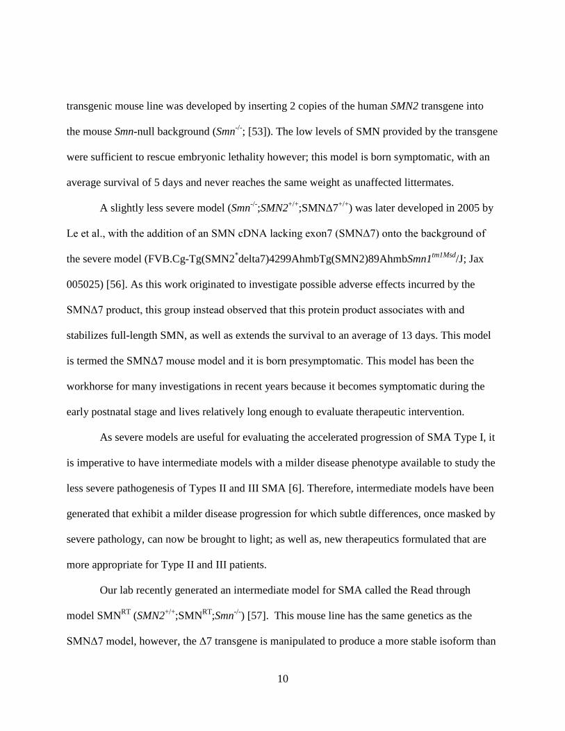
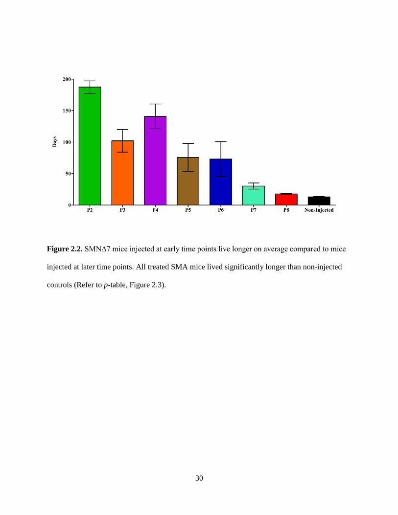
Intracerebroventricular Injection of scAAV9-SMN Increases Survival of SMNΔ7 Mice
On average, mice from all treatment groups lived significantly longer than non-injected
animals however; there was variability within groups (Figure 2.1, 2.2, 2.3). Mice injected on P2
experience no early deaths (all mice lived past 130 days) and they lived the longest with an
average lifespan of 187 days and a median survival of 204 days. The early deaths of the P3 and
P4 injected groups occurred on days 47 and 50, respectively. The P3 injected group lost another
subset of the population between days 60 and 100 but the remaining three mice lived past 150
days (maximum 187 days). The average survival for the P3 group was 102 days with a median
lifespan of 75 days. Mice in the P4 injected group steadily decreased in numbers with the oldest
animal living 211 days; and the average for the group was 141 days with a median lifespan of
167 days. In the P5 injected group, seventy percent of the treated mice died between days 22 and
42, with the remaining three mice living 152, 165 and 211 days. The average and median
survival for the P5 group was 76 and 37 days, respectively. Sixty-two percent of the mice in the
P6 group died between 20 and 35 days while the remaining three mice lived 50, 184 and 211
days; the average and median for the group were 73 and 34 days, respectively. Within the P7
injected group, eighty percent of the animals died between days 21 and 34, while the oldest
animal survived 70 days. Mice from the P7 injected group lived on average, 30 days with a
median survival of 28 days. The P8 injected group had a significant extension in survival,
compared to non-injected animals, although this was not as substantial as the survival extension
exhibited by the earlier-injected mice (Figure 2.3). Both the average and median survival values
for mice in the P8 group were 18 days. A high degree of variability is observed with the survival
data of each group treated at median time points P3 through P7 (Figure 2.4). Overall, mice

28
injected with scAAV9-SMN at earlier time points had a greater extension in survival compared
to mice injected at later time points; and mice from all groups lived significantly longer than
non-injected controls which lived on average, 13 days with a median survival of 14 days.

29
Figure 2.1. ICV injection of scAAV9-SMN increases the survival of SMNΔ7 mice when
administered at early time points. Kaplan-Meier survival curve of untreated SMNΔ7 mice and
those injected with 1x10¹¹
v.g. of scAAV9-SMN on a single day P2-P8.
30
Figure 2.2. SMNΔ7 mice injected at early time points live longer on average compared to mice
injected at later time points. All treated SMA mice lived significantly longer than non-injected
controls (Refer to p-table, Figure 2.3).

31
Figure 2.3. P-table demonstrating statistically significant differences in average survival of mice
in the treatment and non-treatment groups. All treated mice lived significantly longer than non-
injected controls. P-values were calculated using the logrank Mantel-Cox test and the table
illustrates the significance in survival between groups. NI = non-injected. (n.s. no significance, *
p ≤0.05, ** p ≤0.01, *** p ≤0.001, **** p ≤0.0001).

32
Figure 2.4. There is a high degree of variability in survival observed within each group of mice
injected at the median time points. For each treatment group, the line represents the average
survival for the group and the data points represent the minimum, median and maximum survival
within each group. There is a higher degree of variability observed within groups P3 through P7,
while the P2, P8 and the non-injected groups display less variation in survival.

33
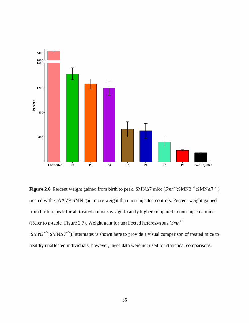
Weight Increase Observed in All Treated Groups
Weight gain was analyzed as the average weight for a given group across days (Figure
2.5) and also as the average percent weight gained from birth to peak weight (Figure 2.6, 2.7). At
birth, all SMA pups and unaffected pups are similar in weight and there are no obvious
phenotypic differences until around day 7 (Figure 2.8). However, by day 11 all treated mice
weighed significantly more than non-injected animals. Even the late injected mice (P7 and P8)
weighed significantly more, although this difference was not as substantial as the difference
observed between early injected mice and the non-injected controls. Compared to non-injected
animals, mice injected on P3 weighed significantly more by day 7, those of P4, P5 and P8 were
significantly higher by day nine, group P2 by day 10 and groups P6 and P7 were significantly
higher than non-treated animals by day 11. Around day 10, the P8 and non-injected mice reach a
plateau and their weight declines from thereon. By day 14 the P8 injected group weighed
significantly less than mice from all other treatment groups. At day 17 an emergence of two
groups within the weight observations can be observed. The first group consisting of P2, P3 and
P4 have similar weight gain and as a group diverge from a second cluster consisting of P5, P6
and P7.
All treated animals exhibited a significantly higher percent weight gain from birth to their
peak weight, compared with non-treated animals; the significance between these groups varied
(Figure 2.6 and 2.7). Animals injected on P2, P3 or P4 displayed similar weight gain from birth
to peak, and there was no significance when compared with each other. Similarly, animals
injected on P5, P6 or P7 had comparable percent weight gain and also did not show significance
when compared to one another. Comparison of either group P2, P3 or P4 with any of group P5,
34
P6 or P7 was significant; for all comparisons p ≤ 0.0001 except P4 compared with P5 or P6 (p ≤
0.001). Mice in the P8 group gained significantly less weight than mice in groups P2 through P6,
but not P7. All treated mice gained weight following treatment with scAAV9-SMN.

35
Figure 2.5. SMNΔ7 mice treated with scAAV9-SMN gain weight throughout their lifespan. The
average weight per group is plotted across days for the surviving animals in each cohort. ni
=
initial number of animals in each group.
36
Figure 2.6. Percent weight gained from birth to peak. SMNΔ7 mice (Smn-/-;SMN2+/+;SMNΔ7+/+)
treated with scAAV9-SMN gain more weight than non-injected controls. Percent weight gained
from birth to peak for all treated animals is significantly higher compared to non-injected mice
(Refer to p-table, Figure 2.7). Weight gain for unaffected heterozygous (Smn+/-
;SMN2+/+;SMNΔ7+/+
) littermates is shown here to provide a visual comparison of treated mice to
healthy unaffected individuals; however, these data were not used for statistical comparisons.

37
Figure 2.7. P-table demonstrating statistically significant differences in average weight gained
from birth to peak all mice in the treatment and non-treated groups. SMNΔ7 mice treated with
scAAV9-SMN gain significantly more weight than untreated littermates. Student’s t-tests were
performed to compare the average percent weight gained from birth to peak between groups.
(n.s. no significance, * p ≤0.05, ** p ≤0.01, *** p ≤0.001, **** p ≤0.0001).

38
Figure 2.8. Representative images of SMNΔ7 mice. A) Phenotypic differences are not apparent
at birth and the pups of the various genotypes cannot be distinguished by observation. WT
(Smn+/+;SMN2+/+;SMNΔ7+/+), HET (Smn+/-;SMN2+/+;SMNΔ7+/+) and SMA (Smn-/-
;SMN2+/+;SMNΔ7+/+
). B) At 9 days old, it can be readily observed that the pup injected at the
earliest time point (P2) has gained weight comparable to the unaffected heterozygous littermate;
and the pups injected at the latest time points (P7 and P8) are slower to gain weight.

39
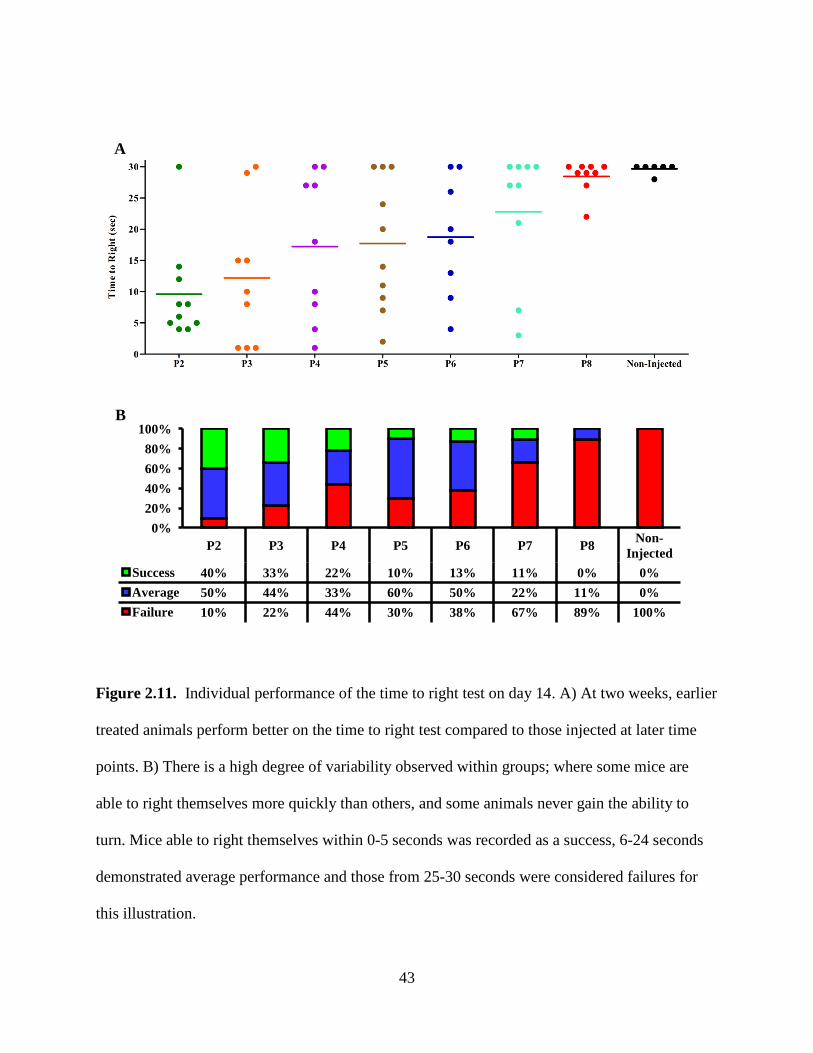
Early Treatment Improves Motor Function in Mice Injected with scAAV9-SMN
Mice have the propensity to right themselves onto all four paws immediately after being
placed on their back; and young, healthy neonates achieve this strength within the first week of
life [95]. Mice treated with scAAV9-SMN at the different time points exhibited differences in
motor function, as assessed by the time to right test. To assess the animals’ strength, a mouse
was placed on its back and the time it took them to right themselves was measured. A mouse
unable to right itself within 30 seconds was recorded as having failed the test for that trial. This
test was repeated daily throughout the lifespan of all treated mice. Over time, a greater
percentage of animals from groups P2 through P7 gained the ability to right themselves however,
this was not the case for the non-injected animals and those injected at the latest time point, P8
(Figure 2.9). Untreated SMNΔ7 mice display weakness beginning around day seven however;
some mice are inherently stronger than others. Therefore, it is not uncommon to observe mice
from the late injection and non-injected groups performing the time to right test on various days.
A high percentage of mice injected on P2, P3, and P4 (80%, 56%, 56%, respectively) gained the
ability to right themselves by day eleven, and they maintained this ability throughout the testing
period and into adulthood. In contrast, a lower percentage of treated mice from the median time
points P5 and P6 (20% and 22%, respectively) are able to right themselves by day 11; but the
ability to right does increase for these groups around day 14 (70% and 75%, respectively), and is
maintained throughout adulthood. Animals from the P7 group display variability in righting
themselves, with few animals turning during the first few days of the testing period. Although
these animals do not live until adulthood (Avg. 30 days, Figure 2.2), they all eventually gain and
maintain the ability to right throughout their shorter lifespan. Only five of the ten mice in the P8

40
injected group were ever able to right themselves, and four of them lost this ability 2-3 days prior
to death. As expected, mice of the non-injected group performed poorly on this test and only two
out of the ten mice gained the ability to right themselves.
Of the mice that are able to right themselves, the early-injected animals tended to turn
over faster than those injected at later time points and the mice in the non-injected group. Figure
2.10 displays the average time to right for each group on days 6-24. During the second week of
postnatal development P2 and P3 reduce their performance time substantially afterwards which
P2 is able to right immediately whereas the P3 group maintains an average time of 4 seconds.
The P4 group steadily reduces its average time to right during the second week and is also able to
turn over immediately starting on the third week. The P5 and P6 injected mice follow a similar
pattern but with less impact; these mice are able to turn over immediately but not until they are
into their third week. Mice in the P7 group were slower to gain this ability and did not perform
well until into the third week however, they finally decreased their average turning time which
they maintained until their death. Only a few animals in the P8 and non-injected groups were
able to turn, but they were slow to do so. Motor function varied for mice on a given day, both
between groups and within groups (Figure 2.11). However, a trend is apparent between groups of
mice injected at different time points, and those injected at earlier time points perform better on
the time to right test and are able to maintain this ability throughout their lifespan.

41
Figure 2.9. Percentage of animals able to right themselves. Assessment of motor function shows
that SMNΔ7 mice treated with scAAV9-SMN at earlier time points perform substantially better
on the time to right test. Animals treated at early time points gain the ability to right themselves
prior to animals injected at later time points.

42
Figure 2.10. Average Time to Right for SMNΔ7 mice treated with scAAV9-SMN. Mice
injected at later time points exhibit less muscle control and turn slower than mice injected at
earlier time points.
43
Figure 2.11. Individual performance of the time to right test on day 14. A) At two weeks, earlier
treated animals perform better on the time to right test compared to those injected at later time
points. B) There is a high degree of variability observed within groups; where some mice are
able to right themselves more quickly than others, and some animals never gain the ability to
turn. Mice able to right themselves within 0-5 seconds was recorded as a success, 6-24 seconds
demonstrated average performance and those from 25-30 seconds were considered failures for
this illustration.
0% 20% 40% 60% 80%
100%
P2 P3 P4 P5 P6 P7 P8 Non-Injected
Success 40% 33% 22% 10% 13% 11% 0% 0% Average 50% 44% 33% 60% 50% 22% 11% 0% Failure 10% 22% 44% 30% 38% 67% 89% 100%
A
B

44
SMN Protein Induction
Increased SMN protein was observed in animals treated with scAAV9-SMN (Figure
2.12). SMNΔ7 mice were injected on P2 or P7 and their tissues harvested on day eleven. The
increased SMN expression observed in P2 and P7 injected mice compared to unaffected mice
was due to the dose of viral particles resulting in higher levels of SMN compared to endogenous
expression levels. Furthermore, low levels of SMN observed in the non-injected animals was
expected because these mice harbor only two copies of human SMN2 and only 10% of
transcripts from each copy of the gene produce functional SMN protein. Mice injected on P2
exhibited significantly higher SMN levels in the brain compared to P7 injected mice (p =
0.0037) and P2 expressed significantly more SMN compared to the non-injected mice (p =
0.0032). P2 and P7 injected animals expressed significantly more SMN in the spinal cord
compared to non-injected animals (p = 0.0003 and p = 0.0082, respectively) and the P2 group
expressed significantly more protein than P7 (p = 0.0007). Robust induction was also observed
in peripheral organs such as the heart. In the heart tissue both P2 and P7 injected animals express
significantly more SMN than non-injected controls (p = 0.0006 and p = 0.0079, respectively).
Furthermore, scAAV9-SMN administration on P2 resulted in significantly higher SMN levels in
the heart compared with the P7 group (p = 0.0023). Protein induction on P2 or P7 greatly
increased protein levels in the brain, spinal cord and heart.

45
Figure 2.12. SMN protein induction is evident in SMNΔ7 mice injected at early and late time
points. In the brain, SMN was upregulated significantly in the P2 mice compared to both the P7
and non-injected groups, but there was no significance observed between P7 and the non-injected
group. There was robust expression observed in the spinal cord where both P2 and P7 injected
mice expressed significantly more protein than non-injected controls and the P2 group exhibited
a significantly higher level of protein than the P7 group. In the heart, both the P2 and P7 injected
mice exhibit a significant induction of SMN. The fold difference of the Western blots (n = 3 for
each group) is shown as the average SMN/actin ratio after normalization of SMN to β-actin.
46
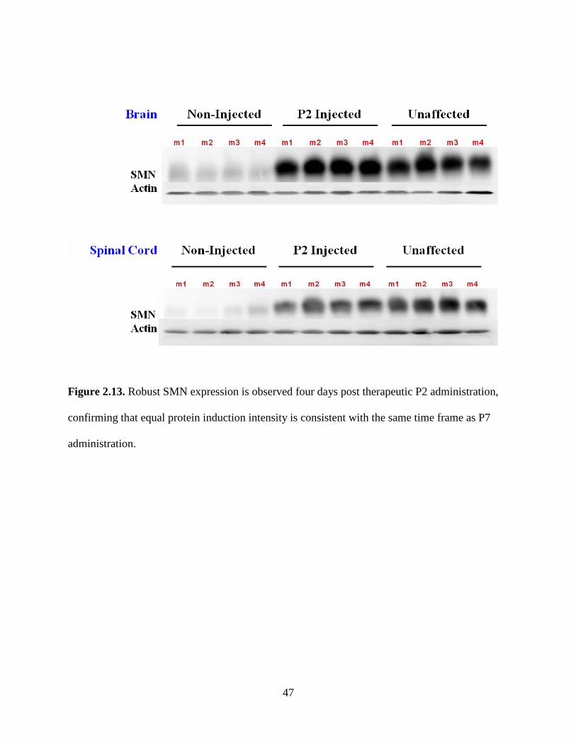
To answer the question of whether the robust expression observed in the P2 group
compared to P7 was due to the fact that the virus was expressing five days longer in the P2
animals, as tissues from both groups were harvested on day eleven, we also harvested tissues
from P2 injected animals on day 6 to account for this discrepancy (Figure 2.13).
47
Figure 2.13. Robust SMN expression is observed four days post therapeutic P2 administration,
confirming that equal protein induction intensity is consistent with the same time frame as P7
administration.

48
Peripheral Distribution of scAAV9-SMN and Transduction Analysis
To determine the extent to which scAAV9-SMN traverses the BBB and is distributed
throughout the periphery we utilized the scAAV9-GFP vector to observe virus dispersion.
Unaffected mice were injected with scAAV9-GFP on P2 or P7 and the heart, liver, spleen and
kidney tissues were harvested on day six or day eleven (Figure 2.14). Results confirm that
scAAV9 is well distributed throughout the periphery with especially strong GFP expression in
the liver and heart tissues.

49
Figure 2.14. Unaffected mice injected with scAAV9-GFP exhibit substantial GFP expression in
the periphery. In animals injected on P2, a stronger visualization was present throughout all
peripheral tissues whereas, in P7 injected animals GFP was expressed the strongest in the liver.

50
Discussion
Timing of therapeutic administration is critical for many diseases, and especially for
SMA. The majority of infants not diagnosed at birth present with acute respiratory failure within
the first year. By this time, the patients are severely compromised, whereas, an early diagnosis
would lead to proper nutritional, therapeutic and even respiratory support, which would improve
the quality of life for SMA children. Multiple reports have demonstrated that early intervention
provides maximal benefit regardless of the mode of therapy: SMN replacement or modulation of
SMN2 splicing. Restoring SMN pre- and post-symptomatically has led to complete or partial
rescue of SMA mice when SMN is reinstated during embryonic or early postnatal development.
Gene therapy using self-complementary adeno-associated virus, serotype 9 (scAAV9-
SMN), to replace SMN, has shown promising results in that it rescues the SMNΔ7 mouse model
when administered within the correct time frame [81, 85]. As SMA is namely a motor neuron
disease, the scAAV9 vector is efficient at transducing a large proportion of the target cell
population. scAAV9-SMN administration to SMNΔ7 mice intravenously on P1 or P2 resulted in
a full rescue (survival >250d) but delaying delivery until P5 decreased survival (median ~25d),
and treatment on P10 had no effect [81]. Comparison of IV and ICV delivery show that route of
administration plays a role in suppressing disease progression. In the severe SMN2 mouse model,
ICV injection significantly extended the survival when compared with IV administration. In the
SMNΔ7 model both injection routes resulted in a complete rescue but the ICV group
experienced fewer early deaths [85, 86].
Transgenic mice harboring an inducible Smn allele have been generated to analyze
temporal requirements of wild-type SMN levels. Early induction provides a more substantial
51
rescue compared to delayed induction, however, reinstatement during embryonic development
(E6) completely ameliorates the disease phenotype.
Antisense technology is utilized in SMA research to increase SMN protein levels by
modulating SMN2 splicing. For example, use of antisense oligomers (ASOs) to block an intron
splice silencer, ISS-N1, has been demonstrated to significantly increase full-length SMN2
transcript levels and SMN protein in brain and spinal cord of the SMNΔ7 model [77, 87]. When
the ISS-N1 ASO was administered by ICV injection on P0, the survival was extended from 15 to
100 days. However, this group found that delaying delivery until P4 decreased the efficacy of the
antisense therapy. Furthermore, IV administration on P0 resulted in survival comparable to the
ICV group however, when delivery was delayed until P4, the IV injected group exhibited
decreased survival.
Taken together, these developments all support the idea that early intervention provides
the maximal benefit for these modes of therapy. Here we have provided an in-depth analysis to
investigate temporal requirements for SMN protein. Multiple time points during the postnatal
period were chosen for analysis in which SMNΔ7 mice were injected with 1x10¹¹
We observed that survival, weight gain and muscle strength correlated with timing of
administration, and that mice injected at earlier time points lived longer, gained more weight and
performed better on the motor function test than mice injected at later time points (Figure 2.15).
All groups lived significantly longer than the P8 injected group and non-injected controls.
Interestingly, mice injected on P2, P3 and P4 lived significantly longer (median: 157, 102, 142
days, respectively) than those injected on P7 (median: 30 days). Despite the more modest effect
v.g. scAAV9-
SMN by ICV injection on a single day, P2 through P8.
52
observed with the P5 and P6 groups, each group had one mouse that lived past 200 days.
Furthermore, groups P2, P3 and P4 did not differ significantly from each other in average weight
gained, but mice from these earliest time points gained significantly more weight than all other
treated mice, and all groups gained significantly more weight than mice from the non-injected
group. Mice in all treatment groups (excluding P7) experienced significant weight gain
compared to P8 injected mice.
We have performed a systematic analysis to investigate the effect of delaying therapeutic
intervention using scAAV9-SMN with direct CNS administration. We utilized the SMNΔ7
mouse model because it exhibits a severe SMA phenotype but lives 13 days [56], allowing
enough time for investigation. Also, we used the self-complementary AAV vector because it
transduces cells efficiently [82, 83], especially motor neurons [84], and induces expression
relatively quickly (within 48 hours) [81]. SMN under the control of the chicken-β-actin promoter
results in ubiquitous expression in the CNS and peripheral tissues [96]. In the investigation
described here we assessed the phenotype of scAAV9-SMN treated mice using assays widely
accepted in the field.
While others have demonstrated that IV injection of scAAV9-SMN provides a complete
rescue in SMNΔ7 mice [81, 85], here we have performed ICV injection for direct delivery into
the CNS, which provided the same result. However, delaying administration by the intravenous
route provided a less substantial rescue [81] than when ICV administration was used, as we have
described here. Furthermore, ICV delivery requires less virus to be used, which is translationally
important for clinical consideration because virus production is costly. Perhaps in the case of
SMA, a combination of virus delivery through both injection routes may demonstrate to be a

53
more efficient means of gene therapy since peripheral tissues are proposed to be affected by low
levels of SMN as well as motor neurons.
The idea that early intervention provides maximal benefit was further supported by the
performance of these mice on the time-to-right test. By day 12, over 50% of mice in groups P2,
P3 and P4 were able to right themselves whereas, only 10-30% of mice in the late injection
groups could perform this test on day 12. Protein induction observed in CNS (robust) and
peripheral (modest) tissues of treated mice lends us to suggest that the resulting extent of rescue
observed in these mice is due, in part, to increased SMN levels in the CNS. Collectively these
data support the importance of early therapeutic intervention and demonstrate that a narrow
therapeutic window exists for which therapy must be applied to provide maximal benefit.
This systematic investigation into the effect of delaying therapeutic intervention using
scAAV9-SMN in the SMNΔ7 mouse model, has laid the foundation for others to elucidate the
correlation between our observations of the overt phenotype of these mice with the molecular
attributes involved in SMA pathogenesis. The results from this study support the idea that early
intervention provides the maximal benefit for ameliorating SMA disease pathogenesis and this is
clinically relevant as the field moves forward with designing therapeutics suitable for SMA
patients in the near future.

54
Figure 2.15. Overt phenotype of P2 treated animal with scAAV9-SMN compared to unaffected
and non-injected littermates.

55
CHAPTER THREE: CONCLUSIONS
As described throughout the literature and clinical assessments, early therapeutic
intervention provides maximum benefit in SMA patients and animal models. It is still unclear as
to the direct progression of SMA disease pathology as to whether direct motor neuron
degeneration causes SMA entirely or whether low SMN levels results in decline of peripheral
organs and tissues which has a negative impact on motor neurons. Differential strategies have
been implemented to further elucidate the biomolecular mechanisms occurring during SMA
pathogenesis and each provides insight to answer these questions.
We have provided a systematic and detailed analysis to determine the timing in which the
therapeutic window of providing maximal benefit closes, in the context of SMA. While
intervention at the earliest stages of postnatal development provides the most robust rescue with
a significant extension of survival, we also observed a reduced degree of rescue in the mice
injected at the later time points. While the majority of mice injected on P2 lived a full lifespan,
only a small cohort of mice from treatment groups P3 through P6 exhibited a full extension of
survival; with administration at P7 only providing a modest extension and P8 only provided
minimal benefit. Furthermore, the variability observed in weight gain and motor function
following treatment at the various time points suggests there is a progressive decline in these
animals following initiation of symptoms and that in order for therapeutic intervention to provide
a maximal benefit, intervention must be applied before disease symptoms progress too far.
Whether the target cell population is lost or too impaired to respond to treatment during late

56
stages of the disease has yet to determined, but with the advancement in gene therapy and now,
RNA-based therapy (ASOs), we will gain a better understanding of SMA disease progression.
Endeavors focused to understand how low SMN levels translate into SMA disease
pathology will be a critical component of future research in the SMA field. Development of
intermediate SMA mouse models will aid in the elucidation of important biochemical and
pathological aspects of SMA disease pathology. These studies can lead to reevaluation of the
timing effects and advance the investigations into the impact of delayed therapeutic intervention.
Furthermore, a combination of pharmacologics and gene therapy may provide a more substantial
effect upon therapeutic intervention [33].
Importantly, investigation into the genetic and molecular basis of SMA has provided
further insight on disease progression for other neurodegenerative disorders such as ALS and
Alzheimer’s, Huntington’s and Parkinson’s disease. Furthermore, the field of SMA research has
widely contributed to, as well as gained insight from, the multitude of other neuromuscular
diseases such as Muscular Dystrophy.

57
BIBLIOGRAPHY
1. Lefebvre, S., et al., Identification and characterization of a spinal muscular atrophy-determining gene. Cell, 1995. 80(1): p. 155-65.
2. Hendrickson, B.C., et al., Differences in SMN1 allele frequencies among ethnic groups within North America. J Med Genet, 2009. 46(9): p. 641-4.
3. Jedrzejowska, M., et al., Incidence of spinal muscular atrophy in Poland--more frequent than predicted? Neuroepidemiology, 2010. 34(3): p. 152-7.
4. Pearn, J., Incidence, prevalence, and gene frequency studies of chronic childhood spinal muscular atrophy. J Med Genet, 1978. 15(6): p. 409-13.
5. Pearn, J., Classification of spinal muscular atrophies. Lancet, 1980. 1(8174): p. 919-22.
6. Lorson, M.A. and C.L. Lorson, SMN-inducing compounds for the treatment of spinal muscular atrophy. Future Med Chem, 2012. 4(16): p. 2067-84.
7. Rothwell, E., et al., Public attitudes regarding a pilot study of newborn screening for spinal muscular atrophy. Am J Med Genet A, 2013. 161(4): p. 679-86.
8. Wilkins, R.H., MD and I.A. Brody, MD, Infantile Spinal Muscular Atrophy. Archives of neurology (Chicago), 1971. 25(3): p. 1.
9. Brzustowicz, L.M., et al., Genetic mapping of chronic childhood-onset spinal muscular atrophy to chromosome 5q11.2-13.3. Nature, 1990. 344(6266): p. 540-1.
10. Novelli, G., et al., Expression study of survival motor neuron gene in human fetal tissues. Biochem Mol Med, 1997. 61(1): p. 102-6.
11. Monani, U.R., et al., A single nucleotide difference that alters splicing patterns distinguishes the SMA gene SMN1 from the copy gene SMN2. Hum Mol Genet, 1999. 8(7): p. 1177-83.

58
12. Coovert, D.D., et al., The survival motor neuron protein in spinal muscular atrophy. Hum Mol Genet, 1997. 6(8): p. 1205-14.
13. Lefebvre, S., et al., Correlation between severity and SMN protein level in spinal muscular atrophy. Nat Genet, 1997. 16(3): p. 265-9.
14. Feldkotter, M., et al., Quantitative analyses of SMN1 and SMN2 based on real-time lightCycler PCR: fast and highly reliable carrier testing and prediction of severity of spinal muscular atrophy. Am J Hum Genet, 2002. 70(2): p. 358-68.
15. Cartegni, L. and A.R. Krainer, Disruption of an SF2/ASF-dependent exonic splicing enhancer in SMN2 causes spinal muscular atrophy in the absence of SMN1. Nat Genet, 2002. 30(4): p. 377-84.
16. Kashima, T. and J.L. Manley, A negative element in SMN2 exon 7 inhibits splicing in spinal muscular atrophy. Nat Genet, 2003. 34(4): p. 460-3.
17. Miyaso, H., et al., An intronic splicing enhancer element in survival motor neuron (SMN) pre-mRNA. J Biol Chem, 2003. 278(18): p. 15825-31.
18. Singh, N.K., et al., Splicing of a critical exon of human Survival Motor Neuron is regulated by a unique silencer element located in the last intron. Mol Cell Biol, 2006. 26(4): p. 1333-46.
19. Lorson, C.L., et al., A single nucleotide in the SMN gene regulates splicing and is responsible for spinal muscular atrophy. Proc Natl Acad Sci U S A, 1999. 96(11): p. 6307-11.
20. Lorson, C.L., et al., SMN oligomerization defect correlates with spinal muscular atrophy severity. Nat Genet, 1998. 19(1): p. 63-6.
21. Pellizzoni, L., B. Charroux, and G. Dreyfuss, SMN mutants of spinal muscular atrophy patients are defective in binding to snRNP proteins. Proc Natl Acad Sci U S A, 1999. 96(20): p. 11167-72.

59
22. Burnett, B.G., et al., Regulation of SMN protein stability. Mol Cell Biol, 2009. 29(5): p. 1107-15.
23. Coady, T.H. and C.L. Lorson, SMN in spinal muscular atrophy and snRNP biogenesis. Wiley Interdiscip Rev RNA, 2011. 2(4): p. 546-64.
24. Eggert, C., et al., Spinal muscular atrophy: the RNP connection. Trends Mol Med, 2006. 12(3): p. 113-21.
25. Meister, G., C. Eggert, and U. Fischer, SMN-mediated assembly of RNPs: a complex story. Trends Cell Biol, 2002. 12(10): p. 472-8.
26. Paushkin, S., et al., The SMN complex, an assemblyosome of ribonucleoproteins. Curr Opin Cell Biol, 2002. 14(3): p. 305-12.
27. Strasswimmer, J., et al., Identification of survival motor neuron as a transcriptional activator-binding protein. Hum Mol Genet, 1999. 8(7): p. 1219-26.
28. Young, P.J., et al., Minute virus of mice small nonstructural protein NS2 interacts and colocalizes with the Smn protein. J Virol, 2002. 76(12): p. 6364-9.
29. Young, P.J., et al., Minute virus of mice NS1 interacts with the SMN protein, and they colocalize in novel nuclear bodies induced by parvovirus infection. J Virol, 2002. 76(8): p. 3892-904.
30. Liu, Q. and G. Dreyfuss, A novel nuclear structure containing the survival of motor neurons protein. EMBO J, 1996. 15(14): p. 3555-65.
31. Young, P.J., et al., Nuclear gems and Cajal (coiled) bodies in fetal tissues: nucleolar distribution of the spinal muscular atrophy protein, SMN. Exp Cell Res, 2001. 265(2): p. 252-61.
32. Young, P.J., et al., The relationship between SMN, the spinal muscular atrophy protein, and nuclear coiled bodies in differentiated tissues and cultured cells. Exp Cell Res, 2000. 256(2): p. 365-74.

60
33. Burghes, A.H. and C.E. Beattie, Spinal muscular atrophy: why do low levels of survival motor neuron protein make motor neurons sick? Nat Rev Neurosci, 2009. 10(8): p. 597-609.
34. Sanchez, G., et al., A novel function for the survival motoneuron protein as a translational regulator. Hum Mol Genet, 2013. 22(4): p. 668-84.
35. Wan, L., et al., Inactivation of the SMN complex by oxidative stress. Mol Cell, 2008. 31(2): p. 244-54.
36. Zou, T., et al., SMN deficiency reduces cellular ability to form stress granules, sensitizing cells to stress. Cell Mol Neurobiol, 2011. 31(4): p. 541-50.
37. Wang, Z.B., X. Zhang, and X.J. Li, Recapitulation of spinal motor neuron-specific disease phenotypes in a human cell model of spinal muscular atrophy. Cell Res, 2013. 23(3): p. 378-93.
38. Rossoll, W., et al., Smn, the spinal muscular atrophy-determining gene product, modulates axon growth and localization of beta-actin mRNA in growth cones of motoneurons. J Cell Biol, 2003. 163(4): p. 801-12.
39. Zhang, Z., et al., SMN deficiency causes tissue-specific perturbations in the repertoire of snRNAs and widespread defects in splicing. Cell, 2008. 133(4): p. 585-600.
40. Pellizzoni, L., Chaperoning ribonucleoprotein biogenesis in health and disease. EMBO Rep, 2007. 8(4): p. 340-5.
41. Terns, M.P. and R.M. Terns, Macromolecular complexes: SMN--the master assembler. Curr Biol, 2001. 11(21): p. R862-4.
42. Battle, D.J., et al., The SMN complex: an assembly machine for RNPs. Cold Spring Harb Symp Quant Biol, 2006. 71: p. 313-20.
43. Pellizzoni, L., J. Yong, and G. Dreyfuss, Essential role for the SMN complex in the specificity of snRNP assembly. Science, 2002. 298(5599): p. 1775-9.

61
44. Matera, A.G., R.M. Terns, and M.P. Terns, Non-coding RNAs: lessons from the small nuclear and small nucleolar RNAs. Nat Rev Mol Cell Biol, 2007. 8(3): p. 209-20.
45. Mouaikel, J., et al., Hypermethylation of the cap structure of both yeast snRNAs and snoRNAs requires a conserved methyltransferase that is localized to the nucleolus. Mol Cell, 2002. 9(4): p. 891-901.
46. Raker, V.A., G. Plessel, and R. Luhrmann, The snRNP core assembly pathway: identification of stable core protein heteromeric complexes and an snRNP subcore particle in vitro. EMBO J, 1996. 15(9): p. 2256-69.
47. Prior, T.W., Spinal muscular atrophy: a time for screening. Curr Opin Pediatr, 2010. 22(6): p. 696-702.
48. Swoboda, K.J., et al., Natural history of denervation in SMA: relation to age, SMN2 copy number, and function. Ann Neurol, 2005. 57(5): p. 704-12.
49. Prior, T.W., Spinal muscular atrophy: newborn and carrier screening. Obstet Gynecol Clin North Am, 2010. 37(1): p. 23-36, Table of Contents.
50. Pyatt, R.E. and T.W. Prior, A feasibility study for the newborn screening of spinal muscular atrophy. Genet Med, 2006. 8(7): p. 428-37.
51. Viollet, L., et al., cDNA isolation, expression, and chromosomal localization of the mouse survival motor neuron gene (Smn). Genomics, 1997. 40(1): p. 185-8.
52. DiDonato, C.J., et al., Cloning, characterization, and copy number of the murine survival motor neuron gene: homolog of the spinal muscular atrophy-determining gene. Genome Res, 1997. 7(4): p. 339-52.
53. Schrank, B., et al., Inactivation of the survival motor neuron gene, a candidate gene for human spinal muscular atrophy, leads to massive cell death in early mouse embryos. Proc Natl Acad Sci U S A, 1997. 94(18): p. 9920-5.
54. Hsieh-Li, H.M., et al., A mouse model for spinal muscular atrophy. Nat Genet, 2000. 24(1): p. 66-70.
62
55. Monani, U.R., et al., The human centromeric survival motor neuron gene (SMN2) rescues embryonic lethality in Smn(-/-) mice and results in a mouse with spinal muscular atrophy. Hum Mol Genet, 2000. 9(3): p. 333-9.
56. Le, T.T., et al., SMNDelta7, the major product of the centromeric survival motor neuron (SMN2) gene, extends survival in mice with spinal muscular atrophy and associates with full-length SMN. Hum Mol Genet, 2005. 14(6): p. 845-57.
57. Cobb, M.S., et al., Development and characterization of an SMN2-based intermediate mouse model of Spinal Muscular Atrophy. Hum Mol Genet, 2013. 22(9): p. 1843-55.
58. Mattis, V.B., et al., A SMNDelta7 read-through product confers functionality to the SMNDelta7 protein. Neurosci Lett, 2008. 442(1): p. 54-8.
59. Bowerman, M., et al., A critical smn threshold in mice dictates onset of an intermediate spinal muscular atrophy phenotype associated with a distinct neuromuscular junction pathology. Neuromuscul Disord, 2012. 22(3): p. 263-76.
60. Chang, J.G., et al., Treatment of spinal muscular atrophy by sodium butyrate. Proc Natl Acad Sci U S A, 2001. 98(17): p. 9808-13.
61. Brichta, L., et al., Valproic acid increases the SMN2 protein level: a well-known drug as a potential therapy for spinal muscular atrophy. Hum Mol Genet, 2003. 12(19): p. 2481-9.
62. Andreassi, C., et al., Phenylbutyrate increases SMN expression in vitro: relevance for treatment of spinal muscular atrophy. Eur J Hum Genet, 2004. 12(1): p. 59-65.
63. Mercuri, E., et al., Randomized, double-blind, placebo-controlled trial of phenylbutyrate in spinal muscular atrophy. Neurology, 2007. 68(1): p. 51-5.
64. Swoboda, K.J., et al., Phase II open label study of valproic acid in spinal muscular atrophy. PLoS One, 2009. 4(5): p. e5268.
63
65. Kissel, J.T., et al., SMA CARNIVAL TRIAL PART II: a prospective, single-armed trial of L-carnitine and valproic acid in ambulatory children with spinal muscular atrophy. PLoS One, 2011. 6(7): p. e21296.
66. Avila, A.M., et al., Trichostatin A increases SMN expression and survival in a mouse model of spinal muscular atrophy. J Clin Invest, 2007. 117(3): p. 659-71.
67. Riessland, M., et al., SAHA ameliorates the SMA phenotype in two mouse models for spinal muscular atrophy. Hum Mol Genet, 2010. 19(8): p. 1492-1506.
68. Narver, H.L., et al., Sustained improvement of spinal muscular atrophy mice treated with trichostatin A plus nutrition. Ann Neurol, 2008. 64(4): p. 465-70.
69. Bricceno, K.V., et al., Histone deacetylase inhibition suppresses myogenin-dependent atrogene activation in spinal muscular atrophy mice. Hum Mol Genet, 2012. 21(20): p. 4448-59.
70. Ting, C.H., et al., Stat5 constitutive activation rescues defects in spinal muscular atrophy. Hum Mol Genet, 2007. 16(5): p. 499-514.
71. Farooq, F., et al., Prolactin increases SMN expression and survival in a mouse model of severe spinal muscular atrophy via the STAT5 pathway. J Clin Invest, 2011. 121(8): p. 3042-50.
72. Baughan, T., et al., Stimulating full-length SMN2 expression by delivering bifunctional RNAs via a viral vector. Mol Ther, 2006. 14(1): p. 54-62.
73. Baughan, T.D., et al., Delivery of bifunctional RNAs that target an intronic repressor and increase SMN levels in an animal model of spinal muscular atrophy. Hum Mol Genet, 2009. 18(9): p. 1600-11.
74. Dickson, A., E. Osman, and C.L. Lorson, A negatively acting bifunctional RNA increases survival motor neuron both in vitro and in vivo. Hum Gene Ther, 2008. 19(11): p. 1307-15.

64
75. Owen, N., et al., Design principles for bifunctional targeted oligonucleotide enhancers of splicing. Nucleic Acids Res, 2011.
76. Skordis, L.A., et al., Characterisation of novel point mutations in the survival motor neuron gene SMN, in three patients with SMA. Hum Genet, 2001. 108(4): p. 356-7.
77. Osman, E.Y., P.F. Yen, and C.L. Lorson, Bifunctional RNAs targeting the intronic splicing silencer N1 increase SMN levels and reduce disease severity in an animal model of spinal muscular atrophy. Mol Ther, 2012. 20(1): p. 119-26.
78. Miyajima, H., et al., Identification of a cis-acting element for the regulation of SMN exon 7 splicing. J Biol Chem, 2002. 277(26): p. 23271-7.
79. Porensky, P.N. and A.H. Burghes, Antisense oligonucleotides for the treatment of spinal muscular atrophy. Hum Gene Ther, 2013. 24(5): p. 489-98.
80. Lorson, C.L., H. Rindt, and M. Shababi, Spinal muscular atrophy: mechanisms and therapeutic strategies. Hum Mol Genet, 2010. 19(R1): p. R111-8.
81. Foust, K.D., et al., Rescue of the spinal muscular atrophy phenotype in a mouse model by early postnatal delivery of SMN. Nat Biotechnol, 2010. 28(3): p. 271-4.
82. Duque, S., et al., Intravenous administration of self-complementary AAV9 enables transgene delivery to adult motor neurons. Mol Ther, 2009. 17(7): p. 1187-96.
83. McCarty, D.M., Self-complementary AAV vectors; advances and applications. Mol Ther, 2008. 16(10): p. 1648-56.
84. Foust, K.D., et al., Intravascular AAV9 preferentially targets neonatal neurons and adult astrocytes. Nat Biotechnol, 2009. 27(1): p. 59-65.
85. Glascock, J.J., et al., Direct central nervous system delivery provides enhanced protection following vector mediated gene replacement in a severe model of spinal muscular atrophy. Biochem Biophys Res Commun, 2012. 417(1): p. 376-81.

65
86. Glascock, J.J., et al., Decreasing disease severity in symptomatic, Smn(-/-);SMN2(+/+), spinal muscular atrophy mice following scAAV9-SMN delivery. Hum Gene Ther, 2012. 23(3): p. 330-5.
87. Porensky, P.N., et al., A single administration of morpholino antisense oligomer rescues spinal muscular atrophy in mouse. Hum Mol Genet, 2012. 21(7): p. 1625-38.
88. Lutz, C.M., et al., Postsymptomatic restoration of SMN rescues the disease phenotype in a mouse model of severe spinal muscular atrophy. J Clin Invest, 2011. 121(8): p. 3029-41.
89. Le, T.T., et al., Temporal requirement for high SMN expression in SMA mice. Hum Mol Genet, 2011.
90. Understanding spinal muscular atrophy. Nat Struct Biol, 2001. 8(1): p. 1.
91. Hua, Y., et al., Peripheral SMN restoration is essential for long-term rescue of a severe spinal muscular atrophy mouse model. Nature, 2011. 478(7367): p. 123-6.
92. Benkhelifa-Ziyyat, S., et al., Intramuscular scAAV9-SMN injection mediates widespread gene delivery to the spinal cord and decreases disease severity in SMA mice. Mol Ther, 2013. 21(2): p. 282-90.
93. Lee, A.J., et al., Limited phenotypic effects of selectively augmenting the SMN protein in the neurons of a mouse model of severe spinal muscular atrophy. PLoS One, 2012. 7(9): p. e46353.
94. Grieger, J.C., V.W. Choi, and R.J. Samulski, Production and characterization of adeno-associated viral vectors. Nat Protoc, 2006. 1(3): p. 1412-28.
95. Butchbach, M.E., J.D. Edwards, and A.H. Burghes, Abnormal motor phenotype in the SMNDelta7 mouse model of spinal muscular atrophy. Neurobiol Dis, 2007. 27(2): p. 207-19.
66
96. Gray, S.J., et al., Optimizing promoters for recombinant adeno-associated virus-mediated gene expression in the peripheral and central nervous system using self-complementary vectors. Hum Gene Ther, 2011. 22(9): p. 1143-53.