deficiency or inhibition of gas6 causes platelet...
TRANSCRIPT

LUND UNIVERSITY
PO Box 117221 00 Lund+46 46-222 00 00
Deficiency or inhibition of Gas6 causes platelet dysfunction and protects mice againstthrombosis
Angelillo-Scherrer, Anne; Garcia de Frutos, Pablo; Aparicio, Cristina; Melis, Els; Savi, Pierre;Lupu, Florea; Arnout, Jef; Dewerchin, Mieke; Hoylaerts, Marc F.; Herbert, Jean-Marc; Collen,Désiré; Dahlbäck, Björn; Carmeliet, PeterPublished in:Nature Medicine
DOI:10.1038/84667
2001
Link to publication
Citation for published version (APA):Angelillo-Scherrer, A., Garcia de Frutos, P., Aparicio, C., Melis, E., Savi, P., Lupu, F., ... Carmeliet, P. (2001).Deficiency or inhibition of Gas6 causes platelet dysfunction and protects mice against thrombosis. NatureMedicine, 7(2), 215-221. https://doi.org/10.1038/84667
General rightsCopyright and moral rights for the publications made accessible in the public portal are retained by the authorsand/or other copyright owners and it is a condition of accessing publications that users recognise and abide by thelegal requirements associated with these rights.
• Users may download and print one copy of any publication from the public portal for the purpose of private studyor research. • You may not further distribute the material or use it for any profit-making activity or commercial gain • You may freely distribute the URL identifying the publication in the public portalTake down policyIf you believe that this document breaches copyright please contact us providing details, and we will removeaccess to the work immediately and investigate your claim.

NATURE MEDICINE • VOLUME 7 • NUMBER 2 • FEBRUARY 2001 215
ARTICLES
Gas6, the product of the growth arrest-specific gene 6 (Gas6), is anew member of the vitamin K-dependent protein family1,2.Proteins belonging to this family are characterized by post-trans-lational γ-carboxylation of certain glutamic acid residues by acarboxylase, using vitamin K as cofactor. The γ-carboxyglutamicacid (Gla)-containing module in prothrombin, coagulation fac-tors VII, IX and X, protein C, protein Z, protein S and Gas6 al-lows these vitamin K-dependent plasma proteins to bind tonegatively charged phospholipid membranes3. Gas6 is struc-turally similar to protein S, but lacks a loop, crucial for the anti-coagulant activity of protein S (ref. 2). The latter is a cofactor foractivated protein C, which inactivates the coagulation factors Vaand VIIIa (ref. 4). Genetic deficiency of protein S in humans isone of the most severe inherited risk factors for thrombosis5. Todate, Gas6 is the only protein among Gla-module–containingproteins which has not been reported to play a role in hemosta-sis or thrombosis.
Apart from a Gla-domain–dependent interaction with phos-pholipid membranes6, Gas6 also binds as a ligand to the receptortyrosine kinases Axl (Ark, Ufo, Tyro7), Sky (Rse, Tyro3, Dtk, Etk,Brt, Tif) and Mer (c-Mer, Eyk, Nyk)7–11 by its carboxy-terminalglobular G domains9. It has been implicated in reversible cellgrowth arrest2, survival12, proliferation12–14 and cell adhesion6,15,16.Mice with a triple deficiency of Axl, Sky and Mer are viable, buthave not been reported to suffer spontaneous bleeding or throm-bosis17.
Here, we generated Gas6–/– mice to investigate the role of Gas6in hemostasis and thrombosis. We found that deficiency of Gas6protected mice against fatal thrombosis, but did not induce
bleeding. Gas6 was found to amplify the aggregation and secre-tion response of platelets to known agonists, and the plateletdysfunction in Gas6-deficient mice resembled the platelet de-fects of patients with primary platelet signal transduction de-fects18. Gas6 antibodies protected mice against fatal thromboembolism without inducing a bleeding tendency, indicating thatinhibition of Gas6 might provide a novel means to safely blockthrombosis.
Results
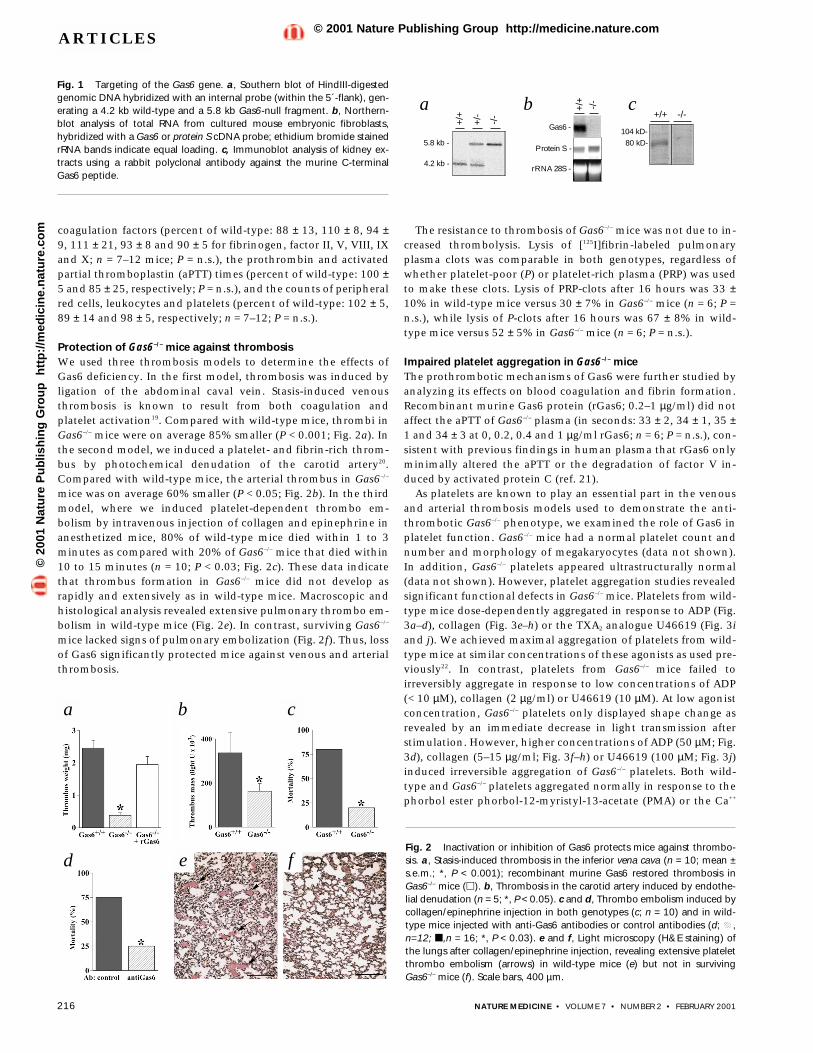
Normal hemostasis in Gas6–/– miceWe inactivated Gas6 by deleting the transcription start site, thetranslation initiation codon, the signal peptide and the Glamodule that is required for binding of Gas6 to phospholipidmembranes. We confirmed correct targeting at the DNA, RNAand protein level (Fig. 1a–c; Fig. 5b). Homozygous Gas6–/– micewere born at the expected mendelian frequency: of 317 offspringfrom heterozygous Gas6+/– breeding pairs, 72 were wild-type(Gas6+/+), 168 were Gas6+/– and 77 were Gas6–/–. Gas6+/– and Gas6–/–
mice were viable, fertile, appeared normal and showed no obvi-ous differences in size, weight or behavior. No genotypic differ-ences in litter size were observed (9.7 ± 2 for wild-type mice; n =23 litters versus 9.3 ± 3 for Gas6–/– mice; n = 44 litters; P = n.s.).
Gas6–/– mice did not suffer spontaneous bleeding or thrombo-sis. Bleeding (estimated as the amount of blood loss) after tailclipping was comparable for both genotypes (166 ± 48 µl in wild-type mice versus 172 ± 68 µl in Gas6–/– mice; n = 10; P = n.s.).There were also no genotypic differences in the plasma levels of
Deficiency or inhibition of Gas6 causes platelet dysfunctionand protects mice against thrombosis
ANNE ANGELILLO-SCHERRER1, PABLO GARCIA DE FRUTOS2, CRISTINA APARICIO2, ELS MELIS1,PIERRE SAVI3, FLOREA LUPU4, JEF ARNOUT1, MIEKE DEWERCHIN1, MARC F. HOYLAERTS1, JEAN-
MARC HERBERT3,DÉSIRÉ COLLEN1, BJÖRN DAHLBÄCK2 & PETER CARMELIET1
1The Center for Transgene Technology and Gene Therapy, Flanders Interuniversity Institute for Biotechnology, Leuven, Belgium
2Department of Clinical Chemistry, Lund University, Wallenberg Laboratory, Malmö University Hospital, Malmö, Sweden
3Cardiovascular/Thrombosis Research Department, Sanofi∼ Synthélabo, Toulouse, France 4Thrombosis Research Institute, Vascular Biology Laboratory, London, UK
A.A.S. and P.G.F. contributed equally to this study.Correspondence should be addressed to P.C.; email: [email protected]
The growth arrest-specific gene 6 product (Gas6) is a secreted protein related to the anticoagu-lant protein S but its role in hemostasis is unknown. Here we show that inactivation of the Gas6gene prevented venous and arterial thrombosis in mice, and protected against fatal collagen/ep-inephrine-induced thrombo embolism. Gas6–/– mice did not, however, suffer spontaneous bleed-ing and had normal bleeding after tail clipping. In addition, we found that Gas6 antibodiesinhibited platelet aggregation in vitro and protected mice against fatal thrombo embolism with-out causing bleeding in vivo. Gas6 amplified platelet aggregation and secretion in response toknown agonists. Platelet dysfunction in Gas6–/– mice resembled that of patients with platelet sig-naling transduction defects. Thus, Gas6 is a platelet-response amplifier that plays a significantrole in thrombosis. These findings warrant further evaluation of the possible therapeutic use ofGas6 inhibition for prevention of thrombosis.
©20
01 N
atu
re P
ub
lish
ing
Gro
up
h
ttp
://m
edic
ine.
nat
ure
.co
m© 2001 Nature Publishing Group http://medicine.nature.com
216 NATURE MEDICINE • VOLUME 7 • NUMBER 2 • FEBRUARY 2001
ARTICLES
coagulation factors (percent of wild-type: 88 ± 13, 110 ± 8, 94 ±9, 111 ± 21, 93 ± 8 and 90 ± 5 for fibrinogen, factor II, V, VIII, IXand X; n = 7–12 mice; P = n.s.), the prothrombin and activatedpartial thromboplastin (aPTT) times (percent of wild-type: 100 ±5 and 85 ± 25, respectively; P = n.s.), and the counts of peripheralred cells, leukocytes and platelets (percent of wild-type: 102 ± 5,89 ± 14 and 98 ± 5, respectively; n = 7–12; P = n.s.).
Protection of Gas6–/– mice against thrombosisWe used three thrombosis models to determine the effects ofGas6 deficiency. In the first model, thrombosis was induced byligation of the abdominal caval vein. Stasis-induced venousthrombosis is known to result from both coagulation andplatelet activation19. Compared with wild-type mice, thrombi inGas6–/– mice were on average 85% smaller (P < 0.001; Fig. 2a). Inthe second model, we induced a platelet- and fibrin-rich throm-bus by photochemical denudation of the carotid artery20.Compared with wild-type mice, the arterial thrombus in Gas6–/–
mice was on average 60% smaller (P < 0.05; Fig. 2b). In the thirdmodel, where we induced platelet-dependent thrombo em-bolism by intravenous injection of collagen and epinephrine inanesthetized mice, 80% of wild-type mice died within 1 to 3minutes as compared with 20% of Gas6–/– mice that died within10 to 15 minutes (n = 10; P < 0.03; Fig. 2c). These data indicatethat thrombus formation in Gas6–/– mice did not develop asrapidly and extensively as in wild-type mice. Macroscopic andhistological analysis revealed extensive pulmonary thrombo em-bolism in wild-type mice (Fig. 2e). In contrast, surviving Gas6–/–
mice lacked signs of pulmonary embolization (Fig. 2f). Thus, lossof Gas6 significantly protected mice against venous and arterialthrombosis.
The resistance to thrombosis of Gas6–/– mice was not due to in-creased thrombolysis. Lysis of [125I]fibrin-labeled pulmonaryplasma clots was comparable in both genotypes, regardless ofwhether platelet-poor (P) or platelet-rich plasma (PRP) was usedto make these clots. Lysis of PRP-clots after 16 hours was 33 ±10% in wild-type mice versus 30 ± 7% in Gas6–/– mice (n = 6; P =n.s.), while lysis of P-clots after 16 hours was 67 ± 8% in wild-type mice versus 52 ± 5% in Gas6–/– mice (n = 6; P = n.s.).
Impaired platelet aggregation in Gas6–/– miceThe prothrombotic mechanisms of Gas6 were further studied byanalyzing its effects on blood coagulation and fibrin formation.Recombinant murine Gas6 protein (rGas6; 0.2–1 µg/ml) did notaffect the aPTT of Gas6–/– plasma (in seconds: 33 ± 2, 34 ± 1, 35 ±1 and 34 ± 3 at 0, 0.2, 0.4 and 1 µg/ml rGas6; n = 6; P = n.s.), con-sistent with previous findings in human plasma that rGas6 onlyminimally altered the aPTT or the degradation of factor V in-duced by activated protein C (ref. 21).
As platelets are known to play an essential part in the venousand arterial thrombosis models used to demonstrate the anti-thrombotic Gas6–/– phenotype, we examined the role of Gas6 inplatelet function. Gas6–/– mice had a normal platelet count andnumber and morphology of megakaryocytes (data not shown).In addition, Gas6–/– platelets appeared ultrastructurally normal(data not shown). However, platelet aggregation studies revealedsignificant functional defects in Gas6–/– mice. Platelets from wild-type mice dose-dependently aggregated in response to ADP (Fig.3a–d), collagen (Fig. 3e–h) or the TXA2 analogue U46619 (Fig. 3iand j). We achieved maximal aggregation of platelets from wild-type mice at similar concentrations of these agonists as used pre-viously22. In contrast, platelets from Gas6–/– mice failed toirreversibly aggregate in response to low concentrations of ADP(< 10 µM), collagen (2 µg/ml) or U46619 (10 µM). At low agonistconcentration, Gas6–/– platelets only displayed shape change asrevealed by an immediate decrease in light transmission afterstimulation. However, higher concentrations of ADP (50 µM; Fig.3d), collagen (5–15 µg/ml; Fig. 3f–h) or U46619 (100 µM; Fig. 3j)induced irreversible aggregation of Gas6–/– platelets. Both wild-type and Gas6–/– platelets aggregated normally in response to thephorbol ester phorbol-12-myristyl-13-acetate (PMA) or the Ca++
+/+ -/-
+/+
5.8 kb -
4.2 kb -
+/-
-/-
Gas6 -
+/+
104 kD-
80 kD-
-/-
Protein S -
rRNA 28S -
Fig. 2 Inactivation or inhibition of Gas6 protects mice against thrombo-sis. a, Stasis-induced thrombosis in the inferior vena cava (n = 10; mean ±s.e.m.; *, P < 0.001); recombinant murine Gas6 restored thrombosis inGas6–/– mice (�). b, Thrombosis in the carotid artery induced by endothe-lial denudation (n = 5; *, P < 0.05). c and d, Thrombo embolism induced bycollagen/epinephrine injection in both genotypes (c; n = 10) and in wild-type mice injected with anti-Gas6 antibodies or control antibodies (d; ,n=12; �,n = 16; *, P < 0.03). e and f, Light microscopy (H&E staining) ofthe lungs after collagen/epinephrine injection, revealing extensive plateletthrombo embolism (arrows) in wild-type mice (e) but not in survivingGas6–/– mice (f). Scale bars, 400 µm.
a b cFig. 1 Targeting of the Gas6 gene. a, Southern blot of HindIII-digestedgenomic DNA hybridized with an internal probe (within the 5´-flank), gen-erating a 4.2 kb wild-type and a 5.8 kb Gas6-null fragment. b, Northern-blot analysis of total RNA from cultured mouse embryonic fibroblasts,hybridized with a Gas6 or protein S cDNA probe; ethidium bromide stainedrRNA bands indicate equal loading. c, Immunoblot analysis of kidney ex-tracts using a rabbit polyclonal antibody against the murine C-terminalGas6 peptide.
a
fd e
b c
©20
01 N
atu
re P
ub
lish
ing
Gro
up
h
ttp
://m
edic
ine.
nat
ure
.co
m© 2001 Nature Publishing Group http://medicine.nature.com

NATURE MEDICINE • VOLUME 7 • NUMBER 2 • FEBRUARY 2001 217
ARTICLES
ionophore A23187 (data not shown).Thrombin stimulated platelet aggregation comparably in both
genotypes at all doses tested (data not shown). However, ultra-structural analysis revealed that thrombin-induced aggregates ofGas6–/– platelets were abnormal. In wild-type aggregates, plateletswere densely packed, made tight contacts with each other andwere completely degranulated (Fig. 4a). In contrast, platelets inGas6–/– aggregates were loosely packed, displayed fewer andsmaller contact sites and were incompletely degranulated (Fig.4b). Fibrinogen is released from α-granules upon platelet activa-tion and forms bridges, linking adjacent activated platelets23.Flow cytometry of washed platelets revealed that fibrinogen wasdetectable on the surface of wild-type platelets after stimulationwith ADP (20 µM; Fig. 4c). In contrast, levels of surface-bound-fibrinogen in Gas6–/– platelets did not increase upon stimulation(Fig. 4d), which may contribute to the loose assembly of Gas6–/–
platelet aggregates. The reduced amount of fibrinogen on Gas6–/–
platelet surfaces was not due to a defect of Gas6–/– platelets to se-quester fibrinogen, as revealed by the comparable amounts ofimmunoreactive fibrinogen in platelet lysates in both genotypes(Fig. 4e). Thus, impaired platelet aggregation in response toknown agonists in Gas6–/– mice contributed to their resistance tothrombosis.
Expression of Gas6 and its receptors in plateletsThe defect of platelet aggregation in Gas6–/– mice indicated thatplatelets produce and respond to Gas6. Therefore, the expressionof Gas6 and its receptors Axl, Sky and Mer was studied in restingand stimulated platelets. By reverse transcriptase (RT)-PCRanalysis, GAS6 mRNA transcripts were detected in humanplatelets (Fig. 5a). In addition, immunoblotting revealed thepresence of Gas6 in platelet extracts and in the releasate ofthrombin-activated platelets from wild-type but not from Gas6–/–
mice (Fig. 5b). Ultrastructural analysis combined with double
immunogold-labeling of resting wild-type platelets revealed thatGas6 colocalized with fibrinogen in α-granules (data not shown).Upon activation of wild-type platelets with thrombin (1 U/ml),Gas6 became detectable on the surface of platelets by immuno-gold-labeling (data not shown). Flow cytometry confirmed thatthe levels of surface-bound Gas6 were minimal in resting humanplatelets but significantly increased upon activation by ADP (5µM; Fig. 5c and d). These results extend previous observations ofGas6 in human megakaryocytes24 and rat platelets25.
Platelets also expressed Gas6 receptors. By RT-PCR analysis,transcripts of Axl, Sky and Mer were detected in human platelets(Fig. 5a). Immunogold-labeling revealed that Axl was localizedon the surface of resting platelets (data not shown). Comparableamounts of Axl and Sky (Fig. 5b) were detectable in wild-typeand Gas6–/– platelets, indicating that the reduced response ofGas6–/– platelets was not due to differences in Gas6 receptor ex-pression. Thus, platelets produce and release Gas6, and expressGas6 receptors.
Impaired secretion in Gas6–/– plateletsUltrastructural analysis of the thrombin-induced platelet aggre-gates indicated a reduced ability of Gas6–/– platelets to degranu-late. Secretion of ADP from dense granules is essential for theformation of stable macro-aggregates after initial formation ofsmall, unstable platelet aggregates26. Secretion of dense granule
a b c d
e f g h
i j k l m
Fig. 3 Effect of Gas6 deficiency or of anti-Gas6 antibodies on platelet ag-gregation. a–j, Aggregation of wild-type (+/+) and Gas6 deficient (–/–)platelet-rich plasma. Platelets from Gas6–/– mice were unable to fully aggre-gate in response to concentrations of ADP at 2.5 µM (a), 5 µM (b) and 10µM (c), of collagen at 2 µg/ml (e) or of the thromboxane A2 analogueU46619 at 10 µM (i), while higher concentrations of ADP (d; 50 µM), colla-gen (f–h; 5–15 µg/ml) or U46619 (j; 100 µM) induced irreversible plateletaggregation. Representative example of 4 independent experiments usingPRP pooled from 4 to 6 wild-type or Gas6–/– mice. Squares represent 2 min(X-axis) and 10% change in light transmission (Y-axis). k, Restoration of theimpaired aggregation of Gas6–/– platelets in response to ADP (5 µM) by re-combinant Gas6 (1 µg/ml) to comparable levels as in wild-type platelets.Representative example of 3 independent experiments. l and m,Aggregation response to ADP (5 µM) of washed human platelets afterpreincubation with anti-Gas6 antibodies (l) or isotype-matched control an-tibodies (m), revealing that anti-Gas6 antibodies prevent platelet aggrega-tion. Representative example of 3 independent experiments. Arrows in allpanels indicate application of the platelet agonists.
Table 1 ATP release in response to various agonists.
Agonist Concentration Wild-type mice Gas6–/– mice
ADP 20 µM 0.8 ± 0.08 0.2 + 0.09*20 µM + rGas6 n.d. 0.75 ± 0.150 µM 2.6 ± 0.8 1.3 ± 0.3*
Collagen 1 µg/ml 1.8 ± 0.3 < 0.110 µg/ml 10 ± 1.0 11 ± 0.8
U46619 10 µM 6.5 ± 1.5 < 0.1100 µM 8.2 ± 2.0 7.6 ± 2.3
Thrombin 1 U/ml 17 ± 1.6 11 ± 1.2*PMA 100 µM 2.3 ± 0.4 1.9 ± 0.6A23187 8 µM 9.2 ± 3.1 7.3 ± 3.0
ATP release expressed in µM. The data represent the mean ± s.e.m. of 3 ex-periments using platelet-rich plasma (for ADP, collagen, U46619, PMA orA23187 stimulation) or washed platelets (for thrombin stimulation). Notethat rGas6 (200 ng/ml) rescued the impaired ATP secretion of Gas6–/–
platelets. Each experiment was performed with a pool of 4 to 6 wild-type orGas6–/– mice. n.d. not done. *, P < 0.05 versus wild-type.
©20
01 N
atu
re P
ub
lish
ing
Gro
up
h
ttp
://m
edic
ine.
nat
ure
.co
m© 2001 Nature Publishing Group http://medicine.nature.com

218 NATURE MEDICINE • VOLUME 7 • NUMBER 2 • FEBRUARY 2001
ARTICLES
stores (evaluated by measuring release of ATP) was significantlyimpaired in Gas6–/– platelets. Compared with wild-type platelets,release of ATP from Gas6–/– platelets was significantly decreasedin response to ADP, collagen or U46619, when these agonistswere used at low concentrations, which only caused plateletshape changes or reversible platelet aggregation (Table 1). ATPrelease from Gas6–/– platelets was also reduced in response tohigh concentrations of ADP (50 µM) or thrombin (1 U/ml), con-sistent with the incomplete degranulation of thrombin-stimu-lated Gas6–/– platelets (ultrastructural analysis). However, releaseof ATP was normal or only slightly reduced when Gas6–/–
platelets were stimulated with high concentrations of collagen(10 µg/ml) or U46619 (100 µM; Table 1), which cause irreversibleplatelet aggregation. PMA and the Ca++ ionophore A23187 in-duced a normal secretory response in both genotypes (Table 1).Secretion of α-granules, as assessed by measurement of surfaceexpression of P-selectin during platelet activation, was also im-paired in Gas6–/– platelets (Fig. 5e and f). Thus, we found a closecorrelation between the defects in the aggregation and secretionresponse of Gas6–/– platelets to various agonists.
Production of TXA2, which contributes to the formation of sta-ble macro-aggregates26, was normal in both genotypes.Production of TXA2 in serum and upon activation of platelets bythrombin (5 U/ml) was estimated by measurement of TXB2.Levels of TXB2 were 110 ± 63 ng/ml in serum and 53 ± 23 ng/mlafter thrombin activation of wild-type mice versus 120 ± 41
ng/ml in serum and 55 ± 16 ng/ml after thrombin activation ofGas6–/– mice (n = 6; P = n.s.).
Restoration of the Gas6–/– phenotype by recombinant Gas6To confirm that the platelet dysfunction in Gas6–/– mice was dueto deficiency of Gas6, we evaluated the effect of recombinantmurine Gas6 (rGas6) on the impaired aggregation and secretionof Gas6–/– platelets in vitro. Whereas rGas6 itself—at a concentra-tion of up to 10 µg/ml—was unable to induce a shape change oraggregation of wild-type or Gas6–/– platelets, a concentration of200 ng/ml restored the defective aggregation (Fig. 3k) and ATPsecretion (Fig. 5g) of Gas6–/– platelets in response to ADP.Moreover, the thrombotic defect in Gas6–/– mice in vivo was re-stored by administering rGas6 at a dose of 100 µg/kg (n = 8; P >0.05 by comparison to wild-type mice; Fig. 2a). These findingsindicate that Gas6 stimulates platelet function by amplifying theresponse to other platelet activators.
Antibodies against Gas6 inhibit platelet functionIn order to examine whether inhibitors of Gas6 might be usefulto prevent thrombosis, we studied the effect of antibodies spe-cific for Gas6 on platelet aggregation in vitro and on thrombo
+/+
-/-
+/+ -/-
num
bero
f cel
lsnu
mbe
rof c
ells
relative fluorescence
ADP 20 µM
resting
ADP 20 µM
Aα
Bβγ
resting
relative fluor
escence
escence
a
b
c
d
e
Fig. 4 Role of Gas6 in the formation of platelet macro-aggregates. a andb, Electron microscopy revealed that platelets at the borders of platelet ag-gregates in wild-type mice (+/+) were densely compacted and completelydegranulated (a). In contrast, Gas6 deficient (–/–) platelet aggregates of acomparable size were loosely packed, had fewer contact sites and were in-completely degranulated (b). Arrows indicate α-granules and arrowheadsindicate dense granules in a and b. Scale bars, 4 µm. c and d, Flow-cytome-try analysis of fibrinogen on washed wild-type (c) and Gas6–/– (d) platelets(black line denotes resting platelets, green line denotes ADP-activatedplatelets), revealing that surface-bound fibrinogen levels only increased instimulated wild-type but not in Gas6–/– platelets. e, Western-blot analysis ofwashed platelet lysates reveals comparable amounts of fibrinogen in wild-type and Gas6–/– platelets.
a
b
c
d
e
f
g
Fig. 5 Expression and role of Gas6 in platelets. a, RT-PCR analysis of Gas6and its receptors Axl, Sky and Mer in human platelets. b, Western-blotanalysis revealing Gas6 in extracts of resting platelets and in releasates ofthrombin-activated platelets from wild-type but not from Gas6–/– mice andcomparable expression of the Gas6 receptors Axl and Sky. c and d, Flow cy-tometry using Gas6-antibodies, revealing minimal Gas6 on the surface ofresting human platelets (c) and increased levels of Gas6 on human plateletsstimulated by ADP 5µM (d). e and f, Flow cytometry of P-selectin on restingplatelets (black line) and on ADP-activated platelets (green line), revealingthat surface expression of P-selectin increased in stimulated wild-type (e)but not in Gas6–/– (f) platelets. g, Rescue by rGas6 (200 ng/ml) of the defec-tive Gas6–/– platelet ATP secretion in response to ADP 20 µM. A representa-tive tracing of ATP secretion by Gas6–/– platelets with and without rGas6,and by wild-type platelets is displayed. The average ATP levels (µM) afterrGas6 rescue are indicated in Table 1. The arrow indicates the time of appli-cation of ADP.
©20
01 N
atu
re P
ub
lish
ing
Gro
up
h
ttp
://m
edic
ine.
nat
ure
.co
m© 2001 Nature Publishing Group http://medicine.nature.com

NATURE MEDICINE • VOLUME 7 • NUMBER 2 • FEBRUARY 2001 219
ARTICLES
embolism after a collagen/epinephrine challenge in vivo. Weused antibodies directed against the C-terminal part of Gas6—re-sponsible for binding of Gas6 to its receptors27. In contrast to iso-type-matched control antibodies, Gas6-neutralizing antibodiesdose-dependently blocked aggregation of washed humanplatelets in response to ADP (5 µM; Fig. 3l and m), but had no ef-fect in the absence of ADP. Since no rGas6 was exogeneouslyadded, the antibodies blocked Gas6 released from platelet storesand acting extracellularly to stimulate platelet aggregation.
Importantly, Gas6-neutralizing antibodies protected wild-typemice against the fatal collagen/epinephrine-induced thromboembolism to the same degree (75% survival, n = 12; Fig. 2d) as ge-netic loss of Gas6 (80% survival in Gas6–/– mice; Fig. 2c). Controlantibodies were ineffective in preventing fatal thrombo em-bolism in wild-type mice (25% survival, n = 16; Fig. 2d). Gas6 an-tibody-treated mice did not show any signs of bleeding. Theseresults indicate that inhibition of Gas6 effectively blocks throm-bosis.
DiscussionHere we provide genetic evidence for a novel role of Gas6 inthrombosis. Gas6–/– mice are protected against arterial and ve-nous thrombosis, but do not suffer spontaneous or trauma-in-duced bleeding. The antithrombotic mechanism of Gas6deficiency is at least partly due to defective platelet aggregationand secretion. Gas6, though ineffective itself, amplifies the re-sponse to known platelet agonists. Neutralizing Gas6 antibodiesprotect wild-type mice against fatal thrombo embolism withoutcausing spontaneous bleeding.
Gas6–/– mice were resistant to thrombosis as assessed usingmodels known to depend on coagulation and platelets19,20,28.Their resistance to thrombosis was not due to differences in co-agulation, fibrinolysis or megakaryopoiesis, but to platelet dys-function. Though ineffective by itself, Gas6 significantlyenhanced the formation of stable platelet macro-aggregates inresponse to several platelet agonists. In the absence of Gas6, lowconcentrations of these agonists could only induce reorganiza-tion of actin filaments, responsible for the shape change preced-ing initial platelet micro-aggregation. Signaling by the ADP,collagen, TXA2 or thrombin receptors was not completelyblocked in Gas6–/– platelets, as a shape change did occur in re-sponse to low concentrations and irreversible platelet aggrega-tion proceeded in response to high concentrations. Onlythrombin induced aggregation of Gas6–/– platelets at low concen-trations, but these aggregates were smaller, loosely packed andincompletely degranulated. Thus, secretion and aggregation ofplatelets could occur, but both were less efficient in Gas6–/–
platelets. As secretion of ADP is essential to secure formation ofstable platelet macro-aggregates, only unstable Gas6–/– plateletmicroaggregates formed at low agonist concentration. Higherconcentrations of platelet agonists or a potent agonist likethrombin were required to induce formation of stable Gas6–/–
platelet macro-aggregates. An additional mechanism that couldcontribute to the defective platelet aggregation in Gas6–/– micemay relate to the reduced formation of fibrinogen bridges link-ing adjacent activated platelets.
An autocrine role for Gas6 in platelets is indicated by the find-ing that Gas6 is present in α-granules and, following platelet ac-tivation, becomes secreted and bound to Gas6 receptors. AsGas6–/– platelets have normal expression of the Gas6 receptorsAxl or Sky, the platelet defects were not related to downregula-tion of these receptors. Collectively, our data are consistent with
a model where Gas6 is released from the α-granules upon initialstimulation of platelets by several agonists. Subsequently, Gas6amplifies—by signaling through one or more of its receptors—the intracellular signals generated from the ADP, collagen, TXA2
and thrombin receptors. Gas6 might exert this amplification sig-nal at the level or downstream of the platelet agonist receptors,but most likely upstream of protein kinase C activation or Ca++
mobilization. Indeed, the downstream pathways mediatinggranule secretion and platelet aggregation18 were functional inGas6–/– platelets, since PMA or the Ca++ ionophore A23186 in-duced normal secretion and aggregation.
Consistent with Evenas et al.21, Gas6 does not seem to prote-olytically activate existing coagulation factors, but we cannot ex-clude the possibility that Gas6 triggers expression of someessential coagulation factors. Indeed, Gas6 could also activateleukocytes and endothelial cells, as these cells express Gas6 re-ceptors29,30. Gas6 might also assist platelet aggregation by physi-cally linking platelets together, for example its C-terminalG-domain could bind a Gas6 receptor on one platelet and form abridge to another platelet via binding of its N-terminal Gla-do-main to phospholipids on an adjacent platelet. This cell-adhe-sion activity of Gas6 may assist, but does not appear to mediate,platelet aggregation, as Gas6 by itself was unable to induceplatelet aggregation. In addition, Gas6–/– platelets formed aggre-gates when stimulated by high concentrations of agonists.
Congenital abnormalities in platelet aggregation and secretionhave been identified in a number of patients suffering from mildbleeding syndromes18. Some of these patients may have a ‘signaltransduction defect’18. Like Gas6–/– mice, patients with primarysignaling transduction defects have impaired secretion of densegranules in response to weak agonists or to low concentrations ofpotent agonists. Fibrinogen levels on platelets are also reduced inthese patients, but their number of platelet granules, TXA2 pro-duction and initial aggregation are normal31. The present investi-gation indicates that Gas6 defects might constitute a possiblemechanism of some of these primary signal transduction defects.
A function for Gas6 or its receptors in thrombosis has not beendemonstrated previously. Deficiency of the purinergic P2Y1 re-ceptor32,33 or of GTP-binding Gαq (ref. 22) displays a comparableprotection against fatal collagen-induced thrombo embolism.However, in contrast to these other mouse models, Gas6 defi-ciency did not increase bleeding time after tail clipping. Thus,Gas6 appears to be redundant for baseline hemostasis, but con-stitutes an important ‘amplification’ system in pathological con-ditions. Precisely because Gas6 only amplifies the response ofother platelet agonists—while not evoking a response itself—in-hibition of Gas6 might constitute an attractive treatment to pre-vent thrombosis without causing bleeding side effects.
MethodsGeneration of Gas6–/– mice. We screened a SVJ mouse genomic libraryin lambda FIX II (Stratagene) with cDNA probes of mouse Gas6 (from C.Schneider). A total homology of 6.2 kb was used to construct the tar-geting vector pPNT.gas6. R1 embryonic stem cells were electroporatedwith NotI-linearized pPNT.gas6. The correctly targeted ES cell cloneswere used for aggregation with Swiss morula embryos to generatechimeric animals, which were test bred for germline transmission, andthe resulting heterozygous mice intercrossed to obtain homozygous off-spring. Genotyping was performed by Southern-blot analysis or PCRamplification of mouse tail DNA using allele-specific probes. Housingand procedures involving experimental animals were approved by theInstitutional Animal Care and Research Advisory Committee of theUniversity in Leuven34.
©20
01 N
atu
re P
ub
lish
ing
Gro
up
h
ttp
://m
edic
ine.
nat
ure
.co
m© 2001 Nature Publishing Group http://medicine.nature.com

220 NATURE MEDICINE • VOLUME 7 • NUMBER 2 • FEBRUARY 2001
ARTICLES
Hemostasis, thrombosis models and thrombolysis. We anesthetizedmice by intraperitoneal injection of sodium pentobarbital (60 mg/kg).Thrombus formation due to stasis was induced by tightening two sutures,separated 0.7 cm apart in the inferior vena cava, for 20 min35. Thrombosiswas quantified by weighing the thrombus after rinsing, blotting on filterpaper and drying overnight at 60 °C (ref. 35). For rescue experiments,Gas6–/– mice received 100 µg/kg of rGas6. Thrombus formation in thecarotid artery by photochemical denudation of the endothelium was estab-lished by irradation of the exposed artery with green light (wavelength: 540nm) from a xenon lamp (L4887, Hamamatsu Photonics, Hamamatsu,Japan) after intravenous administration of Rose Bengal as described37. To in-duce thrombo embolism, a mixture of collagen (0.5 mg/kg, equine colla-gen; Hormon Chemie, München, Germany) and epinephrine (60 µg/kg)was injected into the jugular vein28. When indicated, mice received 100 µggoat-anti-human Gas6 (directed against the C-terminal part of Gas6) orcontrol isotype-matched antibodies (Santa Cruz Biotechnology, SantaCruz, California). Thrombolysis of PRP or P plasma clots embolized into thelungs was measured as described38. Bleeding was measured by tail tip trans-section as described39.
Platelet aggregation and secretion. Whole blood, drawn from anes-thetized mice from the inferior vena cava into 4% citrate (1 volume antico-agulant/9 volumes blood), was centrifuged at 100g (10 min) to obtain PRPand additionally at 2,000g (10 min) to obtain P. PRP and P were pooledfrom four Gas6–/– or wild-type mice. Washed platelets were prepared withblood drawn from the inferior vena cava into acid-citrate-dextrose solution(ACD) (1 volume ACD /6 volumes blood). Apyrase was added to PRP (finalconcentration, 1 U/ml), and platelets were washed by adding 2 vol ACDand centrifuged at 2,000g (10 min). The platelet pellet was resuspended inTyrode’s buffer containing 1% BSA. For experiments using washed humanplatelets, blood from volunteers (9 volumes) was anticoagulated with3.13% citrate (1 volume). Washed platelets were prepared as mentionedabove.
We measured platelet aggregation turbidimetrically using an opticalChronolog aggregometer (model 490, Coulter, Hialeah, Florida). When in-dicated, human washed platelets were preincubated with anti-Gas6 or irrel-evant antibodies (Santa Cruz) before stimulation with ADP (4 min). PlateletATP release was monitored by adding firefly luciferase and luciferin andcomparing the luminescence generated by platelet ATP release or an ATPstandard (Chrono-Lume, Kordia, The Netherlands). When indicated,platelet aggregation or ATP release were performed in the presence of re-combinant murine Gas6. TXA2 production was measured by TXB2 assayusing an EIA Biotrak kit (Amersham International, UK).
Production of rGas6, western-blot analysis and PCR analysis. rGas6 wasproduced in 293 cells stably transfected with the pcDNA3 expression vectorencoding the mouse Gas6 cDNA. rGas6 was purified using a modificationof a calcium affinity method used for Gla-containing proteins40,41. Washedplatelet extracts were immunoblotted after separation by SDS-PAGE usingthe following antibodies: anti-human Gas6 and anti-human Axl (both fromSanta Cruz), anti-murine Sky or anti-murine fibrinogen (NordicImmunology, Tilburg, The Netherlands). PCR was performed on a humanplatelet cDNA library as described42 using the following primers: for Gas6,5´-AGCTGCTCGAGGCGCTGTTGCCGGCGC-3´ and 5´-AGCTGCTCGAG-GACCAGTGCACGCCCAACC-3´ (ref. 27); for axl, 5´-GGTGGCTGTGAA-GACG ATGA-3´ and 5´-CTCAGATACTCCATGCCATCT-3´ (ref. 29); for mer,5´-CACCTCTGCCTTACCACAT CT-3´ and 5´-ATCCACAAAAGCAGCCCAAAGA-3´ (ref. 43); for sky, 5´-CAATCTGAGCACGCTACCAA-3´ and 5´-GGACAGAAGAGGCTGTCCAG-3´.
Electron microscopy and flow cytometry. Washed platelets, stimulatedby thrombin 1 U/ml (5 min) under stirring conditions, were fixed with 2%glutaraldehyde in sodium cacodylate-HCl buffer (pH 7.2-7.4) and post-fixed in 1% osmium tetraoxide in the same buffer before embedding inEpon resin. For immuno EM, 4% paraformaldehyde in 0.1 M phosphatebuffer, pH 7.4 was used as described44. The sections were examined using aPhilips 201 electron microscope. To analyze surface expression of Gas6,washed resting and stimulated platelets were incubated with goat anti-human Gas6 antibody (Santa Cruz) and, subsequently, with fluoresceinisothiocyanate (FITC)-rabbit anti-goat antibody (Dako, Glostrup,
Denmark). For surface expression of fibrinogen or P-selectin, a FITC-fibrino-gen rabbit polyclonal antibody (Dako, Glostrup, Denmark) or a FITC-P-se-lectin rat anti-mouse antibody (BD PharMingen, San Diego, California)were used. After incubation, samples were diluted and immediately ana-lyzed on a FACS calibur flow cytometer (Becton Dickinson, Rungis, France).
AcknowledgmentsWe thank K. Bijnens, A. Bouché, I. Cartois, E. Demarsin, M. De Mol, K.Deroover, E. Gils, B. Hermans, S. Jansen, L. Kieckens, T. Vancoetsem, A.Vandenhoeck, I. Vanlinthout, M. Vanrusselt, P. Vanwesemael, I. Vreys and S.Wyns for technical assistance and J. Vermylen for critical reading of the manu-script. This work was supported by Sanofi Research and the Swedish MedicalResearch Council (Grants 07143, 12561 and 13000) and a SeniorInvestigators Award from the Swedish Foundation for Strategic Research. A.Angelillo-Scherrer is a recipient of awards from the Swiss National Foundationfor Scientific Research and the Fondation Suisse pour les Bourses en Médecineet Biologie.
RECEIVED 19 SEPTEMBER; ACCEPTED 15 DECEMBER 2000
1. Schneider, C., King, R.M. & Philipson, L. Genes specifically expressed at growth ar-rest of mammalian cells. Cell 54, 787–793 (1988).
2. Manfioletti, G., Brancolini, C., Avanzi, G. & Schneider, C. The protein encoded by agrowth arrest-specific gene (gas6) is a new member of the vitamin K-dependentproteins related to protein S, a negative coregulator in the blood coagulation cas-cade. Mol. Cell. Biol. 13, 4976–4985 (1993).
3. Dahlback, B. Blood coagulation. Lancet 355, 1627–1632 (2000).4. Dahlback, B. Protein S and C4b-binding protein: components involved in the regu-
lation of the protein C anticoagulant system. Thromb. Haemost. 66, 49–61 (1991).5. Borgel, D., Gandrille, S. & Aiach, M. Protein S deficiency. Thromb. Haemost. 78,
351–356 (1997).6. Nakano, T. et al. Cell adhesion to phosphatidylserine mediated by a product of
growth arrest-specific gene 6. J. Biol. Chem. 272, 29411–29414 (1997).7. Varnum, B.C. et al. Axl receptor tyrosine kinase stimulated by the vitamin K-depen-
dent protein encoded by growth-arrest-specific gene 6. Nature 373, 623–626(1995).
8. Godowski, P.J. et al. Reevaluation of the roles of protein S and Gas6 as ligands forthe receptor tyrosine kinase Rse/Tyro 3. Cell 82, 355–358 (1995).
9. Nagata, K. et al. Identification of the product of growth arrest-specific gene 6 as acommon ligand for Axl, Sky, and Mer receptor tyrosine kinases. J. Biol. Chem. 271,30022–30027 (1996).
10. Crosier, K.E. & Crosier, P.S. New insights into the control of cell growth; the role ofthe AxI family. Pathology 29, 131–135 (1997).
11. Chen, J., Carey, K. & Godowski, P.J. Identification of Gas6 as a ligand for Mer, aneural cell adhesion molecule related receptor tyrosine kinase implicated in cellulartransformation. Oncogene 14, 2033–2039 (1997).
12. Goruppi, S., Ruaro, E. & Schneider, C. Gas6, the ligand of Axl tyrosine kinase recep-tor, has mitogenic and survival activities for serum starved NIH3T3 fibroblasts.Oncogene 12, 471–80 (1996).
13. Li, R. et al. Identification of Gas6 as a growth factor for human Schwann cells. J.Neurosci. 16, 2012–2019 (1996).
14. Nakano, T. et al. Vascular smooth muscle cell-derived, Gla-containing growth-po-tentiating factor for Ca(2+)-mobilizing growth factors. J. Biol. Chem. 270,5702–5705 (1995).
15. Fridell, Y.W., Villa, J., Jr., Attar, E.C. & Liu, E.T. GAS6 induces Axl-mediated chemo-taxis of vascular smooth muscle cells. J. Biol. Chem. 273, 7123–7126 (1998).
16. McCloskey, P. et al. GAS6 mediates adhesion of cells expressing the receptor tyro-sine kinase Axl. J. Biol. Chem. 272, 23285–23291 (1997).
17. Lu, Q. et al. Tyro-3 family receptors are essential regulators of mammalian sper-matogenesis. Nature 398, 723–728 (1999).
18. Rao, A.K. & Gabbeta, J. Congenital disorders of platelet signal transduction.Arterioscler. Thromb. Vasc. Biol. 20, 285–289 (2000).
19. Herbert, J.M., Bernat, A. & Maffrand, J.P. Importance of platelets in experimentalvenous thrombosis in the rat. Blood 80, 2281–2286 (1992).
20. Matsuno, H. et al. Effects of vapiprost, a novel thromboxane receptor antagonist,on thrombus formation and vascular patency after thrombolysis by tissue-type plas-minogen activator. Br. J. Pharmacol. 106, 533–538 (1992).
21. Evenas, P., Garcia de Frutos, P., Nicolaes, G.A. & Dahlback, B. The second lamininG-type domain of protein S is indispensable for expression of full cofactor activity inactivated protein C-catalysed inactivation of factor Va and factor VIIIa. Thromb.Haemost. 84, 271–277 (2000).
22. Offermanns, S., Toombs, C.F., Hu, Y.H. & Simon, M.I. Defective platelet activationin G α(q)-deficient mice. Nature 389, 183–186 (1997).
23. Bennett, J.S. Integrin structure and function in hemostasis and thrombosis. Ann. NYAcad. Sci. 614, 214–228 (1991).
24. Avanzi, G.C. et al. GAS6, the ligand of Axl and Rse receptors, is expressed inhematopoietic tissue but lacks mitogenic activity. Exp. Hematol. 25, 1219–1226(1997).
25. Ishimoto, Y. & Nakano, T. Release of a product of growth arrest-specific gene 6
©20
01 N
atu
re P
ub
lish
ing
Gro
up
h
ttp
://m
edic
ine.
nat
ure
.co
m© 2001 Nature Publishing Group http://medicine.nature.com

NATURE MEDICINE • VOLUME 7 • NUMBER 2 • FEBRUARY 2001 221
ARTICLES
from rat platelets. FEBS Lett. 466, 197–199 (2000).26. Marcus, A.J. Platelets and their disorders. in Disorders of Hemostasis (eds. Ratnoff,
O.D. & Forbes, C.D.) 79–137 (W.B. Sauders, Philadelphia, Pennsylvania, 1996).27. Mark, M.R., Chen, J., Hammonds, R.G., Sadick, M. & Godowsk, P.J.
Characterization of Gas6, a member of the superfamily of G domain-containingproteins, as a ligand for Rse and Axl. J. Biol. Chem. 271, 9785–9789 (1996).
28. DiMinno, G. & Silver, M.J. Mouse antithrombotic assay: a simple method for theevaluation of antithrombotic agents in vivo. Potentiation of antithrombotic activityby ethyl alcohol. J. Pharmacol. Exp. Ther. 225, 57–60 (1983).
29. Neubauer, A. et al. Expression of axl, a transforming receptor tyrosine kinase, innormal and malignant hematopoiesis. Blood 84, 1931–1941 (1994).
30. O’Donnell, K., Harkes, I.C., Dougherty, L. & Wicks, I.P. Expression of receptor tyro-sine kinase Axl and its ligand Gas6 in rheumatoid arthritis: evidence for a novel en-dothelial cell survival pathway. Am. J. Pathol. 154, 1171–1180 (1999).
31. Gabbeta, J. et al. Abnormal inside-out signal transduction-dependent activation ofglycoprotein IIb-IIIa in a patient with impaired pleckstrin phosphorylation. Blood87, 1368–76 (1996).
32. Fabre, J.E. et al. Decreased platelet aggregation, increased bleeding time and resis-tance to thrombo embolism in P2Y1-deficient mice. Nature Med. 5, 1199–1202(1999).
33. Leon, C. et al. Defective platelet aggregation and increased resistance to thrombo-sis in purinergic P2Y(1) receptor-null mice. J. Clin. Invest. 104, 1731–1737 (1999).
34. Carmeliet, P. et al. Plasminogen activator inhibitor-1 gene-deficient mice. I.Generation by homologous recombination and characterization. J. Clin. Invest. 92,2746–2755 (1993).
35. Vogel, G.M., Meuleman, D.G., Bourgondien, F.G. & Hobbelen, P.M. Comparison
of two experimental thrombosis models in rats effects of four glycosaminoglycans.Thromb. Res. 54, 399–410 (1989).
36. Umemura, K., Nishiyama, H., Kikuchi, S., Kondo, K. & Nakashima, M. Inhibitory ef-fect of a novel orally active GP IIb/IIIa inhibitor, SC-54684A on intimal thickening inthe guinea pig femoral artery. Thromb. Haemost. 76, 799–806 (1996).
37. Kawasaki, T., Kaida, T., Arnout, J., Vermylen, J. & Hoylaerts, M.F. A new animalmodel of thrombophilia confirms that high plasma factor VIII levels are thrombo-genic. Thromb. Haemost. 81, 306–3011 (1999).
38. Stassen, J.M., Vanlinthout, I., Lijnen, H.R. & Collen, D. A hamster pulmonary em-bolism model for the evaluation of the thrombolytic and pharmacokinetic proper-ties of thrombolytic agents. Fibrinolysis 4, Supp., 15–21 (1990).
39. Dejana, E., Callioni, A., Quintana, A. & de Gaetano, G. Bleeding time in laboratoryanimals. II - A comparison of different assay conditions in rats. Thromb. Res. 15,191–197 (1979).
40. Grinnell, B.W. et al. γ-carboxylated isoforms of recombinant human protein S withdifferent biologic properties. Blood 76, 2546–2554 (1990).
41. Yan, S.B. Review of conformation-specific affinity purification methods for plasmavitamin K-dependent proteins. J. Mol. Recognit. 9, 211–218 (1996).
42. Savi, P. et al. Role of P2Y1 purinoceptor in ADP-induced platelet activation. FEBSLett. 422, 291–295 (1998).
43. Graham, D.K., Dawson, T.L., Mullaney, D.L., Snodgrass, H.R. & Earp, H.S. Cloningand mRNA expression analysis of a novel human protooncogene, c-mer. CellGrowth. Differ. 5, 647–657 (1994).
44. Roth, J. Postembedding labeling on Lowicryl K4M tissue sections: detection andmodification of cellular components. Methods Cell. Biol. 31, 513–551 (1989).
45. Lupu, F. et al. Localization and production of plasminogen activator inhibitor-1 in humanhealthy and atherosclerotic arteries. Arterioscler. Thromb. 13, 1090–1100 (1993).
©20
01 N
atu
re P
ub
lish
ing
Gro
up
h
ttp
://m
edic
ine.
nat
ure
.co
m© 2001 Nature Publishing Group http://medicine.nature.com