defect characterization using transmission scanning
TRANSCRIPT
1836doi:10.1017/S1431927618009662
Microsc. Microanal. 24 (Suppl 1), 2018© Microscopy Society of America 2018
Defect Characterization using Transmission Scanning Electron Microscopy Patrick Callahan1, Jean-Charles Stinville1, Eric Yao1, McLean P. Echlin1, Jungho Shin1, Fulin Wang1, Marc De Graef,2 Tresa M. Pollock,1 Daniel S. Gianola1 1. Materials Department, University of California Santa Barbara, Santa Barbara USA. 2. Department of Materials Science and Engineering, Carnegie Mellon University, Pittsburgh USA The past several years has witnessed a surging popularity of scanning transmission electron microscopy (STEM) for defect characterization using diffraction contrast imaging [1]. Advantages of these methods over conventional TEM include the suppression of dynamical effects and spurious contrast, as well as the ability to image relatively thick specimens. In parallel, the use of transmission modalities in the scanning electron microscope (SEM) has attracted recent attention [2]. Here, we link these capabilities by employing an FE-SEM equipped with an annularly-segmented STEM detector for defect characterization – termed transmission SEM (T-SEM) []. Stacking faults and dislocations have been characterized in strontium titanate, a polycrystalline nickel-base superalloy and a single crystal cobalt-base material. Imaging modes that are similar to conventional CTEM bright field (BF) and dark field (DF) and STEM are explored, and some of the differences due to the different accelerating voltages highlighted. Defect images have been simulated for the TSEM configuration using a scattering matrix formulation, and diffraction contrast in the SEM is discussed in comparison to TEM. Interference effects associated with conventional TEM, such as thickness fringes and bending contours are significantly reduced in TSEM by using a convergent probe, similar to a STEM imaging modality, enabling individual defects to be imaged clearly even in high dislocation density regions. We further show that TSEM provides significant advantages for high throughput and dynamic in situ characterization. We employ location-specific in situ tensile experiments to study the nature of dislocations in twin-containing bi- and oligocrystals of a Ni-based superalloy. By selecting specific orientation relationships between the twin and parent domains relative to the tensile axis, we observe the underpinnings of shear localization in the context of damage initiation. To demonstrate the power of this new method for defect-contrast studies, we further show the ability to deduce reciprocal space mapping, and thereby, Burgers vector determination.

Microsc. Microanal. 24 (Suppl 1), 2018 1837
References: [1] P. Phillips, M. Brandes, M. Mills, M. De Graef, Ultramicroscopy 111 (2011) p. 1483. [2] J. Holm, R. Keller, Microscopy Today 25 (2017) p. 12. [3] P.G. Callahan, J.-C. Stinville, E. Yao, M.P. Echlin, M.S. Titus, M. De Graef, D.S. Gianola, T.M. Pollock, Ultramicroscopy 186 (2018) p. 49. [4] The authors acknowledge funding from the NSF MRSEC Program through DMR 1720256 (IRG-1). The research reported here made use of shared facilities of the UCSB MRSEC (NSF DMR 1720256), a member of the Materials Research Facilities Network (www.mrfn.org).
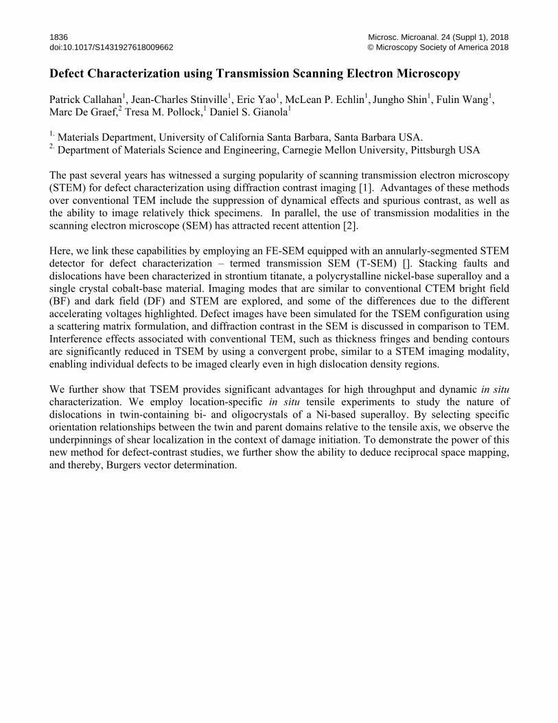
Figure 1. Bright-field transmission scanning electron microscope (TSEM) images of a deformed twin-containing Ni-based superalloy at two different tilts emphasizing (left panel) orientation contrast across twin boundaries and (right panel) dislocation contrast. Note residual dislocations at twin boundaries.