deep neural ensemble for retinal vessel … neural ensemble for retinal vessel segmentation in...
TRANSCRIPT
Deep Neural Ensemble for Retinal Vessel Segmentation in FundusImages towards Achieving Label-free Angiography
Avisek Lahiri, Abhijit Guha Roy, Debdoot Sheet, Prabir Kumar Biswas
Abstract— Automated segmentation of retinal blood vesselsin label-free fundus images entails a pivotal role in com-puted aided diagnosis of ophthalmic pathologies, viz., dia-betic retinopathy, hypertensive disorders and cardiovasculardiseases. The challenge remains active in medical image analysisresearch due to varied distribution of blood vessels, whichmanifest variations in their dimensions of physical appearanceagainst a noisy background. In this paper we formulate thesegmentation challenge as a classification task. Specifically,we employ unsupervised hierarchical feature learning usingensemble of two level of sparsely trained denoised stackedautoencoder. First level training with bootstrap samples ensuresdecoupling and second level ensemble formed by differentnetwork architectures ensures architectural revision. We showthat ensemble training of auto-encoders fosters diversity inlearning dictionary of visual kernels for vessel segmentation.SoftMax classifier is used for fine tuning each member auto-encoder and multiple strategies are explored for 2-level fusion ofensemble members. On DRIVE dataset, we achieve maximumaverage accuracy of 95.33% with an impressively low standarddeviation of 0.003 and Kappa agreement coefficient of 0.708 .Comparison with other major algorithms substantiates the highefficacy of our model.
I. INTRODUCTION
Segmentation and delineation of the articulated topologyof retinal vessel network and structural attributes of ves-sels such as thickness, run length, tortuosity and branchingpatterns provide first level pathological cue for identifyingvarious cardiovascular and opthalmogic diseases such as di-abetes, hypertension, arteriosclerosis [1]. Automatic analysisof retinal vessel topography assists in developing robustscreening systems for diabetic retinopathy [2], localizing offoveal avascular region [3], thinning of arteries [4] and lasersurgery [1]. Vessel tortuosity embeds significant informationabout hypertensive retinopathy [5] while vessel diameter hadbeen studied in connection with hypertension [6]. Besidesclinical areas, retinal topography has also been used forbiometric applications [7].
Related Works: Unsupervised paradigms of automaticvessel detection primarily aims at designing matched filtersby convolving a Gaussian kernel or its derivatives to em-phasize vessel regions [1]. Popular unsupervised multiscaleapproaches assign a ‘vesselness’ metric based on eigenanalysis of the Hessian [8]. Unsupervised model based tech-niques include active contour models and geometric modelguided level sets [9] . Supervised models rely on featureextraction on manually annoted ground truth images followedby classification, usually using Artificial Neural Network
Authors are with Indian Institute of Technology Kharagpur, India.Email: [email protected]
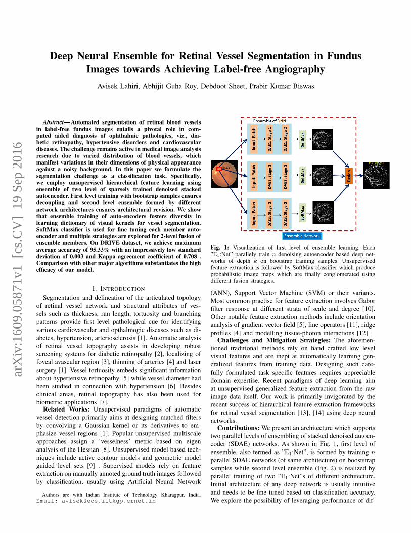
Fig. 1: Visualization of first level of ensemble learning. Each”E1:Net” parallely train n denoising autoencoder based deep net-works of depth k on bootstrap training samples. Unsupervisedfeature extraction is followed by SoftMax classifier which produceprobabilistic image maps which are finally conglomerated usingdifferent fusion strategies.
(ANN), Support Vector Machine (SVM) or their variants.Most common practise for feature extraction involves Gaborfilter response at different strata of scale and degree [10].Other notable feature extraction methods include orientationanalysis of gradient vector field [5], line operators [11], ridgeprofiles [4] and modelling tissue-photon interactions [12].
Challenges and Mitigation Strategies: The aforemen-tioned traditional methods rely on hand crafted low levelvisual features and are inept at automatically learning gen-eralized features from training data. Designing such care-fully formulated task specific features requires appreciabledomain expertise. Recent paradigms of deep learning aimat unsupervised generalized feature extraction from the rawimage data itself. Our work is primarily invigorated by therecent success of hierarchical feature extraction frameworksfor retinal vessel segmentation [13], [14] using deep neuralnetworks.
Contributions: We present an architecture which supportstwo parallel levels of ensembling of stacked denoised autoen-coder (SDAE) networks. As shown in Fig. 1, first level ofensemble, also termed as ”E1:Net”, is formed by training nparallel SDAE networks (of same architecture) on booststrapsamples while second level ensemble (Fig. 2) is realized byparallel training of two ”E1:Net”s of different architecture.Initial architecture of any deep network is usually intuitiveand needs to be fine tuned based on classification accuracy.We explore the possibility of leveraging performance of dif-
arX
iv:1
609.
0587
1v1
[cs
.CV
] 1
9 Se
p 20
16

Fig. 2: Visualization of second level of ensemble learning. We traintwo parallel ”E1:Net”s of different architecture.
ferent architectured networks within the context of ensemblelearning. We study various techniques for combining deci-sions of individual members of the ensemble and we showthat using only simple SoftMax classifier we outperform therecent deep learning based method [13] which uses randomforest classifier after unsupervised feature extraction. Formitigating class imbalance between vessel and background,we present a set of smart sampling procedures for creatinga database congenial for training SDAE.
The rest of the paper is organized as follows. The problemstatement is formally defined in §II and the detailed solutionis presented in §III. In Section §IV we validate and compareour results with state-of-the-art vessel segmentation algo-rithms. Finally, we conclude the paper in §V with a summaryof aptness of our ensemble learning of deep networks forautomatically generating diversified dictionary kernels formedical image analysis.
II. PROBLEM STATEMENT
Given a color fundus image, I ∈ RM×N , our objectiveis to assign a probability to each pixel (x,y) such that itsneighbourhood, N(x, y), centred at (x, y) belongs to eitherof the classes, ω ∈ {vessel, background}. Specifically,given a training set, {Itrain}, we wish to formulate afunction, H(ω|N(x, y), I; {Itrain}), whose response givesus P (ω|N(x, y)). In this paper we learn the function H(.)by hierarchical feature extraction of ∀ N(x, y) ∈ {Itrain}using our proposed fusion of SDAE ensemble networks.
III. PROPOSED APPROACH
A. Image Preprocessing, sampling procedure and databaseformation
Given a RGB fundus image, I, we first extract the greenchannel image, Ig , because it has been reported that the vas-cular structures manifest best contrast in green channel [15].CLAHE is used for compensating irregular illuminations.
Fig. 3 shows an exemplary manually annotated vesselnetwork used for extracting training patches. There existssignificant class imbalance between background and vesselpixels. It is a challenging task to train classifiers in presenceof highly skewed class distributions [16]. We propose a setof intuitive sampling strategies to mitigate this problem. Letthe ground truth binary image be Igr. For sampling vessel
Fig. 3: Example of retinal vessel detection using our proposedalgorithm on test image # 19 of DRIVE [17] dataset.
patches, we first skeletonize the image Igr to Isgr. Imageskeletonization helps in removing the boundary foreground(vessel) pixels but retains the overall topological structureand thereby prevents redundant foreground sampling. Vesseltraining patches, N(x, y), are uniformly sampled from Igat those coordinates for which Isgr = 1. For samplingbackground pixels, we first morphologically dilate Igr to Idgrby a square structural element of dimension 7 × 7. Naivelysampling at Igr = 0 generates samples which are very nearto original vessels and thus the neighbourhood encompassconsiderable region of vessel tissue region. Dilation opera-tions thickens the vessels and thus sampling with Idgr ensuresthat the background training patches are well separated fromoriginal vessel regions. For each training image I, if VIrepresents the set of vessel patches and BI represents set ofbackground patches, we uniformly sample |VI | samples fromBI to form B
′
I . As a final measure to enhance classificationperformance, an autoencoder is trained on a dataset withalternating patches from vessel and background class.
B. Unsupervised layerwise prelearning using SDAEFor automated feature discovery we have used stacked
denoised autoencoders which are analogous in architectureto traditional multilayer perceptron (MLP) networks butin DAE, interconnection weights are learnt sequentiallythrough unsupervised layerwise training (Refer to Sec. III-B[18] for more details). An autoencoder is basically a threelayer fully connected network with one hidden layer whichstores a compressed representation of input x, and outputs,x, which is an approximation of x. Let L denote number oflayers, sl denote number of nodes in layer l, W (l)
ji denoteweight between node i of layer l to node j of layer l + 1and hW,b(.) is sigmoidal activation. According to [18], lossfunction of sparse autoencoder is given by,
L(W,b) =
[1
m
m∑i=1
(1
2||hW,b(xi)− xi||2
)]
+ λ
L−1∑l=1
sl∑i=1
sl+1∑j=1
(W(l)ji )2 + β
s2∑j=1
KL(ρ||ρ) (1)
The first term tries to minimize the discrepancy betweeninput and predicted vector, second term is meant for L2regularization to prevent over fitting and the last term pro-motes sparsity within the network. β is sparsity penalty, mis cardinality of training sample space, ρ is a sparsenessparameter, ρk is th expected activation of node k in hiddenlayer, i.e., ρk = 1
m
∑mi=1 z
(i)k , where, z(i)k is the activation
of node k in hidden layer. DAE is a specialized version of

Fig. 4: Dictionary kernels learnt by DAE1 and DAE2 for ‘En-semble Network 1’. We can clearly see that ensemble learninggenerates diversified kernels. Due to space constraints we refrainfrom showing the other kernels.
autoencoder in which we incorporate random additive noiseat input side to transform x to x + r. Loss function inEq. 1 is minimized by back propagation and the parametersW and b are updated using L-BFGS (Limited MemoryBroyden-Fletcher-Goldfarb-Shanno). After this first step ofpre-training, a stacked DAE is realized by treating thehidden layer node activations as input to a second DAE andretraining the second DAE only. After second phase of pre-training we discard the last output layer and insert a simpleSoftMax classification layer with only a single node whichprovides the probability, P (ω|N(x, y)). Now, this SoftMaxlayer is used for fine tuning the entire stacked architecturesimilar to supervised MLP setting.First level ensemble formation Our first level of ensemble
formation is inspired by success of bagging [19] in ensemblelearning. Given an original set, Z, of training examples, formn sets, Z1, Z2,.. Zn, each of cardinality m by randomlysampling m samples from Z with replacement. We denoteDAEi as a SDAE of depth k trained on Zi and collectionof n such SDAEs is termed as ”E1:Net”. In Fig. 1 and 4and we have used DAEi and DAEi interchangeably. Asshown in Fig. 1, during classification, each DAEi produces aprobabilistic output, P (ω|N(x, y);DAEi). We used multiplestrategies to fuse the probability maps from each DAEi.P (ω|N(x, y))min = argmin
iP (ω|N(x, y);DAEi) (2)
P (ω|N(x, y))max = argmaxi
P (ω|N(x, y);DAEi) (3)
P (ω|N(x, y))avg =1
3
3∑i=1
P (ω|N(x, y);DAEi) (4)
P (ω|N(x, y))wavg =
3∑i=1
αiP (ω|N(x, y);DAEi) (5)
where αk = rk∑3j=1 rj
; rk is the accuracy rate of DAEk oncross validation set.Second level ensemble formation A SDAE (of depth 2)with SoftMax output is characterized by a l − h1 − h2 − c
TABLE I: Performances of individual ”E1:Net” using differentfusion strategies at first level of ensemble.
”E1:Net” Fusion Max. Avg. Accuracy KappaMin (Eq. 2) 0.932 0.687
1 Max (Eq. 3) 0.928 0.679Average (Eq. 4) 0.908 0.654Weighted Average (Eq. 5) 0.948 0.698
Min 0.948 0.6932 Max 0.936 0.684
Average 0.910 0.667Weighted Average 0.950 0.701
TABLE II: Performance comparsion of competing algorithms.(σ :)Standard deviation of max. avg. accuracy. Last four results areobtained from [20].
Method Max. Avg. Accuracy (σ) Kappa AgreementProposed 0.953 (0.003) 0.709Second human observer 0.947 (0.048) 0.758Maji et al. [13] 0.932 (–) 0.628Roy et al. [14] 0.912 (0.026) 0.618Sigurosson et al. [21] 0.942 (0.010) 0.708Yin et al. [15] 0.932 (–) -Sheet et al. [12] 0.976 (–) 0.821Chaudhuri et al. 0.877 (0.0232) 0.33Jiang et al. 0.921 (0.0076) 0.639Martinez-Perez et al. 0.918 (0.0240) 0.638Fraz et al. 0.948 (-) -Zana et al. [22] 0.937 (0.0077) 0.697
network architecture, where l = W ×W is input dimension,c is number of classes and h1, h2 denote number of nodesin first and second hidden layer respectively. We inducefurther diversification in learning by parallel training oftwo ” E1:Net”s ( E1:Net(1) and E1:Net(2)) of differentarchitecture (refer to Fig. 2), using different values of h1and h2. Decisions from each ”E1:Net(i)” is merged by aconvex weighted average; the weight being proportional toits accuracy on validation set.
IV. RESULTS AND DISCUSSIONS
We evaluate the performance of our algorithm on thepopular DRIVE dataset [17] and compare our results withother state-of-the-art vessel segmentation methods. In ourexperiment we have used an architecture of ‘576-400-100-2’for ” E1:Net(1)” and ‘576-200-50-2’ for ” E1:Net(2)”. Wehave used λ = 0.001, β = 3, W = 24; this setting yieldsthe maximum accuracy averaged over test examples (ReferTable II). Pre-training and finetuning of each SDAE is donefor 700 epochs.
In Fig. 4 we show some exemplary visual dictionarykernels learnt by DAE1 and DAE2 of E1:Net. It is evident,specially for Stage 1, that training on bootstrap samplesencourages the ensemble to learn diversified kernels.
We use the two standard metrics, viz., maximum av-erage accuracy and Cohen’s Kappa agreement coefficientfor comparing our results. In Table I we first compare theperformance of individual ” E1:Net” at first level of ensembleusing different fusion strategies as delineated in Sec. III-B. We see that the weighted average method manifestsbest classification accuracy for both the first level ensemblenetworks. For fusing decision at second level of ensemble,we first generate the vessel probability maps by weightedaverage voting from both the level one ”E1:Net”s and averagethem to yield the final soft classification output (see Fig. 2).

Fig. 5: Detection of course (top row) and fine (bottom row) vesselson two exemplary test images of DRIVE dataset.
Binarization of this posterior probability maps is achieved bythresholding the soft classification map at a level Lt whichmaximizes the F − Score.
In Table II we compare our results with state-of-the-artcompeting algorithms. Second column shows the maximumaverage accuracy along with the standard deviation. Com-parison of Table I and II proves that second level ensemblelearning further enhances the classification accuracy andthus justifies our approach. We achieve maximum averageaccuracy of 0.953 with standard deviation of only 0.003 andKappa agreement coefficient of 0.709. It is encouraging toobserve that our proposed unsupervised feature discoverybased model superceeds the human observer in terms ofaccuracy and standard deviation. Fig. 5 manifests the efficacyof our model in detecting both course and fine retinal vessels.Top row magnifies the optic nerve region where many vesselsmerge together and bundle up and segmentation becomesdifficult but our algorithm performs appreciably in suchregion. Our proposed method also achieves high accuracyin segmenting sparsely distributed fine blood vessel (bottomrow).
V. CONCLUSION
In this paper we presented a deep neural ensemble net-work architecture for retinal vessel segmentation. We haveobserved that miscellany of training space and architecturegenerated diversified dictionary of visual kernels for vesseldetection. Each kernel is responsible for identifying a spe-cific orientation of vessel. Learning diversified kernels thusenhances the representation prowess of our ensemble. Exper-imental validations suggest that our unsupervised layerwisefeature discovery based model is not only highly accuratebut also reliable and consistent. Future improvements mightfocus on selecting a better threshold from the proababilitymaps. Another promising direction is to use multiview en-semble learning by extracting features from each stage of astacked autoencoder.
REFERENCES
[1] J. J. Kanski and B. Bowling, Clinical ophthalmology: a systematicapproach. Elsevier Health Sciences, 2011.
[2] T. Teng, M. Lefley, and D. Claremont, “Progress towards automateddiabetic ocular screening: a review of image analysis and intelligentsystems for diabetic retinopathy,” Med. & Biol. Eng. & Comput.,vol. 40, no. 1, pp. 2–13, 2002.
[3] A. Haddouche, M. Adel, M. Rasigni, J. Conrath, and S. Bourennane,“Detection of the foveal avascular zone on retinal angiograms usingmarkov random fields,” Digit. Signal Process., vol. 20, no. 1, pp. 149–154, 2010.
[4] E. Grisan and A. Ruggeri, “A divide et impera strategy for automaticclassification of retinal vessels into arteries and veins,” in EMBC,vol. 1. IEEE, 2003, pp. 890–893.
[5] M. Foracchia, E. Grisan, and A. Ruggeri, “Extraction and quantitativedescription of vessel features in hypertensive retinopathy fundusimages,” in Book Abstr. 2nd Intern. Workshop on Comput. Assist.Fundus Image Anal., vol. 6, 2001.
[6] J. Lowell, A. Hunter, D. Steel, A. Basu, R. Ryder, and R. L. Kennedy,“Measurement of retinal vessel widths from fundus images based on2-d modeling,” IEEE Tran. Med. Imaging, vol. 23, no. 10, pp. 1196–1204, 2004.
[7] C. Marino, M. G. Penedo, M. Penas, M. J. Carreira, and F. Gonzalez,“Personal authentication using digital retinal images,” Pattern Anal. &Appl., vol. 9, no. 1, pp. 21–33, 2006.
[8] C. Kose, C. Iki et al., “A personal identification system using retinalvasculature in retinal fundus images,” Expert Syst. with Appl., vol. 38,no. 11, pp. 13 670–13 681, 2011.
[9] R. J. Winder, P. J. Morrow, I. N. McRitchie, J. Bailie, and P. M.Hart, “Algorithms for digital image processing in diabetic retinopathy,”Computerized Med. Imaging & Graphics, vol. 33, no. 8, pp. 608–622,2009.
[10] M. Niemeijer, J. Staal, B. Ginneken, M. Loog, and M. Abramoff,“Drive: digital retinal images for vessel extraction,” 2004.
[11] A. Hoover, V. Kouznetsova, and M. Goldbaum, “Locating bloodvessels in retinal images by piecewise threshold probing of a matchedfilter response,” IEEE Tran. Med. Imaging, vol. 19, no. 3, pp. 203–210,2000.
[12] D. Sheet, S. P. K. Karri, S. Conjeti, S. Ghosh, J. Chatterjee, and A. K.Ray, “Detection of retinal vessels in fundus images through transferlearning of tissue specific photon interaction statistical physics,” inISBI. IEEE, 2013, pp. 1452–1456.
[13] D. Maji, A. Santara, S. Ghosh, D. Sheet, and P. Mitra, “Deep neuralnetwork and random forest hybrid architecture for learning to detectretinal vessels in fundus images,” in EMBC. IEEE, 2015, pp. 3029–3032.
[14] A. Guha Roy and D. Sheet, “Dasa: Domain adaptation for stackedautoencoders,” in ACPR, 2015.
[15] B. Yin, H. Li, B. Sheng, X. Hou, Y. Chen, W. Wu, P. Li, R. Shen,Y. Bao, and W. Jia, “Vessel extraction from non-fluorescein fundusimages using orientation-aware detector,” Med. Image Anal., vol. 26,no. 1, pp. 232–242, 2015.
[16] M. Galar, A. Fernandez, E. Barrenechea, H. Bustince, and F. Herrera,“A review on ensembles for the class imbalance problem: bagging-, boosting-, and hybrid-based approaches,” IEEE Tran. Syst. Man &Cybernetics, vol. 42, no. 4, pp. 463–484, 2012.
[17] J. Staal, M. D. Abramoff, M. Niemeijer, M. A. Viergever, andB. Van Ginneken, “Ridge-based vessel segmentation in color imagesof the retina,” IEEE Tran. Med. Imaging, vol. 23, no. 4, pp. 501–509,2004.
[18] P. Vincent, H. Larochelle, I. Lajoie, Y. Bengio, and P.-A. Manzagol,“Stacked denoising autoencoders: Learning useful representations ina deep network with a local denoising criterion,” JMLR, vol. 11, pp.3371–3408, 2010.
[19] N. A. H. Mamitsuka, “Query learning strategies using boosting andbagging,” in ICML, vol. 1. Morgan Kaufmann Pub, 1998.
[20] M. Niemeijer, J. Staal, B. van Ginneken, M. Loog, and M. D.Abramoff, “Comparative study of retinal vessel segmentation methodson a new publicly available database,” in SPIE Med. Imaging, 2004,pp. 648–656.
[21] E. M. Sigurosson, S. Valero, J. A. Benediktsson, J. Chanussot, H. Tal-bot, and E. Stefansson, “Automatic retinal vessel extraction based ondirectional mathematical morphology and fuzzy classification,” PatternRecognit. Lett., vol. 47, pp. 164–171, 2014.
[22] M. M. Fraz, P. Remagnino, A. Hoppe, B. Uyyanonvara, A. R. Rud-nicka, C. G. Owen, and S. A. Barman, “An ensemble classification-based approach applied to retinal blood vessel segmentation,” IEEETran. Biomed. Engg., vol. 59, no. 9, pp. 2538–2548, 2012.