deep learning applied to automated chest x-ray...
TRANSCRIPT

Deep Learning applied to automated Chest
X-Ray screening
Fos-Guarinos B., Alberich-Bayarri A.1,2,
Bosch-Roig I., Ten-Esteve A. 2, Martí-Bonmatí L. 3
1 PhD. GIBI Director and QUIBIM CEO2 M. Sc GIBI230
3 MD, PhD. GIBI PI and QUIBIM Founder

Outline
• Introduction
• Purpose
• Materials and methods
• Results
• Conclusion

Introduction
Valdés P., Morales Á. (2015) Posición SERAM sobre la necesidad de informar la radiología simple. SERAM.
75% of the explorations carried out in the Imaging Diagnosis area
are radiographs, being chest X-rays the majority of them because
they contain potential information of the main structures of the
human body (heart, lungs…).
Reporting chest x-rays is a demanding task and very important
medical-legally, sometimes forgettable. So, we wanted to set up a
screening tool in order to aid radiologist by setting a prefilter for
giving priority to the abnormal ones and facilitate the chest x-ray
reporting task.

Purpose
• Designing, developing and evaluating the effectivity of a Computer-
Aided Diagnosis (CAD) system based on artificial intelligence
techniques (deep learning) able to perform automatically a first
screening task of healthy and pathological chest radiographs.

Materials and Methods
MACHINE LEARNING
A type of artificial intelligence that provides
computers the ability to learn and perform
certain tasks without being programmed
explicitly to do so.
DEEP LEARNING
A machine learning technique that can learn
useful representations or features directly from
data such as images, text or sound.
Nehemia A., Prasanna S. (2015). Deep Learning for Computer Vision with Matlab. MATHWORKS. Conference

Materials and Methods
WORKFLOW
CNN SVM
Normal
Abnormal
Preparing the radiological
database
Automatic feature
extraction
Training a classifier

Materials and Methods
• Preparing the radiological database
Indiana University (Open-I)
7470 DICOM chest X-ray images
JSON code containing the MeSH of these images
Python

Materials and Methods
• Preparing the radiological database
Category Total
Abnormal 868
Normal 137
Category Total
Atelectasis 293
Cardiomegaly 331
Nodule 253
Opacity 412
Pleural effusion 144
Materials and Methods
Krizhevsky et al. (2012). ImageNet Classification With Deep Convolutional Neural Networks
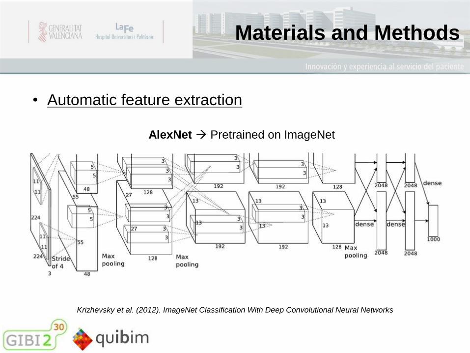
• Automatic feature extraction
AlexNet Pretrained on ImageNet
Materials and Methods
Krizhevsky et al. (2012). ImageNet Classification With Deep Convolutional Neural Networks
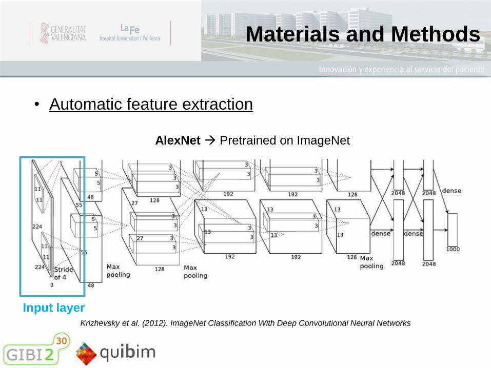
• Automatic feature extraction
AlexNet Pretrained on ImageNet
Input layer
Materials and Methods
Krizhevsky et al. (2012). ImageNet Classification With Deep Convolutional Neural Networks
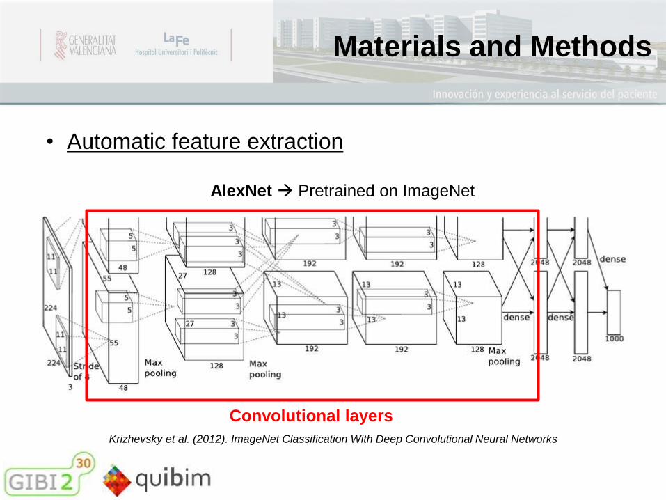
• Automatic feature extraction
AlexNet Pretrained on ImageNet
Convolutional layers

Materials and Methods
Krizhevsky et al. (2012). ImageNet Classification With Deep Convolutional Neural Networks
• Automatic feature extraction
AlexNet Pretrained on ImageNet
Convolutional layers

Materials and Methods
Krizhevsky et al. (2012). ImageNet Classification With Deep Convolutional Neural Networks
• Automatic feature extraction
AlexNet Pretrained on ImageNet
Fully-connected layers

Materials and Methods
• Training a classifier
Automatically
extracted features
Support Vector
Machines
Radiological database
80% for training the
classifier
20% for test and
evaluation

Results
Development of a graphical user interface GUIDE of MATLAB

Results
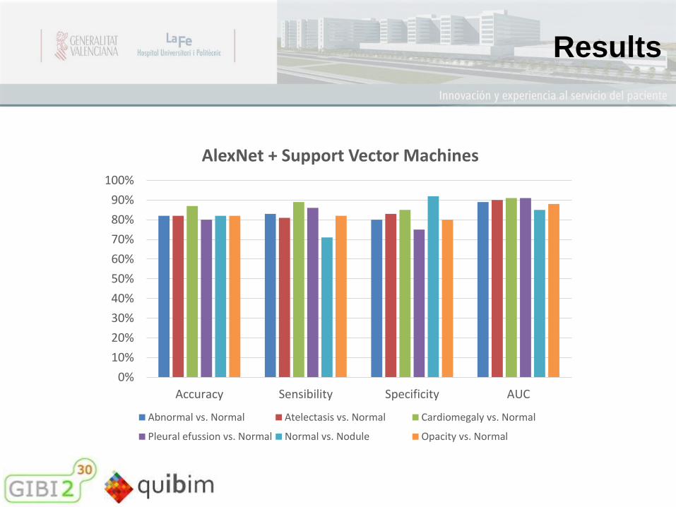
Abnormal
vs. Normal
Atelectasis vs.
Normal
Cardiomegaly vs.
Normal
Pleural efussion
vs. Normal
Normal vs.
Nodule
Opacity vs.
Normal
Accuracy 82% 82% 87% 80& 82% 82%
Sensibility 83% 81% 89% 86% 71% 82%
Specificity 80% 83% 85% 75% 92% 80%
AUC 89% 90% 91% 91% 85% 88%
20% of the data for test and evaluation system (confusion matrix)
Results
0%
10%
20%
30%
40%
50%
60%
70%
80%
90%
100%
Accuracy Sensibility Specificity AUC
AlexNet + Support Vector Machines
Abnormal vs. Normal Atelectasis vs. Normal Cardiomegaly vs. Normal
Pleural efussion vs. Normal Normal vs. Nodule Opacity vs. Normal

Results
89%
91% 91%
79%
84%
91%
86%
91%90%
72%74%76%78%80%82%84%86%88%90%92%
Abnormal vs Normal Cardiomegaly vs. Normal Pleural efussion vs.Normal
AUC
COMPARISON WITH PREVIOUS STUDIES
Our project Bar et al., 2013 Bar et al., 2015
Our study improves or equals the results achieved by classifiers trained
similarly in previous studies.

Conclusions
• A Computer-Aided Diagnosis system has been designed and
developed (DEEPLIR) based on convolutional neural networks,
able to perform automatically a first screening task in healthy and
pathological chest X-rays aimed at solving the problems that have
motivated this project.
• AlexNet has a great potential of knowledge transfering to the
chest x-ray images. From now on, deep learning by CNNs has to be
considered as the first candidate in any essential task of visual
recognition.
Luis Martí Bonmatí – MD, PhD. GIBI PI and QUIBIM FounderÁngel Alberich-Bayarri – PhD. GIBI Director and QUIBIM CEO
QUIBIM StaffFabio García Castro - M.ScRafa Hernández Navarro - B.ScDavid García - M.ScEncarna Sánchez - M.ScKatherine Wilisch R. - M.Sc
GIBI230 StaffEnrique Ruiz Martínez – M.ScAmadeo Ten Esteve – M.ScAna Penadés - Adm.
Internship StudentsBelén Fos GuarinosAlfredo Torregrosa LloretCarlos Moya ClaramuntAna Jiménez PastorIrene Mayorga Ruiz
Team
CSO CTO Back-End Development of Imaging Biomarkers Business Development Coordinator and
CEO support
MS BiomedicalEngineering
Clinical Trials Coord. Administration