deciphering the early events leading to an adaptive immune
TRANSCRIPT

HAL Id: tel-01369028https://tel.archives-ouvertes.fr/tel-01369028
Submitted on 20 Sep 2016
HAL is a multi-disciplinary open accessarchive for the deposit and dissemination of sci-entific research documents, whether they are pub-lished or not. The documents may come fromteaching and research institutions in France orabroad, or from public or private research centers.
L’archive ouverte pluridisciplinaire HAL, estdestinée au dépôt et à la diffusion de documentsscientifiques de niveau recherche, publiés ou non,émanant des établissements d’enseignement et derecherche français ou étrangers, des laboratoirespublics ou privés.
Deciphering the early events leading to an adaptiveimmune response during urinary tract infection
Gabriela Mora Bau
To cite this version:Gabriela Mora Bau. Deciphering the early events leading to an adaptive immune response duringurinary tract infection. Immunology. Université Pierre et Marie Curie - Paris VI, 2015. English.�NNT : 2015PA066666�. �tel-01369028�

Université Pierre et Marie Curie École doctorale Physiologie et Physiopathologie (ED 394)
Immunobiologie des Cellules Dendritiques (Unité Mixte Pasteur/Inserm U818)
Définir le début des événements conduisant à une réponse
immunitaire adaptative lors de l'infection urinaire
Par Gabriela Mora Bau
Thèse de doctorat de Immunologie
Dirigée par Molly A. Ingersoll, PhD
Présentée et soutenue publiquement le 30 Septembre 2015
Devant un jury composé de:
Sebastien Lacroix-Desmazes, PhD Président
Anne HOSMALIN, PhD Rapporteur Jean-Marc CAVAILLON, PhD Rapporteur
Gerard EBERL, PhD Examinateur Molly A. INGERSOLL, PhD Matthew L. ALBERT, MD, PhD
Directeur de thèse Co-directeur de thèse

Page 2
Université Pierre et Marie Curie École doctorale Physiologie et Physiopathologie (ED 394)
Laboratory of Dendritic Cell Biology (Mixed Pasteur/Inserm U818 unit)
Deciphering the early events leading to an adaptive immune
response during urinary tract infection
By Gabriela Mora Bau
Doctoral thesis in Immunology
Directed by Molly A. Ingersoll, PhD
Defended publically on 30 September 2015
In front of a jury composed of:
Sebastien Lacroix-Desmazes, PhD Président Anne HOSMALIN, PhD Rapporteur
Jean-Marc CAVAILLON, PhD Rapporteur Gerard EBERL, PhD Examinateur
Molly A. INGERSOLL, PhD Matthew L. ALBERT, MD, PhD
Directeur de thèse Co-directeur de thèse

Page 3
Table&of&Contents&!Résumé&................................................................................................................................................&5!List&of&figures&....................................................................................................................................&7!List&of&tables&......................................................................................................................................&9!Acknowledgment&..........................................................................................................................&10!Abbreviations&................................................................................................................................&11!Chapter&1:&Introduction&..............................................................................................................&13!The&bladder&mucosa&..............................................................................................................................&14!Urinary&tract&infection&..........................................................................................................................&16!UPEC&is&the&main&causative&agent&of&UTI&.........................................................................................&18!The&UPEC&pathogenic&cycle&..................................................................................................................&22!Therapeutic&options&for&UTI&................................................................................................................&25!The&host&response&during&UTI&............................................................................................................&28!Defense!mechanisms!of!the!urinary!tract!....................................................................................................!28!A!strong!innate!immune!response!is!triggered!during!UTI!.................................................................!30!The!adaptive!immune!response!during!UTI:!the!mystery!starting!to!be!solved!........................!32!
Aims&of&our&study&....................................................................................................................................&38!Chapter&2:&Deciphering&the&early&events&leading&to&an&adaptive&immune&response&in&UTI&................................................................................................................................................&40!Introduction&.............................................................................................................................................&41!Generation&of&fluorescent&UPEC&strains&..........................................................................................&42!UTI&challenge&infection&model&............................................................................................................&44!UPEC&infection&induces&an&adaptive&immune&response&mediated&by&DCs&...........................&44!The&steady&state&bladder&contains&a&heterogeneous&immune&cell&population&..................&47!UPEC&is&acquired&by¯ophages&early&after&infection&............................................................&50!Infiltrating&classical&monocytes&primarily&become¯ophages&during&infection&........&52!BladderRresident¯ophages&negatively&impact&the&induction&of&adaptive&immunity&to&UPEC&.......................................................................................................................................................&54!Effector&cell&infiltration&is&unchanged&during&UPEC&challenge&among¯ophageRdepleted&and&control&treated&mice&...................................................................................................&59!UPECRspecific&antibodies&are&undetectable&during&infection&..................................................&60!Macrophage&depletion&does¬&change&the&cytokineµenvironment&in&the&bladder&during&primary&infection&......................................................................................................................&62!Increasing&the&amount&of&antigen&does¬&affect&the&generation&of&adaptive&immunity&during&UTI&.................................................................................................................................................&63!Macrophage&depletion&leads&to&increased&phagocytosis&of&UPEC&by&DCs&............................&64!
Chapter&3:&Delineating&the&adaptive&immune&response&generated&during&UTI&......&66!Introduction&.............................................................................................................................................&67!CD8α+&and&CD103+&DCs&are&dispensable&for&an&adaptive&immune&response&against&UPEC&.......................................................................................................................................................................&68!T&cells&are&dispensable&for&the&direct&response&to&UPEC&during&challenge&infection&......&69!
Chapter&4:&Discussion&..................................................................................................................&72!How&do¯ophages&limit&the&adaptive&immune&response&against&UPEC?&.......................&73!Macrophages&and&their&relationship&with&the&adaptive&immune&response,&are&they&always&the&“bad&guys”?&..........................................................................................................................&77!Good!guys!..................................................................................................................................................................!78!

Page 4
Bad!guys!.....................................................................................................................................................................!78!What&is&the&nature&of&the&immune&response&against&UPEC?&.....................................................&79!Can&effective&adaptive&immunity&be&evoked&from&the&bladder?&.............................................&81!Why&does&the&adaptive&immunity&generated&during&UTI&“fail”?&.............................................&82!
Materials&and&methods&...............................................................................................................&86!Bacterial&strains&......................................................................................................................................&87!Cell&lines&and&in#vitro&invasion&assay&................................................................................................&87!Mice&and&infections&................................................................................................................................&87!Irradiation,&bone&marrow&cell&transfer,&diphtheria&toxin&treatment&....................................&88!Flow&cytometry&of&bladder&tissue&......................................................................................................&88!Flow&cytometry&of&blood&.......................................................................................................................&89!Monocyte&Bead&labeling&........................................................................................................................&89!Immune&cell&depletion&..........................................................................................................................&89!Luminex&MAP&analysis&..........................................................................................................................&90!ELISA&...........................................................................................................................................................&90!Statistical&analysis&..................................................................................................................................&91!
References&......................................................................................................................................&92!Published&manuscript&...............................................................................................................&111!
&

Page 5
Résumé!L’infection des voies urinaires est l'une des infections bactériennes les plus courantes avec des
coûts de soins de santé très élevés. On estime que 50% des femmes connaîtront une infection
urinaire au cours de leur vie, ceci de manière récurrente chez la moitié d’entre elles. Le
développement de thérapies efficaces a été limité par le manque de connaissance concernant
la mise en place de la réponse immune adaptative lors de cette infection. Dans cette étude,
nous avons démontré qu'une réponse adaptative est générée lors de l'infection urinaire,
cependant celle-ci n’a pas d’action protectrice. Afin de comprendre les mécanismes
aboutissant à ce phénomène, nous avons cherché à caractériser les cellules immunitaires
présentes dans la vessie. Des tests d’absorption bactérienne ont montré que ces macrophages
phagocytent la majorité des bactéries au début de l'infection. Pour évaluer l’influence de ces
cellules sur la mise en place de la réponse immune adaptative, nous avons déplété les
macrophages et évalué la clairance bactérienne lors d’une deuxième infection. En
comparaison avec les animaux non traités, les souris déplétées présentaient une réduction de
la charge bactérienne conséquente lors de la seconde infection, cette clairance dépendant de la
réponse immune adaptative. Pour comprendre ce mécanisme d'inhibition par les
macrophages, nous avons évalué le microenvironnement vésical et la phagocytose au début de
l'infection chez les souris déplétées, et chez les souris non traitées. Bien que nous n’ayons pas
observé de différences dans la production de cytokines, l'absorption bactérienne par les
cellules dendritiques s’avère deux fois plus importante chez les animaux déplétés. Ces
données suggèrent que l'absorption bactérienne par les macrophages tissulaires est néfaste
pour la mise en place de la réponse adaptative, ouvrant de nouvelles options thérapeutiques.
Nous avons également évalué le rôle des lymphocytes T dans ce processus en déplétant ces
cellules au cours de l'infection primaire ou avant la deuxième infection. Ainsi, nous avons
observé que les lymphocytes T sont nécessaires dans la réponse adaptative, mais ne sont
cependant pas indispensables à la clairance bactérienne lors d'une réinfection. De plus,
l'infection des souris Batf3-/-, déplétées en cellules dendritiques spécialisées dans la
présentation croisée, a montré que ces souris contrôlent une seconde infection aussi bien que
les souris contrôle. Ces résultats suggére que la présence lymphocytes T CD8+ n’est pas
nécessaire pour lutter contre l’infection urinaire. Notre étude révèle un mécanisme par lequel
le système immunitaire est compromis lors de l'infection urinaire, offrant un point de départ
intéressant pour une recherche plus approfondie sur le rôle du système immunitaire adaptatif
dans ce contexte, élément fondamental dans le développement de nouvelles thérapies.
Page 6
Abstract&Urinary tract infection (UTI) is one of the most common bacterial infections with exorbitant
health care costs. It is estimated that 50% of women will experience a UTI during their
lifetime and approximately half will suffer recurrent infections. Infected women are treated
with antibiotics, however, antibiotic resistance is increasing, raising the need for new
therapeutic options. Development of efficient therapies has been impeded by the lack of
knowledge of events leading to adaptive immunity. In this study, we demonstrated that an
adaptive immune response is generated during UTI, however this response does not confer
protective immunity. To begin to understand why the response induced during UTI was not
effective, we delineated the immune cell compartment of the bladder and identified
macrophages as the most populous immune cell. We evaluated bacterial acquisition in the
bladder observing that macrophages phagocytize the majority of the bacteria early in
infection. To evaluate the impact of macrophages on the generation of adaptive immunity, we
depleted bladder resident macrophages and evaluated bacterial clearance during a challenge
infection. Interestingly, mice depleted of resident macrophages, prior to primary infection,
exhibited a nearly 2-log reduction in bacterial burden following secondary challenge
compared to untreated animals. This improvement in clearance was dependent on the adaptive
immune system. To shed light on the mechanism of macrophage inhibition, we evaluated the
bladder microenvironment and bacterial acquisition early in infection in macrophage-depleted
and control-treated mice. While we did not observe differences in the cytokine
microenvironment, bacterial uptake by dendritic cells was increased nearly 2-fold in
macrophage-depleted animals. These data suggest that bacterial uptake by tissue macrophages
negatively impacts the development of adaptive immunity, revealing a novel target for
enhancing host responses to bacterial infection of the bladder. We also evaluated the role of T
cells during UTI by depleting these cells during the course of the infection or just prior to
challenge infection. We observed that T cells were necessary to mount an adaptive immune
response to UTI, however, they were dispensable for bacterial killing during challenge
infection. Additionally, infection of Batf3-/- mice, lacking cross-presenting dendritic cells,
suggested that CD8+ T cells are dispensable for the response against UTI as these mice
cleared a challenge infection as well as wildtype mice. Our study has revealed a mechanism
by which the immune system is compromised during UTI, providing an interesting start point
for further investigation of the role of the adaptive immune system during UTI, which will be
fundamental for the development of new therapies to efficiently treat infection.

Page 7
List&of&figures&!Figure 1: Schematic representation of the bladder uroepithelium..………..…..…………… 13
Figure 2: Urothelial plaque formation.……………………..….…………………………… 14
Figure 3: Causative agents of urinary tract infections…..…..……………………………… 17
Figure 4: UPEC-host interactions during UTI..…………………………………...………… 19
Figure 5: A schematic representation of type 1 fimbriae…………………………………… 20
Figure 6: UPEC IBC Pathogenic Pathway Observed in the Murine Cystitis Model..……… 23
Figure 7: B cell immune response………...……….………..……………………………… 35
Figure 8: Proposed model for the generation of an adaptive immune response during UTI.. 38
Figure 9: Fluorescent UPEC strains.…………………………………………………….….. 41
Figure 10: Challenge infection model..………………………………………………….….. 42
Figure 11: An adaptive immune response is necessary for bacterial clearance during UPEC
challenge infection……………..…….……………………..…………………………….… 44
Figure 12: DT-mediated DC ablation.……………………………………………………… 45
Figure 13: The bladder contains a diverse immune cell repertoire.………………………… 47
Figure 14: Among APCs, macrophages predominantly take up UPEC at early times P.I … 49
Figure 15: Classical monocytes robustly enter the bladder and become macrophages…..… 51
Figure 16: Immune cell ablation.……………………………….…………………………… 53
Figure 17: Macrophage depletion improves the adaptive response to UPEC infection…..… 54
Figure 18: CCR2-/- mice are not impaired in bacterial clearance after primary infection…... 55
Figure 19: UPEC reservoirs are not altered in monocyte or macrophage depleted mice…… 57
Figure 20: Macrophage depletion does not impact effector cell infiltration during UPEC
challenge………………………………………………………………………………..…… 58
Figure 21: UPEC-specific IgA remained at the limit of the detection……………….……… 59
Figure 22: Macrophage depletion does not impact cytokine expression in the bladder.….... 60

Page 8
Figure 23: Increasing bacterial inoculum during primary infection does not improve the
response to challenge infection………………………...…………………………………… 62
Figure 24: DCs acquire more bacteria in the absence of macrophages…………..………... 63
Figure 25: CD8α+ and CD103+ DCs absence does not impact the adaptive immune response
against UPEC.……………………………………………………...…..……………..……. 66
Figure 26: T cells are not necessary for UPEC clearance during challenge infection……... 67
Figure 27: Proposed model for the role of macrophages during UTI…………………..…. 72
Figure 28: Proposed model for the generation of adaptive immunity during UTI……….... 79

Page 9
List&of&tables&!Table 1: Previously tested vaccines for UPEC-mediated UTI...………..…..…………...… 27
Table 2: Immune cell populations in naïve bladders…………….………………………… 46
Table 3: Antibodies used for flow cytometry.…..…..……………………………………... 85

Page 10
Acknowledgment& I would like to thank my laboratory for their support during the development of my project. Thanks to Brieuc for always having a smile in his face and for all the nice conversations we had; thanks to Rosa for being not just a wonderful colleague but also a friend, thanks for listening to me and for all the advices you once gave me! Thanks to Nader for your sense of humor and your passion for science which makes everyone to believe that our work is worth it; thanks to Clemence for all the nice moments; thanks to Hana, I laughed so much with you, thanks for all our conversations! Dilay, thank you so much for everything! It was a real pleasure to meet you. I want to thank Matthew for everything, I learnt a lot from you and I appreciate to have been part of your lab. A special thanks to Molly, my supervisor, thank you for all your support, for everything you taught me, for all the conversations we had and for being always there when I needed.& I want to thank my petit classmate Umeshree, thanks a lot for being such a good friend and an amazing woman! I enjoyed every minute I spent with you, you gave me strength in every difficult moment and you taught me so much about everything. Thanks to Mariana and Ai Ing too for all your positive energy and support! I want to finally thank my family and friends in Chili who, from the distance, made this journey easier and enjoyable. Caro, thank you for being there for me every time I needed you and for all your words and our everlasting conversations! Thanks for visiting me and for exploring Europe together, it makes me so happy that we will be living in the same city so soon! My dear Melissa! Thank you very much for being there and for coming to Paris and being part of the last step of my PhD. I want to thank Andrés, my friend, my boyfriend, my everything, you definitely made this happens! Thanks for your patience, for your support, for listening to me, for your advices, for spending these 3 years exploring the word with me! And for being my partner during this Parisian adventure, you are the best! Naty, my little sister, thanks for everything! It was a pleasure to have you in Paris with me! Andrea, my sister and friend! Your words were always there when needed; Tia Maria, always making me laugh and being there for me! Tia Vero and her family, thank you for visiting me and for an incredible summer in Paris! Tia Bea, thanks for coming to close this chapter of my life with me! Ta, my dear grandfather thanks for all your wise words and for share a part of this experience with me in Paris! It was wonderful to have you here; huge thanks to my mom, my dear friend! I would not be here or anywhere without you, you are the best!!! There was no one single day that we did not talk since I am in Paris and I appreciate our conversations so much! I love you like crazy and I am incredibly happy that we will be so close again! Thank you for absolutely everything! I want to thank the PPU program for all their support and for accepting me to perform my PhD at Pasteur.

Page 11
Abbreviations&
APC Antigen presenting cell
BCAM Basal cell adhesion molecule
BCG Bacillus Calmette-Guerin
cAMP Cyclic AMP
CCL Chemokine (C-C motif) ligand
CCR Chemokine (C-C motif) receptor
CDC42 Cell division control 42
CFU Colony forming unit
CNF1 Cytotoxic necrotizing factor 1
CXCL Chemokine (C-X-C motif) ligand
CXCR Chemokine (C-X-C motif) receptor
DC Dendritic cell
DT Diphtheria toxin
ELISA Enzyme-linked immunosorbent assay
ExPEC Extraintestinal pathogenic Escherichia coli
GAGs Glycosaminoglycans
GFP Green fluorescent protein
HlyA Alpha-hemolysin
IBC Intracellular bacterial community
IL Interleukin
LPS Lipopolysaccharide
MHC Major histocompatibility complex
MMP9 Matrix metallopeptidase 9
MOI Multiplicity of infection
NK Natural killer
OVA Ovalbumin
P.I. Post infection
QIR Quiescent intracellular reservoir
RFP Red fluorescent protein
rRNA Ribosomal RNA
Th T helper

Page 12
THP Tamm-Horsfall protein
TLR Toll-like receptor
TNF Tumor necrosis factor
UPEC Uropathogenic Escherichia Coli
UTI Urinary tract infection
WT Wild type
!
Page 13
Chapter&1:&Introduction!
&

Page 14
!;%&#$"99%2&0/3',"&
The bladder is a unique mucosal surface composed of a transitional epithelium containing
three to six layers which are organized as follows: facing the luminal surface of the bladder
are highly differentiated, large and multinuclear superficial cells (referred to as umbrella or
facet cells), one or more intermediate cell layers, and a basal cell layer on top of the lamina
propria (Figure 1) (Apodaca, 2004; Ingersoll and Albert, 2013). The capacity of the bladder to
regenerate after damage was described many years ago (Hicks, 1975), however the presence
of stem cells in the bladder was first proposed only very recently in 2008 (Kurzrock et al.,
2008). In 2011, it was demonstrated that basal cells of the uroepithelium include stem cells.
These stem cells are able to self-renew and differentiate into all the cell types composing the
uroepithelium (Shin et al., 2011). Like in other tissues, resident immune cells are present in
the bladder mucosa. MHC II+ cells were detected by histology in humans, pigs, and mice
(Gardiner et al., 1986; Hart and Fabre, 1981a; Hjelm et al., 1982). Additionally, CD11c+ and
F4/80+ cells (Engel et al., 2008; Schilling et al., 2003), as well as T cells (Christmas, 1994)
have been identified in steady state and infected mouse bladders.
Figure 1: Schematic representation of the bladder uroepithelium. The bladder uroepithelium is composed of 3–6 uroepithelial cell layers. Multinuclear umbrella cells facethe lumen. Intermediate and basal layers are directly underneath. Stem cells, in dark pink, are found in the basal layer, on top of the lamina propria (delineated by the pale blue line). The bladder contains resident "#!T cells and phagocytes such as macrophages and dendritic cells. Figure modified from Ingersoll and Albert, 2013.
Lumen Bladder

Page 15
The apical side of umbrella cells is covered with numerous rigid-looking plaques (2
dimensional crystals of hexagonally packed 16-nm protein particles) (Kachar et al., 1999; Wu
et al., 2009) (Figure 2). These plaques are composed of uroplakins, which are integral
membrane proteins (Apodaca, 2004). Four uroplakin proteins have been identified: UPIa,
UPIb, UPII, and UPIIIa. The plaques are formed when UPII and UPIa dimerize, as well as
UPIII and UPIb; then, the two heterodimers bind to form a tetramer. Finally, 6 tetramers bind
together to form a plaque (Hu et al., 2005) (Figure 2). These uroepithelial plaques are in part
responsible for the barrier function of the uroepithelium as they help to make the bladder
impermeable (Negrete et al., 1996). Additionally, the plaques stabilize the apical surface, and
prevent uroepithelial rupture during bladder distension (Staehelin et al., 1972). Moreover,
they also have a role during infection, discussed later.
Figure 2: Urothelial plaque formation. (A) A model depicting the assembly of the four major uroplakins (UPIa, Ib, II, and IIIa) into 2D crystals, as described in the text. (B) Quick-freeze deep-etch image of the apical surface of a mouse umbrella cell showing urothelial plaques (P) containing hexagonal arrays of 16-nm particles interconnected by particle-free hinge (H) areas. Figure and legend modified from Wu et al., 2009.
!" #"
Cell lumen
Cell membrane
Page 16
The bladder has been considered a sterile mucosa, lacking colonizing microflora (Zasloff,
2007) however, evidence suggests that similar to other organs, like the gut or skin,
commensal bacteria reside in the bladder (Anderson et al., 2004b; Siddiqui et al., 2011). The
bladder was thought to be sterile for so long due the inability to detect bacteria in urine
samples. However, in 2004, viable but nonculturable bacteria were detected in mouse and
human urine samples (Anderson et al., 2004b). Different bacteria have been found in the urine
from healthy women through the study of bacterial 16S rRNA sequences, with
Lactobacillus being the most prominent in the urine microbiota (Siddiqui et al., 2011).
Additionally, bacteria genera found in vaginal microbiota were also detected in urine samples
from healthy women (Ling et al., 2010; Siddiqui et al., 2011). Bacteria commonly considered
as difficult to culture (meaning that they are not generally detectable by conventional culture
methods), such as Aerococcus urinae and the genus Ureaplasma, were also detected in this
study (Siddiqui et al., 2011).
In general, the bladder is not as well studied as other mucosal surfaces. This mucosa is the
most impenetrable organ in the body and has the important function of protecting the body
from toxins accumulated in the urine (Negrete et al., 1996). While the bladder certainly
harbors a microbiota, which needs to be more fully investigated, it is also susceptible to
different uropathogens, which cause urinary tract infection (UTI), one of the most common
infections in the world (Flores-Mireles et al., 2015; Foxman, 2010). Given the importance of
this organ, and our relative lack of knowledge of its biology, it is both a relevant and
interesting topic of study.
Urinary&tract&infection&
UTI is one of the most common bacterial infections, impacting more than 150 million people
annually and resulting in significant health care costs and morbidity (Foxman, 2010; Stamm
and Norrby, 2001). In the United States alone, the cost of health care and time missed from
work reaches 3.5 billion dollars per year (Foxman, 2014). Women are at greater risk for UTI
than men and it is estimated that one out of two women will experience a UTI during their
lifetime, while nearly half of these individuals will experience one or more recurrent

Page 17
infections (Foxman, 2002). Risk factors for UTI include gender, prior UTI, sexual activity,
spermicide use, vaginal infection, diabetes, and catheterization, among others (Hooton, 2000).
UTI can be classified as uncomplicated or complicated. Uncomplicated UTI generally affects
individuals who are otherwise considered healthy and do not present any structural
abnormalities in their urinary tract. Uncomplicated UTI can affect the lower urinary tract
(lower UTI or cystitis) or the upper urinary tract (upper UTI or pyelonephritis). Lower UTI is
characterized by symptoms such as frequent urination, urgency, dysuria, and abdominal
discomfort. If these infections remain untreated, they can progress to upper UTI, which are
associated with additional symptoms including nausea, vomiting, fever, and flank pain.
Notably, upper UTI can eventually progress to bacteremia (Hannan et al., 2012; Hooton,
2012; Nielubowicz and Mobley, 2010). Contrary to uncomplicated UTI, complicated UTI are
associated with individuals who present anatomical or functional abnormalities, or who are
not completely healthy and are suffering from other illness or immunosuppression, are
undergoing long-term catheterization, or have received renal transplantation (Flores-Mireles
et al., 2015; Lichtenberger and Hooton, 2008). Complicated UTI are a significant cause of
bacteremia worldwide and are associated with mortality rates of 20–40% among critically ill
patients (Chan and Yuen, 2015).
The principal causative agent of uncomplicated and complicated UTI is uropathogenic
Escherichia coli (UPEC) (Flores-Mireles et al., 2015; Foxman, 2010). In the case of
uncomplicated UTI, UPEC causes approximately 75% of all community acquired infections
followed by Klebsiella pneumoniae and Staphylococcus saprophyticus. For complicated UTI,
Enterococcus species and Klebsiella pneumonia follow UPEC in prevalence (Flores-Mireles
et al., 2015) (Figure 3). In addition to bacteria, other pathogens can infect the bladder.
Candida albicans can infect the urinary tract producing the same symptoms as bacterial UTI,
however most Candida albicans infections are asymptomatic (Fisher et al., 1982; Malani and
Kauffman, 2007). In addition to Candida albicans, parasitic worms of the Schistosoma genus
(in particular, Schistosoma haematobium) can also infect the urinary tract. These infections
are more common in Africa and the Middle East and are associated with an atypical form of
bladder cancer (Mostafa et al., 1999; Rosin et al., 1994).

Page 18
Figure 3: Causative agents of urinary tract infections. Camembert charts illustrate the most common causative agents for both uncomplicated (left) and complicated (right) UTI as well as risk factors associated with both infections. Figure from Flores-Mireles et al., 2015.
UPEC&is&the&main&causative&agent&of&UTI&
Escherichia coli (E. coli) are Gram-negative rod shaped bacteria, which are incredibly diverse
and can colonize numerous niches. Most E. coli strains are harmless and can even have a
beneficial relationship with their host. However, some strains can cause disease and are
therefore considered pathogenic (Wiles et al., 2008). These pathogenic strains can be
classified as intestinal or extraintestinal E. coli. UPEC is classified as an extraintestinal
pathogenic E. coli (ExPEC) (Russo and Johnson, 2000; Wiles et al., 2008). Although UPEC
strains are well studied, specific factors differentiating E. coli strains unable to infect the
urinary tract from UPEC strains (E. coli able to infect the urinary tract and cause disease)
have not been described. However, it has been observed that UPEC strains share certain
sequences among them, such as iron acquisition genes, that are maintained by positive
evolutionary pressure (positive selection occurring during bladder colonization) conferring
upon UPEC the ability to live in the urinary tract (Chen et al., 2006).

Page 19
In fact, it has been proposed that instead of a common and unique virulence factor, it is the
ability to accumulate and express several virulence genes that defines UPEC strains
(Brzuszkiewicz et al., 2006). UPEC possess different virulence factors, which can be
classified based on their function as: adherence factors, immune evasion, iron acquisition,
toxin, and others (Figure 4). Fimbriae, also known as pili, are complex surface structures that
can be found in many Gram-negative bacteria and mediate adherence of bacteria to specific
receptors expressed by host cells (Krogfelt, 1991; Proft and Baker, 2009). UPEC express
different fimbriae structures including type 1, Dr, P, and S fimbriae (Connell et al., 1996).
Type 1 fimbriae are composed of a major structural subunit FimA and the minor subunits
FimF, FimG, and FimH. The subunits are assembled by a chaperone/usher pathway consisting
of a periplasmic chaperone (FimC) and an integral outer membrane usher protein (FimD)
(Figure 5) (Busch and Waksman, 2012; Jones et al., 1995; Waksman and Hultgren, 2009).
FimH recognizes mannose-containing host glycoprotein receptors, such as UPIa, which are
highly expressed on the apical surface of umbrella cells (Hung et al., 2002). Additionally, β1
and α3 integrins, which are located throughout the uroepithelium, also represent key receptors
for FimH (Eto et al., 2007). These receptors are essential for UPEC invasion of uroepithelial
cells as inhibition of their interaction with UPEC impairs host cell invasion (Eto et al., 2007).
Indeed, type 1 fimbriae are absolutely fundamental for UPEC virulence, as they mediate
bladder colonization (Connell et al., 1996; Proft and Baker, 2009). UPEC mutants lacking
FimH are unable to invade uroepithelial cells (Martinez et al., 2000 ; Wright et al., 2007),
leading to severely reduced colonization (Connell et al., 1996; Rosen et al., 2008b). All UPEC
strains expression type 1 pili and in addition, P fimbriae are expressed by a subset of UPEC
strains and are generally associated with pyelonephritis in humans (Lane and Mobley, 2007).
P fimbriae are composed of a major subunit protein called PapA and the minor subunit
proteins PapE and PapF followed by PapG at the tip of the fimbria (Kuehn et al., 1992; Lane
and Mobley, 2007). It has been shown that type 1 fimbriae work in concert with P fimbriae to
promote kidney colonization (Melican et al., 2011).

Page 20
Figure 4: UPEC-host interactions during UTI. In the bladder, UPEC expression of type 1 fimbriae is essential for colonization, invasion, and persistence. FimH binds mannosylated uroplakins and integrins that coat the surface of umbrella cells. Uroplakin binding by FimH induces actin rearrangement and bacterial internalization via unknown mechanisms. FimH–!3$1 integrin interactions induce actin rearrangement via activation of RHO-family GTPases (such as RAC proteins), resulting in bacterial invasion. Inside the host cell, UPEC can subvert host defenses and resist antibiotic treatment. However, lipopolysaccharide (LPS) released by UPEC is sensed by TLR4, which induces cAMP production via adenylyl cyclase 3 (AC3) activation, resulting in exocytosis of vesicular UPEC across the apical plasma membrane. UPEC subverts this innate defense mechanism by escaping into the cytoplasm, where it then multiplies to form IBCs. Maturation of intracellular bacteria communities (IBCs) causes bacterial dispersal and allows the invasion of other host cells. Alternatively, UPEC can establish quiescent intracellular reservoirs (QIRs) in the underlying transitional cells. In addition, UPEC survives within the harsh bladder environment by secreting several factors that are important for nutrient acquisition. The HlyA promotes host cell lysis and nutrientrelease through pore formation. The siderophores expressed by UPEC allow the bacterium to scavenge iron and thus promote survival. HlyA also triggers epithelial exfoliation. CNF1 is also important for host cell remodelling and functions by binding to the receptor basal cell adhesion molecule (BCAM) on host cells to induce constitutive activation of the RHO GTPases RAC1, RHOA, and cell division control 42 (CDC42), resulting in actin cytoskeletal rearrangements and membrane ruffling. Activation of RAC1 also induces the host cell anti-apoptotic and pro-survival pathways, preventing apoptosis of colonized epithelial cells. The extracellular survival of UPEC also requires evasion of the innate immune system by the adoption of a filamentous morphology, which renders the bacterium more resistant to neutrophil killing. !Figure and legend modified from Flores-Mireles et al., 2015.

Page 21
Figure 5: A schematic representation of type 1 fimbriae. Type 1 fimbriae contain FimA, FimF, FimG and FimH (at the tip of the structure) and the fimbriae assembly system FimD and FimC. The numbers indicate the number of copies of each subunit in the fimbria. Modified from Waksman and Hultgren, 2009.
In addition to virulence factors related to adherence, UPEC is able to produce toxins that
induce host cells lysis, allowing UPEC to capture nutrients, such as iron, from the cell and to
survive in the bladder microenvironment (Dhakal and Mulvey, 2012; Garcia et al., 2013). For
example, UPEC secretes alpha-hemolysin (HlyA), which inserts into the umbrella cell
membrane and induces pore formation, promoting cell lysis (Dhakal and Mulvey, 2012;
Garcia et al., 2013). Additionally, UPEC is able to secrete cytotoxic necrotizing factor 1
(CNF1) that impacts actin remodeling in host cells through the constitutive activation of
members of the Rho family of GTP-binding proteins (Garcia et al., 2013). CNF1 binds to the
receptor basal cell adhesion molecule (BCAM) and enters into the host cell in endocytic
vesicles. It activates Rho GTPases, resulting in cytoskeleton rearrangements and membrane
ruffling, which leads to bacteria internalization. Together with RHO GTPase activation,
CNF1 activates RAC1 and induces anti-apoptotic pathways in the host cell via activation of
the Rac1/PI3K/Akt/IKK/NF-κB pathway (Miraglia et al., 2007).
UPEC also produces a variety of siderophores during infection (Garcia et al., 2011). Iron
availability in the bladder is limited and the role of siderophores is to scavenge iron from the
environment to improve pathogen survival (Neilands, 1995). UPEC is able to produce 4

Page 22
different siderophores including aerobactin, enterobactin, yersiniabactin, and salmochelin
(Henderson et al., 2009). One study tested different UPEC siderophore mutants in mixed
infections to evaluate their role and/or redundancy during infection (Garcia et al., 2011).
Garcia et al. utilized UPEC isogenic mutants lacking individual receptors and performed an in
vivo series of mixed competitive infections, by instilling mice with different combinations of
the mutant strains and evaluating their ability to colonize the bladder. They observed that
yersiniabactin and aerobactin play a more critical role than other siderophores in bladder
infection (Garcia et al., 2011). During infection, the host immune response can target UPEC
bladder colonization mechanisms. However, UPEC deploys different strategies to evade host
defenses. For example, neutrophils release the protein lipocalin-2, which recognizes and binds
enterobactin, interfering with its ability to supply UPEC with iron (Goetz et al., 2002). To
counteract the action of lipocalin-2, UPEC is able to modify enterobactin through
glycosylation to form the related siderophore salmochelin and with this modification
enterobactin is no longer recognized (Smith, 2007).
The&UPEC&pathogenic&cycle&
Once in the urinary tract, UPEC binds and invades umbrella cells, which is critical for
colonization of the bladder and establishment of a UTI (Wiles et al., 2008). As detailed above,
UPEC strains encode filamentous surface adhesive organelles called type 1 pili, which
mediate bacterial attachment to uroepithelial cells (Connell et al., 1996; Langermann et al.,
1997). Once UPEC invade the uroepithelial cells, a pathogenic cascade is initiated (Figure 6)
(Justice et al., 2004). After invasion, replication starts and a loose collection of bacteria is
formed within the cytoplasm of the umbrella cells (Justice et al., 2004). During the first hours
of UPEC replication, the bacteria retain their characteristic rod shape and are nonmotile
(Justice et al., 2004). The doubling time of the bacteria in this phase of the infection is very
fast, ranging between 30-35 minutes. Six to eight hours later, the intracellular bacteria start a
maturation process where organized colonies are formed, which possess several biofilm-like
properties, such as a polysaccharide matrix (Anderson et al., 2003; Kostakioti et al., 2013).
During this phase, bacterial cell length is significantly reduced, generating daughter cells with
a coccoid shape. After 10-14 hours of infection, the bacteria occupy almost the entire
cytoplasm of the umbrella cell, forming bacterial “pods” or a dense and organized community

Page 23
with a globular shape known as intracellular bacteria communities (IBCs) (Anderson et al.,
2003; Justice et al., 2004). Around 12 hours after infection, bacteria localized on the outer
edge of the globular communities start to differentiate into a rod shape, become motile, and
dissociate from the IBC. These rod-shaped bacteria are observed within the cytoplasm of the
cells as well as fluxing out of the cells to the lumen of the bladder (Justice et al., 2004). It is
possible that bacterial fluxing into the lumen of the bladder promotes colonization of
additional uroepithelial cells, facilitating the spread of the infection to neighboring cells. Of
note, it has also been observed that some of the bacteria growing in biofilm-like formation fail
to septate but continue growing, resulting in the formation of filamentous bacteria, which
septate at a later timepoint into rod-shaped daughters (Justice et al, 2004). During the fluxing
and filamentation process, small groups of bacteria are observed in the cytoplasm of healthy
umbrella cells, supporting the idea that fluxing leads to a second round of invasion (Justice et
al., 2004). The IBC pathway utilized by UPEC during UTI has also been observed in other
Gram-negative uropathogen infections that express type 1 pili, such as Klebsiella pneumoniae
(Hannan et al., 2012; Rosen et al., 2008a). In the first hours of infection, the bladder goes
through an exfoliation process and several umbrella cells are lost in the process. Exfoliated
cells containing IBCs and filamentous bacteria have been found in the urine of women with
acute cystitis, suggesting that the IBC pathway may occur in humans as well as in mice
(Rosen et al., 2007). Additionally, UPEC strains obtained from patients with cystitis are able
to go through the IBC cycle in a mouse model of UTI, supporting the idea that this pathway
can occur in humans (Garofalo et al., 2007).

Page 24
Figure 6: UPEC IBC Pathogenic Pathway Observed in the Murine Cystitis Model. The bladder uroepithelium (A) is a pseudostratified transitional epithelium lined by large facet (umbrella) cells. These cells have an apical asymmetric unit membrane containing uroplakins that help form the impermeable bladder barrier and also serve as receptors for UPEC. Bacteria introduced into the bladder adhere to the bladder surface via type 1 pili (B). Upon attachment, bacteria are able to invade (C) and replicate (D) within the facet cell cytoplasm. UPEC form large biofilm-like IBCs within these cells (E). Ultimately the bacteria flux out of their intracellular niche (G), some adopting a filamentous morphology; they then adhere to other host cells and re-enter the infectious cycle. During this process, infected uroepithelial cells are sloughed into the urine (F) and neutrophils are recruited to the site of infection. Figure and legend from Rosen et al., 2007.
Notably, during infection, UPEC is able to establish intracellular reservoirs, which can persist
for months in the bladder tissue. Although not a lot is known about reservoir formation, it has
been demonstrated that they are established early during infection (Mulvey et al., 2001;
Justice et al., 2004). In a C57Bl/6 mouse model of UTI, animals are able to clear acute
infection in a couple of weeks as their urine is sterile and there is no evidence of

Page 25
inflammatory cells (Justice et al., 2004). However, analysis of bladder tissue revealed the
presence of bacteria in umbrella cells arranged in rosette-like clusters, which are defined as
bacteria reservoirs. These clusters of bacteria remain quiescent (they do not replicate) over
weeks and are sequestered within Lamp1+ endosomes inside uroepithelial cells (Mysorekar
and Hultgren, 2006). Antibiotics are unable to eradicate UPEC reservoirs in mice (Blango and
Mulvey, 2010). Given that reservoirs are not eliminated with antibiotic treatment and that
eventually bacteria present in the reservoirs can re-emerge and start a new cycle of infection,
it has been proposed that reservoirs contribute to the high rates of UTI recurrence (Barber et
al., 2013; Schilling et al., 2002). Supporting this hypothesis, recurrence infections are
frequently caused by the same bacterial strain that caused a previous infection in patients
(Brauner et al., 1992; Chen et al., 2013b; Ikaheimo et al., 1996). However, there is not enough
direct evidence to the date to support the existence of bacteria reservoirs in humans, therefore
further studies are needed.
Therapeutic&options&for&UTI&
Antibiotics, such as ampicillin, trimethoprim sulfamethoxazole, and ciprofloxacin are most
commonly used for the treatment of patients with UTI, however antibiotics do not prevent
recurrence (Foxman, 2010). Women suffering from recurrent infections are advised to take
continuous low-dose antibiotic prophylaxis or self-initiated treatment (Nickel, 2005). The
frequency of sexual intercourse is the main risk factor for recurrent UTI in young women and
postcoital antibiotic therapy to prevent UTI episodes can be employed (Kodner and Thomas
Gupton, 2010; Nickel, 2005). Importantly, however, antibiotic resistance in UPEC strains is
increasing and raises the necessity for new therapeutic options (Flores-Mireles et al., 2015;
Foxman, 2010; Hooton et al., 2004). Antibiotic resistant is actually a matter of great concern,
as a recently identified a clone of UPEC, which is globally disseminated, has been described
to be multidrug resistant and associated with urinary tract and bloodstream infections in both
clinical settings and community acquired infections (Petty et al., 2014; Schembri et al., 2015).
The existence of this strain, carrying an easily transmissible resistance cassette, further
emphasizes the urgency to explore new alternatives to treat UTI.

Page 26
Many efforts have been made to develop therapies to specifically target uropathogen
virulence factors. Along this line, vaccine development has focused on disrupting bacterial
adhesion to the urothelium by targeting bacterial pili. While vaccination with the whole pili
structure failed to protect against UTI (Goluszko et al., 2005), it has been observed that
adhesion-based vaccines are effective at preventing the establishment of UTI by limiting host-
pathogen interactions. Notably, vaccination with FimC-FimH chaperone-adhesin complexes
protected nonhuman primates and mice against UTI (Asadi Karam et al., 2013; Langermann
et al., 2000; Langermann et al., 1997). The effectiveness of these FimC-FimH vaccines was
due to their ability to induce an adaptive immune response (particularly, a humoral response)
in vaccinated animals (Asadi Karam et al., 2013; Langermann et al., 2000; Langermann et al.,
1997). Vaccines to target surface structures, bacterial toxins, and iron acquisition systems, as
well as vaccination with whole bacteria, have also been tested in mice and nonhuman
primates with diverse effectiveness (Sivick and Mobley, 2010) (Table 1).
In humans, several immunization treatments have been tested to prevent UTI (Grischke and
Ruttgers, 1987; Sivick and Mobley, 2010; Uehling et al., 2001). In 2007, a phase 2 clinical
trial showed that women vaccinated with vaginal suppositories containing heat-killed
uropathogenic bacteria significantly reduced the recurrence rate of UTI compared with
women vaccinated with placebo suppositories (Hopkins et al., 2007). Importantly, these
women did not suffer from significant adverse effects during the treatment. However,
frequency of sexual intercourse, a significant risk factor for recurrence in young women, was
not taken into consideration in this study and, although improbable, it is possible that the
group receiving placebo had sexual relations more often than treated women (Hopkins et al.,
2007). Moreover, Hopkins et al. evaluated the antibody response during their trial and did not
observe significant differences between placebo and vaccine-treated women. Treatment with
a daily oral capsule composed of a lyophilized mix of membrane proteins from 18 E. coli
strains has also been tested. This treatment reportedly triggers several immunological effects
in vitro, such as NK cell activation and induction of DC maturation, which are able to activate
T cells (Schmidhammer et al., 2002; Van Pham et al., 1990)(Wybran et al., 1989). It also
induces specific antibodies in mice and humans and reduces the incidence of UTI in patients
(Baier et al., 1997; Bauer et al., 2005; Czerwionka-Szaflarska and Pawlowska, 1996; Huber et
al., 2000). Unfortunately, the necessity of daily administration can be unreasonable due to
complications of toxicity of the treatment. As none of these vaccines has been

Page 27
overwhelmingly efficacious, they are not available on the market and further studies to
increase immunogenicity and/or decrease toxic side effects need to be pursued.
In addition to vaccination, other strategies have also been tested to treat UTI. One of the
alternatives includes treating the bladder with protamine sulfate, a highly cationic protein, to
induce umbrella cell exfoliation and in this way, eliminate bound and intracellular UPEC
(Mysorekar and Hultgren, 2006). Of note, protamine sulfate treatment eliminated bacterial
reservoirs in mice (Mysorekar and Hultgren, 2006). However, protamine sulfate treatment
generates high levels of discomfort in healthy volunteers (Lilly and Parsons, 1990). As
another strategy, it has been shown that when UPEC invades uroepithelial cells, it can localize
to CD63+ Rab27b+ secretory lysosomes (Bishop et al., 2007). Secretory lysosomes lack
degradative capacity and can undergo regulated secretion in response to intracellular Ca2+ and
cyclic AMP (cAMP) flux. In vitro experiments have shown that the use of inhibitors of cAMP
activity and Ca2+ flux inhibited the exit of intracellular UPEC from uroepithelial cells (Bishop
et al., 2007). Interestingly, treating UPEC-infected mice with forskolin (a drug that raises
intracellular cAMP levels) induces exocytosis of UPEC-containing intracellular vesicles and
exposes bacteria to the extracellular environment (Bishop et al., 2007). Once in the
extracellular milieu, UPEC is then susceptible to antibiotic action and immune system attack.
Finally, orally active small-molecule FimH antagonists have been tested to treat UTI. These
FimH antagonists consist of mannoside compounds that block the interaction of FimH with
the host uroepithelial cells, preventing UPEC adherence and invasion. Studies using these
antagonists in mice have shown promising results, as they are able to reduce bacterial
colonization to almost undetectable levels and also decrease chronic cystitis (defined as
persistent, high-titer bacteriuria) (Cusumano et al., 2011). Even though major efforts have
been made to find an effective way to treat UTI and to prevent recurrence, to date there is no
effective treatment to prevent recurrence and more studies and clinical trials are necessary to
achieve this goal.

Page 28
Table 1: Previously tested vaccines for UPEC-mediated UTI. From Sivick and Mobley, 2010.
&
!!!;%&;',+&2%,<'*,%&9/25*6&@!?&
D%(%*,%&0%3;"*5,0,&'(&+;%&/25*"2A&+2"3+&
The bladder possesses different mechanisms to prevent the colonization of uropathogens.
These include urine flow and expression of highly sulfated and anionic glycosaminoglycans

Page 29
(GAGs), which line the luminal surface of the bladder, acting as antimicrobial adherence
factors (Lilly and Parsons, 1990; Sivick and Mobley, 2010). While urine flow may be thought
to be an effective mechanism to eliminate bacteria colonizing the urinary tract, it has been
shown that upon an increase in the shear force (like the one occurring with urine flow), FimH
acts as a force sensor and E. coli actually increase their adherence to target cells (Thomas et
al., 2002). Fimbriae are dynamic structures and P pili and type 1 fimbriae are highly
extensible, which positively affects the lifetime of the bonds between the host receptors and
the bacteria (Miller et al., 2006). Type 1 fimbriae can stretched and relaxed repeatedly and at
high shear forces, unraveling increases, resulting in longer FimH-mannose interactions
(Miller et al., 2006).
Uroepithelial cells also secrete potent antimicrobial agents in response to UPEC (Sobel,
1997). Antimicrobial peptides are small, positively charged peptides that bind and disrupt
bacterial membranes, killing the bacteria (Zasloff, 2007). In mammals, the most commonly
produced antimicrobial peptides are the cathelicidins and defensins. Cathelicidins are
constitutively expressed in the urinary tract and the expression of LL-37 (human cathelicidin)
and cathelin-related antimicrobial peptide (CRAMP; murine homolog of LL-37) increases
during UTI (Chromek et al., 2006). Cathelicidins are first produced by the uroepithelial cells,
while later in infection infiltrating immune cells become the main producer of these
antimicrobial peptides (Chromek et al., 2006). Cathelicidins are an important defense
mechanism against UTI as CRAMP-deficient mice are more susceptible to UPEC infection
(Chromek et al., 2006). Mice deficient in defb1 (the murine homolog of human beta-defensin)
display a higher incidence of spontaneous bacteriuria (Morrison et al., 2002), however, defb1
deficiency did not impact UPEC clearance during UTI, suggesting that this antimicrobial
peptide is dispensable during infection (Becknell et al., 2013). Another host strategy to fight
UPEC colonization includes the expression of Tamm-Horsfall protein (THP), also known as
uromodulin, in the urinary tract. THP binds to type 1 fimbriae and limits the interaction of
bacteria with host receptors to prevent bacteria invasion (Zasloff, 2007). THP-deficient mice
posses an impaired capacity to clear UPEC during UTI compared to WT mice. Additionally,
it was observed that THP-deficient mice died during the course of the infection, underlining
the importance of this defense molecule (Bates et al., 2004).
During bacterial colonization, umbrella cells undergo apoptosis, resulting in exfoliation of the
bladder surface, which has been proposed to be a defense mechanism to eliminate infected

Page 30
cells (Mulvey et al., 1998). Only 6 hours after infection, significant exfoliation leads to the
loss of umbrella cells and exposure of underlying, less differentiated uroepithelial cells
(Mulvey et al., 1998). It has also been reported that UPEC suppresses NF-κB activity, which
may enhance apoptosis of uroepithelial cells, as NF-κB activation leads to anti-apoptotic
effects (Klumpp et al., 2001). Additionally, a mechanism by which uroepithelial cells are able
to expel UPEC was reported (Miao et al., 2015). The authors of this study observed that when
UPEC invades uroepithelial cells, the bacteria are targeted by autophagy. However, they can
neutralize lysosomal pH to prevent their own degradation. Lysosome neutralization is sensed
by the cell and lysosome exocytosis is induced, which results in the expulsion of the
intracellular bacteria (Miao et al., 2015).
Sensing of bacteria by uroepithelial and immune cells alerts the host to danger and triggers an
immune response. During UTI, the pattern recognition receptors TLR4, TLR5, and TLR11
recognize the bacteria (Ragnarsdottir et al., 2008). TLR4 stimulation in wild type (WT) mice
results in the activation of NF-κB and subsequently, the expression of proinflammatory genes
including IL-8 and IL-6 (Fischer et al., 2006). When TLR4 KO mice are infected with UPEC,
they present higher bacterial burdens and bacterial clearance is impaired (Ashkar et al., 2008).
Additionally, it was demonstrated, through the use of chimeric mice, that TLR4 expression is
necessary in both stromal and hematopoietic cells to mount a sufficient inflammatory
response and clear UPEC (Schilling et al., 2003). In a murine model of UTI, the absence of
TLR5 resulted in decreased inflammation compared to WT mice early after UPEC infection
and less efficient bacterial clearance (Andersen-Nissen et al., 2007). Finally, mice lacking
TLR11 are also more susceptible to UPEC infection, however this TLR is not functional in
humans due to the presence of an early stop codon (Zhang et al., 2004).
A&strong&innate&immune&response&is&triggered&during&UTI&
During UTI, production of chemokines and cytokines is upregulated in the bladder mucosa in
response to UPEC (Agace et al., 1993; Ingersoll et al., 2008; Samuelsson et al., 2004). UPEC
infection induces a rapid infiltration of neutrophils that is mediated by IL-8 (Godaly et al.,
2001; Godaly et al., 1997). The primary function of these cells is to phagocytize and kill
invading pathogens during infection. CXCR1, the IL-8 receptor, is fundamental for the
migration and activation of neutrophils (Frendeus et al., 2000; Godaly et al., 2000; Hang et

Page 31
al., 2000). During UTI, mice lacking this receptor display reduced neutrophil infiltration,
higher titers of UPEC, and progression to bacteremia as well as renal scarring compared to
WT mice (Frendeus et al., 2000; Godaly et al., 2000; Hang et al., 2000). In addition to
neutrophils, monocytes also infiltrate the bladder during UTI (Engel et al., 2008; Ingersoll et
al., 2008). Monocytes are mononuclear phagocytes found in circulation that infiltrate
inflamed tissues to differentiate to macrophages or dendritic cells (DCs). These cells can be
divided into two subsets depending on their gene and protein expression patterns: classical
(formerly known as inflammatory) and non-classical monocytes (Chow et al., 2011; Shi and
Pamer, 2011; (Geissmann et al., 2003; Ingersoll et al., 2010). Classical monocytes express
high levels of Ly6C and CCR2, are highly infiltrative, and have antimicrobial roles, while
non-classical monocytes display low levels of Ly6C and CCR2 and are involved in tissue
repair and patrolling (Auffray et al., 2007; Serbina et al., 2008; Shi and Pamer, 2011). We
recently reported that when monocytes infiltrate the bladder during infection, the majority
differentiate to macrophages while a small percentage of infiltrating monocytes differentiate
to DCs (Mora-Bau et al., 2015). Depletion of neutrophils and classical monocytes together,
significantly impairs bacteria clearance in the bladder and kidneys (Haraoka et al., 1999).
Surprisingly, blocking neutrophil infiltration into the bladder, by neutralizing G-CSF, results
in improved UPEC clearance (Ingersoll et al., 2008). These results may be due to an increase
in macrophage activating cytokines, as G-CSF neutralization led to an increase of MCP-1,
CCL-2, and IL-1beta in this study (Ingersoll et al., 2008). We recently observed that
monocyte depletion in WT mice modestly improves bacterial clearance during primary UPEC
infection, however we did not observe differences in bacterial clearance when infecting
CCR2-deficient mice (which have greatly reduced numbers of circulating monocytes)
compared to WT mice (Mora-Bau et al., 2015).
The role of other innate immune cells during UTI is not well described. However, a recent
study demonstrated that innate immune cell crosstalk is necessary for a coordinated innate
response, whereby resident macrophages attract monocytes from circulation, which
differentiate to macrophages once in the bladder (Schiwon et al., 2014). These monocyte-
derived macrophages produce TNF-alpha, which induces resident macrophages to produce
CXCL2. CXCL2 induces MMP9 expression in neutrophils, in turn facilitating their trans-
urothelial migration (Schiwon et al., 2014). This mechanism likely works in concert with
cytokine and chemokine expression from the infected uroepithelium, which is known to
mediate neutrophil recruitment and transurothelial migration (Godaly et al., 2000). Despite
Page 32
their role in immune cell recruitment (Schiwon et al., 2014), we recently reported that
macrophages are dispensable for UPEC clearance early in infection, as depletion of these cells
did not affect bacterial burden in the bladder 24 hours post-infection (Mora-Bau et al., 2015).
Additionally, a recent study proposed a role for bladder-resident mast cells, whereby these
cells limit the generation of adaptive immunity during UTI by maintaining a state of immune
privilege in the bladder through IL-10 secretion (Chan et al., 2013). This mechanism may not
be universal, as IL-10 expression is variable in UTI. Some studies report IL-10 protein or
mRNA expression (Duell et al., 2013; Duell et al., 2012) while others have failed to detect its
presence during infection (Ingersoll et al., 2008; Mora-Bau et al., 2015). The reason for this
discrepancy could be the use of different bacteria strains among the studies. In addition, we
observed only a very small number of mast cells resident in the bladder (Mora-Bau et al.,
2015). Finally, γδ+ T cells may also play a role during UTI. These cells, which are resident in
the bladder (Mora-Bau et al., 2015), are the main source of secreted IL-17A during UPEC
infection. Mice lacking IL-17A showed deficient cytokine transcript upregulation and
immune cell infiltration during UTI, resulting in an impaired clearance of UPEC (Sivick et
al., 2010). Of note, the absence of IL-17A did not impact the protective immune response
generated during UTI as IL-17A-deficient mice were as efficient as WT mice at clearing
UPEC after a challenge infection (Sivick et al., 2010).
&
The&adaptive&immune&response&during&UTI:&the&mystery&starting&to&be&solved&
While the innate immune response during UTI has been extensively described, there is little
information regarding the role of the adaptive immune response during this infection.
Notably, the induction of a proper adaptive immune response is the basis for vaccine
development and the more that is known about how these responses are generated, the better
are the chances to generate efficient vaccines. Surprisingly, at the time we started this project,
it was not entirely clear if an adaptive immune response was even generated during UTI. A
significant part of our studies have concentrated on the demonstration that an intact adaptive
immune system is necessary to respond against a challenge infection in a murine model of
UTI (Mora-Bau et al., 2015). Many important questions still remain open in the field of
adaptive immunity and UTI, such as: How this adaptive immune response is generated?
Which cell subsets (e.g., DCs subsets) participate in the generation of an adaptive immune

Page 33
response against UPEC? What kind of effector cells are induced? And very importantly: can
we manipulate and improve this response? Pointing out the paucity of information in the UTI
field regarding the role of adaptive immunity in this infection, Box 1 exemplifies statements
commonly found in UTI-related literature (see Box 1 below).
Box 1: What is the adaptive immune response during UTI? A common question in the field. !- “Existing data regarding the adaptive immune responses to UPEC are relatively limited”!
Sivick and Mobley, 2010 !
!- “A more detail knowledge of adaptive immune response to UPEC is a prerequisite for the development of next-generation candidate vaccines”!
Hustand and Justice, 2010
!- “In general, data regarding the adaptive immune response to UTI are limited” “It is unclear if the immune response is skewed toward a Th1-mediated or Th2-mediated response, and the role of Treg cells has not been elucidated”!
Nielubowicz and Mobley, 2010!
- “Further work is needed to understand the role of lymphoid cells in innate and adaptive immunity of the urinary bladder”!
Hannan et al., 2012!
! !- “Many important questions about the generation of these (adaptive) responses remain unanswered: How do antigen-presenting cells present pathogen antigens? Are UPEC-specific T cells activated? What kind of immunological memory is generated? Can memory T cells be generated to achieve sterilizing immunity?”!
Ingersoll and Albert, 2013!

Page 34
To achieve an adaptive immune response against pathogens, different key players are
required. Antigen presenting cells (APCs), characterized by the expression of MHC II
molecules, are fundamental. These cells induce a humoral (antibody-mediated) and/or cellular
immune (T cell-mediated) response, depending on the nature of the pathogen. APCs include
macrophages, B cells, and DCs and are able to mediate immune responses by capturing,
processing, and presenting antigens to effector cells, such as T cells. DCs are known as the
orchestrators of the immune system, as they mediate communication between the innate and
adaptive immune responses and are proposed to be the only cells able to prime naïve T cells
(Buckwalter and Albert, 2009). They phagocytize pathogens and infected or transformed
cells, sense the microenvironment, and migrate to the lymph node (LN) to deliver information
to naïve lymphocytes. Depending on their origin, function, and distribution, DCs can be
classified into different subsets (Kushwah and Hu, 2011). These DC subsets express different
defining cell surface markers. In mice, LN-resident DC subsets can be distinguished by
expression of CD4 and CD8α molecules (Heath and Carbone, 2009; Turley et al., 2010).
CD8α+ DCs have a greater capacity for cross-presenting antigens to CD8+ T cells, while
CD8α- DCs (including CD4+CD8α- and CD4-CD8α- DCs) are thought to be more efficient at
presenting antigens to CD4+ T cells (Dudziak et al., 2007). In the periphery, at least two
populations of DCs can be identified based on their expression of CD11b and CD103
molecules (Moore and Anderson, 2013). CD103+ DCs are considered to be the equivalent to
the CD8α+ DCs present in LNs, as they share several characteristics (Bedoui et al., 2009;
Dudziak et al., 2007). CD8α+ DCs and CD103+ DCs are both highly efficient in cross-
presenting antigens and therefore inducing a CD8+ T cells response (Bedoui et al., 2009;
Dudziak et al., 2007). Additionally, CD8α+ DCs and CD103+ DCs rely on the same
transcription factors for their development - basic leucine zipper transcription factor ATF-like
3 (BATF3), inhibitor of DNA protein 2 (ID2) and interferon-responsive factor 8 (IRF8)
(Edelson et al., 2010; Jackson et al., 2011). Batf3 is fundamental for the development of
CD8α+ DCs and CD103+ DCs, as mice lacking this protein also lack both of these DC subsets
and have an impaired CD8+ T cell response (Hildner et al., 2008). Early in 1980, MHC II+
cells were described to exist in naïve bladders of human, rat, guinea-pig and mouse (Gardiner
et al., 1986; Hart and Fabre, 1981a; Hjelm et al., 1982), however due to the limited number of
cell markers used in these studies, it is challenging to conclude if these cells are DCs or also
include macrophages. Importantly, we detected DCs in naïve bladders during the course of
our studies, characterized by the expression of CD45, MHC II, CD11c, CD11b and CD103

Page 35
(Miller et al., 2012; Mora-Bau et al., 2015). We described that, as well as in other mucosa
organs (Neyt and Lambrecht, 2013; Rescigno, 2010), the bladder contains the CD11b+ and
CD103+ DC subsets (Mora-Bau et al., 2015). It was reported that DCs are dispensable to clear
UPEC early in infection (Engel et al., 2006); however, we found that DCs are required to
induce an adaptive immune response during UTI as mice only partially depleted of DCs
exhibit compromised clearance during challenge infection (Mora-Bau et al., 2015).
In contrast to DCs, macrophages are not thought to play a significant role in priming naïve T
cells and inducing an adaptive immune response (Buckwalter and Albert, 2009; Hashimoto et
al., 2011). However, it is possible to find literature reporting that macrophages can prime
CD4+ and CD8+ T cells, in vitro and in vivo (Asano et al., 2011; Bernhard et al., 2015; Pozzi
et al., 2005). Macrophages can, however, modulate the induction of adaptive immunity in
other ways, such as by cytokine production (Schreiber et al., 2013; Silva, 2010). For example,
during Citrobacter rodentium infection, macrophages in the gut secrete significant amounts of
IL-12, which biases the generation of an adaptive immunity toward a Th1 response (Schreiber
et al., 2013). Macrophages can also modulate adaptive immunity through antigen
sequestration (Jakubzick et al., 2006; Kradin et al., 1999; MacLean et al., 1996). It has been
shown that after injecting mice intratracheally with heat-killed Listeria, macrophages take up
a majority of the antigen, limiting acquisition by DCs and that an adaptive immune response
is achieved only after macrophage depletion (MacLean et al., 1996). Moreover, we
demonstrated that macrophages negatively impact the generation of an adaptive immune
response during UTI likely by limiting DC access to UPEC (Mora-Bau et al., 2015).
B and T cells are the effector cells of the adaptive immune system; they specifically recognize
pathogens and infected cells through antigen-specific receptors and confer protection against
future infections. After antigen encounter, B cells differentiate to memory B cells or plasma
cells (Figure 7). Memory B cells are long lasting and during secondary infection, they are
quickly reactivated and differentiate to plasma cells. Newly generated plasma cells aid in the
recovery during primary infection through antigen-specific immunoglobulin secretion
(Nothelfer et al., 2015). Antibodies mediate the clearance of extracellular pathogens by
activating the complement cascade, by direct neutralization, or by interacting with other
immune cells (binding to Fc receptors). The induction of antigen-specific antibodies as a way
to protect individuals from reinfection is the hallmark of efficient vaccination. There are
different classes of antibodies including IgG, IgM, IgA, IgD, and IgE; antibody class is

Page 36
defined by the heavy chain constant region of the immunoglobulin and determines the
effector function of the antibody. IgA is the most abundant antibody in mucosal secretions,
providing first line immune protection at mucosal surfaces (Macpherson et al., 2008).
Secretory IgA has been observed in the bladder during UTI (Shi and Pamer, 2011; Svanborg-
Eden and Svennerholm, 1978). There is evidence that a humoral response arises during UTI
as UPEC-specific antibodies have been detected during infection in humans, non-human
primates, and mice, and can inhibit UPEC binding to uroepithelial cells in vitro (Hopkins et
al., 1987; Svanborg-Eden and Svennerholm, 1978; Thumbikat et al., 2006). Additionally, as
discussed above, different vaccination strategies have been tested to treat UTI and some of
them induce a humoral response in mice, non-human primates, and humans (Asadi Karam et
al., 2013; Czerwionka-Szaflarska and Pawlowska, 1996; Langermann et al., 2000;
Langermann et al., 1997).
Figure 7: B cell immune response. In response to activation signals, naive mature B cells proliferate and differentiate into effector cells. B cell activation results from the integration of several infection-related signals, including binding of specific antigens to the B cell receptor (BCR) and pattern recognition receptor (PRR) ligands. In an early polyclonal response, short-lived plasma cells that secrete polyreactive antibodies can be generated. Sustained B cell activation leads to further differentiation and selection in organized lymphoid structures, called germinal centers (GCs). The activation of nuclear factor-%B (NF-%B) and upregulation of activation-induced cytidine deaminase (AID) induce affinity maturation of antibodies through somatic hypermutation and class-switch recombination of the antibody heavy chain. This ultimately results in the differentiation of specific, long-lived plasma cells and memory B cells, which confer protective immunity. Ig: immunoglobulin. Figure and legend modified from Nothelfer et al., 2015.

Page 37
T cells can be generally divided into T “helper”!and “cytotoxic”!cells to identify CD4+ and
CD8+ T cells, respectively. The main function of T helper (Th) cells is to support the immune
response by secretion of cytokines and chemokines to activate neighboring cells and/or to
recruit more immune cells to the site of inflammation/infection. On the other hand, the main
function of cytotoxic T cells is to directly kill infected cells, although they are also capable of
producing a diverse range of cytokines. There is, however, evidence that CD4+ T cells can
also be cytotoxic and eliminate tumor cells and virally infected cells (Fang et al., 2012;
Quezada et al., 2010). Additionally, CD8+ T cells can act as “helper”!instead of cytotoxic by
secreting cytokines and collaborating with CD4+ T cells in asthma and autoimmune
encephalomyelitis (Huber et al., 2013; Huber and Lohoff, 2015; Visekruna et al., 2013).
Upon activation by DCs, CD4+ T cells differentiate into Th cell subsets with distinct effector
functions and cytokine profiles. The different subsets of Th cells include Th1, Th2, and Th17
T cells, among others. Th1 cells are characterized by the production of IFN-γ! and mediate
cellular immunity, while Th2 cells produce IL-5, IL-4, and IL-13 and mediate humoral
immunity. Th17 produce IL-17, IL-21, and IL-22. Th17 T cells can be pro-inflammatory and
play a role in host defense against infection (Korn et al., 2009; Peck and Mellins, 2010).
Additionally, Th17 can also promote autoimmune diseases (Koenders et al., 2005; Langrish et
al., 2005). Naïve T cells develop into one Th cell subset or another depending on the signals
they receive from DCs during the T cell priming phase in the LNs. Generally speaking, DCs
producing IL-12 induce naïve T cells to acquire a Th1 phenotype, whereas DCs producing IL-
4 drive T cell differentiation towards a Th2 phenotype. The Th17 subset is driven by the
cytokines IL-6, IL-23, TGF-β, IL-1, and IL-21 (Korn et al., 2009).
There is little evidence that a cellular response develops during UTI and this area remains
poorly described. Thumbikat et al. used UPEC expressing ovalbumin (OVA) to infect mice
and evaluate the adaptive immune response generated during infection. The authors observed
T cell infiltration into the bladder during the course of the infection, as well as T cell
activation and cytokine secretion during ex vivo OVA stimulation (Thumbikat et al., 2006).
Additionally, OVA-specific antibodies were detected in the serum of infected animals.
Finally, the authors also demonstrated that transfer of serum or T cells from infected animals
limited infection in naïve mice (Thumbikat et al., 2006). Additionally, T cell depletion during
UTI impairs UPEC clearance during a challenge infection (Mora-Bau et al., 2015).
Interestingly, we observed that bacterial clearance is equivalent between WT and Batf3

Page 38
deficient mice during UPEC challenge, suggesting that although T cells are needed for a
response against UPEC, CD8+ T cells, specifically, are dispensable during UTI (Mora-Bau,
unpublished). However, there is no evidence regarding which T cell subsets are generated
during UTI and further studies are needed to advance our knowledge regarding the cellular
response during UTI.
The high rates of recurrence in UTI patients suggest a defect exists in the generation of an
adaptive immune response during this infection. In the context of UTI, it was recently
suggested that mast cell-derived IL-10 impedes the generation of a cellular and humoral-
mediated immune response against UPEC (Chan et al., 2013). We might hypothesize that the
bladder represents an immunoprivileged site, however there is evidence showing that it is
possible to induce an efficient adaptive immune response from the bladder mucosa (Biot et
al., 2012 ; Ratliff, 1992; Ratliff et al., 1993). Bacillus Calmette-Guerin (BCG) (an attenuated
vaccine strain of M. bovis generated to prevent tuberculosis) intravesical instillation has been
used for almost 40 years to treat non-muscle invasive bladder cancer (NMIBC) with high
rates of success (Herr and Morales, 2008; Redelman-Sidi et al., 2014). BCG instillation into
the bladder, induces the generation of BCG-specific CD8+ T cells, which have been shown to
be critical for antitumor immunity (Biot et al., 2012; Ratliff et al., 1993). Additionally, it has
been demonstrated that the depletion of either CD4+ or CD8+ T cells abolishes BCG-mediated
antitumor activity (Ratliff et al., 1993). Moreover, it is noteworthy to consider that despite
high rates of infection, around 50% of patients experiencing one UTI will not experience
recurrence, suggesting that they are able to develop an efficient adaptive immune response.
Aims&of&our&study&&
We are interested in how an adaptive immune responses are induced during UTI. To develop
this project, we first schematized a model that, in a general and simplified way, includes the
steps necessary to generate an adaptive immune response during UPEC infection (Figure 8).
During UTI, UPEC (or another uropathogen) invades the bladder mucosa and induces an
inflammatory response characterized by cytokine secretion and innate immune cell
infiltration, as described above. Early during infection, UPEC is captured by different
immune cells (including APCs) in the bladder, which is a fundamental step for the initiation

Page 39
of adaptive immunity (antigen uptake, Figure 8). To induce an adaptive immune response
against UPEC, DCs should acquire bacteria, migrate to LNs (APC migration, Figure 8), and
present bacterial antigens to naïve T and B cells (effector response induction, Figure 8). Then,
effector cells should migrate to the bladder and clear the infection (effector cell migration,
Figure 8). Surprisingly, at the beginning of our study, none of the proposed steps to induce
adaptive immunity during UTI were described, and we first focused our attention on the early
events occurring during infection - namely, UPEC acquisition in the bladder. We were
interested in defining whether an adaptive immune response existed during UTI as well as the
key players to initiate this response. Additionally, as the rates of UTI recurrence are high, we
also aimed to decipher if a defect in the induction of adaptive immunity could be revealed.
Figure 8: Proposed model for the generation of an adaptive immune response during UTI. In this scheme, we illustrate the different required steps to induce adaptive immunity during UTI including: (1) antigen acquisition in the bladder, (2) DC migration to draining LNs, (3) priming of naïve lymphocytes, and (4) migration of effector cells to the bladder.
!"#$$%&'
()'
!"# $##%&''#
()#!"#*+,-%+''
./0#1%'
*23'45-&#,6+'
78%906&'&%:/6+:%'5+$.9,6+'
$##%&''#
*##%&''#
!"#
78%906&'9%""'45-&#,6+'
+,-./01&2#
;'
<'
='
>'

Page 40
Chapter&2:&Deciphering&the&early&events&leading&to&an&
adaptive&immune&response&in&UTI&
&

Page 41
Introduction&
The results presented in this chapter are part of the manuscript entitled “Macrophages Subvert
Adaptive Immunity to Urinary Tract Infection”!published in Plos Pathogens (Mora-Bau et al.,
2015). Portions of the manuscript have been reproduced here, including the introduction.
It is estimated that one out of two women will experience UTI during their life time and
nearly half of all women infected will suffer recurrence (Foxman, 2010). Currently, there is
little consensus in the field regarding the underlying causes of the high rate of recurrence.
Mechanisms previously proposed to explain this phenomenon include that UPEC forms
protected reservoirs in the bladder, remerging at later time points after initial infection
(Anderson et al., 2004a; Mysorekar and Hultgren, 2006); that UPEC strains colonize the gut
and periodically migrate to the urinary tract (Chen et al., 2013a); or that the immune response
to infection is suppressed by mast cell-derived IL-10 in the bladder (Chan et al., 2013).
Events occurring early in the bladder during infection are key for the development of an
adaptive immune response during UTI. The innate immune response to UPEC infection is
characterized by robust cytokine and chemokine expression, leading to rapid neutrophil and
monocyte infiltration and subsequent bacterial clearance (Engel et al., 2008; Godaly et al.,
2000; Haraoka et al., 1999; Shahin et al., 1987). Depletion of both neutrophils and classical
monocytes, by Gr1 antibody treatment, leads to increased bacterial burden (Daley et al.,
2008), whereas a reduction in circulating neutrophils alone decreases bacterial burden,
suggesting that monocytes help eliminate bacteria in the bladder (Haraoka et al., 1999).
The mechanisms involved in the initiation of adaptive immunity, and indeed the full nature of
the response generated from the bladder during UTI, remain unclear. The majority of studies
have focused on innate immunity to UTI, such as neutrophil or monocyte infiltration, while
only a limited number of studies have focused on adaptive immune mechanisms. For
example, UPEC-specific antibodies arise during UTI in mice, non-human primates, and
human patients, and can inhibit UPEC binding to uroepithelial cells in vitro (Hopkins et al.,
1987; Svanborg-Eden and Svennerholm, 1978; Thumbikat et al., 2006). With respect to the
role of effector cells, only one study has examined the induction of antigen-specific antibody
and T cell responses after UPEC infection, demonstrating that transfer of serum or T cells
from infected animals limits infection in naïve mice (Thumbikat et al., 2006).

Page 42
Our work has focused on the initiation of adaptive immunity to UPEC to determine what
events or mechanisms exist that prevent the induction of sterilizing immunity. Here, we have
demonstrated the necessity of the adaptive immune system to respond to UPEC challenge.
Additionally, we also have shown that macrophages are the main cell type among the bladder
APCs to capture UPEC early in infection. Strikingly, macrophage depletion, prior to primary
infection, improves adaptive immune responses to challenge infection in a macrophage-
replete environment. These data support a model in which bladder-resident macrophages
sequester bacteria, consequently limiting adaptive immune responses, and provide an
explanation for the failure of the immune system to respond effectively to UPEC infection.
Generation&of&fluorescent&UPEC&strains&
To study the immune response generated during UTI, we developed bacteria expressing
antibiotic cassettes and the fluorescent proteins RFP (UTI89-RFP, kanR) or GFP (UTI89-
GFP, ampR) in collaboration with Dr. Ghigo’s laboratory. These strains were engineered
using lambda red recombination (Chaveroche et al., 2000) to introduce an aphA-marsRFP or
bla-GFP cassette in the UTI89 chromosome at the attB lambda phage integration site. The
parental strain UTI89 is sensitive to antibiotics, UTI89-RFP is resistant to kanamycin, and
UTI89-GFP is resistant to ampicillin.
Our goal in producing these strains was to follow the fate of the bacteria by flow cytometry.
Prior to their creation, we had attempted to perform this experiment with an existing UTI89-
GFP strain, generated in the laboratory of Scott Hultgren (Wright et al., 2007). Mice were
infected and, at different time points post-infection (P.I.), bladders were obtained and
analyzed by flow cytometry. However, it was not possible to reliably identify GFP+ cells in
the bladder (UPEC-containing cells) due to the autofluorescence of bladder macrophages and
uroepithelial cells (Figure 9A). To overcome this challenge, we generated new strains and
were able to successfully follow UTI89-RFP to evaluate UPEC acquisition in the bladder.
Importantly, these are isogenic strains, meaning they are identical in every respect except for
their insertion.
UTI89-RFP and UTI89-GFP fluorescence was evaluated by microscopy (Figure 9B). To
evaluate the infectivity of the new fluorescent strains, we performed in vitro invasion assays
with the mouse uroepithelial cell line NUC-1 (De Boer et al., 1993). We observed that UTI89-

Page 43
GFP and UTI89-RFP were able to invade uroepithelial cells with the same efficiency as the
parental UTI89 strain (Figure 9C). Additionally, to evaluate in vivo infectivity, mice were
infected with 107 colony-forming units (CFU) of UTI89, UTI89-GFP, or UTI89-RFP and
sacrificed 24 hours later to evaluate bacterial load. We observed that UTI89-GFP and UTI89-
RFP had similar infectivity in vivo compared to the parental UTI89 strain (Figure 9D).
Figure 9: Fluorescent UPEC strains. (A) Cytometry plots, gated on all CD45+ cells, depict GFP fluorescence (gated in pink with percentages) in mice either uninfected or infected with UTI89-GFP at 4 hours post-infection. (B) Fluorescence of UTI89-GFP and UTI89-marsRFP was confirmed by microscopy. (C) The mouse uroepithelial cell line, NUC-1, was infected with the parental UTI89, UTI89-GFP, or UTI89-RFP at an MOI of 1,10, or 100. Cells were lysed and bacterial titers determined by serial dilution 30 minutes P.I. The percentage of invasion refers to the number of bacteria obtained after infection x 100/number of bacteria in the inoculum. (D) Mice were instilled with 1x10⁷ CFU of UTI89, UTI89-GFP, or UTI89-RFP. CFU per bladder were determined by serial dilution at 24 h P.I. Each dot represents one mouse. Experiments were repeated 2 times.

Page 44
UTI&challenge&infection&model&
To evaluate the role of adaptive immune components, such as B, T, and dendritic cells (DCs),
during UTI, we amended an existing murine model of UPEC-induced cystitis to include
challenge infection. Specifically, female mice were intravesically instilled with 107 CFU of
UPEC isolate UTI89 (Hung et al., 2009), resistant to either ampicillin or kanamycin. The
urine was monitored every 3-5 days to evaluate the resolution of acute infection. Three to four
weeks later, when mice had resolved the primary infection (defined by sterile urine), animals
were challenged with 107 CFU of an isogenic UPEC strain, resistant to a different antibiotic
compared to the strain used for the primary infection. Twenty-four hours post-challenge, mice
were sacrificed and bacterial burden was evaluated (Figure 10). Notably, infecting the mice
with isogenic UPEC strains, differing only by their antibiotic resistance, allows the
differentiation between the quiescent bacterial reservoir established during the primary
infection (Mysorekar and Hultgren, 2006) and the challenge strain. Importantly, this
distinction has not been made in previous studies, therefore it was not possible to conclude if
bacteria measured after challenge infection arise from the primary or the challenge infection
or a mixture of both infections (Thumbikat et al., 2006).
Figure 10: Challenge infection model. Experimental scheme used in the study. Mice were infected with 107 CFU of UPEC, resistant to ampicillin. A group of mice were sacrificed 24 hours later to evaluate CFU per bladder and another group was monitored every 5-7 days to determine the presence of bacteria in their urine. Once the infection resolved (defined by sterile urine), around 3-4 weeks P.I., mice were challenged with an isogenic strain of UPEC resistant to kanamycin. Animals were sacrificed 24 hours post-challenge to assess CFU per bladder. In the course of the study, we varied which bacterial strain was used for the primary or challenge infection, ruling out that the order of infection impacted our results.
UPEC&infection&induces&an&adaptive&immune&response&mediated&by&DCs&
Although surprising, the necessity of the adaptive immune system to respond to UPEC
infection has never been evaluated. Female wildtype (WT) or RAG2-/- mice were instilled

Page 45
with 107 CFU of UTI89, resistant to either ampicillin or kanamycin. Twenty-four hours P.I.
mice were sacrificed to evaluate bacterial titers in the bladder. Additionally, another group of
mice remained alive and their urine was monitored to evaluate the resolution of acute
infection. Once mice cleared the primary infection, animals were challenged with 107 CFU of
an isogenic UPEC strain. Twenty-four hours post-challenge, mice were sacrificed and
bacterial load was evaluated (Figure 11A). When comparing bacterial titers in WT mice
sacrificed 24 hours post-challenge and post-primary infection, we observed a >2 log reduction
in CFU after challenge infection (Figure 11A). By contrast, the bacterial burden after
challenge infection in RAG2-/- mice was comparable to that after primary infection (Figure
11A).
Next, we evaluated the role of DCs during UTI, as these cells prime naïve lymphocytes and
initiate adaptive immune responses. To this end, we utilized CD11c-DTR chimeric mice.
Chimeric mice were generated by irradiating WT mice with a single dose of 5-6 gray in an x-
ray irradiator. These mice were reconstituted with total bone marrow cells from CD11c-DTR
mice. Twelve weeks post-reconstitution, we depleted CD11c-expressing cells prior to primary
infection, by administration of two doses of diphtheria toxin (DT) (Figure 11B). As has been
previously reported (Probst et al., 2005), DT treatment impacted bladder resident
macrophages as well (Figure 12), however this reduction was not significant as the number of
macrophages present in DT-treated animals was within the range observed in naïve untreated
animals (Table 2). Twenty-four hours after depletion, mice were infected as described in
Figure 10 and urine was evaluated every 3-5 days to determine resolution of the infection.
Once the primary infection was cleared, animals were challenged with an isogenic UTI89
strain, as described in Figure 10. Importantly, to determine bacterial burden after a primary
infection specifically in the chimeric mice, an additional group of naïve and untreated
chimeric animals received a primary infection at the same time as the infected mice received
the challenge infection (Figure 11B, 1°!group). Mice were sacrificed 24 hours post-primary or
challenge infection and we observed that animals treated with PBS were better able to clear
UPEC after challenge than DC-depleted mice (Figure 11B).
These results suggest that the improvement in bacterial clearance observed after a challenge
infection is mediated by an adaptive immune response, dependent on DCs and lymphocytes.
Interestingly, bacteria titers derived from the primary infections, representing the UPEC
reservoirs, were comparable between WT and RAG2-/- mice, as well as between PBS and DT-

Page 46
treated animals, suggesting that reservoirs remain untouched during an adaptive immune
response.
Figure 11: An adaptive immune response is necessary for bacterial clearance during UPEC challenge infection. (A) Female C57Bl/6 (WT) or RAG2-/- mice were instilled with UTI89 and sacrificed 24 hours P.I. (1°) or challenged with an isogenic UPEC strain carrying a different antibiotic marker and sacrificed 24 hours P.I. (2°) to evaluate bacterial burden. (B) 12 weeks post-reconstitution, chimeric CD11c-DTR mice were treated with PBS or diphtheria toxin (DT) to eliminate DCs and infected with UTI89 24 hours post treatment. Mice were challenged with the isogenic UPEC strain and sacrificed 24 hours P.I. to measure CFU/bladder (2°). At the time of the challenge infection in (B), an additional group of naïve CD11c-DTR chimeric mice was infected with UTI89 to evaluate CFU after a primary UPEC infection (1°). (C-D) Graphs depict CFU/bladder of the primary strain from the infections in (A) and (B), respectively. Each dot represents one mouse, lines are medians. Experiments were performed 2 times with 5-7 mice per group in each experiment. *p=0.0221, Mann-Whitney.
A! B!
C! D!

Page 47
&
!!&
&
&
&
Figure 12: DT-mediated DC ablation. Irradiated C57Bl/6 mice were reconstituted with bone marrow from CD11c-DTR animals and allowed to rest for 12 weeks. Prior to infection, mice were treated two times with PBS (-) or 4 ng/g diphtheria toxin (+). 24 hours post-treatment, a cohort of animals were analyzed by flow cytometry to assess the extent of depletion in the bladder. Graph depicts the number of DCs in the bladder in PBS or DT treated mice. Each dot is one mouse, lines are medians. Depletion efficiency was tested in each batch of chimeric mice prior to experimentation, n=2-4 mice per group. &
!The&steady&state&bladder&contains&a&heterogeneous&immune&cell&population&
After demonstrating the necessity of the adaptive immune system to respond against a
secondary UPEC infection, we were interested in how adaptive immune responses are
initiated from the bladder mucosa. To accomplish this, we first defined the resident immune
cell compartment of the bladder. While some immune cell populations have been described in
naïve bladder, such as DCs and γδ+ T cells (Christmas, 1994; Gardiner et al., 1986; Hart and
Fabre, 1981a, b; Hjelm et al., 1982), a comprehensive analysis of all immune cell populations
has not been performed. To define the immune cell populations residing in steady state
bladder tissue, we evaluated the CD45+ cell compartment from naïve female mice. Bladders
were enzymatically digested and the expression of different proteins (Table 3), commonly
used to define immune cell populations, was evaluated by flow cytometry. APCs, defined as
MHC II+, represented the majority of CD45+ cells (69% ±!7.5 of CD45+ cells, Figure 13A, E).
Among the APCs, macrophages, defined by CD64 and F4/80 co-expression (Gautier et al.,
2012; Gordon et al., 2011; Tamoutounour et al., 2012) comprised the largest population

Page 48
(~40% of CD45+ cells) (Figure 13B, E). The DC compartment contained CD11b+ and
CD103+ DCs, corresponding to the 15% and 5% of CD45+ cells, respectively (Figure 13B, E).
Within the MHC II- CD11b- gate, we identified CD11blow-int cKit+ IgE+ mast cells (Jonsson
and Daeron, 2012), NK1.1+ NK cells, CD3+ CD4+ and CD3+ γδ+ T cells (Figure 13C, E). Of
note, we never observed CD8+ T cells in naïve bladders. It was recently shown that classical
Ly6C+ monocytes are found in steady state within naïve nonlymphoid tissues, such as skin
(Jakubzick et al., 2013; Tamoutounour et al., 2013). In line with these observations, we
identified resident Ly6C+ monocytes as well as SiglecF+ eosinophils (Dyer et al., 2011) in the
MHC II- CD11b+ gate (Figure 13D, E). Notably, we did not observed neutrophils in naïve
bladders.
Table 2: Immune cell populations in naïve bladders.
Naïve bladder!
Cell subset! Cell numbera!
CD45+! 35714 ±!11161!
Macrophages! 9035 ±!3480!
CD11b+ DCs! 4046 ±!1785!
CD103+ DCs! 1011 ±!443!
Monocytes! 720 ±!467!
a Cell numbers are displayed as the mean ±!the standard deviation. Values are derived from at least 8 mice, analyzed in separate experiments.

Page 49
Figure 13: The bladder contains a diverse immune cell repertoire. Naïve bladders from female C57Bl/6 mice were digested for flow cytometry. Single cell preparations were stained with antibodies indicated in Table 3. (A) Single, CD45+ cells were gated into 3 groups (i-iii) according to their CD11b and MHC II expression levels. (B) MHC II+ cells from gate (i) were divided into 3 populations, F480+ CD64+ macrophages (pink gate), CD11b+ DCs (blue gate)

Page 50
and CD103+ DCs (green gate). The expression levels of MHC II, CD64, F4/80, CD11c, CD103, and CD11b are depicted in the histograms (macrophages - pink lines, CD11b+ DCs - blue lines, CD103+ DCs - green lines). (C) MHC II- CD11b- cells from gate (ii) were subdivided by their expression of CD3 and NK1.1. CD3+ cells were divided into CD4+ and TCR + and the CD3- gate shows cKit+ IgE+ mast cells. (D) The dot plot, from gate (iii) depicts MHC II- CD11b+ F480int/+ eosinophils (purple gate) and monocytes (orange gate). Histograms depict expression levels of SiglecF and Ly6C from gate (iii) (eosinophils - purple lines, monocytes - orange lines). (E) Graphs depict immune cell populations in the naïve bladder as percentage of all CD45+ cells. Representative cytometry dot plots and histograms from different individual bladders are depicted, with the exception of the mast cell plot in (C), which depicts three pooled bladders due to the low number of mast cells. In (E), each dot represents one bladder, lines are medians, plots are pooled from three or more experiments.
UPEC&is&acquired&by¯ophages&early&after&infection&&
Having defined the composition of the steady state immune cell compartment of the bladder,
we sought to determine which of these cell populations acquire UPEC early after infection.
Antigen acquisition is a fundamental step during infection and development of an adaptive
immune response. Mice were instilled with 107 CFU of UTI89-RFP or UTI89 and sacrificed
at 4, 24, and 48 hours P.I. to analyze their bladders by flow cytometry. UTI89-RFP-
containing cells were identified by gating CD45+ cells with RFP fluorescence levels greater
than those in bladders infected with the non-fluorescent UTI89 strain (Figure 14A) and their
phenotypes were defined according to the expression of cell-specific markers as in Figure 13.
At 4 hours P.I., the majority of UTI89-RFP+ cells localized to the MHC II+ cell compartment.
At 24 and 48 hours P.I., UTI89-RFP+ cells were evenly distributed between the MHC II+ and
II- gates (Figure 14B). As our objective was to investigate initiation of adaptive immune
responses, we focused our attention on APCs acquiring UPEC in the bladder (UPEC+ MHC
II+ cells). Among this compartment, we observed that the majority displayed a macrophage
phenotype (MHC II+ CD64+ F4/80+) (Figure 14C). Notably, two subpopulations were
distinguishable in the macrophage gate at 24 and 48 hours P.I., representing resident (CD64hi
F4/80hi Ly6C-) and monocyte-derived macrophages (CD64int F4/80int Ly6C+) (Figure 14C).
Importantly, while the number of UPEC-containing DCs changed very little during the course
of infection, the number of macrophages harboring UTI89-RFP increased more than 7-fold at
24 hours and remained elevated at 48 hours (Figure 14D). At all the evaluated timepoints,

Page 51
macrophages harbored approximately 60-80% of the bacteria found within the APC
compartment, demonstrating that they were the main cell type within this compartment to
phagocytose UPEC at early timepoints post-infection (Figure 14E).
Figure 14: Among APCs, macrophages predominantly take up UPEC at early times post-infection. Female C57Bl/6 mice were instilled with UTI89-RFP and bladders were processed for flow cytometry at 4, 24, and 48 hours. (A) Representative plots, gated on CD45+ cells, depict the fluorescence level of UTI89-RFP in bladders, as compared to bladders infected with the nonfluorescent parental UTI89 strain. (B) Graphs depict the distribution of UPEC between MHC II+ and MHC II- cell populations (gated first on CD45+ cells) over time. (C) Representative flow plots display the distribution of bacteria in the MHC II+ cell populations by overlay of bacteria+ cells (blue) on top of all CD45+ cells (gray). (D) Graph depicts the number of UPEC+ macrophages and DCs over time. (E) Graphs show the distribution of UPEC in CD45+ MHC II+ macrophages and DCs as a percentage of UPEC acquired by all MHC II+ cells. In (B) and (E), each dot represents one mouse, lines are medians. In (A) and (C), single representative bladders are depicted. Experiments were performed 4 times with 4-7 mice per group and results pooled in (B), (D), (E).

Page 52
Infiltrating&classical&monocytes&primarily&become¯ophages&during&
infection&&
During the course of our infections, CD64 expression exhibited differential staining patterns
within the macrophage gate (Figure 14C). As it has been reported that monocytes infiltrate the
bladder during UPEC infection (Ingersoll et al., 2008), we hypothesized that CD64int cells
observed in the macrophage gate corresponded to monocyte-derived macrophages. Therefore,
we investigated the fate of infiltrating monocytes. We used an in vivo labeling technique to
monitor the trafficking and fate of classical and nonclassical monocytes into the bladder
(Engel et al., 2008; Tacke et al., 2006). Using fluorescent nondegradable beads, classical or
nonclassical monocytes can be labeled in circulation and then the tissue of interest is
evaluated by flow cytometry to identify the presence of infiltrating monocytes. Mice were
infected 24 hours after labeling either the classical or nonclassical monocytes in circulation
and sacrificed at 4, 24 and 48 hours P.I. to analyze their bladders by flow cytometry. We
observed a greater infiltration of classical monocytes than the nonclassical subset, which is in
line with previous observations from other infection models (Ingersoll et al., 2011; Serbina
and Pamer, 2006) (Figure 15A). As the majority of the infiltrating monocytes corresponded to
classical monocytes, we focused our attention on this subset. We evaluated the expression of
specific cell surface markers to define the phenotype of bead+ cells. From 24 to 48 hours P.I.
CD11c, MHC II, and CD64 expression was upregulated in the majority of bead+ cells, while
CD11b and Ly6C levels decreased (Figure 15B). Indeed, the percentage of bead+ cells with a
monocytic phenotype decreased from 24 to 48 hours P.I. while bead+ cells with a macrophage
phenotype increased (Figure 15C). Additionally, a smaller percentage, approximately 10% of
all bead+ cells, had a DC phenotype. Taken together, these data show that upon infiltration
into infected bladder, classical monocytes mainly differentiate to macrophages.

Page 53
Figure 15: Classical monocytes robustly enter the bladder and become macrophages. In naïve C57Bl/6 mice, monocyte subsets were labeled in vivo as described in Materials and Methods. The designation “classical mo”! and “nonclassical mo”! indicates the monocyte subset labeled. Mice were infected with UTI89 24 hours after monocyte labeling and sacrificed at 4, 24, and 48 hours P.I. for flow cytometry. (A) Representative cytometry plots are shown, where gated bead+ cells (blue) are overlaid on all CD45+ bladder cells (gray). Graphs depict the quantitation of infiltrating monocytes over time, note difference in y-axis. (B) Histograms show the bead+ cell phenotype in the bladder over time after classical monocyte labeling (red lines - 24 hours P.I., blue lines - 48 hours P.I., and gray histograms - resident macrophages at 4 hours P.I., for reference). (C) Graphs show the percentage of all bead+ CD45+ cells after classical monocyte labeling in infected bladders by immune cell subset at 24 and 48 hours P.I. Each dot is one mouse, lines are medians. In (A) and (B), single representative bladders are displayed. Experiments were performed 3 times with 3-5 mice per group.

Page 54
BladderIresident¯ophages&negatively&impact&the&induction&of&adaptive&
immunity&to&UPEC&&
Macrophages represent the majority of immune cells in the bladder and they are also the
predominant APC acquiring UPEC early in infection. Additionally, during infection,
infiltrating monocytes contributed to the macrophage compartment. Macrophages can
influence the generation of an adaptive immune response through the modulation of the
cytokine microenvironment (Schreiber et al., 2013; Silva, 2010) or through antigen
sequestration (Kradin et al., 1999; MacLean et al., 1996). Thus we hypothesized that
monocyte-derived and/or resident macrophages might impact the induction of adaptive
immunity during UTI. To test this hypothesis, we depleted each population separately and
evaluated the impact on bacterial clearance after primary and challenge infection. Circulating
monocytes were depleted with clodronate liposomes (Van Rooijen and Sanders, 1994)
(Figure 16A-B) and 15-18 hours later mice were infected with UPEC. Of note, clodronate
treatment did not eliminate bladder-resident macrophages (Figure 16B). Mice were sacrificed
24 hours P.I. and bladder homogenates were plated to determine bacterial load. Monocyte-
depleted animals had a small (<1-log) but significant improvement in bacterial clearance at 24
hours P.I. (Figure 17A). This difference, however, was lost if mice were infected 24 hours
post-clodronate treatment, when monocytes have begun to repopulate the circulation,
suggesting that monocyte depletion has a transient impact on bacterial burden. Supporting this
conclusion, there were no differences in bacterial burden 24 hours P.I. in CCR2-/- mice
(Figure 18), which have greatly reduced, but not absent, numbers of circulating monocytes
(Serbina and Pamer, 2006), as compared to WT mice.
Bladder-resident macrophages were eliminated by two consecutive injections of anti-CSF1R
depleting antibody 24 hours prior to infection (Figure 16C) (Hashimoto et al., 2013). The
anti-CFS1R antibody also targeted monocytes, however these cells were not completely
eliminated at the moment of infection (Figure 16D). Macrophage-depleted and control mice
had comparable bacterial burden at 24 hours P.I. (Figure 17B) suggesting that the absence of
macrophages does not impact UPEC clearance at this time P.I.

Page 55
Figure 16: Immune cell ablation. (A-B) Mice were treated with PBS or clodronate liposomes (Clod) I.V. and 15-18 hours later, blood and bladder samples were obtained to evaluate immune cell depletion. Graphs depict the percentage of (A) monocytes and neutrophils in blood and (B) monocytes, macrophages, and DCs in the bladder after treatment. (C-D) Mice received two injections of anti-CSF1R antibody (Ab) or control isotype antibody (Iso) and 24 hours post-treatment, naive bladders were isolated to evaluate immune cell depletion. Graphs show the (C) percentage and cell number of macrophages and DCs in the bladder and (D) percentage of monocytes and neutrophils in the blood. (E) Mice were depleted of macrophages as in (C-D), however, bladders were evaluated for repopulation by macrophages 4 weeks after depletion, prior to challenge infection in additional cohorts of treated mice. Each dot represents one mouse. Experiments were repeated 2-4 times with 2-7 mice per group.

Page 56
Figure 17: Macrophage depletion improves the adaptive response to UPEC infection. Graphs show CFU per bladder 24 hours post primary infection (A) in mice treated with PBS or clodronate liposomes (Clod) to deplete monocytes 15-18 hours prior to infection and (B) in mice treated 24 hours prior to primary infection with isotype control (Iso) or CSF1R antibody (Ab). (C-F) Mice were given a primary infection, allowed to resolve, and 3 to 4 weeks P.I.,

Page 57
mice were challenged with an isogenic UPEC strain, as in Fig. 1A. Graphs show CFU per bladder 24 hours post challenge infection in (C) wildtype mice treated with PBS or clodronate-loaded liposomes (Clod) to deplete monocytes 15-18 hours prior to primary infection, (D) isotype (Iso) or CSF1R antibody (Ab) treated mice to deplete resident macrophages 24 hours prior to primary infection, or left untreated (Untx), (E) RAG2-/- mice treated with isotype (Iso) or CSF1R antibody (Ab) prior to primary infection or, (F) wildtype
mice treated with isotype (Iso) or CSF1R antibody (Ab) to deplete resident macrophages and isotype (Iso) or anti-CD4 and anti-CD8 antibodies (Abs) to deplete T cells, prior to primary infection. Each dot is one mouse, lines are medians. Experiments were performed 2-4 timeswith 4-7 mice per group. **p=0.0021, Mann-Whitney; ns: not significant.
Fig 18: CCR2-/- mice are not impaired in bacterial clearance after primary infection. Graph depicts the CFU/bladder 24 hours post-primary infection in wildtype (WT) or CCR2-/-
mice. Experiment was repeated 2 times with 4-5 mice per group.
We then evaluated the role of monocytes and macrophages in the generation of an adaptive
immune response. Indeed, macrophages can impact adaptive immunity via cytokine secretion
or antigen sequestration (Kradin et al., 1999; Schreiber et al., 2013). To directly evaluate the
influence of these cells on the induction of adaptive immunity during UTI, we depleted each
population separately, as above, prior to primary infection. Monocytes were depleted by
clodronate treatment and 15-18 hours post-depletion mice were infected, allowed to clear
their infection, and then challenged with an isogenic UTI89 strain. Twenty-four hours post-
challenge mice were sacrificed to determine bacteria titers. We did not observe differences in

Page 58
bacterial burden post-challenge (Figure 17C) or in reservoir formation (Figure 19A),
suggesting that the absence of monocytes at the moment of the primary infection did not
influence the response to UPEC challenge.
To determine if resident macrophages impact bacterial clearance after challenge, we treated
mice with anti-CSF1R as above and infected with UPEC. Once the primary infection was
cleared, mice were challenged with an isogenic UTI89 strain and CFU were measured at 24
hours post-challenge. While macrophage absence did not impact UPEC clearance during a
primary infection (Figure 17B), we observed a surprising reduction of nearly 2 orders of
magnitude in CFU after challenge infection in macrophage-depleted mice compared to
control isotype-treated and untreated (infected and challenged but not receiving any antibody
injection) mice (Figure 17D). Importantly, at the time of challenge infection, macrophages
had repopulated the bladder in depleted animals (Figure 16E), discarding the possibility that
reduced bacterial clearance during challenge was directly due to the absence of macrophages.
Similar to that observed in the RAG2-/- or CD11c-depleted mice, reservoir formation was not
impacted by macrophage depletion (Figure 19B).
To evaluate if this improvement in bacterial clearance in the absence of macrophages was
dependent on the adaptive immune system, we depleted macrophages and performed the same
experiment as above in RAG2-/- mice. We observed no difference in bacterial clearance
following challenge infection between macrophage-depleted or control treated RAG2-/- mice
(Figure 17E). To specifically address the necessity of T cells, we depleted macrophages in
mice depleted or not of T cells. To deplete T cells, mice were treated with anti-CD4 and CD8
depleting antibodies prior to primary infection and at 5 days intervals throughout infection.
We observed an improvement in bacterial clearance in the absence of macrophages only in
mice with an intact T cell compartment. Mice depleted of CD4+ and CD8+ T cells had similar
bacterial burden independently of macrophage depletion (Figure 17F). Bacteria titers in T cell
depleted mice were similar to titers observed during a primary infection, supporting our
findings in RAG2-/- mice, and demonstrating that T cells are necessary to mount an adaptive
immune response against UPEC. Taken together, these results support the conclusion that
macrophage depletion at the prior to primary infection positively impacts the generation of an
adaptive immune response against UPEC.

Page 59
Figure 19: UPEC reservoirs are not altered in monocyte or macrophage depleted mice. Graphs depict CFU/bladder arising from the primary infecting strain in an experiment in which (A) monocytes or (B) macrophages were depleted prior to primary infection and then challenged with an isogenic strain and sacrificed 24 hours post-challenge. Each dot represents one mouse. Experiments were repeated 2-4 times with 2-7 mice per group.
Effector&cell&infiltration&is&unchanged&during&UPEC&challenge&among&
macrophageIdepleted&and&control&treated&mice&&
To investigate potential mechanisms mediating improved clearance after macrophage
depletion in a challenge infection, we evaluated the infiltration of effector cells during UPEC
challenge in macrophage-depleted and control animals. We depleted macrophages and
infected the mice as in Figure 10. Upon resolution of the primary infection, mice were
challenged and sacrificed 24 hours P.I. for analysis by flow cytometry. We evaluated the
infiltration of T and B cells, as well as neutrophils and monocytes into the bladder.
Surprisingly, we did not observe differences in the number of T or B cells infiltrating the
tissue (Figure 20A) nor any difference in the number of neutrophils or monocytes (Figure
20B). These results suggest that improved bacterial clearance infection is not mediated by
increased numbers of innate or effector cells infiltrating the bladder during challenge.

Page 60
Figure 20: Macrophage depletion does not impact effector cell infiltration during UPEC challenge. (A-B) Female C57Bl/6 mice were infected and then challenged with UPEC after resolution of their primary infection, as in figure 10. Twenty-four hours post-challenge, cellular infiltration into the bladder was evaluated by flow cytometry. Graphs depict cell number per bladder of the indicated populations. Experiment was repeated 3 times with 5-7 mice per group.
UPECIspecific&antibodies&are&undetectable&during&infection&
In addition to cell infiltration, we evaluated UPEC-specific antibodies as a measure of the
adaptive immune response generated during primary infection in macrophage-depleted and
control treated mice. Urine samples were collected during the course of different experiments
to evaluate UPEC-specific IgA by ELISA. We observed that UPEC-specific IgA was at the
limit of detection of the assay and never significantly higher than the levels detected in
uninfected mice (Figure 21A) and for this reason we could not draw any conclusions
regarding the generation of UPEC-specific antibodies in macrophage-depleted and control
mice. As an alternative approach, we evaluated UPEC-specific IgA in bladder lysates,
however the levels were also at the limit of detection of the assay (Figure 21B). Of note, it has
been reported that UPEC-specific antibodies are not generated during bladder infection (Chan
et al., 2013 ; Ratner et al., 1981).
A

Page 61
Figure 21: UPEC-specific IgA remained at the limit of the detection. (A-B) Female C57Bl/6 mice were depleted or not of macrophages and infected 24 hours post-depletion with107 CFU of UPEC. (A) Urine was collected during the course of the infection and IgA was evaluated at different days (d) post-infection (P.I.). (B) Once the primary infection was resolved, mice were challenged with UPEC. Twenty-four hours post-challenge, bladders were isolated and homogenized. UPEC-specific IgA in bladder homogenates was evaluated by ELISA. Graph depicts the mean of IgA levels in isotype (Iso) and antibody (Ab) treated mice. Experiment was repeated 2 times with 5-6 mice per group.
&
!!
Iso Ab Iso Ab0.0
0.2
0.4
0.6
0.8
IgA
(OD
450)
IgA bladder lysates
!-CSFR1Control T cell depletion
IgA bladder lysates
!-CSF1R
B
Iso Ab Iso Ab Iso Ab Iso Ab0
50
100
150Ig
A (n
g/m
L)
Naive 9 d P.I. 14 d P.I. 16 d P.I.
IgA urine
!-CSF1R
A
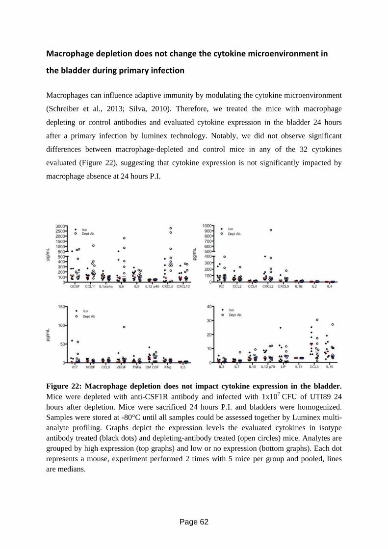
Page 62
J"32'<;"6%&9%<$%+5'*&9'%,&*'+&3;"*6%&+;%&3A+'75*%&0532'%*:52'*0%*+&5*&
+;%&#$"99%2&9/25*6&<250"2A&5*(%3+5'*&
Macrophages can influence adaptive immunity by modulating the cytokine microenvironment
(Schreiber et al., 2013; Silva, 2010). Therefore, we treated the mice with macrophage
depleting or control antibodies and evaluated cytokine expression in the bladder 24 hours
after a primary infection by luminex technology. Notably, we did not observe significant
differences between macrophage-depleted and control mice in any of the 32 cytokines
evaluated (Figure 22), suggesting that cytokine expression is not significantly impacted by
macrophage absence at 24 hours P.I.
Figure 22: Macrophage depletion does not impact cytokine expression in the bladder. Mice were depleted with anti-CSF1R antibody and infected with 1x10⁷ CFU of UTI89 24 hours after depletion. Mice were sacrificed 24 hours P.I. and bladders were homogenized. Samples were stored at -80°C until all samples could be assessed together by Luminex multi-analyte profiling. Graphs depict the expression levels the evaluated cytokines in isotype antibody treated (black dots) and depleting-antibody treated (open circles) mice. Analytes are grouped by high expression (top graphs) and low or no expression (bottom graphs). Each dot represents a mouse, experiment performed 2 times with 5 mice per group and pooled, lines are medians.

Page 63
Increasing&the&amount&of&antigen&does¬&affect&the&generation&of&adaptive&
immunity&during&UTI&
In the lung, it has been observed that macrophages can influence the generation of an adaptive
immune response by antigen sequestration, hindering antigen presentation and subsequent T
cell priming by DCs (Jakubzick et al., 2008; Jakubzick et al., 2006; Kradin et al., 1999;
MacLean et al., 1996). It has been proposed that DC migration and induction of adaptive
immunity can only occur in the lung after macrophages have been saturated and there is an
excess of antigen available to the DCs (Kradin et al., 1999; MacLean et al., 1996). To test the
hypothesis that antigen availability during primary infection impacts the generation of an
adaptive immune response during UTI, we infected untreated mice with 107, 108, or 109 CFU
of UPEC. Once the primary infection was resolved, mice were challenged with 107 CFU of an
isogenic UPEC strain and 24 hours P.I., sacrificed to determine bacterial burden. We did not
observe a difference in the number of bacteria per bladder after a primary infection despite
increasing the inoculum 100-fold (Figure 23A). Furthermore, we did not observe any
differences in bacterial load after challenge among the three groups (Figure 23B). These data
suggest that increasing the number of bacteria during primary infection does not improve the
generation of adaptive immunity during UTI, however it is possible that we did not saturate
bladder resident macrophages with bacteria and that it is necessary to increase the number of
bacteria used for infection. We did not check if the number of DCs acquiring UPEC among
the different groups (mice infected with 107, 108, or 109 CFU of UPEC) increased, therefore
an alternative explanation for this result could be that independently of the number of bacteria
used to infect the mice, the amount of DCs harboring UPEC was the same.

Page 64
Figure 23: Increasing bacterial inoculum during primary infection does not improve the response to challenge infection. Female C57Bl/6 mice were instilled with 1x10⁷, 1x10⁸, or 1x10⁹ CFU of UPEC and challenged with 1x10⁷ CFU after resolution of the primary infection as in figure 10. (A) Plot depicts CFU at 24 hours post-primary infection. (B) Graph shows CFU 24 hours post-challenge. Each dot represents one mouse. Experiments were repeated 2 times with 5-7 mice per group.
Macrophage&depletion&leads&to&increased&phagocytosis&of&UPEC&by&DCs&
As the bladder microenvironment was unchanged at 24 hours P.I. in the absence of
macrophages, we evaluated whether macrophages were physically sequestering UPEC in the
bladder during UTI by measuring bacterial acquisition in the bladder in the context of
macrophage depletion. Mice were depleted or not of macrophages by anti-CSF1R antibody
treatment, infected with UTI89-RFP, and sacrificed 24 hours P.I. We then evaluated the
immune cells containing UPEC (RFP+) by flow cytometry as in Figure 13. The distribution of
UPEC was altered in mice depleted of macrophages as compared to the control group. A
greater percentage of MHC II- cells contained UPEC compared to MHC II+ cells (Figure
24A). Additionally, the percentage of neutrophils containing bacteria increased in
macrophage-depleted mice, likely compensating for the lack of monocytes and resident
macrophages (Figure 24B). In MHC II+ cell populations, while the total number of DCs in
infected bladders was unchanged (Figure 24C), we observed that a significantly greater
percentage of DCs had taken up UPEC in macrophage-depleted animals (Figure 24D).
Moreover, the percentage of UPEC-containing resident macrophages and monocyte-derived
macrophages remained unchanged (Figure 24E). These results suggest that macrophages
sequester UPEC from DCs and in their absence, more DCs are now able to acquire UPEC
positively impacting the induction of an adaptive immune response.

Page 65
Figure 24: DCs acquire more bacteria in the absence of macrophages. Macrophages were depleted or not and mice were infected. Twenty-four hours post-primary infection, bladders were analyzed by flow cytometry to determine distribution of UPEC in (A) the MHC II- and MHC II+ compartments, (B) neutrophils and monocytes. (C) Plot depicts the total number of DCs present in the bladder 24 hours post-primary infection. (D-E) Plots depict the percentage distribution of UPEC in the MHC II+ compartment by cell population (D) DCs and (E) resident macrophages and monocyte-derived macrophages. Each dot represents one mouse, lines are medians. Experiments were repeated 3 times with 4-7 mice per group in each experiment and results pooled. p-values indicated on graphs, Mann-Whitney test. !

Page 66
Chapter&3:&Delineating&the&adaptive&immune&response&
generated&during&UTI&
&

Page 67
Introduction&
The results presented in this chapter are part of our ongoing efforts to further delineate the
adaptive immune response to UTI. We are currently focusing on unraveling the role of the T
cell. The unpublished work presented here represents our first findings.
We found that the adaptive immune system was necessary for improved bacterial clearance
after UPEC challenge. The adaptive immune responses generated against pathogens can be
classified as cellular or humoral, depending whether they are mediated by T or B cells,
respectively. In general, intracellular pathogens, such as viruses and intracellular bacteria,
trigger a cellular response that includes the generation of specific CD8+ T cells, which are
necessary to detect, and clear infected cells. On the other hand, extracellular pathogens
commonly induce an antibody-mediated response. Of note, both responses can be generated
during infection; as an example, HIV infection induces virus-specific T cells as well as virus-
specific antibodies (Xu et al., 2013). It is not completely clear whether a cellular response
alone or a humoral response alone is able to control UPEC infection during challenge or if
both responses are needed. Therefore, we focused our attention on dissecting the adaptive
immune response generated during UTI, considering that broadening our knowledge is key to
the development of future therapeutic options.
DCs mediate communication between the innate and adaptive immune response. They
phagocytize pathogens and infected cells, sense the microenvironment, and migrate to LNs to
deliver information to naïve lymphocytes. Depending on their origin, function, and
distribution, DCs can be classified into different subsets including CD4+ and CD8α+ DCs
(Kushwah and Hu, 2011). CD103+ DCs, which are particularly important to induce a CD8+ T
cell response, reside in nonlymphoid tissues and cross-present antigens (Bedoui et al., 2009;
del Rio et al., 2010; Ginhoux et al., 2009). The transcription factor Batf3 has been shown to
be fundamental for the development of CD8α+ DCs (LN-resident DCs) and CD103+ DCs, as
mice lacking this protein also lack both of these DC subsets and have an impaired CD8+ T
cell response (Hildner et al., 2008). Over the course of our studies, we reported that DCs and
T and B cells are necessary to reduce CFU after UPEC challenge (Mora-Bau et al., 2015),
however, which DC subset is involve in the induction of adaptive immunity during UTI or
what type of T cells are generated during this response is unknown. We observed that CD103+
DCs reside in the bladder mucosa, and thus evaluated the role of these DCs during UTI.
Page 68
CD8α+&and&CD103+&DCs&are&dispensable&for&an&adaptive&immune&response&
against&UPEC&
CD8α+ DCs and CD103+ DCs are more efficient cross-presenting cells and therefore
fundamental for the induction of CD8+ T cell responses (Bedoui et al., 2009; Dudziak et al.,
2007; Hildner et al., 2008). The role of CD8+ T cells during UTI has not been addressed
before. This is surprising considering that UPEC invades uroepithelial cells and a CD8+ T cell
response would be beneficial to fight intracellular bacteria. To evaluate the role of CD8α+
DCs and CD103+ DCs during UTI, we utilized Batf3 KO mice. Batf3 KO and WT mice were
infected with 107 CFU of UPEC and their urine was monitored every 5-6 days to evaluate the
presence of bacteria. When no bacteria in the urine were detected, mice received a challenge
infection with an isogenic UPEC strain as in Figure 10. We did not observe any difference in
bacterial clearance between Batf3 KO and WT mice suggesting that CD8α+ DCs and CD103+
DCs are not necessary to induce an adaptive immune response against UPEC (Figure 25A).
We did not observe any difference in the reservoir either (Figure 25B). As CD8α+ DCs and
CD103+ DCs are thought to be essential to induce cytotoxic T cells, CD8+ T cell responses
should be absent in Batf3 KO mice. The use of these mice can be considered to be an indirect
means to evaluate CD8+ T cell responses. With this in mind, our results suggest that a CD8+ T
cell response is dispensable for UPEC clearance during challenge infection. The lack of a
CD8 T cell response may explain why bacterial reservoirs persist, even in the face of a
challenge infection. It has been reported, however, that CD8+ T cell responses can arise in
Batf3 KO mice, as CD8α+ DCs are induced in the absence of the Batf3 transcription factor
and cross-present soluble antigens and cell-associated antigens during GVHD (Seillet et al.,
2013).

Page 69
Figure 25: The absence of CD8α+ and CD103+ DCs does not impact the adaptive immune response against UPEC. WT and Batf3 KO mice were infected with 107 CFU of UPEC. Once the mice resolved the primary infection, they were challenged with an isogenic strain of UPEC. Twenty-four hours post-challenge, mice were sacrificed and bacterial load was evaluated. Graphs depict bacterial load derived from the challenge (A) and primary infecting strains (bacteria reservoirs) (B). Experiment was repeated 3 times with 5-6 mice per group.
&
T&cells&are&dispensable&for&the&direct&response&to&UPEC&during&challenge&
infection&
In previous experiments, we observed that a lack of T cells over the course of primary
infection negatively impacted bacterial clearance during UPEC challenge (Fig 17F).
However, this experimental approach did not allow us to determine whether T cells are
necessary to directly kill bacteria during challenge infection or if they are required to generate
a humoral response against UPEC. To directly test whether T cells, and in particular CD8+ T
cells, were necessary as part of the challenge response, we modified our T cell depletion
experiment. We depleted T cells one day prior to challenge infection, instead of prior to
primary infection, a strategy that would permit the induction of T cell immunity. Mice were
infected with 107 CFU of UPEC and urine was monitored every 5-6 days to evaluate the
resolution of primary infection. When the acute infection was resolved, mice were treated
with anti-CD4 and CD8 depleting antibodies or isotype controls 24 hours prior to challenge
infection. Twenty-four hours post-challenge, mice were sacrificed and bacterial burden was
evaluated. We observed no difference in bacterial clearance between T cell depleted and

Page 70
control isotype treated mice, suggesting that T cells are not necessary to clear UPEC during
challenge infection (Figure 26).
Figure 26: T cells are not necessary for UPEC clearance during challenge infection. Female C57Bl/6 mice were infected with 107 CFU of UPEC. Once mice had resolved the primary infection, CD4+ and CD8+ T cells were depleted by administration of anti-CD4 and anti-CD8 depleting antibodies. Twenty-four hours post-depletion, mice were challenged with an isogenic strain of UPEC and 24 hours later sacrificed to evaluate bacterial load. Experiment was repeated 3 times with 5-6 mice per group.
In summary, the results presented here, suggest that T cells are not necessary to directly clear
UPEC during challenge infection and/or to clear bacterial reservoirs. However, the response
during a challenge infection is impaired when T cells were depleted at the prior to infection
(Figure 17F) suggesting a role for these cells in the development of an adaptive immune
response during UTI. We hypothesize that T cells are required for an antibody-mediated
response against UPEC and that these antibodies are sufficient to clear UPEC during a
challenge infection. It will be interesting to continue studying the role of T and B cells during
UTI to answer questions such as: 1) what kind of T cells are generated during UTI? 2) Are
UPEC-specific CD8+ T cells generated during UTI? If so, 3) are they truly dispensable during
infection? 4) Can we improve the antibody response generated during UTI to improve
bacterial clearance? 5) Can we induce or augment the CD8+ T cell response to eliminate
UPEC reservoirs? To answer these questions, we are in critical need of specific tools to be
able to identify UPEC-specific lymphocytes, including UPEC-specific MHC tetramers. To
facilitate our search for specific lymphocytes, we have recently engineered UPEC strains to

Page 71
express OVA peptides, which can be recognized by transgenic CD4+ and CD8+ T cells (T
cells from OT II and OT I mice) and an altered flagellin, which can be recognized by
transgenic CD4+ T cells (T cells from SM1 mice). With these tools, we will be able to
evaluate if UPEC-specific T cells responses are generated and further characterize this
response to achieve greater understanding of the adaptive immune response occurring during
UTI to provide better solutions to treat UTI patients.
!!
&
Page 72
Chapter&4:&Discussion&

Page 73
Portions of the manuscript “Macrophages Subvert Adaptive Immunity to Urinary Tract
Infection” published in Plos Pathogens (Mora-Bau et al., 2015) have been reproduced here.
UTI is unusual in that it is a common infection, affecting otherwise healthy
individuals, that recurs with high frequency, suggesting a defect in the ability to build an
adaptive immune response to UPEC. We observed that the absence of B and/or T cells or DCs
impaired the host’s capacity to clear bacteria after a challenge infection, confirming that
adaptive immune responses are primed and necessary during UTI. Although this was an
expected result, it has never been formally demonstrated before. Notably, while we found that
an adaptive immune response is primed, and bacterial clearance after a challenge infection is
impaired in the absence of DCs, B and T cells, intact mice were not fully protected from
challenge infection. To shed light on potential mechanisms preventing the development of a
more protective adaptive immune response to UPEC infection, we focused on the role of
MHC II+ cells, as they are the central initiators of immunity. In the course of our work, our
key finding was that, in the context of a challenge infection, resident macrophage depletion
improved the host’s ability to eliminate bacterial load. Importantly, macrophages were
depleted prior to the first infection; however, they were present in normal numbers at the time
of challenge infection indicating that it was not the absence of macrophages during the second
infection that impacted bacterial clearance during challenge. This improvement was
dependent on the adaptive immune system as the phenotype was lost when macrophages were
depleted in RAG2-/- mice or in mice depleted of T cells. Given that macrophages were the
principal APC to acquire UPEC early in infection, these data suggest a model in which
macrophages sequester the bacteria from the DCs subverting the initiation of a robust
adaptive immune response during UTI.
How&do¯ophages&limit&the&adaptive&immune&response&against&UPEC?&
To understand how macrophages limit induction of an adaptive immune response
during UTI, we evaluated events post-challenge and post-primary infection. We were
specifically interested in answering two fundamental questions: (1) which cells are
responsible for improved clearance during bacterial challenge in macrophage-depleted mice
and (2) what happens in the bladder during primary infection, in the absence of macrophages,

Page 74
that improves adaptive immunity against UPEC? To address the first question, we evaluated
immune cell infiltration into the bladder during challenge infection. We observed a significant
infiltration of T cells and a smaller infiltration of B cells post-challenge but no major
differences in cell numbers between the control and treated groups. At this time, we cannot
rule out potential qualitative differences in the activation or specificity of the infiltrating
effector cells, and this is an active line of investigation. To gain more knowledge in this
regard, it would be ideal to characterize what kind of T cells are infiltrating the bladder during
challenge infection (e.g., T helper bias, regulatory T cells) and determine if these T cells are
different between macrophage-depleted and control-treated mice (e.g., activation status,
cytotoxic potential).
To answer the second question, we evaluated events occurring in the bladder in the
first hours following primary infection, in the absence or presence of macrophages, which
may impact the generation of an adaptive immune response. It has been shown that
macrophages can influence the tissue microenvironment and in this way, modulate the
induction of adaptive immunity (Schreiber et al., 2013; Silva, 2010). For example, during
infection with Citrobacter rodentium, macrophages induce Th1 polarization of CD4 T cells by
secreting IL-12 (Schreiber et al., 2013). Furthermore, a recent study suggested that IL-10
expression from mast cells suppresses adaptive immunity to UPEC (Chan et al., 2013). Thus,
hypothesizing that the absence of macrophages might influence the microenvironment, either
directly or indirectly, we evaluated cytokine production in the bladder during infection in
macrophage-depleted and control-treated mice. Multi-analyte cytokine analysis revealed no
striking differences between control and depleted mice and in contrast to the study mentioned
above, we did not detect IL-10 expression. The reasons for the different finding in our study
and Chan et al. study are unclear. In the course of our study, we found few mast cells in naive
bladder tissue. In addition, the differences may be due to the significant variation that exists in
the genomes of commonly used strains such as cystitis strain UTI89, pyelonephritis strains
J96, 563, CFT073, and clinical isolates (see phylogenetic tree in (Petty et al., 2014)).
Together, our results suggest that macrophage absence does not modulate the bladder
microenvironment during UTI, however, as we only evaluated cytokine expression at 24
hours P.I., we cannot rule out the possibility that the microenvironment was altered at a
different time point.

Page 75
Additionally, it has been observed that macrophages can limit the adaptive immune
response by antigen sequestration (McLean et al. 1996; Kradin et al. 1999; Jakubzick et al.,
2006). For example, after injecting mice intratracheally with heat-killed Listeria,
macrophages acquire the majority of the antigen, limiting heat-killed Listeria acquisition by
DCs; therefore, an adaptive immune response is achieved only after macrophage depletion
(MacLean et al., 1996). Jakubzick and colleagues found that DC migration and antigen
presentation only occurred when alveolar macrophages were over-saturated with antigen,
allowing DC antigen uptake to occur. To explore the hypothesis of antigen sequestration
during UTI, we evaluated which immune cell populations acquired UPEC in the bladder in
the absence of macrophages. We observed an increase in the percentage of DCs containing
bacteria in macrophage-depleted mice compared to control-treated animals. DCs are key
players in initiating adaptive immune responses, and we found that they can do so from the
bladder mucosa in the context of UTI. Indeed, even a partial depletion of bladder-resident
DCs, prior to primary infection, rendered animals less capable of clearing bacteria after
challenge infection. The intermediate clearance phenotype observed in DT-treated mice may
have been mediated by DCs remaining after depletion or by DCs repopulating the tissues
before the primary infection was resolved, permitting delayed antigen presentation; this
suggests that a small number of DCs is sufficient to mount an adaptive immune response
against UPEC. Thus, our data suggest that the more efficient adaptive immune response
against UPEC observed during macrophage depletion may be mediated by the increase in the
percentage and number of DCs carrying bacteria. If DCs in the bladder have limited access to
UPEC due to macrophage sequestration, we could hypothesize that depleting neutrophils,
which also acquire a significant number of bacteria during infection (see MHC II- cells
containing UPEC in Figure 14) (Mora-Bau et al., 2015), would also increase UPEC
availability for DCs and improve the adaptive immune response in UTI. However, we would
have to consider that in the absence of neutrophils, mice display improved bacterial clearance
during primary infection (Ingersoll et al., 2008), which could affect the induction of adaptive
immunity.
While depletion of bladder resident macrophages impacted the outcome in UPEC
infection, monocyte depletion had no effect. The majority of monocytes infiltrating the
bladder gave rise to macrophages, however some of these cells also became DCs. Therefore,
monocyte depletion impacted the number of macrophages in the bladder but it also impacted
the number of DCs and this may be the reason why the adaptive immune response against

Page 76
UPEC was not improved in monocyte-depleted mice. We might imagine that a decrease in
DC number would negatively impact the generation of an adaptive immune response against
UPEC, however, as the total number of macrophages was also decreased as an indirect effect
of clodronate treatment, remaining DCs may have had more access to UPEC. This hypothesis
could be tested in a rigorous manner, by monocyte depletion and quantification of the total
number of DCs as well as the number of DCs carrying bacteria, over several timepoints.
It is possible that macrophages are the main APC in the bladder to acquire UPEC
because they outnumber DCs in both naïve and infected bladders. Alternatively, it is possible
that the localization of macrophages and DCs in the bladder impact phagocytosis. If
macrophages are positioned closer to the bladder lumen, they will have a greater sampling
capacity during infection. To shed some light onto these questions, we are currently
performing ex vivo experiments to evaluate UPEC acquisition by DCs and macrophages. By
varying the proportion of these two cell types in the cultures, we can determine whether, for
example, when we have the same number of DCs and macrophages then both cell types can
acquire UPEC equally or there is still a bias in phagocytosis. Additionally, in this set-up, we
have removed differences in cellular localization. To more directly address the question of
localization, we will image the bladder.
Having defined an early role for resident macrophages and DCs during UTI (Figure
27), our work significantly advances the understanding of how adaptive responses to UPEC
are achieved. Our data point to a barrier in the immune system that must be overcome,
particularly for patients with recurrent UTI. Although macrophage sequestration of particulate
antigen in the lung has been described (Jakubzick et al., 2006; Kradin et al., 1999; MacLean
et al., 1996), this is, to the best of our knowledge, the first study to propose a role for the
physical sequestration of antigen during live bacterial infection. Strategies that increase DC
number or DC migration may overcome the subversion imposed by macrophages, providing a
viable solution to treat patients with recurrent UTI.
!!

Page 77
Figure 27: Proposed model for the role of macrophages during UTI. This scheme depicts our model regarding how an adaptive immune response is generated during UPEC infection in the presence (left, control) and absence (right, macrophage depletion) of macrophages. While in the control situation, macrophages acquire the majority of UPEC, limiting antigen acquisition by DCs and the generation of an adaptive immune response, macrophage depletion leads to an increase of UPEC-containing DCs and improves the adaptive immunity against UPEC.
J"32'<;"6%,&"*9&+;%52&2%$"+5'*,;5<&85+;&+;%&"9"<+5:%&500/*%&2%,<'*,%S&"2%&
+;%A&"$8"A,&+;%&T#"9&6/A,UR&&
In our study, we observed that macrophages negatively impact adaptive immunity
generated during UTI (Mora-Bau et al., 2015), however, there are other examples describing
the negative and positive impact of macrophages on adaptive immune responses. Thus, it may
not be straightforward to describe the macrophage as a “good guy” or a “bad guy” during
infection, and likely many variables impact the behavior and role of activated macrophages.
!"#$$%&' !"#$$%&' ()'
!"#
*+',-.&#/01' 23%450&'&%67016%'-1$84/01'
$%&##'%((#
)*+,-./%0#
91/.%1'':75#;%'<!"='
12#
12#
12#$##'%((#
3##'%((#
!"#
2-*45-(# !)'5-67)8%#.%6(%+-*#
91/.%1'':75#;%'<12='
- Increase UPEC uptake by DCs - Increase of adaptive immunity against UPEC - Increase UPEC uptake by DCs - Increase of adaptive immunity against UPEC
- Limited UPEC availability for DCs
12#

Page 78
Good&guys&
Macrophages were thought to have little or no direct T cell priming capability;
however, a recent report has shown that macrophages can present adenovirus particle-derived
antigen to T cells in vivo in the presence or absence of DCs (Bernhard et al., 2015). Moreover,
in the presence of both DCs and macrophages, this study shows that MHCI+ CD169+
macrophages were able to prime a more comprehensive immune reaction than cross-
presenting DCs. Macrophages activated T cells specific for all the epitopes presented,
whereas cross-presentation by DCs stimulated only those T cells that recognize
immunodominant epitopes. Moreover, lymph node resident CD169+ macrophages have a
prominent role in the priming of CTL-mediated antitumor immunity (Asano et al., 2011).
CD169+ macrophages phagocytize dead tumor cells transported via lymphatic flow and
subsequently cross-present tumor antigens to CD8+ T cells. In the absence of these
macrophages, antitumor immunity is impaired (Asano et al., 2011). These results thus reveal a
previously unknown role for CD169+ macrophages in the induction of adaptive immune
responses, in the context of viral infection and tumor immunity.
Macrophages can also collaborate with DCs to promote health (Mazzini et al., 2014).
Gut resident CD103+ DCs are responsible for inducing oral tolerance to fed antigens, however
only the CX3CR1+ macrophages and intestinal epithelial cells can efficiently capture these
antigens from the lumen. How DCs acquire the antigens from the lumen then? It was recently
demonstrated that CX3CR1+ macrophages, quickly transfer these antigens after their capture
to CD103+ DCs in a gap junction dependent manner (Mazzini et al., 2014). As we have
observed that naïve bladders contain many CX3CR1+ positive cells, it would be interesting to
study whether resident macrophages and DCs can communicate with each other through these
channels during UTI. For example, macrophages could transfer inhibitory signals to the DCs,
as regulatory T cells transfer cAMP to effector CD4+ T cells to suppress their effector
functions. Alternatively, if macrophages and DCs do not communicate during UTI, it might
be that inducing their communication would lead to antigen transfer and an improved
adaptive immune response.
Bad&guys&
In addition to sequestration of antigens from DCs (McLean et al. 1996; Kradin et al.
1999), macrophages can act as “bad guys” while interacting with the adaptive immune

Page 79
response. This can be exemplified in the context of obesity-induced inflammation (Cho et al.,
2014; Morris et al., 2013). Obesity-induced inflammation is associated with metabolic
syndrome, cardiovascular disease, and the development of type 2 diabetes (Apovian et al.,
2008; Lumeng and Saltiel, 2011; Ouchi et al., 2011). Adipose tissue macrophages possess all
the functional activities of APCs and can promote antigen-specific T cell activation and Th1
polarization of CD4+ T cells in the fat of obese mice, helping to coordinate an adaptive
immune response to obesity (Morris et al., 2013). Additionally, adipose tissue macrophages
are required for the induction of effector/memory adipose tissue-specific CD4+ T cells, which
are involved in obesity-induced inflammation and insulin resistance (Cho et al., 2014).
Deficient expression of MHC II in adipose tissue macrophages (which can no longer present
antigens to CD4+ T cells) leads to an improvement in glucose intolerance by increasing
adipose tissue insulin sensitivity (Cho et al., 2014). Macrophages can also negatively impact
the adaptive immune response to ovarian carcinoma, as it has been observed that with tumor
cells, they attract regulatory T cells through the production of the chemokine CCL22 (Curiel
et al., 2004).
During UTI, we demonstrated that macrophages act as the “bad guys” because they
negatively impact the generation of an adaptive immune response against UPEC.
Macrophages are professional phagocytic cells, however, their main role is to maintain the
homeostasis of tissues, in part by secretion of cytokines to alert other immune cells to
infection or inflammation. In the bladder, macrophages are the most abundant immune cells.
They may phagocytize the majority of UPEC simply because they outnumber other
phagocytes in the bladder at early timepoints. Thus, by fulfilling their role as phagocytic cells
trying to clear pathogens, they exert a detrimental effect on the development of an adaptive
immune response.
What&is&the&nature&of&the&immune&response&against&UPEC?&
We still do not completely understand how the adaptive immune system eliminates
UPEC. Although we were not able to detect UPEC-specific antibodies above the limit of
detection of our assay, others have identified that antibodies against bacteria are generated
during infection (Hopkins et al., 1987; Svanborg-Eden and Svennerholm, 1978; Thumbikat et

Page 80
al., 2006). Additionally, some studies evaluating vaccination strategies for UTI have
measured the induction of a humoral response after vaccination and found that this response
positively correlates with a positive outcome of the therapy (Asadi Karam et al., 2013;
Langermann et al., 2000; Langermann et al., 1997). Thus, we hypothesized that the protection
induced during UTI following a primary infection is mediated by an antibody response;
however we also considered that T cells might play a direct role in the killing of infected
cells. To directly evaluate the role of T cells during challenge, CD4+ and CD8+ T cells were
depleted right before challenge infection and bacterial clearance was evaluated. We observed
no difference in bacteria titers between T cell-depleted versus control treated mice, suggesting
that T cells are not necessary to clear UPEC during challenge. Interestingly, when T cells
were depleted before primary infection instead of challenge infection, mice were not
protected against an UPEC challenge. The explanation for this may be that T cell depletion at
the beginning of infection also impairs the generation of specific B cells and a humoral
response, while T cell depletion right before challenge infection does not affect the humoral
response already generated. Together, these results suggest that a humoral response is
sufficient to confer protection during UPEC challenge.
Additionally, as UPEC is able to invade urothelial cells and even remain there for
months as bacterial reservoirs, we hypothesized that a CD8+ T cell response is required to
clear intracellular UPEC. We evaluated the role of CD8+ T cells indirectly by using Batf3 KO
mice, which lack CD8α+ and CD103+ DCs impairing CD8+ T cell responses (Hildner et al.,
2008). We did not observe any difference in bacterial clearance during challenge infection
between WT and Batf3 KO mice, suggesting that CD8+ T cells are dispensable for fighting
UPEC during a challenge infection. Importantly, it has been shown that CD8α+ and CD103+
DCs can develop in Batf3 mice, thus a CD8+ T cell response could be induce in these mice.
To formally rule out a role for CD8+ T cells, we would need to measure whether they
infiltrate during infection in the Batf3 mouse as well as to eliminate them directly prior to
challenge infection.
Interestingly, we did not observe any difference in the reservoir either in these
experiments, suggesting that once reservoirs are formed (which occurs early during infection
(Mulvey et al., 2001)) they remained untouched by the immune system. To explain why the
reservoirs are not affected by the adaptive immune response generated during UTI we
propose two hypothesis: (1) UPEC-specific CD8+ T cells are not generated during UTI or (2)

Page 81
UPEC-specific CD8+ T cells are generated, however, they do not recognize UPEC-infected
cells as these cells are not be presenting UPEC antigens. It would be interesting to perform ex
vivo experiments and evaluate if CD8+ T cells obtained from UPEC-infected mice are able to
kill UPEC-infected uroepithelial cells, as well as to evaluate the induction of UPEC-specific
CD8+ T cells in infected mice through tetramer staining. Thus, while this is still a work in
progress, we demonstrated that mice unable to induce a CD8+ T cell response (Batf3 KO
experiments) and mice lacking T cells during UPEC challenge infection are able to clear
infection as well as control mice, suggesting that even though we were unable to detect
UPEC-specific antibodies during the course of our experiments, a humoral response during
UTI likely mediates protection during (challenge) infection.
Can&effective&adaptive&immunity&be&evoked&from&the&bladder?&
The early defense against bacterial infections, as well as the induction of an adaptive
immune response, is dependent mainly on resident tissue cells (Iwasaki and Medzhitov, 2004;
Medzhitov and Janeway, 2000), which initiate the immune response against invading
pathogens. In this study, we showed that the bladder is equipped with different resident
immune cell population including DCs, which are essential to mount an adaptive immune
response. In addition to finding the right players to induce adaptive immune responses in the
bladder, we observed that adaptive immunity can be primed from the bladder mucosa during
UPEC infection (Mora-Bau et al., 2015). While we formally demonstrated the necessity for
DCs, T and B cells in response to UTI, the fact that adaptive immune responses can be raised
from the bladder is not a completely novel concept. Indeed, successful immunotherapy for
bladder cancer is reliant upon robust adaptive immune responses. Bladder cancer is the fifth
more common cancer in the US and Europe and with extraordinarily high health care costs
(Ferlay et al., 2007; Siegel et al., 2013). Bacillus Calmette-Guerin (BCG) instillation is the
primary treatment option for nonmuscle invasive bladder cancer patients after tumor resection
and has been used for almost 40 years with high success rates (Dovedi and Davies, 2009;
Fuge et al., 2015). Both UTI and bladder cancer have in common the fact that bacteria are
introduced into the bladder lumen, as part of the infection process in UTI or as a therapy
during bladder cancer. Interestingly, UPEC and BCG instillation appear to evoke very
different immunological outcomes. Mouse experiments have demonstrated that following

Page 82
BCG instillation into the bladder, BCG-specific CD8+ T cells are primed (Biot et al., 2012).
Additionally, analysis of patient bladder biopsies has shown T cell infiltration during BCG
therapy and that the degree of infiltration correlates with treatment response (Prescott et al.,
1992). Moreover, the depletion of either CD4+ or CD8+ T cells abolishes BCG-mediated
antitumor activity, suggesting that T cells play an important role during BCG immunotherapy
(Ratliff et al., 1993). During UTI, we observe that an adaptive immune response is primed
and confers protection against a challenge infection. However, this response neither prevents
reinfection nor eliminates the bacterial reservoir established during primary infection. It has
been shown that, during BCG treatment, live BCG can be found in the LNs and this
phenomenon correleates with the priming of BCG-specific CD8+ T cells (Biot et al., 2012).
Our preliminary experiments failed to detect the presence of live UPEC in the LNs during
UTI. With this in mind, we could speculate that bladder APCs are unable to mount an
efficient adaptive immune response and to achieve this response it would be necessary to have
bacterial presence in the LNs. The necessity of the pathogen in the LNs to induce an adaptive
immune response has also been shown during lung infection with Mycobacterium
tuberculosis, suggesting that this phenomenon is not specific to the bladder and it is related to
the nature of the pathogen (Chackerian et al., 2002; Wolf et al., 2008). The response
generated during BCG treatment seems to be an “efficient” response as the presence of total
T cells (T cell infiltrates in bladder biopsies) and/or BCG-specific T cells is associated with a
better outcome for the disease (Biot et al., 2012; Prescott et al., 1992; Ratliff et al., 1993).
Why&does&the&adaptive&immunity&generated&during&UTI&“fail”?&
Mice can develop an adaptive immune response during UTI however this response
does not prevent reinfection. Therefore, the important questions are: why is the adaptive
immune response generated during UTI not efficient? Why does this response not prevent
recurrence? There are several possibilities to answer to these remaining open questions. The
steps to induce an adaptive immune response during UTI include (1) UPEC acquisition in the
bladder, (2) DC migration to the LNs, (3) priming of naive T and B cells, and (4) migration of
effector cells to the bladder (Figure 28). We imagine that a failure in any of these steps could
impair adaptive immunity generated during UTI. For example, during UPEC acquisition in
the bladder, we demonstrated that macrophages sequester UPEC and limit bacteria acquisition

Page 83
by DCs (Mora-Bau et al., 2015). In the next step, DC migration to the LNs is essential to
induce an adaptive immune response, therefore a defect in DC migration could also add up to
poor induction of adaptive immunity against UPEC. During tuberculosis, the generation of an
adaptive immune response against M. tuberculosis is delayed, allowing bacterial growth and
dissemination (Roberts and Robinson, 2014; Urdahl et al., 2011). Evidence suggests that this
impaired response to M. tuberculosis is due to the fact that DC migration from the lung to the
LNs is inhibited, which limits the initiation of an adaptive immune response (Roberts and
Robinson, 2014; Urdahl et al., 2011). Following migration, DCs prime naive lymphocytes in
the LNs to induce an adaptive immune response specific to the infecting pathogen. If the
priming of UPEC-specific T cells is not efficient, the adaptive immune response will be
impaired. As an example, during BCG immunotherapy, it has been observed that the
induction of Th1 T cells is associated with the success of therapy; however, a Th2 response is
associated with BCG failure (Nadler et al., 2003; Riemensberger et al., 2002). Finally, it is
also possible that UPEC-specific effector cells do not migrate and/or infiltrate the bladder to
fight infection. In addition, it is possible that, although specific T and B cells are generated,
they are not long lasting and when women experience recurrence they do not have specific
lymphocytes to fight the infection. Thus, there are many reasons that could give explain why
the adaptive immune response induced during UTI is not efficient, leading to high rates of
recurrence. Each of these possibilities needs to be tested in a rigorous experimental fashion, which is challenging given the number of different ways the immune response can fail. It is
fundamental to continue studying how adaptive immunity is achieved against UPEC to
understand what fails and how this failure can be overcome to improve the response.

Page 84
Figure 28: Proposed model for the generation of adaptive immunity during UTI. In this scheme, we illustrate the different required steps to induce adaptive immunity during UTI including: (1) antigen acquisition in the bladder mucosa, (2) DC migration to draining LNs, (3) priming of naïve lymphocytes and (4) migration of effector cells to the bladder.
In addition to these potential issues during the primary immune response, we have an
additional layer of complexity: bacterial reservoirs. Different hypotheses have been proposed
to explain UTI recurrence. These hypotheses include that UPEC strains colonize the gut and
periodically migrate to the urinary tract (Chen et al., 2013), that the immune response to
infection is suppressed by mast cell-derived IL-10 in the bladder (Chan et al., 2013), or that
UPEC forms protected reservoirs in the bladder, remerging at later time points after initial
infection (Anderson et al., 2004; Mysorekar and Hultgren, 2006). It could also be a mix of the
proposed explanations as they are not mutually exclusive. With this in mind, it would be
fundamental to eliminate bacterial reservoirs from the bladder as we could be eliminating a
major source of recurrence. To eliminate the reservoirs, a CD8+ T cell response would be
necessary, however, the generation of UPEC-specific CD8+ T cells during UTI has not been
demonstrated yet. Our challenge experiments in Batf3 KO mice suggest that CD8+ T cells are
dispensable during UTI; however, further experiments are needed to fully demonstrate a role
of these cells during infection. For example, through tetramer staining, we could evaluate if
!"#$$%&'
()'
!"# $##%&''#
()#!"#*+,-%+''
./0#1%'
*23'45-&#,6+'
78%906&'&%:/6+:%'5+$.9,6+'
$##%&''#
*##%&''#
!"#
78%906&'9%""'45-&#,6+'
+,-./01&2#
;'
<'
='
>'

Page 85
UPEC-specific CD8+ T cells are generated during UTI. If these cells are generated, we can
evaluate their ability to infiltrate the bladder. In addition, we could directly deplete CD8+ T
cells and evaluate bacterial clearance during challenge and in UPEC reservoirs, as this is a
more direct way to evaluate their role during infection than using Batf3 KO animals.
Unfortunately, it may be that UPEC-specific CD8+ T cells are not generated during UTI,
rendering the immune system incapable of detecting and eliminating infected cells. It is
critical to remember, though, that although the rates of recurrence are high, more than 50% of
UTI patients will not recur. We should consider studying these women, as it would be
interesting to detect differences between them and women that tend to have recurrent
infections to identify possible explanations for the failure of the response in those who recur.
Along this line, there are some known genetic components in humans that may be associated
with a major risk of UTI (Zaffanello et al., 2010). For example, it was observed that UTI-
prone children express lower levels of CXCR1 (IL-8 receptor) than healthy children
(Lundstedt et al., 2007). Additionally, low expression of TLR4 on neutrophils has been
associated with asymptomatic bacteriuria in children suffering UTI with difficulties in
clearing infection (Ragnarsdottir et al., 2007).
UTI affects millions of people every year and induce high health care costs (Foxman,
2010; Stamm and Norrby, 2001). Patients suffering from this infection are treated with
antibiotics, however antibiotics do not prevent recurrence, which is a major problem among
infected women. Importantly, antibiotic resistance is increasing, as well as the necessity of
new therapeutic options (Flores-Mireles et al., 2015; Foxman, 2010; Hooton et al., 2004).
Mechanism to increase the number of DCs and/or to increase the trafficking of T cells to the
bladder could be considered as a way to improve the response to UTI. Additionally, we could
consider that BCG instillation might be beneficial for the host response against UPEC as it is
against tumor cells during BCG treatment for bladder cancer. As a short-term goal, to
continue studying how an adaptive immune response is initiated and how it operates during
UTI is critical. It is important to define, for example, which effector cells are fundamental for
bacterial clearance during challenge and how we can eradicate UPEC reservoir. Once we have
more knowledge about these fundamental questions it will be possible to define new
therapeutic options to treat UTI patients and, very importantly, to prevent recurrence.
!

Page 86
Materials&and&methods&!

Page 87
&Bacterial&strains&
The human UPEC cystitis isolate, UTI89 (kind gift from Scott Hultgren) (Mulvey et al.,
2001), the fluorescent protein-expressing strains UTI89-RFP and UTI89-GFP (generated at
Institut Pasteur (Mora-Bau et al., 2015)) were used for infection. Briefly, fluorescent bacteria
were engineered using lambda red recombination (Chaveroche et al., 2000) to introduce an
aphA-marsRFP or bla-GFP cassette in the UTI89 chromosome at the attB lambda phage
integration site. UTI89 is sensitive to antibiotics, UTI89-RFP is resistant to kanamycin, and
UTI89-GFP is resistant to ampicillin, Top52 is sensitive to antibiotics. Bacteria were grown
overnight in static cultures at 37°C in Luria-Bertani broth (LB) in the presence of antibiotics
(kanamycin 50 mg/mL or ampicillin 100 mg/mL) when appropriate.
Cell&lines&and&in#vitro&invasion&assay&
The mouse uroepithelial cell line NUC-1 (De Boer et al., 1993) was used to evaluate in vitro
invasion of the newly generated UTI89 fluorescent strains (UTI89-GFP and UTI89-RFP).
Fifty thousand NUC-1 cells were infected with UTI89, UTI89-GFP, or UTI89-RFP at
increasing MOIs of bacteria. Thirty minutes post-infection, cells were washed, lysed, and
serial dilutions were plated in LB plates or antibiotic plates as appropriate. Percentage of
invasion was calculated by dividing the number of bacteria inside the cells by the inoculum x
100.
Mice&and&infections&
Female C57BL/6 mice between 6 and 8 weeks old were from The Jackson Laboratory or
Charles River. CD11c-DTR mice were a kind gift from Marc Lecuit and Claude LeClerc
(Institut Pasteur). RAG2-/- mice and CD3-/- mice were a kind gift from Antonio Freitas
(Institut Pasteur). Batf3-/- mice were purchased from The Jackson Laboratory. Briefly, mice
anesthetized with 100 mg/kg ketamine and 5 mg/kg xylazine were infected with 107 colony-
forming units (CFU) of TOP52 or UTI89 strains (UTI89, UTI89-GFP or UTI89-RFP) in 50
uL PBS via a catheter introduced into the urethra (Hung et al., 2009). In the inoculum
escalating experiment, mice received 107, 108 or 109 CFU in 50 µL PBS. To calculate CFU,

Page 88
bladders were aseptically removed and homogenized in 1 mL PBS. Serial dilutions were
plated on LB agar, with or without antibiotics, as required and CFU in each plate were
counted 16-18 hours later. For challenge infection experiments, mice were infected with one
of the two fluorescent strains of UTI89, expressing antibiotic resistance (kanamycin or
ampicillin). Once the primary infection cleared, 3 to 4 weeks later, mice were infected with
107 CFU of an isogenic UTI89 strain with a different antibiotic resistance (See Figure 10).
The strain used for the challenge infection was determined by that used in the primary
infection, such that the antibiotic resistances were different between the primary and
challenge infection, e.g., UTI89-GFP for the primary and UTI89-RFP for the challenge
infection. Importantly, both strains were used as the primary or the challenge strain in
different experiments. Resolution of primary infection was monitored by plating urine every
5-6 days on antibiotic-containing plates.
Irradiation,&bone&marrow&cell&transfer,&diphtheria&toxin&treatment&
C57Bl/6 mice were irradiated with a single dose of 5-6 gray in an x-ray irradiator at 6 weeks
of age. Animals were reconstituted with 1.6-3.2 x 10⁶ total bone marrow cells from CD11c-
DTR mice 6 hours after irradiation. Mice were allowed to reconstitute for a minimum of 12
weeks and reconstitution was evaluated by flow cytometry of congenic markers. 48 and 24
hours prior to infection, mice were administered 4 ng/g of diphtheria toxin I.V. Depletion
efficiency was tested in each batch of chimeric mice prior to experimentation.
Flow&cytometry&of&bladder&tissue&
At indicated timepoints, bladders were removed and minced with dissection scissors into
tubes containing 1 mL of digestion buffer (0.34 U/mL of Liberase TM (Roche) and 100
mg/mL of DNase in PBS) kept at 4°C. Minced tissue was then incubated at 37°C. Tubes were
vigorously shaken by hand every 15 minutes. 45 minutes to 1 hour post-incubation, when the
tissue had a glassy, transparent appearance and was almost entirely digested, digestion was
stopped by adding several mL of PBS supplemented with 2% FBS and 0.2 mM EDTA. The
bladder digest was then passed through a 100 µm cell strainer to obtain a single cell
suspension. Gentle pressure was applied to any tissue remaining in the strainer. Samples were
washed, Fc receptors were blocked by adding 1:50 Fc block during 15 minutes, and stained

Page 89
with antibodies listed in Table 2. Total cell counts in the bladder were determined by the
addition of AccuCheck Counting beads (Invitrogen) to a known volume of sample after
staining, just prior to cytometer acquisition. Samples were acquired on a BD LSRFortessa
using DIVA software and data were analyzed by FlowJo (Treestar) software. Gating
strategies for all cell populations except for neutrophils are depicted in figure 13. Neutrophils
were identified as MHC II-, CD11b+, Ly6G+, Ly6C-, SiglecF-, and F4/80-.
Flow&cytometry&of&blood&
To identify cell populations in the circulation, whole blood was incubated with BD
PharmLyse, (BD Bioscience) and subsequently stained with antibodies indicated in the Table
2. Samples were acquired on a BD LSRFortessa using DIVA software and data were analyzed
by FlowJo (Treestar) software. Total cell counts in the blood were determined by the addition
of AccuCheck Counting beads (Invitrogen) to 10 µL of whole blood diluted in 1-step
Fix/Lyse Solution (eBioscience).
Monocyte&Bead&labeling&
In vivo bead labeling of classical and nonclassical monocytes was performed as previously
described (Tacke et al., 2006). Briefly, classical monocytes were labeled by I.V.
administration of 200 mL clodronate liposomes to transiently deplete all monocytes and then
by I.V. injection of 1 µM nondegradable fluorescent particles 24 hours later. Nonclassical
monocytes were labeled by injection of 1 µM nondegradable fluorescent particles without
prior monocyte depletion. Labeling efficiency was confirmed by flow cytometry.
Immune&cell&depletion&
To deplete monocytes, wildtype C57BL/6 mice received I.V. injection of 200 µL of
clodronate liposomes (or PBS control liposomes) 15-18 hours prior to infection (Van Rooijen
and Sanders, 1994). To deplete bladder-resident macrophages anti-CSF1R antibody (2
mg/mL, clone AFS98, eBioscience) was used. Animals received two I.V. injections, on two
consecutive days, of anti-CSF1R antibody or isotype control (clone eBR2a, eBioscience). We
administered 400 µg/mouse on day 1 and 200 µg/mouse on day 2, to decrease the impact on

Page 90
circulating monocytes. To deplete T cells, 100 µg of CD4 (clone GK1.5, Bio X Cell) and 100
µg of CD8 (clone YTS 169.4, Bio X Cell) per mouse were injected together intraperitoneally
24 hours prior to primary infection. As a control, 200 mg of isotype control (clone LTF-2, Bio
X Cell) per mouse was injected intraperitoneally 24 hours prior to primary infection. The
depletion was repeated 5 days post-infection, and once a week to maintain the depletion until
the moment of challenge infection.
Luminex&MAP&analysis&
Mice were infected with UTI89 and bladders removed 24 hours P.I. Bladders were
homogenized with a handheld tissue grinder in 1 mL PBS on ice. After removal of a 100 µL
aliquot to calculate CFU by serial dilution, bladder homogenates were clarified by
microcentrifugation (13K, 4, 5 minutes) and stored at -80°C until assessment by Luminex
Milliplex MAP Mouse Cytokine/Chemokine Magnetic Bead Panel, Premixed 32-Plex,
according to the manufacturer’s recommendations (Merck Millipore) (11 paper). All samples
were assessed together to avoid inter-assay variability. Just prior to analysis, after thawing,
samples were centrifuged a second time to remove any cell debris.
ELISA&&
Urine and plasma samples were collected during the course of experiments and preserved at -
20°C. At the time of sacrifice, bladder lysates were collected and preserved at -20°C. Total
UPEC-specific IgA was evaluated by a homemade ELISA, which was developed using the
mouse IgA total ELISA Ready-SET-go (eBioscience). 96-well microwell plates (Nunc) were
coated with UPEC lysate (50 ug/mL in coating buffer) and incubated overnight at 4°C. Then,
plates were washed and blocked with blocking buffer. After blocking, plates were washed,
incubated with urine and later incubated with HRP-conjugated anti-mouse IgA detection
antibody followed by incubation with the substrate solution. Plates were read in LabSystems
Multiskan MS plate reader.

Page 91
Statistical&analysis&
GraphPad Prism was used to evaluate statistical significant. Graphs depict medians and
statistical significance was determined by the nonparametric Mann-Whitney test.
Table 3: Antibodies used for flow cytometry.
Molecule! Clone! Vendor!
CD3! 145-2C11! BD Bioscience!
CD4! RM4-5! BD Bioscience!
CD4! GK1.5! BD Bioscience!
CD8! 53-6.7! BD Bioscience!
CD45! 30-F11! BD Bioscience!
CD64! X54-5/7.1! BD Bioscience!
CD103! M290! BD Bioscience!
CD115! AFS98! eBioscience!
CD11b! M1/70! BD Bioscience!
CD11c! N418! eBioscience!
c-Kit! 2B8! BD Bioscience!
F4/80! CI:A3-1! AbD Serotec!
γδ TCR! GL3! eBioscience!
Gr1! RB6-8C5! BD Bioscience!
IgE! R35-72! BD Bioscience!
Ly6G! 1A8! BD Bioscience !
Ly6C! AL-21! BD Bioscience!
MHC-II (I-A/I-E)! M5/114.15.2! eBioscience!
NK 1.1! PK136! BD Bioscience!
Nkp46! 29A1.4! BD Bioscience!
SiglecF! E50-2440! BD Bioscience!
!

Page 92
References&!

Page 93
!Agace,! W.W.,! Hedges,! S.R.,! Ceska,! M.,! and! Svanborg,! C.! (1993).! InterleukinR8! and! the!neutrophil!response!to!mucosal!gramRnegative!infection.!J!Clin!Invest!92,!780R785.!
AndersenRNissen,! E.,!Hawn,! T.R.,! Smith,! K.D.,!Nachman,! A.,! Lampano,! A.E.,! Uematsu,! S.,!Akira,! S.,! and! Aderem,! A.! (2007).! Cutting! edge:! Tlr5R/R! mice! are! more! susceptible! to!Escherichia!coli!urinary!tract!infection.!J!Immunol!178,!4717R4720.!
Anderson,! G.,! Palermo,! J.,! Schilling,! J.,! Roth,! R.,! Heuser,! J.,! and! Hultgren,! S.! (2003).!Intracellular!bacterial! biofilmRlike!pods! in!urinary! tract! infections.! Science! (New!York,!N.Y.)!301,!105R107.!
Anderson,! G.G.,! Dodson,! K.W.,! Hooton,! T.M.,! and! Hultgren,! S.J.! (2004a).! Intracellular!bacterial! communities!of!uropathogenic!Escherichia! coli! in!urinary! tract!pathogenesis.!Trends!in!microbiology!12,!424R430.!
Anderson,!M.,!Bollinger,!D.,!Hagler,!A.,!Hartwell,!H.,!Rivers,!B.,!Ward,!K.,!and!Steck,!T.R.!(2004b).! Viable! but! nonculturable! bacteria! are! present! in! mouse! and! human! urine!specimens.!J!Clin!Microbiol!42,!753R758.!
Apodaca,!G.!(2004).!The!uroepithelium:!not!just!a!passive!barrier.!Traffic!5,!117R128.!
Apovian,! C.M.,! Bigornia,! S.,!Mott,!M.,!Meyers,!M.R.,! Ulloor,! J.,! Gagua,!M.,!McDonnell,!M.,!Hess,!D.,!Joseph,!L.,!and!Gokce,!N.!(2008).!Adipose!macrophage!infiltration!is!associated!with! insulin! resistance! and! vascular! endothelial! dysfunction! in! obese! subjects.!Arteriosclerosis,!thrombosis,!and!vascular!biology!28,!1654R1659.!
Asadi! Karam,! M.R.,! Oloomi,! M.,! Mahdavi,! M.,! Habibi,! M.,! and! Bouzari,! S.! (2013).!Vaccination!with!recombinant!FimH!fused!with!flagellin!enhances!cellular!and!humoral!immunity!against!urinary!tract!infection!in!mice.!Vaccine!31,!1210R1216.!
Asano,!K.,!Nabeyama,!A.,!Miyake,!Y.,!Qiu,!C.H.,!Kurita,!A.,!Tomura,!M.,!Kanagawa,!O.,!Fujii,!S.,!and!Tanaka,!M.!(2011).!CD169Rpositive!macrophages!dominate!antitumor!immunity!by!crosspresenting!dead!cellRassociated!antigens.!Immunity!34,!85R95.!
Ashkar,!A.,!Mossman,!K.,!Coombes,!B.,!Gyles,!C.,!and!Mackenzie,!R.!(2008).!FimH!adhesin!of!type!1!fimbriae!is!a!potent!inducer!of!innate!antimicrobial!responses!which!requires!TLR4!and!type!1!interferon!signalling.!PLoS!pathogens!4.!
Auffray,!C.,!Fogg,!D.,!Garfa,!M.,!Elain,!G.,!JoinRLambert,!O.,!Kayal,!S.,!Sarnacki,!S.,!Cumano,!A.,! Lauvau,!G.,! and!Geissmann,!F.! (2007).!Monitoring!of!blood!vessels! and! tissues!by!a!population!of!monocytes!with!patrolling!behavior.!Science!317,!666R670.!
Baier,!W.,!Sedelmeier,!E.A.,!and!Bessler,!W.G.!(1997).!Studies!on!the!immunogenicity!of!an! Escherichia! coli! extract! after! oral! application! in! mice.! ArzneimittelRForschung! 47,!980R985.!
Barber,!A.E.,!Norton,!J.P.,!Spivak,!A.M.,!and!Mulvey,!M.A.!(2013).!Urinary!tract!infections:!current!and!emerging!management!strategies.!Clin!Infect!Dis!57,!719R724.!

Page 94
Bates,!J.M.,!Raffi,!H.M.,!Prasadan,!K.,!Mascarenhas,!R.,!Laszik,!Z.,!Maeda,!N.,!Hultgren,!S.J.,!and!Kumar,!S.!(2004).!TammRHorsfall!protein!knockout!mice!are!more!prone!to!urinary!tract!infection:!rapid!communication.!Kidney!international!65,!791R797.!
Bauer,!H.W.,!Alloussi,!S.,!Egger,!G.,!Blumlein,!H.M.,!Cozma,!G.,!and!Schulman,!C.C.!(2005).!A! longRterm,!multicenter,! doubleRblind! study!of! an!Escherichia! coli! extract! (OMR89)! in!female!patients!with!recurrent!urinary!tract! infections.!European!urology!47,!542R548;!discussion!548.!
Becknell,! B.,! Spencer,! J.D.,! Carpenter,! A.R.,! Chen,! X.,! Singh,! A.,! Ploeger,! S.,! Kline,! J.,!Ellsworth,!P.,!Li,!B.,!Proksch,!E.,!et!al.! (2013).!Expression!and!antimicrobial! function!of!betaRdefensin!1!in!the!lower!urinary!tract.!PloS!one!8,!e77714.!
Bedoui,! S.,!Whitney,! P.G.,!Waithman,! J.,! Eidsmo,! L.,!Wakim,! L.,! Caminschi,! I.,! Allan,!R.S.,!Wojtasiak,!M.,!Shortman,!K.,!Carbone,!F.R.,!et!al.!(2009).!CrossRpresentation!of!viral!and!self!antigens!by!skinRderived!CD103+!dendritic!cells.!Nat!Immunol!10,!488R495.!
Bernhard,!C.A.,!Ried,!C.,!Kochanek,!S.,!and!Brocker,!T.!(2015).!CD169+!macrophages!are!sufficient!for!priming!of!CTLs!with!specificities!left!out!by!crossRpriming!dendritic!cells.!Proc!Natl!Acad!Sci!U!S!A!112,!5461R5466.!
Biot,! C.,! Rentsch,! C.,! Gsponer,! J.,! Birkhäuser,! F.,! JusforguesRSaklani,! H.,! Lemaître,! F.,!Auriau,!C.,!Bachmann,!A.,!Bousso,!P.,!Demangel,!C.,!et!al.!(2012).!Preexisting!BCGRspecific!T! cells! improve! intravesical! immunotherapy! for! bladder! cancer.! Science! translational!medicine!4.!
Bishop,!B.L.,!Duncan,!M.J.,!Song,!J.,!Li,!G.,!Zaas,!D.,!and!Abraham,!S.N.!(2007).!Cyclic!AMPRregulated!exocytosis!of!Escherichia!coli! from! infected!bladder!epithelial!cells.!Nat!Med!13,!625R630.!
Blango,!M.G.,!and!Mulvey,!M.A.!(2010).!Persistence!of!uropathogenic!Escherichia!coli!in!the!face!of!multiple!antibiotics.!Antimicrobial!agents!and!chemotherapy!54,!1855R1863.!
Brauner,! A.,! Jacobson,! S.H.,! and! Kuhn,! I.! (1992).! Urinary! Escherichia! coli! causing!recurrent! infectionsRRa! prospective! followRup! of! biochemical! phenotypes.! Clinical!nephrology!38,!318R323.!
Brzuszkiewicz,!E.,!Bruggemann,!H.,!Liesegang,!H.,!Emmerth,!M.,!Olschlager,!T.,!Nagy,!G.,!Albermann,! K.,! Wagner,! C.,! Buchrieser,! C.,! Emody,! L.,! et! al.! (2006).! How! to! become! a!uropathogen:! comparative! genomic! analysis! of! extraintestinal! pathogenic! Escherichia!coli!strains.!Proc!Natl!Acad!Sci!U!S!A!103,!12879R12884.!
Buckwalter,! M.R.,! and! Albert,! M.L.! (2009).! Orchestration! of! the! immune! response! by!dendritic!cells.!Curr!Biol!19,!R355R361.!
Busch,! A.,! and! Waksman,! G.! (2012).! ChaperoneRusher! pathways:! diversity! and! pilus!assembly!mechanism.!Philosophical!transactions!of!the!Royal!Society!of!London.!Series!B,!Biological!sciences!367,!1112R1122.!

Page 95
Chackerian,! A.A.,! Alt,! J.M.,! Perera,! T.V.,! Dascher,! C.C.,! and! Behar,! S.M.! (2002).!Dissemination!of!Mycobacterium!tuberculosis!is!influenced!by!host!factors!and!precedes!the!initiation!of!TRcell!immunity.!Infect!Immun!70,!4501R4509.!
Chan,! C.Y.,! St! John,! A.L.,! and! Abraham,! S.N.! (2013).! Mast! cell! interleukinR10! drives!localized!tolerance!in!chronic!bladder!infection.!Immunity!38,!349R359.!
Chan,!J.F.,!and!Yuen,!K.Y.!(2015).!A!new!ASPECT!for!complicated!urinary!tract!infections.!Lancet!385,!1920R1922.!
Chaveroche,!M.K.,!Ghigo,!J.M.,!and!d'Enfert,!C.!(2000).!A!rapid!method!for!efficient!gene!replacement! in! the! filamentous! fungus!Aspergillus!nidulans.!Nucleic!acids!research!28,!E97.!
Chen,!S.L.,!Hung,!C.S.,!Xu,!J.,!Reigstad,!C.S.,!Magrini,!V.,!Sabo,!A.,!Blasiar,!D.,!Bieri,!T.,!Meyer,!R.R.,! Ozersky,! P.,! et! al.! (2006).! Identification! of! genes! subject! to! positive! selection! in!uropathogenic!strains!of!Escherichia!coli:!a!comparative!genomics!approach.!Proc!Natl!Acad!Sci!U!S!A!103,!5977R5982.!
Chen,! S.,! Wu,! M.,! Henderson,! J.,! Hooton,! T.,! Hibbing,! M.,! Hultgren,! S.,! and! Gordon,! J.!(2013a).!Genomic!Diversity!and!Fitness!of!E.!coli!Strains!Recovered!from!the!Intestinal!and! Urinary! Tracts! of! Women! with! Recurrent! Urinary! Tract! Infection.! Science!translational!medicine!5.!
Chen,!S.L.,!Wu,!M.,!Henderson,!J.P.,!Hooton,!T.M.,!Hibbing,!M.E.,!Hultgren,!S.J.,!and!Gordon,!J.I.!(2013b).!Genomic!diversity!and!fitness!of!E.!coli!strains!recovered!from!the!intestinal!and! urinary! tracts! of!women!with! recurrent! urinary! tract! infection.! Sci! Transl!Med!5,!184ra160.!
Cho,!K.W.,!Morris,!D.L.,!DelProposto,!J.L.,!Geletka,!L.,!Zamarron,!B.,!MartinezRSantibanez,!G.,! Meyer,! K.A.,! Singer,! K.,! O'Rourke,! R.W.,! and! Lumeng,! C.N.! (2014).! An! MHC! IIRdependent! activation! loop! between! adipose! tissue! macrophages! and! CD4+! T! cells!controls!obesityRinduced!inflammation.!Cell!reports!9,!605R617.!
Chow,! A.,! Brown,! B.D.,! and! Merad,! M.! (2011).! Studying! the! mononuclear! phagocyte!system!in!the!molecular!age.!Nat!Rev!Immunol!11,!788R798.!
Christmas,! T.J.! (1994).! Lymphocyte! subRpopulations! in! the! bladder! wall! in! normal!bladder,!bacterial!cystitis!and!interstitial!cystitis.!Br!J!Urol!73,!508R515.!
Chromek,! M.,! Slamova,! Z.,! Bergman,! P.,! Kovacs,! L.,! Podracka,! L.,! Ehren,! I.,! Hokfelt,! T.,!Gudmundsson,!G.H.,!Gallo,!R.L.,!Agerberth,!B.,!and!Brauner,!A.!(2006).!The!antimicrobial!peptide! cathelicidin! protects! the! urinary! tract! against! invasive! bacterial! infection.!Nature!medicine!12,!636R641.!
Connell,!I.,!Agace,!W.,!Klemm,!P.,!Schembri,!M.,!Marild,!S.,!and!Svanborg,!C.!(1996).!Type!1!fimbrial!expression!enhances!Escherichia!coli!virulence!for!the!urinary!tract.!Proc!Natl!Acad!Sci!U!S!A!93,!9827R9832.!
!

Page 96
Curiel,! T.J.,! Coukos,!G.,! Zou,! L.,! Alvarez,! X.,! Cheng,! P.,!Mottram,!P.,! EvdemonRHogan,!M.,!ConejoRGarcia,!J.R.,!Zhang,!L.,!Burow,!M.,!et!al.!(2004).!Specific!recruitment!of!regulatory!T! cells! in! ovarian! carcinoma! fosters! immune! privilege! and! predicts! reduced! survival.!Nature!medicine!10,!942R949.!
Cusumano,!C.K.,! Pinkner,! J.S.,!Han,!Z.,!Greene,! S.E.,! Ford,!B.A.,! Crowley,! J.R.,!Henderson,!J.P.,! Janetka,! J.W.,! and!Hultgren,! S.J.! (2011).!Treatment!and!prevention!of!urinary! tract!infection!with!orally!active!FimH!inhibitors.!Sci!Transl!Med!3,!109ra115.!
CzerwionkaRSzaflarska,!M.,!and!Pawlowska,!M.!(1996).!Influence!of!UroRVaxom!on!sIgA!level! in! urine! in! children! with! recurrent! urinary! tract! infections.! Archivum!immunologiae!et!therapiae!experimentalis!44,!195R197.!
Daley,! J.M.,! Thomay,! A.A.,! Connolly,!M.D.,! Reichner,! J.S.,! and! Albina,! J.E.! (2008).! Use! of!Ly6GRspecific!monoclonal!antibody!to!deplete!neutrophils!in!mice.!J!Leukoc!Biol!83,!64R70.!
De! Boer,! W.I.,! Rebel,! J.M.,! Foekens,! J.A.,! Vermey,! M.,! and! Van! der! Kwast,! T.H.! (1993).!Characterization! of! mouse! urothelial! cell! lines! in! different! phases! of! transitionalRcell!carcinogenesis.!International!journal!of!cancer.!Journal!international!du!cancer!54,!1022R1027.!
del!Rio,!M.L.,!Bernhardt,!G.,!RodriguezRBarbosa,!J.I.,!and!Forster,!R.!(2010).!Development!and!functional!specialization!of!CD103+!dendritic!cells.!Immunol!Rev!234,!268R281.!
Dhakal,! B.K.,! and!Mulvey,!M.A.! (2012).! The! UPEC! poreRforming! toxin! alphaRhemolysin!triggers!proteolysis!of!host!proteins!to!disrupt!cell!adhesion,!inflammatory,!and!survival!pathways.!Cell!Host!Microbe!11,!58R69.!
Dovedi,!S.J.,!and!Davies,!B.R.!(2009).!Emerging!targeted!therapies!for!bladder!cancer:!a!disease!waiting!for!a!drug.!Cancer!Metastasis!Rev!28,!355R367.!
Dudziak,!D.,!Kamphorst,!A.O.,!Heidkamp,!G.F.,!Buchholz,!V.R.,!Trumpfheller,!C.,!Yamazaki,!S.,!Cheong,!C.,!Liu,!K.,!Lee,!H.W.,!Park,!C.G.,!et!al.! (2007).!Differential!antigen!processing!by!dendritic!cell!subsets!in!vivo.!Science!315,!107R111.!
Duell,!B.L.,!Carey,!A.J.,!Dando,!S.J.,!Schembri,!M.A.,!and!Ulett,!G.C.!(2013).!Human!bladder!uroepithelial! cells! synergize! with! monocytes! to! promote! ILR10! synthesis! and! other!cytokine!responses!to!uropathogenic!Escherichia!coli.!PloS!one!8,!e78013.!
Duell,! B.L.,! Carey,! A.J.,! Tan,! C.K.,! Cui,! X.,! Webb,! R.I.,! Totsika,! M.,! Schembri,! M.A.,!Derrington,! P.,! IrvingRRodgers,! H.,! Brooks,! A.J.,! et! al.! (2012).! Innate! transcriptional!networks! activated! in! bladder! in! response! to! uropathogenic! Escherichia! coli! drive!diverse! biological! pathways! and! rapid! synthesis! of! ILR10! for! defense! against! bacterial!urinary!tract!infection.!J!Immunol!188,!781R792.!
Dyer,! K.D.,! GarciaRCrespo,! K.E.,! Killoran,! K.E.,! and! Rosenberg,! H.F.! (2011).! Antigen!profiles! for! the! quantitative! assessment! of! eosinophils! in! mouse! tissues! by! flow!cytometry.!J!Immunol!Methods!369,!91R97.!

Page 97
Edelson,! B.T.,! Kc,! W.,! Juang,! R.,! Kohyama,! M.,! Benoit,! L.A.,! Klekotka,! P.A.,! Moon,! C.,!Albring,!J.C.,!Ise,!W.,!Michael,!D.G.,!et!al.!(2010).!Peripheral!CD103+!dendritic!cells!form!a!unified!subset!developmentally!related!to!CD8alpha+!conventional!dendritic!cells.!J!Exp!Med!207,!823R836.!
Engel,!D.,!Dobrindt,!U.,!Tittel,!A.,!Peters,!P.,!Maurer,!J.,!Gutgemann,!I.,!Kaissling,!B.,!Kuziel,!W.,!Jung,!S.,!and!Kurts,!C.!(2006).!Tumor!necrosis!factor!alphaR!and!inducible!nitric!oxide!synthaseRproducing!dendritic!cells!are!rapidly!recruited!to!the!bladder!in!urinary!tract!infection!but!are!dispensable!for!bacterial!clearance.!Infect!Immun!74,!6100R6107.!
Engel,!D.R.,!Maurer,! J.,!Tittel,!A.P.,!Weisheit,!C.,!Cavlar,!T.,! Schumak,!B.,!Limmer,!A.,! van!Rooijen,!N.,! Trautwein,! C.,! Tacke,! F.,! and!Kurts,! C.! (2008).! CCR2!mediates! homeostatic!and! inflammatory! release! of! Gr1(high)! monocytes! from! the! bone! marrow,! but! is!dispensable! for!bladder! infiltration! in!bacterial!urinary! tract! infection.! J! Immunol!181,!5579R5586.!
Eto,!D.S.,!Jones,!T.A.,!Sundsbak,!J.L.,!and!Mulvey,!M.A.!(2007).!IntegrinRmediated!host!cell!invasion!by!type!1Rpiliated!uropathogenic!Escherichia!coli.!PLoS!Pathog!3,!e100.!
Fang,!M.,! Siciliano,! N.A.,! Hersperger,! A.R.,! Roscoe,! F.,! Hu,! A.,! Ma,! X.,! Shamsedeen,! A.R.,!Eisenlohr,! L.C.,! and! Sigal,! L.J.! (2012).! PerforinRdependent! CD4+! TRcell! cytotoxicity!contributes!to!control!a!murine!poxvirus!infection.!Proc!Natl!Acad!Sci!U!S!A!109,!9983R9988.!
Ferlay,!J.,!Autier,!P.,!Boniol,!M.,!Heanue,!M.,!Colombet,!M.,!and!Boyle,!P.!(2007).!Estimates!of!the!cancer!incidence!and!mortality!in!Europe!in!2006.!Ann!Oncol!18,!581R592.!
Fuge,! O.,! Vasdev,! N.,! Allchorne,! P.,! and! Green,! J.S.! (2015).! Immunotherapy! for! bladder!cancer.!Research!and!reports!in!urology!7,!65R79.!
Fischer,!H.,!Yamamoto,!M.,!Akira,!S.,!Beutler,!B.,!and!Svanborg,!C.!(2006).!Mechanism!of!pathogenRspecific! TLR4! activation! in! the!mucosa:! fimbriae,! recognition! receptors! and!adaptor!protein!selection.!Eur!J!Immunol!36,!267R277.!
Fisher,! J.F.,!Chew,!W.H.,!Shadomy,!S.,!Duma,!R.J.,!Mayhall,!C.G.,! and!House,!W.C.! (1982).!Urinary!tract!infections!due!to!Candida!albicans.!Reviews!of!infectious!diseases!4,!1107R1118.!
FloresRMireles,! A.L.,!Walker,! J.N.,! Caparon,! M.,! and! Hultgren,! S.J.! (2015).! Urinary! tract!infections:! epidemiology,! mechanisms! of! infection! and! treatment! options.! Nat! Rev!Microbiol!13,!269R284.!
Foxman,!B.! (2002).!Epidemiology!of!urinary! tract! infections:! incidence,!morbidity,! and!economic!costs.!The!American!journal!of!medicine!113!Suppl!1A,!5SR13S.!
Foxman,!B.!(2010).!The!epidemiology!of!urinary!tract!infection.!Nat!Rev!Urol!7,!653R660.!
Foxman,! B.! (2014).! Urinary! tract! infection! syndromes:! occurrence,! recurrence,!bacteriology,! risk! factors,! and! disease! burden.! Infectious! disease! clinics! of! North!America!28,!1R13.!

Page 98
Frendeus,!B.,!Godaly,!G.,!Hang,!L.,!Karpman,!D.,!Lundstedt,!A.C.,!and!Svanborg,!C.!(2000).!Interleukin! 8! receptor! deficiency! confers! susceptibility! to! acute! experimental!pyelonephritis!and!may!have!a!human!counterpart.!J!Exp!Med!192,!881R890.!
Garcia,! E.C.,! Brumbaugh,! A.R.,! and!Mobley,! H.L.! (2011).! Redundancy! and! specificity! of!Escherichia!coli!iron!acquisition!systems!during!urinary!tract!infection.!Infect!Immun!79,!1225R1235.!
Garcia,!T.A.,!Ventura,!C.L.,!Smith,!M.A.,!Merrell,!D.S.,!and!O'Brien,!A.D.!(2013).!Cytotoxic!necrotizing! factor!1!and!hemolysin! from!uropathogenic!Escherichia!coli!elicit!different!host!responses!in!the!murine!bladder.!Infect!Immun!81,!99R109.!
Gardiner,! R.A.,! Seymour,! G.J.,! Lavin,! M.F.,! Strutton,! G.M.,! Gemmell,! E.,! and! Hazan,! G.!(1986).!Immunohistochemical!analysis!of!the!human!bladder.!Br!J!Urol!58,!19R25.!
Garofalo,! C.K.,! Hooton,! T.M.,! Martin,! S.M.,! Stamm,! W.E.,! Palermo,! J.J.,! Gordon,! J.I.,! and!Hultgren,! S.J.! (2007).! Escherichia! coli! from!urine!of! female!patients!with!urinary! tract!infections! is! competent! for! intracellular!bacterial! community! formation.! Infect! Immun!75,!52R60.!
Gautier,! E.L.,! Shay,! T.,! Miller,! J.,! Greter,! M.,! Jakubzick,! C.,! Ivanov,! S.,! Helft,! J.,! Chow,! A.,!Elpek,! K.G.,! Gordonov,! S.,! et! al.! (2012).! GeneRexpression! profiles! and! transcriptional!regulatory! pathways! that! underlie! the! identity! and! diversity! of! mouse! tissue!macrophages.!Nat!Immunol!13,!1118R1128.!
Geissmann,! F.,! Jung,! S.,! and! Littman,! D.R.! (2003).! Blood! monocytes! consist! of! two!principal!subsets!with!distinct!migratory!properties.!Immunity!19,!71R82.!
Ginhoux,! F.,! Liu,! K.,! Helft,! J.,! Bogunovic,!M.,! Greter,!M.,! Hashimoto,! D.,! Price,! J.,! Yin,! N.,!Bromberg,! J.,!Lira,!S.,!et!al.! (2009).!The!origin!and!development!of!nonlymphoid! tissue!CD103+!DCs.!The!Journal!of!experimental!medicine!206,!3115R3130.!
Godaly,!G.,!Bergsten,!G.,!Hang,!L.,!Fischer,!H.,!Frendeus,!B.,!Lundstedt,!A.C.,!Samuelsson,!M.,! Samuelsson,! P.,! and! Svanborg,! C.! (2001).! Neutrophil! recruitment,! chemokine!receptors,!and!resistance!to!mucosal!infection.!J!Leukoc!Biol!69,!899R906.!
Godaly,! G.,! Hang,! L.,! Frendeus,! B.,! and! Svanborg,! C.! (2000).! Transepithelial! neutrophil!migration!is!CXCR1!dependent!in!vitro!and!is!defective!in!ILR8!receptor!knockout!mice.!J!Immunol!165,!5287R5294.!
Godaly,! G.,! Proudfoot,! A.E.,! Offord,! R.E.,! Svanborg,! C.,! and!Agace,!W.W.! (1997).! Role! of!epithelial!interleukinR8!(ILR8)!and!neutrophil!ILR8!receptor!A!in!Escherichia!coliRinduced!transuroepithelial!neutrophil!migration.!Infect!Immun!65,!3451R3456.!
Goetz,!D.H.,!Holmes,!M.A.,!Borregaard,!N.,!Bluhm,!M.E.,!Raymond,!K.N.,!and!Strong,!R.K.!(2002).! The! neutrophil! lipocalin! NGAL! is! a! bacteriostatic! agent! that! interferes! with!siderophoreRmediated!iron!acquisition.!Molecular!cell!10,!1033R1043.!
Goluszko,! P.,! Goluszko,! E.,! Nowicki,! B.,! Nowicki,! S.,! Popov,! V.,! and!Wang,! H.Q.! (2005).!Vaccination!with!purified!Dr!Fimbriae!reduces!mortality!associated!with!chronic!urinary!tract!infection!due!to!Escherichia!coli!bearing!Dr!adhesin.!Infect!Immun!73,!627R631.!

Page 99
Gordon,!S.,!Hamann,!J.,!Lin,!H.H.,!and!Stacey,!M.!(2011).!F4/80!and!the!related!adhesionRGPCRs.!European!journal!of!immunology!41,!2472R2476.!
Grischke,!E.M.,!and!Ruttgers,!H.! (1987).!Treatment!of!bacterial! infections!of! the! female!urinary!tract!by!immunization!of!the!patients.!Urol!Int!42,!338R341.!
Hang,! L.,! Frendeus,! B.,! Godaly,! G.,! and! Svanborg,! C.! (2000).! InterleukinR8! receptor!knockout!mice! have! subepithelial! neutrophil! entrapment! and! renal! scarring! following!acute!pyelonephritis.!J!Infect!Dis!182,!1738R1748.!
Hannan,!T.J.,! Totsika,!M.,!Mansfield,!K.J.,!Moore,!K.H.,! Schembri,!M.A.,! and!Hultgren,! S.J.!(2012).! HostRpathogen! checkpoints! and! population! bottlenecks! in! persistent! and!intracellular! uropathogenic! Escherichia! coli! bladder! infection.! FEMS! microbiology!reviews!36,!616R648.!
Haraoka,!M.,!Hang,!L.,!Frendeus,!B.,!Godaly,!G.,!Burdick,!M.,!Strieter,!R.,!and!Svanborg,!C.!(1999).!Neutrophil!recruitment!and!resistance!to!urinary!tract!infection.!J!Infect!Dis!180,!1220R1229.!
Hart,! D.N.,! and! Fabre,! J.W.! (1981a).! Demonstration! and! characterization! of! IaRpositive!dendritic!cells!in!the!interstitial!connective!tissues!of!rat!heart!and!other!tissues,!but!not!brain.!J!Exp!Med!154,!347R361.!
Hart,! D.N.,! and! Fabre,! J.W.! (1981b).! Major! histocompatibility! complex! antigens! in! rat!kidney,!ureter,!and!bladder.!Localization!with!monoclonal!antibodies!and!demonstration!of!IaRpositive!dendritic!cells.!Transplantation!31,!318R325.!
Hashimoto,! D.,! Miller,! J.,! and! Merad,! M.! (2011).! Dendritic! cell! and! macrophage!heterogeneity!in!vivo.!Immunity!35,!323R335.!
Hashimoto,!D.,!Chow,!A.,!Noizat,!C.,!Teo,!P.,!Beasley,!M.B.,!Leboeuf,!M.,!Becker,!C.D.,!See,!P.,!Price,! J.,! Lucas,! D.,! et! al.! (2013).! TissueRresident! macrophages! selfRmaintain! locally!throughout!adult! life!with!minimal!contribution!from!circulating!monocytes.! Immunity!38,!792R804.!
Heath,!W.R.,!and!Carbone,!F.R.!(2009).!Dendritic!cell!subsets!in!primary!and!secondary!T!cell!responses!at!body!surfaces.!Nat!Immunol!10,!1237R1244.!
Henderson,! J.P.,! Crowley,! J.R.,! Pinkner,! J.S.,! Walker,! J.N.,! Tsukayama,! P.,! Stamm,! W.E.,!Hooton,!T.M.,!and!Hultgren,!S.J.!(2009).!Quantitative!metabolomics!reveals!an!epigenetic!blueprint! for! iron! acquisition! in! uropathogenic! Escherichia! coli.! PLoS! Pathog! 5,!e1000305.!
Herr,!H.,!and!Morales,!A.!(2008).!History!of!bacillus!CalmetteRGuerin!and!bladder!cancer:!an!immunotherapy!success!story.!The!Journal!of!urology!179,!53R56.!
Hicks,! R.M.! (1975).! The! mammalian! urinary! bladder:! an! accommodating! organ.!Biological!reviews!of!the!Cambridge!Philosophical!Society!50,!215R246.!
Hildner,! K.,! Edelson,! B.T.,! Purtha,! W.E.,! Diamond,! M.,! Matsushita,! H.,! Kohyama,! M.,!Calderon,!B.,! Schraml,!B.U.,!Unanue,! E.R.,!Diamond,!M.S.,!et!al.! (2008).!Batf3!deficiency!

Page 100
reveals!a!critical!role!for!CD8alpha+!dendritic!cells!in!cytotoxic!T!cell!immunity.!Science!322,!1097R1100.!
Hjelm,!E.,!Forsum,!U.,!and!Klareskog,!L.!(1982).!AntiRIaRreactive!cells!in!the!urinary!tract!of!man,!guineaRpig,!rat!and!mouse.!Scandinavian!journal!of!immunology!16,!531R538.!
Hooton,!T.M.!(2000).!Pathogenesis!of!urinary!tract!infections:!an!update.!The!Journal!of!antimicrobial!chemotherapy!46!Suppl!A,!1R7.!
Hooton,!T.M.! (2012).!Clinical!practice.!Uncomplicated!urinary! tract! infection.!The!New!England!journal!of!medicine!366,!1028R1037.!
Hooton,! T.M.,! Besser,! R.,! Foxman,! B.,! Fritsche,! T.R.,! and! Nicolle,! L.E.! (2004).! Acute!uncomplicated!cystitis!in!an!era!of!increasing!antibiotic!resistance:!a!proposed!approach!to!empirical!therapy.!Clin!Infect!Dis!39,!75R80.!
Hopkins,!W.J.,!Elkahwaji,!J.,!Beierle,!L.M.,!Leverson,!G.E.,!and!Uehling,!D.T.!(2007).!Vaginal!mucosal! vaccine! for! recurrent! urinary! tract! infections! in!women:! results! of! a! phase! 2!clinical!trial.!J!Urol!177,!1349R1353;!quiz!1591.!
Hopkins,!W.J.,!Uehling,!D.T.,!and!Balish,!E.!(1987).!Local!and!systemic!antibody!responses!accompany! spontaneous! resolution! of! experimental! cystitis! in! cynomolgus! monkeys.!Infect!Immun!55,!1951R1956.!
Hu,!C.C.,!Liang,!F.X.,!Zhou,!G.,!Tu,!L.,!Tang,!C.H.,!Zhou,!J.,!Kreibich,!G.,!and!Sun,!T.T.!(2005).!Assembly! of! urothelial! plaques:! tetraspanin! function! in!membrane! protein! trafficking.!Molecular!biology!of!the!cell!16,!3937R3950.!
Huber,! M.,! Baier,!W.,! Serr,! A.,! and! Bessler,!W.G.! (2000).! Immunogenicity! of! an! E.! coli!extract! after! oral! or! intraperitoneal! administration:! induction! of! antibodies! against!pathogenic!bacterial!strains.!International!journal!of!immunopharmacology!22,!57R68.!
Huber,!M.,!Heink,!S.,!Pagenstecher,!A.,!Reinhard,!K.,!Ritter,!J.,!Visekruna,!A.,!Guralnik,!A.,!Bollig,! N.,! Jeltsch,! K.,! Heinemann,! C.,! et! al.! (2013).! ILR17A! secretion! by! CD8+! T! cells!supports!Th17Rmediated!autoimmune!encephalomyelitis.!J!Clin!Invest!123,!247R260.!
Huber,!M.,!and!Lohoff,!M.!(2015).!Change!of!paradigm:!CD8+!T!cells!as!important!helper!for! CD4+! T! cells! during! asthma! and! autoimmune! encephalomyelitis.! Allergo! journal!international!24,!8R15.!
Hung,! C.RS.,! Dodson,! K.,! and! Hultgren,! S.! (2009).! A! murine! model! of! urinary! tract!infection.!Nature!protocols!4,!1230R1243.!
Hung,! C.S.,! Bouckaert,! J.,! Hung,! D.,! Pinkner,! J.,! Widberg,! C.,! DeFusco,! A.,! Auguste,! C.G.,!Strouse,!R.,!Langermann,!S.,!Waksman,!G.,! and!Hultgren,!S.J.! (2002).!Structural!basis!of!tropism! of! Escherichia! coli! to! the! bladder! during! urinary! tract! infection.! Molecular!microbiology!44,!903R915.!
Ikaheimo,!R.,!Siitonen,!A.,!Heiskanen,!T.,!Karkkainen,!U.,!Kuosmanen,!P.,!Lipponen,!P.,!and!Makela,! P.H.! (1996).! Recurrence! of! urinary! tract! infection! in! a! primary! care! setting:!analysis!of!a!1Ryear!followRup!of!179!women.!Clin!Infect!Dis!22,!91R99.!

Page 101
Ingersoll,!M.A.,!and!Albert,!M.L.!(2013).!From!infection!to!immunotherapy:!host!immune!responses!to!bacteria!at!the!bladder!mucosa.!Mucosal!immunology!6,!1041R1053.!
Ingersoll,!M.A.,!Spanbroek,!R.,!Lottaz,!C.,!Gautier,!E.L.,!Frankenberger,!M.,!Hoffmann,!R.,!Lang,!R.,!Haniffa,!M.,!Collin,!M.,!Tacke,!F.,! et!al.! (2010).!Comparison!of!gene!expression!profiles!between!human!and!mouse!monocyte!subsets.!Blood!115,!e10R19.!
Ingersoll,!M.A.,!Kline,!K.A.,!Nielsen,!H.V.,!and!Hultgren,!S.J.!(2008).!GRCSF!induction!early!in! uropathogenic! Escherichia! coli! infection! of! the! urinary! tract! modulates! host!immunity.!Cell!Microbiol!10,!2568R2578.!
Ingersoll,!M.A.,!Platt,!A.M.,!Potteaux,!S.,!and!Randolph,!G.J.!(2011).!Monocyte!trafficking!in!acute!and!chronic!inflammation.!Trends!in!Immunology!32,!470R477.!
Iwasaki,!A.,!and!Medzhitov,!R.!(2004).!TollRlike!receptor!control!of!the!adaptive!immune!responses.!Nat!Immunol!5,!987R995.!
Jackson,! J.T.,! Hu,! Y.,! Liu,! R.,! Masson,! F.,! D'Amico,! A.,! Carotta,! S.,! Xin,! A.,! Camilleri,! M.J.,!Mount,!A.M.,!Kallies,!A.,!et!al.!(2011).!Id2!expression!delineates!differential!checkpoints!in! the! genetic! program! of! CD8alpha+! and! CD103+! dendritic! cell! lineages.! EMBO! J! 30,!2690R2704.!
Jakubzick,!C.,!Gautier,!E.L.,!Gibbings,!S.L.,!Sojka,!D.K.,!Schlitzer,!A.,!Johnson,!T.E.,!Ivanov,!S.,!Duan,!Q.,!Bala,!S.,!Condon,!T.,!et!al.!(2013).!Minimal!differentiation!of!classical!monocytes!as!they!survey!steadyRstate!tissues!and!transport!antigen!to!lymph!nodes.!Immunity!39,!599R610.!
Jakubzick,!C.,!Helft,! J.,!Kaplan,!T.,! and!Randolph,!G.! (2008).!Optimization!of!methods! to!study! pulmonary! dendritic! cell! migration! reveals! distinct! capacities! of! DC! subsets! to!acquire!soluble!versus!particulate!antigen.!Journal!of!immunological!methods!337,!121R131.!
Jakubzick,!C.,!Tacke,!F.,!Llodra,!J.,!van!Rooijen,!N.,!and!Randolph,!G.J.!(2006).!Modulation!of!dendritic!cell!trafficking!to!and!from!the!airways.!J!Immunol!176,!3578R3584.!
Jones,!C.H.,!Pinkner,!J.S.,!Roth,!R.,!Heuser,!J.,!Nicholes,!A.V.,!Abraham,!S.N.,!and!Hultgren,!S.J.!(1995).!FimH!adhesin!of!type!1!pili! is!assembled!into!a!fibrillar!tip!structure!in!the!Enterobacteriaceae.!Proc!Natl!Acad!Sci!U!S!A!92,!2081R2085.!
Jonsson,!F.,!and!Daeron,!M.!(2012).!Mast!cells!and!company.!Front!Immunol!3,!16.!
Justice,! S.S.,! Hung,! C.,! Theriot,! J.A.,! Fletcher,! D.A.,! Anderson,! G.G.,! Footer,! M.J.,! and!Hultgren,! S.J.! (2004).! Differentiation! and! developmental! pathways! of! uropathogenic!Escherichia!coli!in!urinary!tract!pathogenesis.!Proc!Natl!Acad!Sci!U!S!A!101,!1333R1338.!
Kachar,!B.,!Liang,!F.,!Lins,!U.,!Ding,!M.,!Wu,!X.R.,!Stoffler,!D.,!Aebi,!U.,!and!Sun,!T.T.!(1999).!ThreeRdimensional! analysis! of! the! 16! nm! urothelial! plaque! particle:! luminal! surface!exposure,! preferential! headRtoRhead! interaction,! and! hinge! formation.! Journal! of!molecular!biology!285,!595R608.!

Page 102
Klumpp,! D.J.,! Weiser,! A.C.,! Sengupta,! S.,! Forrestal,! S.G.,! Batler,! R.A.,! and! Schaeffer,! A.J.!(2001).! Uropathogenic! Escherichia! coli! potentiates! type! 1! pilusRinduced! apoptosis! by!suppressing!NFRkappaB.!Infect!Immun!69,!6689R6695.!
Kodner,! C.M.,! and! Thomas! Gupton,! E.K.! (2010).! Recurrent! urinary! tract! infections! in!women:!diagnosis!and!management.!American!family!physician!82,!638R643.!
Koenders,!M.I.,! Lubberts,!E.,!OppersRWalgreen,!B.,! van!den!Bersselaar,!L.,!Helsen,!M.M.,!Kolls,!J.K.,!Joosten,!L.A.,!and!van!den!Berg,!W.B.!(2005).!Induction!of!cartilage!damage!by!overexpression! of! T! cell! interleukinR17A! in! experimental! arthritis! in!mice! deficient! in!interleukinR1.!Arthritis!and!rheumatism!52,!975R983.!
Korn,!T.,!Bettelli,!E.,!Oukka,!M.,!and!Kuchroo,!V.K.!(2009).!ILR17!and!Th17!Cells.!Annu!Rev!Immunol!27,!485R517.!
Kostakioti,! M.,! Hadjifrangiskou,! M.,! and! Hultgren,! S.J.! (2013).! Bacterial! biofilms:!development,!dispersal,!and!therapeutic!strategies!in!the!dawn!of!the!postantibiotic!era.!Cold!Spring!Harbor!perspectives!in!medicine!3,!a010306.!
Kradin,!R.L.,! Liu,!H.W.,! van!Rooijen,!N.,! Springer,!K.,! Zhao,! L.H.,! and!Leary,! C.P.! (1999).!Pulmonary! immunity! to! Listeria! is! enhanced! by! elimination! of! alveolar!macrophages.!American!journal!of!respiratory!and!critical!care!medicine!159,!1967R1974.!
Krogfelt,!K.A.!(1991).!Bacterial!adhesion:!genetics,!biogenesis,!and!role!in!pathogenesis!of!fimbrial!adhesins!of!Escherichia!coli.!Reviews!of!infectious!diseases!13,!721R735.!
Kuehn,!M.J.,!Heuser,!J.,!Normark,!S.,!and!Hultgren,!S.J.!(1992).!P!pili! in!uropathogenic!E.!coli!are!composite!fibres!with!distinct!fibrillar!adhesive!tips.!Nature!356,!252R255.!
Kurzrock,!E.A.,!Lieu,!D.K.,!Degraffenried,!L.A.,!Chan,!C.W.,!and!Isseroff,!R.R.!(2008).!LabelRretaining!cells!of!the!bladder:!candidate!urothelial!stem!cells.!Am!J!Physiol!Renal!Physiol!294,!F1415R1421.!
Kushwah,!R.,!and!Hu,!J.!(2011).!Complexity!of!dendritic!cell!subsets!and!their!function!in!the!host!immune!system.!Immunology!133,!409R419.!
Lane,! M.C.,! and! Mobley,! H.L.! (2007).! Role! of! PRfimbrialRmediated! adherence! in!pyelonephritis! and! persistence! of! uropathogenic! Escherichia! coli! (UPEC)! in! the!mammalian!kidney.!Kidney!international!72,!19R25.!
Langermann,! S.,! Mollby,! R.,! Burlein,! J.E.,! Palaszynski,! S.R.,! Auguste,! C.G.,! DeFusco,! A.,!Strouse,!R.,!Schenerman,!M.A.,!Hultgren,!S.J.,!Pinkner,!J.S.,!et!al.!(2000).!Vaccination!with!FimH! adhesin! protects! cynomolgus! monkeys! from! colonization! and! infection! by!uropathogenic!Escherichia!coli.!J!Infect!Dis!181,!774R778.!
Langermann,!S.,!Palaszynski,!S.,!Barnhart,!M.,!Auguste,!G.,!Pinkner,!J.S.,!Burlein,!J.,!Barren,!P.,! Koenig,! S.,! Leath,! S.,! Jones,! C.H.,! and! Hultgren,! S.J.! (1997).! Prevention! of! mucosal!Escherichia! coli! infection! by! FimHRadhesinRbased! systemic! vaccination.! Science! 276,!607R611.!

Page 103
Langrish,! C.L.,! Chen,! Y.,! Blumenschein,! W.M.,! Mattson,! J.,! Basham,! B.,! Sedgwick,! J.D.,!McClanahan,! T.,! Kastelein,! R.A.,! and! Cua,! D.J.! (2005).! ILR23! drives! a! pathogenic! T! cell!population!that!induces!autoimmune!inflammation.!J!Exp!Med!201,!233R240.!
Lichtenberger,!P.,!and!Hooton,!T.M.!(2008).!Complicated!urinary!tract!infections.!Current!infectious!disease!reports!10,!499R504.!
Lilly,! J.D.,! and! Parsons,! C.L.! (1990).! Bladder! surface! glycosaminoglycans! is! a! human!epithelial!permeability!barrier.!Surg!Gynecol!Obstet!171,!493R496.!
Ling,!Z.,!Kong,!J.,!Liu,!F.,!Zhu,!H.,!Chen,!X.,!Wang,!Y.,!Li,!L.,!Nelson,!K.E.,!Xia,!Y.,!and!Xiang,!C.!(2010).! Molecular! analysis! of! the! diversity! of! vaginal! microbiota! associated! with!bacterial!vaginosis.!BMC!genomics!11,!488.!
Lumeng,! C.N.,! and! Saltiel,! A.R.! (2011).! Inflammatory! links! between! obesity! and!metabolic!disease.!J!Clin!Invest!121,!2111R2117.!
Lundstedt,! A.C.,! McCarthy,! S.,! Gustafsson,! M.C.,! Godaly,! G.,! Jodal,! U.,! Karpman,! D.,!Leijonhufvud,!I.,!Linden,!C.,!Martinell,!J.,!Ragnarsdottir,!B.,!et!al.!(2007).!A!genetic!basis!of!susceptibility!to!acute!pyelonephritis.!PloS!one!2,!e825.!
MacLean,! J.A.,! Xia,! W.,! Pinto,! C.E.,! Zhao,! L.,! Liu,! H.W.,! and! Kradin,! R.L.! (1996).!Sequestration!of! inhaled!particulate!antigens!by!lung!phagocytes.!A!mechanism!for!the!effective! inhibition! of! pulmonary! cellRmediated! immunity.! The! American! journal! of!pathology!148,!657R666.!
Macpherson,! A.J.,!McCoy,! K.D.,! Johansen,! F.E.,! and! Brandtzaeg,! P.! (2008).! The! immune!geography!of!IgA!induction!and!function.!Mucosal!immunology!1,!11R22.!
Malani,! A.N.,! and! Kauffman,! C.A.! (2007).! Candida! urinary! tract! infections:! treatment!options.!Expert!review!of!antiRinfective!therapy!5,!277R284.!
Martinez,!J.J.,!Mulvey,!M.A.,!Schilling,!J.D.,!Pinkner,!J.S.,!and!Hultgren,!S.J.!(2000).!Type!1!pilusRmediated!bacterial!invasion!of!bladder!epithelial!cells.!EMBO!J!19,!2803R2812.!
Mazzini,!E.,!Massimiliano,!L.,!Penna,!G.,! and!Rescigno,!M.! (2014).!Oral! tolerance!can!be!established! via! gap! junction! transfer! of! fed! antigens! from!CX3CR1(+)!macrophages! to!CD103(+)!dendritic!cells.!Immunity!40,!248R261.!
Melican,! K.,! Sandoval,! R.M.,! Kader,! A.,! Josefsson,! L.,! Tanner,! G.A.,! Molitoris,! B.A.,! and!RichterRDahlfors,!A.!(2011).!Uropathogenic!Escherichia!coli!P!and!Type!1!fimbriae!act!in!synergy! in!a! living!host! to! facilitate!renal!colonization! leading! to!nephron!obstruction.!PLoS!Pathog!7,!e1001298.!
Medzhitov,!R.,!and!Janeway,!C.,!Jr.!(2000).!Innate!immunity.!The!New!England!journal!of!medicine!343,!338R344.!
Miao,! Y.,! Li,! G.,! Zhang,! X.,! Xu,! H.,! and! Abraham,! S.N.! (2015).! A! TRP! Channel! Senses!Lysosome!Neutralization!by!Pathogens!to!Trigger!Their!Expulsion.!Cell!161,!1306R1319.!

Page 104
Miller,!J.C.,!Brown,!B.D.,!Shay,!T.,!Gautier,!E.L.,!Jojic,!V.,!Cohain,!A.,!Pandey,!G.,!Leboeuf,!M.,!Elpek,! K.G.,! Helft,! J.,! et! al.! (2012).! Deciphering! the! transcriptional! network! of! the!dendritic!cell!lineage.!Nat!Immunol!13,!888R899.!
Miller,!E.,!Garcia,!T.,!Hultgren,!S.,!and!Oberhauser,!A.F.!(2006).!The!mechanical!properties!of! E.! coli! type! 1! pili! measured! by! atomic! force! microscopy! techniques.! Biophysical!journal!91,!3848R3856.!
Miraglia,! A.G.,! Travaglione,! S.,! Meschini,! S.,! Falzano,! L.,! Matarrese,! P.,! Quaranta,! M.G.,!Viora,!M.,! Fiorentini,! C.,! and! Fabbri,! A.! (2007).! Cytotoxic! necrotizing! factor! 1! prevents!apoptosis!via!the!Akt/IkappaB!kinase!pathway:!role!of!nuclear!factorRkappaB!and!BclR2.!Molecular!biology!of!the!cell!18,!2735R2744.!
Moore,!A.J.,!and!Anderson,!M.K.!(2013).!Dendritic!cell!development:!a!chooseRyourRownRadventure!story.!Advances!in!hematology!2013,!949513.!
MoraRBau,!G.,!Platt,!A.M.,!van!Rooijen,!N.,!Randolph,!G.J.,!Albert,!M.L.,!and!Ingersoll,!M.A.!(2015).! Macrophages! Subvert! Adaptive! Immunity! to! Urinary! Tract! Infection.! PLoS!Pathog!11,!e1005044.!
Morris,! D.L.,! Cho,! K.W.,! Delproposto,! J.L.,! Oatmen,! K.E.,! Geletka,! L.M.,! MartinezRSantibanez,!G.,!Singer,!K.,!and!Lumeng,!C.N.!(2013).!Adipose!tissue!macrophages!function!as!antigenRpresenting!cells!and!regulate!adipose!tissue!CD4+!T!cells!in!mice.!Diabetes!62,!2762R2772.!
Morrison,!G.,!Kilanowski,! F.,!Davidson,!D.,! and!Dorin,! J.! (2002).!Characterization!of! the!mouse!beta!defensin!1,!Defb1,!mutant!mouse!model.!Infect!Immun!70,!3053R3060.!
Mostafa,! M.H.,! Sheweita,! S.A.,! and! O'Connor,! P.J.! (1999).! Relationship! between!schistosomiasis!and!bladder!cancer.!Clinical!microbiology!reviews!12,!97R111.!
Mulvey,! M.,! Schilling,! J.,! and! Hultgren,! S.! (2001).! Establishment! of! a! persistent!Escherichia! coli! reservoir! during! the! acute! phase! of! a! bladder! infection.! Infection! and!immunity!69,!4572R4579.!
Mulvey,! M.A.,! LopezRBoado,! Y.S.,! Wilson,! C.L.,! Roth,! R.,! Parks,! W.C.,! Heuser,! J.,! and!Hultgren,! S.J.! (1998).! Induction! and! evasion! of! host! defenses! by! type! 1Rpiliated!uropathogenic!Escherichia!coli.!Science!282,!1494R1497.!
Mysorekar,! I.,! and! Hultgren,! S.! (2006).! Mechanisms! of! uropathogenic! Escherichia! coli!persistence!and!eradication!from!the!urinary!tract.!Proceedings!of!the!National!Academy!of!Sciences!of!the!United!States!of!America!103,!14170R14175.!
Nadler,!R.,!Luo,!Y.,!Zhao,!W.,!Ritchey,! J.K.,!Austin,! J.C.,!Cohen,!M.B.,!O'Donnell,!M.A.,! and!Ratliff,! T.L.! (2003).! Interleukin! 10! induced! augmentation! of! delayedRtype!hypersensitivity! (DTH)!enhances!Mycobacterium!bovis!bacillus!CalmetteRGuerin! (BCG)!mediated!antitumour!activity.!Clin!Exp!Immunol!131,!206R216.!
Negrete,! H.O.,! Lavelle,! J.P.,! Berg,! J.,! Lewis,! S.A.,! and! Zeidel,! M.L.! (1996).! Permeability!properties! of! the! intact! mammalian! bladder! epithelium.! The! American! journal! of!physiology!271,!F886R894.!

Page 105
Neilands,! J.B.! (1995).! Siderophores:! structure!and! function!of!microbial! iron! transport!compounds.!The!Journal!of!biological!chemistry!270,!26723R26726.!
Neyt,!K.,!and!Lambrecht,!B.N.!(2013).!The!role!of!lung!dendritic!cell!subsets!in!immunity!to!respiratory!viruses.!Immunol!Rev!255,!57R67.!
Nickel,! J.C.! (2005).! Practical! management! of! recurrent! urinary! tract! infections! in!premenopausal!women.!Reviews!in!urology!7,!11R17.!
Nielubowicz,!G.R.,!and!Mobley,!H.L.!(2010).!HostRpathogen!interactions!in!urinary!tract!infection.!Nature!reviews.!Urology!7,!430R441.!
Nothelfer,!K.,!Sansonetti,!P.J.,!and!Phalipon,!A.!(2015).!Pathogen!manipulation!of!B!cells:!the!best!defence!is!a!good!offence.!Nature!reviews.!Microbiology!13,!173R184.!
Ouchi,!N.,!Parker,!J.L.,!Lugus,!J.J.,!and!Walsh,!K.!(2011).!Adipokines!in!inflammation!and!metabolic!disease.!Nat!Rev!Immunol!11,!85R97.!
Peck,!A.,!and!Mellins,!E.D.!(2010).!Precarious!balance:!Th17!cells!in!host!defense.!Infect!Immun!78,!32R38.!
Petty,!N.K.,!Ben!Zakour,!N.L.,!StantonRCook,!M.,!Skippington,!E.,!Totsika,!M.,!Forde,!B.M.,!Phan,!M.D.,!Gomes!Moriel,!D.,!Peters,!K.M.,!Davies,!M.,!et!al.!(2014).!Global!dissemination!of!a!multidrug!resistant!Escherichia!coli!clone.!Proc!Natl!Acad!Sci!U!S!A!111,!5694R5699.!
Pozzi,!L.A.,!Maciaszek,!J.W.,!and!Rock,!K.L.!(2005).!Both!dendritic!cells!and!macrophages!can! stimulate! naive! CD8! T! cells! in! vivo! to! proliferate,! develop! effector! function,! and!differentiate!into!memory!cells.!J!Immunol!175,!2071R2081.!
Prescott,!S.,!James,!K.,!Hargreave,!T.B.,!Chisholm,!G.D.,!and!Smyth,!J.F.!(1992).!Intravesical!Evans! strain! BCG! therapy:! quantitative! immunohistochemical! analysis! of! the! immune!response!within!the!bladder!wall.!J!Urol!147,!1636R1642.!
Probst,!H.C.,! Tschannen,! K.,! Odermatt,! B.,! Schwendener,! R.,! Zinkernagel,! R.M.,! and!Van!Den! Broek,! M.! (2005).! Histological! analysis! of! CD11cRDTR/GFP! mice! after! in! vivo!depletion!of!dendritic!cells.!Clin!Exp!Immunol!141,!398R404.!
Proft,! T.,! and! Baker,! E.N.! (2009).! Pili! in! GramRnegative! and! GramRpositive! bacteria! R!structure,!assembly!and!their!role!in!disease.!Cellular!and!molecular!life!sciences!:!CMLS!66,!613R635.!
Quezada,! S.A.,! Simpson,! T.R.,! Peggs,! K.S.,! Merghoub,! T.,! Vider,! J.,! Fan,! X.,! Blasberg,! R.,!Yagita,! H.,! Muranski,! P.,! Antony,! P.A.,! et! al.! (2010).! TumorRreactive! CD4(+)! T! cells!develop!cytotoxic!activity!and!eradicate!large!established!melanoma!after!transfer!into!lymphopenic!hosts.!J!Exp!Med!207,!637R650.!
Ragnarsdottir,! B.,! Fischer,! H.,! Godaly,! G.,! GronbergRHernandez,! J.,! Gustafsson,! M.,!Karpman,!D.,! Lundstedt,!A.C.,! Lutay,!N.,!Ramisch,! S.,! Svensson,!M.L.,!et!al.! (2008).!TLRR!and! CXCR1Rdependent! innate! immunity:! insights! into! the! genetics! of! urinary! tract!infections.!Eur!J!Clin!Invest!38!Suppl!2,!12R20.!

Page 106
Ragnarsdottir,! B.,! Samuelsson,!M.,! Gustafsson,!M.C.,! Leijonhufvud,! I.,! Karpman,! D.,! and!Svanborg,! C.! (2007).! Reduced! tollRlike! receptor! 4! expression! in! children! with!asymptomatic!bacteriuria.!J!Infect!Dis!196,!475R484.!
Ratliff,!T.L.! (1992).!Role!of! the! immune!response! in!BCG! for!bladder!cancer.!European!urology!21!Suppl!2,!17R21.!
Ratliff,!T.L.,!Ritchey,!J.K.,!Yuan,!J.J.,!Andriole,!G.L.,!and!Catalona,!W.J.!(1993).!TRcell!subsets!required!for!intravesical!BCG!immunotherapy!for!bladder!cancer.!J!Urol!150,!1018R1023.!
Ratner,! J.J.,! Thomas,! V.L.,! Sanford,! B.A.,! and! Forland,! M.! (1981).! BacteriaRspecific!antibody!in!the!urine!of!patients!with!acute!pyelonephritis!and!cystitis.!J!Infect!Dis!143,!404R412.!
RedelmanRSidi,!G.,!Glickman,!M.S.,!and!Bochner,!B.H.!(2014).!The!mechanism!of!action!of!BCG!therapy!for!bladder!cancerRRa!current!perspective.!Nature!reviews.!Urology!11,!153R162.!
Rescigno,!M.!(2010).!Intestinal!dendritic!cells.!Advances!in!immunology!107,!109R138.!
Riemensberger,!J.,!Bohle,!A.,!and!Brandau,!S.!(2002).!IFNRgamma!and!ILR12!but!not!ILR10!are! required! for! local! tumour! surveillance! in!a! syngeneic!model!of!orthotopic!bladder!cancer.!Clin!Exp!Immunol!127,!20R26.!
Roberts,! L.L.,! and! Robinson,! C.M.! (2014).! Mycobacterium! tuberculosis! infection! of!human! dendritic! cells! decreases! integrin! expression,! adhesion! and! migration! to!chemokines.!Immunology!141,!39R51.!
Rosen,! D.,! Hooton,! T.,! Stamm,!W.,! Humphrey,! P.,! and!Hultgren,! S.! (2007).! Detection! of!intracellular!bacterial!communities!in!human!urinary!tract!infection.!PLoS!medicine!4.!
Rosen,! D.A.,! Pinkner,! J.S.,! Jones,! J.M.,!Walker,! J.N.,! Clegg,! S.,! and! Hultgren,! S.J.! (2008a).!Utilization! of! an! intracellular! bacterial! community! pathway! in! Klebsiella! pneumoniae!urinary!tract!infection!and!the!effects!of!FimK!on!type!1!pilus!expression.!Infect!Immun!76,!3337R3345.!
Rosen,!D.A.,! Pinkner,! J.S.,!Walker,! J.N.,! Elam,! J.S.,! Jones,! J.M.,! and!Hultgren,! S.J.! (2008b).!Molecular!variations!in!Klebsiella!pneumoniae!and!Escherichia!coli!FimH!affect!function!and!pathogenesis!in!the!urinary!tract.!Infect!Immun!76,!3346R3356.!
Rosin,! M.P.,! Saad! el! Din! Zaki,! S.,! Ward,! A.J.,! and! Anwar,! W.A.! (1994).! Involvement! of!inflammatory! reactions! and! elevated! cell! proliferation! in! the! development! of! bladder!cancer!in!schistosomiasis!patients.!Mutation!research!305,!283R292.!
Russo,! T.A.,! and! Johnson,! J.R.! (2000).! Proposal! for! a! new! inclusive! designation! for!extraintestinal! pathogenic! isolates! of! Escherichia! coli:! ExPEC.! J! Infect! Dis! 181,! 1753R1754.!
Samuelsson,!P.,!Hang,!L.,!Wullt,!B.,!Irjala,!H.,!and!Svanborg,!C.!(2004).!TollRlike!receptor!4!expression!and!cytokine!responses!in!the!human!urinary!tract!mucosa.!Infect!Immun!72,!3179R3186.!

Page 107
Schembri,!M.A.,!Zakour,!N.L.,!Phan,!M.D.,!Forde,!B.M.,!StantonRCook,!M.,!and!Beatson,!S.A.!(2015).! Molecular! Characterization! of! the! Multidrug! Resistant! Escherichia! coli! ST131!Clone.!Pathogens!4,!422R430.!
Schilling,!J.,!Martin,!S.,!Hung,!C.,!Lorenz,!R.,!and!Hultgren,!S.!(2003).!TollRlike!receptor!4!on! stromal! and! hematopoietic! cells! mediates! innate! resistance! to! uropathogenic!Escherichia!coli.!Proceedings!of!the!National!Academy!of!Sciences!of!the!United!States!of!America!100,!4203R4208.!
Schilling,! J.D.,! Lorenz,! R.G.,! and! Hultgren,! S.J.! (2002).! Effect! of! trimethoprimRsulfamethoxazole! on! recurrent! bacteriuria! and! bacterial! persistence! in! mice! infected!with!uropathogenic!Escherichia!coli.!Infect!Immun!70,!7042R7049.!
Schiwon,!M.,!Weisheit,!C.,!Franken,!L.,!Gutweiler,!S.,!Dixit,!A.,!MeyerRSchwesinger,!C.,!Pohl,!J.M.,!Maurice,!N.J.,!Thiebes,! S.,! Lorenz,!K.,!et!al.! (2014).!Crosstalk!between! sentinel! and!helper!macrophages!permits!neutrophil!migration!into!infected!uroepithelium.!Cell!156,!456R468.!
Schmidhammer,!S.,!Ramoner,!R.,!Holtl,!L.,!Bartsch,!G.,!Thurnher,!M.,!and!ZelleRRieser,!C.!(2002).!An!Escherichia!coliRbased!oral!vaccine!against!urinary!tract! infections!potently!activates!human!dendritic!cells.!Urology!60,!521R526.!
Schreiber,!H.A.,!Loschko,! J.,!Karssemeijer,!R.A.,!Escolano,!A.,!Meredith,!M.M.,!Mucida,!D.,!Guermonprez,!P.,!and!Nussenzweig,!M.C.!(2013).!Intestinal!monocytes!and!macrophages!are!required!for!T!cell!polarization!in!response!to!Citrobacter!rodentium.!J!Exp!Med!210,!2025R2039.!
Seillet,!C.,!Jackson,!J.T.,!Markey,!K.A.,!Brady,!H.J.,!Hill,!G.R.,!Macdonald,!K.P.,!Nutt,!S.L.,!and!Belz,!G.T.!(2013).!CD8alpha+!DCs!can!be!induced!in!the!absence!of!transcription!factors!Id2,!Nfil3,!and!Batf3.!Blood!121,!1574R1583.!
Serbina,! N.V.,! Jia,! T.,! Hohl,! T.M.,! and! Pamer,! E.G.! (2008).! MonocyteRmediated! defense!against!microbial!pathogens.!Annu!Rev!Immunol!26,!421R452.!
Serbina,!N.V.,!and!Pamer,!E.G.! (2006).!Monocyte!emigration! from!bone!marrow!during!bacterial!infection!requires!signals!mediated!by!chemokine!receptor!CCR2.!Nat!Immunol!7,!311R317.!
Shahin,! R.D.,! Engberg,! I.,! Hagberg,! L.,! and! Svanborg! Eden,! C.! (1987).! Neutrophil!recruitment!and!bacterial!clearance!correlated!with!LPS!responsiveness! in! local!gramRnegative!infection.!J!Immunol!138,!3475R3480.!
Shi,! C.,! and! Pamer,! E.G.! (2011).! Monocyte! recruitment! during! infection! and!inflammation.!Nat!Rev!Immunol!11,!762R774.!
Shin,!K.,!Lee,! J.,!Guo,!N.,!Kim,!J.,!Lim,!A.,!Qu,!L.,!Mysorekar,!I.U.,!and!Beachy,!P.A.!(2011).!Hedgehog/Wnt! feedback!supports! regenerative!proliferation!of!epithelial! stem!cells! in!bladder.!Nature!472,!110R114.!
Siegel,!R.,!Naishadham,!D.,!and!Jemal,!A.!(2013).!Cancer!statistics,!2013.!CA!Cancer!J!Clin!63,!11R30.!

Page 108
Siddiqui,! H.,! Nederbragt,! A.J.,! Lagesen,! K.,! Jeansson,! S.L.,! and! Jakobsen,! K.S.! (2011).!Assessing!diversity!of!the!female!urine!microbiota!by!high!throughput!sequencing!of!16S!rDNA!amplicons.!BMC!microbiology!11,!244.!
Silva,! M.T.! (2010).! Neutrophils! and! macrophages! work! in! concert! as! inducers! and!effectors! of! adaptive! immunity! against! extracellular! and! intracellular! microbial!pathogens.!J!Leukoc!Biol!87,!805R813.!
Sivick,!K.,!Schaller,!M.,!Smith,!S.,!and!Mobley,!H.!(2010).!The!innate!immune!response!to!uropathogenic! Escherichia! coli! involves! ILR17A! in! a! murine! model! of! urinary! tract!infection.!Journal!of!immunology!(Baltimore,!Md.!:!1950)!184,!2065R2075.!
Sivick,!K.E.,!and!Mobley,!H.L.!(2010).!Waging!war!against!uropathogenic!Escherichia!coli:!winning!back!the!urinary!tract.!Infect!Immun!78,!568R585.!
Smith,!K.D.!(2007).!Iron!metabolism!at!the!host!pathogen!interface:! lipocalin!2!and!the!pathogenRassociated!iroA!gene!cluster.!The!international!journal!of!biochemistry!&!cell!biology!39,!1776R1780.!
Sobel,! J.D.! (1997).!Pathogenesis!of!urinary! tract! infection.!Role!of!host!defenses.! Infect!Dis!Clin!North!Am!11,!531R549.!
Staehelin,!L.A.,!Chlapowski,!F.J.,!and!Bonneville,!M.A.!(1972).!Lumenal!plasma!membrane!of! the! urinary! bladder.! I.! ThreeRdimensional! reconstruction! from! freezeRetch! images.!The!Journal!of!cell!biology!53,!73R91.!
Stamm,!W.E.,! and!Norrby,! S.R.! (2001).! Urinary! tract! infections:! disease! panorama! and!challenges.!J!Infect!Dis!183!Suppl!1,!S1R4.!
SvanborgREden,! C.,! and!Svennerholm,!A.M.! (1978).! Secretory! immunoglobulin!A! and!G!antibodies!prevent!adhesion!of!Escherichia!coli! to!human!urinary! tract!epithelial!cells.!Infect!Immun!22,!790R797.!
Tacke,!F.,!Ginhoux,!F.,!Jakubzick,!C.,!van!Rooijen,!N.,!Merad,!M.,!and!Randolph,!G.J.!(2006).!Immature!monocytes!acquire!antigens!from!other!cells!in!the!bone!marrow!and!present!them!to!T!cells!after!maturing!in!the!periphery.!J!Exp!Med!203,!583R597.!
Tamoutounour,!S.,!Guilliams,!M.,!Montanana!Sanchis,!F.,!Liu,!H.,!Terhorst,!D.,!Malosse,!C.,!Pollet,! E.,! Ardouin,! L.,! Luche,! H.,! Sanchez,! C.,! et! al.! (2013).! Origins! and! functional!specialization!of!macrophages!and!of!conventional!and!monocyteRderived!dendritic!cells!in!mouse!skin.!Immunity!39,!925R938.!
Tamoutounour,!S.,!Henri,!S.,!Lelouard,!H.,!de!Bovis,!B.,!de!Haar,!C.,!van!der!Woude,!C.J.,!Woltman,! A.M.,! Reyal,! Y.,! Bonnet,! D.,! Sichien,! D.,! et! al.! (2012).! CD64! distinguishes!macrophages! from! dendritic! cells! in! the! gut! and! reveals! the! Th1Rinducing! role! of!mesenteric! lymph! node!macrophages! during! colitis.! European! journal! of! immunology!42,!3150R3166.!
Thomas,!W.E.,!Trintchina,!E.,!Forero,!M.,!Vogel,!V.,!and!Sokurenko,!E.V.!(2002).!Bacterial!adhesion!to!target!cells!enhanced!by!shear!force.!Cell!109,!913R923.!

Page 109
Thumbikat,! P.,!Waltenbaugh,! C.,! Schaeffer,! A.,! and!Klumpp,! D.! (2006).! AntigenRspecific!responses! accelerate! bacterial! clearance! in! the! bladder.! Journal! of! immunology!(Baltimore,!Md.!:!1950)!176,!3080R3086.!
Turley,! S.J.,! Fletcher,! A.L.,! and! Elpek,! K.G.! (2010).! The! stromal! and! haematopoietic!antigenRpresenting!cells!that!reside!in!secondary!lymphoid!organs.!Nat!Rev!Immunol!10,!813R825.!
Uehling,!D.T.,!Hopkins,!W.J.,!Beierle,!L.M.,!Kryger,! J.V.,! and!Heisey,!D.M.! (2001).!Vaginal!mucosal! immunization! for! recurrent!urinary! tract! infection:! extended!phase! II! clinical!trial.!J!Infect!Dis!183!Suppl!1,!S81R83.!
Urdahl,! K.B.,! Shafiani,! S.,! and! Ernst,! J.D.! (2011).! Initiation! and! regulation! of! TRcell!responses!in!tuberculosis.!Mucosal!immunology!4,!288R293.!
Van!Pham,!T.,!Kreis,!B.,!CorradinRBetz,!S.,!Bauer,! J.,!and!Mauel,! J.! (1990).!Metabolic!and!functional! stimulation!of! lymphocytes!and!macrophages!by!an!Escherichia! coli! extract!(OMR89):!in!vitro!studies.!Journal!of!biological!response!modifiers!9,!231R240.!
Van!Rooijen,!N.,!and!Sanders,!A.!(1994).!Liposome!mediated!depletion!of!macrophages:!mechanism! of! action,! preparation! of! liposomes! and! applications.! J! Immunol! Methods!174,!83R93.!
Visekruna,!A.,!Ritter,!J.,!Scholz,!T.,!Campos,!L.,!Guralnik,!A.,!Poncette,!L.,!Raifer,!H.,!Hagner,!S.,!Garn,!H.,! Staudt,!V.,!et!al.! (2013).!Tc9! cells,! a!new!subset!of!CD8(+)!T! cells,! support!Th2Rmediated!airway!inflammation.!European!journal!of!immunology!43,!606R618.!
Waksman,! G.,! and! Hultgren,! S.J.! (2009).! Structural! biology! of! the! chaperoneRusher!pathway!of!pilus!biogenesis.!Nature!reviews.!Microbiology!7,!765R774.!
Wiles,!T.J.,!Kulesus,!R.R.,!and!Mulvey,!M.A.!(2008).!Origins!and!virulence!mechanisms!of!uropathogenic!Escherichia!coli.!Exp!Mol!Pathol!85,!11R19.!
Wolf,! A.J.,! Desvignes,! L.,! Linas,! B.,! Banaiee,! N.,! Tamura,! T.,! Takatsu,! K.,! and! Ernst,! J.D.!(2008).! Initiation! of! the! adaptive! immune! response! to! Mycobacterium! tuberculosis!depends!on!antigen!production! in! the! local! lymph!node,!not! the! lungs.! J!Exp!Med!205,!105R115.!
Wright,!K.J.,!Seed,!P.C.,!and!Hultgren,!S.J.! (2007).!Development!of! intracellular!bacterial!communities!of!uropathogenic!Escherichia!coli!depends!on!type!1!pili.!Cell!Microbiol!9,!2230R2241.!
Wu,! X.R.,! Kong,! X.P.,! Pellicer,! A.,! Kreibich,! G.,! and! Sun,! T.T.! (2009).! Uroplakins! in!urothelial!biology,!function,!and!disease.!Kidney!international!75,!1153R1165.!
Wybran,!J.,!Libin,!M.,!and!Schandene,!L.!(1989).!Enhancement!of!cytokine!production!and!natural!killer!activity!by!an!Escherichia!coli!extract.!Onkologie!12!Suppl!3,!22R25.!
Xu,!H.,!Wang,!X.,!and!Veazey,!R.S.!(2013).!Mucosal!immunology!of!HIV!infection.!Immunol!Rev!254,!10R33.!
Page 110
Zaffanello,!M.,!Malerba,!G.,!Cataldi,!L.,!Antoniazzi,!F.,!Franchini,!M.,!Monti,!E.,!and!Fanos,!V.!(2010).! Genetic! risk! for! recurrent! urinary! tract! infections! in! humans:! a! systematic!review.!Journal!of!biomedicine!&!biotechnology!2010,!321082.!
Zasloff,! M.! (2007).! Antimicrobial! peptides,! innate! immunity,! and! the! normally! sterile!urinary!tract.!Journal!of!the!American!Society!of!Nephrology!:!JASN!18,!2810R2816.!
Zhang,!D.,!Zhang,!G.,!Hayden,!M.S.,!Greenblatt,!M.B.,!Bussey,!C.,!Flavell,!R.A.,!and!Ghosh,!S.!(2004).! A! tollRlike! receptor! that! prevents! infection! by! uropathogenic! bacteria.! Science!303,!1522R1526.!

Page 111
Published&manuscript&

RESEARCH ARTICLE
Macrophages Subvert Adaptive Immunity toUrinary Tract InfectionGabriela Mora-Bau1, AndrewM. Platt2, Nico van Rooijen3, Gwendalyn J. Randolph2¤,Matthew L. Albert1, Molly A. Ingersoll1*
1 Unité d’Immunobiologie des Cellules Dendritiques, Department of Immunology, Institut Pasteur andINSERMU818, Paris, France, 2 Department of Gene and Cell Medicine and the Immunology Institute, MountSinai School of Medicine, New York, New York, United States of America, 3 Department of Molecular CellBiology, Free University Medical Center, Amsterdam, The Netherlands
¤ Current Address: Department of Pathology, Washington University Medical School, St. Louis, Missouri,United States of America* [email protected]
AbstractUrinary tract infection (UTI) is one of the most common bacterial infections with frequentrecurrence being a major medical challenge. Development of effective therapies has beenimpeded by the lack of knowledge of events leading to adaptive immunity. Here, we establishconclusive evidence that an adaptive immune response is generated during UTI, yet thisresponse does not establish sterilizing immunity. To investigate the underlying deficiency,we delineated the naïve bladder immune cell compartment, identifying resident macro-phages as the most populous immune cell. To evaluate their impact on the establishment ofadaptive immune responses following infection, we measured bacterial clearance in micedepleted of either circulating monocytes, which give rise to macrophages, or bladder residentmacrophages. Surprisingly, mice depleted of resident macrophages, prior to primary infec-tion, exhibited a nearly 2-log reduction in bacterial burden following secondary challengecompared to untreated animals. This increased bacterial clearance, in the context of a chal-lenge infection, was dependent on lymphocytes. Macrophages were the predominant anti-gen presenting cell to acquire bacteria post-infection and in their absence, bacterial uptakeby dendritic cells was increased almost 2-fold. These data suggest that bacterial uptake bytissue macrophages impedes development of adaptive immune responses during UTI,revealing a novel target for enhancing host responses to bacterial infection of the bladder.
Author SummaryUrinary tract infection is a common infection with a high propensity for recurrence. Themajority of infections are caused by uropathogenic E. coli, a growing public health concernwith increasing prevalence of antibiotic resistant strains. Finding therapeutic options thatcircumvent the need for antibiotics, while boosting patients’ immune response to infectionis desirable to counteract further increases in antibiotic resistance and to provide long-lasting resistance to infection. Currently, little is known about how adaptive immune
PLOS Pathogens | DOI:10.1371/journal.ppat.1005044 July 16, 2015 1 / 23
a11111
OPEN ACCESS
Citation: Mora-Bau G, Platt AM, van Rooijen N,Randolph GJ, Albert ML, Ingersoll MA (2015)Macrophages Subvert Adaptive Immunity to UrinaryTract Infection. PLoS Pathog 11(7): e1005044.doi:10.1371/journal.ppat.1005044
Editor:William C Gause, University of Medicine &Dentistry New Jersey, UNITED STATES
Received: March 5, 2015
Accepted: June 23, 2015
Published: July 16, 2015
Copyright: © 2015 Mora-Bau et al. This is an openaccess article distributed under the terms of theCreative Commons Attribution License, which permitsunrestricted use, distribution, and reproduction in anymedium, provided the original author and source arecredited.
Data Availability Statement: All relevant data arewithin the paper and its Supporting Information files.
Funding: This project was supported in part byfunding from a Ruth Chemers Neustein PostdoctoralFellowship award, European Union SeventhFramework Programme Marie Curie Action (PCIG11-GA- 2012-3221170, and the Immuno-OncologyLabEx (MAI). GMB is a scholar from the Pasteur-Paris University (PPU) International PhD program.GJR was supported by National Institutes of HealthAI049653. The funders had no role in study design,data collection and analysis, decision to publish, orpreparation of the manuscript.

responses, which typically prevent recurrent infection in other organs, arise from the blad-der during urinary tract infection. Here, we investigated the initial interactions betweenimmune cell populations of the bladder and uropathogenic E. coli, finding that macro-phages are the principal cell population to engulf bacteria. Interestingly, these same cellsappear to inhibit the development of adaptive immunity to the bacteria, as their depletion,prior to primary infection, results in a stronger immune response during bacterial chal-lenge. We found that in the absence of macrophages, dendritic cells, which are the mostpotent initiators of adaptive immunity, are able to take up more bacteria for presentation.Our study has revealed a mechanism in which specific immune cells may act in a mannerdetrimental to host immunity.
IntroductionUrinary tract infection (UTI) is one of the most common bacterial infections, impacting morethan 130 million people annually worldwide [1,2]. The principal causative agent is uropatho-genic Escherichia coli (UPEC), accounting for more than 75% of all community acquired infec-tions, particularly among a seemingly healthy population (e.g., premenopausal women) [1]. Inuncomplicated UTI (i.e., cystitis), nearly half of all women infected will experience recurrence[3]. Currently, there is little consensus in the field regarding the underlying causes of the highrate of recurrence. Mechanisms previously proposed to explain this phenomenon include thatUPEC forms protected reservoirs in the bladder, remerging at later time points after initialinfection [4,5]; that UPEC strains colonize the gut and periodically migrate to the urinary tract[6]; or that the immune response to infection is suppressed by mast cell-derived IL-10 in thebladder [7].
The innate immune response to UPEC infection is characterized by robust cytokine andchemokine expression, leading to rapid neutrophil and monocyte infiltration and subsequentbacterial clearance [8–12]. Depletion of both neutrophils and monocytes, by Gr1 antibodytreatment [13], leads to increased bacterial burden, whereas a reduction in circulating neutro-phils alone decreases bacterial burden, suggesting that monocytes help eliminate bacteria in thebladder [10,11]. A recent study demonstrated that innate immune cell crosstalk is necessary fora coordinated innate response, whereby resident macrophages, responding to signals frominfiltrating monocytes, induce MMP9 expression in neutrophils, in turn facilitating their trans-urothelial migration [14]. This mechanism likely works in concert with cytokine and chemo-kine expression from infected urothelium that mediates neutrophil recruitment and trans-urothelial migration [9].
The mechanisms involved in the initiation of adaptive immunity, and indeed the full natureof the response generated from the bladder during UTI, remain unclear [15]. The majority ofstudies have focused on innate immunity to UTI, such as neutrophil or monocyte infiltration,while only a limited number of studies have focused on adaptive immune mechanisms [16].For example, UPEC-specific antibodies arise during UTI in mice, non-human primates, andhuman patients, and can inhibit UPEC binding to urothelial cells in vitro [17–19]. With respectto the role of effector cells, only one study has examined the induction of antigen-specific anti-body and T cell responses after UPEC infection, demonstrating that transfer of serum or Tcells from infected animals limits infection in naïve mice [19].
In this study, we investigated the initiation of adaptive immunity to UPEC to determinewhether defects exist preventing the induction of sterilizing immunity. We conclusively dem-onstrated that adaptive immune responses are generated in response to UPEC infection;
Macrophages Limit Adaptive Immunity to UTI
PLOS Pathogens | DOI:10.1371/journal.ppat.1005044 July 16, 2015 2 / 23
Competing Interests: The authors have declaredthat no competing interests exist.

however, they are insufficient to prevent reinfection. We performed the first systematic analysisof the tissue-resident immune cell compartment in the steady state bladder of mice and investi-gated the role of macrophages, and their precursors, in the adaptive immune response duringUTI. Strikingly, macrophage depletion, prior to primary infection, improved adaptive immuneresponses to challenge infection in a macrophage-replete environment. We observed that uponinfection, macrophages were the principal population, among the antigen presenting cells, toacquire UPEC early in infection, and in their absence, bacterial uptake by dendritic cells (DCs)was increased. These data support a model in which bladder-resident macrophages sequesterbacteria, consequently limiting adaptive immune responses, and provides an explanation forthe failure of the immune system to respond effectively to UPEC infection.
ResultsUPEC infection primes an adaptive immune response mediated by DCsSurprisingly, no study has directly tested the necessity of an adaptive immune response to limitUPEC reinfection or the role of specific components of the adaptive immune system in gener-ating these responses. We employed a model of UPEC-induced cystitis in which 107 colony-forming units (CFU) of UPEC isolate UTI89, made resistant to either ampicillin or kanamycin,were instilled intravesically into 7–8 week old female wildtype C57Bl/6 or C57Bl/6 RAG2-/-
mice [20]. Animals were sacrificed at 24 hours post-infection (P.I.) to assess bacterial burdenor monitored for bacteriuria to evaluate the resolution of acute infection, defined by theabsence of bacteria in the urine. Three to four weeks later, when the mice had resolved the pri-mary infection, animals were challenged with 107 CFU of an isogenic UPEC strain, resistant tothe antibiotic not employed for primary infection, and sacrificed 24 hours P.I. to evaluate bac-terial clearance (Fig 1A). Importantly, the use of isogenic UPEC strains, differing only by anti-biotic resistance and fluorescent marker, permitted differentiation between quiescent bacteriaresiding in reservoirs established during primary UPEC infection [5] and the challenge strain.Of note, this distinction has not been made in previous reports, and thus it has remainedunclear whether bacteria measured in the bladder after challenge infection derive from the pri-mary or challenge infection, or represent a mixture of both infections [19]. After UPEC chal-lenge in wildtype mice, we observed a>2 log reduction in CFU of the challenge UPEC straincompared to the bacterial burden 24 hours after primary infection (Fig 1B). By contrast, thebacterial burden after challenge infection in RAG2-/- mice was similar to that after primaryinfection (Fig 1B).
As DCs are the principal cells to present antigen to lymphocytes, we investigated their rolein inducing an adaptive immune response following a primary infection. Utilizing CD11c-DTR chimeric mice, we depleted CD11c-expressing DCs, prior to primary infection, by admin-istration of two doses of diphtheria toxin (S1 Fig). As previously reported, diphtheria toxintreatment also impacted the number of tissue resident macrophages (S1 Fig and [21]); however,this reduction was minimal and the number of macrophages present in toxin-treated mice waswithin the range of normal variance (Table 1). Twenty-four hours after depletion, we infectedmice as described in Fig 1A, and followed resolution of infection by assessing bacteriuria.Upon resolution of the primary infection, mice were challenged with an isogenic UTI89 strain,as described above. To measure the bacterial burden following primary infection in the chime-ric mice, an additional cohort of naïve, untreated chimeric mice received a primary infection atthe same time as the infected animals received the challenge infection (Fig 1C, 1° group). Weassessed bacterial burden at 24 hours following primary or challenge infection and observedthat animals treated with PBS were better able to clear UPEC after challenge compared to DC-depleted animals (Fig 1C). Together, these results suggest that the reduction in CFU observed
Macrophages Limit Adaptive Immunity to UTI
PLOS Pathogens | DOI:10.1371/journal.ppat.1005044 July 16, 2015 3 / 23

Fig 1. An adaptive immune response is necessary for bacterial clearance during UPEC challengeinfection. (A) Experimental scheme used in the study. (B) Female C57Bl/6 (WT) or RAG2-/- mice wereinstilled with UTI89 and sacrificed 24 hours P.I. (1°) or challenged with an isogenic UPEC strain carrying adifferent antibiotic marker and sacrificed 24 hours P.I. (2°) to evaluate bacterial burden. (C) 12 weeks post-reconstitution, chimeric CD11c-DTRmice were treated with PBS or diphtheria toxin (DT) to eliminate DCsand infected with UTI89 24 hours post treatment. Mice were challenged with the isogenic UPEC strain andsacrificed 24 hours P.I. to measure CFU/bladder (2°). At the time of the challenge infection in (C), anadditional group of naïve CD11c-DTR chimeric mice was infected with UTI89 to evaluate CFU after a primaryUPEC infection (1°). (D-E) Graphs depict CFU/bladder of the primary strain from the infections in (B) and (C),respectively. Each dot represents one mouse, lines are medians. Experiments were performed 2 times with5–7 mice per group in each experiment. *p = 0.0221, Mann-Whitney.
doi:10.1371/journal.ppat.1005044.g001
Macrophages Limit Adaptive Immunity to UTI
PLOS Pathogens | DOI:10.1371/journal.ppat.1005044 July 16, 2015 4 / 23

after a challenge infection is mediated by an adaptive immune response, dependent upon DCsand lymphocytes. Interestingly, this response to UPEC reduces bacterial burden but does notprevent re-infection after challenge (Fig 1 and [19]).
Finally, in our model, approximately 10% of the total CFU measured after a challenge infec-tion arose from the primary infecting strain, representing the reservoir formed during infec-tion. These bacteria appeared to be protected from host clearance mechanisms, as nodifferences in the number of bacteria present in the reservoir between wildtype and RAG2-/-
mice (Fig 1D), or PBS and DT-treated CD11c-DTR mice were observed (Fig 1E).
The bladder contains a diverse immune cell repertoireTo understand how adaptive immune responses are initiated in the bladder, we began by inves-tigating the resident immune cell compartment of the bladder. Earlier studies have describedbladder-resident DCs [22–25], however, a comprehensive analysis of all immune cell popula-tions has not been previously performed. Thus, we executed a systematic analysis of the blad-der-resident CD45+ immune cell compartment. Bladders from naïve C57Bl/6 mice wereenzymatically digested and immunostained (Materials and Methods, Table 2). As the bladder’sbroad autofluorescent signal interfered with immune cell detection, we developed a gatingstrategy to exclude nonhematopoietic autofluorescent cells during analysis (S2A–S2C Fig).Antigen presenting cells (APCs), defined as MHC II+, comprised the majority of CD45+ cells(69% ± 7.5 of CD45+ cells, Fig 2A and 2E). Macrophages, delineated by CD64 [26,27] and F4/80 co-expression, were by far the largest APC population (~40% of CD45+ cells) (Fig 2B and2E). The CD11b+ and CD103+ dendritic cell (DC) subsets represented 15% and 5% of CD45+
cells, respectively (Fig 2B and 2E). Within the MHC II- CD11b- gate, we identified NK1.1+ NKcells, CD11blo-int cKit+ IgE+ mast cells [28], CD3+ CD4+, and CD3+ γδ+ T cells, but neverobserved CD8+ T cells in naïve bladders (Fig 2C and 2E). Recently, it was reported that classicalLy6C+ monocytes constitutively traffic into naïve nonlymphoid tissues, such as skin, in thesteady state [29,30]. Accordingly, in the MHC II- CD11b+ gate, we identified resident Ly6C+
monocytes as well as SiglecF+ eosinophils [31] (Fig 2D and 2E). Notably, no neutrophils wereobserved in naïve bladders.
Infiltrating classical monocytes differentiate to macrophages duringinfectionIn addition to neutrophils, monocytes infiltrate the bladder upon UPEC infection [8,11]. Todetermine the fate of infiltrating monocytes, we employed in vivo labeling of circulating
Table 1. Immune cell populations in naïve bladders.
Naïve bladder
Cell subset Cell numbera
CD45+ 35714 ± 11161
Macrophages 9035 ± 3480
CD11b+ DCs 4046 ± 1785
CD103+ DCs 1011 ± 443
Monocytes 720 ± 467
a Cell numbers are displayed as the mean ± the standard deviation. Values are derived from at least 8mice, analyzed in separate experiments.
doi:10.1371/journal.ppat.1005044.t001
Macrophages Limit Adaptive Immunity to UTI
PLOS Pathogens | DOI:10.1371/journal.ppat.1005044 July 16, 2015 5 / 23

monocytes to monitor their entry into infected bladders [8,32]. Mice were infected 24 hoursafter labeling circulating classical or nonclassical monocytes and sacrificed at 4, 24, and 48hours P.I., for analysis by flow cytometry. In line with observations from other infection mod-els [33,34], a greater number of classical monocytes infiltrated the bladder over time than non-classical monocytes (Fig 3A, note the scales of the y-axes). In the classical labeling protocol, amajority of infiltrated bead+ cells upregulated CD11c, MHC II, and CD64 expression, anddownregulated CD11b and Ly6C from 24 to 48 hours P.I., phenotypically resembling residentmacrophages (gray histograms) (Fig 3B). The percentage of bead+ cells that were identified asmacrophages increased from 24 to 48 hours, while only 10% of all bead+ cells had a DC pheno-type (Fig 3C), supporting the conclusion that infiltrating classical monocytes predominantlydifferentiate into macrophages during UTI.
Depletion of bladder-resident macrophages improves adaptive immunityto UPECDuring infection, infiltrating monocytes increased the already substantial macrophage com-partment, thus we hypothesized that monocyte-derived and/or resident macrophages mightplay an important role during UPEC infection. To test this hypothesis, we depleted each popu-lation separately to determine the impact on bacterial burden. Circulating monocytes, but notbladder-resident macrophages, were depleted with clodronate liposomes [35] (S3A and S3BFig), and mice were infected 15–18 hours later with UPEC. Mice were sacrificed 24 hours P.I.to determine CFU. Monocyte-depleted animals had a small (<1-log) but significant improve-ment in bacterial elimination 24 hours P.I. (Fig 4A). This difference, however, was lost if micewere infected 24 hours post-clodronate treatment, when monocytes have begun to repopulate
Table 2. Antibodies used in this study.
Molecule Clone Vendor
CD3 145-2C11 BD Bioscience
CD4 RM4-5 BD Bioscience
CD4 GK1.5 BD Bioscience
CD8 53–6.7 BD Bioscience
CD45 30-F11 BD Bioscience
CD64 X54-5/7.1 BD Bioscience
CD103 M290 BD Bioscience
CD115 AFS98 eBioscience
CD11b M1/70 BD Bioscience
CD11c N418 eBioscience
c-Kit 2B8 BD Bioscience
F4/80 CI:A3-1 AbD Serotec
gammadelta TCR GL3 eBioscience
Gr1 RB6-8C5 BD Bioscience
IgE R35-72 BD Bioscience
Ly6G 1A8 BD Bioscience
Ly6C AL-21 BD Bioscience
MHC-II (I-A/I-E) M5/114.15.2 eBioscience
NK 1.1 PK136 BD Bioscience
Nkp46 29A1.4 BD Bioscience
SiglecF E50-2440 BD Bioscience
doi:10.1371/journal.ppat.1005044.t002
Macrophages Limit Adaptive Immunity to UTI
PLOS Pathogens | DOI:10.1371/journal.ppat.1005044 July 16, 2015 6 / 23

Fig 2. The bladder contains a diverse immune cell repertoire. Naïve bladders from female C57Bl/6 mice were digested for flow cytometry. Single cellpreparations were stained with antibodies indicated in Table 2. (A) Single, CD45+ cells were gated into 3 groups (i-iii) according to their CD11b and MHC IIexpression levels. (B) MHC II+ cells from gate (i) were divided into 3 populations, F480+ CD64+ macrophages (pink gate), CD11b+ DCs (blue gate) andCD103+ DCs (green gate). The expression levels of MHC II, CD64, F4/80, CD11c, CD103, and CD11b are depicted in the histograms (macrophages—pinklines, CD11b+ DCs—blue lines, CD103+ DCs—green lines). (C) MHC II- CD11b- cells from gate (ii) were subdivided by their expression of CD3 and NK1.1.CD3+ cells were divided into CD4+ and TCR γδ+ and the CD3- gate shows cKit+ IgE+ mast cells. (D) The dot plot, from gate (iii) depicts MHC II- CD11b+
Macrophages Limit Adaptive Immunity to UTI
PLOS Pathogens | DOI:10.1371/journal.ppat.1005044 July 16, 2015 7 / 23

the circulation, suggesting that monocyte depletion has a transient impact on bacterial burden.Supporting this conclusion, there were no differences in bacterial burden 24 hours P.I. inCCR2-/- mice (S4 Fig), which have greatly reduced numbers of circulating monocytes [33], ascompared to wildtype mice.
Bladder-resident macrophages were eliminated by administration of anti-CSF1R depletingantibody 24 hours prior to infection (S3C Fig) [36]. Importantly, the anti-CFS1R antibody alsotargeted monocytes, however these cells were not completely eliminated from circulation at thetime of infection (compare S3A Fig to S3D Fig). Mice depleted of macrophages had a similarbacterial burden at 24 hours P.I. compared to non-depleted mice (Fig 4B) suggesting that theabsence of macrophages does not impact UPEC clearance at early time points P.I.
As macrophage depletion had no impact on the primary infection, we considered whethertheir absence might influence the generation of an adaptive immune response. Indeed, macro-phages can impact adaptive immunity via cytokine secretion or antigen sequestration. Todirectly test the influence of monocytes and macrophages on the generation of adaptive immu-nity during UTI, we depleted each of these cell types, as above. To address the role of mono-cytes, mice were depleted by clodronate treatment, infected, and subsequently challenged withan isogenic UTI89 strain and sacrificed at 24 hours P.I. to determine CFU, as in (Fig 1A). Wedid not observe a difference in bacterial burden at 24 hours post-challenge (Fig 4C) or in reser-voir formation (S5 Fig), suggesting that the absence of monocytes, before primary infection,did not influence bacterial clearance after challenge infection. Further, these data demonstratethat the small improvement observed in bacterial clearance at 24 hours post-primary infectionafter clodronate treatment (Fig 4A), did not influence the development of an adaptive immuneresponse to the bacteria.
To determine whether resident macrophages influence bacterial clearance after challenge,we treated mice with anti-CSF1R and infected with UPEC. Upon resolution of the primaryinfection, mice were challenged with an isogenic UTI89 strain and bacterial burden determined24 hours post-challenge. In the course of the experiment, macrophage depletion did not impactreservoir formation (S5 Fig). Despite similar clearance during the primary infection, weobserved a surprising reduction of nearly 2 orders of magnitude in CFU after challenge infec-tion of mice depleted of macrophages prior to the primary infection as compared to controlisotype-treated and untreated (infected and challenged, but not receiving antibody injection)mice (Fig 4D). Importantly, at the time of the challenge infection, macrophages had repopu-lated the bladder in depleted animals, ruling out the possibility that bacterial burden was influ-enced by the absence of macrophages during challenge infection (S3E Fig).
To test whether this improvement in bacterial clearance in the absence of macrophages wasdependent upon components of the adaptive immune system, we depleted macrophages inRAG2-/- mice. We observed no difference in the CFU per bladder after UPEC challengebetween macrophage-depleted or control treated RAG2-/- mice (Fig 4E). To specifically assessthe necessity of T cells, we depleted macrophages in mice that had been treated with CD4 andCD8 depleting antibodies prior to primary infection. We observed fewer UPEC post-challengeonly in macrophage-depleted mice that were replete of their T cells. However, mice depleted ofCD4+ and CD8+ T cells did not demonstrate improved bacterial clearance after challenge inde-pendently of macrophage depletion (Fig 4F). Together, these results support the conclusion
F480int/+ eosinophils (purple gate) and monocytes (orange gate). Histograms depict expression levels of SiglecF and Ly6C from gate (iii) (eosinophils—purple lines, monocytes—orange lines). (E) Graphs depict immune cell populations in the naïve bladder as percentage of all CD45+ cells. Representativecytometry dot plots and histograms from different individual bladders are depicted, with the exception of the mast cell plot in (C), which depicts three pooledbladders due to the low number of mast cells. In (E), each dot represents one bladder, lines are medians, plots are pooled from three or more experiments.
doi:10.1371/journal.ppat.1005044.g002
Macrophages Limit Adaptive Immunity to UTI
PLOS Pathogens | DOI:10.1371/journal.ppat.1005044 July 16, 2015 8 / 23

Fig 3. Classical monocytes robustly enter the bladder and becomemacrophages. In naïve C57Bl/6 mice, monocyte subsets were labeled in vivo asdescribed in Materials and Methods. The designation “classical mo” and “nonclassical mo” indicates the monocyte subset labeled. Mice were infected withUTI89 24 hours after monocyte labeling and sacrificed at 4, 24, and 48 hours P.I. for flow cytometry. (A) Representative cytometry plots are shown, wheregated bead+ cells (blue) are overlaid on all CD45+ bladder cells (gray). Graphs depict the quantitation of infiltrating monocytes over time, note difference in y-axis. (B) Histograms show the bead+ cell phenotype in the bladder over time after classical monocyte labeling (red lines—24 hours P.I., blue lines—48 hoursP.I., and gray histograms—resident macrophages at 4 hours P.I., for reference). (C) Graphs show the percentage of all bead+ CD45+ cells after classicalmonocyte labeling in infected bladders by immune cell subset at 24 and 48 hours P.I. Each dot is one mouse, lines are medians. In (A) and (B), singlerepresentative bladders are displayed. Experiments were performed 3 times with 3–5 mice per group.
doi:10.1371/journal.ppat.1005044.g003
Macrophages Limit Adaptive Immunity to UTI
PLOS Pathogens | DOI:10.1371/journal.ppat.1005044 July 16, 2015 9 / 23

Fig 4. Macrophage depletion improves the adaptive response to UPEC infection.Graphs show CFU perbladder 24 hours post primary infection (A) in mice treated with PBS or clodronate liposomes (Clod) todeplete monocytes 15–18 hours prior to infection and (B) in mice treated 24 hours prior to primary infectionwith isotype control (Iso) or CSF1R antibody (Ab). (C-F) Mice were given a primary infection, allowed toresolve, and 3 to 4 weeks P.I., mice were challenged with an isogenic UPEC strain, as in Fig 1A. Graphsshow CFU per bladder 24 hours post challenge infection in (C) wildtype mice treated with PBS or clodronate-loaded liposomes (Clod) to deplete monocytes 15–18 hours prior to primary infection, (D) isotype (Iso) or
Macrophages Limit Adaptive Immunity to UTI
PLOS Pathogens | DOI:10.1371/journal.ppat.1005044 July 16, 2015 10 / 23

that macrophage depletion at the time of primary infection positively impacts the capacity togenerate a T cell-dependent adaptive immune response against UPEC.
Effector cell infiltration and cytokine secretion are unchanged aftermacrophage depletion during infectionTo investigate potential mechanisms mediating improved clearance after macrophage deple-tion in a challenge infection, we first focused on events following the challenge. We depletedmacrophages and infected mice as above. When all mice had resolved the primary infection,we challenged the mice and 24 hours post-challenge, mice were sacrificed. We assessedimmune cell infiltration into the bladder by flow cytometry and surprisingly, we did notobserve differences in the number of T cells, B cells, neutrophils, or monocytes infiltrating thebladder after challenge infection (Fig 5A and 5B). These data suggest that improved bacterialclearance is not mediated by increased numbers of innate or effector cells. In addition to cellinfiltration, we evaluated UPEC-specific IgA in the urine over time, however, the levels ofUPEC-specific IgA were at the limit of detection of the assay, as has been reported in otherstudies [7].
Macrophages can influence the generation of an adaptive immune response through themodulation of the cytokine microenvironment [37,38]. Thus, we evaluated cytokine expressionin the bladder 24 hours after a primary infection in control or antibody-treated mice by lumi-nex technology. Notably, we did not observe significant differences between isotype-treatedand antibody-depleted mice in any of the 32 cytokines evaluated (Fig 5C and S6 Fig), suggest-ing that cytokine expression is not significantly impacted at this timepoint in infection.
Macrophages are the predominant APC to acquire UPEC early afterinfectionGiven that we found no differences in effector cell infiltration or cytokine expression, we con-sidered whether macrophages sequester bacteria during infection. To test which immune cellsacquire UPEC during infection, we utilized our kanamycin-resistant UTI89 strain, which alsoexpresses MARS red fluorescent protein (UTI89-RFP) (S7A and S7B Fig). Importantly, whenwe infected mice with our ampicillin-resistant UTI89 strain expressing GFP, we could notclearly differentiate between UTI89-GFP-containing cells and the background autofluores-cence of bladder macrophages (S7A Fig) or urothelial cells (S2 Fig). Importantly, UTI89-RFPhad similar infectivity in vitro and in vivo compared to the parental UTI89 strain andUTI89-GFP (S7C and S7D Fig). Mice were instilled with 107 CFU of UTI89-RFP and sacrificedat 4, 24, and 48 hours P.I. to analyze their bladders by flow cytometry. UTI89-RFP-containingcells were identified by gating CD45+ cells with RFP fluorescence levels greater than those inbladders infected with the non-fluorescent parental UTI89 strain (Fig 6A) and their pheno-types were determined based on expression of cell-specific markers as in Fig 2. At 4 hours P.I.,the majority of UTI89-RFP+ cells were in the MHC II+ cell compartment. At 24 and 48 hoursP.I., UTI89-RFP+ cells were evenly distributed between the MHC II+ and II- gates (Fig 6B). TheMHC II- cells containing bacteria at 24 and 48 hours were primarily CD11b+ Ly6G+ Ly6C+
CSF1R antibody (Ab) treated mice to deplete resident macrophages 24 hours prior to primary infection, or leftuntreated (Untx), (E) RAG2-/- mice treated with isotype (Iso) or CSF1R antibody (Ab) prior to primary infectionor, (F) wildtype mice treated with isotype (Iso) or CSF1R antibody (Ab) to deplete resident macrophages andisotype (Iso) or anti-CD4 and anti-CD8 antibodies (Abs) to deplete T cells, prior to primary infection. Each dotis one mouse, lines are medians. Experiments were performed 2–4 times with 4–7 mice per group.**p = 0.0021, Mann-Whitney; ns: not significant.
doi:10.1371/journal.ppat.1005044.g004
Macrophages Limit Adaptive Immunity to UTI
PLOS Pathogens | DOI:10.1371/journal.ppat.1005044 July 16, 2015 11 / 23

neutrophils and CD11b+ Ly6G- Ly6C+ monocytes. Among the UPEC+ MHC II+ cells, themajority exhibited a macrophage phenotype (Fig 6C). Notably, two subpopulations were dis-tinguishable in the macrophage gate at 24 and 48 hours P.I., representing resident (CD64hi F4/80hi Ly6C-) and monocyte-derived macrophages (CD64int F4/80int Ly6C+) (Fig 6C). Indeed,while the number of DCs harboring UTI89-RFP changed very little, the number of macro-phages containing bacteria increased more than 7-fold at 24 hours and remained elevated at 48hours (Fig 6D). At all timepoints analyzed, macrophages harbored approximately 60–80% ofthe bacteria found within the MHC II+ APC compartment, demonstrating that macrophageswere the primary cell type to phagocytose bacteria at early timepoints post-infection (Fig 6E).
Fig 5. Macrophage depletion does not impact effector cell infiltration or cytokine expression. (A-B) Female C57Bl/6 mice were infected and thenchallenged with UPEC after resolution of their primary infection, as in Fig 1A. Twenty-four hours post-challenge, cellular infiltration into the bladder wasevaluated by flow cytometry. Graphs depict cell number per bladder of the indicated populations. Experiment was repeated 3 times with 5–7 mice per group.(C) Mice were depleted with anti-CSF1R antibody and infected with 1x10⁷ CFU of UTI89 24 hours after depletion. Mice were sacrificed 24 hours P.I. andbladders were homogenized. Samples were stored at -80°C until all samples could be assessed together by Luminex multi-analyte profiling. Graphs depictthe expression levels of selected cytokines in isotype antibody treated (black dots) and depleting-antibody treated (open circles) mice. Analytes are groupedby high expression (left graph) and low or no expression (right graph). Each dot represents a mouse, experiment performed 2 times with 5 mice per group andpooled, lines are medians. Additional analytes are shown in S6 Fig.
doi:10.1371/journal.ppat.1005044.g005
Macrophages Limit Adaptive Immunity to UTI
PLOS Pathogens | DOI:10.1371/journal.ppat.1005044 July 16, 2015 12 / 23

Macrophage depletion leads to increased phagocytosis of UPEC byDCsMacrophages can influence the generation of an adaptive immune response by antigen seques-tration, as has been observed in the lung, hindering antigen presentation and subsequent T cellpriming by DCs [39–42]. More specifically, it has been proposed that DC migration and theinduction of adaptive immunity can only occur in the lung after the phagocytic capacity ofmacrophages has been saturated and excess antigen is available to DCs [42,43]. To test thehypothesis that antigen availability during primary infection impacted initiation of an adaptiveresponse during UTI, we infected untreated mice with 107, 108, or 109 CFU of UPEC. Whenthe animals had resolved the infection, they were challenged with 107 CFU of an isogenicUPEC strain and 24 hours post-challenge, sacrificed to determine bacterial burden. Notably,we did not observe a difference in the number of bacteria per bladder after a primary challengedespite increasing the inoculum 100-fold (S8A Fig). Furthermore, we did not observe any
Fig 6. Macrophages preferentially take up UPEC at early times post-infection. Female C57Bl/6 mice were instilled with UTI89-RFP and bladders wereprocessed for flow cytometry at 4, 24, and 48 hours. (A) Representative plots, gated on CD45+ cells, depict the fluorescence level of UTI89-RFP in bladders,as compared to bladders infected with the nonfluorescent parental UTI89 strain. (B) Graphs depict the distribution of UPEC between MHC II+ and MHC II- cellpopulations (gated first on CD45+ cells) over time. (C) Representative flow plots display the distribution of bacteria in the MHC II+ cell populations by overlayof bacteria+ cells (blue) on top of all CD45+ cells (gray). (D) Graph depicts the number of UPEC+ macrophages and DCs over time. (E) Graphs show thedistribution of UPEC in CD45+ MHC II+ macrophages and DCs as a percentage of UPEC acquired by all MHC II+ cells. In (B) and (E), each dot representsone mouse, lines are medians. In (A) and (C), single representative bladders are depicted. Experiments were performed 4 times with 4–7 mice per group andresults pooled in (B), (D), (E).
doi:10.1371/journal.ppat.1005044.g006
Macrophages Limit Adaptive Immunity to UTI
PLOS Pathogens | DOI:10.1371/journal.ppat.1005044 July 16, 2015 13 / 23
differences in bacterial burden after challenge among the three groups (S8B Fig). Thus, increas-ing the number of bacteria during primary infection did not improve the generation of anadaptive immune response during UTI, however it is possible that we did not saturate bladderresident macrophages with bacteria.
To directly test whether macrophages were physically sequestering UPEC in the bladderduring UTI, we evaluated bacteria uptake in the bladder in the context of macrophage deple-tion. Mice were depleted or not of macrophages by anti-CSF1R antibody, infected withUTI89-RFP and sacrificed 24 hours P.I. The distribution of UPEC was altered in mice depletedof macrophages as compared to the control group. More bacteria localized to MHC II- cells(Fig 7A and 7B), potentially explaining why macrophage depletion did not impact bacterialclearance after primary infection. The percentage of neutrophils containing bacteria was specif-ically increased in macrophage-depleted mice, likely compensating for the lack of monocytesand resident macrophages (Fig 7B). In MHC II+ cell populations, while the total number ofDCs in infected bladders was unchanged (Fig 7C), we observed a significantly greater percent-age of DCs had taken up UPEC in macrophage-depleted animals (Fig 7D). Notably, the per-centage of UPEC-containing resident and MHC II+ monocyte-derived macrophages was notdifferent between the isotype and depleting antibody groups (Fig 7E).
DiscussionUTI is unusual in that it is a common infection that recurs with high frequency, particularly inotherwise healthy adult women [1], suggesting a defect exists in the ability to mount an adap-tive immune response to UPEC. We observed that the absence of B and/or T cells or DCsimpaired the host’s capacity to clear bacteria after a challenge infection, confirming that adap-tive immune responses are primed during UTI. Although this was an expected result, surpris-ingly, it has never been formally demonstrated in the literature until now. We furtherdemonstrated that while immune responses are primed, they neither prevent reinfection noreliminate the bacterial reservoir established during primary infection. To shed light on poten-tial mechanisms preventing the development of effective adaptive immunity to UPEC infec-tion, we focused on the role of MHC II+ cells as they are the key initiators of adaptiveimmunity. Unexpectedly, in the context of a challenge infection, resident macrophage deple-tion improved the host’s ability to eliminate bacterial load. Importantly, macrophages weredepleted prior to the first infection; however, they were present in normal numbers at the timeof challenge infection. Notably, this improvement was dependent on the adaptive immune sys-tem as the phenotype was lost when macrophages were depleted in RAG2-/- mice or in micedepleted of T cells. Given that macrophages were the principal APC to acquire UPEC early ininfection, these data suggest that macrophages subvert initiation of a robust adaptive immuneresponse during UTI.
To understand the mechanism of macrophage subversion of adaptive immunity duringUTI, we evaluated events post-challenge and post-primary infection. We observed a significantinfiltration of T cells and a smaller infiltration of B cells post-challenge but no major differ-ences in cell numbers between the control and treated groups. At this time, we cannot rule outpotential qualitative differences in the activation or specificity of the infiltrating effector cells,including T cells or possibly NK T cells, which may play a role in kidney infection [44]. Asmacrophages repopulated the bladder before challenge infection, we hypothesized that theimpact on adaptive immunity occurred in the first few hours or days following primary infec-tion. To explore the possibility of antigen sequestration, we evaluated which immune cell popu-lations acquired UPEC in the bladder in the absence of macrophages. We observed an increasein the percentage of DCs containing bacteria. DCs are key players in initiating adaptive
Macrophages Limit Adaptive Immunity to UTI
PLOS Pathogens | DOI:10.1371/journal.ppat.1005044 July 16, 2015 14 / 23

Fig 7. Dendritic cells acquire more bacteria in the absence of macrophages.Macrophages weredepleted or not and mice were infected. Twenty-four hours post-primary infection, bladders were analyzed byflow cytometry to determine distribution of UPEC in (A) the MHC II- and MHC II+ compartments, (B)neutrophils and monocytes. (C) Plot depicts the total number of DCs present in the bladder 24 hours post-primary infection. (D-E) Plots depict the percentage distribution of UPEC in the MHC II+ compartment by cellpopulation (D) DCs and (E) resident macrophages and monocyte-derived macrophages. Each dotrepresents one mouse, lines are medians. Experiments were repeated 3 times with 4–7 mice per group ineach experiment and results pooled. p-values indicated on graphs, Mann-Whitney test.
doi:10.1371/journal.ppat.1005044.g007
Macrophages Limit Adaptive Immunity to UTI
PLOS Pathogens | DOI:10.1371/journal.ppat.1005044 July 16, 2015 15 / 23

immune responses, and we found that they can do so from the bladder mucosa in the contextof UTI. Indeed, even a partial depletion of bladder-resident DCs, prior to primary infection,rendered animals less capable of clearing bacteria after challenge infection. The intermediateclearance phenotype observed in DT-treated mice may have been mediated by DCs remainingafter depletion or by DCs repopulating the tissues before the primary infection was resolved,permitting delayed antigen presentation. Thus, our data suggest that the more efficacious adap-tive immune response against UPEC observed during macrophage depletion may be mediatedby the increase in the percentage and number of DCs carrying bacteria. Notably, however, theproportion of DCs containing UPEC in infected bladders was less than 5% of total DCs presentin mice, suggesting that only a very small number of antigen-carrying DCs are required tomount an adaptive immune response during UTI.
Macrophages outnumbered DCs in both naïve and infected bladders and were the principalcell to acquire UPEC. Our findings contradict a recent study in which Schiwon et al. suggestthat bladder-resident macrophages sense UPEC infection, but do not phagocytose bacteria[14]. As an explanation for this apparent discrepancy, we found that MHC II+ cells containingGFP-expressing bacteria were indistinguishable from autofluorescent but uninfected cells. Weengineered UTI89 to express a red fluorescent protein to specifically overcome the challenge ofdistinguishing naturally autofluorescent cells in the bladder (e.g., macrophages and urothelialcells) from those containing UPEC. Lending credence to this interpretation, the authors alsodid not detect GFP-expressing UPEC in urothelial cells [14], which are invaded during thecourse of UTI, as their autofluorescence also likely masked the GFP signal [45,46].
We evaluated macrophages because of their prominent role in bacterial acquisition. How-ever, even when the majority of UPEC was captured by macrophages at early timepoints P.I.,their depletion did not impact bacterial clearance after the primary infection. This apparentcontradiction may be explained by the increased bacterial uptake by neutrophils observed inthe absence of macrophages. Our data support a model in which macrophages sequester bacte-ria from DCs early in infection, however we cannot rule out that depletion of macrophagesalters the microenvironment during infection, despite our negative findings in bladder homog-enates. Indeed, a recent study suggests that IL-10 expression from mast cells suppresses adap-tive immunity to UPEC [7]. However, in the course of our study, we found few mast cells innaïve bladder tissue. Furthermore, multi-analyte cytokine analysis revealed no striking differ-ences between control and depleted mice and we could not detect IL-10 expression in this or aprior study [11]. The reasons for this are unclear, however may be due to the significant varia-tion that exists in the genomes of commonly used strains such as cystitis strain UTI89, pyelone-phritis strains J96, 563, CFT073, and clinical isolates (see phylogenetic tree in [47]).
Having defined an early role for resident macrophages and DCs during UTI, our work sig-nificantly advances the understanding of how adaptive responses to UPEC are achieved. How-ever, we still do not completely understand how the adaptive immune system eliminatesUPEC. Though we were not able to detect UPEC-specific antibodies above the limit of detec-tion of our assay, others have identified that antibodies against the bacteria are generated dur-ing infection [17–19]. Thus, we hypothesize that the protection induced during UTI followinga primary infection is mediated by an antibody response; however T cells may also play a criti-cal role in the killing of infected cells. With respect to the role of the bladder macrophage, ourdata point to a barrier in the immune system that must be overcome, particularly for patientswith recurrent UTI. Although macrophage sequestration of particulate antigen in the lung hasbeen described, this is, to the best of our knowledge, the first study to propose a role for thephysical sequestration of antigen during live bacterial infection. Strategies that increase DCnumber or migration may overcome the subversion imposed by macrophages, providing a via-ble solution to treat patients with recurrent UTI.
Macrophages Limit Adaptive Immunity to UTI
PLOS Pathogens | DOI:10.1371/journal.ppat.1005044 July 16, 2015 16 / 23

Materials and MethodsEthics statementAt Mount Sinai School of Medicine, mouse experiments were conducted in accordance withapproval of protocol number LA11-00003 by the Institutional Animal Care and Use Commit-tee at Mount Sinai School of Medicine, which adheres to the guidelines put forth by the AnimalWelfare Act and the Public Health Service policy on Humane Care and Use of Laboratory Ani-mals. At Institut Pasteur, mouse experiments were conducted in accordance with approval ofprotocol number 2012–0024 by the Comité d’éthique en expérimentation animale Paris Centreet Sud (the ethics committee for animal experimentation), in application of the EuropeanDirective 2010/63 EU. In our experiments, mice were anesthetized either by inhalation of iso-flurance (3–4%) or by injection of 100 mg/kg ketamine and 5 mg/kg xylazine and sacrificedeither by cervical dislocation or carbon dioxide inhalation.
Bacterial strainsThe human UPEC cystitis isolate, UTI89 (kind gift from Scott Hultgren) [46], and the fluores-cent protein-expressing strains UTI89-RFP and UTI89-GFP were used for infection. Briefly,fluorescent bacteria were engineered using lambda red recombination [48] to introduce anaphA-marsRFP or bla-GFP cassette in the UTI89 chromosome at the attB lambda phage inte-gration site. UTI89 is sensitive to antibiotics, UTI89-RFP is resistant to kanamycin, andUTI89-GFP is resistant to ampicillin. Bacteria were grown overnight in static cultures at 37°Cin Luria-Bertani broth (LB) in the presence of antibiotics (kanamycin 50 μg/mL or ampicillin100 μg/mL) where appropriate.
Cell lines and in vitro invasion assayThe mouse urothelial cell line NUC-1 [49] was used to evaluate the in vitro invasion efficacy ofeach UTI89 strain. Fifty thousand cells were infected with UTI89, UTI89-GFP, or UTI89-RFPat increasing MOIs. Thirty minutes post-infection, cells were washed, lysed, and serial dilutionswere plated. Percent invasion was calculated by dividing the number of bacteria inside the cellsby the inoculum x 100.
Mice and infectionsFemale C57BL/6 mice between 6 and 8 weeks old were from The Jackson Laboratory or CharlesRiver. CD11c-DTR mice were a kind gift fromMarc Lecuit and Claude LeClerc (Institut Pas-teur). RAG2-/- mice were a kind gift from Antonio Freitas (Institut Pasteur). Briefly, mice anes-thetized with isoflurance (4%) or 100 mg/kg ketamine and 5 mg/kg xylazine were infected with107 colony-forming units (CFU) of one of two UTI89 strains in 50 μL PBS via a catheter intro-duced into the urethra [20] except in the inoculum escalating experiment, where mice received107, 108 or 109 CFU in 50 μL PBS. To calculate CFU, bladders were aseptically removed andhomogenized in 1 mL PBS. Serial dilutions were plated on LB agar, with or without antibiotics,as required. For challenge infection experiments, mice were infected with one of the two fluo-rescent strains of UTI89, expressing antibiotic resistance (kanamycin or ampicillin) (See Fig1A). Once the primary infection cleared, 3 to 4 weeks, mice were infected with 107 CFU of anisogenic UTI89 strain with a different antibiotic resistance. The strain used for the challengeinfection was determined by that used in the primary infection, such that the antibiotic resis-tances were different between the primary and challenge infection, e.g., UTI89-GFP for the pri-mary and UTI89-RFP for the challenge infection. Importantly, both strains were used as the
Macrophages Limit Adaptive Immunity to UTI
PLOS Pathogens | DOI:10.1371/journal.ppat.1005044 July 16, 2015 17 / 23

primary or the challenge strain in different experiments. Resolution of infection was monitoredby plating urine every 5–6 days on antibiotic-containing plates.
Irradiation, bone marrow cell transfer, diphtheria toxin treatmentC57Bl/6 mice were irradiated with a single dose of 5–6 gray in an x-ray irradiator at 6 weeks ofage. Animals were reconstituted with 1.6–3.2 x 10⁶ total bone marrow cells from CD11c-DTRmice 6 hours after irradiation. Mice were allowed to reconstitute for a minimum of 12 weeksand reconstitution was evaluated by flow cytometry of congenic markers. 24 and 48 hoursprior to infection, mice were administered 4 ng/g of diphtheria toxin I.V. Depletion efficiencywas tested in each batch of chimeric mice prior to experimentation.
Flow cytometry of bladder tissueAt indicated timepoints, bladders were removed and minced with dissection scissors into tubescontaining digestion buffer kept at 4°C. Minced tissue was then incubated at 37°C in 1 mL ofdigestion buffer containing 0.34 U/mL of Liberase TM (Roche) and 100 μg/mL of DNase inPBS. Tubes were vigorously shaken by hand every 15 minutes. 45 minutes to one hour post-incubation, when the tissue had a glassy, transparent appearance and was almost entirelydigested, digestion was stopped by adding several mL of PBS supplemented with 2% FBS and0.2 μM EDTA. The entire bladder digest was passed through a 100 μM cell strainer to obtain asingle cell suspension. Gentle pressure was applied to any tissue remaining in the strainer. Sam-ples were washed, Fc receptors blocked, and stained with antibodies listed in Table 2. Total cellcounts in the bladder were determined by the addition of AccuCheck Counting beads (Invitro-gen) to a known volume of sample after staining, just prior to cytometer acquisition. Gatingstrategies for all cell populations except for neutrophils are depicted in Fig 2. Neutrophils wereidentified as MHC II-, CD11b+, Ly6G+, Ly6C-, SiglecF-, and F4/80-.
Flow cytometry of bloodTo identify cell populations in the circulation, whole blood was incubated with BD PharmLyse,(BD Bioscience) and subsequently stained with antibodies indicated in the Table 2. Sampleswere acquired on a BD LSRFortessa using DIVA software and data were analyzed by FlowJo(Treestar) software. Total cell counts in the blood were determined by the addition of Accu-Check Counting beads (Invitrogen) to 10 μL of whole blood diluted in 1-step Fix/Lyse Solution(eBioscience).
Monocyte bead labelingIn vivo bead labeling of classical and nonclassical monocytes was performed as previouslydescribed [32]. Briefly, classical monocytes were labeled by I.V. administration of 200 μL clo-dronate liposomes to transiently deplete all monocytes and then by I.V. injection of 1 μM non-degradable fluorescent particles 24 hours later. Nonclassical monocytes were labeled byinjection of 1 μM nondegradable fluorescent particles without prior monocyte depletion.Labeling efficiency was confirmed by flow cytometry.
Immune cell depletionTo deplete monocytes, wildtype C57BL/6 mice received I.V. injection of 200 μL of clodronateliposomes (or PBS control liposomes) 15–18 hours prior to infection [35]. Anti-CSF1R anti-body (2 mg/mL, clone AFS98, eBioscience) was used to deplete bladder-resident macrophages.Animals received two I.V. injections, on consecutive days, of anti-CSF1R antibody or isotype
Macrophages Limit Adaptive Immunity to UTI
PLOS Pathogens | DOI:10.1371/journal.ppat.1005044 July 16, 2015 18 / 23

control (clone eBR2a, eBioscience). We administered 400 μg/mouse on day 1 and 200 μg/mouse on day 2, to decrease the impact on circulating monocytes. To deplete T cells, 100 μg ofCD4 (clone GK1.5, Bio X Cell) and 100 μg of CD8 (clone YTS 169.4, Bio X Cell) per mousewere injected together intraperitoneally 24 hours prior to primary infection. 200 μg of isotypecontrol (clone LTF-2, Bio X Cell) per mouse was injected intraperitoneally. The depletion wasrepeated 5 days post-infection, and once a week to maintain the depletion until challengeinfection.
Luminex MAP analysisMice were infected with UTI89 and bladders removed 24 hours P.I. Bladders were homoge-nized with a handheld tissue grinder in 1 mL PBS on ice. After removal of a 100 μL aliquot tocalculate CFU by serial dilution, bladder homogenates were clarified by microcentrifugation(13K, 4, 5 minutes) and stored at -80°C until assessment by Luminex Milliplex MAPMouseCytokine/Chemokine Magnetic Bead Panel, Premixed 32-Plex, according to the manufactur-er’s recommendations (Merck Millipore) [11]. All samples were assessed together to avoidinter-assay variability. Just prior to analysis, after thawing, samples were centrifuged a secondtime to remove any cell debris.
Statistical analysisGraphPad Prism was used to evaluate statistical significant. Graphs depict medians and statisti-cal significance was determined by the nonparametric Mann-Whitney test.
Supporting InformationS1 Fig. DT-mediated DC ablation. Irradiated C57Bl/6 mice were reconstituted with bonemarrow from CD11c-DTR animals and allowed to rest for 12 weeks. Prior to infection, micewere treated two times with PBS (-) or 4 ng/g diphtheria toxin (+). 24 hours post-treatment, acohort of animals were analyzed by flow cytometry to assess the extent of depletion in the blad-der. Graph depicts the number of DCs in the bladder in PBS or DT treated mice. Each dot isone mouse, lines are medians. Depletion efficiency was tested in each batch of chimeric miceprior to experimentation, n = 2–4 mice per group.(TIF)
S2 Fig. Bladder autofluorescence. (A) Gating strategy for whole bladder digests. Bladdersfrom naïve mice were processed as described in Materials and Methods. The entire bladderpreparation was acquired and live cells were gated based on their forward and side scatterproperties (top left). CD45+ cells were identified (top, middle), however, this population con-tained a large contaminating cell population (gated in pink, top right), particularly when CD45was conjugated to fluorophores that emit near the emission wavelength of GFP. To eliminatethe contaminating autofluorescent cells from our analyses, we selected single cells (FSC-W,SSC-W) versus MHC II staining (bottom, left, middle). The autofluorescent population wasreduced by this strategy while immune cell populations remained (bottom, right) (B) Micro-graph of the luminal surface of an en face whole mount prepared bladder stained only withDAPI (blue) to reveal DNA, to illustrate the intrinsic autofluorescence in this tissue. (C)Graphs depict the percentage decrease in the contaminating cell population (left) and the rela-tive change in the myeloid cell populations in the bladder after each gating step (right, CD11b+
cells are derived from black gates and CD11c+ cells are derived from blue gates in (A), and con-taminating cells are gated in pink).(TIF)
Macrophages Limit Adaptive Immunity to UTI
PLOS Pathogens | DOI:10.1371/journal.ppat.1005044 July 16, 2015 19 / 23

S3 Fig. Immune cell ablation. (A-B) Mice were treated with PBS or clodronate liposomes(Clod) I.V. and 15–18 hours later, blood and bladder samples were obtained to evaluateimmune cell depletion. Graphs depict the percentage of (A) monocytes and neutrophils inblood and (B) monocytes, macrophages, and DCs in the bladder after treatment. (C-D) Micereceived two injections of anti-CSF1R antibody (Ab) or control isotype antibody (Iso) and 24hours post-treatment, naive bladders were isolated to evaluate immune cell depletion. Graphsshow the (C) percentage and cell number of macrophages and DCs in the bladder and (D) per-centage of monocytes and neutrophils in the blood. (E) Mice were depleted of macrophages asin (C-D), however, bladders were evaluated for repopulation by macrophages 4 weeks afterdepletion, prior to challenge infection in additional cohorts of treated mice. Each dot repre-sents one mouse. Experiments were repeated 2–4 times with 2–7 mice per group.(TIF)
S4 Fig. CCR2-/- mice are not impaired in bacterial clearance after primary infection. Graphdepicts the CFU/bladder 24 hours post-primary infection in wildtype (WT) or CCR2-/- mice.Experiment was repeated 2 times with 4–5 mice per group.(TIFF)
S5 Fig. UPEC reservoirs are not altered in monocyte or macrophage depleted mice. Graphsdepict CFU/bladder arising from the primary infecting strain in an experiment in which (A)monocytes or (B) macrophages were depleted prior to primary infection and then challengedwith an isogenic strain and sacrificed 24 hours post-challenge. Each dot represents one mouse.Experiments were repeated 2–4 times with 2–7 mice per group.(TIFF)
S6 Fig. Macrophage depletion does not impact cytokine expression post-primary infection.Mice were depleted with anti-CSF1R antibody and infected with 1x10⁷ CFU of UTI89 24 hoursafter depletion. Mice were sacrificed 24 hours P.I. and bladders were homogenized. Sampleswere stored at -80°C until all samples could be assessed together by Luminex multi-analyte pro-filing, to avoid inter-assay variability. Graphs depict the expression levels of selected cytokinesin isotype antibody treated (black dots, red medians) and depleting-antibody treated (open cir-cles, blue medians) mice. Analytes are grouped by high expression (top) to low or no expres-sion (bottom). Each dot represents a mouse, experiment performed 2 times with 5 mice pergroup and all data pooled.(TIFF)
S7 Fig. Fluorescent UPEC strains. (A) Cytometry plots, gated on all CD45+ cells, depict GFPfluorescence (gated in pink with percentages) in mice either uninfected or infected withUTI89-GFP at 4 hours post-infection. (B) Fluorescence of UTI89-GFP and UTI89-marsRFPwas confirmed by microscopy. (C) The mouse urothelial cell line, NUC-1, was infected withthe parental UTI89, UTI89-GFP, or UTI89-RFP at an MOI of 1,10, or 100. Cells were lysedand bacterial titers determined by serial dilution 30 minutes P.I. The percentage of invasionrefers to the number of bacteria obtained after infection x 100/number of bacteria in the inocu-lum. (D) Mice were instilled with 1x10⁷ CFU of UTI89, UTI89-GFP, or UTI89-RFP. CFU perbladder were determined by serial dilution at 24 h P.I. Each dot represents one mouse. Experi-ments were repeated 2 times.(TIF)
S8 Fig. Increasing bacterial inoculum in a primary infection does not improve the responseto challenge infection. Female C57Bl/6 mice were instilled with 1x10⁷, 1x10⁸, or 1x10⁹ CFU ofUPEC and challenged with 1x10⁷ CFU after resolution of the primary infection as in Fig 1A.
Macrophages Limit Adaptive Immunity to UTI
PLOS Pathogens | DOI:10.1371/journal.ppat.1005044 July 16, 2015 20 / 23

(A) Plot depicts CFU 24 hours post-primary infection. (B) Graph shows CFU 24 hours post-challenge. Each dot represents one mouse. Experiments were repeated 2 times with 5–7 miceper group.(TIFF)
AcknowledgmentsWe gratefully acknowledge the insightful discussions and critical reading of the manuscript byAndrés Tittarelli, Bjorn Albrecht, and Darragh Duffy. We thank Scott Hultgren for kindly pro-viding UPEC strain UTI89, Marc Lecuit for the CD11c-DTR mice, and Antonio Freitas for theRAG2-/- mice. We are particularly grateful to David Lebeaux and Christophe Beloin from Jean-Marc Ghigo’s laboratory at Institut Pasteur for constructing the UTI89-GFP and UTI89-RFPstrains.
Author ContributionsConceived and designed the experiments: GMB AMP GJR MLAMAI. Performed the experi-ments: GMB AMPMAI. Analyzed the data: GMB AMP GJR MLAMAI. Contributed reagents/materials/analysis tools: NvR. Wrote the paper: GMB AMP GJR MLAMAI.
References1. Foxman B (2010) The epidemiology of urinary tract infection. Nature reviews Urology 7: 653–660. doi:
10.1038/nrurol.2010.190 PMID: 21139641
2. Russo TA, Johnson JR (2003) Medical and economic impact of extraintestinal infections due to Escher-ichia coli: focus on an increasingly important endemic problem. Microbes Infect 5: 449–456. PMID:12738001
3. Johnson CC (1991) Definitions, classification, and clinical presentation of urinary tract infections. MedClin North Am 75: 241–252. PMID: 1996031
4. Anderson GG, Dodson KW, Hooton TM, Hultgren SJ (2004) Intracellular bacterial communities of uro-pathogenic Escherichia coli in urinary tract pathogenesis. Trends Microbiol 12: 424–430. PMID:15337164
5. Mysorekar IU, Hultgren SJ (2006) Mechanisms of uropathogenic Escherichia coli persistence and erad-ication from the urinary tract. Proc Natl Acad Sci U S A 103: 14170–14175. PMID: 16968784
6. Chen SL, WuM, Henderson JP, Hooton TM, Hibbing ME, et al. (2013) Genomic diversity and fitness ofE. coli strains recovered from the intestinal and urinary tracts of women with recurrent urinary tractinfection. Sci Transl Med 5: 184ra160.
7. Chan CY, St John AL, Abraham SN (2013) Mast cell interleukin-10 drives localized tolerance in chronicbladder infection. Immunity 38: 349–359. doi: 10.1016/j.immuni.2012.10.019 PMID: 23415912
8. Engel DR, Maurer J, Tittel AP, Weisheit C, Cavlar T, et al. (2008) CCR2mediates homeostatic andinflammatory release of Gr1(high) monocytes from the bone marrow, but is dispensable for bladder infil-tration in bacterial urinary tract infection. J Immunol 181: 5579–5586. PMID: 18832716
9. Godaly G, Hang L, Frendeus B, Svanborg C (2000) Transepithelial neutrophil migration is CXCR1dependent in vitro and is defective in IL-8 receptor knockout mice. J Immunol 165: 5287–5294. PMID:11046063
10. Haraoka M, Hang L, Frendeus B, Godaly G, Burdick M, et al. (1999) Neutrophil recruitment and resis-tance to urinary tract infection. J Infect Dis 180: 1220–1229. PMID: 10479151
11. Ingersoll MA, Kline KA, Nielsen HV, Hultgren SJ (2008) G-CSF induction early in uropathogenicEscherichia coli infection of the urinary tract modulates host immunity. Cell Microbiol 10: 2568–2578.doi: 10.1111/j.1462-5822.2008.01230.x PMID: 18754853
12. Shahin RD, Engberg I, Hagberg L, Svanborg Eden C (1987) Neutrophil recruitment and bacterial clear-ance correlated with LPS responsiveness in local gram-negative infection. Journal of immunology 138:3475–3480.
13. Daley JM, Thomay AA, Connolly MD, Reichner JS, Albina JE (2008) Use of Ly6G-specific monoclonalantibody to deplete neutrophils in mice. J Leukoc Biol 83: 64–70. PMID: 17884993
Macrophages Limit Adaptive Immunity to UTI
PLOS Pathogens | DOI:10.1371/journal.ppat.1005044 July 16, 2015 21 / 23

14. Schiwon M, Weisheit C, Franken L, Gutweiler S, Dixit A, et al. (2014) Crosstalk between sentinel andhelper macrophages permits neutrophil migration into infected uroepithelium. Cell 156: 456–468. doi:10.1016/j.cell.2014.01.006 PMID: 24485454
15. Ingersoll MA, Albert ML (2013) From infection to immunotherapy: host immune responses to bacteria atthe bladder mucosa. Mucosal immunology 6: 1041–1053. doi: 10.1038/mi.2013.72 PMID: 24064671
16. Nielubowicz GR, Mobley HL (2010) Host-pathogen interactions in urinary tract infection. Nat Rev Urol7: 430–441. doi: 10.1038/nrurol.2010.101 PMID: 20647992
17. Hopkins WJ, Uehling DT, Balish E (1987) Local and systemic antibody responses accompany sponta-neous resolution of experimental cystitis in cynomolgus monkeys. Infect Immun 55: 1951–1956. PMID:3305357
18. Svanborg-Eden C, Svennerholm AM (1978) Secretory immunoglobulin A and G antibodies preventadhesion of Escherichia coli to human urinary tract epithelial cells. Infection and immunity 22: 790–797. PMID: 83303
19. Thumbikat P, Waltenbaugh C, Schaeffer AJ, Klumpp DJ (2006) Antigen-specific responses acceleratebacterial clearance in the bladder. J Immunol 176: 3080–3086. PMID: 16493067
20. Hung CS, Dodson KW, Hultgren SJ (2009) A murine model of urinary tract infection. Nat Protoc 4:1230–1243. doi: 10.1038/nprot.2009.116 PMID: 19644462
21. Probst HC, Tschannen K, Odermatt B, Schwendener R, Zinkernagel RM, et al. (2005) Histological anal-ysis of CD11c-DTR/GFPmice after in vivo depletion of dendritic cells. Clin Exp Immunol 141: 398–404. PMID: 16045728
22. Gardiner RA, Seymour GJ, Lavin MF, Strutton GM, Gemmell E, et al. (1986) Immunohistochemicalanalysis of the human bladder. Br J Urol 58: 19–25. PMID: 2936415
23. Hart DN, Fabre JW (1981) Demonstration and characterization of Ia-positive dendritic cells in the inter-stitial connective tissues of rat heart and other tissues, but not brain. J Exp Med 154: 347–361. PMID:6943285
24. Hart DN, Fabre JW (1981) Major histocompatibility complex antigens in rat kidney, ureter, and bladder.Localization with monoclonal antibodies and demonstration of Ia-positive dendritic cells. Transplanta-tion 31: 318–325. PMID: 6785911
25. Hjelm E, Forsum U, Klareskog L (1982) Anti-Ia-reactive cells in the urinary tract of man, guinea-pig, ratand mouse. Scand J Immunol 16: 531–538. PMID: 6818686
26. Gautier EL, Shay T, Miller J, Greter M, Jakubzick C, et al. (2012) Gene-expression profiles and tran-scriptional regulatory pathways that underlie the identity and diversity of mouse tissue macrophages.Nat Immunol 13: 1118–1128. doi: 10.1038/ni.2419 PMID: 23023392
27. Tamoutounour S, Henri S, Lelouard H, de Bovis B, de Haar C, et al. (2012) CD64 distinguishes macro-phages from dendritic cells in the gut and reveals the Th1-inducing role of mesenteric lymph node mac-rophages during colitis. European journal of immunology 42: 3150–3166. doi: 10.1002/eji.201242847PMID: 22936024
28. Jonsson F, Daeron M (2012) Mast cells and company. Front Immunol 3: 16. doi: 10.3389/fimmu.2012.00016 PMID: 22566901
29. Jakubzick C, Gautier EL, Gibbings SL, Sojka DK, Schlitzer A, et al. (2013) Minimal differentiation ofclassical monocytes as they survey steady-state tissues and transport antigen to lymph nodes. Immu-nity 39: 599–610. doi: 10.1016/j.immuni.2013.08.007 PMID: 24012416
30. Tamoutounour S, Guilliams M, Montanana Sanchis F, Liu H, Terhorst D, et al. (2013) Origins and func-tional specialization of macrophages and of conventional and monocyte-derived dendritic cells inmouse skin. Immunity 39: 925–938. doi: 10.1016/j.immuni.2013.10.004 PMID: 24184057
31. Dyer KD, Garcia-Crespo KE, Killoran KE, Rosenberg HF (2011) Antigen profiles for the quantitativeassessment of eosinophils in mouse tissues by flow cytometry. Journal of immunological methods369: 91–97. doi: 10.1016/j.jim.2011.04.009 PMID: 21565196
32. Tacke F, Ginhoux F, Jakubzick C, van Rooijen N, Merad M, et al. (2006) Immature monocytes acquireantigens from other cells in the bone marrow and present them to T cells after maturing in the periphery.J Exp Med 203: 583–597. PMID: 16492803
33. Serbina NV, Pamer EG (2006) Monocyte emigration from bone marrow during bacterial infectionrequires signals mediated by chemokine receptor CCR2. Nat Immunol 7: 311–317. PMID: 16462739
34. Ingersoll MA, Platt AM, Potteaux S, Randolph GJ (2011) Monocyte trafficking in acute and chronicinflammation. Trends in Immunology 32: 470–477. doi: 10.1016/j.it.2011.05.001 PMID: 21664185
35. Van Rooijen N, Sanders A (1994) Liposome mediated depletion of macrophages: mechanism of action,preparation of liposomes and applications. J Immunol Methods 174: 83–93. PMID: 8083541
Macrophages Limit Adaptive Immunity to UTI
PLOS Pathogens | DOI:10.1371/journal.ppat.1005044 July 16, 2015 22 / 23

36. Hashimoto D, Chow A, Noizat C, Teo P, Beasley MB, et al. (2013) Tissue-resident macrophages self-maintain locally throughout adult life with minimal contribution from circulating monocytes. Immunity38: 792–804. doi: 10.1016/j.immuni.2013.04.004 PMID: 23601688
37. Schreiber HA, Loschko J, Karssemeijer RA, Escolano A, Meredith MM, et al. (2013) Intestinal mono-cytes and macrophages are required for T cell polarization in response to Citrobacter rodentium. J ExpMed 210: 2025–2039. doi: 10.1084/jem.20130903 PMID: 24043764
38. Silva MT (2010) Neutrophils and macrophages work in concert as inducers and effectors of adaptiveimmunity against extracellular and intracellular microbial pathogens. J Leukoc Biol 87: 805–813. doi:10.1189/jlb.1109767 PMID: 20110444
39. Jakubzick C, Helft J, Kaplan TJ, Randolph GJ (2008) Optimization of methods to study pulmonary den-dritic cell migration reveals distinct capacities of DC subsets to acquire soluble versus particulate anti-gen. J Immunol Methods 337: 121–131. doi: 10.1016/j.jim.2008.07.005 PMID: 18662693
40. Jakubzick C, Tacke F, Llodra J, van Rooijen N, Randolph GJ (2006) Modulation of dendritic cell traffick-ing to and from the airways. J Immunol 176: 3578–3584. PMID: 16517726
41. Kradin RL, Liu HW, van Rooijen N, Springer K, Zhao LH, et al. (1999) Pulmonary immunity to Listeria isenhanced by elimination of alveolar macrophages. Am J Respir Crit Care Med 159: 1967–1974. PMID:10351946
42. MacLean JA, Xia W, Pinto CE, Zhao L, Liu HW, et al. (1996) Sequestration of inhaled particulate anti-gens by lung phagocytes. A mechanism for the effective inhibition of pulmonary cell-mediated immu-nity. Am J Pathol 148: 657–666. PMID: 8579128
43. Jakubzick C, Bogunovic M, Bonito AJ, Kuan EL, Merad M, et al. (2008) Lymph-migrating, tissue-derived dendritic cells are minor constituents within steady-state lymph nodes. J Exp Med 205: 2839–2850. doi: 10.1084/jem.20081430 PMID: 18981237
44. Minagawa S, Ohyama C, Hatakeyama S, Tsuchiya N, Kato T, et al. (2005) Activation of natural killer Tcells by alpha-galactosylceramide mediates clearance of bacteria in murine urinary tract infection. JUrol 173: 2171–2174. PMID: 15879881
45. Anderson GG, Palermo JJ, Schilling JD, Roth R, Heuser J, et al. (2003) Intracellular bacterial biofilm-like pods in urinary tract infections. Science 301: 105–107. PMID: 12843396
46. Mulvey MA, Schilling JD, Hultgren SJ (2001) Establishment of a persistent Escherichia coli reservoirduring the acute phase of a bladder infection. Infection and immunity 69: 4572–4579. PMID: 11402001
47. Petty NK, Ben Zakour NL, Stanton-Cook M, Skippington E, Totsika M, et al. (2014) Global dissemina-tion of a multidrug resistant Escherichia coli clone. Proc Natl Acad Sci U S A 111: 5694–5699. doi: 10.1073/pnas.1322678111 PMID: 24706808
48. ChaverocheMK, Ghigo JM, d'Enfert C (2000) A rapid method for efficient gene replacement in the fila-mentous fungus Aspergillus nidulans. Nucleic Acids Res 28: E97. PMID: 11071951
49. De Boer WI, Rebel JM, Foekens JA, Vermey M, Van der Kwast TH (1993) Characterization of mouseurothelial cell lines in different phases of transitional-cell carcinogenesis. Int J Cancer 54: 1022–1027.PMID: 7687588
Macrophages Limit Adaptive Immunity to UTI
PLOS Pathogens | DOI:10.1371/journal.ppat.1005044 July 16, 2015 23 / 23