december, 1996 19970717 217 · jose j. villalobos, b.a., b.s., d.d ... 1215 jefferson davis...
TRANSCRIPT
ION EXCHANGE STRENGTHENING OF A LEUCITE-REINFORCED
DENTAL CERAMIC
A
THESIS
Presented to the faculty of
The University of Texas Graduate School of Biomedical Sciences
at San Antonio
in Partial Fulfillment
of the Requirements
for the Degree of
MASTER OF SCIENCE
By
Jose J. Villalobos, B.A., B.S., D.D.S.
San Antonio, Texas
December, 1996
19970717 217
I I.
REPORT DOCUMENTATION PAGE Form ApprovedR R DOMB No. 07040188
Public reporting burden for this collection of information is estimated to average 1 hour per response, including the time for reviewing instructions, searching existing data sources, gathering and maintaining the data needed, and completing andreviewing the collection of information. Send comments regarding this burden estimate or any other aspect of this collection of information, including suggestions for reducing this burden, to Washington Headquarters Services, Directorate forInformation Operations and Reports, 1215 Jefferson Davis Highway, Suite 1204, Arlington, VA 22202-4302, and to the Office of Management and Budget, Paperwork Reduction Project (0704-0188), Washington, DC 20503.
1. AGENCY USE ONLY (Leaveblank) 2. REPORT DATE 3. REPORT TYPE AND DATES COVERED1. AENC USEONL 08vblak) - T -R 11 JUL 974. TITLE AND SUBTITLE 5. FUNDING NUMBERSION EXCHANGE STRENGTHENING OF A LEUCITE-REINFORCED DENTALCERAMIC
6. AUTHOR(S)
JOSE J. VILLALOBOS
7. PERFORMING ORGANIZATION NAME(S) AND ADDRESS(ES) 8. PERFORMING ORGANIZATIONUNIVERSITY OF TEXAS GRADUATE SCHOOL OF BIOMEDICAL SCEINCES REPORT NUMBER
97-087
9. SPONSORINGIMONITORING AGENCY NAME(S) AND ADDRESS(ES) 10. SPONSORINGIMONITORINGDEPARTMENT OF THE AIR FORCE AGENCY REPORT NUMBER
AFIT/CI2950 P STREETWRIGHT-PAT'ERSON AFB OH 45433-7765
11. SUPPLEMENTARY NOTES
12a. DISTRIBUTION AVAILABILITY STATEMENT 12b. DISTRIBUTION CODE
13. ABSTRACT (Maximum 200 words
14. SUBJECT TERMS 15. NUMBER OF PAGES
8116. PRICE CODE
17. SECURITY CLASSIFICATION 18. SECURITY CLASSIFICATION 19. SECURITY CLASSIFICATION 20. LIMITATION OF ABSTRACTOF REPORT OF THIS PAGE OF ABSTRACT
Standard Form 298(Rev. 2-89) (EG)Prescribed by ANSI Std. 2 09.18
L TIC QUALI TY EIPET',D a, Designed using Perform Pro, WHSIDIOR, Oct94
ION EXCHANGE STRENGTHENING OF A LEUCITE-REINFORCED CERAMIC
Jose de Jesus Villalobos
APPROVED:
Su e rvig
Date
APPROVED:
Professor and Dean

ION EXCHANGE STRENGTHENING OF A LEUCITE-REINFORCED
DENTAL CERAMIC
Jose J. Villalobos, M.S.
The University of Texas Graduate School of Biomedical Sciences
at San Antonio
Supervising Professor: Barry K. Norling, Ph.D.
Demand for improved dental esthetics has led to an increased use of ceramics in
dentistry, specifically all-ceramic restorations. Unfortunately, the brittle characteristics of
dental ceramics remain an area of concern and limit their applications. Metal-ceramic
restorations have proven successful, but problems associated with the use of metal
substructures exist. Numerous all-ceramic systems have been introduced, however strengths
equivalent to metal-ceramic restorations have not been obtained. As a result, strengthening
techniques are being actively investigated.
A popular method for strengthening glass and ceramic materials is through creation of
residual stress in the form of a surface compressive layer. This can be accomplished by
overglazing, tempering, or chemical treatment via ion exchange. Traditional ion exchange
involves the replacement of small sodium ions present in the glass, by larger potassium ions
applied to the surface. Crowding of atoms occurs in the surface microstructure while the bulk
V
material remains unchanged. Flexural strength of the ceramic increases as a result of this
induced surface compression.
Successful strengthening of conventional feldspathic porcelains by means of potassium
for sodium ion exchange is well documented. Ion exchange strengthening of feldspathic
porcelains used with metal-ceramic restorations, however, is of questionable value. Although
metal substructures compromise esthetics by altering natural light transmission, they provide
excellent protection against crack propagating tensile forces.
Leucite-reinforced all-ceramic restorations are increasing in popularity due to
improved translucency, color control and strength. The high potassium content characteristic
of leucite-reinforced ceramics led to the hypothesis tested in this investigation, that rubidium-
for-potassium exchange will yield higher fiexural strength than conventional potassium-for-
sodium ion exchange. The purpose of this investigation was to determine the effect of five ion
exchange treatments on the biaxial flexural strength of a leucite-reinforced dental ceramic.
The glass transition temperature (Tg) of the ceramic was determined. A range of
temperatures, relative to Tg, was employed to determine optimal conditions for treatment.
One hundred sixty disks of a high leucite porcelain (Optec HSP, Jeneric/Pentron) were
prepared according to the manufacturer's directions. The disks were lapped to provide
parallel faces. Subsequently, the specimens were finished and randomly divided into 32
groups (n=5). Fourteen groups were treated using conventional potassium-for-sodium ion
exchange (Tuf-Coat, GC International) and 14 groups were treated using a rubidium nitrate
paste. Optimal treatment temperatures for both ions were identified using 150 C temperature
increments over a range of 250'C - 510' C. All groups were treated for 30 minutes.
i

Following identification of optimal treatment temperatures, consecutive ion exchange
treatment was performed on additional groups using the determined temperatures. A final
group was treated with a 50-50 mixture (by weight) of both agents. An untreated group of
specimens served as a control group. All specimens were loaded to fracture using a pin-on-
three-ball fixture at a crosshead speed of 0.5 mm/minute.
Among the rubidium exchanged groups, the peak strength value was 194.1 + 10.9
MPa at 4650 C (an increase of 57.7% over the control group). For potassium, the peak was
170.3 + 13.5 MPa at 405' C (3 8.5% increase). Consecutive treatment with both ions yielded
a strength increase of 64%, with values of 202.5 + 17.4 (potassium followed by rubidium) and
202.3 + 21.1 MPa (rubidium followed by potassium). A mixture of both agents yielded a.mean strength value of 204.4 + 12.9 MPa at 4290 C (66.1% increase).
Statistical analysis and evaluation of the data revealed that ion exchange with rubidium
nitrate yielded significantly higher flexural strength values than potassium exchange at similar
treatment conditions (Student Newman-Keuls analysis, p < 0.05). Consecutive treatments
with potassium and rubidium, as well as a mixture of the two did not significantly increase
strength over the use of rubidium alone. Ion exchange produced no visible change in
appearance of the ceramic.
According to the results of this investigation, rubidium exchange provides more
effective strengthening than conventional potassium exchange of leucite-reinforced dental
ceramic. Strengthening is highly dependent on the temperature of the treatment. The internal
surfaces of dental restorations are subject to high tensile forces which can lead to failure. Ion
exchange treatment of leucite-reinforced restorations, particularly the internal surfaces,
significantly improves strength without changing the appearance of the ceramic. Further
vii

research to evaluate clinical pre-cementation surface preparation procedures, and their effects
on ion exchange treated surfaces, are needed in order to fully exploit the advantages of ion
exchange strengthening.
0
TABLE OF CONTENTS
Page
Title ......................................................................................... 1
Approval........................................................................................ ii
Dedication...................................................................................iii
Acknowledgments............................................................................. iv
Abstract ......................................................................................... v
Table of Contents.............................................................................. ix
List of Tables ................................................................................ xii
List of Figures............................................................................... xiii
*List of Plates ................................................................................ xiv
1. INTRODUCTION................................................................... 15
II. LITERATURE REVIEW........................................................... 18
A. The Chemistry of Dental Porcelain ......................................... 18
1. The Nature of Glass...................................................... 18
2. Dental Porcelain Composition........................................... 20
3. Classification of Dental Porcelain....................................... 21
B. Strength Limiting Factors ................................................... 23
1. Fracture Mechanics of Ceramic Materials ............................. 23
2. Dental Porcelain Bulk Texture Limitations............................. 24
3. Fatigue ................................................................... 25
C. Strengthening Methods..................................................... 30
1. Fusion of Porcelain to Metals........................................... 30
ix
Page
2. Dispersion Strengthening ............................................................ 33
a. Leucite-Reinforced Feldspathic Porcelain ......................... 35
3. Surface Compression ................................................................... 36
a. Thermal Tempering .......................................................... 36
b. Glazing and Lamination ................................................... 37
c. Ion Exchange ..................................................................... 38
i) Ion Exchange Above Glass Transition Temperature ... 38
ii) Ion Exchange Below Glass Transition Temperature ... 39
D. Ion Exchange and Dental Ceramics ................................................... 39
1. Effect of Surface Insult on Treated Porcelain ............................... 42
2. The Internal Tensile Surface ......................................................... 43
E. Experimental Objectives ................................................................... 46
III. METHODS AND MATERIALS .................................................................. 47
A. Experimental Plan .............................................................................. 47
1. M aterials ...................................................................................... 47
a. Fabrication of Specimens ................................................. 47
b. Ion Exchange Agents Investigated .................................... 47
2. M ethods ...................................................................................... 48
a. P art 1 ............................................................................ . . . 48
b . P art 2 .............................................................................. . . 49
c. Ion Exchange Agent Application ...................................... 49
X

Page
3. Biaxial Flexural Strength Analysis .............................................. 50
4. Qualitative Analysis .................................................................... 51
IV . RESULTS ................................................................................................. 59
V . DISCU SSION .......................................................................................... 68
VI. SUM M ARY ............................................................................................... 73
VII. SIGNIFICAN CE ........................................................................................ 74
Literature Cited ..................................................................................................... 75
V ita ............................................................................................................................. 8 1
A

LIST OF TABLES
Page
Table 1. Effect of Treatment Temperature On Flexural Strength .......................... 55
Table 2. Flexural Strength as a Function of Treatment ......................................... 56
0
LIST OF FIGURES
Page
Figure 1. Flexural Strength as a Function of Temperature for Potassium Silicate(Tuf-Coat) and RbNO Treated Porcelain Specimens ............................. 57
Figure 2. Differential Scanning Calorimetry for Optec HSP Porcelain ..................... 58
Figure 3. Elemental Analysis of Leucite-Reinforced Porcelain Beforeand After Ion Exchange With Potassium Silicate (Tuf-Coat) .................. 59
Figure 4. Elemental Analysis of Leucite-Reinforced Porcelain Beforeand After Ion Exchange With RbNO3 .......................... . . . . . . . . . . . . . . . . . . . . . . . . . . . 60
Figure 5. Elemental Analysis of Leucite-Reinforced Porcelain Beforeand After Ion Exchange With Potassium Silicate (Tuf-Coat)and R bN O 3 M ixture ............................................................................... 61
0
xii
LIST Of PLATES
Page
Plate 1. Porcelain Disks Loaded to Failure Using a Pin-On-Three-BallFixture at a Crosshead Speed of 0.05 mm/min ......................................... 51
Plate 2. Representative Specimen Fractured Under Biaxial FlexureConditions Allowing Maximum Tensile Stresses Within theCentral Loading A rea ............................................................................. 52
Plate 3. Leucite-Reinforced Porcelain, Hand-Lapping Fixture and FinishedD isk Specim ens ...................................................................................... 53
Plate 4. Potassium Silicate (Tuf-Coat) (A) and Rubidium Nitrate Paste (B)Applied to Porcelain Disk Specimens Prior to Heat Treatment ................ 54
Plate 5. Potassium Silicate (Tuf-Coat) (A) and Rubidium Nitrate Paste (B)Dried for 20 minutes at 1500 C ................................................................ 55
Plate 6. Potassium Silicate (Tuf-Coat) (A) and Rubidium Nitrate Paste (B)After Heat Treatment (Ion Exchange) for 30 minutes at 4370 C ............... 56
Plate 7. Potassium (A) and Rubidium (B) Ion Exchange Treated Specimens(Residue Removed) Along With Untreated Specimen ............................. 57
Xiv

15
I. INTRODUCTION
During the early 1970's, tremendous interest in metal-ceramic restorations produced
rapid advancements. Base metal (nickel-chromium) alloys were introduced and palladium-
silver systems quickly followed. Popularity grew as improvements were made and by 1974,
metal-ceramic restorations accounted for 90 percent of all fixed prostheses fabricated in the
United States (Jones, 1985). During the same period, ion exchange strengthening was first
applied to dental porcelains, but drew limited attention.
Although metal-ceramic systems fulfill the physical requirements for single and
multiple-unit restorations, metal substructures significantly alter light transmission, decreasing
translucency and compromising esthetics. In an attempt to overcome this difficulty, a number
of all-ceramic restorative systems have been introduced. Unfortunately, all-ceramic
restorations are more susceptible to fracture. Therefore, techniques for strengthening all-
ceramic systems while maintaining their optical properties are extremely desirable.
Alumina-reinforced materials (e.g., In-Ceram, Vitadur N core; Vita Zahnfabrik, Bad
Sackingen, Germany) exhibit the highest fracture toughness values among all-ceramic systems
currently available. They are, however, the least translucent (Seghi et al., 1995). In order to
achieve acceptable esthetic results, the highly opaque core materials must be veneered with
weaker translucent porcelains which limit load capacity. Because they are used in such thin
cross sections, core materials may contribute only moderately to strength (Southan, 1987a;
Hondrum, 1992). The quality of the core-veneer interface, a potential source of failure, also is
critical to the success of layered restorations (Kelly et al., 1995; Carrier et al., 1995).

16
Leucite-reinforced dental ceramics are becoming increasingly popular. They are
available as conventional sintered powder systems, or in ingot form for heat-pressing using the
lost wax technique. Leucite-reinforced restorations are composed of a single material
requiring no separate core. Consequently, strength values can surpass those of layered
structures (Seghi et al., 1995). Nevertheless, excellent translucency and color control are
their principal advantages.
Ion exchange has no visible effects on dental ceramics and can enhance the strength of
leucite-reinforced materials without affecting their desirable optical properties. Traditional ion
exchange involves substitution of larger potassium ions for smaller sodium ions at elevated
temperatures. Residual compressive stresses develop within the exchanged surface upon
cooling that must be exceeded for cracks to propagate from the surface of the ceramic
material. Conventional feldspathic porcelains can be strengthened significantly using this
process (Seghi et al., 1990; Anusavice et al., 1991). When applied to leucite-reinforced
ceramic, however, only modest increases in strength result (Anusavice et al., 1992).
Leucite-reinforced ceramics display higher potassium contents than conventional
feldspathic porcelains. High potassium content already present in certain dental porcelains is
thought to reduce the driving force for potassium-for-sodium exchange (Piddock et al.,
1991). Thus, substitution of a larger monovalent ion for the potassium ion already present in
leucite-reinforced material, is the logical solution.
Kistler (1962), first demonstrated the use of rubidium for substitution of smaller
monovalent ions in industrial glasses. Denry et al. (1993), demonstrated the use of rubidium
for potassium exchange in a conventional feldspathic porcelain. The greater volume of
rubidium ion (33%) relative to potassium ion, and the fact that feldspathic porcelains often
17
contain twice as much potassium as sodium, made rubidium a logical choice (Denry et al.,
1993). In fact, the investigators, obtained a maximum flexural strength increase of 82% using
rubidium compared to 50% using potassium at the same time/temperature parameters.
If mobile ions are available for substitution in both the glass and crystalline phases, the
high leucite content (and an attendant high potassium content) in leucite-reinforced ceramic
should facilitate ion exchange with rubidium. Consequently, the objectives of this
investigation were to evaluate the relative changes in flexural strength of a leucite-reinforced
dental ceramic following treatment with potassium and rubidium ion exchange. This study
addressed the following questions:
1) Does the high potassium content characteristic of a leucite-reinforced dental
ceramic allow for significant strength increase with rubidium exchange relative to potassium
exchange?
2) What is the optimal treatment temperature yielding the maximum strength in a
leucite-reinforced ceramic treated with potassium exchange?
3) What is the optimal treatment temperature yielding the maximum strength in a
leucite-reinforced ceramic treated with rubidium exchange?
4) What is the effect of subsequent ion exchange treatment using a) potassium
exchange followed by rubidium exchange, and b) rubidium exchange followed by potassium
exchange?
5) What is the effect of ion exchange treatment using a 50-50 mixture of potassium
and rubidium on a leucite-reinforced ceramic?
0

18
II. LITERATURE REVIEW
A. The Chemistry of Dental Porcelain
The development of porcelain by Chinese ceramists dates back as early as 1000 A.D.
(Jones, 1985). In the seventeenth century, European artisans were repeatedly unsuccessful in
imitating the Chinese porcelain after numerous attempts. In 1717, a Jesuit priest named
d'Entrecolles gained the trust of Chinese ceramists, and learned their secrets. Shortly
thereafter, d'Entrecolles returned to Europe with the secrets of this material. Sixty years
later, porcelain was used for the first time as a dental restorative material (Jones, 1985).
True porcelains are described as scattered islands of undissolved minerals in a sea of
non-crystalline glass. This is the general character of porcelains used for denture teeth and
pontics, also classified as high- and medium-fusing porcelains (Lacy, 1977). Because fusion
of true porcelain to metal is fraught with problems, development of lower-fusing porcelains
emerged that were formulated specifically for this purpose..
Dental porcelain contains a minimum of approximately 60 percent Si0 2 (McLean,
1979). Because of the close relationship to glass, an understanding of the nature of glass is
essential.
1. The Nature of Glass
Fused silica (Si0 2) is chemically the most elementary glass, and the basic glass-
forming material of dental interest (Lacy, 1977). As Si0 2 melts, its crystalline structure
breaks down into long polymer-like chains resulting in a very viscous liquid. When cooled
below the melting point, the entangled polymers are unable to reorient into their crystalline
positions, subsequently freezing in an amorphous, "liquid" configuration. The resulting glass,

19
known as a "supercooled" liquid, is homogeneous in composition with no fixed melting point
(Lacy, 1977).
Fused silica is a strong solvent for other oxides. The effect of adding Na2O,
extra oxygen balanced electrically by Na+ cations, results in breakdown of the Si-O polymer
chains. This has very important effects on the physical and chemical properties of the glass.
Addition of Na2O reduces the thermal energy needed to break down the molecular structure
of silica, resulting in a reduction in melting temperature. The coefficient of thermal expansion
is increased by loosening of the atomic structure and the addition of large cations. Shortening
of the Si-O polymer chains also increases the fluidity of the liquid (Lacy, 1977).
Commonly used glass modifying oxides, known also as fluxes, include Na20,
O K20, Li20, MgO and CaO. A limit exists on how much modifying oxide can be added to the
glass without inducing detrimental effects. As polymer chains are broken down, mobility of
the molecules increases, thereby making crystallization more likely (Lacy, 1977). A return
towards a crystalline state is termed devitrification.
Metal-ceramic technology requires dental porcelains with high thermal
expansion coefficients for compatibility with specially formulated metal alloys. This is
attainable by adding large quantities of modifying oxides to the porcelain formulation. Low-
fusing, highly fluxed porcelains are relatively unstable, requiring careful control during
manufacture and use to prevent crystallization. Crystallized glass exhibits poor optical
properties and loss of surface glaze. Devitrification can occur as a result of prolonged heating
or "too many bakes," allowing molecules to assume their lowest energy or crystalline
arrangement (Lacy, 1977). Contaminants in the porcelain can also induce crystallization by
acting as nuclei upon which silicate crystals can grow (Lacy, 1977). Stability of dental
20
porcelain is dependent on the Si-O covalent bond. Excessive reduction of these bonds can
lead to problems.
2. Dental Porcelain Composition
Potassium and sodium feldspar are naturally occurring minerals composed of
potash (K20), soda (Na2O), alumina (A120 3), and silica (SiO 2). Feldspar (75 - 85%) is the
primary ingredient used in the preparation of many dental porcelains. When potassium
feldspar is mixed with various metal oxides and fired at high temperatures (1150' C - 1530'
C), it can form the crystalline mineral leucite and a glass phase. Leucite is a potassium-
aluminum-silicate mineral with a high coefficient of thermal expansion (Anusavice, 1996)..
Quartz (12 - 22%), a high-fusion material, consisting of pure SiO2, contributes. stability during heating by providing a framework for the other ingredients (Craig, 1993). It
helps prevent slumping and also acts to strengthen the porcelain (Naylor, 1992).
Kaolin (3 - 5%), a clay produced in nature by the weathering of feldspar, is
found as a residue deposited along the banks and bottoms of streams. Pure kaolin is an almost
white powder with the formula of A12 0 3 " 2SiO 2 " 2H20 (Craig, 1993). Historically, this
material was used as a binder, added to help form a workable mass during molding. Kaolin
imparts opacity to porcelain and is consequently used in limited quantities if at all (Phillips,
1982; Naylor, 1992).
Dental porcelain with specific properties can be made by the careful addition of
modifying oxides as previously mentioned. Once formulated, the mixture is heated to
temperatures well above those used in the dental laboratory. The melt is quenched in cold
water, immediately breaking up the glass into fragments called "fit" (McLean, 1979).

21
Fritting, the process of blending, melting and quenching, can be repeated several times to
ensure homogeneity in the glass. The flit is then ground to specific particle sizes ranging from
2 to 75 microns in different porcelains as established by the manufacturer (Binns, 1983).
The colorless ground flit is further prepared into a multitude of colors and
opacities according to the designated role for that particular material. This is accomplished by
the addition of metallic oxides that act as pigments and opacifying agents. Metallic pigments
include titanium oxide for yellow-brown shades, manganese oxide for lavender, iron oxide for
brown and red, cobalt oxide for blue and copper or chromium oxide for green (Craig, 1993).
Opacifying agents consist of metal oxides ground to very fine particle size (<5 microns).
These materials screen out the underlying surface color of metal-ceramic restorations.
Commonly used opacifying agents include: cerium oxide, titanium oxide, zirconium oxide and
tin oxide (McLean, 1979; Craig, 1993).
In the dental laboratory, complete fusion of the glass flit is neither
accomplished nor desired. The bottled glass powder is molded, compacted, dried, then
reheated under vacuum. The fusion temperatures used (8710 - 1,066' C) are much lower than
those used for the original glass melt. The particles soften and coalesce at all points of
contact without any further chemical change (Lacy, 1977). This process is known as
condensation and sintering. The final product depends on the quality and quantity of
ingredients, as well as the particle size and firing conditions chosen (Claus, 1989).
3. Classification of Dental Porcelain
Dental porcelains are traditionally classified according to their fusion
temperatures (Anusavice, 1996):

22
1) High-fusing porcelains fuse at a temperature of 1,300' C (2,3720 F). These
materials are used almost exclusively by manufacturers in the fabrication of porcelain denture
teeth.
2) Medium-fusing porcelains have fusion temperatures ranging from 1,1010 to
1,3000 C (2,0130 to 2,072' F). Medium-fusing porcelains are used by manufacturers to
fabricate pontic facings (e.g., Trupontics), as well as denture teeth.
3) Low-fusing porcelains have fusion temperatures ranging from 850' to
1,1000 C (1,5620 to 2,0120 F). Due to the popularity of metal-ceramic restorations, low-
fusing porcelains represent the most widely used class of dental porcelain.
4) Ultra-low fusing porcelains display fusion ranges below 8500 C (15620 F).. They are formulated specifically for use with titanium and specialized Type IV gold alloys.
Low-fusing dental porcelains are not true porcelains, but a variety of powdered
and fused glass. These materials, more appropriately referred to as dental glasses, represent a
variety of nearly homogeneous glasses with profound tendencies to react chemically with
other metallic oxides (McLean, 1979). These reactions greatly modify the appearance and
behavior of low-fusing porcelains. The modified silica glass can be transformed into a
material which, when layered with other similar glasses, looks very much like the true dental
porcelains that were used for many years (Lacy, 1977).
The development of new dental ceramic systems, along with alternative
methods of fabrication, has made classification difficult. Classification of dental ceramics
according to fusion temperature alone is inadequate. Besides fusion temperature, dental
ceramics can be categorized according to type, usage, processing methods, and substructure
material.
23
B. Strength Limiting Factors
To better understand techniques for strengthening dental ceramics, a discussion of
factors which limit the strength of glass and ceramic materials is helpful.
1. Fracture Mechanics of Ceramic Materials
The importance of surface condition on practical strength is widely accepted.
This concept was referred to by Preston (1933) in a statement: "We do not measure the
strength of glass, we measure the weakness of the surface." Griffith (1920) postulated the
existence of minute cracks and scratches, submicroscopic in size, on the surface of glass.
These surface defects, termed "Griffith flaws," act as stress concentration centers when the
glass is subjected to tensile loading. Fracture occurs when the critical breaking stress,
concentrated at the flaw tip, is exceeded, resulting in crack propagation (Stookey, 1965).
Brittle fracture involves both crack formation and crack propagation. The
longer the crack, the less energy is required to continue its growth, making it self propagating
(McLean and Hughes, 1965). Compressive forces tend to approximate the edges of surface
cracks, explaining the characteristic high strength of ceramic materials seen in compression.
Tensile forces open crack sites, explaining ceramics' characteristic weakness in tension.
The theoretical strength of glass is calculated to be 1 x 10³ to 3 x 10³ kg/mm2
(Sugerman, 1967). Surface flaws (or microcracks) are responsible for the discrepancies
between observed and theoretical strengths (Kingery, 1976). In actuality, observed strengths
are 1/100 to 1/1000 the theoretical strength values for bulk glasses (Jones, 1983).
Experimental glass specimens prepared in a controlled laboratory setting illustrate this point.
Surface flaws may be removed from glass fibers by repeated dipping in a variety of acids.
Glass fibers treated in this manner exhibit tensile strengths approaching 2 x 10⁶ psi, or 13,847

24
. MPa (Nordberg et al., 1964; Stookey, 1965). Simply touching the prepared surface with
one's fingers reduces strength from 106 to 105 if no strengthening measures are taken
(Kingery, 1976). Ceramic materials in everyday use have, in general, 1/10 of the strength of
similar materials prepared in controlled settings (Jones, 1983).
There are many potential causes of microcracks in dental ceramic restorations.
Mismatches in coefficients of thermal expansion between veneer and core porcelains can
produce such difficulties. Heat generation during grinding and adjustment also can lead to
microcrack formation and propagation. Heat concentration during grinding procedures often
creates differences in expansion in different areas of the porcelain. Microcracks resulting from
excessive heat generation are more detrimental than the surface changes (scratches) produced. by grinding (Riley, 1977). Certain factors leading to microcrack formation in dental
restorations may be unavoidable, such as simple handling, as previously mentioned. The most
significant unavoidable factor, however, is the destructive, repetitive masticatory force that
occurs in the harsh environment of the oral cavity (Riley, 1977).
2. Dental Porcelain Bulk Texture Limitations
Griffith (1920) postulated that surface flaws are more important than internal
flaws. Nevertheless, internal flaws which occur during fabrication also limit the strength of
dental restorations (Jones, 1983).
The bulk texture of dental porcelain is characterized by imperfections between
interfaces of the original frit particles. These imperfections may result from incomplete fusion
during sintering. Thermal stresses also can create internal flaws, causing fused particles to
separate at their interface during cooling. The sizes and shapes of frit particles also can be a
factor. Rounded particles exhibit better packing densities than angular grains (McLean,

25
1979). Smaller particles may lead to smaller flaws and a reduction in their total number due to
more rapid and efficient sintering. Firing time and temperature parameters also can affect the
sizes of internal flaws (Jones, 1983).
The presence of porosity in dental porcelain generally is believed to reduce
strength. A tenfold reduction in porosity due to vacuum firing, surprisingly showed no
significant effect on strength (Jones, 1983). This was thought to be due to the spherical type
of porosity associated with glassy feldspathic porcelains. Irregular, nonspherical porosity,
however, may facilitate crack initiation when subjected to critical stresses. Aluminous
porcelains, unlike feldspathic porcelains, exhibit an inverse relationship between porosity and
strength. This may be due to irregularly shaped porosity often seen in many pure oxide
ceramic systems (Jones, 1983).
A more significant effect of porosity in dental porcelain is a highly undesirable
increase in opacity. The introduction of vacuum firing in 1940 largely overcame this feature
of sintered dental porcelain.
Porosity is most detrimental if present at the surface, if irregularly shaped, or, if
seen at the metal-ceramic interface (Jones, 1983). Southan (1977) summarized the strength
phenomenon by saying, "the observed strength of dental porcelain is principally determined by
the presence of surface flaws, whose effect overshadows other variables present."
3. Fatigue
Dental ceramics, like other materials, are subject to fatigue. Fatigue refers to
the degradation of strength over time. Two types of loading conditions can lead to fatigue:
1) cyclic (repetitive) loading and 2) static loading, which may be potentiated by the presence
of a chemically active agent such as water. In the oral environment, there is a combination of
26
both conditions (Reid et al., 1990). The progressive loss of strength that accompanies cyclic
loading is due to the gradual propagation of one or more pre-existing cracks. Strength of the
material falls as the crack-free section of the ceramic decreases in area (Reid et al., 1990).
The presence of water is known to enhance this process (Charles, 1958;
Zijlstra and Burggraaf, 1968; Kingery et al., 1975). In some cases, the strengths of ceramic
materials may depend more on the chemical environments to which they are exposed rather
than their surface flaws (Wiederhorn, 1968). The effects of environmental moisture can be
illustrated experimentally. Identical glass rods, for example, are three times stronger when
tested in a vacuum environment rather than when tested in moist air (Hallig, 1962).
Dental ceramics also are susceptible to crack growth enhanced by moisture. (Jones, 1983; Morena et al., 1986; Anusavice and Lee, 1989; Fairhust et al., 1993).
Feldspathic and aluminous porcelain samples have been shown to exhibit a 27% reduction in
strength when tested while submerged in water (Sherrill and O'Brian, 1974). Clinically,
crowns must function in the presence of moisture, externally from saliva and internally from a
cementing agent. An apparent time factor seems to be involved in the fracture process of
porcelain jacket crowns (Southan, 1983). Lehman (1967) observed that after two years of
clinical service, approximately 5% of porcelain jacket crowns failed. This time-dependent
reduction in strength, aided by the combined influence of water and stress, is termed "static
fatigue".
The mechanism for static fatigue, also known as stress corrosion, is believed to
be a chemical reaction between water molecules and glass surfaces (Charles, 1958). The
reaction rate is greatest at crack tips, where the stresses are highest. The absorbed moisture
lowers the energy required for crack propagation (Hasselman, 1968). The pre-existing flaws

27
grow to critical dimensions. Since stress concentration increases with length, crack
propagation continues until the load is removed or fracture occurs (Kingery et al., 1975).
In 1972, Weiderhorn hypothesized that stress corrosion was the result of
hydroxyl ions, attacking Si-O bonds:
OH + R-Si-O-Si-R -> R-SiOH + R-SiO"
The silonate groups, which are highly basic, are hydrolyzed by water to form
silanol groups and hydroxyl ions:
R-SiO" + H20 -> R-SiOH + OH"
Weiderhorn supported his hypothesis with data indicating an increase in crack
velocity with increasing hydroxyl ion concentration.
The degree to which static fatigue proceeds depends on the glass composition,
temperature, humidity, time allowed and nature of the surface damage (Charles, 1958;
Weiderhorn, 1972). Concentration of reactants also plays a role, as well as the structural state
of the glass (Charles 1958). Glass has lower density or "expanded" structure at elevated
temperatures. An expanded structure also results when a large number of metal cations such
as Na+ are present. This behavior is characteristic of highly fluxed, low-fusing dental
porcelains commonly used today. Charles (1958) concluded that stress corrosion of an
expanded glass structure proceeded faster than corrosion of a compacted glass structure, even
though external conditions and glass compositions were identical.
28
Temperature is believed to play a role in the stress corrosion process. The
temperature range encountered in the oral environment is sufficiently elevated to promote this
process. Surface flaws undergo slow crack growth when subjected to forces of mastication
along with the wide range of temperature fluctuation seen in the oral cavity (Ritter et al.,
1985; Morena et al., 1986).
Various dental porcelains are influenced by loading rates. In general, glass
materials loaded at rapid rates require higher forces for fracture. Static or slow loading of
glass materials cause fracture at stresses lower than expected. Porcelain specimens were
tested using a slow bend test at a speed of 0.01 cm per minute, and again dynamically at a
constant rate of 800 cm per minute. The modulus of rupture values were between 43 and
97% higher when the rate was increased to 800 cm per minute (Jones, 1972). Dental
porcelain subjected to rapid or short-term loading is stronger than porcelain placed under slow
or long-term loading conditions (Jones, 1983).
Static loading is a detrimental form of stress to dental ceramic restorations. Ill-
fitting restorations forced to place are in a constant state of stress. Constant stress, enhanced
by moisture in the oral environment, will lead to early failure at stresses much lower than the
reported tensile strengths of the porcelains utilized (Riley, 1977). Subcritical crack growth
parameters for three different dental ceramics using a dynamic fatigue (constant stressing rate)
method were obtained by Morena et al. (1986). Feldspathic and aluminous porcelains along
with a fine-grain polycrystalline core material (Cerestore) were tested in distilled water at
37°C. The feldspathic porcelain was also tested in artificial saliva. Considerable differences in
crack growth values were found. Feldspathic porcelain exhibited the lowest value while the
fine-grain polycrystalline exhibited the highest value. Lifetime prediction curves, constructed

29
from crack growth values and inert strengths, showed fatigue failure within 5 years was a
good possibility for feldspathic porcelain at stress levels consistent with the oral environment.
Little likelihood of failure was predicted for the fine-grain ceramic. The low value for
feldspathic porcelain, the most adversely affected by water, was comparable to that of silicate
glass.
Silicate glass is known to be among the most fatigue susceptible glasses
(Morena, 1986). Similarities between silicate glass and feldspathic porcelain suggest that the
mechanical properties of feldspathic porcelain are controlled largely by the glass matrix and
not by the crystalline phase (Morena, 1986). High values obtained for the fine-grain ceramic
suggest this material should be relatively unaffected by the oral environment, except at high
stresses maintained for long duration. This data was consistent with previous data of fine-
grain, alpha-A120 3 ceramics (Pletka and Weiderhorn, 1982). In terms of resistance to
subcritical crack growth, aluminous porcelain was positioned between feldspathic porcelain
and the fine-grain ceramic. Despite a glassy content comparable to feldspathic porcelain (30-
40%), crack growth value was much higher than feldspathic porcelain. This indicated direct
interaction occurring between cracks and dispersed alumina phase (Morena et al., 1986).
Certain oxides present within the glass decrease the incidence of static fatigue.
Incorporation of tin into the glass, for example, can cause the material to be less prone to
static fatigue (Jones, 1983).
The use of a thin chemical barrier to prevent moisture from reaching fracture
initiating surface microcracks, was investigated by Rosenstiel et al. (1993). A flurosilane
agent, commercially used as a water repellent, was chosen due to its ability to form a strong
bond with glass. The flurosilane coat was seen to effectively reduce stress corrosion in soda-
30
lime glass. When tested on feldspathic porcelain, strength increased significantly from 56 to
71 MPa. Unfortunately, the fluorochemical was deemed extremely toxic by the manufacturer,
and withdrawn. Thin-film coating, however, may be a practical method of increasing the
longevity of ceramic restorations.
C. Strengthening Methods
Strengthening mechanisms are designed to resist initiation and propagation of surface
flaws. Southan once stated, "any strengthening process must either remove flaws, stiffen the
material or protect its surface from moisture contamination (1977). There are three basic
methods by which the strength of dental ceramics can be improved:
1) A porcelain surface can be protected from destructive tensile forces by fusing onto
a ductile metal substrate.
2) Mechanical properties of the porcelain bulk itself may be improved by
incorporation of a crystalline ceramic into its' glassy matrix.
3) The ceramic can be strengthened by development of compressive stresses at the
surface (Jones, 1983).
1. Fusion of Porcelain to Metals
All-ceramic crowns predominantly fail from tensile stresses occurring on their
internal surfaces, that propagate surface flaws through the restoration (McLean, 1979).
Propagation of such flaws can be prevented by fusion of the porcelain to an oxide-coated
metal surface. The metal at the internal surface distributes stresses and provides rigid support
. (Yamamoto, 1985).

31
Fusion of ceramics to metal has been used in industrial manufacturing
processes for centuries (Jones, 1983). In 1886, Charles Land first introduced the use of fused
porcelain for dental restorations. Lands' patented method utilizes a burnished platinum foil
matrix to which the porcelain is fused (Jones, 1985). A significant development in dental
ceramic to metal bonding occurred in 1956, with the fusion of porcelain to gold alloy
(Brecker, 1956). Crowns and fixed partial dentures were fabricated with the esthetics of
porcelain and the strength of the gold alloy. Difficulties with these early alloys however, soon
rendered them obsolete. Differences in coefficient of thermal expansion between metal alloy
and ceramic was a major obstacle. In 1962, Weinstein et al., patented a gold alloy formulation
and a feldspathic porcelain designed for porcelain-fused-to metal restorations.
Porcelain systems containing at least 11% K20, produce high-expansion
suitable for metal-ceramic bonding when subjected to heat treatments of 700 to 12000C. High
thermal expansion is the result of leucite crystallization which is controlled by K20 content,
temperature and treatment time (Jones, 1991). The amount of leucite formation is also
affected by the cooling rate of the fired porcelain (Mackert and Evans, 1991). Current
porcelain systems rely upon this leucite phase to provide the thermal expansion necessary for
compatibility with metal-ceramic alloys (Mackert et al., 1994).
Metal-ceramic systems depend on a strong bond between the metal and fused
ceramic. This bond is thought to be largely mechanical in nature however, other mechanisms
besides mechanical interlocking may be involved such as van der Waals forces, compression,
and chemical bonding. (McLean, 1979; Bagby et al., 1990).
1) Mechanical Interlocking - According to McLean (1979), a roughened alloy
surface produced by sandblasting provides an easily wettable surface which will assist in
32
mechanical retention. Besides increasing surface area, the roughened surface provides
irregularities into which porcelain can flow (Dykema et al., 1986). Excessive surface
roughness however, may compromise adhesion if voids occur at the interface (Craig, 1993).
The effect of surface roughness reported in the literature is difficult to interpret The degree of
surface roughness is not defined and is only sparsely used in the dental laboratory (Bagby et
al., 1990).
2) Van der Waal forces or "wetting bonds", involve surface tension of
porcelain in the liquid state, as well as contact angle. Wettability of the liquid porcelain can be
measured by its contact angle. A contact angle greater then 90 degrees indicates a lack of
wetting and, consequently, lack of adhesion (Dykema et al., 1986). This surface energy
approach considers adhesion in wetting to be a source of bonding (O'Brien, 1977).
3) External surface compression - Thermal expansion of the veneered
porcelain must be slightly less than that of the metal alloy. Porcelains formulated for metal-
ceramics have typical coefficients of thermal expansion between 13.0 and 14.0 X 10- 6 /C and
metals between 13.5 and 14.5 X 10⁶ / C . During cooling, the porcelain is held in a state of
compression as shrinkage of the metal occurs.
4) Chemical bonding - The chemical bond involves an intermediate oxide layer
present at the porcelain-metal interface. Porcelain at the interface partially dissolves and is
saturated with the metal oxide. The metal oxide is saturated with metal. As a result, a
continuous structure is formed from the porcelain through the oxide layer to the metal,
chemically bonding the porcelain to the metal (Bagby et al., 1990).
Metal-ceramic restorations have been accepted almost universally. The
0excellent fit, strength and improved esthetics of metal-ceramic restorations have made them
33
the most commonly used complete coverage dental restoration (Christensen, 1986). Although
a metal substructure seems to adequately strengthen, there are disadvantages to metal-ceramic
restorations.
Human enamel transmits up to 70% light, whereas dentin varies between 20 to
40% (McLean, 1979). Metal substructures markedly diminish light transmission resulting in
loss of translucency found in natural teeth. In addition, the underlying metal color often
penetrates the porcelain, lowering the value of the restoration and making it appear grayer
then the surrounding teeth (Wall and Cipra, 1992). Opaque porcelains are used to mask the
metal coping; however, they are highly reflective causing a less than natural appearance
(McLean, 1979). In an attempt to avoid this reflection, metal-ceramic restorations are often
overcontoured during fabrication. Overcontouring, especially in the cervical region of the
restoration, interferes with natural cleansing, leading to poor gingival health
Metal-ceramic restorations require significant removal of tooth structure.
Inadequate preparation due to overly conservative tooth reduction is a common problem.
Highly esthetic, natural looking, and properly contoured metal-ceramic restorations require
adequate space for restorative materials. This often necessitates intentional devitalization of
teeth. Esthetic metal-ceramic restorations are difficult to achieve. Strict attention to detail is
required during both clinical and laboratory phases of restorative procedures.
2. Dispersion Strengthening
The mechanical properties of feldspathic porcelain were significantly improved
by McLean and Hughes in 1965. Addition of 40 to 50% alumina to a low-fusing glass
produced a glass-crystal composite with twice the flexural strength of feldspathic porcelain.
0Theoretically, the energy required for crack propagation through both phases is thought to be
34
higher than that required to fracture the weaker glass phase alone (McLean and Hughes,
1965). The reinforced material, designed for use as a core replacing the metal substructure, is
veneered with a thermal expansion-matched porcelain. Alumina-reinforced porcelain has been
used for the fabrication of porcelain jacket all-ceramic crowns for the last 30 years.
Properties of multiphase ceramic materials have been studied by several
investigators. Binns (1962) demonstrated the presence of internal stresses resulting from
differences in thermal expansion of dispersed alumina and zirconia in a glass. He stated that
strength of crystal-glass solids are dependent on the interaction between the two phases. In
his work, the properties of the glass matrix were modified by the presence of crystalline
grains. Strength and elasticity of the glass increased considerably when interaction took place. with an included phase having high modulus of elasticity. Binns found the modulus of
elasticity increase was independent of grain size of the included phase.
Hasselman and Fulrath (1966) observed that crack propagation took place
preferentially through the glass matrix, and that alumina spheres offered considerable
resistance to crack propagation. Their proposed fracture theory hypothesized that hard
crystalline dispersions present within glass will limit the size of Griffith flaws and strengthen
the composite. The flaw size of glass was calculated, using Griffith theory, to be
approximately 50 microns. For a high strength ceramic (such as alumina) flaw size would be
only a few microns.
The effect of a dispersed alumina phase on glass was investigated
quantitatively. The volume fraction of alumina within the glass was varied and a range of
0 particle sizes were used. At low volume, flaw size was statistically reduced independent of

35
particle size. At high volume, flaw size was governed by the distance between particles
dispersed in the matrix. Therefore, strength should be a function of volume fraction of
dispersed phase at lower volumes, and dependent on both volume and particle size of
dispersed phase at high volume fractions.
According to Jones (1982), the wide deviation in coefficient of thermal
expansion between a quartz crystalline phase and feldspathic glass explains the lower strength
observed when these materials are present. The coefficient of thermal expansion of dispersed
alumina, on the other hand, is only slightly higher then the glass. This allows the crystals to
remain in intimate contact with the glass. Upon cooling, the glass is placed into compression,
increasing the strength of the glass-alumina composite.
a. Leucite-Reinforced Feldspathic Porcelain
Alumina particles strengthen dental porcelain; however, they also
increase opacity limiting their use to core materials. Research has concentrated on the
development of high-strength ceramic restorations that allow light transmission similar to the
human tooth. This has led to the development of a new class of dispersion strengthened
systems known as leucite-reinforced feldspathic porcelain. This material has a typical
crystalline content of 45%, with a grain size less then 5 microns (Katz, 1989). Addition of a
crystalline phase with high modulus of elasticity results in a glass with considerable increase in
strength and elasticity. The high strength crystals probably bear a greater proportion of an
applied load and act as a reinforcing phase.
36
As in alumina-reinforced porcelain, differences in thermal expansion
coefficient between the glass and crystalline phases also play a role in leucite-reinforced
ceramics. The dispersed leucite crystalline phase, with thermal expansion slightly greater than
the glass, will place the glass matrix in compression upon cooling (Jones, 1982; Dong et al.,
1989). The uneven stress distribution between the two phases may be responsible for the
increased strength observed in these materials.
Despite a high crystalline content, the material retains its translucency
due to the close match between the refractive index of leucite and that of the glass matrix
(Hondrum, 1992). Excellent translucency and color control are major advantages of these
systems allowing high potential for creating very esthetic restorations.
3. Surface Compression
It is essentially impossible to prevent surface abrasion and crack development
in ceramic restorations while in service. The employment of processes which will prevent
these surface defects from becoming sources of restoration failures is highly desirable. The
most effective methods for strengthening glasses and ceramics currently involve introducing a
compressive layer at the surface (Olcott, 1963; Stookey, 1965). For breakage to occur,
applied tensile stresses must first overcome the surface compressive stresses (Stookey, 1965).
Various physical and chemical methods for achieving compressive surface layers are available.
These methods, used in the glass and ceramic industry, are currently being investigated for
application into dental ceramic materials.
a) Thermal Tempering
37
Thermal tempering, perhaps the most familiar method for introducing
surface compression, dates back to the seventeenth century (Olcott, 1963). The best known
application for this technique is in the production of automotive glass. Thermal tempering
involves heating a glass to a temperature just below its glass transition temperature, then
rapidly cooling it using air jets or a liquid medium. The rapidly cooled surface freezes in a
low-density state, characteristic of high temperature, while the slower cooling interior
densifies before freezing. The shrinking interior pulls on the exterior surface placing it into
compression, while the interior develops a compensating tension (Stookey, 1965; and Olcott,
1963).
Strengthening by thermal tempering has certain limitations. Simple
shapes are required such that uniform stress distributions can occur. In industry, for example,
windows and automotive glass are successfully strengthened by this technique. Glass objects
must also have a minimum thickness and minimal wall thickness variation (Nordberg et al.,
1964; Zijlstra and Burggraaf, 1968). Thermal tempering has been applied to dental ceramic
materials with reported increases in flexural strength of 158% (Anusavice and Hojjatie, 1991).
Dental restorations, however, are characterized by complex shapes, sharp angles and varying
thickness. Strengthening of dental ceramics by means of thermal tempering involve challenges
which must be overcome.
b) Glazing and Lamination
Lamination, or glazing, with low expansion glass is routinely used in
industry. Surface compression is obtained by coating a glass or ceramic object with a thin
layer of a glass having lower thermal expansion. During cooling, the low-expansion glass coat
shrinks less then the substrate and is consequently placed in compression (Eppler, 1983).
38
Optimum results can be obtained when coefficients of thermal expansion between glass and
surface coat differ as much as possible.
Silicate glass tends to increase in volume as a result of moisture
absorption. Expansion of glass decreases the compression of the glaze, transforming the
surface stress into tension. After sufficient time, the ceramic will then tend to craze (Kingery
et al., 1975). Delayed crazing-type failures occur while in service and have been observed on
overglazed ceramic crowns some years after functioning in the mouth (Jones, 1983).
In dentistry, the benefit of applying a low-fusing glass coat or
"overglaze" is controversial. Some investigators feel dental ceramic restorations are better
fabricated without the use of overglazes, especially since self-glazing can be accomplished
satisfactorily. According to McLean (1979), overglazes are difficult to apply evenly, detailed
surface characteristics are difficult to obtain, they produce too high a gloss and they eventually
erode, leaving a rough surface.
c) Ion Exchange
Ion exchange, a well-established technique used in the glass and ceramics industry, involves
ion substitution between the glass surface and an applied reagent while maintaining electrical
neutrality. A surface compression later results, which strengthens the ceramic by opposing
crack propagation. This technique differs from physical strengthening methods because the
chemical composition at the surface differs from the interior bulk after treatment (Nordberg et
al., 1964).
i) Ion Exchange Above Glass Transition Temperature
An early unconventional method of ion exchange, which has not
found much commercial application, involves substituting the monovalent ions present in the
39
. glass for smaller ions provided by a lithium salt bath (Hood and Stookey, 1957). This
technique differs from subsequent techniques in that: 1) ion exchange is conducted at
temperatures above the transition temperature of the glass, and 2) a large ion is replaced by a
smaller ion. Strengthening ultimately occurs because a surface with lower thermal expansion
than the interior glass results after treatment, placing the surface in compression during
cooling. The high temperatures (above glass transition) and the associated risk of
deformation, limit application of this technique.
ii) Ion Exchange Below Glass Transition Temperature
Kistlers' (1962) ion exchange technique involves the
substitution of monovalent alkali ions (sodium) occupying spaces or "holes" in the irregular
silicate network by larger ions (potassium) provided by a molten salt bath. At temperatures
approximating 4000 C, the monovalent ions from both salt bath and glass become mobile. Ion
exchange occurs by an inward flow of potassium ions down a concentration gradient which
provides the driving force. At this low treatment temperature the glass' rigidity is maintained,
therefore, residual stress induced from crowding in the surface microstructure is not lost due
to structural relaxation. Because Kistler's method is executed at temperatures below glass
transition, the risk of viscous deformation associated with higher temperatures is eliminated
(Kistler, 1962; Nordberg et al., 1964; Stookey, 1965).
D. Ion Exchange and Dental Ceramics
Ion exchange strengthening was first applied to dental ceramics by Southan in 1970.
Southan found, by means of semi-quantitative spectrographic analysis, that sodium and
potassium ions were present in comparable amounts in two popular brands of dental

40
porcelain. Flexural strength increases of approximately 122% were obtained by immersion of
abraded porcelain specimens into molten potassium nitrate at 4750 C for 19 hours (Southan,
1970). The strength increase obtained by Southan, although impressive, was marred by the
excessive amount of treatment time required.
Dunn et al. (1977), investigated a variety of time-temperature parameters in an
attempt to obtain adequate strengthening in a reasonable period of time. Ceramco body
porcelain and a potassium nitrate molten salt bath were utilized. Temperatures from 375' C to
4750 C, and treatment times from 30 minutes to 9 hours were tested. High temperatures
yielded fast diffusion rates, therefore, greater strengths were achieved quickly. Strengthening,
however, dropped after prolonged treatment at these higher temperatures. Strength values
significantly decreased at 4750 C, as the glass transition temperature of the porcelain was
approached. The optimum treatment temperature, for the particular brand of porcelain used,
was 400' C. Maximum strength was achieved in four hours, however, adequate strength
almost twice that of untreated samples was obtained in one hour.
Compressive stress generated by ion exchange predominates at lower treatment
temperatures and/or shorter treatment time, while stress relaxation occurs at higher
temperature and/or longer treatment time, reducing the degree of surface compression
obtained (Dunn et al.,). Stress relaxation occurring within the glass microstructure acts as a
competing mechanism to compressive strength produced by ion exchange. The low viscosity
associated with high temperature allows rearrangement of the surface microstructure in order
to accommodate larger ions.
As illustrated in the previous study, surface compressive stress generated by ion
exchange is diffusion controlled. Other studies investigating ion exchange and diffusion
41
kinetics support this hypothesis. White and Seghi (1992), also evaluated time and
temperature variables associated with ion exchange. Temperature was varied by 500 C
increments from 300' C to 6000 C. The glass transition temperatures (Tg) coincided with
temperatures in which strength began to drop. Glass transition temperature (Tg) is defined as
that temperature at which there is an abrupt increase in the thermal expansion coefficient. It is
characteristic of each particular ceramic and indicates increased molecular mobility
(Anusavice, 1996). Residual compressive stress generated within the ceramic surface is
controlled by the opposing phenomena of diffusion and relaxation, which is directly effected
by the selected temperature (White and Seghi, 1992). The authors suggested precise control
of temperature was more critical than control of time..
Although ion exchange strengthening of dental porcelain has proven effective, the
technique has yet to be utilized in a broad-scale manner by the dental community. The
excessive time requirement reported may be a reason, another may be hazards associated with
use of molten salts. A commercial product which minimizes these difficulties was introduced
in 1985 (Tuf-Coat, GC International, Tokyo, Japan). GC Tuf-Coat is a potassium-silicate
solution, as revealed by energy dispersive x-ray analysis (Wassenaar, 1990). The product is
painted on the completed ceramic restoration, dried, then heat-treated. A conventional
porcelain oven, already present in most dental labs and many dental offices, is the only
equipment required. The time/temperature parameters recommended by the manufacturer
produced maximum flexural strengths in various porcelains, when tested by an outside source
(Seghi and White, 1992).
Introduction of the potassium-based ion exchange product by GC sparked a great deal
of interest, and many studies resulted. One study applied this product to seven different

42
. feldspathic porcelains (Seghi et al., 1990). Specimens were dried at 1500 C for 20 minutes
then heat-treated for 30 minutes at 450' C, as recommended by the manufacturer. Flexural
strength significantly increased in all porcelains tested with strength increases ranging from
20% to 83%. The variable range of strength increases obtained were attributed to differences
in glass transition temperatures (Tg), which in turn were attributed to differing chemical
compositions among the porcelains tested. It is interesting to note that Will-Ceram, a low
sodium porcelain, yielded the highest strength improvement, illustrating once again that
factors other than ion concentration within the chemical composition play a role in this
process.
The compressive layer thickness is thought to be important in limiting crack
propagation. The depth must extend beyond the surface flaws in order to be effective. Dunn
et al. (1977) advocated a minimum thickness of 50 microns was needed. Electron probe
microanalysis after potassium for sodium exchange, revealed ion penetration of 30 to 100
microns (Piddock et al., 1991; Anusavice et al, 1992). Although the compressive layer
thickness is related to the depth of ion penetration, it is not necessarily identical with it
(Kingery et al., 1975). Greater ion penetration is possible with higher temperatures closer to
the Tg, but not without risk of stress relaxation.
2. Effect of Surface Insult on Ion Exchange Treated Porcelain
Surface defects, in the form of scratches and abrasion, on both chemically
treated and untreated porcelains were investigated (Southan, 1987b). Controlled scratches,
30 to 40 microns deep, were shown to severely decrease strength on untreated tensile
surfaces. Chemically treated samples with identical scratches also experienced strength
reduction. However, this group was over three times stronger than the comparably damaged
43
* untreated group. Treated samples abraded to a depth of 60 to 74 microns, using 220 grit
silicon carbide paper, were still stronger than non-abraded untreated controls, revealing that
the weakening effect of grinding ion exchange treated samples to these depths, is only
marginal (Southan, 1987b).
A more recent study found grinding to a depth of 50 microns, on ion
exchange-treated specimens, had no significant effect on strength (Anusavice et al., 1994).
However, grinding to depths of 100 to 250 microns resulted in significant reduction in
strength. The compressive stress layer thickness typically obtained by ion exchange appears
adequate, however, its thin nature may be a limitation of this technique especially if
procedures which greatly alter the surface become necessary.
Certain clinical procedures may compromise a chemically treated surface.
Grinding the internal surface during fitting of restorations may decrease the strength if the
abrasion exceeds the stress layer thickness. Air abrasion, used to divest, clean and roughen a
surface to enhance retention, not only introduces surface flaws, but can also reduce the
compressive stress layer. Additional heat-treatments such as staining and glazing may anneal
out any residual stress, diminishing the strength of the treated porcelain. These problems can
be avoided by completing any surface altering procedures, such as fitting, staining and glazing,
prior to ion exchange treatment (Giordano et al., 1994). Ion exchange should be the final
procedure prior to actual cementation of the dental restoration.
Cementation of most all-ceramic restorations is accomplished using resin based
systems. Acid etching followed by silination of the fit surface just prior to cementation is
standard procedure. In industry, acid-etching has been utilized to remove surface flaws in
order to obtain higher strength properties in glass (Stookey, 1965). Anusavice et al. (1994),
44
studied the effect of etching with 1.23% acidulated fluoride gel on a chemically strengthened
ceramic surface. Acid-etch treatment times of 30 minutes, 60 minutes and 300 minutes were
utilized. Etching in excess of 60 minutes was shown to significantly decrease the effect
produced by ion exchange.
Acid-etching dissolves a portion of the surface glass and may compromise
strengthening obtained by ion exchange. The depth of surface penetration, or etch, will
depend on several factors including the acid concentration and length of application. Clinical
cementation of all-ceramic restorations call for much shorter etching time. Acid-etching
associated with clinical precementation procedures, and its effect on ion exchange treated
surfaces, should be investigated.
2. The Internal Tensile Surface
The internal or "fit" surface of the restoration, has long been implicated as the
site of major tensile forces most likely to cause failure (Southan, 1972; Southan, 1977;
McLean, 1979; Marquis, 1985). More recently, fracture surface analysis of clinically failed
all-ceramic restorations reveals that failure often occurs from the internal surface, due to
internal surface flaws (Kelly et, al., 1989; Kelly et al., 1990). Finite-element-stress analysis
reveals that the occlusal region of the internal surface is subject to the highest tensile stress
(Anusavice et al., 1992).
The nature of occlusal forces, stress location and distribution, must be considered
when applying ion exchange clinically. For example, the external surface of an all-ceramic
restoration is subject mainly to compressive stresses intraorally. The benefit of ion exchange
treatment on this surface would not be dramatic. This was demonstrated by Piddock et al.
(1991). In this investigation, improved strength was only measured when the porcelain disk

45
specimens used were loaded to failure with the ion exchange treated surface in tension (facing
downward). Ion exchange of the opposite surface placed in compression during loading
(facing upward), had no beneficial effect on strength. Ion exchange treatment of the internal
surface, however, would be beneficial due to high tensile stresses experienced in this area.
The ability to treat the internal surface and margin of all-ceramic restorations, those surfaces
experiencing the highest tensile stresses, represents the greatest potential advantage of ion
exchange strengthening.
Various feldspathic porcelains are known to be conducive to ion exchange with
significant flexural strength increases reported (Seghi et al., 1990; Piddock et al., 1991;
Anusavice et al., 1992). The majority of dental ceramics investigated, however, are intended
for use with metal-ceramic systems. Ion exchange strengthening of metal-ceramic restorations
is of questionable value since the metal substrate provides the best known protection against
tensile failure from internal sources. Ion exchange strengthening studies should therefore
emphasize non-metal systems. Not only does the ceramic composition of all-ceramic systems
differ from those designed for fusion with metal, there are also differences among the various
all-ceramic systems available. Different treatment parameters and/or ionic species for
substitution are required in order to achieve optimal strengthening.
Potassium-for-sodium exchange of leucite-reinforced ceramic has yielded relatively
low increases in flexural strength, as previously mentioned (Anusavice et al., 1992). This may
be due to the high potassium content already present in these materials. In order to induce
effective surface compression in this high potassium ceramic, the need for a monovalent ion
larger than potassium seems logical.
46
Ion exchange using a rubidium salt has been demonstrated in industrial glass (Kistler,
1961). Rubidium-for-potassium ion exchange was applied to Ceramco II, conventional
feldspathic porcelain, yielding flexural strength increases of up to 82% (Denry et al., 1993).
Rubidium-for-potassium ion exchange of leucite-reinforced ceramics may lead to significant
strengthening and should be investigated.
E. Experimental Objectives
The objectives of this investigation are:
1) To evaluate the relative changes in flexural strength of a leucite-reinforced ceramic
following treatment with potassium and rubidium ion exchange.
2) To determine the optimal treatment temperatures for both potassium ion exchange
and rubidium ion exchange.
3) To investigate the effect of the following consecutive ion exchange treatments: a)
potassium followed by rubidium, and b) rubidium followed by potassium ion exchange.
4) To investigate the effect of treatment with a 50-50 mixture of both potassium and
rubidium ion exchange agents.

47
II. Materials and Methods
A. Experimental Plan
1. Materials
The dental ceramic selected for this investigation was Optec HSP
(Jeneric/Pentron, Wallingford, CT) a leucite-reinforced feldspathic porcelain used for
fabricating all-ceramic dental restorations. Optec HSP was the first leucite-reinforced
porcelain introduced. Other systems, now available on the market, are similar in composition.
Optec HSP porcelain is filled with 50.6% leucite particles by weight, which are less then 5
microns in size. The restorations are fabricated on foil matrices or refractory dies using
traditional condensation and sintering techniques.
* a. Fabrication of Specimens
One-hundred sixty disks of Optec HSP porcelain (shade A3, Lot #
M0832), 12 mm in diameter and 1 mm thick, were prepared (Plate 1). A cylindrical plastic
mold approximately 15 mm in diameter and 2 mm in depth was used to form the porcelain
disks prior to firing. The disks were fired once under vacuum at a heating rate of 550 C/min to
a temperature of 10160 C, as recommended by the manufacturer. The resultant specimens
were individually wet-ground to parallel sides and desired thickness using 60, 180, 240, 320,
400, and 600 grit silicon carbide abrasive paper and a hand-lapping fixture (Model 150, South
Bay Technology Inc., San Clemente, CA) on a rotary lapping table. Each disk was finished to
600 grit on the test surface and 180 grit on the loading surface. The prepared specimens were
randomly divided into 32 groups (n=5).
b. Ion Exchange Agents Investigated
48
Two chemical agents were evaluated. Tuf-Coat (GC International
Corp., Tokyo, Japan), the first commercially available ion exchange agent, strengthens
feldspathic porcelain via a Na+-K process. Energy dispersive x-ray analysis suggests Tuf-
Coat is a potassium silicate solution (Wassenaar, 1990). The potassium-based paste was
developed for final treatment of glazed porcelain surfaces. It allows a simple, time efficient
method of ion exchange strengthening compared to 4 - 24 hour molten salt bath immersion, as
used in the past.
The second agent evaluated for ion exchange was rubidium nitrate
(Alfa Aesar Chemicals, Ward Hill, MA). In the 1880's, rubidium was used therapeutically (as
a bromide salt) for treatment of epilepsy, syphilis and cardiac conditions. In cardiac patients
an incidental finding of subjective well-being demonstrated rubidium's mood altering effect
(Linter, 1985). Rubidium exerts biologic and pharmacological effects similar to those of
classic antidepressant drugs. Administered orally as rubidium chloride, it appears to be
nontoxic and therapeutically effective in several types of depressive disorders. The
therapeutic role of rubidium in clinical psychiatry, however, requires further investigation to
understand it's mode of action (Williams, 1987).
2. Methods
a. Part 1:
Fourteen groups of specimens were treated with Tuf-Coat ion
exchange agent. In addition to the manufacturer's recommended heat treatment of 4500C, ion
exchange using various temperatures ranging from 2500C - 5100C were employed as shown in
Table 1. The rubidium nitrate crystals were finely ground and mixed with distilled water to
form a paste (Denry et al., 1993). Fourteen additional groups were treated with rubidium

49
nitrate using the same temperature range. Treatment duration was held constant at 30
minutes for all groups. Thirty minutes is feasible, both clinically and in the laboratory.
Flexural strength vs temperature data was collected, plotted then
analyzed using two dimensional curve fitting (TableCurve 2D, Jandel Scientific, San Rafael,
CA). Treatment temperatures yielding maximum flexural strength increases were identified
for both potassium and rubidium exchange (Figure 1).
b. Part 2:
The specimens were then subjected to consecutive treatments with both
chemical agents at the optimal temperatures taken from the fitted curve (Figure 1). The first
group was treated with Tuf-Coat at 4200C subsequently followed by rubidium nitrate at
437 0C. The second group was treated with rubidium nitrate at 4370C followed by Tuf-Coat at
4200C. Both chemical agents were mixed together in equal amounts by weight then used to
treat a third group at 4290 C (the average of both optimal temperatures). A group of
specimens was left untreated and served as a control. To evaluate for possible residual stress
induced during surface grinding and finishing, a group was annealed following surface
preparation by heat treatment at 540' C for 15 minutes.
c. Ion Exchange Agent Application
Ion exchange was carried out by applying a uniform 0.25 mm layer of
ion exchange paste to the 600 grit finished surface. To control the thickness, each disk was
stabilized on soft putty (Ticene) and positioned in a recessed channel machined into an
aluminum block. The treatment surface was positioned parallel to and 0.25 mm below the
surface of the block using a 0.25 mm feeler gauge. The slurry agent was deposited on the
disk until flush with the top of the block. All coated specimens were then placed on a tray in
50
. groups of five and dried in a small furnace (Jelenko Accu-Therm 11 1000, Armonk, NY) at
150 0C for 20 minutes. Upon drying, the disks were transferred immediately to a porcelain
oven (Programat P95, Ivoclar, Amherst, NY) for 30 minute heat treatments at the various
temperatures. Specimens were removed from the oven after heat treatment, and fast-cooled
in ambient air. When cool, the slurry residue was removed with a brush, and each disc was
rinsed with running water for 30 seconds. All specimens were stored at ambient conditions
prior to being tested.
3. Biaxial Flexural Strength Analyses
Biaxial flexural strength was determined on all 32 groups using a pin-on-three
ball fixture (Wachtman et al., 1972). Disk specimens were positioned with the treated side
facing downward (in tension) and loaded to failure using a universal testing machine (Instron
Model 1125, Instron Corp., Canton, MA). The load was applied centrally using a flat ended
indenter at a crosshead speed of 0.5 mm per minute. Polyethylene film, 0.1mm thick, was
placed between the disk and loading piston of the test fixture to obtain uniform loading at the
contact area. Failure stress, a, was calculated for all specimens using the following equations
developed by Marshall (1980):
r=A~t2
and
S(1 v)(2a 2 - 2)A = r[2(1 + v)ln--. + +(1+v)]4 2b2
where P is the applied load at failure, v (Poisson's ratio) is 0.28 (Ban and Anusavice, 1990), a
is the radius of the support circle, b is the radius of the disk specimen, t is the thickness of the

51
disk specimen and r0 is the radius of the piston at the surface of contact. For small r0 values,
such as used in this investigation, ro* = (1.6 r02 + - .675t.
4. Qualitative Analyses
Qualitative analysis of the relative changes in potassium, sodium and rubidium
concentration after ion exchange treatment was accomplished using energy dispersive x-ray
spectroscopy (EDS) analysis. An Amray 1610T scanning electron microscope (Amray,
Bedford, MA), coupled to a Noran 5500 energy dispersive spectrometer (Tracor Northern,
Middleton, WI) was used. Representative disk fragments from all test groups and the control
group were carbon coated and analyzed.

52
Plate 1. Porcelain Disks Loaded to Failure Using a Pin-On-Three-Ball
Fixture at a Crosshead Speed of 0.05 mm/min
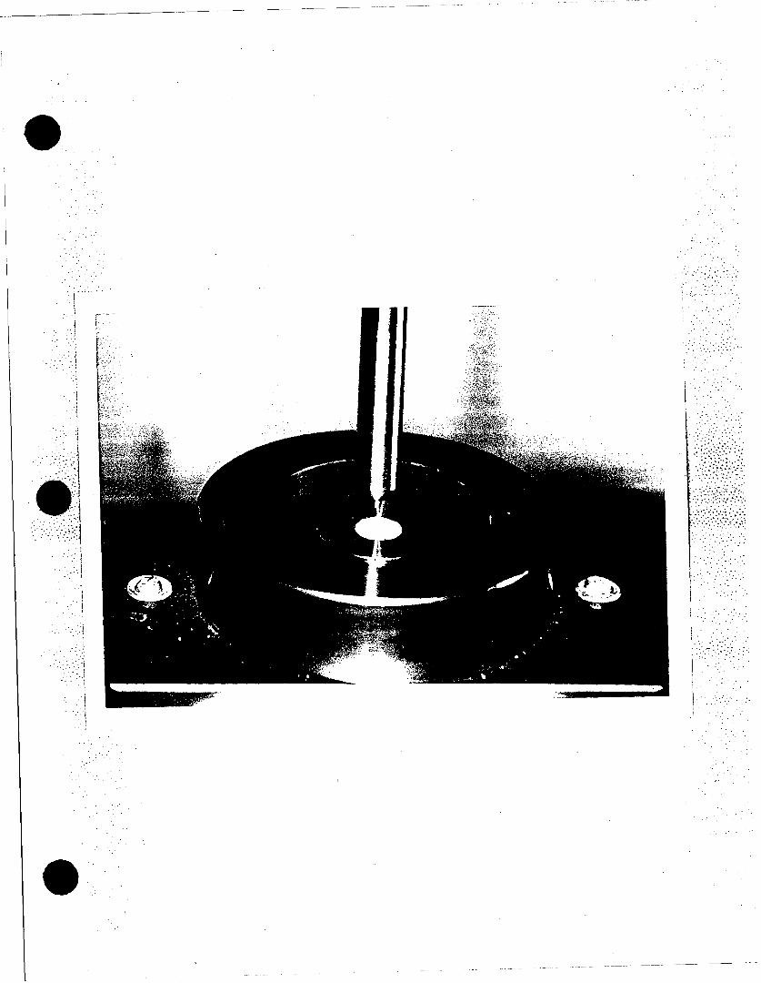
0
I
01

53
Plate 2. Representative Specimen Fractured Under Biaxial Flexure
Conditions Allowing Maximum Tensile Stresses Within the
Central Loading Area
0

1 AA

54
Plate 3. Leucite-Reinforced Porcelain, Hand-Lapping Fixture and Finished
Disk Specimens
0

0 F A ;j

55
Plate 4. Potassium Salt (Tuf-Coat) (A) and Rubidium Nitrate Paste (B)
Applied to Porcelain Disk Specimens Prior to Heat Treatment
0
0

K r
V~
_ - U -
~ '~*, - -,
- ~-.tt
tt. A
56
Plate 5. Potassium Salt (Tuf-Coat) (A) and Rubidium Nitrate Paste (B)
Dried for 20 minutes at 1500 C

r44
JS<
57
Plate 6. Potassium Salt (Tuf-Coat) (A) and Rubidium Nitrate Paste (B)
After Heat Treatment (Ion Exchange) for 30 minutes at 4370 C

.ii 77
IfI
Vt

58
Plate 7. Potassium (A) and Rubidium (B) Ion Exchange Treated Specimens
(Residue Removed) Along With Untreated Specimen
0
0

VN~.
~e '~ -. IC
~ 7.' tW.4V>4x%.t - .<.2*
'P k -~ . ,.,
44,
4 ~7'!X!A ' .'44 ' I,
I
' %t4*7L2.7 V~WA ~ >4'
-, . - ..
I~t pr
C' * L4t
0
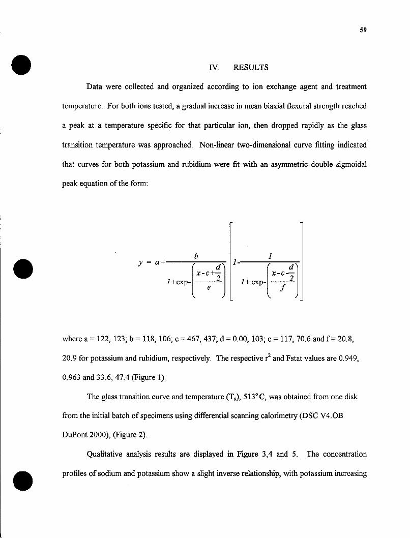
59
IV. RESULTS
Data were collected and organized according to ion exchange agent and treatment
temperature. For both ions tested, a gradual increase in mean biaxial flexural strength reached
a peak at a temperature specific for that particular ion, then dropped rapidly as the glass
transition temperature was approached. Non-linear two-dimensional curve fitting indicated
that curves for both potassium and rubidium were fit with an asymmetric double sigmoidal
peak equation of the form:
b 1- 1y = a-+ ' x -
lexp-X-C+ 2 1+exp-,
where a = 122, 123; b = 118, 106; c = 467, 437; d = 0.00, 103; e = 117, 70.6 and f= 20.8,
20.9 for potassium and rubidium, respectively. The respective r2 and Fstat values are 0.949,
0.963 and 33.6, 47.4 (Figure 1).
The glass transition curve and temperature (Tg), 5130 C, was obtained from one disk
from the initial batch of specimens using differential scanning calorimetry (DSC V4.OB
DuPont 2000), (Figure 2).
Qualitative analysis results are displayed in Figure 3,4 and 5. The concentration
profiles of sodium and potassium show a slight inverse relationship, with potassium increasing
60
and sodium decreasing after treatment with the potassium-based paste (Figure 3). In the
rubidium ion exchanged specimen (Figure 4), marked decreases in concentrations for
potassium and sodium are seen. Because spectral deconvolution was not performed, the
concentration profile for rubidium cannot be discerned due to overlap between the rubidium,
aluminum and silicon peaks (Figure 4). In the specimen treated with a mixture of both ions
(Figure 5), potassium is also greatly reduced. The high sodium peak suggests that ion
exchange with sodium did not occur to any notable extent.
Modulus of rupture, mean and standard deviation values for each group as well as
percentage change in strength, relative to control specimens, are listed in Tables 1 and 2. An
analysis of variance (ANOVA) was followed by a Student Newman-Keuls test. Results are
summarized in Table 2. Vertical lines connect subgroup means that were not significantly
different at the p < 0.05 level.
The treatment temperatures yielding maximum flexural strengths over the untreated
controls were 405'C for the potassium-based slurry (38.5% increase), and 4650C for
rubidium nitrate (57.7% increase). In contrast, the optimal temperatures taken from the fitted
curve (Figure 1) were 4200 C for potassium and 465' C for rubidium nitrate. Consecutive
treatment with both slurries yielded a strength increase of 64% regardless of order. Treatment
with a mixture of both chemical agents yielded the highest strength increase of 66.1%,
although this was not significantly different from any of the treatments involving the rubidium
ion at p < 0.05. Visual examination of ion exchange treated specimens revealed no apparent
changes when compared with untreated specimens.
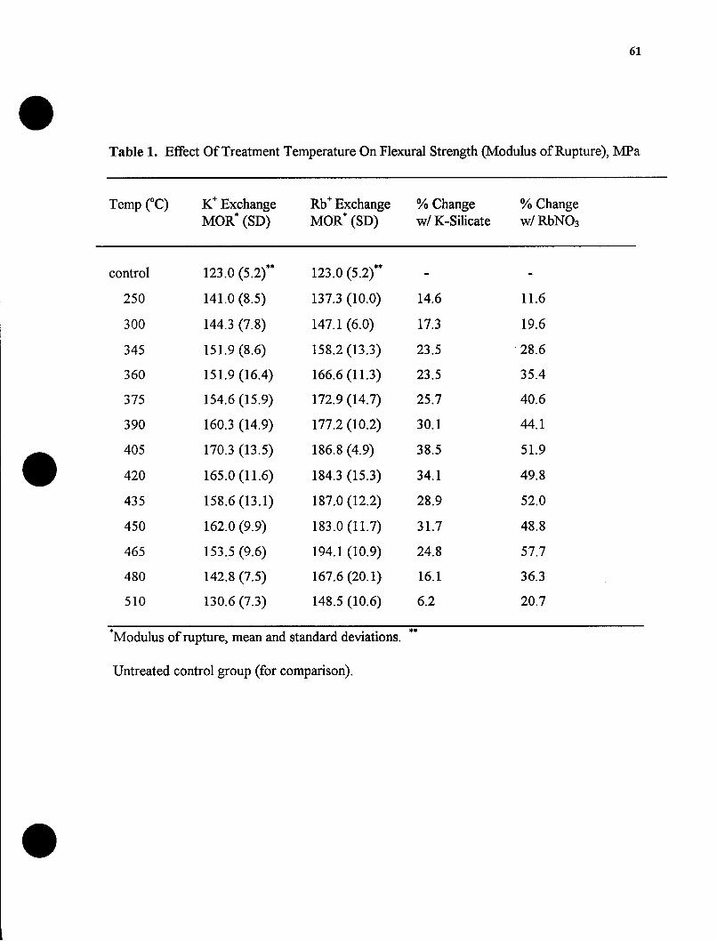
61
0Table 1. Effect Of Treatment Temperature On Flexural Strength (Modulus of Rupture), MPa
Temp (C) K Exchange Rb Exchange % Change % ChangeMOR* (SD) MOR* (SD) w/ K-Silicate w/ RbNO3
control 123.0 (5.2)** 123.0 (5.2)** - -
250 141.0 (8.5) 137.3 (10.0) 14.6 11.6
300 144.3 (7.8) 147.1 (6.0) 17.3 19.6
345 151.9 (8.6) 158.2 (13.3) 23.5 -28.6
360 151.9 (16.4) 166.6 (11.3) 23.5 35.4
375 154.6 (15.9) 172.9 (14.7) 25.7 40.6
390 160.3 (14.9) 177.2 (10.2) 30.1 44.1
405 170.3 (13.5) 186.8 (4.9) 38.5 51.9
420 165.0 (11.6) 184.3 (15.3) 34.1 49.8
435 158.6 (13.1) 187.0 (12.2) 28.9 52.0
450 162.0 (9.9) 183.0 (11.7) 31.7 48.8
465 153.5 (9.6) 194.1 (10.9) 24.8 57.7
480 142.8 (7.5) 167.6 (20.1) 16.1 36.3
510 130.6 (7.3) 148.5 (10.6) 6.2 20.7
*Modulus of rupture, mean and standard deviations. **
Untreated control group (for comparison).
0
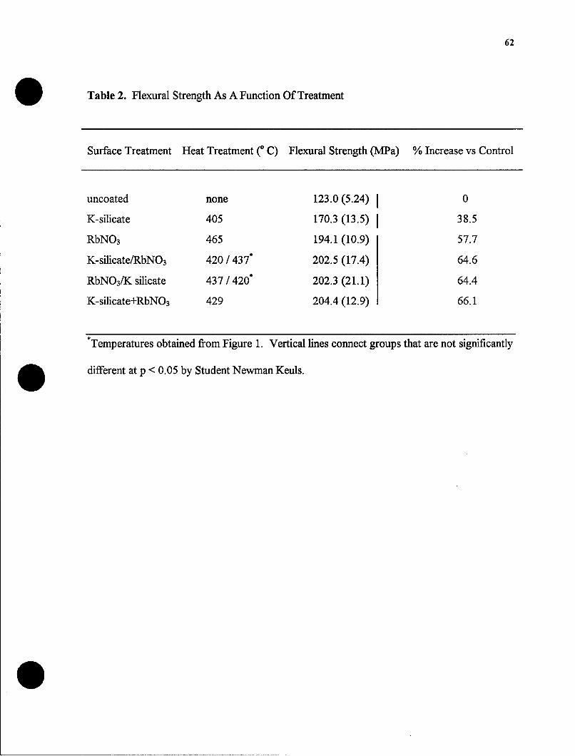
62
Table 2. Flexural Strength As A Function Of Treatment
Surface Treatment Heat Treatment (0 C) Flexural Strength (MPa) % Increase vs Control
uncoated none 123.0 (5.24) 0
K-silicate 405 170.3 (13.5) 38.5
RbNO3 465 194.1 (10.9) 57.7
K-silicate/RbNO 3 420 / 437* 202.5 (17.4) 64.6
RbNO3/K silicate 437 / 420* 202.3 (21.1) 64.4
K-silicate+RbNO3 429 204.4 (12.9) 66.1
*Temperatures obtained from Figure 1. Vertical fines connect groups that are not significantly
different at p < 0.05 by Student Newman Keuls.
0

63
Figure 1. Flexural Strength as a Function of Temperature for Potassium
Salt (Tuf-Coat) and RbNO3 Treated Porcelain Specimens
0
0
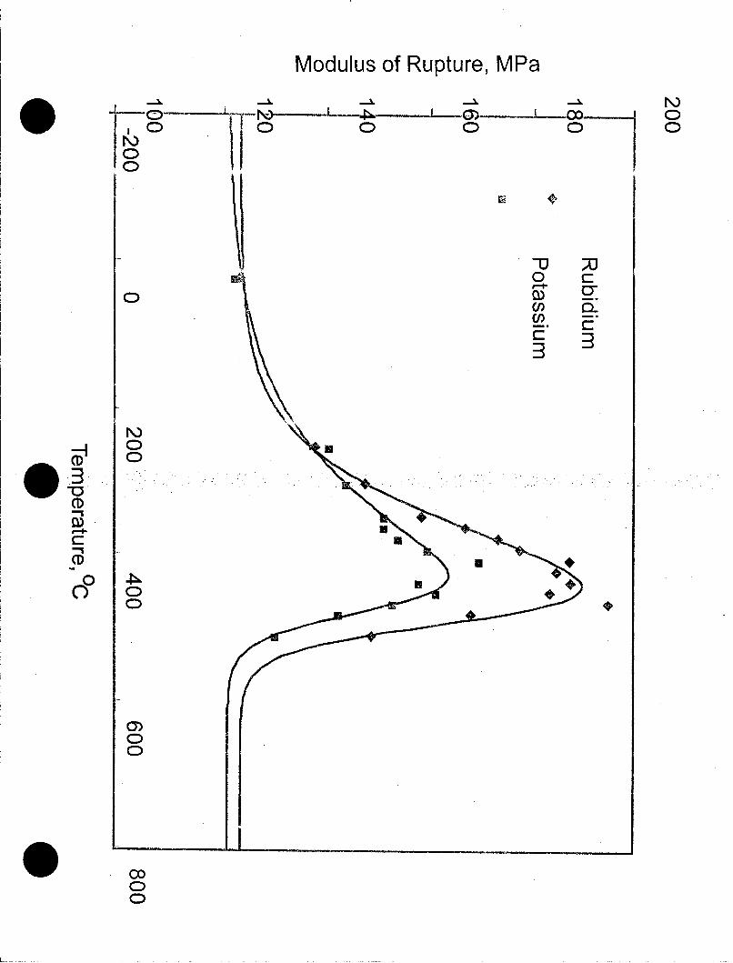
Modulus of Rupture, MPa
co- CD
C) C)o 0 0
N)-H
CMS
CD
CD
0C)
C)
_C__ _

64
Figure 2. Differential Scanning Calorimetry for Optec HSP Porcelain

Heat Flow (mW)
I Io
CD6
Is. F _______
CD /
~~On

65
Figure 3. Elemental Analysis of Leucite-Reinforced Porcelain Before and
After Ion Exchange With Potassium Salt (Tuf-Coat)
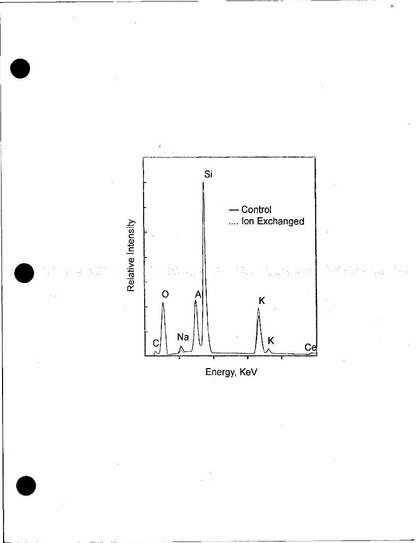
Si
-Control
... Ion Exchanged
-4-a
aU.4" 0 A K
C Na K C
Energy, KeV

66
Figure 4. Elemental Analysis of Leucite-Reinforced Porcelain Before
and After Ion Exchange With RbNO3

-Control
>-. Ion ExchangedCn Rb
* A,K
~JNakJ P K Ce
Energy, KeV

67
Figure 5. Elemental Analysis of Leucite-Reinforced Porcelain Before
and After Ion Exchange With Potassium Salt (Tuf-Coat)
and RbNO3 Mixture
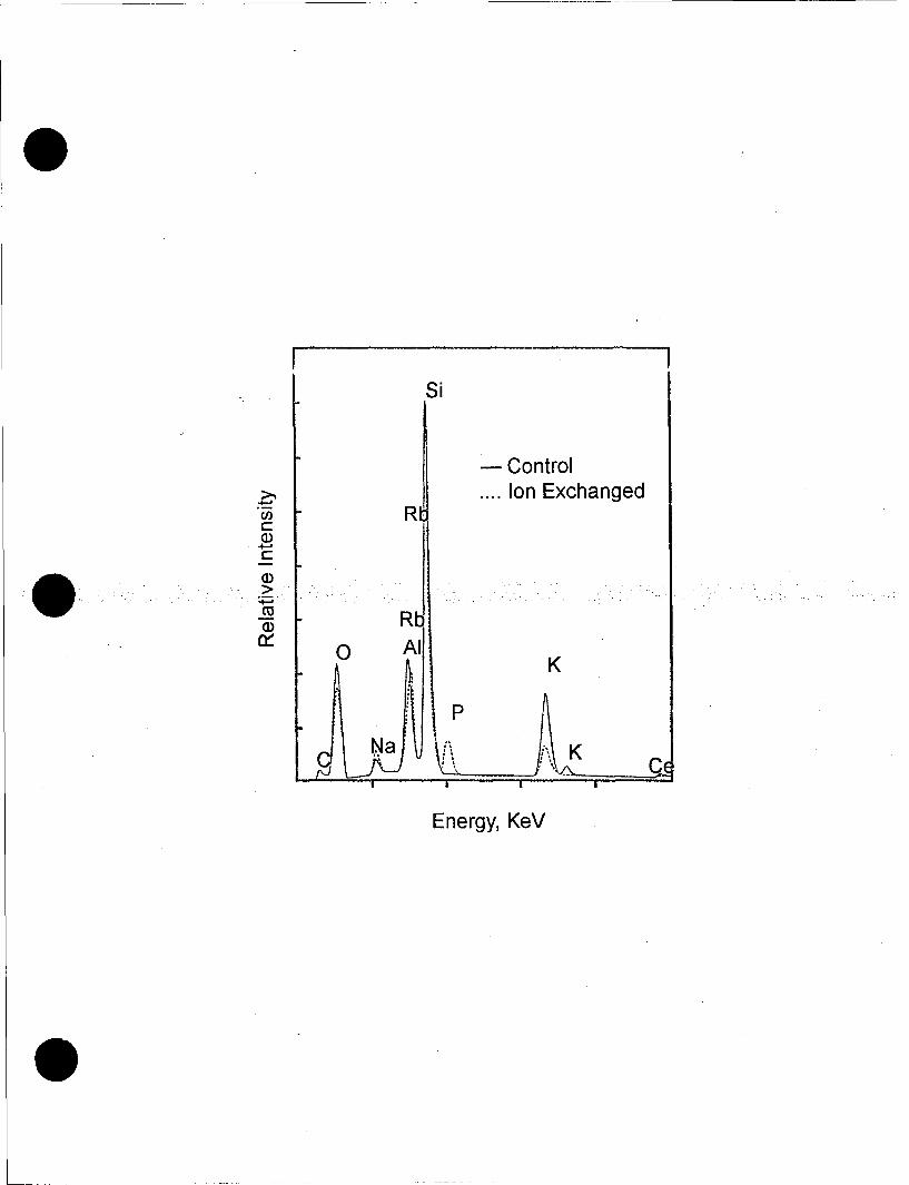
Si
-Control
...Ion Exchanged
C' A
CCu
Energy, KeV

68
* V. DISCUSSION
Ion exchange strengthening of glasses and ceramic materials is a diffusion-controlled
process. Concentration of diffusing ions, temperature, and time are the major interacting
variables, however composition of the substrate also plays an important role (Kingery, 1976).
Because the driving force for diffusion is an ion concentration gradient, ceramic composition
should influence the amount of ion exchange that is possible. Previous studies have
demonstrated this variation in degree of strengthening among ceramics of differing
composition (Seghi et al,. 1990).
Elements within the ceramic other than diffusible species influence the reaction. For
example, boron in borosilicate glass is thought to depress ion exchange, since no
strengthening results when either low or high sodium content samples are subjected to
treatment. Alkali-alumino-silicates, on the other hand, are characterized as being highly
conducive to ion exchange strengthening. It has been suggested that the effect of alumina
within the glass network, rather than an increased exchange reaction, is responsible for this
observation (Nordberg et al, 1964). In a study of seven dental porcelains, ion exchange
yielded the highest flexural strength increase in a porcelain exhibiting relatively low sodium
content (Seghi et al., 1990). Consequently, ion mobility may be of greater importance than
actual ion concentration.
Besides determining the concentration of diffusing ions and their relative mobilities,
the ceramic composition influences the glass transition temperature (Tg) (Seghi et. aL, 1990).
Knowledge of Tg for the particular ceramic is required when use of the ion exchange process
is being considered. As the treatment temperature approaches T., residual stress developed
during the exchange is quickly lost, due to structural rearrangement and stress relaxation

69
(annealing). An abrupt drop in biaxial flexural strength for both potassium and rubidium ion
exchange treatments, coincides with the Tg (5130 C) for Optec HSP porcelain, as seen in
Figure 1. The optimal temperature for potassium ion exchange was approximately 100 C
below Tg, coinciding with conditions previously reported (Denry et al., 1993). In contrast, the
optimal temperature for rubidium exchange occurred at approximately 50' C below Tg.
Rubidium ion, because of its' relative larger size, requires higher temperatures for diffusion.
Consequently, a narrower temperature range for effective treatment exists.
The biaxial flexural strength increase of 38.5%, obtained with potassium ion exchange
of leucite-reinforced porcelain, is in agreement with increases of 37% and 38% previously
reported (Holloway et al., 1993; Crimaldi et al., 1994). In comparison, strength increases
from 32% to 97% have been reported after potassium ion exchange with conventional
feldspathic porcelains (Anusavice et al., 1991). In leucite-reinforced materials low sodium
concentration may be a factor however, lower matrix glass volume (due to a high crystalline
content), may account for relatively low strengthening seen with potassium ion exchange.
Qualitative analysis results with energy dispersive x-ray spectroscopy (EDS) suggest
potassium exchange occurred to a minimal extent relative to rubidium exchange (Figure 3 and
4). Low relative strengthening along with minimal exchange evidenced by qualitative analysis,
suggests ion exchange using potassium may not have involved the leucite crystalline phase.
Because the glass matrix composition in leucite-reinforced porcelain is similar to that of
conventional feldspathic porcelains, however, potassium ion exchange is expected to some
degree.
The mean biaxial flexural strength increase after rubidium exchange was significantly
higher than with potassium exchange, as expected (Table 2). The marked reduction in
70
concentration profile for potassium seen in Figure 4, suggests rubidium exchange occurred to
a greater extent. In addition, the decrease in concentration profile for sodium suggests
rubidium exchange can occur with sodium as well as potassium ions. Increased strength
values observed after rubidium exchange may also be due to the relative size of the rubidium
ion, reported to be 33% larger in volume than potassium (Denry et al., 1993). Employing a
larger ion, with more opportunities for exchange with smaller ions (i.e., sodium and/or
potassium), would result in greater crowding of the surface microstructure and higher surface
compressive stress.
Rubidium exchange of leucite-reinforced ceramic in this investigation yielded a mean
biaxial flexural strength increase of 58%. In comparison, rubidium exchange of a conventional
feldspathic porcelain yielded a strength increase of 82%, in a previous investigation (Denry et
al., 1993). Lower glass content may be a factor accounting for a lower percentage strength
increase. However, whether rubidium exchange directly or indirectly, involved the crystalline
leucite cannot be determined from this investigation. The marked reduction in potassium
concentration profile after rubidium exchange (Figure 4), however, may be suggestive of
leucite involvement.
Leucite is characterized by a high potassium content, higher than the surrounding glass
matrix (Rouf et al., 1980; Denry et al., 1993). In addition, Optec HSP, leucite-reinforced
ceramic, contains at least 45% leucite by weight (Katz, 1989). The possibility of ion exchange
within the crystalline phase is intriguing, when the high volume of leucite present in these
materials is considered.
Leucite's open crystalline structure may aid in ion exchange. According to a ceramic
publication, "Leucite is a potassium aluminosilicate and is in a group of minerals characterized

71
by large cavities in their anionic lattice that allow good ion-exchanging properties" (Naray-
Szabo, 1969; quoted by Denry et al., 1993). Chemical strengthening techniques not only
improve the strength of the matrix glass, but also improve the strength of the crystalline phase
(Kingery etal., 1975).
Substitution of intermediate-sized ions for small ions prior to exchange with larger
ions would, theoretically, simplify the total reaction, allowing it to proceed to a greater extent.
In this investigation, potassium exchange followed by rubidium exchange yielded only slightly
greater strength values than treatment with rubidium exchange alone. A small difference in
sodium and potassium concentration profiles after potassium exchange seen in Figure 3, may
explain the small difference between rubidium exchange of untreated disks, and rubidium
* exchange of disks treated first with potassium (Table 2).
Specimens treated with potassium followed by rubidium were not statistically different
in strength than specimens treated with rubidium followed by potassium. The low treatment
temperature used may explain the similar outcomes for both consecutive treatments (Table 2).
Substitution of large ions within the glass by smaller ions requires a more fluid glass state.
The low temperature (420 C) used may not have been sufficient to permit potassium
exchange with rubidium ions previously substituted. Higher temperatures above the Tg of the
glass may be required for potassium for rubidium (i.e., small ion for large ion) exchange to
proceed.
Ion exchange treatment with multiple ionic species would appear to be a quicker and
more convenient way of improving strength. In this investigation, treatment with a mixture of
* potassium and rubidium ions yielded strength equivalent to that obtained with consecutive
treatments (Table 2). The relatively low concentration profile for potassium seen in Figure 5

72
suggests rubidium for potassium exchange occurred. However, because of the high
concentration profile for sodium, it is uncertain whether potassium for sodium exchange
occurred to any extent. Differences in ion mobilities are a factor along with other variables
previously discussed.
Ion exchange involving multiple ions requires further investigation. Experimentation
including ion exchange sequencing, concentration of exchange agents, and treatment
temperatures, is needed in order to determine peak treatment conditions for specific porcelain
systems. In addition, the porcelains themselves might be specifically formulated to allow for
maximum strengthening through subsequent ion exchange.
0
0
73
VI. SUMMARY
Ion exchange is a strengthening technique that is applicable to most dental ceramics.
Previous work has shown that conventional feldspathic and alumina reinforced porcelains can
be strengthened using ion exchange treatment with rubidium nitrate as well as conventionally
using a potassium salt. Leucite-reinforced all-ceramic restorations are increasing in popularity
due to their translucency, color control, bondability and improved strength. Ion exchange
may be a practical way of strengthening these restorations without affecting their desirable
optical properties. The following results and conclusions can be drawn from this
investigation:
1) Ion exchange with rubidium nitrate yielded significantly higher mean biaxial flexural
strength values than with the potassium exchange (p < 0.05).
2) The optimal treatment temperature for potassium exchange was 4050 C,
approximately 1000 C below the Tg (5130 C).
3) The optimal treatment temperatures for rubidium exchange was 4650 C,
approximately 50' C below the Tg.
4) Consecutive ion exchange treatment yielded a 64% increase in mean biaxial flexural
strength over untreated controls, regardless of order.
5) Treatment with a mixture of both rubidium nitrate and potassium paste yielded the
greatest increase in strength (66%) over untreated controls, although not significantly greater
than with rubidium alone.
6) Potassium and rubidium ion exchange treatments were accomplished without
visible changes in color and translucency to the ceramic.

74
VII. SIGNIFICANCE
Ion exchange was introduced into dentistry in 1970, when metal-ceramic restorations
were in vogue. This may be the main reason this process has been relatively ignored. Today,
strengthening by ion exchange is more applicable than ever due to esthetic emphasis and
growing popularity of all-ceramic restorations.
It is well established that the internal surfaces of all-ceramic restorations are subject to
high tensile forces which can lead to failure. Ion exchange may be a practical technique for
improving the strength and durability of these restorations while retaining the desirable optical
properties characteristic of leucite-reinforced, as well as other all-ceramic restorative systems.
75
LITERATURE CITED
Anusavice KJ. 1996. Phillips' Science of Dental Materials, Tenth edition. Philadelphia. WBSaunders Co.
Anusavice KJ, Shen C, Lee RB. 1991. Strengthening of feldspathic porcelain by ion exchangeand tempering. J. Dent. Res., 71:1134-1148.
Anusavice KJ, Shen C, Vermost B, Chow B. 1992. Strengthening of porcelain by ionexchange subsequent to thermal tempering. Dent. Mater., 8: 149-152.
Anusavice KJ, Hojjatie B, Chang TC. 1994. Effect of grinding and fluoride-gel exposure onstrength of ion-exchanged porcelain. J. Dent. Res., 73:1444-1449.
Bagby M. 1990. Metal ceramic compatibility. J. Prosthet Dent., 63:21-25.
Ban S and Anusavice KJ. 1990. Influence of test method on failure stress of brittle materials.J. Dent. Res., -62:1791-1799.
Binns DB. 1962. Some physical properties of two-phase crystal-glass solids. In: Stewart GH,ed. Science of Ceramics. Volume 1. London. Academic Press. 315-334.
Binns DB. 1983. The chemical and physical properties of dental porcelain. In: DentalCeramics: Proceedings of the First International Symposium on Ceramics. McLeanJW. ed. Chicago. Quintessence Publishing Co. 41-82.
Brecker CS. 1956. Porcelain baked to gold-a new medium in prosthodontics. J. ProsthetDent., 6:801-810.
Carrier DD and Kelly JR. 1995. In-Ceram failure behavior and core veneer interface quality asinfluenced by residual infiltration glass. J. Prosthet Dent., 4:237-242.
Charles RJ. 1958. Static fatigue of glass I. J. Applied Phys., 29:1549-1560.
Christensen GJ. 1986. The use of porcelain-fused-to-metal restorations in current dentalpractice: A survey. J. Prosthet Dent., 56:1-3.
Claus H. 1989. The structure and microstructure of dental porcelain in relationship to thefiring conditions. Int. J. Prosthodont., 2:376-384.
Craig RG. 1993. Restorative Dental Materials. Ninth edition. St. Louis. CV Mosby Co.
Crimaldi AJ, Denry IL, Rosenstiel SF. 1994. Strengthening of leucite-reinforced porcelain bydouble ion-exchange. J. Dent. Res., 73:268.

76
. Denry IL, Rosenstiel SF, Holloway JA, Niemiec MS. 1993. Enhanced chemical strengtheningof feldspathic dental porcelain. J. Dent. Res., 72:1429-1433.
Dong JK, Luthy H, Wohlwend A, Scharer P. 1992. Heat-pressed ceramics: Technology andstrength. Int. J. of Prosthodont., 5:9-16.
Dunn B, Levy MN, Reisbick MH. 1977. Improving the fracture resistance of dental ceramic.J. Dent. Res., 56:1209-1213.
Dykema RW, Goodacre CJ, Phillips RW. 1986. Johnston's Modem Practice in FixedProsthodontics. Fourth ed. Philadelphia. WB Saunders Co.
Eppler RA. 1983. Glass Science and Technology. Volume 1. New York. Academic Press.301-336.
Giordano RA, Campbell S, Pober R. 1994. Flexural strength of feldspathic porcelain treatedwith ion exchange, overglaze, and polishing. J. Prosthet Dent., 71:468-472.
Griffith AA. 1920. The phenomena of rupture and flow in solids. Phil. Trans. R. Soc. LondonSer. A. 163-198.. Hallig WB. 1962. Source of weakness and the ultimate strength of brittle amorphous solids.In: Mackenzie JD. ed. Modem Aspects of the Vitreous State. London. Butterworths.152-194.
Hasselman DPH and Fulrath RM. 1965. Proposed fracture theory of a dispersion-strengthened glass matrix. J. Am. Ceram. Soc., 49:68-72.
Holloway JA, Denry IL, Rosenstiel SF. 1993. Strengthening of dental porcelain by double ionexchange. J. Dent. Res., 2:887.
Hondrum SO. 1992. A review of the strength properties of dental ceramics. J. Prosthet Dent.,67:859-865
Jones DW. 1982. Ceramics and Glass as Restorative Materials. In: Smith DC, Williams DF.eds. Biocompatibility of Dental Material Biocompatibility of Prosthodontic Materials.Boca Raton, Fla. CRC Press. 79-122.
Jones DW. 1983. The strength and strengthening mechanisms of dental ceramics. In: DentalCeramics: Proceedings of the First International Symposium on Ceramics. McLeanJW. ed. Chicago. Quintessence Publishing Co. 83-141.
Jones DW. 1985. Development of dental ceramics. Dent. Clin. North. Am., 29:621-644.
Katz S. 1989. American Thermocraft Corp., Assignee. High Strength Feldspathic DentalPorcelains Containing Crystalline Leucite. U.S. Patent 4,798,536.

77
Kelly JR, Cambell SD, Bowen KH. 1989. Fracture-surface analysis of dental ceramics. J.Prosthet Dent., 62:536-541.
Kelly JR, Tesk JA, Sorenson JA. 1995. Failure of all-ceramic fixed partial dentures in vivo andin vitro: analysis and remodeling. J. Dent. Res., .74:1253-1258.
Kelly JR, Giordano R, Pober R, Cima MJ. 1990. Fracture surface analysis of dental ceramics:clinically failed restorations. Int. J. of Prosthodont., 3:430-440.
Kingery WD, Bowen IK, Uhlmann DR. 1976. Introduction to Ceramics. 2nd ed. New York:John Wiley and Sons.
Kistler SS. 1961. Stresses in glass produced by nonuniform exchange of monovalent ions. J.Am. Ceram. Soc. 45:59-68.
Lacy AM. 1977. The chemical nature of dental porcelain. In: Dental Porcelain: The State ofthe Art-1977, A Compendium of the Colloquium held at the University of SouthernCalifornia School of Dentistry. Yamada RN and Grenoble PB. eds. Los Angeles. 47-53.. Lehman ML. 1967. Stability and durability of porcelain jacket crowns. Br. Dent. J., 123:419-426.
Linter CM. 1985. Neuropsychiatric aspects of trace elements. Br. J. Hosp. Med., 34:361-365.
Mackert JR, Evans AL. 1991. Effect of cooling rate on leucite volume fraction in dentalporcelains. J.Dent. Res., 70:137-139.
Mackert JR, Rueggeberg FA, Lockwood PE, Evans AL. 1994. Isothermal anneal effect onmicrocrack density around leucite particles in dental porcelain. J. Dent. Res., 73:1221 -1227.
Marquis PM. Optimizing the strength of all-ceramic jacket crowns. In: Perspectives in DentalCeramics: Proceedings of the Fourth International Symposium on Ceramics. Preston J.ed. Chicago. Quintessence Publishing Co.
Marshall DB. 1980. An improved biaxial flexure test for ceramics. J. Am. Ceram. Soc.,59:551-553.
McLean JW and Hughes TH. 1965. The reinforcement of dental porcelain with ceramicoxides. Br. Dent. J., 112:251-267.. McLean JW. 1979. The Science and Art of Dental Ceramics. Volume 1: The Nature of DentalCeramics and their Clinical Use. Chicago. Quintessence Publishing Co.

78
McLean JW. 1983. Dental Ceramics: Proceedings of the First International Symposium onCeramics. Chicago. Quintessence Publishing Co.
Messer PF. 1990. The strength of dental ceramics. J. Dent., 19:51-55.
Morena R, Lockwood PE, Fairhurst CW. 1986. Fracture toughness of commercial dentalporcelains. Dent. Mater., 2:58-62.
Naray-Szabo I. 1969. Inorganic crystal chemistry. Budapest. Akademiai Kiado. 375.
Naylor WP. 1992. Introduction to Metal Ceramic Technology. Chicago. QuintessencePublishing Co.
Nordberg ME, Mochel EL, Garfinkel HM, Olcott JS. 1964. Strengthening by ion exchange. J.Am. Ceram. Soc., 47:215-219.
Olcott JS. 1963. Chemical strengthening of glass. Science., 140:1189-1193.
Phillips RW. 1982. Skinner's Science of Dental Materials, Eighth edition. Philadelphia. WBSaunders Co.
O Piddock V, Qualtrough AJE, Brough I. 1991. An investigation of an ion strengthening pastefor dental porcelains. Int. J. Prosthodont., 4:132-137.
Pletka BJ and Weiderhorn SM. 1982. A comparison of failure predictions by strength andfracture mechanics techniques. J. Mater. Sci., 17:1247-1268.
Preston JD. 1985. Perspectives in Dental Ceramics: Proceedings of the Fourth InternationalSymposium on Ceramics. Chicago. Quintessence Publishing Co.
Reid CN, Fisher J, Jacobson PH. 1990. Fatigue and wear of dental materials. J. Dent., 18:209-215.
Riley EJ. 1977. Ceramo-metal restoration state of the science. Dent. Clin. North Am., 21:669-682.
Ritter JE, Vicedomine M, Breder K, Jakus K. 1985. Dynamic fatigue of indented, soda-limeglass as a function of temperature. J. Mater. Sci., 2Q:2868-2872.
Rosenstiel SF, Denry IL, Zhu W, Gupta PK, Van der Sluys RA. 1993. Fluoroalkylethyl silanecoating as a moisture barrier for dental ceramics. J. Biomed. Mater. Res., 27:415-417.
Rothermal DL. 1976. Effect of stress on durability of ion-exchanged surfaces. J. Am. Ceram.Soc., 50:574-577.

79
.Rouf MA, Hermansson L, Carlson R. 1980. Crystallization of glasses in the primary phasefield of leucite in the K20-A1203-SiO 2 system. Trans. J Br. Ceram. Soc., 7U:36-39.
Seghi RR, Crispin BC, Mito W. 1990. The effect of ion exchange on the flexural strength offeldspathic porcelains. Int. J. Prosthodont., 3:130-134.
Seghi RR, Denry I, Brajevic F. 1992. Effects of ion exchange on hardness and fracturetoughness of dental ceramics. Int. J. Prosthodont., 5:309-314.
Seghi RR, Denry I, Rosenstiel SF. 1995. Relative fracture toughness and hardness of newdental ceramics. J. Prosthet Dent., 74:145-150.
Sherrill CA and O'Brian WJ. 1974. Transverse strength of aluminous and feldspathicporcelain. J. Dent. Res., 53:683-690.
Southan DE. 1970. Strengthening modem dental porcelain by ion exchange. Austr. Dent. J.,15:507-510.
Southan DE. 1972. Faulty porcelain jacket crowns. Austr. Dent. J., 17:436-440.
Southan DE. 1977. Defects in porcelain at the porcelain-to-metal interface. In: DentalPorcelain: The State of the Art-1977, A Compendium of the Colloquium held at theUniversity of Southern California School of Dentistry. Yamada HN and Grenoble PB.eds. Los Angeles. 143-149.
Southan DE. 1983. The porcelain jacket crown. In: Dental Ceramics: Proceedings of the FirstInternational Symposium on Ceramics. McLean JW. ed. Chicago. QuintessencePublishing Co. 207-230.
Southan DE. 1987a. Optical density-strength relationships in dental porcelain. QuintessenceInt., 18:261-263.
Southan DE. 1987b. Laminate strength of dental porcelains. Quintessence Int., 18:357-359.
Southan DE. 1987c. Effect of surface injury on chemically strengthened dental porcelain.Quintessence Int., 18:575-580.
Stookey SD. 1965. Strengthening glass and glass-ceramics by built-in surface compression.In: High Strength Materials. Zackay VF. ed. New York. Wiley J. and Sons Inc. 669-681.
Sugerman B. 1967. Strength of glass (a review). J. Mater. Sci., 2:275-283.
Wachtman JB, Capps W, Mandel J. 1972. Biaxial flexure tests of ceramic substrates. I. ofMater. 7:188-194.
80
Wall JG and Cipra DL. 1992. Alternative crown systems: Is the metal-ceramic crown alwaysthe restoration of choice?. Dent. Clin. North. Am., M:765-782.
Wassenaar P and Randell DM. 1990. G-C Ceramicoat: An ion exchange hardener forceramics. Austr. Dent. J., 35:474.
Wiederhorn SM. 1968. Moisture assisted crack growth in ceramics. Int. J. Frac. Mech. 4:171-177.
White SN and Seghi RR. 1992. The effect of ion strengthening time/temperature kinetics onthe flexural strength of feldspathic porcelains. Dent. Mater., 8:320-323.
Williams RH. 1987. Pharmacologic role of rubidium in psychiatric research. Compr. Ther.,13:46-54.
Yamamoto M. 1985. Metal-Ceramics: Principles and Methods of Makoto Yamamoto.Chicago. Quintessence Publishing Co.
Zijlstra AL and Burggraaf AJ. 1968. Fracture phenomena and strength properties ofchemically and physically strengthened glass. I. General survey of strength and fracturebehavior of strengthened glass. J. Non-Cryst. Solids., 1:49-68.
I
* VITA
Jose do Jesus Villalobos
At one year of age, his family irmrigMed to the United States and
setled in Southern California. He graduated from Palm Springs figh School in 1979. That
same year he entered College of the Dee who he spent one year. He transferred to The
University of San Diego where he majored in Biology. He was awarded a Bachelor of Arts
degree from The University of San Diego in December 1983.
In July 1984 he entered the University ofCalifornia, San Francisco School of Dentistry
and received a Doctorate of Dental Surgery degree in June 1988.
In July IM, bebgan General Practice Residency in Hospital Dentistry at Rancho
Los Am*s Medical Center. Downey, California. On December 17, 1988, while in residency,
he married Cybn M arte. Upon completion ofhi residency in July 1989, he obtained his
Unied States Citizenship and was commissioned In the United States Air Force Dental Corps.
Ais first assignoent was at Nelis Air Force Base, Las Vegas, Nevada, where he served as a
-staff general dentt
first daughter, Nicole Marie was born.
his second daughterTerea Ann was born. In June of 1994, he entered the University of
Fexas Heath Science Center at San Antonio, Texas where he is presently pursuing a Post-
Doctoral-degree in Prosthodontics t third daughter, Lia Renee was
born In June of 1995, he was admitted as a candidate for the Masters ofScience Degree at. the Graduate School of Biomedical Sciences.

82
0
0
0