dca protects against oxidation injury attributed to
TRANSCRIPT

Research ArticleDCA Protects against Oxidation Injury Attributed to CerebralIschemia-Reperfusion by Regulating Glycolysis through PDK2-PDH-Nrf2 Axis
Xiaoyong Zhao,1,2 Shan Li,1 Yunchang Mo,1 Ruru Li,1 Shaoyi Huang,1 Anqi Zhang,1
Xuqing Ni,1 Qinxue Dai ,1 and Junlu Wang 1
1Department of Anesthesiology, The First Affiliated Hospital of Wenzhou Medical University, Wenzhou,325000 Zhejiang Province, China2Shandong Provincial Medicine and Health Key Laboratory of Clinical Anesthesia, School of Anesthesiology,Weifang Medical University, Weifang 261021, China
Correspondence should be addressed to Qinxue Dai; [email protected] and Junlu Wang; [email protected]
Received 15 July 2021; Revised 6 September 2021; Accepted 4 October 2021; Published 19 October 2021
Academic Editor: Wen-Jun Tu
Copyright © 2021 Xiaoyong Zhao et al. This is an open access article distributed under the Creative Commons AttributionLicense, which permits unrestricted use, distribution, and reproduction in any medium, provided the original work isproperly cited.
Cerebral ischemic stroke (IS) is still a difficult problem to be solved; energy metabolism failure is one of the main factors causingmitochondrion dysfunction and oxidation stress damage within the pathogenesis of cerebral ischemia, which producesconsiderable reactive oxygen species (ROS) and opens the blood-brain barrier. Dichloroacetic acid (DCA) can inhibit pyruvatedehydrogenase kinase (PDK). Moreover, DCA has been indicated with the capability of increasing mitochondrial pyruvateuptake and promoting oxidation of glucose in the course of glycolysis, thereby improving the activity of pyruvatedehydrogenase (PDH). As a result, pyruvate flow is promoted into the tricarboxylic acid cycle to expedite ATP production.DCA has a protective effect on IS and brain ischemia/reperfusion (I/R) injury, but the specific mechanism remains unclear.This study adopted a transient middle cerebral artery occlusion (MCAO) mouse model for simulating IS and I/R injury inmice. We investigated the mechanism by which DCA regulates glycolysis and protects the oxidative damage induced by I/Rinjury through the PDK2-PDH-Nrf2 axis. As indicated from the results of this study, DCA may improve glycolysis, reduceoxidative stress and neuronal death, damage the blood-brain barrier, and promote the recovery of oxidative metabolismthrough inhibiting PDK2 and activating PDH. Additionally, DCA noticeably elevated the neurological score and reduced theinfarct volume, brain water content, and necrotic neurons. Moreover, as suggested from the results, DCA elevated the contentof Nrf2 as well as HO-1, i.e., the downstream antioxidant proteins pertaining to Nrf2, while decreasing the damage of BBB andthe degradation of tight junction proteins. To simulate the condition of hypoxia and ischemia in vitro, HBMEC cells receivedexposure to transient oxygen and glucose deprivation (OGD). The DCA treatment is capable of reducing the oxidative stressand blood-brain barrier of HBMEC cells after in vitro hypoxia and reperfusion (H/R). Furthermore, this study evidenced thatHBMEC cells could exhibit higher susceptibility to H/R-induced oxidative stress after ML385 application, the specific inhibitorof Nrf2. Besides, the protection mediated by DCA disappeared after ML385 application. To sum up, as revealed from thementioned results, DCA could exert the neuroprotective effect on oxidative stress and blood-brain barrier after brain I/R injuryvia PDK2-PDH-Nrf2 pathway activation. Accordingly, the PDK2-PDH-Nrf2 pathway may play a key role and provide a newpharmacology target in cerebral IS and I/R protection by DCA.
1. Introduction
Stroke refers to a vital cause of death and permanent dis-ability globally [1], of which ischemic stroke (IS) takes up
more than 87% of its incidence [2]. The early interventionstrategy of IS refers to restoring the blood supply ofinfarcted and ischemic areas. Nevertheless, reperfusion islikely to further aggravate ischemic brain injury, i.e.,
HindawiOxidative Medicine and Cellular LongevityVolume 2021, Article ID 5173035, 12 pageshttps://doi.org/10.1155/2021/5173035

cerebral ischemia/reperfusion (I/R) injury [3, 4]. Accordingto related studies and reports, CIRI displays a relationshipwith energy metabolism disorder [5, 6], oxidative stress [7,8], Ca2+ overload, excitatory neurotransmitters, apoptosis,and necrosis [9]. Energy metabolism disorder can lead toconsiderable ROS generation, and oxidative stress attrib-uted to ROS displays a close relationship with IS patho-genesis. In CIRI, the excessive production of ROS willcause DNA, proteins, and brain lipids to undergo oxida-tive damage, thereby causing cell death and neurologicaldysfunction [10]. Thus, energy metabolizers are taken intoaccount to prevent and treat IS.
During reperfusion after cerebral ischemia, the bloodsupply will restore glucose and oxygen levels, produce exces-sive ROS, promote responses of promoting oxidative stresssuch as leukocyte and proinflammatory neutrophil infiltra-tion and complement and platelet activation, and damagethe blood-brain barrier (BBB) [11, 12], which are all compo-nents of reperfusion injury. Accordingly, it is necessary toreduce reperfusion injury to promote cell repair and ische-mic tissue regeneration.
Dichloroacetic acid (DCA) is a small molecule, whichhas been used as a therapeutic agent for many genetic mito-chondrial diseases [13, 14]. Dichloroacetic acid (DCA) is aninhibitor of pyruvate dehydrogenase kinase (PDK), andPDK2 is the most abundant isoenzyme in the rat brain[15]. DCA can inhibit mitochondrial PDK2 and activatepyruvate dehydrogenase (PDH), which is a gatekeeperenzyme combining anaerobic (glycolysis) with aerobic(Krebs cycle) metabolism [16, 17]. After ischemia and hyp-oxia, the activity of PDH decreases, pyruvate cannot decar-boxylate and will produce lactic acid via glycolysis, andeach glucose molecule produces two moles of ATP. How-ever, when PDH is activated, pyruvate can decarboxylate toacetyl-CoA, enter the tricarboxylic acid cycle, and produceup to 36 moles of ATP per glucose molecule within mito-chondria [18]. DCA exerts protecting effects on I/R injury[12], cancer [19], and pulmonary hypertension [20]. Never-theless, it is not clear if DCA exerts a protecting effect on ISand CIRI.
In this study, we found that mitochondrial-relatedenzymes are inactivated after cerebral ischemia-reperfusion,and then, glycolysis produces considerable ROS. DCA canimprove glycolysis by inhibiting PDK2 and activatingPDH, so as to activate Nrf2, reduce oxidative stress, andreduce the permeability of the blood-brain barrier. Thus,as suggested from the results of this study, DCA is likelyto be a new therapeutic approach in terms of IS andCIRI.
2. Materials and Methods
2.1. Sigma-Aldrich (St. Louis, Missouri, USA) OfferedMaterials. Dichloroacetic acid (347795), 2,3,5-triphenylte-trazole chloride (TTC), and ML385 (GC19254) originatedfrom GLPBIO (Montclair, California, the United States ofAmerica). Gibco (Grand Island, NY) provided fetal bovineserum (FBS) and trypsin.
2.2. Animal and Animal Experiments. An animal and focalcerebral ischemia model and male C57BL/6 mice aged 6 to8 weeks were used for this study. The animal operationsreceived approval from the Animal Experimentation EthicsCommittee (No. WYDW2019-0559). In addition, thehumanistic care was carried out according to the animalexperiment guidelines of Wenzhou Medical University.The researchers carried out the transient MCAO model ofmice by occluding MCA [21]. In terms of sham-operatedmice, the isolation was conducted on the right commonand external carotid artery, whereas there was no MCA liga-tion. The mice received the random allocation in 4 cohorts,i.e., sham cohort, MCAO cohort, DCA (100mg/kg) cohort,and DCA (200mg/kg) cohort. After 90min of occlusion,100mg/kg and 200mg/kg DCA were given to the DCAcohort as soon as the plug was released.
2.3. Neurological Deficit Assessment. When the 24 h reperfu-sion was conducted, the neurological deficit received theassessment in accordance with the scoring standards [22,23]: 0: no neurological deficit; 1: falling to contralateral sides;2: failing to have spontaneous activities; 3: failing to stretchthe contralateral forelimb; 4: circling to paretic sides.
2.4. Infarct Volume Assessment. After neurological assess-ment, as mentioned before [24], mice were euthanized with2% pentobarbital sodium, and the mice received the decapi-tation. The brains received the removal to measure infarctvolume. Coronal section slices were taken from the wholebrain and then received the staining process with 2% TTCunder the temperature of 37°C for 20min. To conduct theinvestigation, the pictures of slices were captured by usinga digital camera, and all images were collected and analyzedwith the use of ImageJ (National Institutes of Health, USA).The relative infarct volume rate was obtained, and the edemawas corrected. In brief, the calculating process is conductedbelow: corrected infarct volume ratio = ½contralateralhemisphere area − ðischemic hemisphere area − infarct areaÞ/contralateral hemisphere area� × 100%.
2.5. Brain Water Level. The mice received the sacrifice 24hwhen MCAO was caused. The brains received carefulremoval. By weighing the ischemic hemisphere, the wetweight received the rapid measurement. By weighing thesamples dried under the temperature of 105°C for 24 h, thedry weight received the measurement. The brain water con-tent is expressed as brainwater content ð%Þ = ðwet weight −dry weightÞ/wet weight × 100%.
2.6. Nissl Staining. The mice were deeply anesthetized whenthe neurological deficit test was performed. The left ventriclereceived the perfusion by using 4% paraformaldehyde.When the perfusion was achieved, the brain received the48 h fixing process, the dehydration, and the embedmentin wax. Coronal sections with a thickness of 10μm wereset for Nissl staining. The experiment was carried outaccording to the instructions of the Nissl staining tool(Solarbio, China). Brain slices received dehydration by usingalcohol and were impregnated with xylene and stained withthiophane. Then, the morphological changes of cortical
2 Oxidative Medicine and Cellular Longevity
neurons were observed under a microscope. The number ofsurviving neurons was recorded by neuron count.
2.7. TUNEL Assay. By complying with the manufacturer’sguidelines, the researchers carried out the TUNEL test usingthe in situ cell death detection tool (Roche) and detectedunder the fluorescence microscope (DM2500; Leica Micro-systems, Germany). With the use of Image-Pro Plus version6.0 (Media Cybernetics, USA), the researchers without anyknowledge regarding the grouping assignment measuredTUNEL positive cells with green fluorescence. Results hadthe expression of labeled cell numbers.
2.8. Assessment of BBB Permeability. Based on the measure-ment of the penetration of Evans blue (Sigma) in brain tis-sues, the researchers examined BBB permeability [25].Evans blue (2% saline, 4mL/kg body weight) was adminis-tered intravenously through the tail vein 1 h before measure-ment. The anesthetized animals were perfused with normalsaline before sampling. The respective sample received theweighing and homogenizing processes by using 400μLPBS, and subsequently, the sample received the precipitationthroughout the night by using 50% trichloroacetic acid. The
sample underwent 30min centrifugation at 10,000 rpm toprecipitate the brain tissues. EB absorbance received themeasurement at 610nm using one microplate reader (Bio-Tek, Winooski, Vermont). The concentration was then cal-culated according to the standard curve, with theexpression of μg/g brain tissue.
2.9. Electron Microscope Study. The brain slices were fixedwith 0.1mol/L methylarsonic acid buffer of 4% glutaralde-hyde (pH7.4). The slices were then immersed in 1% osmiumtetroxide in 0.1M methyl arsenate buffer for 2 h and stainedovernight with 1% uranyl acetate aqueous solution. The tis-sue sections received the dehydration to 100% with ascend-ing series of ethanol and subsequently with acetone andunderwent the embedding process within an epoxy resin.The ultrathin sections received the restaining by using leadcitrate before the examination under transmission electronmicroscopy (H7650).
2.10. Cell Culture and Treatment. HBMEC (HUM-CELL-0101) cells were purchased from PriCells (Wuhan, China).The cells received the culture under the temperature of37°C, 5% CO2, and 95% humidity supplemented with 1%
Sham MCAO 100 200
DCA mg/kg
(a)
Sham MCAO 100 2000
20
40
60
DCA mg/kg
⁎⁎
Infa
rct v
olum
e (%
)
⁎
⁎
(b)
Sham MCAO 100 2000
1
2
3
4
5
DCA mg/kg
Neu
rolo
gica
l sco
re
⁎⁎⁎
⁎
(c)
Sham MCAO 100 20060
70
80
90
100
Brai
n w
ater
cont
ent (
%)
DCA mg/kg
⁎⁎ ⁎
⁎
(d)
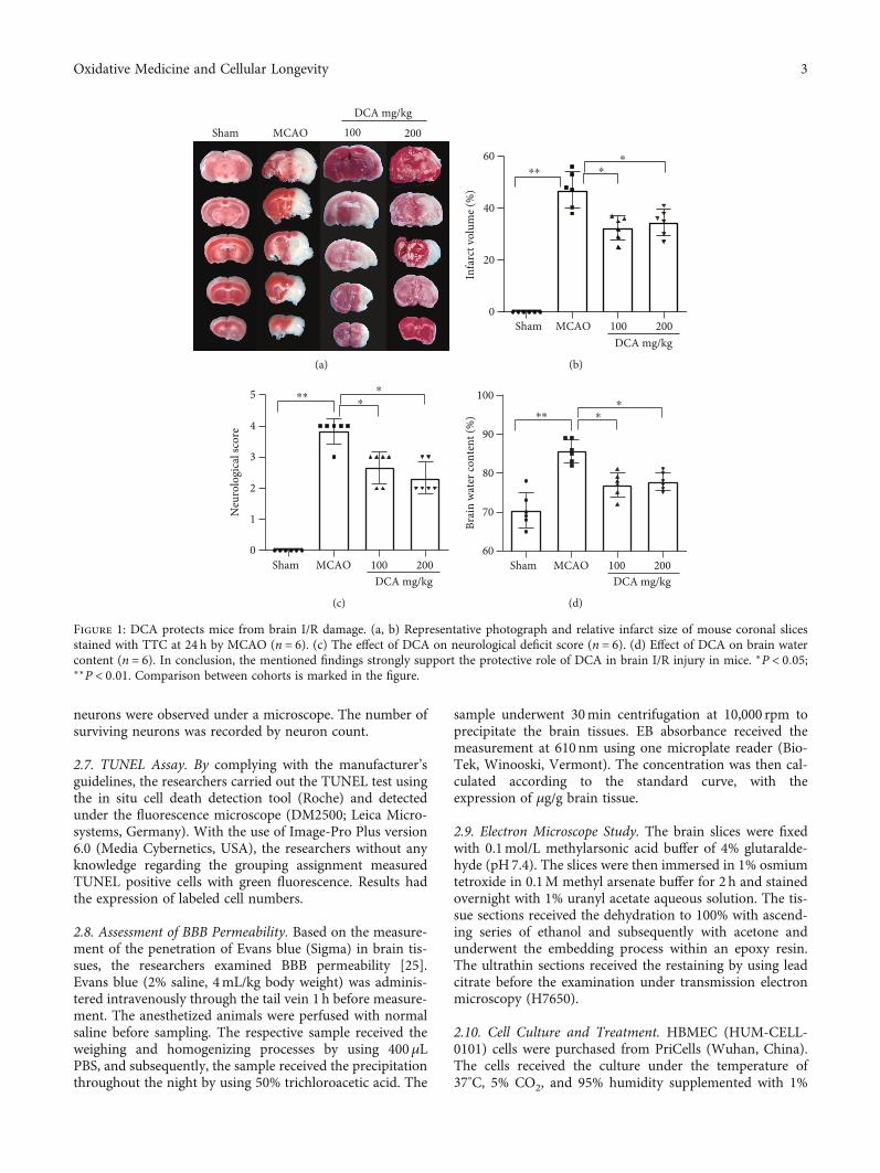
Figure 1: DCA protects mice from brain I/R damage. (a, b) Representative photograph and relative infarct size of mouse coronal slicesstained with TTC at 24 h by MCAO (n = 6). (c) The effect of DCA on neurological deficit score (n = 6). (d) Effect of DCA on brain watercontent (n = 6). In conclusion, the mentioned findings strongly support the protective role of DCA in brain I/R injury in mice. ∗P < 0:05;∗∗P < 0:01. Comparison between cohorts is marked in the figure.
3Oxidative Medicine and Cellular Longevity

penicillin/streptomycin solution (P/S, Sclencell), 1% endo-thelial cell growth supplement (ECGS, Sclencell), and 10%fetal bovine serum (FBS, Sclencell). To simulate ischemia-like conditions in vitro, HBMEC cells received the transfertoward sugar-free medium and the culture within the Tri-GAS (1% O2/5% CO2/94% N2) incubating tool for 4 h. Sub-sequently, the glucose-free medium received the replace-ment by using fresh maintenance medium and therecovery based on normoxic conditions for 24h.
2.11. Determination of Cell Viability (Cell-Counting-Kit 8(CCK-8) Colorimetric Assay). Cell viability was determinedby CCK-8 (Dojindo Molecular Technologies, Inc., Kuma-moto, Japan). For HBMEC cells, the cells are inoculated in96-well plates with a density of 5,000 cells per well. The nextday, HBMEC cells were pretreated with different concentra-tions of DCA, i.e., 2.5mM, 5mM, and 10mM, 6h beforehypoxia, and then exposed to OGD for 4 h, followed by oxy-genation for 24 h. Next, the addition of 20μL CCK-8 was
Sham MCAO
500 𝜇m
100 200
DCA mg/kg
500 𝜇m 500 𝜇m 500 𝜇m
50 𝜇m50 𝜇m50 𝜇m50 𝜇m
(a)
Sham MCAO 100 2000
50
100
150
200
Niss
l pos
itive
neu
rons
DCA mg/kg
⁎⁎
⁎
⁎
(b)
Sham MCAO 100 2000
20
40
60
80
100
TUN
EL p
ositi
ve n
euro
ns (%
)
DCA mg/kg
⁎⁎ ⁎
⁎
(c)
Sham 100 200MCAO
Mer
geD
API
TUN
EL
DCA mg/kg
50 𝜇m
(d)
Figure 2: DCA attenuates neuronal apoptosis after brain I/R injury. (a, b) Nissl staining and (c, d) TUNEL staining and quantitative analysisof coronal sections of ischemic cerebral cortex (n = 6; scale, 50μm). ∗P < 0:05 and ∗∗P < 0:01. The comparison between cohorts is marked inthe figure.
4 Oxidative Medicine and Cellular Longevity

used to each well, and the incubation was achieved under thetemperature of 37°C. Lastly, the absorbance at 450nmreceived the measurement with a microplate analyzer.
2.12. Oxidative Stress Detection. The brain tissue andHBMEC cells were homogenized and centrifuged with12000mg × g for 15min. The supernatant was collected forspectrophotometric study. The BCA assay kit was adoptedfor determining the protein concentration. The contents ofsuperoxide dismutase (SOD) and malondialdehyde (MDA)in brain tissue and HBMEC cells were detected by usingthe appropriate kit (Beyotime Biotechnology, China) andin accordance with the producer’s guideline.
2.13. Determination of Intracellular Reactive Oxygen Species.To determine the production of intracellular reactive oxygenspecies, the 2′,7′-dichlorofluorescein diacetate (DCFH-DA)assay was used to measure ROS according to the manufac-turer’s instructions (Solarbio, Beijing, China). In the pres-ence of ROS, DCFH reacts with ROS to form DCF, afluorescent product. Intracellular detection of ROS in differ-ent groups was achieved by incubating cells with 10μmol/LDCFH-DA at 37°C in darkness for 30min. The fluorescenceof DCFH-DA is inspired at 488 nm, and the emission is col-lected at 525nm. The fluorescence microscope (Olympus,Japan) is used to detect the fluorescence value.
2.14. Western Blotting Assay. Overall proteins from theischemic side cerebral cortex and HBMEC cells receivedthe collection and the fractionation by using SDS-PAGE gels
[23]. Subsequently, the protein received the incubation byusing primary antibodies against PDK2 (1 : 1000, Abcam,USA), PDH (1 : 1000, Abcam, USA), ZO-1 (1 : 1000, Abcam,USA), occludin (1 : 1000, Abcam, USA), Nrf2 (1 : 500,Proteintech, Chicago, USA), HO-1 (1 : 500, Proteintech,Chicago, USA), and Tubulin (1 : 10000, BaoDragon, Hefei,China). The BCA test kit (P0012; Beyotime Biotechnology)was used to measure protein concentrations. After the dena-turation, the same amount of protein was separated by SDS-PAGE and transferred to PVDF membranes (Millipore,Billerica, MA). After the membrane transfer, the membranewas sealed with 5% skim milk at ambient temperatures for2 h. Next, the membrane received the incubation under thetemperature of 4°C with primary antibody and then withappropriate secondary antibody at ambient temperaturesfor 1 h. Image Lab Software (Bio-Rad Laboratories Inc.,Berkeley, CA) was used to detect the protein bands.
2.15. Statistical Analysis. All data, in addition to the neuro-logic score, had the expression of the mean ± standarddeviation ðS:D:Þ and received the comparison by ANOVAand then with Tukey’s multiple-comparison examination.The neurologic scores had the expression of the median(range) and received the comparison with a nonparametricmethod (Kruskal-Wallis test) as well as the Mann–WhitneyU statistic with Bonferroni correction. The researchersemployed GraphPad Prism 7.0 (GraphPad, San Diego, CA,USA) to achieve the statistical investigation. A value of P <0:05 was statistically significant.
100Sham MCAO 200
DCA mg/kg
(a)
Sham MCAO 100 2000123
20406080
Evan
s blu
e con
cent
ratio
n
DCA mg/kg
⁎⁎⁎
⁎
(b)
Occludin
Zo-1
Tubulin
MCAODCA mg/kg
+ + +–
– 100 200–
(c)
Sham MCAO 100 2000.0
0.5
1.0
1.5
DCA mg/kg
Occ
ludi
n/tu
bulin
(fold
of s
ham
)
⁎⁎
⁎
⁎
(d)
Sham MCAO 100 2000.0
0.5
1.0
1.5
Zo-1
/Tub
ulin
(fold
of s
ham
)DCA mg/kg
⁎⁎
⁎
⁎
(e)
Figure 3: DCA can reduce the damage of the blood-brain barrier attributed to ischemia. (a, b) Representative photograph of Evans blue-stained mouse brains 24 h after sham surgery or MCAO (n = 6). (c–e) Western blotting assay of the representative proteins of TJproteins occludin and ZO-1 in mouse brain and the band strength of the respective protein relative to Tubulin. ∗P < 0:05 and ∗∗P < 0:01.The comparison between cohorts is marked in the figure.
5Oxidative Medicine and Cellular Longevity

3. Results
3.1. DCA Protects Mice from Cerebral Ischemia-ReperfusionInjury. According to Figure 1, to study the potential effectexerted by DCA in CIRI, neurological score, cerebral infarctarea rate, and brain edema content were examined 24 hwhen MCAO was caused. In contrast to sham operation,the MCAO cohort had an increase in cerebral infarct sizeand cerebral edema and a decrease in neurological scores.The cerebral infarction area and neurological score were sig-nificantly improved in the DCA cohort (Figures 1(a)–1(c)).In addition, the DCA treatment significantly improved cere-bral edema (Figure 1(d)).
3.2. DCA Attenuates Neuronal Apoptosis after I/R Injury.Nissl staining showed a decrease in the number of neuronsin the MCAO cohort compared with the sham cohort anda significant improvement in the number of neurons in theDCA treatment cohort (Figures 2(a) and 2(b)). TUNELstaining showed that neuronal apoptosis increased in the
MCAO cohort compared with the sham operation cohortbut decreased in the DCA cohort (Figures 2(c) and 2(d)).
3.3. DCA Attenuates BBB Damage after Cerebral I/R Injury.The permeability of Evans blue dye is shown in Figures 3(a)and 3(b). Compared with the sham operation cohort, thepermeability of Evans blue dye increased in the MCAOcohort but decreased in the DCA cohort. Compared withthe sham operation cohort, the MCAO cohort also signifi-cantly reduced the expressions of major TJ membrane pro-teins occludin and ZO-1 (Figures 3(c)–3(e)), which wereimproved in the DCA treatment cohort, and they interactedto maintain BBB integrity. The mentioned results suggestthat DCA inhibits ischemia-induced BBB destruction.
3.4. DCA Improves Mitochondrial Metabolism after CerebralI/R Injury. According to electron microscopy (Figure 4(a)),compared with the sham operation cohort, the volume ofmitochondria in the MCAO cohort increased, the electrondensity of matrix decreased, the matrix particles decreasedor disappeared, and the cristae became shorter, reduced,
Sham MCAODCA mg/kg
200100
(a)
+ + +–
– 200–
PDK2
Tubulin
MCAODCA mg/kg 100
(b)
0
1
2
3
100
PDK2
/tubu
lin(fo
ld o
f sha
m)
DCA mg/kgMCAOSham 200
⁎⁎
⁎⁎
(c)
0.0
0.5
1.0
1.5
PDH
/Tub
ulin
(fold
of s
ham
)
100DCA mg/kg
MCAOSham 200
⁎⁎⁎
⁎
(d)
PDH
Tubulin
+ + +–
– 200–
MCAODCA mg/kg 100
(e)
0
50
100
150
200
100DCA mg/kg
MCAOSham 200
SOD
(U/m
g)
⁎⁎
⁎⁎
(f)
0
5
10
15
20
25
30
100DCA mg/kg
MCAOSham 200
MD
A(n
mol
/mg)
⁎⁎
⁎⁎
(g)
Figure 4: DCA improves mitochondrial metabolism after brain I/R injury. (a) Mitochondrial morphology was observed by electronmicroscopy 24 h after sham surgery or MCAO (n = 6). (b–e) Western blotting assay of key metabolic proteins PDK2 and PDH ofmitochondrial TCA cycle in mouse brain as well as the band strength of the respective protein relative to Tubulin. (f, g) Effects of DCAon the contents of oxidative stress products SOD and MDA after brain I/R injury. ∗P < 0:05 and ∗∗P < 0:01. The comparison betweencohorts has been shown in the figure.
6 Oxidative Medicine and Cellular Longevity

and moved to the edge, which was improved in the DCAtreatment cohort. In contrast to the sham-operated cohort(Figures 4(b)–4(e)), the expression of mitochondrialmetabolism-related protein PDK2 increased, and PDHdecreased in the MCAO cohort, PDK2 in the DCA cohortwas lower than that in the MCAO cohort, and PDH in theMCAO cohort was higher than that in the MCAO cohort.Oxidative damage was assessed by measuring SOD andMDA productions. As shown in Figures 4(f) and 4(g), asopposed to the sham operation cohort, SOD activity signifi-cantly decreased, and MDA content increased in the H/Rcohort. Furthermore, DCA administration significantlyrestored the activity of SOD and decreased the content ofMDA after H/R.
3.5. DCA Activates the Nrf2/HO-1 Signaling Channel toReduce Oxidative Damage. The Western blotting assay wasconducted to examine the expressions of oxidative stress-related proteins Nrf2 and HO-1 in the ischemic cerebral cor-tex. According to Western blotting analysis of the ischemiccerebral cortex after MCAO, DCA significantly upregulatedNrf2 and HO-1 expressions (Figures 5(a)–5(d)).
3.6. DCA Improves Mitochondrial Metabolism after I/RInjury and Activates Nrf2/HO-1 Signaling Channel toReduce Oxidative Damage In Vitro. The researchers specifi-cally investigated the neuroprotective influence exerted byDCA in HBMEC cells using the OGD model. The viabilityof the injured cells was measured by the CCK8 assay. Forinstance (Figure 6(a)), cell viability was significantly reducedby OGD-induced treatment compared with the controls,while cell viability was significantly increased by the DCAtreatment. As indicated from the results, DCA could protectdifferentiated HBMEC cells from OGD injury, and the rela-tive optimal dose was 5mM.
Oxidative damages were assessed through the measure-ment of in vitro SOD, MDA, and ROS productions. As
shown in Figures 6(b) and 6(c), compared with the shamoperation cohort, the SOD activity of the H/R cohort signif-icantly decreased, and the MDA content increased. In addi-tion, DCA significantly restored the activity of SOD andreduced the MDA content after H/R (Supplementary mate-rial online, Figure S). DCA reduced the ROS content afterH/R. DCA inhibited PDK2, i.e., the key metabolic proteinof mitochondrial TCA cycle, and activated PDH.Furthermore, DCA activated Nrf2 and HO-1 expressions(Figures 6(d) and 6(e)). Oxidative damages were assessedthrough the measurement of SOD, MDA, and ROSproductions in vitro.
3.7. DCA Attenuates the Damage of Blood-Brain Barrier afterI/R Injury In Vitro and the Disruption of Tight Protein. DCAattenuates the expressions of occludin and ZO-1, the key ofTJ membrane proteins, in vitro to maintain BBB integrity.The mentioned results indicated that DCA inhibitsischemia-induced BBB destruction (Figures 7(a)–7(d)).
4. Discussion
Previous studies have shown that DCA plays an importantrole in vascular protection [26], promoting vascular revascu-larization and improving vascular calcification in patientswith atherosclerosis [27]. However, the mechanism ofDCA regulating mitochondrial metabolism and oxidativestress in cerebral IS and I/R has not been clarified. For ISand I/R, energy metabolism disorder and mitochondrial dys-function are able to result in considerable free radical forma-tions, thereby triggering oxidative damages, inhibiting theactivity of antioxidant enzymes, breaking down the blood-brain barrier, and aggravating brain injury. This studyreveals the protective role of DCA in mediating brain I/Rinjury. DCA can improve CIRI by reducing infarct volume,neurological score, and cerebral water content. DCA admin-istration attenuated mitochondrial metabolism, oxidative
Nrf2
Tubulin
+ + +–
– 200–
MCAODCA mg/kg 100
(a)
+ + +–
– 200–
MCAODCA mg/kg 100
HO-1
Tubulin
(b)
Nrf2
/tubu
lin(fo
ld o
f sha
m)
0
1
2
3
4
100DCA mg/kg
MCAOSham 200
⁎⁎⁎
⁎⁎
(c)
HO
-1/tu
bulin
(fold
of s
ham
)
0
1
2
3
4
5
100DCA mg/kg
MCAOSham 200
⁎ ⁎⁎
⁎⁎
(d)
Figure 5: DCA activates the Nrf2/HO-1 signaling channel. (a–d) Western blotting assay of Nrf2 and HO-1 in mouse brains and the bandstrength of the respective protein relative to Tubulin. ∗P < 0:05 and ∗∗P < 0:01. The comparison between cohorts is marked in the figure.
7Oxidative Medicine and Cellular Longevity

stress, neuronal apoptosis, and blood-brain barrier perme-ability after I/R injury in mice. In addition, the DCA treat-ment also reduced the mitochondrial metabolism, blood-brain barrier permeability, and oxidative stress in OGD-induced HBMEC cells. This study confirmed that the brainprotective function of DCA was related to the activation ofthe PDK2-PDH-Nrf2 pathway, and the Nrf2-mediated anti-oxidant stress and blood-brain barrier protection disap-peared when ML385, i.e., the specific inhibitor of Nrf2, wasused.
Under physiological conditions, ATP required by thebrain is mainly produced by pyruvate oxidation (PO) andglucose oxidation (GO) within mitochondria [28]. Pyruvateformation increases the rate of glycolysis and promotes theglucose oxidizing process via the PDH activation [29], thus
converting pyruvate to acetyl-coA. Nevertheless, based onpathophysiological conditions (e.g., I/R), due to mitochon-drial dysfunction, it can facilitate the expressions of PDKand phosphorylate PDH, thereby inhibiting PDH regulatedglucose metabolism and reducing glucose oxidation rate[30, 31]. Studies have found that in the brain, PDK activitymainly has a correspondence to isoenzyme PDK2. Further-more, DCA could inhibit PDK, the inhibition order wasPDK2 > PDK1 > PDK4 > PDK3 [15], which improved theactivity of PDH.
Previous studies have shown that DCA is a pharmaco-logical agent that activates PDH by inhibiting PDK and alsoshows significant neuroprotective potential. The administra-tion of DCA has been suggested to facilitate local lactic acidremoval [32], tumor therapy [33], and pulmonary
Con OGD 2.5 5 100
50
100
150
OGD+DCAmM
Cell
viab
ility
(% o
f con
trol
)
⁎
⁎
(a)
Sham H/R DCA ML3850
10
20
30
40
50
H/R
SOD
(U/m
g)
⁎⁎
⁎
⁎
(b)
0
10
20
30
40
50
MD
A(n
mol
/mg)
Sham H/R DCA ML385H/R
⁎⁎ ⁎
⁎
(c)
H/RDCA(mM)
+ +__
1.955 0
1
2
3
4
0.0
0.5
1.0
1.5
Tubulin
PDH
PDK2
+_
_ _ _
PDK2
/tubu
lin(fo
ld o
f sha
m)
PDH
/Tub
ulin
(fold
of s
ham
)
Sham H/R DCA ML385 Sham H/R DCA ML385H/R H/R
ML3 85(uM)
⁎
⁎
⁎⁎
⁎⁎
⁎⁎
⁎
(d)
55
0
1
2
3
0
1
2
3
4
5
H/RDCA (mM)
+ +__
1.9
+_
_ _ _
Tubulin
Nrf2
HO-1
Nrf2
/tubu
lin(fo
ld o
f sha
m)
HO
-1/tu
bulin
(fold
of s
ham
)
Sham H/R DCA ML385 Sham H/R DCA ML385H/RH/R
ML385(𝜇M)
⁎
⁎⁎ ⁎⁎
⁎⁎
⁎⁎ ⁎⁎
(e)
Figure 6: Neuroprotective effect of DCA on HBMEC cells. (a) CCK8 assay was used to detect the viability of cells after injury. (b, c) Theproductions of SOD and MAD were measured to assess oxidative damage. (d) DCA inhibits PDK2, a key metabolic protein ofmitochondrial TCA cycle, and activates the expression of PDH. (e) DCA activated the protein expressions of Nrf2 and HO-1. ∗P < 0:05and ∗∗P < 0:01. The comparison between cohorts is marked in the figure.
8 Oxidative Medicine and Cellular Longevity

hypertension [34]. However, the protective effect, molecularmechanism, and blood-brain barrier permeability of DCA incerebral IS and I/R injury have been rarely investigated. Thisstudy reported that DCA could exert a protective effect by
inhibiting PDK2 and activating PDH to regulate mitochon-drial metabolism. The transmission electron microscopewas adopted to observe the mitochondria of the cerebral cor-tex after brain I/R injury, and the results suggested that the
Tubulin
Occludin
H/RDCA (mM)
+ +–
–
1.9
+–
– – –ML385 (𝜇M)55
(a)
Tubulin
Zo-1
H/RDCA(mM)
+ +–
–
1.955
+–
– – –ML385(𝜇M)
(b)
0.0
0.5
1.0
1.5O
cclu
din/
tubu
lin(fo
ld o
f sha
m)
Sham H/R DCA ML385H/R
⁎⁎
⁎
⁎
(c)
0.0
0.5
1.0
1.5
Zo-1
/Tub
uiln
(fold
of s
ham
)
Sham H/R DCA ML385H/R
⁎
⁎
⁎⁎
(d)
Figure 7: DCA attenuates the expressions of key occludin and ZO-1 of TJ membrane proteins in vitro to maintain BBB integrity. (a–d)Western blotting assay of occludin and ZO-1 and the band strength of the respective protein relative to Tubulin in mouse brain. Thementioned results indicate that DCA inhibits ischemia-induced BBB destruction. ∗P < 0:05 and ∗∗P < 0:01. The comparison betweencohorts is marked in the figure.
Pyruvate
Acetyl-CoA
ATP
PDK2ROS
Mitochondria
SOD MDA
Disrupted BBB
PromoteInhibit
StrokeDCA
Glucose
Glucose
Ischemicstroke
DCALactate
TCAcycle
PDH
Nrf2
HO-1
Figure 8: Sodium dichloroacetate (DCA) improves glycolysis by inhibiting PDK2 and activating PDH and promotes the conversion ofpyruvate into acetyl-CoA, which then enters the TCA cycle so as to produce ATP, thus improving glycolysis. Meanwhile, it activates theNrf2/HO-1 signaling pathway to increase the activity of SOD, inhibit the generation of MDA, and reduce oxidative stress, therebyalleviating the damage of the blood-brain barrier and facilitating the recovery of oxidative metabolism.
9Oxidative Medicine and Cellular Longevity

mitochondrial injury was alleviated. In IS, after the recoveryof cerebral blood flow, considerable free radicals and reactiveoxygen species will be produced, leading to the aggravationof I/R injury. The mechanism is that IS leads to the abnor-mal activities of some enzymes of mitochondrial metabolism(PDK2 and PDH). However, DCA can inhibit PDK2 andactivate PDH, thus improving mitochondrial metabolism.
Oxidative stress acts as the vital actor of brain I/R injury,capable of causing neuronal damage and death [35]. Variousantioxidants can improve brain I/R injury [36–38]. Accord-ing to existing studies, DCA exhibits broad biological activ-ity and can cross the blood-brain barrier in mice [39].Accordingly, we investigated whether DCA affects oxidativestress after brain I/R injury. In addition, Nrf2 refers to a crit-ical antioxidant defense mechanism [40, 41]. Under cerebralI/R, excessive oxidative stress facilitates Keap1 and Nrf2 sep-arating processes, thereby activating Nrf2, and the activatedNrf2 is translocated to the nucleus and binds with ARE, thusactivating the transcription of several downstream antioxi-dant genes [42, 43]. Thus, Nrf2 acts as the vital transcriptionelement for maintaining redox homeostasis. According toexisting researches, Nrf2 has a cytoprotective effect withina wide range of I/R-induced brain and kidney injury models[44, 45]. However, there are few studies on how DCA andNrf2 regulate oxidative stress. We assumed that DCA regu-lates mitochondrial metabolism by inhibiting PDK2 andactivating PDH, thus activating the Nrf2-HO-1 channel toproduce antioxidant stress effect. As revealed from theresults, the activities of Nrf2 and HO-1 significantlyincreased after the DCA treatment. We used ML385, a spe-cific inhibitor of Nrf2 in cells, and the protective effect ofDCA disappeared, indicating that DCA could alleviate oxi-dative stress through the PDK2-PDH-Nrf2 channel.
In this study, SOD and MDA were employed to assessoxidative damage. SOD can catalyze superoxide anion freeradicals to be transformed into hydrogen peroxide. As aproduct of lipid peroxidation, MDA has been adopted toassess the level of free radicals within brain I/R injury [46].Consistent with existing studies, SOD activity declined sig-nificantly and the MDA level rose significantly after brainI/R injury. Next, DCA significantly improved the activityof SOD and downregulated MDA levels within brain tissuesand HBMEC cells of I/R mice. The mentioned resultsdirectly reveal that DCA attenuates brain I/R damage byinhibiting oxidative stress.
The blood-brain barrier (BBB) refers to a selection-related osmotic membrane comprising endothelial cells,extracellular matrix unit pertaining to the basement mem-brane, pericyte, and endings of astrocytes. The tight junctionof endothelial cells is the gatekeeper, restricting the entry ofsubstances from the blood into the brain, thereby maintain-ing brain homeostasis [47]. After the aggravation of I/Rinjury, impaired BBB integrity increases paracellular perme-ability, which allows toxins, a wide range of immune cells,and inflammation-related cytokines to enter the brain,which leads to risen cerebrovascular edema, hemorrhagictransformation, and increased mortality [25]. As indicatedfrom the data of this study, DCA reduces extravasation ofEvans blue dye in the I/R cerebral cortex of mice. Moreover,
ischemia noticeably reduced the expressions of major TJmembrane protein, including occludin and ZO-1, andDCA significantly improved the expressions of occludinand ZO-1 for maintaining BBB integrity.
The present study also had some defects; for example,BBB only observed changes in endothelial cells but did notobserve the relationship between the extracellular matrixcomponents of the basement membrane, pericytes andastrocytes, and BBB. In addition, we did not observe the rel-evant studies on DCA in patients, and the relevant mecha-nisms need to be further studied.
In brief, our study proved that after brain I/R injury,DCA can improve the activity of mitochondrial-relatedenzymes PDK2/PDH to promote energy generation andactivate the Nrf2 pathway to inhibit oxidative stress andneuronal apoptosis and increase BBB permeability(Figure 8). The PDK2-PDH-Nrf2 pathway may play a keyrole and provide a new pharmacology target in cerebral ISand I/R protection by DCA.
Data Availability
The data used to support the findings of this study are avail-able from the corresponding authors upon request.
Conflicts of Interest
The authors declare no conflicts of interest.
Authors’ Contributions
Xiaoyong Zhao, Shan Li, and Yunchang Mo are co-firstcoauthors.
Acknowledgments
This work was supported by the Wenzhou Science & Tech-nology Bureau Foundation, China (No. 2018ZY003), and theNational Natural Science Foundation of China, China(Grant No. 81704180). This work was also supported bythe Shandong provincial project of medical and health tech-nology development program, China (2019WS605), andZhejiang Province Science and Technology Plan Researchand Xinmiao Talent Program, China (Grant No.2020R413079).
Supplementary Materials
Supplementary Figure: effect of DCA on ROS levels inHBMEC cells induced by H/R. (a) Reactive oxygen specieslevels were analyzed by DCFH-DA kit. (b) Compared withthe sham operation cohort, the ROS content increased. Inaddition, DCA reduced the ROS content after H/R. ∗P <0:05 and ∗∗P < 0:01. The comparison between cohorts ismarked in the figure. (Supplementary Materials)
References
[1] E. J. Benjamin, M. J. Blaha, S. E. Chiuve et al., “Heart diseaseand stroke statistics-2017 update: a report from the American
10 Oxidative Medicine and Cellular Longevity

Heart Association,” Circulation, vol. 135, no. 10, pp. e146–e603, 2017.
[2] B. Liao, L. Geng, F. Zhang et al., “Adipocyte fatty acid-bindingprotein exacerbates cerebral ischaemia injury by disrupting theblood-brain barrier,” European Heart Journal, vol. 41, no. 33,pp. 3169–3180, 2020.
[3] C. Tang, J. Hong, C. Hu et al., “Palmatine protects against cere-bral ischemia/reperfusion injury by activation of theAMPK/Nrf2 pathway,” Oxidative Medicine and Cellular Lon-gevity, vol. 2021, Article ID 6660193, 12 pages, 2021.
[4] M. Dai, L. Wu, K. Yu et al., “D-Carvone inhibit cerebral ische-mia/reperfusion induced inflammatory responseTLR4/NLRP3 signaling pathway,” Biomedicine & Pharmaco-therapy, vol. 132, p. 110870, 2020.
[5] W. Xie, T. Zhu, P. Zhou et al., “Notoginseng Leaf TriterpenesAmeliorates OGD/R-Induced Neuronal Injury viaSIRT1/2/3-Foxo3a-MnSOD/PGC-1α Signaling PathwaysMediated by the NAMPT-NAD Pathway,”Oxidative Medicineand Cellular Longevity, vol. 2020, Article ID 7308386, 15 pages,2020.
[6] J. P. Bolanos, M. A. Moro, I. Lizasoain, and A. Almeida, “Mito-chondria and reactive oxygen and nitrogen species in neuro-logical disorders and stroke: therapeutic implications,”Advanced Drug Delivery Reviews, vol. 61, no. 14, pp. 1299–1315, 2009.
[7] Q. Yang, Q. Huang, Z. Hu, and X. Tang, “Potential neuropro-tective treatment of stroke: targeting excitotoxicity, oxidativestress, and inflammation,” Frontiers in Neuroscience, vol. 13,p. 1036, 2019.
[8] L. Wu, X. Xiong, X. Wu et al., “Targeting oxidative stress andinflammation to prevent ischemia-reperfusion injury,” Fron-tiers in Molecular Neuroscience, vol. 13, p. 28, 2020.
[9] B. Xu, T. Wang, J. Xiao et al., “FCPR03, a novel phosphodies-terase 4 inhibitor, alleviates cerebral ischemia/reperfusioninjury through activation of the AKT/GSK3β/ β-catenin sig-naling pathway,” Biochemical Pharmacology, vol. 163,pp. 234–249, 2019.
[10] T. Kalogeris, C. P. Baines, M. Krenz, and R. J. Korthuis, “Cellbiology of ischemia/reperfusion injury,” International Reviewof Cell and Molecular Biology, vol. 298, pp. 229–317, 2012.
[11] D. N. Granger and P. R. Kvietys, “Reperfusion injury and reac-tive oxygen species: the evolution of a concept,” Redox Biology,vol. 6, pp. 524–551, 2015.
[12] D. K. Hong, A. R. Kho, B. Y. Choi et al., “Combined treatmentwith dichloroacetic acid and pyruvate reduces hippocampalneuronal death after transient cerebral ischemia,” Frontiers inNeurology, vol. 9, p. 137, 2018.
[13] Y. Sun, T. Li, C. Xie et al., “Dichloroacetate treatment improvesmitochondrial metabolism and reduces brain injury in neona-tal mice,” Oncotarget, vol. 7, no. 22, pp. 31708–31722, 2016.
[14] P. W. Stacpoole, T. L. Kurtz, Z. Han, and T. Langaee, “Role ofdichloroacetate in the treatment of genetic mitochondrial dis-eases,” Advanced Drug Delivery Reviews, vol. 60, no. 13-14,pp. 1478–1487, 2008.
[15] M. M. Bowker-Kinley, I. W. Davis, P. Wu, A. R. Harris, andM. K. Popov, “Evidence for existence of tissue-specific regula-tion of the mammalian pyruvate dehydrogenase complex,”Biochemical Journal, vol. 329, no. 1, pp. 191–196, 1998.
[16] Y. Itoh, T. Esaki, K. Shimoji et al., “Dichloroacetate effects onglucose and lactate oxidation by neurons and astrogliain vitro and on glucose utilization by brain in vivo,” Proceed-
ings of the National Academy of Sciences of the United Statesof America, vol. 100, no. 8, pp. 4879–4884, 2003.
[17] A. R. Kho, B. Y. Choi, S. H. Lee et al., “The effects of sodiumdichloroacetate on mitochondrial dysfunction and neuronaldeath following hypoglycemia-induced injury,” Cells, vol. 8,no. 5, p. 405, 2019.
[18] P. Wang, M. Chen, Z. Yang et al., “Activation of pyruvatedehydrogenase activity by dichloroacetate improves survivaland neurologic outcomes after cardiac arrest in rats,” Shock,vol. 49, no. 6, pp. 704–711, 2018.
[19] P. W. Stacpoole, “Therapeutic targeting of the pyruvate dehy-drogenase complex/pyruvate dehydrogenase kinase(PDC/PDK) axis in cancer,” JNCI: Journal of the NationalCancer Institute, vol. 109, no. 11, 2017.
[20] E. D. Michelakis, V. Gurtu, L. Webster et al., “Inhibition ofpyruvate dehydrogenase kinase improves pulmonary arterialhypertension in genetically susceptible patients,” ScienceTranslational Medicine, vol. 9, no. 413, 2017.
[21] W. Geng, L. Cai, K. Han et al., “Electroacupuncture Pretreat-ment Alleviates Cerebral Ischemia-Reperfusion Injury byIncreasing GSK-3β Phosphorylation Level via Adenosine A1Receptor,” BioMed Research International, vol. 2020, ArticleID 6848450, 9 pages, 2020.
[22] E. Z. Longa, P. R. Weinstein, S. Carlson, and R. Cummins,“Reversible middle cerebral artery occlusion without craniect-omy in rats,” Stroke, vol. 20, no. 1, pp. 84–91, 1989.
[23] L. Buscemi, C. Blochet, P. J. Magistretti, and L. Hirt, “Hydro-xycarboxylic acid receptor 1 and neuroprotection in a mousemodel of cerebral ischemia-reperfusion,” Frontiers in Physiol-ogy, vol. 12, p. 689239, 2021.
[24] M. Sun, B. Deng, X. Zhao et al., “Isoflurane preconditioningprovides neuroprotection against stroke by regulating theexpression of the TLR4 signalling pathway to alleviate micro-glial activation,” Scientific Reports, vol. 5, no. 1, p. 11445, 2015.
[25] D. N. Doll, H. Hu, J. Sun, S. E. Lewis, J. W. Simpkins, andX. Ren, “Mitochondrial crisis in cerebrovascular endothelialcells opens the blood-brain barrier,” Stroke, vol. 46, no. 6,pp. 1681–1689, 2015.
[26] T. Deuse, X. Hua, D. Wang et al., “Dichloroacetate preventsrestenosis in preclinical animal models of vessel injury,”Nature, vol. 509, no. 7502, pp. 641–644, 2014.
[27] Y. Yang, Y. Sun, J. Chen et al., “AKT-independent activation ofp38 MAP kinase promotes vascular calcification,” Redox Biol-ogy, vol. 16, pp. 97–103, 2018.
[28] W. C. Stanley, G. D. Lopaschuk, J. L. Hall, and J. G. Mccor-mack, “Regulation of myocardial carbohydrate metabolismunder normal and ischaemic conditions. Potential for pharma-cological interventions,” Cardiovascular Research, vol. 33,no. 2, pp. 243–257, 1997.
[29] X. Zhu, D. Long, M. Zabalawi et al., “Stimulating pyruvatedehydrogenase complex reduces itaconate levels and enhancesTCA cycle anabolic bioenergetics in acutely inflamed mono-cytes,” Journal of Leukocyte Biology, vol. 107, no. 3, pp. 467–484, 2020.
[30] K. Subramani, S. Lu, M. Warren et al., “Mitochondrial target-ing by dichloroacetate improves outcome following hemor-rhagic shock,” Scientific Reports, vol. 7, no. 1, p. 2671, 2017.
[31] J. R. Ussher, W. Wang, M. Gandhi et al., “Stimulation of glu-cose oxidation protects against acute myocardial infarctionand reperfusion injury,” Cardiovascular Research, vol. 94,no. 2, pp. 359–369, 2012.
11Oxidative Medicine and Cellular Longevity

[32] S. J. DeVience, X. Lu, J. L. Proctor et al., “Enhancing metabolicimaging of energy metabolism in traumatic brain injury usinghyperpolarized [1-13C]Pyruvate and dichloroacetate,”Metab-olites, vol. 11, no. 6, p. 335, 2021.
[33] J. Parczyk, J. Ruhnau, C. Pelz et al., “Dichloroacetate and PX-478 exhibit strong synergistic effects in a various number ofcancer cell lines,” BMC Cancer, vol. 21, no. 1, p. 481, 2021.
[34] P. Liu, W. Huang, Y. Ding et al., “Fasudil dichloroacetate alle-viates SU5416/hypoxia-induced pulmonary arterial hyperten-sion by ameliorating dysfunction of pulmonary arterialsmooth muscle cells,” Drug Design, Development and Therapy,vol. Volume 15, pp. 1653–1666, 2021.
[35] C. Izzo, P. Vitillo, P. di Pietro et al., “The role of oxidative stressin cardiovascular aging and cardiovascular diseases,” Life,vol. 11, no. 1, p. 60, 2021.
[36] S. U. Rehman, M. Ikram, N. Ullah et al., “Neurologicalenhancement effects of melatonin against brain injury-induced oxidative stress, neuroinflammation, and neurode-generation via AMPK/CREB signaling,” Cells, vol. 8, no. 7,p. 760, 2019.
[37] R. Rodrigo, R. Fernandez-Gajardo, R. Gutierrez et al., “Oxida-tive stress and pathophysiology of ischemic stroke: novel ther-apeutic opportunities,” CNS & Neurological Disorders DrugTargets, vol. 12, no. 5, pp. 698–714, 2013.
[38] T. Wang, H. Chen, S. Xia, X. Chen, H. Sun, and Z. Xu, “Ame-liorative effect of parishin C against cerebral ischemia-inducedbrain tissue injury by reducing oxidative stress and inflamma-tory responses in rat model,” Neuropsychiatric Disease andTreatment, vol. Volume 17, pp. 1811–1823, 2021.
[39] T. Tataranni and C. Piccoli, “Dichloroacetate (DCA) and can-cer: an overview towards clinical applications,”Oxidative Med-icine and Cellular Longevity, vol. 2019, Article ID 8201079, 14pages, 2019.
[40] I. Bellezza, I. Giambanco, A. Minelli, and R. Donato, “Nrf2-Keap1 signaling in oxidative and reductive stress,” Biochimicaet Biophysica Acta (BBA) - Molecular Cell Research, vol. 1865,no. 5, pp. 721–733, 2018.
[41] W. Tu, H. Wang, S. Li, Q. Liu, and H. Sha, “The anti-inflammatory and anti-oxidant mechanisms of the Keap1/Nr-f2/ARE signaling pathway in chronic diseases,” Aging and Dis-ease, vol. 10, no. 3, pp. 637–651, 2019.
[42] B. I. Ucar, G. Ucar, S. Saha, B. Buttari, E. Profumo, and L. Saso,“Pharmacological protection against ischemia-reperfusioninjury by regulating the Nrf2-Keap1-ARE signaling pathway,”Antioxidants (Basel), vol. 10, no. 6, p. 823, 2021.
[43] H. Wang, W. Wei, X. Lan et al., “Neuroprotective effect ofswertiamain on cerebral ischemia/reperfusion injury by induc-ing the Nrf2 protective pathway,” ACS Chemical Neuroscience,vol. 10, no. 5, pp. 2276–2286, 2019.
[44] F. Zhou, M. Wang, J. Ju et al., “Schizandrin A protects againstcerebral ischemia-reperfusion injury by suppressing inflam-mation and oxidative stress and regulating the AMPK/Nrf2pathway regulation,” American Journal of TranslationalResearch, vol. 11, no. 1, pp. 199–209, 2019.
[45] M. Nezu, N. Suzuki, and M. Yamamoto, “Targeting theKEAP1-NRF2 system to prevent kidney disease progression,”American Journal of Nephrology, vol. 45, no. 6, pp. 473–483,2017.
[46] Z. Yang, C. Weian, H. Susu, and W. Hanmin, “Protectiveeffects of mangiferin on cerebral ischemia-reperfusion injuryand its mechanisms,” European Journal of Pharmacology,vol. 771, pp. 145–151, 2016.
[47] B. Obermeier, R. Daneman, and R. M. Ransohoff, “Devel-opment, maintenance and disruption of the blood-brainbarrier,” Nature Medicine, vol. 19, no. 12, pp. 1584–1596,2013.
12 Oxidative Medicine and Cellular Longevity