dc-6 - medtecshop · · 2013-05-26owns the intellectual property rights to this mindray product...
TRANSCRIPT
DC-6
Diagnostic Ultrasound System
Specifications

I-1
Intellectual Property Statement SHENZHEN MINDRAY BIO-MEDICAL ELECTRONICS CO., LTD. (hereinafter called Mindray)
owns the intellectual property rights to this Mindray product and this manual. This manual may
refer to information protected by copyrights or patents and does not convey any license under
the patent rights of Mindray, nor the rights of others. Mindray does not assume any liability
arising out of any infringements of patents or other rights of third parties.
Mindray intends to maintain the contents of this manual as confidential information. Disclosure
of the information in this manual in any manner whatsoever without the written permission of
Mindray is strictly forbidden.
Release, amendment, reproduction, distribution, rent, adaptation and translation of this
manual in any manner whatsoever without the written permission of Mindray is strictly
forbidden.
C-1
Contents
1 Intended Use .........................................................................................1-1
2 Configuration ........................................................................................2-1
2.1 Standard Configuration ................................................................................... 2-1
2.2 Transducers Available and Applications.......................................................... 2-1
2.3 Optional Units ................................................................................................. 2-1
2.4 Recommended Peripheral Devices ................................................................ 2-2
3 Performance Specifications.................................................................3-1
3.1 Scan Modes.................................................................................................... 3-1
3.2 Exam Modes................................................................................................... 3-1
3.3 Acoustic Power ............................................................................................... 3-2
3.4 Image Specifications....................................................................................... 3-2
3.4.1 Tissue Specific Imaging (TSI) .......................................................................... 3-2
3.4.2 Tissue Harmonic Imaging (THI) ....................................................................... 3-2
3.4.3 iTouch .............................................................................................................. 3-2
3.4.4 Zoom................................................................................................................ 3-2
3.4.5 Image depth..................................................................................................... 3-2
3.4.6 Scan control..................................................................................................... 3-2
3.4.7 Post processing ............................................................................................... 3-3
3.4.8 SV (Sample Volume)........................................................................................ 3-3
3.4.9 Doppler scanning parameters.......................................................................... 3-3
3.4.10 Doppler audio ................................................................................................ 3-3
4 Preset .....................................................................................................4-1
4.1 System Preset ................................................................................................ 4-1
4.2 Preset of a Transducer and an Exam mode.................................................... 4-1
4.3 Preset of Image Parameters ........................................................................... 4-1
4.4 Preset of Measurements................................................................................. 4-1
4.5 Preset of Comments and Body Marks ............................................................ 4-1
5 Management of Patient Files................................................................5-1

C-2
5.1 Patient Information.......................................................................................... 5-1
5.2 iStation ............................................................................................................ 5-1
5.3 Graph/Text Workstation .................................................................................. 5-1
6 Cine Review...........................................................................................6-1
6.1 Freeze............................................................................................................. 6-1
6.2 Image Storage ................................................................................................ 6-1
6.3 Cine Review.................................................................................................... 6-1
7 Comments and Body Marks.................................................................7-1
7.1 Comments ...................................................................................................... 7-1
7.2 Body Marks..................................................................................................... 7-1
8 Measurements.......................................................................................8-1
8.1 General Measurements .................................................................................. 8-1
8.1.1 B mode ............................................................................................................ 8-1
8.1.2 M mode............................................................................................................ 8-1
8.1.3 PW mode ......................................................................................................... 8-1
8.1.4 Color mode ...................................................................................................... 8-1
8.2 Application Measurements.............................................................................. 8-2
9 User Interface ........................................................................................9-1
9.1 Power On/Off .................................................................................................. 9-1
9.2 I/O Interfaces .................................................................................................. 9-1
9.3 Monitor............................................................................................................ 9-2
10 External Dimensions and Weight ......................................................10-1
10.1 External Dimensions (excluding optional devices)........................................ 10-1
10.2 Weight........................................................................................................... 10-1
11 Environmental Conditions .................................................................11-1
11.1 Power Supply................................................................................................ 11-1
11.2 Operating Conditions .................................................................................... 11-1
11.3 Conditions of Storage and Transport ............................................................ 11-1
12 Safety Classification...........................................................................12-1

1-1
1 Intended Use
The DC-6 Diagnostic Ultrasound System is intended for use in exams of adult
abdomen, adult cardiology, pediatric abdomen, pediatric cardiology, fetal cardiology,
gynecology, obstetrics (first trimester, second and third trimester), kidney, prostate,
thyroid, breast, and other small parts, and peripheral vascular (peripheral artery and
peripheral vein), and carotid.

2-1
2 Configuration
2.1 Standard Configuration (1) Main unit
(2) 15″ color CRT monitor
(3) Accessories
(4) Standard transducer configuration (big convex array 3C5 and linear array 7L4)
2.2 Transducers Available and Applications Model Type Intended Use Region Applied
3C5 Convex Gynecology, obstetrics, abdomen, pediatrics
Body surface
3C1 Convex Gynecology, obstetrics, abdomen, pediatrics, cardiology
Body surface
7L4 Linear Small parts, neonatal cephalic, peripheral vascular, superficial surface, general musculoskeletal
Body surface
6CV1 Convex Gynecology, obstetrics, urology Transvaginal, Transrectal
2.3 Optional Units No Name Model
1 Footswitch 971-SWNOM
2 DICOM module DICOM 3.0
3 ECG module /
4 HPRF module /
Configuration
2-2
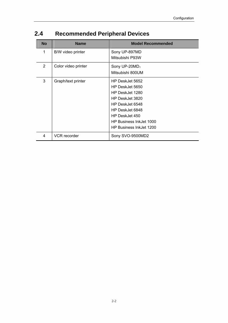
2.4 Recommended Peripheral Devices No Name Model Recommended
1 B/W video printer Sony UP-897MD Mitsubishi P93W
2 Color video printer Sony UP-20MD; Mitsubishi 800UM
3 Graph/text printer HP DeskJet 5652 HP DeskJet 5650 HP DeskJet 1280 HP DeskJet 3820 HP DeskJet 6548 HP DeskJet 6848 HP DeskJet 450 HP Business InkJet 1000 HP Business InkJet 1200
4 VCR recorder Sony SVO-9500MD2
3-1
3 Performance Specifications
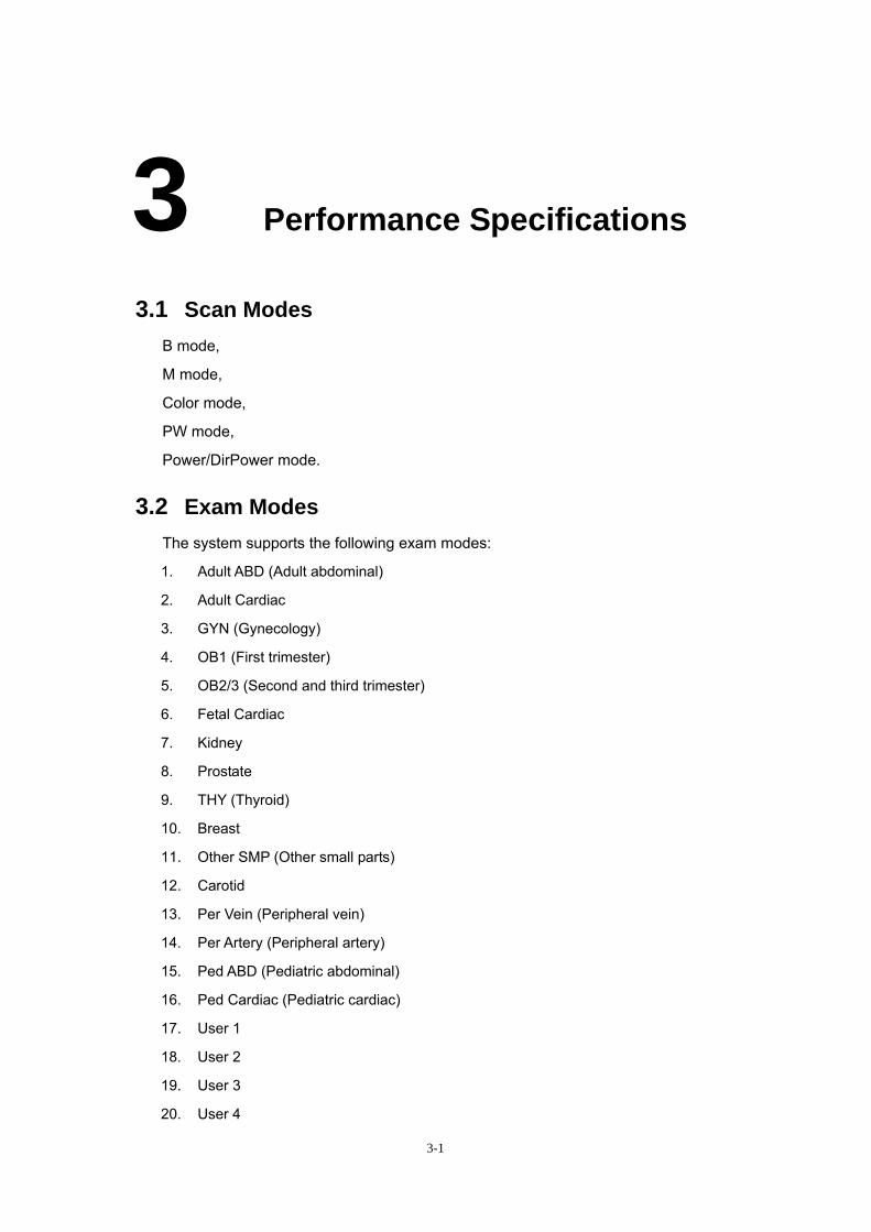
3.1 Scan Modes B mode,
M mode,
Color mode,
PW mode,
Power/DirPower mode.
3.2 Exam Modes The system supports the following exam modes:
1. Adult ABD (Adult abdominal)
2. Adult Cardiac
3. GYN (Gynecology)
4. OB1 (First trimester)
5. OB2/3 (Second and third trimester)
6. Fetal Cardiac
7. Kidney
8. Prostate
9. THY (Thyroid)
10. Breast
11. Other SMP (Other small parts)
12. Carotid
13. Per Vein (Peripheral vein)
14. Per Artery (Peripheral artery)
15. Ped ABD (Pediatric abdominal)
16. Ped Cardiac (Pediatric cardiac)
17. User 1
18. User 2
19. User 3
20. User 4

Performance Specifications
3-2
3.3 Acoustic Power The acoustic power is user-adjustable, ranging 16 levels between AP0 and AP15.
The system can display MI and TI (TIB, TIC and TIS) in real time.
3.4 Image Specifications
3.4.1 Tissue Specific Imaging (TSI) The system is configured with four TSI: general, muscle, fat and fluid.
3.4.2 Tissue Harmonic Imaging (THI) All transducers support Tissue Harmonic Imaging (THI), and harmonic frequencies
adjustable.
3.4.3 iTouch The system supports one-key optimization function (iTouch).
3.4.4 Zoom The magnification scale is 100-400%.
3.4.5 Image depth The scan depth is adjustable. Different transducers have different display depths in
terms of intended use, as follows:
High-frequency transducer: 1.4~11.8cm;
Low-frequency transducer: 4.9~26.3cm.
3.4.6 Scan control
1) TGC: 8-segment adjustment.
2) Gain
3) Scan range
4) The linear transducers support steering scan. 5) Focus number and position
6) Scan density
7) MBP (Multi-beam Parallel Imaging)
8) Contrast
9) Dynamic range
10) Edge enhancement

Performance Specifications
3-3
11) Frame average
12) M soften
13) Smooth
14) 8 IP combinations, and independent IP for THI
3.4.7 Post processing
1) Image reversal: up/down, left/right, and rotation
2) Gray rejection
3) Gray transform
4) γ correction
3.4.8 SV (Sample Volume)
1) Position of sample volume is adjustable;
2) Size of sample volume is adjustable.
3.4.9 Doppler scanning parameters
1) PRF (Pulse repetition frequency)
2) Doppler Frequency,
3) Persistence,
4) Color focus position,
5) Baseline;
6) Wall filter;
7) Colorize
8) Map (Power map and DirPower map)
3.4.10 Doppler audio
1) Doppler audio: ON/OFF;
2) The volume is adjustable without steps.
4-1
4 Preset
4.1 System Preset Field setup: time, date, language, and hospital name, etc.
Presets of image format,
Presets of measurement parameters,
Biopsy presets,
ECG-relevant presets,
Presets of peripheral devices,
And so on.
4.2 Preset of a Transducer and an Exam mode The user can preset selectable exam modes for a transducer.
The user can define an exam mode.
4.3 Preset of Image Parameters The user can preset the image parameters according to modes and transducers.
The user can preset IP and post process maps.
4.4 Preset of Measurements The user can preset the measurement packages.
The user can preset the measurement items.
4.5 Preset of Comments and Body Marks The user can preset the general comments and user-defined comments.
The user can preset the general body marks and user-defined body marks.
5-1
5 Management of Patient Files
5.1 Patient Information Basic patient information
Detailed patient information (Including OB/GYN)
5.2 iStation Supports quick storage and review of files and Cineloop sequences;
Supports review images;
Supports image demonstration (iVision);
The patient images and reports can be stored and reviewed;
The historical images and reports can be inquired.
All patient data can be edited, exported, deleted or backed up.
5.3 Graph/Text Workstation Supports color video printer, graph/text printer, and B/W video printer;
Supports immediate printing of video images;
Supports DVD and VCR recording of screen images;
Remote transfer: the system supports DICOM3.0.

6-1
6 Cine Review
6.1 Freeze The system supports the freeze status in each mode.
6.2 Image Storage The supported storage formats of image files include: BMP, JPG, FRM, CIN, AVI, and
DCM.
The system supports portable hard disk, internal hard disk, and CD-R/W backup.
6.3 Cine Review The system supports the manual and automatic Cine review of images in the B/Color
mode.
The system supports the Cine review of images in the M mode and PW mode.
The system supports the Cine review of images in the linked modes.
The system supports ECG-wave review.
7-1
7 Comments and Body Marks
7.1 Comments The system supports the entry of characters in Chinese and English.
General comments: the user can give comments by means of inputting characters or
Chinese words through the keyboard.
Automatic comments: the user can give comments by means of selecting comment
texts through the comment library.
The user can preset general comments.
The user can define comments.
7.2 Body Marks The body marks are grouped into different categories: Abdomen, Urology, OB/GYN,
Cardiology, Small Part, Blood Vessel, and Orthopaedics. The user can select body marks
or preset them in the exam modes.
The user can define a body mark.
The position of a body mark and its transducer mark are adjustable.
The user can simultaneously add body marks in multiple image windows.

8-1
8 Measurements
8.1 General Measurements
8.1.1 B mode Distance Depth Angle Area (Ellipse, Trace, Spline and Cross-line) Volume (Ellipse, Ellipse distance and Three-distance) Cross line Parallel line Tracing length (Trace, Spline) Ratio IMT B histogram B profile
8.1.2 M mode Distance Time Slope Heart rate
8.1.3 PW mode Time Heart rate Speed Acceleration Resistance index Spectrum trace
8.1.4 Color mode Distance Depth Angle

1.1 Application Measurements The system supports measurements of gynecology, obstetrics, cardiology, small
parts, urology, orthopedics and peripheral vessels.
8-2
Area (Ellipse, Trace, Spline and Cross-line) Volume (Ellipse, Ellipse distance and Three-distance) Cross line Parallel line Tracing length (Trace, Spline) Ratio IMT Color speed Color profile
8.2 Application Measurements The system supports measurements of gynecology, obstetrics, cardiology, small
parts, urology, orthopedics and peripheral vessels.
9-1
9 User Interface
9.1 Power On/Off The system has two switches for power on/off; one is the power switch and the
other is circuit breaker.
The power switch is located on the left side of the machine, and used for turning
on/off the AC power.
The circuit breaker is located on the power panel at the back of the machine.
9.2 I/O Interfaces
1) Transducer socket : 3 ;
2) ECG interface : 3-lead input interface;
3) Footswitch interface : 1;
4) USB interface : 2;
5) Video out interface : 1;
6) Video in interface : 1;
7) Audio in interface : 2;
8) Audio out interface : 2;
9) S-video in interface : 3;
10) S-video out interface : 2;
11) VGA output interface : 1;
12) VGA in interface : 1;
13) RGB in interface : 1;
14) RGB output interface : 1;
15) Ethernet interface : 2;
16) Remote control interface: 2;
17) Serial port : 2;
18) Input of the main unit power : 1;
User Interface
9-2
19) Output of the auxiliary power : 3;
20) Parallel port : 1;
21) Equipotential terminal : 1;
22) Functional grounding terminal : 1;
23) MIC interface : 1;
24) Reset : 1
9.3 Monitor The monitor is a color 15” CRT.
The monitor has the functions of elevation and rotation.
The monitor supports screen saver, and the user can set the duration and graphic of
the screen saver.

10-1
10 External Dimensions and Weight
10.1 External Dimensions (excluding optional devices) 1390mm(height)×790mm (length)×480mm (width)
10.2 Weight Approx. 132Kg

11-1
11 Environmental Conditions
11.1 Power Supply Power supply voltage: 100 to 127V~ or 220 to 240V~ Power supply frequency: 50/60Hz Power consumption: 800VA
11.2 Operating Conditions Ambient temperature : 0°C ~40°C Relative humidity : 30% ~ 85% (no condensation) Atmospheric pressure : 700 hPa ~ 1060 hPa
11.3 Conditions of Storage and Transport Ambient temperature : -20°C ~ 55°C Relative humidity : 30% ~ 95% (no condensation) Atmospheric pressure : 700 hPa ~ 1060 hPa

12-1
12 Safety Classification
(1) According to the type of protection against electric shock:
CLASS I EQUIPMENT
(2) According to the degree of protection against electric shock: TYPE-BF EQUIPMENT
(3) According to the degree of protection against harmful ingress of water:
The main unit belongs to IPX0, and the transducers belong to IPX7.
(4) According to the degree of safety of application in the presence of a FLAMMABLE ANESTHETIC MIXTURE WITH AIR or WITH OXYGEN OR NITROUS OXIDE: EQUIPMENT not suitable for use in the presence of a FLAMMABLE ANESTHETIC MIXTURE WITH AIR or WITH OXYGEN OR NITROUS OXIDE
(5) According to the mode of operation:
CONTINUOUS OPERATION
(6) According to the installation and use:
MOBILE EQUIPMENT


P/N:2105-20-40465(V1.0)