dbios anatomy models & skeletons -...
TRANSCRIPT
Dbios
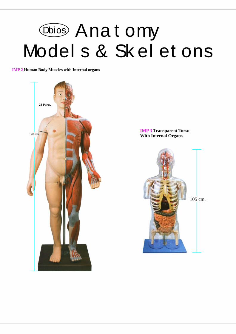
IMP 2 Human Body Muscles with Internal organs
170 cm.
28 Parts.
105 cm.
IMP 3 Transparent TorsoWith Internal Organs
AnatomyModels & Skeletons

Anatomy Musculature
86cm.
IMP 1 Male Muscle FigureFeatures: 27 Parts.1/2 life size., Height 86cm., Width 49cm. Thickness 38cm.
IMP 1
IMP 347 Muscles of Arm with Main Vessels and NervesFeatures: 6 Parts.Length 85cm.Width 23cm.Height 18 cm. N.W. : 8.7 Kg.
IMP 347
IMP 349 Muscles of the LegLength 870cm.Width 12cm. Height 17cm.
80cm.
IMP 349
20cm.
IMP 350
IMP 350 Muscles of FootFeatures: 9Parts. Height 20cm.Width 9cm. Thickness 33 cm.
IMP 348 Muscles of HandHeight 22.5cm.Width 13.5cm. Thickness 5.5cm.
IMP 348
Separated into 5 Parts.
1

Anatomy DbiosTorso
2
IMP 302 Dual-Sex Torso with Head and Open Back
333 positions are displayed.
Features: This model consists of interchangeable male & female genitalias, abdomen covers, open back muscles of head and open & neck internal organ, skull and brain, etc. It also shows the anatomical structure of head, neck, trunk, some part of upper limbs, muscles, thorax, celiac cavity.
Height 80cm. Width 37cm.
80cm.
IMP 302
IMP 301 Torso with Head and Interchangeable Male and Female Genitals
The model separated 20 parts.
Height 90cm.* Width 40cm.* Thickness 25cm.Material : Advanced PVC. 90cm.
IMP 301
23cm.
IMP 352 Block Skin
Enlarged 70 times.
Height 23cm.Width 22cm. Thickness 11cm. N.W.: 1.3 Kg.
IMP 352
23.5cm.
IMP 351
IMP 351 Skin Section
Enlarged approx. 70 times.Separated into 5 Parts.
Height 22.5cm.Width 23.5cm. Thickness 4cm.
IMP 329B A Brain Lobe Model
4 Parts.
IMP 329C Functional Localization
4 Parts.
IMP 329A Functional Zones of Cerebral Cortex
4 Parts.

3
Anatomy HeadDbios
38cm.
IMP 330 Half Head with Blood VesselsHeight 38cm. Width 20cm. Thickness 9cm.
IMP 331 Head with Muscles
Separated into 10 parts.
Height 30cm. Width 18cm. Thickness 21cm.
30cm.
IMP 330A Median section of the Head
IMP 332 Head & Neck with Blood Vessels, Nerves and BrainSeparated into 19 parts.Height 36cm. Width 26cm. Thickness 12cm.
18cm.
IMP-330B Human Half Head, Brain and Neck Region Brain (Sagittal Section)
28cm.
IMP-331A Human Head With Brain
23cm.
Separated into 9 parts.
IMP - 331B Muscular Head with Brain
16cm.
Separated into 5 parts.

Anatomy DbiosBrain/Nervous
4
13.5cm.
IMP 328 BrainFeatures : 8 Parts.Height 13.5cm. Width 12.5cm. Thickness 16cm.
IMP 328A Human BrainFeatures : 8 Parts.
IMP 329 Brain with Arteries
Separated into 9 parts.
Height 15cm*. Width 14cm*. Thickness 16cm.
15cm. 26cm.
IMP 322 Nervous System1/2 life size. Height 81cm, Width 28cm. Thickness 3.5cm.
81cm.
IMP 322A Spinal Cord in Spinal Canal
75cm.
IMP 321 Neuron
Enlarged 2500 times.
Features: 2 PartsHeight 38.5cm, Width 28cm. Thickness 13cm.
38.5cm

5
Anatomy Nervous/EyeDbios
IMP 327 Nerves and Blood-vessels in the Facial SkullHeight 21cm*, Width 32cm*. Thickness 19cm. N.W.: 1Kg.
80cm.
IMP 285 MICROanatomy EyeThe model illustrates the microscopc structure of the retina with choroid and sclera. The left block-like, layered side to the model side shows the complete structure of the retina including the supplying vascular layer and parts of the sclera from a light microscopic view. The right part of the model is a sectional enlargement. It shows the microscope structure of the photoreceptors and the cells of the pigmented layer25x23x18.5 cm ; 1.2 kg
IMP 285 A r
are
olion
clec
t
19cm.
IMP 314 Eyeball
Separated into 7 parts.
Height 19cm, Width 15cm. Thickness 13.5cm.Enlarged approx. 5 times located horizontally
IMP 315 Eyeball With Part of Orbit
Separated into 8 parts,
Height 19cm, Width 18cm. Thickness 22cm.Enlarged approx. 3 times.
19cm.
IMP 316 Topography of the Orbit
Separated into 9 parts,
Enlarged Height 30cm, Width 38.5cm. Thickness 26.5cm.
30cm.
IMP 354 Sympathetic Nervous System.
IMP 316 A Working Eye
30cm.
45cm.

Anatomy DbiosNasal/Ear
6
lThe outer nasal cartilageslThe nasal, maxillary, frontal and sphenoidal sinuses lThe opened maxillary sinus when the zygomatic arch
is removed the following structures are shown in a median section:
lThe nasal cavity, lined with mucosa, with the nasal conchae (removable)
lThe arteries of the mucous membranelThe olfactory nerveslThe innervation of the lateral wall of the nasal cavity,
the nasal conchae and the roof of mouth (palate)
IMP 261
IMP261 Nose with Paranasal Sinuses, 4-partThis model illustrates the structure of the nose with the paranasal sinuses in the upper right half of a face in 1.5 fold enlargement. The following structure can be seen from the out side, differentiated by colour (also visible through the removable transparent skin);
32cm.
IMP 317 Anatomical Ear
Separated into 6 parts,
Height 21cm, Width 32cm. Thickness 13cm.Enlarged approx. 3 times.
IMP 320 Ear Labyrinth
Separated into 2 parts,
Height 16cm, Width 23cm. Thickness 9cm.Enlarged approx. 18 times. 16cm.
IMP 319 Labyrinth with Auditory Ossicles and Tympanic MembraneHeight 18cm. Width 17cm. Thickness 19cm.Enlarged approx. 4 times.
18cm.
IMP 318 Anatomical Ear with PinnaHeight 41cm, Width 44cm. Thickness 26cm.Enlarged approx. 4 times.
41cm.
Enlarged approx. 1.5 times
Ask for Rare Histology Microscopic Slides
15cm.
IMP 319A Auditory Ossicles

Anatomy DbiosTeeth
8
IMP 218 Enlarge model showing the clinical and prosthetic rehabilitation of a carious premolar tooth with an apical granuloma.
IMP 218
IMP 206 Dental Disease, magnified 2 times, 21 partsThis model is based on a lifelike illustration of a lower jaw with 16 removable teeth of an adult magnified two times.One half of the model shows eight healthy teeth and healthy gums. The other half of the model shows the following dental diseases : * Dental plaque* Dental calculus (tartar)* Periodontitis* Inflammation of the root* Fissure, approximal and smooth surface caries. One part of the front bone section can be removedto view the roots, vessels and nerves. Two molarsare sectioned along the length to show the inside ofthe tooth.Delivered on a base. 25.5 x 18.5 x 18cm; 0.6 kg.
IMP 206
IMP 207 Human mouth model made from transparent resin on a full scale, assembled on an occlusor, higlighting various pathologiesDemonstrative of caries, tartar, gingivae retractions and granuloms.
IMP 207
IMP 208 Full scale transparent resin model of human mouth highlighting abscesses round the apex, cysts and teeth incorporated in the bone.Indispensable as a guide for surgical practice on a manikin
IMP 208

9
Anatomy TeethDbios
IMP 212 deal occlusion I
IMP 212
IMP 213class I, overcrowding
Malocclusion
IMP 213
IMP 214class II, division-I
Malocclusion
IMP214
IMP 215class II, division-II
Malocclusion
IMP 215
IMP 216class III
Malocclusion
IMP 216
IMP 217inexcact bite
Malocclusion
IMP 217
IMP 250 6-part Giant Molar with Dental Caries, 15 times life-size, This model depicts an upper triple-root molar and
. it features a longitudinal section through the crown, two roots and the pulp cavity. Contains removable pulp and three tooth inserts with different stages of advanced caries. On stand.
separates into 6 parts
IMP 250
IMP 262 Lower Jaw of a 12-year-old
Enlarged in 3 times.
Features: 11-14 years Old.Size: Length 17cm.Width 29cm.Thickness 6cm. N.W.: 2.6 Kg.
IMP 262
IMP 263 Molar with Caries
Separated into 3 Parts.
Size: Height 11cm.Width 15cm.Thickness 4cm. N.W.: 0.9 Kg.
IMP 263

7
Anatomy TeethDbios
IMP 201 Model for conservative practice, with 32 screw- fixed teeth and rigid gums model featuring the four wisdom teeth
IMP 201
IMP 202 Model with gum gingivae for practice on manikin and with channelled transparent- root teeth.
at the same time conservative practice and endodontics practice.
The teeth, fixed with a screw, are made with root canals containing red wax, in order to enable
Moreover they feature a two-layer crown (composite on the surface and acrylic in depth) and a transparent root. The model is applied to the articulator by using the adapting supports.
IMP 202
IMP 203 Model of jaw and mandible, with articulation and in white resin, with transparent gum gingivae and rooted channelled removable teeth, transparent at root levelFull scale
IMP 203
IMP 204 Human mouth model with pathologies mounted on an occlucer on a full scaleEquipped with gum gingivae and removable teeth, it shows caries and parodonthopathies, and is vary accurately made, especially as far as carious teeth, tartar, pouches and gingivae retraction are concerned, with more or less intense colours according to the inflammation degree.
IMP 204

Anatomy DbiosTeeth
10
IMP 266 Pathologic TeethFeatures: 2 Parts.Size: Height 11cm.Width11cm.Thickness 4cm.
IMP 264 A Set of Five Teeth ModelsEnlarged approx. 8 times.
lower incisor lower canine
lower molar with one root
lower incisor with two root
Lower first upper molar with three root
IMP 265 Development of A Set of TeethThis model consists of 4 parts, which shows the development from temporary teeth to the germination of the permanent teeth, demonstrating the teeth at the ages of less than 6 months, 2 years, 5 to 7 years, 17 to 26 years.
IMP 267 Case of Series Model of teeth AffectionFeatures: 12 Parts.Size: Height 25cm.,Width 32cm.,Thickness 2cm.
IMP 26911-part
Half Lower Jaw, 3 times full-size,
The front section of bone and all the teeth are removable , one incisor is longitudinally sectioned. Nerves, blood vessels, the sublingual and submandibular glands are shown.
IMP 269
IMP 268 Case of Teeth OdontopathiesFeatures: 25 kinds of teeth.Size: Height 25cm.Width 32cm.Thickness 2cm.
Ask for Anatomy Dissection Videos

11
Anatomy Teeth/LarynxDbios
IMP 270 Transparent resin anatomical mandible with seven teeth and three free sellae, on a support, suitable for implantation demonstrations and to highlight the internal vascular patterns.
IMP 270IMP 303 Cavities of Nose, Mouth and Pharynx with Larynx 10 parts, such as tongue, rodent muscles, etc. Skull, nose, mouth, larynx, pharynx, are also demonstrated in sagittal sections. 133 position are displayed.Enlarged 2 times.Height 42cm. Width 25cm.Thickness 20.5cm.
42cm.IMP 304 Larynx with Tongue This model consists of 5 parts, and shows the anatomical structure of laryngeal cartilages, laryngeal muscles, laryngeal cavity . Total in life size and 55 positions are displayed.Height 18 cm.Width 6.5cm.thickness 10 cm.
18cm.
IMP 305 Larynx Material: Advanced PVCHeight 14 cm. Width 6.5cm.Thickness 6 cm, N.W.: 0.16 Kg. The model shows cartilaginous Median section skeleton, ligamentous apparatus, muscles, relief of membrane, thryroid gland. Separated into 2 parts,
14cm.
IMP 306 Functional Model of Larynx Features: This model shows the anatomical structure of laryngeal cartilages, laryngeal commissure, laryngeal muscles and laryngeal cavity. It demon strates the movement of oricoarytenoid joint with the simulation of open and close glottis, and the epiglottis cartilages can work to close the outlet of larynx. 24 positions are displayed. Height 30 cm.Width 15 cm.Thickness 14 cm.Material: Advanced PVC and painted with imported paint.
30cm.
IMP 326 Interior model of Mouth, Nose, Pharynx and Larynx with Blood VesselsHeight 21cm, Width 3.5cm. Thickness 14.5cm. N.W.: 0.7Kg.
21cm.

Anatomy DbiosNasal/Ear
12
IMP 307 Transparent LungMaterial: Advanced PVC The magnified model shows 10 segments of bronchi of right and left lung.
IMP 309 Heart, lung and LarynxFeatures: The consists of several parts as larynx, lung, heart and blood vessels. It shows the anatomical structure of the heart; lung as well as the viscus in thorax in follow: left and right lungs, bifurcation of the trachea, esophageal hiatus with aortic hiatus in the diaphragm. 70 positions are displayed.Height 36 cm. Width 20cm. ,Thickness 10 cm.
36cm.
27cm.
IMP 308 Heart, lung and Bronchial Tree
Separated into 4 parts.
Height 27cm. Width 27cm. Thickness 19cm.The model shows in 2/3 life size the tracheobronchial system, heart, major vessels. Pulmonary vessels. IMP 312 Anatomical Heart Model
Height 25cm, Width 23cm. ,Thickness 30cm.
Separated into 3 parts,
25cm.
IMP 313 Anatomical Heart ModelHeight 15cm, Width 12cm. Thickness 18cm.Enlarged approx. 1.5 times.
15cm.
IMP 307A Human Lung
35cm.
IMP 307B Lung Normal
35cm.
IMP 312A Thoracic Cavity Model
43cm.

13
Anatomy DigestiveDbios
IMP 310 Circulatory SystemSize: 1/2 life size. Height 82cm, Width 29cm. Thickness 5cm. 82cm.
76cm.
IMP 311 Lymphatic SystemHeight 76cm, Width 49cm. Thickness 8.5cm.
IMP287 MICRO anatomy Artery and VeinThe model shows a medium-sized muscular artery with two adjacent veins from the antebrachial area with adjoining fat tissue and muscle enlarged 14 times. The model illustrates the reciprocal anatomical relationship of artery and vein and the basic functional techniques of the venous valves ("valve function" and"muscle pump"). The left vein and the middle artery are fenestrated in the upper anterior segment, revealing the various layers of the wall structure in a cross and longitudinal section and in top view. The right vein is opened throughout in the anterior segment, revealing the orifice of a feeder vein and two venous valves, i.e. "flap valves" formed by a duplication of the tunica intima. On the rear of the model, the relief of two veins is shown to illustrate the functional aspect of the venous valves. Supplied on base.26x19x18.5 cm; 0.9 kg
rare
Aoll
ct on
ce
i
IMP 287
IMP 309A Human Endocrine System
IMP 309A
45cm.
50cm.
IMP 309C Digestive System
75cm.
IMP 309B Stomach on Stand
21cm.
IMP 309D Stomach wall

Anatomy DbiosKidney
14
e A r
ar
colc ion
let
IMP 286
IMP 286 MICRO anatomy Digestive SystemThe model illustrations the structure of the fine tissues of four characteristic sections of the digestive system:l Oesophagus l Stomachl Small intesting l Large intestineThe front of the model, from top to bottom, shows a magnified view in histological section of the individual sections of the digestive system and their fine tissue structures. On the back of the model, highly magnified views of didactically interesting areas of each of the digestive system sections shown on the front are emphasized.29.5x26x18.5 cm; 1.5 kg
IMP 282 MICRO anatomy LiverThis 2-part model shows a highly magnified diagrammatic view of a section of the liver. It illustrates the structure components of the liver in two different enlargements. The left part of the model shows a section of the liver that comprises several liver lobules. The right part of the model is a highly magnified view of the sectioned liver lobule on the left.15x26x18.5 cm: 0.7 kg
IMP282
e A r
ar
olion
clec
t
IMP 333 Liver and Gall BladderHeight 19cm. Width 26.5cm. Thickness 12cm.
19cm.
IMP 333
Ask for Rare Histology
Microscopic Slides
IMP 337A Human Excretory System
IMP 337A
40cm.
IMP 333C Kidney on Stand
40cm.
IMP433Liver, Pancreas, Duodenum

15
Anatomy KidneyDbios
IMP 334B Inguinal Hernia This natural-sized, graphic model shows the anatomical structures of a male groin with an indirect inguinal hernia, opened in layers Two diagrammatic illustrations on the base allow for a comparison of direct and indirect hernia Mounted on base.
l
l
l
l
l
Section of renal cortex and renal medullaWedge-shaped section of a kidney lobe with a diagrammatic depiction of three nephrons with Henle's loop and didactic/ diagrammatic illustration of the vascular supplyDiagrammatic illustration of a nephron with a short Henle's loop and didactic/diagrammatic illustration of the vascular supplyDiagrammatic illustration of an opened renal corpuscle with nephron and light-microscopic tranverse sections of the proximal, attenuated and distal segments of a renal tubuleDiagrammatic / didactic illustration of an opened renal corpuscle Mounted on a base.23.5x25.5x19 cm ; 1.3 kg
IMP 284 MICRO anatomy KidneyThis extremely detailed model shows the morphologic/functional units of the kidney greatly magnified. Six model zones illustrate the following fine-tissue structure that serve the production or urine :
Longitudinal section of a kidneyl
IMP 284 rare
Acol
ection
l
IMP 332A Spleen, Pancreas & Duodenum.
20cm.
IMP 333A Kidney L.S.
35cm.
IMP 333B Kidney, Nephron & Corpuscle.
30cm.
68cm.
35cm.
35cm

Anatomy DbiosGenital/Gynae
16
IMP 335 Male Genital OrgansHeight 21cm. Width 18cm. Thickness 20cm. 21cm.
IMP 337 Female Genital OrgansFeatures: 4 Parts.Height 12cm. Width 14cm. Thickness 15cm.
21cm.
IMP 334 Median Section of Male PelvisFeatures: 4 Parts.Height 27cm. Width 10cm. Thickness 25cm.
27cm.
AM 80A Imported-Enlarged Model of PlacentaHeight 50cm.Width 52cm. Thickness 2cm.
25cm.
IMP 336 Median Section of Female Pelvis
Separated into 2 parts.
Height 25cm. Width 10.5cm. Thickness 26cm.
IMP 339 Skeleton of Male Pelvis Length 24cm.Width 17cm. Height 15cm.
IMP 340 Skeleton of Female Pelvis Length 20cm.Width 15cm. Height 19cm.
AM 80B Human Placenta.
30cm.
30cm.
IMP 337AFemale Inner Genital Organ
32cm.
40cm.
HT. 17cm
IMP 336A Female Pelvis
24cm.

17
Anatomy PregnancyDbios
AM 79B Imported- Human development up to the Embryo at the End of the 1st MonthFeatures: 13 Parts. Height 45cm.Width 55cm. Thickness 5cm.
AM 79A Imported- Fertilization and Development of Human Ovum Up to the 3rd MonthFeatures: The total sets consists of 16 different collected in a showcase with removable cover. It shows the development from ovum to the fetus in third month. Height 38cm. ,Width 65cm. ,Thickness 6.5cm.
AM 83B Imported-Series Demonstration Model of PregnancyHeight 36cm.Width 18cm. Thickness 36cm.
First Month Second Month Third Month
Fourth Month Fifth Month Sixth Month
Seventh Month Twin fetus of Second Month
AM 53A Imported-Fetal Circulatory System Size: Natural size.
IMP 353 Pelvis with Uterus in Ninth Month of Pregnancy Features: 2 Parts.Height 36cm.Width 18cm. Thickness 36cm.
36cm.
Human EmbryoIMP 353C
22cm.

Anatomy DbiosPregnancy
18
AM 70A IMP- Mammary Gland in Resting Period
Separated into 2 patrs.
Height 14.5cm. Width 12.5cm. Thickness 11cm.
AM 70B IMP- Mammary Gland in Lactation Height 14.5cm.Width 12.5cm. Thickness 11cm.
Separated into 2 patrs.
AM 70C IMP- Ovary ModelFeatures: 5 Parts.Height 21.5cm.Width 36.5cm. Thickness 11cm.
21.5cm.
AM 84A Imported-Demonstration Model of Childbirth This model consists of uterus, fetus, placenta. It shows the procedures of the delivery.
IMP 353B Stages of Pregnancy Uterus with Fetus.
IMP 353B
AM 84A
IMP 280A Stages of Fertilization.
40cm.
40cm.
IMP 380B Fertilization process
27cm.
85cm.

27
Anatomy JointsDbios
IMP 341 Section through the Shoulder JointHeight 18cm.Width 17cm. Thickness 2cm.
IMP 343 Section through the Knee JointHeight 14cm.Width 15cm. Thickness 2cm.
IMP 342 Section through the Elbow JointHeight 14cm.Width 10cm. Thickness 2cm.
IMP 344 Section through the Hip JointHeight 22cm.Width 17cm. Thickness 2cm.
IMP 345 Section through the HandHeight 24cm.Width 15cm. Thickness 2cm.
IMP 346 Section through a Normal FootHeight 23cm.Width 17cm. Thickness 2cm.
IMP 289 Micro Anatomy Muscle FibreThe model illustrates a section of a skeleton muscle fibre and its neuromuscular end plate magnified approx. 10.000 times. The muscle fibre is the basic element of the diagonally striped skeletal muscle. 23.5x26x18.5 cm.;1.1kg.

Anatomy DbiosJoints
28E-mail us : [email protected]
A-87 Imported
ADULT ARM BONE WITH
THREE JOINTS & LIGAMENTS
A-86 Imported
ADULT LEG BONE WITH
THREE JOINTS & LIGAMENTS
A-92 Imported FUNCTIONAL
ANKLE JOINT (RIGHT)
A-91 Imported FUNCTIONAL
WRIST JOINT (RIGHT)
A-90 Imported FUNCTIONAL
ELBOW JOINT (RIGHT)
A-93 Imported FUNCTIONAL
KNEE JOINT (RIGHT)
A-89 Imported FUNCTIONAL
HIP JOINT (RIGHT)
IMP. 131 IMPORTED VERTEBRAL COLUMN WITH
FEMORAL HEADS & STAND :
Same as IMP. 130 with
addition of movable femoral
heads size 32" Tall
IMP.130
Size : 29" tall.
IMPORTED VERTEBRAL COLUMN WITH STAND:
Flexible spine, with pelvis,
occipital bone, vertebral artery
and dorsal herniated disc
between the 3rd and 4th lumbar
vertebrae.
A-88 Imported FUNCTIONAL
SHOULDER JOINT (RIGHT)
All Skeleton Parts are Near to Original
Hand Phone - 09729010431(Harish Mahajan)
Skull With Mandible 3parts Ribs (L&R) Sternum Humerus, Radius, Ulna (Left)Humerus, Radius, Ulna (Right)Pelvis (L&R)Sacrum Femur, Tibia, Fibula (Left)Femur, Tibia, Fibula (Right)Vertebral Column (Disarticulated)Cervical Vertebrae (Set of 7) Thoracic Vertebrae (Set of 12)Lumbar Vertebrae (Set of 5)Hand Disarticulated (L&R)Foot Disarticulated (L&R)

29
Anatomy VertebrateDbios
IMP 275 Flexible Spine with Female Pelvis 74 cm; 1.8kg. without stand.
IMP275
IMP 276 Flexible Spine with Male Pelvis74 cm; 1.8kg. without stand.
IMP 276
IMP 278 Stages of Disc Prolapse and Vertebral DegenerationThe model can be disassembled into its components (vertebral bodies, intervertebral discs and spinal nerves).22 cm; 0.5 kg
IMP 278
IMP 323 Spinal Cord with Nerve BranchesFeatures: 2 Parts, Enlarged 5 times.
7.5cm.
IMP 324 Fifth Cervical VertebraHeight 7.5cm, Width 32cm. Thickness 24cm. N.W.: 1.8Kg.Enlarged approx. 7 times.
9cm.
IMP 325 Thoracic Vertabra (TH II) with Spinal CordHeight 9cm, Width 18.5cm*. Thickness 18.5cm. N.W.: 1.5Kg.
IMP 104A Adult SkullIMP 258A Infant Skull
Charts for Histology,Physiology, Embryology,
Diseases
Must Ask

Anatomy DbiosSukll
30
IMP 255 Deluxe Demonstration Skull, 10 - partsThis replica of the human skull is of an exceptional quality. The skull cap is removable and the base of skull is mid-sagitally divided. The frontal sinus, perpendicular lamina and vomer are fitted with flaps which can be opened to view the lateral nose wall and sphenoidal sinus. On the left half, the temporal bone can be removed and folded up in the area of the tympanic membrane. Maxilla and mandible are opened to reveal the alveolar nerves. On the right side the temporal bone is opened to reveal the sigmoid sinus, the facial nerve canal and the semicircular ducts. Additional flaps are located at the maxillary sinus and the right half of the mandible, so that the dental roots of the premolars and molars of the lower jaw can also be viewed. The natural occlusion and the individual removal and replacement of the each tooth also make this skull especially interesting for dentists.
IMP 255
IMP 256 System Skull - Bony Skull, 6-partThis version represents a complete midsagitally sectioned skull. It can be disassembled into both halves of the skullcap and the base of skull, the nasal septum and the complete mandible. To demonstrate masticatory movement, the lower jaw is mounted flexibly. An excellent skull to study the bony structure and the complicated anatomy of the human skull.
IMP 256
IMP 253B Skull Dissected IMP 253B Coulored Skull Dissected

31
Anatomy SkullDbios
IMP 257 Classic Skull, 3-partAll featuring the following, unless otherwise specified : lHigh quality original casts of real human skullslHand-made from hard, unbreakable plasticlHighly accurate representation of the fissures, foramina,
processes, sutures etc.lDisassemble into at least 3 parts for detailed studies
IMP 258 Classic Skull, with opened Lower jaw, 3-partThis dental skull with opened mandible exposes the dental roots with vessels and nerves. The cranial bones, bone components, fissures, formamina and other structure and numbered. The cranial sutures are shown in colour, as are the meningeal vessels and venous sinuses.
IMP 258
IMP 257
IMP 260 Comparative Study of Skulls, Set of 5
Fetal
Child Young
AdultOld
IMP 260A Comparative Study of lower jaw, Set of 5
For Image visit our website

Anatomy DbiosSkull
32
IMP281 MICRO anatomy Bone Structure This extremely detailed model depicts a three-dimensional section of a lamellar bone, showing the typical structure of a tubular bone enlarged 80 times. Various planers are shown in cross and longitudinal section through all levels of the bone, as well as a2-plane section through the inner structure of the bone marrow. The typical elements of a lamellar bone are easily identified and help to understand its structure and function with the characteristic osteons , also referred to as Haversian systems. This model allows a graphics illustration of the interplay of the individual components, such as spongy and compact substance, endosteum, cortical substance, osteocytes, Volkmann and Haversian canals. Supplied on base.26x19x14.5 cm; 0.8 kg IMP 281
e A r
ar
colec
ionl
t
IMP 251
IMP 251 Skull model decomposable into ten parts with anatomical parts highlighted through colour.
Different views
IMP 252 Skull decomposable into ten parts with anatomical parts highlighted through colours.Human skull resin model, full scale, decomposable into 10 parts : it shows the muscular insertion areas - Chromatically highlighted - of the masticatory system, as well as some vascular and nerve terminations. The numbers refer to a more analytical description listed separately.
IMP 252
Different views

33
Anatomy SkeletonsDbios
IMP. 128
IMP. 128 IMPORTED MR. THRIFTY SKELETON 85 cm. : An economical teaching
skeleton with user-friendly personality that will encourage children to learn the names of
the bones, with details to satisfy students, doctors or anyone interested in the human
skeleton. Key card and a heavy metal stand included. Removable calvarium.
J U S T L I K E O R I G I N A L
AM103.
IMP. 126A
IMP126A Fetus Skeleton Model
AM103. DISARTICULATED HUMAN SKELETON WITH SKULLLife-size, disarticulated adult skeleton includes 3-part skull, Hand
and foot are completely disarticulated

Anatomy DbiosSkeleton
34
IMP.- 126
IMPORTED BUDGET BUCKY SKELETON : This economical , l i fe-s ize articualted adult plasdetailtic skelton is ideal for teaching the basics of anatomy when intricate textural nuances of the bone are not required. The arms and legs are removable for study. Features nerve branches, vertebral artery, and herniated lumbar disk. Skull included movable jaw, cut calvarium, suture lines, and 3 removable lower teeth. Mounted on stand. Complete with dust cover and skeltal system chart.
IMP.- 127
IMP.- 126
IMP.- 127 IMPORTED MR. SUPERSKELTON : The world's most complete skeleton, featuring joint ligaments, a flexible spine with nerve endings and full indication of muscle origins and insertions painted on one-half of the body. The skull dissects into three pieceswith a removable calvarium and lower jaw. The mounting of the asial skeleton allows natural movement of the skull on the 1st and 2nd cervical vertebrae. Teh flexible spine includes all spinal nerves and teh vertebral artery. A special mouting of the ribe cage prevents sagging. Teh skull, left arm and leg are fully detachable. Muscle insertions in red and origins in blue are painted on teh left side of the skelton. The iliocostal and longissimus muscles are painted differently for clearer understanding. Teh right side has th eligaments of the shoulde, elbow, hip and knee reproduced in a lifelike manner. The left side of the skeleton is provided with numerical notation of the major boens, bone parts, fissures and foramen. The skeleton is mounted on a mobile stand. Dust cover, key card and skeleton system chart are included.
Male Female
IMP.- 126 F