david chiluiza1,2, sneha krishna1 2,3 - jbc.org chiluiza1,2, sneha krishna1, valérie a....
TRANSCRIPT

Mutant TRPC6 activates Erk1/2
1
Gain-of-function mutations in transient receptor potential C6 (TRPC6) activate extracellular-signal-
regulated kinases Erk1/2*
David Chiluiza1,2
, Sneha Krishna1, Valérie A. Schumacher
2,3 and Johannes Schlöndorff
1,2
1From the Division of Nephrology, Department of Medicine, Beth Israel Deaconess Medical Center,
Boston, MA 02215
2Harvard Medical School, Boston, MA 02215
3Department of Medicine, Boston Children’s Hospital, Boston, MA 02215.
*Running title: Mutant TRPC6 activates Erk1/2
To whom correspondence should be addressed: Johannes Schlondorff, Division of Nephrology, RN304B,
99 Brookline Ave, Boston, MA 02215, USA, Tel.:(617) 667-0508; Fax: (617) 667-0495; E-mail:
Keywords: Calcium, Epidermal growth factor receptor, Focal segmental glomerulosclerosis, cAMP-
dependent protein kinase.
Background: Signaling events affected by
disease-associated mutations in TRPC6 are
poorly defined.
Results: Expression of mutant TRPC6 induces
Erk1/2 activation via both cell autonomous and
non-autonomous mechanisms.
Conclusion: Mutant TRPC6 activates complex
signaling pathways that lead to the release of
paracrine factors activating Erk.
Significance: Understanding the signaling
pathways downstream of gain-of-function
TRPC6 is crucial for understanding TRPC6-
mediated biology and pathology.
SUMMARY
Gain-of-function mutations in the
canonical transient receptor potential 6
(TRPC6) gene are a cause of autosomal
dominant focal segmental glomerulosclerosis
(FSGS4). The mechanisms whereby
abnormal TRPC6 activity results in
proteinuria remain unknown. The Erk1/2
MAP kinases are activated in glomeruli and
podocytes in several proteinuric disease
models. We therefore examined whether
FSGS-associated mutations in TRPC6 result
in activation of these kinases. In 293T cells
and cultured podocytes, overexpression of
gain-of-function TRPC6 mutants resulted in
increased Erk1/2 phosphorylation, an effect
dependent upon channel function.
Pharmacologic inhibitor studies implicated
several signaling mediators, including
calmodulin and calcineurin, supporting the
importance of TRPC6-mediated calcium
influx in this process. Through media
transfer experiments, we uncovered two
distinct mechanisms for Erk activation by
mutant TRPC6, a cell autonomous, EGFR-
independent mechanism and a cell non-
autonomous mechanism involving
metalloprotease-mediated release of a
presumed EGFR ligand. The inhibitors KN-
92 and H89 were able to block both pathways
in mutant TRPC6 expressing cells, as well as
the prolonged elevation of intracellular
calcium levels upon carbachol stimulation
seen in these cells. However, these effects
appear independent of their effects on
CaMKII and PKA, respectively.
Phosphorylation of T70, S282 and Y31/Y285
were not necessary for Erk activation by
mutant TRPC6, though a phosphomimetic
TRPC6 S282E mutant was capable of Erk
activation. Taken together, these results
identify two pathways downstream of mutant
http://www.jbc.org/cgi/doi/10.1074/jbc.M113.463059The latest version is at JBC Papers in Press. Published on May 3, 2013 as Manuscript M113.463059
Copyright 2013 by The American Society for Biochemistry and Molecular Biology, Inc.
by guest on May 27, 2018
http://ww
w.jbc.org/
Dow
nloaded from

Mutant TRPC6 activates Erk1/2
2
TRPC6 leading to Erk activation that may
play a role in the development of FSGS.
The canonical transient receptor potential 6
(TRPC6) channel is a member of the transient
receptor potential (TRP) superfamily of six
transmembrane cation channels (1,2). TRPC
subunits assemble to form functional homo-
and/or heterotetramers, with TRPC6 capable of
oligomerizing with TRPC1, 3, 7, and itself (3).
Several stimuli have been shown to activate the
channel, including Gq coupled receptors (4,5)
and receptor tyrosine kinases (6), as well as
direct binding of diacyl glycerol (7). In the case
of receptor-mediated activation, regulated
exocytosis of the channel is at least partially
responsible for increasing channel activity
(5,6,8). Membrane deformation has also been
reported to activate the channel directly (9),
though this mechanism remains controversial
(10).
Mutations in the TRPC6 gene are a cause of
autosomal dominant focal segmental
glomerulosclerosis (FSGS), a progressive kidney
disease frequently leading to end-stage kidney
disease (11,12). The majority of mutations
identified to date have been shown to be gain-of-
function, increasing whole cell current
amplitudes and/or decreasing channel
inactivation (11-14). The mutations map to
various segments of the protein, predominantly
the ankyrin repeats in the amino-terminal
cytoplasmic domain and a putative coiled-coil
sequence in the carboxyl-terminal cytoplasmic
domain. The mechanism(s) whereby these
mutations enhance channel activity remains
unclear, though there are suggestions that some
mutations may alter cell surface localization (12)
and binding to nephrin (8). Additional evidence
suggests TRPC6 plays an important role in
podocyte function. TRPC6 is upregulated in
several acquired proteinuric kidney diseases
(15), transgenic overexpression of wild-type or
mutant TRPC6 in podocytes induces a mild
glomerular phenotype (16), and TRPC6-
deficient mice show some resistance to
angiotensin II induced proteinuria (17). TRPC6
has also been implicated in regulating blood
pressure (18), pulmonary vascular tone and
permeability (19-24), cardiac hypertrophy (25-
27), myofibroblast differentiation and associated
wound healing (28,29), neuronal growth cone
guidance (30) and platelet function (31). Why
mutations in TRPC6 lead specifically to kidney
disease is unknown.
Several signaling pathways have been shown
to be regulated downstream of TRPC6. For
example, in cardiac myoblasts and fibroblasts
TRPC6 activates the calcineurin-NFAT pathway
(25-27), while in podocytes, FSGS-associated
mutations in TRPC6 increase basal activation of
this pathway (32). Calcineurin activation in
podocytes has, in turn, been implicated in
mediating proteinuria in several murine models
(33), while forced expression of constitutively
active NFATc1 in podocytes rapidly leads to
podocyte effacement and proteinuria (34). In
addition to activating calcineurin, TRPC6
activates the small GTPase Rho, while
antagonizing the activation of Rac, in podocytes
and other cell types (35-37). Excess Rho
activity in podocytes induces proteinuria and
glomerular dysfunction (38,39). The importance
of calcineurin and Rho activation in mediating
TRPC6-induced glomerular pathology has not
been established in vivo and awaits investigation
in an animal model. Finally, TRPC6 has been
shown to be involved in activation of Erk
downstream of PDGF signaling in dopaminergic
neurons (40).
The Raf-Mek-Erk pathway is one of three
canonical mitogen activated protein kinase
signaling pathways, and plays a central role in
multiple essential cellular functions, including
proliferation, survival and differentiation (41-
44). The pathway represents one of the first
kinase cascades identified, with Raf
phosphorylating Mek, which in turn
phosphorylates, and thereby activates, Erk1 and
2. Numerous proteins and signaling pathways
can influence this cascade through input at its
various levels, with activation of Raf by the
small GTPase Ras representing the classical
pathway. Similarly, the output can be wildly
disparate, with over 160 substrates for Erk1 and
2 identified (45-47) capable of affecting diverse
responses (46). The mechanisms whereby the
pathway is able to execute specific responses to
different stimuli remains incompletely
understood, though the use of scaffolding
proteins and distinct pools of kinases in different
subcellular locales appears critical (48).
by guest on May 27, 2018
http://ww
w.jbc.org/
Dow
nloaded from
Mutant TRPC6 activates Erk1/2
3
Several lines of evidence suggest that Erk
activation occurs in glomeruli, and in podocytes
and mesangial cells specifically, in response to
pathological stresses. Erk phosphorylation, and
thereby activation, is increased in glomeruli and
podocytes in response to angiotensin II infusion
(49-52), salt-sensitive hypertension (53),
puromycin aminoglycoside nephropathy (54-58),
passive Heymann nephritis (59,60) rapidly
progressive glomerulonephritis (61-64), and
diabetic nephropathy (65-67). Phospho-Erk
levels in mesangial cells have been correlated
with glomerular damage in IgA nephropathy
(68). In podocytes, Erk has been implicated in
mediating actin reorganization, cell migration
and proliferation (61), and acting as a both pro-
apoptotic (56,69-71) and anti-apoptotic/pro-
survival (58,60,72-74) signal.
In the present study, we demonstrate that
disease-associated, gain-of-function mutations in
TRPC6 induce Erk activation in several cell
types, including cultured podocytes, with at least
two distinct pathways involved, one cell
autonomous and one non-autonomous. Both
pathways, and calcium influx downstream of
mutant TRPC6 activation, are inhibited by the
small molecules KN-92 and H89, though the
targets of their actions remain unclear. Finally,
several reported phosphorylation sites on
TRPC6 are not required for the action of mutant
TRPC6, though a phosphomimetic S282E
mutation is sufficient to induce Erk activation.
These results identify Erk as a potential mediator
of focal segmental glomerulosclerosis induced
by TRPC6 mutations.
EXPERIMENTAL PROCEDURES
Reagents and plasmids - Pharmacologic
reagents were purchased from the following
vendors: carbachol, cyclosporine A, forskolin,
H89, KN-92, KN-93, tetracycline and Y-27632
from Sigma-Aldrich; U0126 and Tyrphostin AG
1478 from Cell Signaling Biotechnology;
bisindolylmaleimide I/Gö6850, Compound C,
Gö6976, Gö6983, GW 5074, STO-609, and W-7
from EMD Biosciences; Batimastat, Marimastat
and GW6001 from Santa Cruz Biotechnology,
conjugated, cell permeable C3 transferase (Rho
Inhibitor I) from Cytoskeleton, Inc.
Antibodies used in this study were from the
following commercial sources: Cell Signaling
Technology (rabbit phospho-Erk1/2, #4370;
rabbit Erk1/2, #4695; mouse Erk1/2, #9107;
rabbit and mouse anti-HA antibodies, #3724 and
2367; phospho-(Ser/Thr) PKA substrate
antibody, #9621; HRP conjugated anti-rabbit
and anti-mouse IgG secondary antibodies, #7074
and 7076), Santa Cruz Biotechnology (rabbit
PKA C, sc-903), Sigma-Aldrich (FLAG M2,
unconjugated, F3165, FITC-labeled, F4049, and
conjugated to agarose, A2220; HA mouse
monoclonal conjugated to agarose, A2095).
Fluorescence-conjugated secondary antibodies
and Phalloidin were from Jackson
ImmunoResearch and Invitrogen, respectively.
The plasmids pcDNA3.1 and
pcDNA4/TO/myc-His B (Invitrogen) containing
the full-length human TRPC6 ORF with an
amino-terminal FLAG tag sequence and various
FSGS-associated mutations, as well as the
dominant-negative pore mutation changing
amino acids LFW to AAA, have been described
previously (32). HA tagged wild-type and
R895C mutant TRPC6 were generated by
standard PCR based strategies and cloned into
pcDNA3/myc-His A (Invitrogen).
Phosphorylation site mutations were introduced
using Invitrogen’s Quikchange XL kit following
the manufacturer’s protocols, and confirmed by
sequencing. Plasmids for expressing GFP fused
to the CaMKII inhibitory peptide (GFP-AC3-I)
or scrambled control peptide (GFP-AC3-C) were
provided by Dr. M. Anderson (75). FLAG-
tagged wild-type and kinase mutant (K73M)
PKA Cα expression constructs were kindly
provided by Drs. Murshid and Calderwood (76).
Cell culture - M1R cells, generated by
expressing the M1 acetylcholine receptor in T-
REx-293 cells (Invitrogen), as well as stable
clones of M1R cells expressing tetracycline
inducible wild-type FLAG-TRPC6 or mutant
FLAG-TRPC6 R895C, were maintained as
previously described (32). TRPC6 expression
was induced by growing cells in media
supplemented with 2 μg/ml tetracycline or 0.1
g/ml doxycycline for 18-48 hours prior to use.
HEK 293T cells were obtained from
GeneHunter and maintained in DMEM with 4.5
g/L glucose and supplemented with 10% fetal
bovine serum (Atlanta Biologicals) and
antibiotics. A conditionally immortalized
murine podocyte cell line was generated from a
by guest on May 27, 2018
http://ww
w.jbc.org/
Dow
nloaded from

Mutant TRPC6 activates Erk1/2
4
temperature-sensitive SV40 large T antigen
transgenic mouse (Charles River, St. Louis) as
previously reported (77) and was maintained on
collagen I coated tissue culture plates in RPMI
media with 10% FBS and interferon (20
units/ml; Sigma Aldrich) at 33º C; cells were
shifted to media without interferon and cultured
at 37º C to induce differentiation. Human
conditionally immortalized podocytes were
provided by Dr. M. Saleem and cultured as
recommended (78).
Transient transfections of M1R cell
derivatives and 293T cells were performed using
FuGENE6 or XtremeGENE 9 (Roche) following
the manufacturer’s protocol. Cells were
processed 18-48 hours after transfection.
Murine podocytes were transfected 48-72 hours
after differentiation using an Amaxa
Nucleofector apparatus with program T-13 and
the Basic Nucleofector Kit for Primary
Mammalian Epithelial Cells (Lonza) and
replated onto collagen I coated plates for 24
hours prior to analysis.
The siRNA experiments were performed by
reverse transfection using Lipofectamine
RNAiMax and pools of either control siRNA or
siRNA targeting PKA C (Santa Cruz
Biotechnology). 36-48 hours after transfection,
cells were treated with or without doxycycline
overnight to induced TRPC6 expression.
For inhibitor studies, cells were first treated
with or without tetracycline (2μg/ml) for 16-24
hours. The media was then replaced with
complete media containing the indicated
inhibitors at the following concentrations: 25
μM W-7, 5 μM KN-92 and -93, 5 μM STO-609,
10 μM cyclosporine A (CsA), 40 μM Compound
C, 1 μM Gö6976, 1 μM Gö6983, 5 μM U0126,
10 μM GW5074, 10 M H89, 100 nM
Tyrphostin AG 1478, 20 M Batimastat, 20 M
Marimastat, 20 M GW6001, 1 g/ml C3
transferase, 10 M Y-27632. Equal final
concentrations of DMSO carrier were present
under all conditions. After three hours of
treatment with inhibitors, cells were lysed and
processed for analysis.
Immunoprecipitation and immunoblotting -
Cells were rinsed once in phosphate buffered
saline with calcium and phosphate, and lysed in
TBS with 1% Triton X-100 (50 mM Tris, pH
7.4, 150 mM NaCl, 1%(v/v) Triton X-100),
modified RIPA lysis buffer (50 mM Tris, pH
8.0, 150 mM NaCl, 1% NP-40, 0.5% sodium
deoxycholate), or low-salt lysis buffer (50 mM
Tris, pH 8.0, 100 mM NaCl, 5 mM EDTA, 1%
(v/v) NP-40, 0.5% (w/v) sodium deoxycholate),
all supplemented with cOmplete Protease
Inhibitor Cocktail and PhosStop (Roche).
Lysates were cleared by centrifugation at
17,000xg for 15 minutes at 4ºC. An aliquot was
mixed with 4x SDS-sample loading buffer with
beta-mercaptoethanol, incubated at 95ºC for 5
minutes and used to assay protein expression by
Western blot.
For immunoprecipitation, cleared lysates
were incubated with 15 µl of FLAG M2 or anti-
HA agarose slurry (Sigma-Aldrich), and
incubated with constant agitation at 4ºC for 2-3
hours. Immunoprecipitated complexes were
washed three times with lysis buffer and eluted
off of the beads by boiling in SDS-sample
loading buffer.
Cell lysates and immunoprecipitated
materials were separated by SDS-PAGE using
TGX gels (Bio-Rad) and transferred to PVDF
membrane (Bio-Rad). The membrane was
blocked with 5% bovine serum albumin (BSA)
in TBST (TBS with 0.05% Tween-20) for 1 hour
at room temperature, followed by incubation
with primary antibody in 5% BSA TBST using
dilutions recommended by the supplier. After
three washes in TBST, blots were incubated with
the appropriate secondary antibody conjugated
to HRP (Cell Signaling Technology) in TBST at
room temperature, followed by detection with
SuperSignal West Dura chemiluminescent
substrate (Pierce). Images were obtained using
autoradiography film or with a FluorChem Q
imager (Cell Biosciences).
In vitro kinase assay - HA-TRPC6 was
immunoprecipitated from cell lysates with anti-
HA antibody conjugated agarose and washed
extensively with lysis buffer followed by 1x
PKA phosphorylation buffer (50 mM Tris-HCl,
10mM MgCl2, pH 7.5). The sample was divided
into two sets and incubated in PKA buffer with
200 μM ATP with or without 2,500 units of
PKA catalytic subunit (New England Biolabs) at
30°C for one hour. The samples were then
washed twice with lysis buffer, incubated in 2x
by guest on May 27, 2018
http://ww
w.jbc.org/
Dow
nloaded from

Mutant TRPC6 activates Erk1/2
5
sample loading buffer at 95°C, and processed for
Western blot analysis.
Imunnofluorescence - Cells were cultured on
12-mm poly-D-lysine-covered coverslips (BD
354086) in 24-well plates until 60-70%
confluent, followed by the addition of
tetracycline were indicated. Twenty-four hours
later, coverslips were passed to clean 24-well
plates and cells were washed twice with cold
PBS, fixed in 2% formaldehyde and
permeabilized with 0.3% Triton X-100. Cells
were blocked for 1 hour in PBS with 1% BSA
and 0.5% Tween 20, then incubated sequentially
with primary and secondary antibodies in the
same blocking solution for 1 hour each. Nuclei
were counterstained with 2 μg/ml Hoechst
solution and coverslips mounted on glass slides
with Prolong Gold antifade reagent (Invitrogen).
Digital images were captured using an RT Slider
Diagnostic instrument digital camera connected
to an Olympus BX60 microscope.
Calcium Imaging - Intracellular calcium
concentration analysis of cell populations was
performed using Fura-2 AM (Life Technologies)
following the manufacturer’s general
recommendations. Briefly, cells were cultured
in 96-well black, clear bottom plates (Costar)
until 70% densities, and then treated with or
without Tetracycline (2 µg/ml) for 24 hours to
induce FLAG-TRPC6 WT or R895C expression.
Cells were dye loaded with 2 µM Fura-2 in 50 µl
calcium uptake (CU) buffer (1X Hank’s
balanced salt solution, 20 mM HEPES, 2.5 mM
probenecid) for 30 min at 37ºC in a cell
incubator. Media was then exchanged for 75 µl
CU buffer alone for another 30 min. For
inhibitor studies, inhibitors or DMSO carrier
was present during this incubation.
Fura-2 fluorescence signal capture was
performed in a FlexStation 3 Multi-Mode
microplate reader (Molecular Devices)
calibrated to 37ºC using SoftMax Pro v5.4
software. Fluorescence signals were
sequentially measured with excitation at 340 nm
and 380 nm, and emission at 510 nm to obtain a
340/380 ratio as a surrogate for intracellular
calcium concentrations. Data was collected
every 5 seconds. Baseline ratio was obtained for
one minute, followed by addition of 25 µl of CU
buffer with or without carbachol, and signal
captured every 5 seconds for another 5 minutes.
Transactivation assay - Tetracycline-
inducible FLAG-TRPC6 cells were grown with
or without tetracycline or doxycycline for 18-24
hours. Conditioned media from the cells was
harvested and cleared of cell debris by
centrifugation. Various inhibitors were either
present during the entire media conditioning
step, or added after media harvest, as indicated.
HEK 293T cells were exposed to either
unconditioned or conditioned media as indicated
followed by lysis and Western blot analysis.
Statistical Analysis - Western blot band
intensities were quantified using AlphaView Q
SA v.3.2.2 (Cell Biosciences) and normalized to
control conditions. Statistical analysis was
performed using GraphPad Prism 5.
RESULTS
Gain-of-function TRPC6 mutations activate
Erk. We examined the levels of phosphorylated,
activated Erk1/2 in stable cell lines expressing
either no TRPC6 (M1R), FLAG-tagged wild-
type TRPC6 (WT) or FLAG-tagged TRPC6
harboring the FSGS-associated R895C mutation
(R895C) under a tetracycline inducible promoter
(Fig. 1A, B). Inducing expression of wild-type
TRPC6 led to a small increase in basal Erk
activation, though this did not reach statistical
significance. Similarly, transient transfection of
wild-type TRPC6 had only a minimal effect on
phospho-Erk levels (Fig. 1C, and Figure 9,
below). In contrast, expression of the R895C
mutant TRPC6 induced a pronounced increase in
phospho-Erk levels (Fig. 1A-C). The reason for
the higher basal level of phospho-Erk seen in the
R895C inducible cell line even in the absence of
tetracycline is unclear, but may be due to low
level expression of the channel under uninduced
conditions (as ascertained by RT-PCR and
immunoprecipitation/Western blotting; data not
shown).
To ascertain whether the effect on Erk
phosphorylation was specific to the R895C
mutant or a common effect of disease-
associated, gain-of-function mutations, we
transiently transfected the parental cell line
(M1R cells) with either wild-type or one of
several mutant forms of TRPC6 (Fig. 1C).
Expression of three well-established gain-of-
function disease mutants located on opposite
ends of the protein (P112Q, R895C and E897K)
by guest on May 27, 2018
http://ww
w.jbc.org/
Dow
nloaded from

Mutant TRPC6 activates Erk1/2
6
led to an increase in Erk phosphorylation.
Introduction of a dominant negative pore
mutation (LFW to AAA, (3)) into the R895C
mutant protein (R895C DN) abolished Erk1/2
activation, suggesting that the channel activity of
TRPC6 is necessary for Erk activation.
To ascertain whether mutant TRPC6 had a
similar effect in a more disease-relevant cell
line, we ascertained phospho-Erk levels in
murine podocytes transiently transfected with
either control vector or HA-tagged wild-type or
R895C mutant TRPC6 (Fig. 1D, E). Again,
expression of mutant TRPC6 led to a significant
increase in phospho-Erk levels compared to both
control and wild-type TRPC6 expressing cells.
The more modest relative increase in phospho-
Erk levels in podocytes relative to 293T cells
may in part be due to the higher basal levels of
phospho-Erk in these cells (Fig. 5J and data not
shown).
We further examined Erk activation in
TRPC6 R895C expressing M1R cells by
immunocytochemistry (Fig. 1F). In the absence
of tetracycline, minimal phospho-Erk signal was
seen in cells. Expressing wild-type TRPC6 did
not appreciably increase phospho-Erk levels. In
contrast, when mutant TRPC6 expression was
induced, the majority of cells showed an
increase in phospho-Erk signal, though there
was significant heterogeneity in phospho-Erk
levels.
Pharmacologic inhibition of mutant TRPC6
mediated Erk activation. To begin to address
what pathways might be involved in Erk1/2
activation downstream of mutant TRPC6, we
examined the effect of various pharmacologic
inhibitors (Table 1) on this process (Fig. 2A, B).
As expected, U0126, an inhibitor of MEK, the
kinase directly responsible for Erk
phosphorylation, effectively abrogated the
increase in phospho-Erk due to TRPC6 R895C.
The calmodulin inhibitor W-7, the calcium-
calmodulin-dependent protein kinase II
(CaMKII) inhibitor KN-93, and the calcineurin
inhibitor cyclosporine A (CsA), but not the
calcium-calmodulin-dependent protein kinase
kinase (CaMKK) inhibitor STO-609, all
abolished the effect of R895C TRPC6 on Erk
activation. Compound C, an inhibitor of AMPK
(though with additional off-target effects (79))
also blocked Erk activation. Interestingly, the
broad PKC inhibitor Gö6983 (and
Bisindolylmaleimide I/Gö6850; data not shown),
but not Gö6976, which targets the calcium-
dependent, conventional PKCs, also inhibited
Erk activation, suggesting that one of the
atypical PKCs may be required for this effect of
TRPC6. Finally, GW5074, which inhibits c-Raf
in vitro (80), was not an effective inhibitor of
mutant TRPC6-mediated Erk activation.
The effect of KN-93 on phospho-Erk levels
suggested that CaMKII might be involved in this
signaling cascade. To further address this
possibility, we first tested the effect of KN-92, a
derivative of KN-93 without inhibitory activity
against CaMKII (81), on phospho-Erk levels
(Fig. 3A, B). Surprisingly, we found that KN-92
was almost as effective at decreasing phospho-
Erk levels in cells expressing TRPC6 R895C as
KN-93. To further examine the potential role of
CaMKII, we made use of a CaMKII inhibitory
peptide fused to GFP (GFP-AC3-I) (75).
Expressing either this inhibitory peptide, or a
scrambled control peptide (GFP-AC3-C),
together with TRPC6 R895C did not appreciably
alter phospho-Erk levels (Fig. 3C), further
arguing against a role for CaMKII in mediating
Erk activation by mutant TRPC6, and suggesting
that the effect of KN-93 on this process is
mediated, at least in part, independently of its
effect on CaMKII activity.
Previous studies have suggested that PKA
and cGMP-dependent protein kinase (PKG)
might play a role in limiting the activity of
TRPC6 channel (82-87). We therefore
examined whether inhibition of PKA through
H89 might further enhance Erk phosphorylation
in cells expressing TRPC6 R895C. Contrary to
our expectations, treating cells with H89
abolished the increase in phospho-Erk in cells
expressing mutant TRPC6 (Fig. 4A). To
investigate the potential role of PKA in
mediating Erk activation by mutant TRPC6, we
co-transfected TRPC6 R895C together with
either wild-type or dominant negative, kinase
dead PKA C (Fig. 4B). Unlike the effect of
H89, dominant negative PKA C did not
diminish phospho-Erk levels, though
overexpression of wild-type PKA C did
increase phospho-Erk levels both in the presence
and absence of TRPC6. To further address the
discrepancy between the effect of H89 and
by guest on May 27, 2018
http://ww
w.jbc.org/
Dow
nloaded from

Mutant TRPC6 activates Erk1/2
7
dominant negative PKA, we knocked down the
expression of endogenous PKA C. PKA
levels, normalized to actin, were significantly
decreased after transfecting PKA siRNA
compared to scrambled control siRNA
(PKA/actin ratio: 46.8±2.3% vs. 100±6.1%,
respectively; p<0.0001 by paired t test, n=10).
Upon PKA knockdown, phospho-Erk levels
were modestly increased in R895C TRPC6
expressing cells compared to control siRNA
treated cells (Fig. 4C, D). The increase in
phospho-Erk levels seen upon PKA knockdown
in the absence of TRPC6 induction did not reach
statistical significance. Taken together, these
results suggest that H89 is able to inhibit mutant
TRPC6-mediated increases in phospho-Erk
levels independently of its effect on PKA. We
cannot exclude that PKA may, in fact, have a
small inhibitory effect on mutant TRPC6.
H89 has been reported to inhibit kinases in
addition to PKA, including Rho-dependent
protein kinase II (ROCK-II) (88). TRPC6 is
known to activate RhoA (35,37). We therefore
examined whether the Rho-ROCK pathway was
necessary for mutant TRPC6 mediated Erk
activation. Neither membrane permeable C3
transferase (a Rho inhibitor) nor the ROCK
inhibitor Y-27632 significantly altered phospho-
Erk levels (Fig. 4E, F). These results suggest
that mutant TRPC6 mediated Erk activation
occurs independent of PKA or the Rho-ROCK
pathway.
Mutant TRPC6 induces paracrine Erk
activation via release of EGFR ligand. Calcium
activated signals play a role in G-protein
coupled receptor autocrine and paracrine
signaling pathways, including those activating
Erk through tyrosine kinase receptor
transactivation (reviewed in (89,90)).
Furthermore, TRPC1 has been shown to be
involved in the amplification of the EGFR
signaling cascade, including the activation of
Erk (91). We therefore investigated whether a
paracrine signaling pathway might be involved
in Erk activation due to TRPC6 R895C
expression (Fig. 5). HEK293T cells were
exposed to unconditioned media or media
conditioned by uninduced or induced TRPC6
wild-type or R895C cells. Exposure to
conditioned media from cells expressing TRPC6
R895C induced Erk activation within 5 minutes,
while media from uninduced cells, cells
expressing wild-type TRPC6, or control media
had no effect (Fig. 5A). A time course analysis
revealed peak activation of Erk after 5 minutes
of exposure to conditioned media from TRPC6
R895C cells, with a lower level of Erk activation
persisting through sixty minutes (Fig. 5B). We
next examined whether TRPC6-mediated Erk
activation was dependent upon EGFR kinase
activity. Treating TRPC6 R895C expressing
cells with Tyrphostin AG 1478, an EGFR (92)
and ErbB4 (93) kinase inhibitor, failed to
decrease phospho-Erk levels (Fig. 5C). In
contrast, adding Tyrphostin AG 1478 to TRPC6
R895C conditioned media inhibited Erk
activation in 293T cells exposed to the media
(Fig. 5D), suggesting that distinct signaling
pathways are involved in cell autonomous and
non-autonomous Erk activation by mutant
TRPC6.
As metalloprotease mediated shedding of
EGF family growth factors is a frequent
mechanism involved in transactivation (89,94),
we next examined the ability of metalloprotease
inhibitors to affect Erk activation. Incubating
FLAG-TRPC6 R895C expressing cells with the
metalloprotease inhibitor Batimastat did not alter
Erk activation in these cells (Fig. 5E), but the
conditioned media generated from these cells
failed to activate Erk in 293T cells (Fig. 5F).
Adding Batimastat after conditioning did not
alter Erk activation in the 293T cells (Fig. 5G).
Similar results were obtained with two
additional metalloprotease inhibitors,
Marimastat and GM6001 (data not shown).
Together these results strongly suggest that a
metalloprotease dependent signaling step is
required in TRPC6 R895C expressing cells to
allow efficient media conditioning.
We also examined the ability of KN-92,
KN-93 and H89 to affect paracrine Erk
activation. Consistent with a model in which
these inhibitors block early signaling
downstream of mutant channel, media
conditioned in the presence of these inhibitors
did not activate Erk in 293T cells (Fig. 5H),
while adding inhibitors to pre-conditioned media
did not prevent Erk activation (Fig. 5I).
Cultured human podocytes were also
exposed to cultured media from various TRPC6
expressing 293T cell lines (Fig. 5J). Similar to
by guest on May 27, 2018
http://ww
w.jbc.org/
Dow
nloaded from

Mutant TRPC6 activates Erk1/2
8
results with 293T cells, phospho-Erk levels were
increased only by media conditioned by TRPC6
R895C expressing cells. The time course of
activation was slightly different, with higher
phospho-Erk levels seen at 15 minutes compared
to 5 minutes. In addition, the relative increase in
Erk phosphorylation was lower than in 293T
cells, due to higher basal phospho-Erk levels in
podocytes compared to 293T cells (compare Fig.
5J, second and fourth lanes from the right).
In aggregate, these results suggest that
mutant TRPC6 can lead to Erk activation
through at least two distinct mechanisms: 1) a
cell autonomous mechanism independent of
metalloproteases and EGFR, and 2) a non-
autonomous process which involves the
metalloprotease dependent release of a soluble
factor which signals through a Tyrphostin AG
1478 sensitive receptor kinase.
TRPC6 R895C mediated calcium elevations
are inhibited by H89 and KN-92. The ability of
KN-92 and H89 to inhibit both cell autonomous
Erk activation in TRPC6 R895C expressing cells
and release of paracrine signaling factors
suggests that they are acting relatively proximal
to channel activation and resulting calcium
influx. We therefore addressed whether these
inhibitors might interfere with TRPC6 mediated
calcium influx.
As measured by the ratiometric fluorescent
dye Fura-2 (Fig. 6A), we found that in the
presence of extracellular calcium, uninduced
cells had similar basal fluorescence ratios
measured at 30 seconds (Fig. 6A, B). Cells
expressing TRPC6 R895C had, on average, a
higher basal calcium level, though this
difference was variable and not seen consistently
in all experiments (e.g. compare basal levels in
Fig. 6A and C), nor in our previously published
findings (32). In the absence of extracellular
calcium (-Ca; Fig. 6A), all cells had a similar
basal fluorescence ratio that was significantly
lower than in the presence of calcium (p<0.0001,
one-way ANOVA with Tukey’s multiple
comparison test, n=8).
Stimulating cells with carbachol (at time 60
seconds, Fig. 6A) led to a rapid rise in
intracellular calcium in all cells in the presence
or absence of extracellular calcium. Peak
340/380 ratios (measured at 70 seconds) in the
presence of extracellular calcium were
significantly higher than in the absence of
extracellular calcium (p<0.0001, one-way
ANOVA with Bonferroni’s multiple comparison
test; n=8). Inducing wild-type or mutant TRPC6
expression did not significantly alter peak ratios
among the groups of cells treated with
extracellular calcium or among the groups
treated without extracellular calcium. In the
absence of extracellular calcium, intracellular
calcium levels rapidly returned back to baseline.
In contrast, in the presence of extracellular
calcium, the TRPC6 R895C expressing cells
showed a slower and smaller drop in the
fluorescence ratio over the next several minutes
(p<0.0001, one-way ANOVA with Tukey’s
multiple comparison test, compared to all other
groups at 210 seconds; n=8). These results are
consistent with previously published
electrophysiological data suggesting that this
mutation leads to increased current amplitudes
(11) and suggests a possible effect on time-
dependent channel inactivation similar to that
seen with other FSGS-associated mutations (13).
We next addressed whether KN-92 and H89
have any effect on TRPC6 R895C mediated
calcium influx (Fig. 6C). Both inhibitors
prevented the prolonged elevation in the Fura-2
fluorescence ratio seen with expression of
TRPC6 R895C (p<0.01 by one-way ANOVA
comparing (+Tet)(DMSO) versus all other
groups at 210 seconds). The apparent blunting
of the initial peak rise in the fluorescence ratio
upon carbachol stimulation in cells treated with
H89 or KN-92 did not reach statistical
significance. Taken together, these data suggest
that the ability of both inhibitors to block Erk
activation by mutant TRPC6 may be due to their
ability to blunt calcium influx mediated by
mutant TRPC6 activation. Our data do not
allow us to differentiate between a direct effect
of these compounds on channel
activation/activity and an indirect effect on
calcium influx (such as through reversal of the
sodium-calcium exchanger (95,96)).
PKA C binds and phosphorylates TRPC6.
To further investigate the disparate effects of
H89 and dominant negative PKA C or PKA
siRNA on phospho-Erk levels, we examined
whether TRPC6 and PKA can interact. TRPC6
was readily co-immunoprecipitated with PKA
C when co-expressed in 293T cells (Fig. 7A).
by guest on May 27, 2018
http://ww
w.jbc.org/
Dow
nloaded from

Mutant TRPC6 activates Erk1/2
9
The ability of PKA C to co-precipitate TRPC6
was not affected by introducing the R895C
mutation into TRPC6, or by abolishing the
kinase activity of PKA C (Fig. 7B). In contrast
to mutating the kinase domain of PKA C,
treating cells with H89 did abolish the TRPC6-
PKA interaction (Fig. 7C). This result was
surprising in light of reports that H89 acts by
competing for ATP, but not substrate, binding to
PKA (97). Forskolin-treating cells to activate
adenylate cyclase did not appreciably enhance
the interaction between the two overexpressed
proteins (Fig. 7C).
We next examined whether PKA could
phosphorylate TRPC6 making use of a phospho-
PKA substrate antibody developed to
specifically recognize phospho-serine and –
threonine residues within the context of the
consensus PKA site RRXS/T (Cell Signaling
Technology). Under basal conditions, some
amount of TRPC6 phosphorylation could be
detected using this antibody (Fig. 8A). This
signal was greatly increased by treating cells
with forskolin to activate adenylated cyclase. To
confirm that these phosphorylation events were
mediated by PKA, cells were treated with H89.
The presence of this compound abolished both
the basal and forskolin-induced phospho-S/T
signals (Fig. 8A, compare lanes 1 to 3, and 2 to
4). TRPC6 contains several potential consensus
PKA phosphorylation sites, including S13, T70
and S322 (84). Phosphorylation of threonine 70
in TRPC6, and of the corresponding threonine in
TRPC3, by both PKA and PKG has been
reported (85,86,98). The role of threonine 70 as
a site for PKA phosphorylation was therefore
investigated (Fig. 8B). Both wild-type and
R895C mutant TRPC6 showed increased
phospho-S/T signal upon treating cells with
forskolin. In contrast, introducing the T70V
mutation into the R895C TRPC6 protein
completely abolished this effect of forskolin,
indicating that threonine 70 is a major TRPC6
phosphorylation site after forskolin treatment.
To ascertain whether threonine 70 is in fact a
direct target of PKA phosphorylation, we
performed in vitro kinase assays (Fig. 8C).
Incubating wild-type and R895C mutant TRPC6
with recombinant PKA increased signal using
the phospho-PKA substrate antibody. In
contrast, no phospho-serine or threonine was
detected in TRPC6 containing the T70V
mutation. While threonine 70 appears to be a
site of PKA mediated phosphorylation in vitro
and in vivo, the site was not required for binding
of PKA Cα (Fig. 8D).
Evaluation of TRPC6 phosphorylation site
mutations on Erk activation. Several
phosphorylation sites on TRPC6 have been
reported to play potential roles in regulating
channel function. The potential Erk
phosphorylation site on S282 has been reported
to be required for activating TRPC6 currents in
response to the cAMP analogue, 8-Br-cAMP
(99). Introduction of the S282A mutation into
the TRPC6 R895C construct did not
significantly alter phospho-Erk levels (Fig. 9A,
C). In addition, abolishing the PKA/PKG
phosphorylation site on T70 (86,100), or the
dual tyrosine phosphorylation sites at Y31 and
Y285 (8), failed to significantly alter Erk
activation by TRPC6 R895C (Fig. 9A, C).
However, when we introduced a
phosphomimetic S282E mutation into wild-type
TRPC6, phospho-Erk levels were elevated
comparable to that of the R895C mutation (Fig.
9B, D). Neither the S282E nor the S282A
mutation significantly altered phospho-Erk
levels in the context of the R895C mutation.
This suggests that while the S282E mutation
induces Erk activation similar to R895C and
other disease-associated mutations,
phosphorylation of the S282 residue is not
required for Erk activation in the context of the
R895C mutant.
DISCUSSION
Here we report several novel observations:
1. Gain-of-function TRPC6 mutants activate Erk
in the absence of exogenous stimuli. 2. Erk
activation by mutant TRPC6 can be mediated by
two distinct pathways, a cell autonomous
pathway resistant to metalloprotease and EGFR
inhibitors, and a cell non-autonomous pathway
sensitive to these inhibitors. 3. Two
pharmacological inhibitors, KN-92 and H89 are
able to block the prolonged elevations in
intracellular calcium concentrations mediated by
mutant TRPC6, as well as block both pathways
leading to Erk activation. 4. Dominant negative
PKA C or knockdown of endogenous PKA C
does not replicate the effect of H89, while H89
by guest on May 27, 2018
http://ww
w.jbc.org/
Dow
nloaded from

Mutant TRPC6 activates Erk1/2
10
disrupts the interaction of TRPC6 and PKA C.
5. Introducing a phosphomimetic S282E
mutation into TRPC6 induces Erk activation, but
abolishing potential phosphorylation sites at
T70, S282 and Y31/Y285 does not prevent Erk
activation by TRPC6 R895C.
While our results clearly demonstrate Erk
activation downstream of gain-of-function
TRPC6 mutants, it remains unclear whether
activation of wild-type TRPC6 is also able to
activate the two signaling pathways leading to
Erk phosphorylation. Addressing this point has
been hampered by the lack of specific TRPC6
agonists; treatment with stimuli known to
activate TRPC6, including Hyp9 (101), lead to
Erk activation even in cells not expressing
TRPC6 (D.C., unpublished observation).
However, several prior studies have reported
links between TRPC channels and Erk. A role
for TRPC channels, including TRPC6, in
activating Erk was first suggested in the context
of PDGF-mediated neuroprotection (40). More
recently, TRPC1 has been shown to augment
EGFR mediated signaling, including Erk
activation (91), while TRPC3 appears to play a
role in Erk activation in cardiac fibroblasts
(102). Our data suggest that multiple distinct
pathways may connect TRPC6 activity to Erk
activation, including a paracrine signaling
pathway likely involving an EGFR ligand. It is
interesting to speculate whether a similar
paracrine signaling mechanism is involved in the
reported augmentation of EGFR-mediated Erk
activation by TRPC1 (91).
Several lines of evidence implicate calcium
influx in mutant TRPC6-mediated Erk
activation. Specifically, pore mutations
abolishing channel activity prevent Erk
activation, inhibitors of the calcium dependent
proteins calmodulin and calcineurin block Erk
activation, and KN-92 and H89 block both Erk
activation and calcium influx downstream of
mutant TRPC6. However, while average basal
calcium levels are slightly higher in cells
expressing TRPC6 R895C, the differences are
small and not consistently seen either in this or
previous (32) studies. How might these
discrepant results be reconciled? We postulate
that in contrast to the global and simultaneous
activation of TRPC6 channels upon stimulation
with carbachol, under basal conditions there may
be stochastic activation of TRPC6 within a
subset of cells and/or localized to a small area of
a cell. These localized increases in calcium
concentration may be sufficient to induce Erk
activation, but not to always significantly alter
the average calcium concentration as measured
across the entire cell population. Consistent
with this possibility, in our prior study (32) a
small minority of TRPC6 R895C expressing
cells demonstrate elevated basal calcium levels
without significantly altering the average
calcium level across the population. Transient
calcium spikes could induce prolonged periods
of Erk activation, and therefore lead to an
increase in global phospho-Erk levels.
Confirming this hypothesis will require the
development of assays to simultaneously
monitor intracellular calcium concentrations and
Erk activity within individual cells across time.
Besides its potential role downstream of
TRPC signaling, Erk has been reported to be
required for TRPC6 activation by cAMP
analogues (99). In addition, disrupting a
putative Erk phosphorylation site at S281 in
mouse TRPC6 (corresponding to S282 in
human) prevented TRPC6 activation by cAMP,
though phosphorylation of this site was not
demonstrated (99). While we found that S282 is
not necessary for Erk activation by TRPC6
R895C, a phosphomimetic S282E mutation in
wild-type TRPC6 did induce Erk activation.
These results suggest that phosphorylation at
S282, while not necessary for TRPC6 R895C
actions, might activate signaling pathways
downstream of TRPC6. The effect of the S282E
mutation on TRPC6 channel activity and
evidence that the S282 residue is in fact
phosphorylated in vivo await further
investigation.
In aggregate, the studies focusing on the
interplay between TRPC6 and Erk suggest the
possibility of a potential feed-forward loop
involving these two proteins. Such a loop could
potentially parallel or augment the well-
established positive feedback loop involving the
TRPC6-calcineurin-NFAT pathway (25,26,103),
especially as Erk activation in our system was
inhibited by cyclosporine A.
Our data add to the conflicting reports
regarding the potential effect of PKA, its
inhibitors, and phosphorylation of T70 on
by guest on May 27, 2018
http://ww
w.jbc.org/
Dow
nloaded from

Mutant TRPC6 activates Erk1/2
11
TRPC6 activity. TRPC6 was first reported to be
phosphorylated by PKA in human platelets but
this does not appear to alter OAG-stimulated
calcium influx (104). Similarly, PKA has been
reported to inhibit TRPC5 but not TRPC6
currents when the proteins are expressed in 293T
cells (105), and activation of TRPC6 by cAMP
analogues is insensitive to H89 (99). In
contrast, in pituitary cells, non-specific cation
channels, likely including TRPC6, are involved
in PKA stimulated calcium influx (106). PKA
has been reported to phosphorylate mouse
TRPC6 at two sites, S28 and T69
(corresponding to T70 in human) leading to
channel inhibition (86,100). However, in one
study S28 was felt to be the critical inhibitory
site (100), while the other study noted that
phosphorylation of T69 was key (98). In
support of the role of T69, the same site has
been reported to be the target of inhibitory
phosphorylation by PKG (86,87). We confirmed
that PKA is capable of phosphorylating TRPC6
at T70 in vivo and in vitro, and that in vivo
phosphorylation at this site is inhibited by H89.
In our system, H89 blocked mutant TRPC6
mediated Erk activation, media conditioning and
prolonged intracellular calcium elevations after
carbachol stimulation, and also interfered with
the interaction of TRPC6 and the catalytic
subunit of PKA. Overexpression of dominant
negative, kinase dead PKA C, in contrast, did
not block Erk activation, while knockdown of
endogenous PKA modestly enhanced Erk
activation by mutant TRPC6. These results
strongly suggest that the ability of H89 to inhibit
mutant TRPC6 mediated Erk activation is
independent of its inhibition of PKA. Our
results are consistent with an inhibitory role for
PKA as well, though if there is such a role, it
does not depend upon phosphorylation of T70
on TRPC6. While our data do not fully
elucidate the role of PKA or H89 in regulating
TRPC6, they strongly caution against the use of
H89 in studying the interplay of TRPC6 and
PKA.
The ability of KN-92, initially used as a
negative control for KN-93, to inhibit mutant
TRPC6-mediated Erk activation and calcium
influx was unexpected. The effect on calcium
influx, in particular, suggests that it may be
acting either on TRPC6 channel
activation/activity directly or indirectly on
calcium influx subsequent to TRPC6 activation.
Consistent with the later possibility, KN-92 and
KN-93 have been shown to both inhibit L-type
calcium channels (107), and nimodipine-
sensitive L-type calcium channels have been
proposed to be activated in response to TRPC6-
mediated depolarization in smooth muscle cells
(108). Dedicated electrophysiological studies
will be needed to address these possibilities.
Ultimately, these studies beget the question
of whether Erk activation plays a role in the
pathogenesis of TRPC6-mediated FSGS. As
noted earlier, Erk activation is clearly a common
manifestation in multiple human glomerular
diseases and mouse models, though whether its
role is as a mediator or mitigator of disease is
uncertain. No studies addressing the effect of
Erk1/2 deletion in the kidney or podocytes on
glomerular function have been published to date.
Addressing the importance of Erk activation in
TRPC6-mediated FSGS will need to be
addressed in mouse models.
In summary, we have identified activation of
the Erk pathway via at least two distinct
pathways as a consequence of FSGS-associated
mutations in TRPC6. These results open up
novel lines of investigation, including those
focusing on a role for TRPC6 in EGFR
transactivation and paracrine signaling, and on a
role for Erk activation in TRPC6-mediated
kidney disease.
by guest on May 27, 2018
http://ww
w.jbc.org/
Dow
nloaded from

Mutant TRPC6 activates Erk1/2
12
REFERENCES
1. Clapham, D. E., Runnels, L. W., and Strubing, C. (2001) Nature reviews. Neuroscience 2, 387-
396
2. Ramsey, I. S., Delling, M., and Clapham, D. E. (2006) Annual review of physiology 68, 619-647
3. Hofmann, T., Schaefer, M., Schultz, G., and Gudermann, T. (2002) Proceedings of the National
Academy of Sciences of the United States of America 99, 7461-7466
4. Boulay, G., Zhu, X., Peyton, M., Jiang, M., Hurst, R., Stefani, E., and Birnbaumer, L. (1997) The
Journal of biological chemistry 272, 29672-29680
5. Cayouette, S., Lussier, M. P., Mathieu, E. L., Bousquet, S. M., and Boulay, G. (2004) The
Journal of biological chemistry 279, 7241-7246
6. Hisatsune, C., Kuroda, Y., Nakamura, K., Inoue, T., Nakamura, T., Michikawa, T., Mizutani, A.,
and Mikoshiba, K. (2004) The Journal of biological chemistry 279, 18887-18894
7. Hofmann, T., Obukhov, A. G., Schaefer, M., Harteneck, C., Gudermann, T., and Schultz, G.
(1999) Nature 397, 259-263
8. Kanda, S., Harita, Y., Shibagaki, Y., Sekine, T., Igarashi, T., Inoue, T., and Hattori, S. (2011)
Molecular biology of the cell 22, 1824-1835
9. Spassova, M. A., Hewavitharana, T., Xu, W., Soboloff, J., and Gill, D. L. (2006) Proceedings of
the National Academy of Sciences of the United States of America 103, 16586-16591
10. Gottlieb, P., Folgering, J., Maroto, R., Raso, A., Wood, T. G., Kurosky, A., Bowman, C., Bichet,
D., Patel, A., Sachs, F., Martinac, B., Hamill, O. P., and Honore, E. (2007) Pflugers Archiv :
European journal of physiology 455, 1097-1103
11. Reiser, J., Polu, K. R., Moller, C. C., Kenlan, P., Altintas, M. M., Wei, C., Faul, C., Herbert, S.,
Villegas, I., Avila-Casado, C., McGee, M., Sugimoto, H., Brown, D., Kalluri, R., Mundel, P.,
Smith, P. L., Clapham, D. E., and Pollak, M. R. (2005) Nature genetics 37, 739-744
12. Winn, M. P., Conlon, P. J., Lynn, K. L., Farrington, M. K., Creazzo, T., Hawkins, A. F.,
Daskalakis, N., Kwan, S. Y., Ebersviller, S., Burchette, J. L., Pericak-Vance, M. A., Howell, D.
N., Vance, J. M., and Rosenberg, P. B. (2005) Science 308, 1801-1804
13. Heeringa, S. F., Moller, C. C., Du, J., Yue, L., Hinkes, B., Chernin, G., Vlangos, C. N., Hoyer, P.
F., Reiser, J., and Hildebrandt, F. (2009) PloS one 4, e7771
14. Zhu, B., Chen, N., Wang, Z. H., Pan, X. X., Ren, H., Zhang, W., and Wang, W. M. (2009)
Mutation research 664, 84-90
15. Moller, C. C., Wei, C., Altintas, M. M., Li, J., Greka, A., Ohse, T., Pippin, J. W., Rastaldi, M. P.,
Wawersik, S., Schiavi, S., Henger, A., Kretzler, M., Shankland, S. J., and Reiser, J. (2007) J Am
Soc Nephrol 18, 29-36
16. Krall, P., Canales, C. P., Kairath, P., Carmona-Mora, P., Molina, J., Carpio, J. D., Ruiz, P.,
Mezzano, S. A., Li, J., Wei, C., Reiser, J., Young, J. I., and Walz, K. (2010) PloS one 5, e12859
17. Eckel, J., Lavin, P. J., Finch, E. A., Mukerji, N., Burch, J., Gbadegesin, R., Wu, G., Bowling, B.,
Byrd, A., Hall, G., Sparks, M., Zhang, Z. S., Homstad, A., Barisoni, L., Birbaumer, L.,
Rosenberg, P., and Winn, M. P. (2011) J Am Soc Nephrol 22, 526-535
18. Dietrich, A., Mederos, Y. S. M., Gollasch, M., Gross, V., Storch, U., Dubrovska, G., Obst, M.,
Yildirim, E., Salanova, B., Kalwa, H., Essin, K., Pinkenburg, O., Luft, F. C., Gudermann, T., and
Birnbaumer, L. (2005) Molecular and cellular biology 25, 6980-6989
19. Tauseef, M., Knezevic, N., Chava, K. R., Smith, M., Sukriti, S., Gianaris, N., Obukhov, A. G.,
Vogel, S. M., Schraufnagel, D. E., Dietrich, A., Birnbaumer, L., Malik, A. B., and Mehta, D.
(2012) The Journal of experimental medicine 209, 1953-1968
20. Weissmann, N., Dietrich, A., Fuchs, B., Kalwa, H., Ay, M., Dumitrascu, R., Olschewski, A.,
Storch, U., Mederos y Schnitzler, M., Ghofrani, H. A., Schermuly, R. T., Pinkenburg, O., Seeger,
W., Grimminger, F., and Gudermann, T. (2006) Proceedings of the National Academy of Sciences
of the United States of America 103, 19093-19098
by guest on May 27, 2018
http://ww
w.jbc.org/
Dow
nloaded from

Mutant TRPC6 activates Erk1/2
13
21. Weissmann, N., Sydykov, A., Kalwa, H., Storch, U., Fuchs, B., Mederos y Schnitzler, M.,
Brandes, R. P., Grimminger, F., Meissner, M., Freichel, M., Offermanns, S., Veit, F., Pak, O.,
Krause, K. H., Schermuly, R. T., Brewer, A. C., Schmidt, H. H., Seeger, W., Shah, A. M.,
Gudermann, T., Ghofrani, H. A., and Dietrich, A. (2012) Nature communications 3, 649
22. Samapati, R., Yang, Y., Yin, J., Stoerger, C., Arenz, C., Dietrich, A., Gudermann, T., Adam, D.,
Wu, S., Freichel, M., Flockerzi, V., Uhlig, S., and Kuebler, W. M. (2012) American journal of
respiratory and critical care medicine 185, 160-170
23. Loot, A. E., and Fleming, I. (2011) Journal of cardiovascular pharmacology 57, 140-147
24. Fuchs, B., Rupp, M., Ghofrani, H. A., Schermuly, R. T., Seeger, W., Grimminger, F.,
Gudermann, T., Dietrich, A., and Weissmann, N. (2011) Respiratory research 12, 20
25. Kuwahara, K., Wang, Y., McAnally, J., Richardson, J. A., Bassel-Duby, R., Hill, J. A., and
Olson, E. N. (2006) The Journal of clinical investigation 116, 3114-3126
26. Onohara, N., Nishida, M., Inoue, R., Kobayashi, H., Sumimoto, H., Sato, Y., Mori, Y., Nagao, T.,
and Kurose, H. (2006) The EMBO journal 25, 5305-5316
27. Wu, X., Eder, P., Chang, B., and Molkentin, J. D. (2010) Proceedings of the National Academy of
Sciences of the United States of America 107, 7000-7005
28. Davis, J., Burr, A. R., Davis, G. F., Birnbaumer, L., and Molkentin, J. D. (2012) Developmental
cell 23, 705-715
29. Nishida, M., Onohara, N., Sato, Y., Suda, R., Ogushi, M., Tanabe, S., Inoue, R., Mori, Y., and
Kurose, H. (2007) The Journal of biological chemistry 282, 23117-23128
30. Li, Y., Jia, Y. C., Cui, K., Li, N., Zheng, Z. Y., Wang, Y. Z., and Yuan, X. B. (2005) Nature 434,
894-898
31. Paez Espinosa, E. V., Murad, J. P., Ting, H. J., and Khasawneh, F. T. (2012) Biochemical and
biophysical research communications 417, 853-856
32. Schlondorff, J., Del Camino, D., Carrasquillo, R., Lacey, V., and Pollak, M. R. (2009) American
journal of physiology 296, C558-569
33. Faul, C., Donnelly, M., Merscher-Gomez, S., Chang, Y. H., Franz, S., Delfgaauw, J., Chang, J.
M., Choi, H. Y., Campbell, K. N., Kim, K., Reiser, J., and Mundel, P. (2008) Nature medicine 14,
931-938
34. Wang, Y., Jarad, G., Tripathi, P., Pan, M., Cunningham, J., Martin, D. R., Liapis, H., Miner, J.
H., and Chen, F. (2010) J Am Soc Nephrol 21, 1657-1666
35. Tian, D., Jacobo, S. M., Billing, D., Rozkalne, A., Gage, S. D., Anagnostou, T., Pavenstadt, H.,
Hsu, H. H., Schlondorff, J., Ramos, A., and Greka, A. (2010) Science signaling 3, ra77
36. Singh, I., Knezevic, N., Ahmmed, G. U., Kini, V., Malik, A. B., and Mehta, D. (2007) The
Journal of biological chemistry 282, 7833-7843
37. Jiang, L., Ding, J., Tsai, H., Li, L., Feng, Q., Miao, J., and Fan, Q. (2011) Exp Biol Med
(Maywood) 236, 184-193
38. Wang, L., Ellis, M. J., Gomez, J. A., Eisner, W., Fennell, W., Howell, D. N., Ruiz, P., Fields, T.
A., and Spurney, R. F. (2012) Kidney international 81, 1075-1085
39. Zhu, L., Jiang, R., Aoudjit, L., Jones, N., and Takano, T. (2011) J Am Soc Nephrol 22, 1621-1630
40. Yao, H., Peng, F., Fan, Y., Zhu, X., Hu, G., and Buch, S. J. (2009) Cell death and differentiation
16, 1681-1693
41. Rubinfeld, H., and Seger, R. (2004) Methods Mol Biol 250, 1-28
42. Widmann, C., Gibson, S., Jarpe, M. B., and Johnson, G. L. (1999) Physiological reviews 79, 143-
180
43. Aouadi, M., Binetruy, B., Caron, L., Le Marchand-Brustel, Y., and Bost, F. (2006) Biochimie 88,
1091-1098
44. Roskoski, R., Jr. (2012) Pharmacol Res 66, 105-143
45. Carlson, S. M., Chouinard, C. R., Labadorf, A., Lam, C. J., Schmelzle, K., Fraenkel, E., and
White, F. M. (2011) Science signaling 4, rs11
46. Yoon, S., and Seger, R. (2006) Growth factors (Chur, Switzerland) 24, 21-44
by guest on May 27, 2018
http://ww
w.jbc.org/
Dow
nloaded from

Mutant TRPC6 activates Erk1/2
14
47. Kosako, H., Yamaguchi, N., Aranami, C., Ushiyama, M., Kose, S., Imamoto, N., Taniguchi, H.,
Nishida, E., and Hattori, S. (2009) Nature structural & molecular biology 16, 1026-1035
48. Wortzel, I., and Seger, R. (2011) Genes & cancer 2, 195-209
49. Li, X. C., Carretero, O. A., and Zhuo, J. L. (2006) Biochemical pharmacology 71, 1711-1719
50. Flannery, P. J., and Spurney, R. F. (2006) Nephron. Experimental nephrology 103, e109-118
51. Gorin, Y., Ricono, J. M., Wagner, B., Kim, N. H., Bhandari, B., Choudhury, G. G., and Abboud,
H. E. (2004) The Biochemical journal 381, 231-239
52. Hamaguchi, A., Kim, S., Yano, M., Yamanaka, S., and Iwao, H. (1998) J Am Soc Nephrol 9, 372-
380
53. Hamaguchi, A., Kim, S., Izumi, Y., and Iwao, H. (2000) J Am Soc Nephrol 11, 39-46
54. Bijian, K., Takano, T., Papillon, J., Khadir, A., and Cybulsky, A. V. (2004) American journal of
physiology. Renal physiology 286, F255-266
55. Koshikawa, M., Mukoyama, M., Mori, K., Suganami, T., Sawai, K., Yoshioka, T., Nagae, T.,
Yokoi, H., Kawachi, H., Shimizu, F., Sugawara, A., and Nakao, K. (2005) J Am Soc Nephrol 16,
2690-2701
56. Liu, S., Ding, J., Fan, Q., and Zhang, H. (2010) Molecular biology reports 37, 2477-2484
57. Park, S. J., and Jeong, K. S. (2004) Biochemical and biophysical research communications 323,
1-8
58. Wada, T., Pippin, J. W., Nangaku, M., and Shankland, S. J. (2008) Nephron. Experimental
nephrology 109, e8-19
59. Cybulsky, A. V., Takano, T., Papillon, J., Bijian, K., and Guillemette, J. (2005) American journal
of physiology. Renal physiology 289, F593-603
60. Pippin, J. W., Durvasula, R., Petermann, A., Hiromura, K., Couser, W. G., and Shankland, S. J.
(2003) The Journal of clinical investigation 111, 877-885
61. Bollee, G., Flamant, M., Schordan, S., Fligny, C., Rumpel, E., Milon, M., Schordan, E., Sabaa,
N., Vandermeersch, S., Galaup, A., Rodenas, A., Casal, I., Sunnarborg, S. W., Salant, D. J.,
Kopp, J. B., Threadgill, D. W., Quaggin, S. E., Dussaule, J. C., Germain, S., Mesnard, L.,
Endlich, K., Boucheix, C., Belenfant, X., Callard, P., Endlich, N., and Tharaux, P. L. (2011)
Nature medicine 17, 1242-1250
62. Bokemeyer, D., Guglielmi, K. E., McGinty, A., Sorokin, A., Lianos, E. A., and Dunn, M. J.
(1997) The Journal of clinical investigation 100, 582-588
63. Bokemeyer, D., Ostendorf, T., Kunter, U., Lindemann, M., Kramer, H. J., and Floege, J. (2000) J
Am Soc Nephrol 11, 232-240
64. Bokemeyer, D., Panek, D., Kramer, H. J., Lindemann, M., Kitahara, M., Boor, P., Kerjaschki, D.,
Trzaskos, J. M., Floege, J., and Ostendorf, T. (2002) J Am Soc Nephrol 13, 1473-1480
65. Sakai, N., Wada, T., Furuichi, K., Iwata, Y., Yoshimoto, K., Kitagawa, K., Kokubo, S.,
Kobayashi, M., Hara, A., Yamahana, J., Okumura, T., Takasawa, K., Takeda, S., Yoshimura, M.,
Kida, H., and Yokoyama, H. (2005) American journal of kidney diseases : the official journal of
the National Kidney Foundation 45, 54-65
66. Toyoda, M., Suzuki, D., Honma, M., Uehara, G., Sakai, T., Umezono, T., and Sakai, H. (2004)
Kidney international 66, 1107-1114
67. Awazu, M., Ishikura, K., Hida, M., and Hoshiya, M. (1999) J Am Soc Nephrol 10, 738-745
68. Tamouza, H., Chemouny, J. M., Raskova Kafkova, L., Berthelot, L., Flamant, M., Demion, M.,
Mesnard, L., Paubelle, E., Walker, F., Julian, B. A., Tissandie, E., Tiwari, M. K., Camara, N. O.,
Vrtovsnik, F., Benhamou, M., Novak, J., Monteiro, R. C., and Moura, I. C. (2012) Kidney
international 82, 1284-1296
69. Zhang, H., Ding, J., Fan, Q., and Liu, S. (2009) Exp Biol Med (Maywood) 234, 1029-1036
70. Chen, C. A., Chen, T. S., and Chen, H. C. (2012) Exp Biol Med (Maywood) 237, 777-783
71. Wang, Y., Deb, D. K., Zhang, Z., Sun, T., Liu, W., Yoon, D., Kong, J., Chen, Y., Chang, A., and
Li, Y. C. (2012) J Am Soc Nephrol 23, 1977-1986
by guest on May 27, 2018
http://ww
w.jbc.org/
Dow
nloaded from

Mutant TRPC6 activates Erk1/2
15
72. Brinkkoetter, P. T., Olivier, P., Wu, J. S., Henderson, S., Krofft, R. D., Pippin, J. W.,
Hockenbery, D., Roberts, J. M., and Shankland, S. J. (2009) The Journal of clinical investigation
119, 3089-3101
73. Bijian, K., Takano, T., Papillon, J., Le Berre, L., Michaud, J. L., Kennedy, C. R., and Cybulsky,
A. V. (2005) American journal of physiology. Renal physiology 289, F1313-1323
74. Doublier, S., Lupia, E., Catanuto, P., Periera-Simon, S., Xia, X., Korach, K., Berho, M., Elliot, S.
J., and Karl, M. (2011) Kidney international 79, 404-413
75. Zhang, R., Khoo, M. S., Wu, Y., Yang, Y., Grueter, C. E., Ni, G., Price, E. E., Jr., Thiel, W.,
Guatimosim, S., Song, L. S., Madu, E. C., Shah, A. N., Vishnivetskaya, T. A., Atkinson, J. B.,
Gurevich, V. V., Salama, G., Lederer, W. J., Colbran, R. J., and Anderson, M. E. (2005) Nature
medicine 11, 409-417
76. Murshid, A., Chou, S. D., Prince, T., Zhang, Y., Bharti, A., and Calderwood, S. K. (2010) PloS
one 5, e13830
77. Schumacher, V. A., Schlotzer-Schrehardt, U., Karumanchi, S. A., Shi, X., Zaia, J., Jeruschke, S.,
Zhang, D., Pavenstadt, H., Drenckhan, A., Amann, K., Ng, C., Hartwig, S., Ng, K. H., Ho, J.,
Kreidberg, J. A., Taglienti, M., Royer-Pokora, B., and Ai, X. (2011) J Am Soc Nephrol 22, 1286-
1296
78. Saleem, M. A., O'Hare, M. J., Reiser, J., Coward, R. J., Inward, C. D., Farren, T., Xing, C. Y., Ni,
L., Mathieson, P. W., and Mundel, P. (2002) J Am Soc Nephrol 13, 630-638
79. Emerling, B. M., Viollet, B., Tormos, K. V., and Chandel, N. S. (2007) FEBS letters 581, 5727-
5731
80. Lackey, K., Cory, M., Davis, R., Frye, S. V., Harris, P. A., Hunter, R. N., Jung, D. K., McDonald,
O. B., McNutt, R. W., Peel, M. R., Rutkowske, R. D., Veal, J. M., and Wood, E. R. (2000)
Bioorganic & medicinal chemistry letters 10, 223-226
81. Tombes, R. M., Grant, S., Westin, E. H., and Krystal, G. (1995) Cell growth & differentiation :
the molecular biology journal of the American Association for Cancer Research 6, 1063-1070
82. Chen, J., Crossland, R. F., Noorani, M. M., and Marrelli, S. P. (2009) American journal of
physiology. Heart and circulatory physiology 297, H417-424
83. Kinoshita, H., Kuwahara, K., Nishida, M., Jian, Z., Rong, X., Kiyonaka, S., Kuwabara, Y.,
Kurose, H., Inoue, R., Mori, Y., Li, Y., Nakagawa, Y., Usami, S., Fujiwara, M., Yamada, Y.,
Minami, T., Ueshima, K., and Nakao, K. (2010) Circulation research 106, 1849-1860
84. Koitabashi, N., Aiba, T., Hesketh, G. G., Rowell, J., Zhang, M., Takimoto, E., Tomaselli, G. F.,
and Kass, D. A. (2010) Journal of molecular and cellular cardiology 48, 713-724
85. Kwan, H. Y., Huang, Y., and Yao, X. (2004) Proceedings of the National Academy of Sciences of
the United States of America 101, 2625-2630
86. Nishida, M., Watanabe, K., Sato, Y., Nakaya, M., Kitajima, N., Ide, T., Inoue, R., and Kurose, H.
(2010) The Journal of biological chemistry 285, 13244-13253
87. Takahashi, S., Lin, H., Geshi, N., Mori, Y., Kawarabayashi, Y., Takami, N., Mori, M. X., Honda,
A., and Inoue, R. (2008) The Journal of physiology 586, 4209-4223
88. Davies, S. P., Reddy, H., Caivano, M., and Cohen, P. (2000) The Biochemical journal 351, 95-
105
89. Liebmann, C. (2011) Molecular and cellular endocrinology 331, 222-231
90. Werry, T. D., Sexton, P. M., and Christopoulos, A. (2005) Trends in endocrinology and
metabolism: TEM 16, 26-33
91. Tajeddine, N., and Gailly, P. (2012) The Journal of biological chemistry 287, 16146-16157
92. Levitzki, A., and Gazit, A. (1995) Science 267, 1782-1788
93. Fukazawa, R., Miller, T. A., Kuramochi, Y., Frantz, S., Kim, Y. D., Marchionni, M. A., Kelly, R.
A., and Sawyer, D. B. (2003) Journal of molecular and cellular cardiology 35, 1473-1479
94. Ohtsu, H., Dempsey, P. J., and Eguchi, S. (2006) American journal of physiology 291, C1-10
95. Lemos, V. S., Poburko, D., Liao, C. H., Cole, W. C., and van Breemen, C. (2007) Biochemical
and biophysical research communications 352, 130-134
by guest on May 27, 2018
http://ww
w.jbc.org/
Dow
nloaded from

Mutant TRPC6 activates Erk1/2
16
96. Rosker, C., Graziani, A., Lukas, M., Eder, P., Zhu, M. X., Romanin, C., and Groschner, K. (2004)
The Journal of biological chemistry 279, 13696-13704
97. Engh, R. A., Girod, A., Kinzel, V., Huber, R., and Bossemeyer, D. (1996) The Journal of
biological chemistry 271, 26157-26164
98. Nishioka, K., Nishida, M., Ariyoshi, M., Jian, Z., Saiki, S., Hirano, M., Nakaya, M., Sato, Y.,
Kita, S., Iwamoto, T., Hirano, K., Inoue, R., and Kurose, H. (2011) Arteriosclerosis, thrombosis,
and vascular biology 31, 2278-2286
99. Shen, B., Kwan, H. Y., Ma, X., Wong, C. O., Du, J., Huang, Y., and Yao, X. (2011) The Journal
of biological chemistry 286, 19439-19445
100. Horinouchi, T., Higa, T., Aoyagi, H., Nishiya, T., Terada, K., and Miwa, S. (2012) The Journal of
pharmacology and experimental therapeutics 340, 143-151
101. Leuner, K., Heiser, J. H., Derksen, S., Mladenov, M. I., Fehske, C. J., Schubert, R., Gollasch, M.,
Schneider, G., Harteneck, C., Chatterjee, S. S., and Muller, W. E. (2010) Molecular
pharmacology 77, 368-377
102. Harada, M., Luo, X., Qi, X. Y., Tadevosyan, A., Maguy, A., Ordog, B., Ledoux, J., Kato, T.,
Naud, P., Voigt, N., Shi, Y., Kamiya, K., Murohara, T., Kodama, I., Tardif, J. C., Schotten, U.,
Van Wagoner, D. R., Dobrev, D., and Nattel, S. (2012) Circulation 126, 2051-2064
103. Nijenhuis, T., Sloan, A. J., Hoenderop, J. G., Flesche, J., van Goor, H., Kistler, A. D., Bakker,
M., Bindels, R. J., de Boer, R. A., Moller, C. C., Hamming, I., Navis, G., Wetzels, J. F., Berden,
J. H., Reiser, J., Faul, C., and van der Vlag, J. (2011) The American journal of pathology 179,
1719-1732
104. Hassock, S. R., Zhu, M. X., Trost, C., Flockerzi, V., and Authi, K. S. (2002) Blood 100, 2801-
2811
105. Sung, T. S., Jeon, J. P., Kim, B. J., Hong, C., Kim, S. Y., Kim, J., Jeon, J. H., Kim, H. J., Suh, C.
K., Kim, S. J., and So, I. (2011) American journal of physiology 301, C823-832
106. Tomic, M., Kucka, M., Kretschmannova, K., Li, S., Nesterova, M., Stratakis, C. A., and
Stojilkovic, S. S. (2011) American journal of physiology. Endocrinology and metabolism 301,
E370-379
107. Gao, L., Blair, L. A., and Marshall, J. (2006) Biochemical and biophysical research
communications 345, 1606-1610
108. Soboloff, J., Spassova, M., Xu, W., He, L. P., Cuesta, N., and Gill, D. L. (2005) The Journal of
biological chemistry 280, 39786-39794
by guest on May 27, 2018
http://ww
w.jbc.org/
Dow
nloaded from

Mutant TRPC6 activates Erk1/2
17
Acknowledgements - We would like to thank M. Anderson, A. Murshid and S. Calderwood for sharing
expression plasmids and M. Saleem for providing the human podocyte cell line. We are grateful to the
Institute of Chemistry and Cell Biology-Longwood screening facility and the Beth Israel Deaconess
Microscopy Core for providing excellent core facilities and technical support.
FOOTNOTES
*This work was supported by the NIH/NIDDK (K08DK80947) and a Carl W. Gottschalk Research Grant
from the American Society of Nephrology. 1From the Division of Nephrology, Department of Medicine, Beth Israel Deaconess Medical Center,
Boston, MA 02215 2Harvard Medical School, Boston, MA 02215 3Department of Medicine, Boston Children’s Hospital, Boston, MA 02215. 4The abbreviations used are: CaMKII, calmodulin-dependent protein kinase II; CaMKK, calcium-
calmodulin-dependent protein kinase kinase; CsA, cyclosporine A; EGFR, epidermal growth factor
receptor; FSGS, focal segmental glomerulosclerosis; NFAT, nuclear factor of activated T-cells; OAG,
1,oleoyl-2-acetyl-glycerol; PKA C, cAMP-dependent protein kinase catalytic subunit ; PKG, cGMP-
dependent protein kinase; ROCK, Rho-dependent protein kinase.
FIGURE LEGENDS
FIGURE 1. Mutant TRPC6 induces Erk activation. A. Parental M1R cells or subclones expressing the
indicated FLAG-TRPC6 construct under a tetracycline inducible promoter were treated with or without
tetracycline for 16-24 hours as indicated before lysis. Whole lysate blots were probed for FLAG,
phosphorylated Erk1/2 (p-Erk), and total Erk1/2 as indicated. B. Average normalized ratio of p-Erk to
total Erk signal in different stable cell lines without or with tetracycline induction. One way ANOVA
with Tukey’s multiple comparison test; * p value <0.001 vs. all other groups; n=5. C. Phosphorylated
Erk, total Erk and FLAG-TRPC6 levels in M1R cells transiently transfected with the indicated FLAG-
TRPC6 constructs. D. Whole cell lysates from differentiated murine podocytes transfected with the
indicated HA-TRPC6 expression constructs or control vector were blotted for the expression of HA-
TRPC6, p-Erk, total Erk and -actin as indicated. E. Average normalized ratio of p-Erk to total Erk
signal in podocytes expressing the indicated HA-TRPC6 protein. One way ANOVA with Tukey’s
multiple comparison test; n.s., not statistically significant; n=7. F. Immunofluorescence of inducible
M1R subclones expressing wild-type or R895C mutant FLAG-TRPC6 treated without (-) or with (+)
tetracycline as indicated. Images taken under identical exposure conditions.
FIGURE 2. Pharmacological inhibition of Erk activation. M1R cells stably expressing tetracycline
inducible FLAG-TRPC6 R895C were treated without (-Tet) or with tetracycline (all other samples)
overnight before being treated with the indicated inhibitors for 3 hours prior to cell lysis. A. Western blot
analysis of phosphorylated Erk, total Erk1/2 and FLAG-TRPC6. B. Average normalized phospho-Erk
levels in cells exposed to various inhibitors. One way ANOVA with Dunnett’s multiple comparison test;
* p value <0.05 vs. DMSO treated; n=3. Table 1 summarizes the major targets of inhibitors used.
FIGURE 3. Effect of KN-92 and KN-93 on Erk activation by TRPC6 R895C. M1R cells expressing
FLAG-TRCP6 R895C, or uninduced cells (-Tet) were treated with the indicated inhibitors or DMSO as a
carrier control for 3 hours prior to cell lysis. A. Western blot analysis of phospho-Erk, Erk and FLAG-
TRPC6 levels. B. Average normalized phospho-Erk levels in cells exposed to the indicated inhibitors.
One way ANOVA with Tukey’s multiple comparison test; ** p value <0.001 vs. all other conditions; all
other comparisons, p>0.05; n=4. C. Western blot analysis of 293T cells transfected with HA-TRPC6
R895C and either GFP (-), GFP-AC3 inhibitor (I) or control (C) constructs as indicated.
by guest on May 27, 2018
http://ww
w.jbc.org/
Dow
nloaded from

Mutant TRPC6 activates Erk1/2
18
FIGURE 4. PKA inhibitor H89 but not dominant negative PKA C, PKA knockdown or RhoA pathway
inhibitors block Erk activation by R895C TRPC6. A. M1R cells expressing R895C TRPC6 in a
tetracycline inducible manner were treated without (-Tet) or with tetracycline, followed by incubation
with H89 at increasing concentrations or control carrier (DMSO). Phospho-Erk, Erk and FLAG-TRPC6
expression were examined by Western blot of whole cell lysates. B. Western blot for phosphorylated Erk,
Erk, HA and FLAG of 293T cells transfected with HA-TRPC6 R895C and FLAG-PKA C wild type
(WT) or dominant negative (DN) constructs as indicated. C. R895C TRPC6 expressing cells were
transfected with control (C) siRNA or siRNA targeting PKA C (P) followed by treatment with or
without doxycycline to induce TRPC6 expression. Levels of phosphorylated and total Erk, FLAG-
TRPC6 and endogenous PKA C were analyzed by Western blot. D. Average normalized phospho-
Erk/Erk levels in M1R cells expressing R895C TRPC6 under doxycycline treatment and transfected with
the indicated siRNA. One way ANOVA with Tukey’s multiple comparison test; all pairwise comparisons
with p value <0.001 except where indicated by ns; n=3-7. E. Tetracycline inducible FLAG-TRPC6
R895C expressing cells were treated without (-Tet) or with tetracycline, followed by incubation with
carrier only (DMSO), Rho inhibitor (C3 transferase), ROCK inhibitor (Y-27632), or MEK inhibitor
(U0126). Phospho-Erk and total Erk levels were analyzed by Western blot. F. Average normalized
phospho-Erk/Erk levels in cells treated as in E. One way ANOVA with Dunnett’s multiple comparison
test vs. DMSO, ***p value <0.0001, ns p>0.05; n=3-5.
FIGURE 5. Soluble factor induces TRPC6 R895C mediated Erk activation. A. 293T cells were treated
for 5 minutes with unconditioned media (M) or conditioned media from cells uninduced (-) or induced (+)
to express the indicated FLAG-TRPC6. Phospho-Erk and total Erk levels were assessed by Western blot.
B. 293T cells were incubated for the indicate times with media conditioned by FLAG-TRPC6 R895C
cells cultured in the presence or absence of tetracycline as indicated. C. FLAG-TRPC6 R895C inducible
M1R cells were treated with 100 nM Tyrphostin or DMSO as indicated for 3 hours prior to lysis and
Western blotting. D. Media conditioned by FLAG-TRPC6 R895C expressing cells was supplemented
with the indicated inhibitors immediately prior to addition to 293T cells. Phosphorylated and total Erk
levels in 293T cells were assessed after a 5 minute incubation. E. Western blot analysis of FLAG-
TRPC6 R895C cells grown overnight in the absence (-Dox) or presence of doxycycline and either carrier
(DMSO) or 20 M Batimastat. Media from the cells was collected and used to treat 293T cells in (F) for
5 minutes. G. Conditioned media from TRPC6 R895C expressing cells was supplemented with carrier
(DMSO) or 20 M Batimastat immediately prior to treating 293T cells for 5 minutes. H, I. 293T cells
were treated for 5 minutes with FLAG-TRPC6 R895C conditioned media which was supplemented with
5 M KN-92, 5 M KN-93 or 10 M H89 either (H) during the overnight media conditioning period, or
(I) immediately prior to transfer onto 293T cells. J. Podocytes or 293T cells (as indicated at bottom)
were exposed to unconditioned media (M), media conditioned by cells inducibly expressing FLAG-
TRPC6 wild-type (WT) or R895C, or left in standard podocyte media (Basal) for the times indicated prior
to lysis and immunoblotting.
FIGURE 6. Enhanced calcium influx by TRPC6 R895C is inhibited by H89 and KN-92. Ratio of Fura-2
fluorescence upon excitation at 340 and 380 nm in the indicated M1R stable cell lines. Carbachol was
added to a final concentration of 100 M after 60 seconds. A. Comparison of tetracycline-inducible
TRPC6 wild-type and R895C cell lines in the presence or absence of extracellular calcium. Basal Fura-2
ratios (measured at 30 seconds) were not different between cell types in the absence of extracellular
calcium, but were significantly higher in the presence of extracellular calcium. In addition, with
extracellular calcium TRPC6 R895C expressing cells had slightly higher basal calcium levels than other
cells. After carbachol stimulation, the peak ratio (70 seconds) was significantly higher in the presence
rather than absence of extracellular calcium, but was not significantly different among cells within these
groups. At 210 seconds, R895C (+Tet) cells demonstrated higher Fura-2 ratios than the other three cell
by guest on May 27, 2018
http://ww
w.jbc.org/
Dow
nloaded from

Mutant TRPC6 activates Erk1/2
19
populations in the presence of calcium but not in the absence of exogenous calcium. See text for details
of statistical analysis. B. Average basal Fura-2 fluorescence ratios (measured at 30 seconds) in the
indicated M1R cell lines in the presence of extracellular calcium. Paired one way ANOVA with Tukey’s
multiple comparison test; ***p value <0.0001, * p value <0.05, all other pairwise comparisons with
p>0.05; n=21. C. Fura-2 fluorescence ratios of TRPC6 R895C expressing cells pretreated with H89 or
KN-92 as indicated. Cells were stimulated with carbachol at 60 seconds. At 210 seconds, R895C (+Tet)
cells had higher Fura-2 ratios relative to the other three cell populations; one way ANOVA with
Dunnett’s multiple comparison test, p<0.01; n=9-12. Peak ratio differences between the groups did not
reach statistical significance.
FIGURE 7. TRPC6 binds PKA Cα. A. 293T cells were transfected with expression plasmids for HA-
TRPC6 and FLAG-PKA Cα as indicated. Expression was confirmed by Western blotting of lysates with
anti-HA and anti-FLAG antibodies. HA-TRPC6 was detected in FLAG immunoprecipitated complexes
only in the presence of FLAG-PKA Cα. B. Co-immunoprecipitation of wild-type and R895C mutant
HA-TRPC6 with both wild-type and kinase inactive mutant FLAG-tagged PKA Cα. Lysates were probed
to confirm expression of the various proteins. The * in the second panel indicates IgG heavy chain. C.
Effect of PKA inhibitor H89 and adenylate cyclase activator Forskolin on the interaction of overexpressed
TRPC6 and PKA Cα.
FIGURE 8. TRPC6 is phosphorylated by PKA at threonine 70. A. TRPC6 phosphorylation at PKA
consensus sites. 293T cells expressing HA-TRPC6 were treated with 20 μM forskolin and/or 20 μM H89
as indicated for 30 minutes prior to cell lysis and immunoprecipitation. Immunoprecipitated HA-TRPC6
was probed with phospho-PKA substrate antibody. B. Threonine 70 is a major site of TRPC6
phosphorylation in response to forskolin treatment. The indicated HA-TRPC6 constructs were expressed
in 293T cells exposed to forskolin or carrier for 30 minutes prior to lysis and HA immunoprecipitation.
TRPC6 phosphoyrlation at PKA consensus sites was monitored with phospho-PKA substrate antibody.
C. In vitro phosphorylation of TRPC6 by PKA catalytic subunit. The indicated HA-TRPC6 proteins were
purified from cell lysates and incubated with or without recombinant PKA catalytic subunit, followed by
immunoblotting with phospho-PKA substrate antibody. D. Threonine 70 is not required for TRPC6
binding to PKA Cα. The indicated HA-TRPC6 constructs were co-immunoprecipiated with FLAG-PKA
Cα.
FIGURE 9. Effect of TRPC6 phosphorylation site mutations on activation of Erk by R895C TRPC6.
293T cells were transfected with the indicated HA-tagged TRPC6 constructs; YY>FF refers to a Y31F,
Y285F double mutation. A. and B. Western blots for phospho-Erk, total Erk and HA-TRPC6. C. and D.
Average normalized phospho-Erk/total Erk ratios for cells expressing the indicated HA-TRPC6
constructs. One way ANOVA with (C) Dunnett’s multiple comparison test; ** p value <0.01 vs. R895C;
n=7; and (D) Bonferoni multiple comparison test against wild-type and R895C; * p value <0.05 vs. WT; #
p value <0.05 vs. R895C; n=9-17.
by guest on May 27, 2018
http://ww
w.jbc.org/
Dow
nloaded from
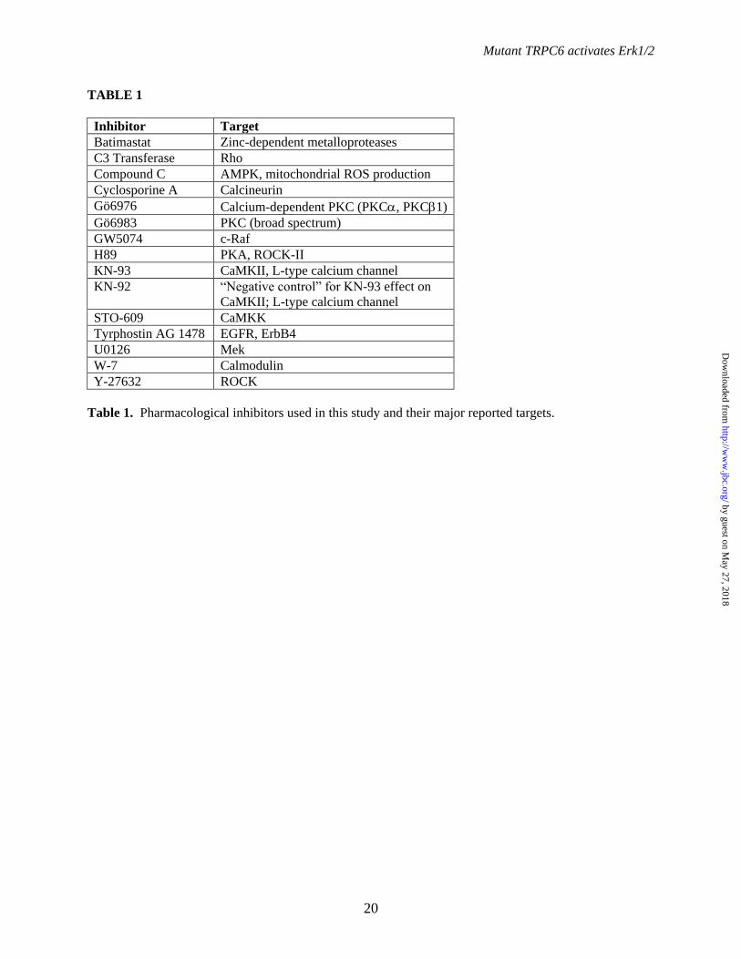
Mutant TRPC6 activates Erk1/2
20
TABLE 1
Inhibitor Target
Batimastat Zinc-dependent metalloproteases
C3 Transferase Rho
Compound C AMPK, mitochondrial ROS production
Cyclosporine A Calcineurin
Gö6976 Calcium-dependent PKC (PKC, PKC1)
Gö6983 PKC (broad spectrum)
GW5074 c-Raf
H89 PKA, ROCK-II
KN-93 CaMKII, L-type calcium channel
KN-92 “Negative control” for KN-93 effect on
CaMKII; L-type calcium channel
STO-609 CaMKK
Tyrphostin AG 1478 EGFR, ErbB4
U0126 Mek
W-7 Calmodulin
Y-27632 ROCK
Table 1. Pharmacological inhibitors used in this study and their major reported targets.
by guest on May 27, 2018
http://ww
w.jbc.org/
Dow
nloaded from

P-Erk
Erk
- + - +- + Tet- WT R895C FLAG-C6
37
50
37
50
100
150
FLAG
*
39
39198131
83
- WT
P112
QR8
95C
E897
KR8
95C
DN
P-Erk
Erk
FLAG
A.
C.
B.
Control WT R895C0
1
2
3
n.s.
p<0.001
p<0.05
P-Er
k/Er
k
E.
D.
F.
DAPI FLAG P-Erk
WT
R895
C
-
+
-
+
Figure 1
100
150
37
50
37
50
- WT R895C
HA
P-Erk
Erk
HA-TRPC6
37
50Actin
by guest on May 27, 2018
http://ww
w.jbc.org/
Dow
nloaded from

** * * * * *
P-Erk
Erk
FLAG
- Tet
DM
SOW
-7
KN93
STO
609
CsA
Com
poun
d C
Gö6
976
Gö6
983
U01
26G
W50
74
A.
B.
Figure 2
by guest on May 27, 2018
http://ww
w.jbc.org/
Dow
nloaded from

P-Erk
Erk
FLAG
- Tet
DM
SOKN
92
KN93
A.
B.
C.
Figure 3
I C-I C-HA-C6 R895C-
GFP-AC3
P-Erk
Erk
HA
GFP
by guest on May 27, 2018
http://ww
w.jbc.org/
Dow
nloaded from
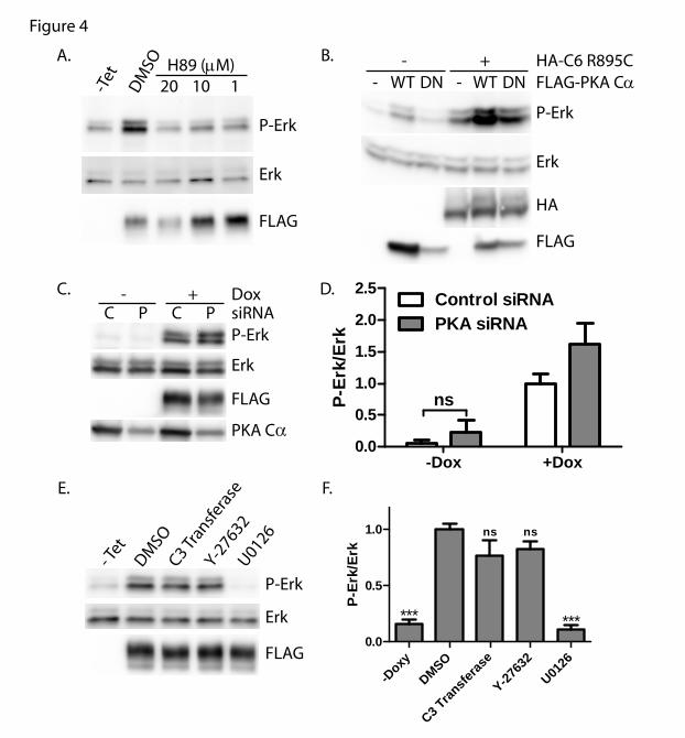
-Doxy
DMSO
C3 Tran
sferas
e
Y-2763
2U01
260.0
0.5
1.0
*** ***
ns ns
P-Er
k/Er
k
-Dox +Dox0.0
0.5
1.0
1.5
2.0
2.5 Control siRNAPKA siRNA
nsP-Er
k/Er
k
P-Erk
Erk
FLAG
-Tet
DM
SO
20 110H89 (µM)
A.
P-Erk
Erk
HA
FLAG
FLAG-PKA CαHA-C6 R895C
WT DN- WT DN-- +B.
C.
Figure 4
- Tet
U0126
C3 Tran
sfera
se
DMSO
Y-27632
P-Erk
Erk
FLAG
D.
C P- +
C P siRNADox
P-Erk
Erk
FLAG
PKA Cα
E. F.
by guest on May 27, 2018
http://ww
w.jbc.org/
Dow
nloaded from

P-Erk
Erk
FLAG
-Tet
DM
SOTy
rpho
stin
A. B.
C.
P-Erk
Erk
M - +WT R895C
DoxCell Line
- +
D.
- +15’ 30’
P-Erk
Erk
Dox- + - +60’1’
- +5’
- +
P-Erk
Erk
-Tet
DM
SO
U01
26
1 10 50 100
Tyrphostin (nM)
E.
P-Erk
Erk
-Dox
DM
SOU
0126
KN92
KN93
H-8
9Ty
rpho
stin
P-Erk
Erk
-Dox
DM
SOU
0126
KN92
KN93
H-8
9
Tyrp
host
in
-Dox
DM
SOBa
timas
tat
P-Erk
Erk
FLAG
-Dox
DM
SOBa
timas
tat
P-Erk
Erk
-Dox
DM
SOBa
timas
tat
P-Erk
Erk
F. G.
H. I.
J. 15’
P-Erk
Erk
Dox
5’
- +- + - +- + - +
5’Media
Podocyte 293T
WT R895C WT R895C
Bas
al
M M
Figure 5
by guest on May 27, 2018
http://ww
w.jbc.org/
Dow
nloaded from

0.5
1.0
1.5
2.0
2.5
3.0
3.5
0 30 60 90 120 150 180 210 240 270 300 330 360
Rat
io (3
40/3
80)
T im e (S ec)
W T (-C a) (-Te t)W T (-C a) (+T et)R 895C (-C a) (-Te t)R 895C (-C a) (+T et)W T (+C a) (-T e t)W T (+C a) (+Tet)R 895C (+C a) (-T e t)R 895C (+C a) (+Tet)
Figure 6
A.
B.
WT -Tet WT +Tet R895C -Tet R895C +Tet1 .0
1 .1
1 .2
1 .3
1 .4
1 .5
*
****
***
Rat
io (3
40/3
80)
C.
by guest on May 27, 2018
http://ww
w.jbc.org/
Dow
nloaded from

100
37
50
37
50
150
100
150LysateWB: HA
LysateWB: FLAG
IP: FLAGWB: HA
IP: FLAGWB: PKA Cα
HA-TRPC6FLAG-PKA Cα+
+ -+-
+
100
150
37
50
IP: FLAGWB: HA
IP: FLAGWB: FLAG
*
FLAG-PKA CαHA-TRPC6
- WT MutR895C
- WT MutWT
37
50 LysateWB: FLAG
100
150LysateWB: HA
Fors
kolin
H89
100
150
100
150
37
50
37
50
HA-TRPC6FLAG-PKA Cα+
+-+-
+++
++
IP: FLAGWB: HA
LysateWB: HA
IP: FLAGWB: FLAG
LysateWB: FLAG
A. B.
C.
Figure 7
by guest on May 27, 2018
http://ww
w.jbc.org/
Dow
nloaded from

- + - + - +
100
150
100
150
WT R895CR895CT70V HA-TRPC6
Forskolin
WB: HA
WB: P-S/T (PKA)
- +-
- + - + - +
100
150
100
150
WT R895CR895CT70V HA-TRPC6
PKA Cα
WB: HA
WB: P-S/T (PKA)
37
50
100
150
100
150
IP: FLAGWB: HA
LysateWB: HA
LysateWB: FLAG
FLAG-PKA CαHA-TRPC6R895C
- + - +T70V
R895C
- Fors
kolin
H89
Fors
kolin
+ H
89
100
150IP: HAWB: P-S/T (PKA)
100
150
LysateWB: HA
A. B.
C. D.
Figure 8
by guest on May 27, 2018
http://ww
w.jbc.org/
Dow
nloaded from

P-Erk
Erk
HA
HA-TRPC6- WT R895
CR8
95C
S282
AR8
95C
T70V
R895
C YY
>FF
-WT
R895C
R895C
S282A
R895C
T70V
R895C
YY>FF
0
2
4
6
P-Er
k/Er
k
****
P-Erk
Erk
HA
HA-TRPC6- WT R895
CR8
95C
S282
AR8
95C
S282
E
S282
AS2
82E
A.
C.
B.
D.
Figure 9
by guest on May 27, 2018
http://ww
w.jbc.org/
Dow
nloaded from

David Chiluiza, Sneha Krishna, Valerie A. Schumacher and Johannes Schlondorffextracellular-signal-regulated kinases Erk1/2
Gain-of-function mutations in transient receptor potential C6 (TRPC6) activate
published online May 3, 2013J. Biol. Chem.
10.1074/jbc.M113.463059Access the most updated version of this article at doi:
Alerts:
When a correction for this article is posted•
When this article is cited•
to choose from all of JBC's e-mail alertsClick here
by guest on May 27, 2018
http://ww
w.jbc.org/
Dow
nloaded from