daptomycin is effective in the treatment of experimental...
TRANSCRIPT

1
Daptomycin Is Effective in the Treatment of Experimental Endocarditis Due to
Methicillin-Resistant and Glycopeptide-Intermediate Staphylococcus aureus
Francesc Marco,1 Cristina García de la Mària,
1 Yolanda Armero,
1 Eurídice Amat,
1 Dolors
Soy,2 Asunción Moreno,
3 Ana del Río,
3 Manel Almela,
1 Carlos A. Mestres,
4 José M.
Gatell,3 María Teresa Jiménez de Anta,
1 José M. Miró,
3* for the Hospital Clinic
Experimental Endocarditis Study Group†
1Microbiology Service, Hospital Clínic, Institut d’Investigacions Biomèdiques August Pi-
Sunyer, University of Barcelona School of Medicine, Barcelona, Catalunya, Spain
2 Pharmacy Service, Hospital Clínic, Institut d’Investigacions Biomèdiques August Pi-
Sunyer, University of Barcelona School of Medicine, Barcelona, Catalunya, Spain
3 Infectious Diseases Service, Hospital Clínic, Institut d’Investigacions Biomèdiques
August Pi-Sunyer, University of Barcelona School of Medicine, Barcelona, Catalunya,
Spain
4Cardiovascular Institute, Institut d’Investigacions Biomèdiques August Pi-Sunyer,
University of Barcelona School of Medicine, Barcelona, Catalunya, Spain
†Members of the Hospital Clinic Endocarditis Study Group, Hospital Clínic-IDIBAPS,
University of Barcelona School of Medicine, Barcelona, Spain: Miró JM, Moreno A, del
Río A, Gatell JM (Infectious Diseases Service); Marco F, García de la Mària C, Armero
Y, Almela M, Jiménez de Anta MT (Microbiology Service), Mestres CA, Cartañá R,
Ninot S, Pomar JL, Azqueta M, Sitges M, Paré JC, Sanz G (Cardiovascular Institute),
ACCEPTED
Copyright © 2008, American Society for Microbiology and/or the Listed Authors/Institutions. All Rights Reserved.Antimicrob. Agents Chemother. doi:10.1128/AAC.00510-07 AAC Accepts, published online ahead of print on 21 April 2008
on July 17, 2018 by guesthttp://aac.asm
.org/D
ownloaded from

2
Pérez N, Ramírez J, Ribalta T (Pathology Department), Brunet M (Toxicology Service),
Soy D (Pharmacy Service), and de Lazzari E (UASP).
*Corresponding author:
Jose M. Miró
Infectious Diseases Service
Hospital Clinic Universitari
Villarroel 170
08036-Barcelona
Spain
Phone: 34-93-2275586
Fax: 34-93-4514438
E-mail: [email protected]
Presented in part at the Interscience Conference on Antimicrobial Agents and
Chemotherapy (ICAAC), held December 16-19, 2005, in Washington, DC, USA.
Abstract number B-2002.
Suggested running head: Daptomycin for MRSA/GISA Experimental Endocarditis
Conflicts of interest: None of the authors have any potential conflicts of interest with
this study.
ACCEPTED
on July 17, 2018 by guesthttp://aac.asm
.org/D
ownloaded from

3
Abstract
Daptomycin is a lipopeptide antibiotic with potent in vitro activity against gram-positive cocci,
including S. aureus. This study evaluated in vitro and in vivo efficacy of daptomycin against 2
methicillin-resistant clinical S. aureus (MRSA) isolates: MRSA 277 (vancomycin MIC 2
µg/ml) and GISA ATCC 700788 (vancomycin MIC 8 µg/ml). Time-kill experiments
demonstrated that daptomycin was bactericidal in vitro against these 2 strains. The in vivo
activity of daptomycin (6 mg/kg q 24 h) was evaluated using a rabbit model of infective
endocarditis and compared with high-dose (HD, 1 g i.v. q 6 h) and recommended-dose (RD, 1
g i.v. q 12 h) vancomycin regimens for 48 hours and an untreated control group. Daptomycin
was significantly more effective than vancomycin-RD in reducing the density of bacteria in the
vegetations for the MRSA (0 [0-1.5] vs. 2 [0-5.6] log CFU/g veg; P = 0.02) and GISA (2 [0-2]
vs. 6.6 [2.0-6.9] log CFU/g veg; P < 0.01) strains studied. In addition, daptomycin sterilized
more vegetations than vancomycin-RD against MRSA (13/18 [72%] vs. 7/20 [35%]; P = 0.02)
and both vancomycin regimens against GISA (12/19 [63%] vs. 4/20 [20%]; P < 0.01). No
statistical difference between vancomycin-HD and vancomycin-RD for MRSA treatment was
noted. These results support the use of daptomycin for treatment of aortic valve endocarditis
caused by GISA and MRSA.
Word count = 220
Keywords: experimental endocarditis, GISA, MRSA, Staphylococcus aureus,
vancomycin MIC 2 µg/ml, recommended-dose vancomycin, high-dose vancomycin,
daptomycin.
ACCEPTED
on July 17, 2018 by guesthttp://aac.asm
.org/D
ownloaded from

4
Introduction
Methicillin-resistant Staphylococcus aureus (MRSA) strains are a common cause of
severe community-acquired and healthcare-associated infections, causing over one
quarter of all S. aureus infective endocarditis (IE) cases (15). When treating IE caused by
MRSA, vancomycin may exhibit slow bactericidal activity and diffuse poorly into
bacterial vegetations (26, 33). Over the years, vancomycin use has been associated with
treatment failures in patients with IE and bacteremia, even in instances where patients’
MRSA isolates remained in the susceptible minimum inhibitory concentration (MIC)
range (16, 22). Furthermore, recent studies showed a vancomycin MIC shift from MRSA
with a vancomycin MIC of < 0.5 µg/ml to strains with MIC values of 1 or 2 µg/ml (21,
35). Sakoulas and colleagues found that clinical success correlated with vancomycin MIC
values: vancomycin treatment failure for MRSA bacteremia was significantly more likely
when the vancomycin MIC was 1-2 µg/ml compared with < 0.5 µg/ml (32). Two other
studies showed similar results (21, 34). Moreover, the emergence of glycopeptide-
intermediate S. aureus (GISA) and heterogeneous GISA (hGISA) (in addition to
vancomycin-resistant S. aureus [VRSA]) strains has mandated the development of
alternatives to vancomycin for the clinical care of drug-resistant S. aureus infections (1,
11, 36, 37).
Daptomycin is a cyclic lipopeptide antibiotic with rapid bactericidal activity against most
antibiotic-resistant gram-positive pathogens in vitro, including GISA and MRSA strains
(7, 17). It is approved by the FDA and EMEA for the treatment of complicated skin and
skin-structure infections and bacteremia, including those involving right-sided IE caused
by MSSA and MRSA (9). A randomized clinical study (14) found that daptomycin is not
ACCEPTED
on July 17, 2018 by guesthttp://aac.asm
.org/D
ownloaded from
5
inferior to standard therapy for S. aureus bacteremia and right-sided IE. Against MRSA,
44% of daptomycin-treated patients had a successful clinical outcome, compared with
32% of patients receiving vancomycin (14).
Compared with MRSA data, there are far fewer data available regarding the activity of
daptomycin against S. aureus with reduced susceptibility to glycopeptides (GISA)
including vancomycin. The objective of this study was to compare daptomycin with 2
vancomycin regimens (using the recommended dose [RD] or a high dose [HD] of
vancomycin) in an experimental model of aortic valve endocarditis in rabbits infected
with MRSA or GISA using a humanized pharmacokinetics model.
Materials and Methods
Bacterial isolates. The MRSA 277 strain used in the study described here was isolated
from a patient with bacteremia at Hospital Clínic Universitari in Barcelona, while the
GISA strain was isolated from a patient in the United States (30) and is currently
included in the American Type Culture Collection of strains (ATCC 700788). Both
strains were stored at -80ºC in skim milk.
Antibiotics. Daptomycin powder was supplied by Cubist Pharmaceuticals (Lexington,
MA, USA). Vancomycin, gentamicin, and rifampin were purchased from Sigma (St.
Louis, MO, USA).
Susceptibility testing. Gentamicin, rifampin, and vancomycin MICs and minimal
bactericidal concentrations (MBCs) were determined using the microdilution method in
liquid media cation-adjusted Mueller-Hinton broth (Oxoid, Hampshire, England),
ACCEPTED
on July 17, 2018 by guesthttp://aac.asm
.org/D
ownloaded from

6
according to the procedures of the Clinical and Laboratory Standards Institute (CLSI;
formerly National Committee for Clinical Laboratory Standards [NCCLS]) (29).
Daptomycin susceptibility testing was in Mueller-Hinton broth adjusted to 50 µg/ml of
calcium per standard methodology. S. aureus ATCC 29213 was used as the test control
strain.
Time-kill studies. All time-kill curves were performed in duplicate according to CLSI
criteria. An inoculum of 105 colony-forming units (CFU)/ml was used. Bactericidal
activity was defined as at least a 3-log reduction in CFU at 24 hours in comparison with
the initial inoculum. Viability counts were performed at 0, 4, and 24 hours (13).
Study animals. An experimental aortic valve endocarditis was induced in New Zealand
White rabbits (body weight, 2 kg), which were obtained from San Bernardo Farm
(Pamplona, Spain). The animals were housed in the animal facilities of Facultad de
Medicina, Universidad de Barcelona, which is equipped with automatic air exchange
with a high-efficiency particulate air (HEPA) filter and a circadian light cycle. They were
nourished ad libitum.
Human pharmacokinetics simulation studies. Antibiotics were administered using a
computer-controlled infusion pump system, designed to reproduce human serum
pharmacokinetics in rabbits after an intravenous (i.v.) infusion. Animal drug doses were
chosen to simulate the pharmacokinetic profile of daptomycin (6 mg/kg i.v. q 24 h) (4,
10, 11) and vancomycin (30 mg/kg i.v. divided into 2 doses; for an adult of 70 kg, 1 g i.v.
ACCEPTED
on July 17, 2018 by guesthttp://aac.asm
.org/D
ownloaded from

7
q 12 h) in humans. These are the current recommended doses to treat S. aureus
endocarditis (2, 14). In addition, vancomycin-HD was also tested in order to achieve the
proposed optimal pharmacokinetic/pharmacodynamic (PK/PD) index of AUC/MIC > 350
(28) for the treatment of serious infections due to MRSA. For this vancomycin-HD
treatment, the animals received 60 mg/kg i.v. vancomycin divided into 4 doses
(equivalent to 1 g i.v. q 6 h, for a 70-kg adult). Assuming an MIC for MRSA of 2 µg/ml,
this dose would simulate the human PK profile necessary for the aforementioned PK/PD
ratio.
To determine the concentrations of daptomycin and vancomycin in serum samples in 5
healthy rabbits, blood was drawn from a carotid catheter at different times after a single
i.v. injection of the antibiotic. Rabbits received 25 mg/kg of vancomycin and 15 mg/kg of
daptomycin. Daptomycin concentrations in plasma were assayed by the disk-plate
bioassay method (12) with Micrococcus luteus (ATCC 9341) as the bioassay
microorganism and Mueller-Hinton agar as the growth medium. The sensitivity of
daptomycin bioassay was 0.5 µg/ml. Vancomycin concentrations were assayed at the
Centre de Diagnòstic Biomèdic, Hospital Clínic, Barcelona. The concentration was
measured by immunoturbidimetric test, boosted by latex method (ADVIA Chemistry
System, Bayer Health Care LLC, Leverkusen, Germany). The limit of detection of the
procedure was 0.8 µg/ml. The range was from 0.8 to 72.9 µg/ml, and the inter- and intra-
assay coefficient of variation was from 2.2% to 2.7%, evaluated for 3 levels of
concentrations: 10.9, 39.3, and 73.1 µg/ml.
ACCEPTED
on July 17, 2018 by guesthttp://aac.asm
.org/D
ownloaded from

8
In order to ensure that rabbits were dosed appropriately in the experimental IE model, it
was necessary to first obtain the pharmacokinetic parameters of daptomycin and
vancomycin in healthy, uninfected rabbits. Different pharmacokinetic parameters were
estimated on the basis of an open 2-compartment model in order to compare the
pharmacokinetics in rabbits, the human-adapted model, and humans (19, 20) as
previously described (27).
An infusion pump system was set up to deliver antibiotics intravenously into rabbits at
previously calculated flow rates to simulate the human kinetics of 6 mg/kg daptomycin
daily (n = 5); 1 g vancomycin every 12 hours (n = 5); or 1 g vancomycin every 6 hours (n
= 4). To determine the antibiotic concentrations in rabbits’ serum, 1 ml of blood was
sampled at different times after the start of drug infusion.
Endocarditis model. Experimental aortic valve infective endocarditis was induced
according to the method described by Garrison and Freedman (18). A catheter was
inserted through the right carotid artery into the left ventricle, and the catheter for the
antibiotic administration was placed into the inferior vena cava through the jugular vein,
as previously described (27).
The infusion pump delivered 2 ml/hour of 0.9% saline solution until the beginning of
antimicrobial administration. Twenty-four hours after placement of the intracardiac
catheter, all animals were infected via the marginal ear vein with 1 ml of saline solution
containing 7 × 10⁵
CFU/ml of the MRSA strain or 8 × 10⁵ CFU/ml of the GISA strain.
One ml of blood was obtained 16 hours after infection and immediately before the
initiation of antimicrobial therapy to confirm the bacteremia, which was interpreted to
ACCEPTED
on July 17, 2018 by guesthttp://aac.asm
.org/D
ownloaded from

9
indicate IE. The infected rabbits were then randomized to the following treatment groups:
control with saline solution only (n = 20 for MRSA, n = 17 for GISA); daptomycin (n =
19 for MRSA, n = 19 for GISA); vancomycin-RD (n = 20 for MRSA, n = 23 for GISA);
and vancomycin-HD (n = 18 for MRSA, n = 22 for GISA). Eighteen hours after
inoculation with the MRSA/GISA, daptomycin and vancomycin treatments were
initiated; also at that time, control animals were sacrificed and vegetations quantitated for
bacterial CFUs. Antibiotics were administered via the computer-controlled infusion pump
system for 48 hours. After finishing the 48-hour treatment, animals were sacrificed after
an additional 6 half-lives of either daptomycin (48 hours) or vancomycin (6 hours) had
elapsed. This provided growing time for residual viable bacteria contained within the
endocardial vegetations.
Analysis of endocardial vegetations. After antibiotic treatment, rabbits were killed and
aortic valve vegetations were removed and processed as previously described (27).
The recovered bacteria were retested to measure daptomycin and vancomycin MICs for
comparison to pretreatment MIC values. The results were expressed as the number of
log10 CFUs of MRSA or GISA per gram of vegetation. The result was assigned a value of
2 if there was no growth on the quantitative plates but there was growth in the qualitative
culture (the rest of the homogenate in tryptic soy broth). The result was assigned a value
of zero, and the vegetation was considered sterile if there was no growth from the initial
quantitative culture or from the homogenates cultured for a week.
ACCEPTED
on July 17, 2018 by guesthttp://aac.asm
.org/D
ownloaded from

10
Statistical analysis. The results were expressed as the median and the interquartile range
(IQR) interval of the number of log10 CFUs of MRSA or GISA per gram of vegetation.
The Mann-Whitney rank-sum test was used to compare the log10 CFU/g values between
the different treatment groups. The Fisher exact test was used to compare the rate of
sterilization of vegetations and assess whether there were differences between treatment
groups.
Results
Susceptibility testing. The MRSA strain was susceptible to vancomycin and daptomycin
(MIC, 2 µg/ml and 0.12 µg/ml; MBC, 2 µg/ml and 0.25 µg/ml, respectively) but resistant
to gentamicin and rifampin (MIC > 256 µg/ml for both), according to the CLSI standard
MIC breakpoints (29). The GISA strain was susceptible to daptomycin and gentamicin
(MIC, 0.5 µg/ml and 0.25 µg/ml; MBC, 1 µg/ml and 128 µg/ml, respectively), but
presented intermediate resistance to vancomycin (MIC, 8 µg/ml) and resistance to
rifampin (MIC > 256 µg/ml). The MBC/MIC ratio (14) for the GISA isolate (128/8)
indicated it was tolerant to vancomycin (23).
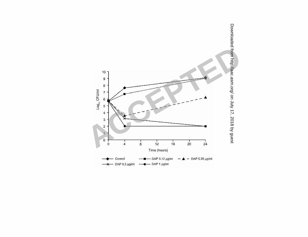
In vitro time-kill experiments. The in vitro activity of daptomycin and vancomycin for
MRSA 277 and GISA ATCC 700788 is presented in Fig. 1A-1B and Fig. 2A-2B,
respectively. At the concentrations tested (between 4 and 0.5 times the MIC), daptomycin
showed a bactericidal activity over the MIC for both strains, while vancomycin was
bacteriostatic.
ACCEPTED
on July 17, 2018 by guesthttp://aac.asm
.org/D
ownloaded from

11
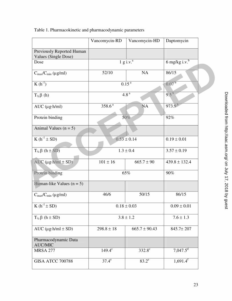
Pharmacokinetic studies. The pharmacokinetic parameters obtained from the human-
adapted model were similar to those in humans (3) (Table 1). The mean trough
concentration of daptomycin was well above the daptomycin MICs of both the MRSA
and GISA strains, and the mean trough concentration of vancomycin-RD and
vancomycin-HD exceeded the vancomycin MIC of MRSA (2 µg/ml). Vancomycin
studies were also performed in GISA strains for a negative control. The mean trough
concentration in the 38 treated rabbits (18 MRSA and 20 GISA) of the vancomycin-HD
arms was 20 µg/ml at 24 hours and 19 µg/ml at 48 hours. We do not have concentrations
from treated rabbits in the vancomycin-RD arm.
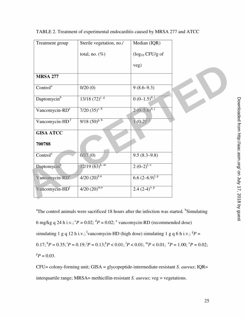
Treatment of established endocarditis. The relative effectiveness of the daptomycin
and vancomycin therapeutic regimens for the treatment of established endocarditis
(control, daptomycin, vancomycin-RD, and vancomycin-HD) is shown in Table 2. One
rabbit treated with daptomycin (from MRSA arm), 3 treated with vancomycin-RD (from
GISA arm), and 2 with vancomycin-HD (from GISA arm) died during the experiment
and were not included in the analyses.
In the 2 strains of S. aureus tested, all control rabbits had infected aortic valve
vegetations with a high median bacterial titer per gram of vegetation (>9 log10 CFU).
Comparisons between treated groups revealed that after 48 hours of treatment,
daptomycin was significantly more effective than vancomycin-RD in sterilizing the
MRSA and GISA vegetations (P = 0.02 and P < 0.01, respectively). In comparison with
vancomycin-HD, daptomycin sterilized more vegetations in the MRSA group, but the
ACCEPTED
on July 17, 2018 by guesthttp://aac.asm
.org/D
ownloaded from

12
differences did not reach statistical significance (P = 0.17), and compared to HD-
vancomycin, daptomycin was significantly more effective against the GISA isolates (P <
0.01). Daptomycin also reduced the median number of MRSA and GISA CFUs in the
vegetations of treated animals to a greater extent than both vancomycin regimens. These
differences were statistically significant for both strains in the vancomycin-RD arm
(MRSA, P = 0.02; GISA, P < 0.01) and against GISA in the vancomycin-HD arm (P =
0.02).
The vancomycin-HD was more active than the vancomycin-RD for the MRSA strain, but
there were no statistically significant differences either in the proportion of sterilized
vegetations (P = 0.35) or in the reduction of the density of the bacteria within the
vegetations (P = 0.13). In the GISA experimental endocarditis, vancomycin-HD showed
more activity than vancomycin-RD (P = 0.03), but the proportion of sterilized vegetations
was low and the same (20%) in both arms.
All the MRSA and GISA isolates from the endocardial vegetations had the same MIC of
the study strain after daptomycin, vancomycin-RD, and vancomycin-HD treatments.
Discussion
The in vitro susceptibility and time-kill results provided here indicate that daptomycin
has bactericidal activity against MRSA 277 and GISA ATCC 700788 strains, having
MIC/MBC values of 0.12/0.25 µg/ml and 0.5/1 µg/ml, respectively. This is consistent
with previous results in which daptomycin was found to be active against GISA isolates.
ACCEPTED
on July 17, 2018 by guesthttp://aac.asm
.org/D
ownloaded from
13
Animal model experiments with daptomycin have been published previously in the peer-
reviewed literature for IE infections caused by enterococci (6, 25), as well as S. aureus
and MRSA (5, 31), but there are limited animal model data available with GISA IE
infections (24). The present study used a rabbit model simulating human-like
pharmacokinetics to investigate the efficacy of daptomycin in S. aureus aortic valve IE
caused by MRSA or GISA. Here daptomycin was significantly more effective in treating
GISA aortic IE than were both vancomycin regimens after 48 hours of treatment (P <
0.01). Daptomycin was also significantly more effective in treating MRSA aortic IE than
was vancomycin-RD (P < 0.01) but did not significantly differ from vancomycin-HD in
this regard. For both the MRSA and the GISA isolates studied, vancomycin-HD (60
mg/kg i.v. divided into 4 doses; for an adult of 70 kg, 1 g i.v. q 6 h) was not significantly
different from vancomycin-RD (30 mg/kg i.v. divided into 2 doses; for an adult of 70 kg,
1 g i.v. q 12 h) except for the reduction of bacteria within the vegetations in the GISA
animals (P = 0.03). In order to reach an optimal pharmacokinetic/pharmacodynamic
(PK/PD) index of AUC/MIC > 350 (28), we needed to simulate in rabbits a schedule of 1
g i.v. q 6 h. With this regimen, we obtained in the vancomycin-HD an AUC/MIC of 333,
and a mean steady-state trough serum concentrations of vancomycin close to 20 µg/ml in
the 38 treated rabbits. In humans, the efficacy of this vancomycin-HD regimen in treating
episodes of MRSA endocarditis with vancomycin MIC of 2 µg/ml isolates must be
counterbalanced with the increased risk of renal toxicity of this regimen (21).
The activity of daptomycin in our study is in partial agreement with 2 earlier studies. In a
rat model that did not use human-like pharmacokinetics, Voorn et al. (37) showed that
ACCEPTED
on July 17, 2018 by guesthttp://aac.asm
.org/D
ownloaded from

14
daptomycin (5 mg/kg b.i.d.) was more effective than vancomycin (100 mg/kg b.i.d.) or
teicoplanin (15 mg/kg b.i.d.) to treat cloxacillin-tolerant or cloxacillin-nontolerant aortic
S. aureus endocarditis. In addition, Cha and Rybak (8) used an in vitro simulated
endocardial vegetations model, where daptomycin (6 mg/kg) achieved 99.9% kill of
GISA by 8 hours, while vancomycin was ineffective after the same period. In contrast,
Kaatz et al. demonstrated that daptomycin (8 mg/kg q 8h) and teicoplanin (12.5 and 40
mg/kg q 12h) were as efficacious as vancomycin (17.5 mg/kg q 6h) in reducing MRSA
found in rabbit endocardial vegetations after 4 days of treatment (24), but that study did
not use a human-like pharmacokinetic model. While the computer-controlled drug
infusion system used in the present study can simulate human drug pharmacokinetics and
is automated for technical ease, it has been only recently developed and, thus, confounds
interstudy comparisons using different vegetations models, animals, and different dosing
methods and frequencies. The other important limitation is bacterial strain heterogeneity.
Preclinical studies such as those presented here will continue to be important as
physicians look to optimize vancomycin dosage for MRSA endocarditis caused by
isolates with a vancomycin MIC = 2 µg/ml and to look for vancomycin alternatives to
treat MRSA endocardial infections, and potentially those caused by VRSA. There are
limited clinical data available on the use of other antibiotics in lieu of vancomycin for
MRSA IE. The recent clinical study with daptomycin published by Fowler et al.—the
largest study performed in patients with bacteremia and/or IE caused by S. aureus,
including isolates with methicillin resistance—demonstrated that daptomycin alone is
comparable to dual antibiotic therapy with semisynthetic penicillin or vancomycin plus
gentamicin in these infections (14). In this trial, the efficacy of daptomycin for right-
ACCEPTED
on July 17, 2018 by guesthttp://aac.asm
.org/D
ownloaded from

15
sided endocarditis caused by MSSA or MRSA was good. However, for left-sided
endocarditis, its efficacy was as poor as current treatment options—antistaphylococcal
penicillin (nafcillin, oxacillin, flucloxacillin) or vancomycin—both with initial
gentamicin therapy. Furthermore, the MIC increase for daptomycin and vancomycin in
some patients with microbiological failure is a cause for concern. In our experimental
study, daptomycin was better than vancomycin-RD for the treatment of MRSA or GISA
IE and was also more effective than vancomycin-HD for the treatment of GISA IE. We
did not detect an increase in the MIC of the strains after 2 days of therapy. However, we
cannot rule out the possibility of the development of heteroresistance to vancomycin or
daptomycin, because population analyses were not performed on bacteria harvested from
vegetations.
In conclusion, in the rabbit experimental system used here, daptomycin was effective in
sterilizing GISA and MRSA aortic valve endocarditis vegetations, was more effective
than vancomycin-HD in treating MRSA experimental endocarditis, was more potent than
vancomycin-RD and vancomycin-HD in treating GISA experimental endocarditis, and
may provide a foundation for the use of daptomycin in treating left-sided MRSA/GISA
endocarditis in humans.
ACCEPTED
on July 17, 2018 by guesthttp://aac.asm
.org/D
ownloaded from

16
Acknowledgments and Financial Disclosure/Conflict of Interest: This work was
supported in part by a medical school grant from Cubist Pharmaceuticals, Inc., 65
Hayden Avenue, Lexington, MA 02421 (USA); and a grant from the Ministerio de
Sanidad y Consumo, Instituto de Salud Carlos III Madrid (Spain), Spanish Network for
the Research in Infectious Diseases (REIPI RD06/0008), and CIBER CB 06/06/2008.
This work was also supported in part by Fondo de Investigaciones Sanitarias (FIS)
(Madrid, Spain) grants FIS 00/0475, FIS 02/0322, and FIS 05/0170. The authors also
thank Fundación Máximo Soriano Jiménez for technical support. Dr. J.M. Miró was a
recipient of a research grant from the Institut d’Investigacions Biomèdiques August Pi i
Sunyer (IDIBAPS) and the Conselleria de Salut de la Generalitat de Catalunya,
Barcelona (Spain).
ACCEPTED
on July 17, 2018 by guesthttp://aac.asm
.org/D
ownloaded from

17
Reference List
1. Appelbaum, P. C. 2006. MRSA--the tip of the iceberg. Clin Microbiol Infect. 12
Suppl 2:3-10.
2. Baddour, L. M., W. R. Wilson, A. S. Bayer, V. G. Fowler, Jr., A. F. Bolger, M.
E. Levison, P. Ferrieri, M. A. Gerber, L. Y. Tani, M. H. Gewitz, D. C. Tong, J.
M. Steckelberg, R. S. Baltimore, S. T. Shulman, J. C. Burns, D. A. Falace, J. W. Newburger, T. J. Pallasch, M. Takahashi, and K. A. Taubert. 2005.
Infective endocarditis: diagnosis, antimicrobial therapy, and management of
complications: a statement for healthcare professionals from the committee on
rheumatic fever, endocarditis, and Kawasaki disease, council on cardiovascular
disease in the young, and the councils on clinical cardiology, stroke, and
cardiovascular surgery and anesthesia, American Heart Association--executive
summary: endorsed by the Infectious Diseases Society of America. Circulation.
111:3167-3184.
3. Benvenuto, M., D. P. Benziger, S. Yankelev, and G. Vigliani. 2006.
Pharmacokinetics and tolerability of daptomycin at doses up to 12 milligrams per
kilogram of body weight once daily in healthy volunteers. Antimicrob Agents
Chemother. 50:3245-3249.
4. Blouin, R. A., L. A. Bauer, D. D. Miller, K. E. Record, and W. O. Griffen, Jr. 1982. Vancomycin pharmacokinetics in normal and morbidly obese subjects.
Antimicrob Agents Chemother. 21:575-580.
5. Cantoni, L., M. P. Glauser, and J. Bille. 1990. Comparative efficacy of
daptomycin, vancomycin, and cloxacillin for the treatment of Staphylococcus
aureus endocarditis in rats and role of test conditions in this determination.
Antimicrob Agents Chemother. 34:2348-2353.
6. Caron, F., M. D. Kitzis, L. Gutmann, A. C. Cremieux, B. Maziere, J. M. Vallois, A. Saleh-Mghir, J. F. Lemeland, and C. Carbon. 1992. Daptomycin or
teicoplanin in combination with gentamicin for treatment of experimental
endocarditis due to a highly glycopeptide-resistant isolate of Enterococcus faecium.
Antimicrob Agents Chemother. 36:2611-2616.
7. Cha, R., W. J. Brown, and M. J. Rybak. 2003. Bactericidal activities of
daptomycin, quinupristin-dalfopristin, and linezolid against vancomycin-resistant
ACCEPTED
on July 17, 2018 by guesthttp://aac.asm
.org/D
ownloaded from

18
Staphylococcus aureus in an in vitro pharmacodynamic model with simulated
endocardial vegetations. Antimicrob Agents Chemother. 47:3960-3963.
8. Cha, R. and M. J. Rybak. 2003. Daptomycin against multiple drug-resistant
staphylococcus and enterococcus isolates in an in vitro pharmacodynamic model
with simulated endocardial vegetations. Diagn Microbiol Infect Dis. 47:539-546.
9. Cubicin® (daptomycin for injection) [package insert]. 2006. Cubist
Pharmaceuticals, Lexington, MA.
10. Cubist Pharmaceuticals. 2003. Cubicin® (daptomycin for injection) prescribing
information. 1108090803.Lexington, MA.
11. Cunha, B. A. 2005. Methicillin-resistant Staphylococcus aureus: clinical
manifestations and antimicrobial therapy. Clin Microbiol Infect. 11 Suppl 4:33-42.
12. Edberg, S. 1996. The measurement of antibiotics in human body fluids: techniques
and signifigance, In: V. Lorian (ed.), Antibiotics in laboratory medicine. William
and Wilkins, Co., Baltimore, MD.
13. Eliopoulos, G. and R. Moellering. 1996. Antimicrobial combinations, p. 330-396.
In: V. Lorian (ed.), Antibiotics in laboratory medicine. William and Wilkins, Co.,
Baltimore, MD.
14. Fowler, V. G., Jr., H. W. Boucher, R. Corey, E. Abrutyn, A. W. Karchmer, M.
E. Rupp, D. P. Levine, H. F. Chambers, F. P. Tally, G. A. Vigliani, C. H.
Cabell, A. S. Link, I. Demeyer, S. G. Filler, M. Zervos, P. Cook, J. Parsonnet,
J. M. Bernstein, C. S. Price, G. N. Forrest, G. Fatkenheuer, M. Gareca, S. J. Rehm, H. R. Brodt, A. Tice, and S. E. Cosgrove. 2006. Daptomycin versus
standard therapy for bacteremia and endocarditis caused by Staphylococcus aureus.
N Engl J Med. 355:653-665.
15. Fowler, V. G., Jr., J. M. Miro, B. Hoen, C. H. Cabell, E. Abrutyn, E.
Rubinstein, G. R. Corey, D. Spelman, S. F. Bradley, B. Barsic, P. A. Pappas, K.
J. Anstrom, D. Wray, C. Q. Fortes, I. Anguera, E. Athan, P. Jones, J. T. van der Meer, T. S. Elliott, D. P. Levine, and A. S. Bayer. 2005. Staphylococcus
aureus endocarditis: a consequence of medical progress. JAMA. 293: 3012-3021.
16. Fowler, V. G., Jr., G. Sakoulas, L. M. McIntyre, V. G. Meka, R. D. Arbeit, C.
H. Cabell, M. E. Stryjewski, G. M. Eliopoulos, L. B. Reller, G. R. Corey, T.
ACCEPTED
on July 17, 2018 by guesthttp://aac.asm
.org/D
ownloaded from

19
Jones, N. Lucindo, M. R. Yeaman, and A. S. Bayer. 2004. Persistent bacteremia
due to methicillin-resistant Staphylococcus aureus Infection is associated with agr
dysfunction and low-level in vitro resistance to thrombin-induced platelet
microbicidal protein. J Infect Dis. 190:1140-1149.
17. Fuchs, P. C., A. L. Barry, and S. D. Brown. 2002. In vitro bactericidal activity of
daptomycin against staphylococci. J Antimicrob Chemother. 49: 467-470.
18. Garrison, P. K. and L. R. Freedman. 1970. Experimental endocarditis I.
Staphylococcal endocarditis in rabbits resulting from placement of a polyethylene
catheter in the right side of the heart. Yale J Biol Med. 42:394-410.
19. Gavalda, J., P. J. Cardona, B. Almirante, J. A. Capdevila, M. Laguarda, L. Pou, E. Crespo, C. Pigrau, and A. Pahissa. 1996. Treatment of experimental
endocarditis due to Enterococcus faecalis using once-daily dosing regimen of
gentamicin plus simulated profiles of ampicillin in human serum. Antimicrob
Agents Chemother. 40:173-178.
20. Gavalda, J., C. Torres, C. Tenorio, P. Lopez, M. Zaragoza, J. A. Capdevila, B.
Almirante, F. Ruiz, N. Borrell, X. Gomis, C. Pigrau, F. Baquero, and A. Pahissa. 1999. Efficacy of ampicillin plus ceftriaxone in treatment of experimental
endocarditis due to Enterococcus faecalis strains highly resistant to
aminoglycosides. Antimicrob Agents Chemother. 43:639-646.
21. Hidayat, L. K., D. I. Hsu, R. Quist, K. A. Shriner, and A. Wong-Beringer. 2006. High-dose vancomycin therapy for methicillin-resistant Staphylococcus
aureus infections: efficacy and toxicity. Arch Intern Med. 166:2138-2144.
22. Howden, B. P., P. B. Ward, P. G. Charles, T. M. Korman, A. Fuller, P. du
Cros, E. A. Grabsch, S. A. Roberts, J. Robson, K. Read, N. Bak, J. Hurley, P. D. Johnson, A. J. Morris, B. C. Mayall, and M. L. Grayson. 2004. Treatment
outcomes for serious infections caused by methicillin-resistant Staphylococcus
aureus with reduced vancomycin susceptibility. Clin Infect Dis. 38:521-528.
23. Jones, R. N. 2006. Microbiological features of vancomycin in the 21st century:
minimum inhibitory concentration creep, bactericidal/static activity, and applied
breakpoints to predict clinical outcomes or detect resistant strains. Clin Infect Dis.
42 Suppl 1:S13-S24.
24. Kaatz, G. W., S. M. Seo, V. N. Reddy, E. M. Bailey, and M. J. Rybak. 1990.
Daptomycin compared with teicoplanin and vancomycin for therapy of
ACCEPTED
on July 17, 2018 by guesthttp://aac.asm
.org/D
ownloaded from

20
experimental Staphylococcus aureus endocarditis. Antimicrob Agents Chemother.
34:2081-2085.
25. Kennedy, S. and H. F. Chambers. 1989. Daptomycin (LY146032) for prevention
and treatment of experimental aortic valve endocarditis in rabbits. Antimicrob
Agents Chemother. 33:1522-1525.
26. Levine, D. P., B. S. Fromm, and B. R. Reddy. 1991. Slow response to
vancomycin or vancomycin plus rifampin in methicillin-resistant Staphylococcus
aureus endocarditis. Ann Intern Med. 115:674-680.
27. Miró JM, García de la Mária C, Armero Y, de Lazzari E, Soy D, Moreno A,
del Rio A, Almela M, Mestres CA, Gatell JM, Jiménez de Anta MT, Francesc
Marco and the Hospital Clínic Experimental Endocarditis Study Group. 2007. Efficacy of telavancin in the treatment of experimental endocarditis due to
glycopeptide-intermediate Staphylococcus aureus. Antimcrob Agents Chemother.
51: 2373-2377.
28. Moise, P. A., A. Forrest, S. M. Bhavnani, M. C. Birmingham, and J. J. Schentag. 2000. Area under the inhibitory curve and a pneumonia scoring system
for predicting outcomes of vancomycin therapy for respiratory infections by
Staphylococcus aureus. Am J Health Syst Pharm. 57 Suppl 2:S4-S9.
29. National Committee for Clinical Laboratory Standards (NCCLS). 2006.
Performance standards for antimicrobial susceptibility testing, ninth informational
supplement. M100-S11. National Committee for Clinical Laboratory Standards,
Villanova, PA..
30. Rotun, S. S., V. McMath, D. J. Schoonmaker, P. S. Maupin, F. C. Tenover, B. C. Hill, and D. M. Ackman. 1999. Staphylococcus aureus with reduced
susceptibility to vancomycin isolated from a patient with fatal bacteremia. Emerg
Infect Dis. 5:147-149.
31. Sakoulas, G., G. M. Eliopoulos, J. Alder, and C. T. Eliopoulos. 2003. Efficacy
of daptomycin in experimental endocarditis due to methicillin-resistant
Staphylococcus aureus. Antimicrob Agents Chemother. 47:1714-1718.
32. Sakoulas, G., P. A. Moise-Broder, J. Schentag, A. Forrest, R. C. Moellering, Jr., and G. M. Eliopoulos. 2004. Relationship of MIC and bactericidal activity to
efficacy of vancomycin for treatment of methicillin-resistant Staphylococcus aureus
bacteremia. J Clin Microbiol. 42:2398-2402.
ACCEPTED
on July 17, 2018 by guesthttp://aac.asm
.org/D
ownloaded from

21
33. Small, P. M. and H. F. Chambers. 1990. Vancomycin for Staphylococcus aureus
endocarditis in intravenous drug users. Antimicrob Agents Chemother. 34:1227-
1231.
34. Soriano, A., F. Marco, J. A. Martinez, E. Pisos, M. Almela, V. P. Dimova, D. Alamo, M. Ortega, J. Lopez, and J. Mensa. 2008. Influence of vancomycin
minimum inhibitory concentration on the treatment of methicillin-resistant
Staphylococcus aureus bacteremia. Clin Infect Dis. 46:193-200.
35. Steinkraus, G., R. White, and L. Friedrich. 2007. Vancomycin MIC creep in
non-vancomycin-intermediate Staphylococcus aureus (VISA), vancomycin-
susceptible clinical methicillin-resistant S. aureus (MRSA) blood isolates from
2001-05. J Antimicrob Chemother. 60:788-794.
36. Tenover, F. C., J. W. Biddle, and M. V. Lancaster. 2001. Increasing resistance to
vancomycin and other glycopeptides in Staphylococcus aureus. Emerg Infect Dis.
7:327-332.
37. Voorn, G. P., J. Kuyvenhoven, W. H. Goessens, W. C. Schmal-Bauer, P. H. Broeders, J. Thompson, and M. F. Michel. 1994. Role of tolerance in treatment
and prophylaxis of experimental Staphylococcus aureus endocarditis with
vancomycin, teicoplanin, and daptomycin. Antimicrob Agents Chemother. 38:487-
493.
ACCEPTED
on July 17, 2018 by guesthttp://aac.asm
.org/D
ownloaded from
22
[[Figure legends]]
FIG. 1A. Results of time-kill experiments for MRSA 277 strain incubated with
daptomycin. Cell density (log 10 CFU/ml) was determined at indicated time points. CFU
= colony-forming unit; DAP = daptomycin.
FIG. 1B. Results of time-kill experiments for MRSA 277 strain incubated with
vancomycin. Cell density (log 10 CFU/ml) was determined at indicated time points. CFU
= colony-forming unit; VAN = vancomycin.
FIG. 2A. Results of time-kill experiments for GISA ATCC 700788 strain incubated with
daptomycin. Cell density (log 10 CFU/ml) was determined at indicated time points. CFU
= colony-forming unit; DAP = daptomycin.
FIG. 2B. Results of time-kill experiments for GISA ATCC 700788 strain incubated with
vancomycin. Cell density (log 10 CFU/ml) was determined at indicated time points. CFU
= colony-forming unit; VAN = vancomycin. ACCEPTED
on July 17, 2018 by guesthttp://aac.asm
.org/D
ownloaded from
23
Table 1. Pharmacokinetic and pharmacodynamic parameters
Vancomycin-RD Vancomycin-HD Daptomycin
Previously Reported Human
Values (Single Dose)
Dose 1 g i.v.a 6 mg/kg i.v.
b
Cmax/Cmin (µg/ml) 52/10 NA 86/15
K (h-1
) 0.15 a 0.07
b
T½ β (h) 4.8 a 9.5
b
AUC (µg⋅h/ml) 358.6 a NA 973.9
b
Protein binding 50% 92%
Animal Values (n = 5)
K (h-1
± SD) 0.53 ± 0.14 0.19 ± 0.01
T½ β (h ± SD) 1.3 ± 0.4 3.57 ± 0.19
AUC (µg⋅h/ml ± SD) 101 ± 16 665.7 ± 90 439.8 ± 132.4
Protein binding 65% 90%
Human-like Values (n = 5)
Cmax/Cmin (µg/ml) 46/6 50/15 86/15
K (h-1 ± SD) 0.18 ± 0.03 0.09 ± 0.01
T½ β (h ± SD) 3.8 ± 1.2 7.6 ± 1.3
AUC (µg⋅h/ml ± SD) 298.8 ± 18 665.7 ± 90.43 845.7± 207
Pharmacodynamic Data
AUC/MIC
MRSA 277 149.4c 332.8
c 7,047.5
d
GISA ATCC 700788 37.4e 83.2
e 1,691.4
f
ACCEPTED
on July 17, 2018 by guesthttp://aac.asm
.org/D
ownloaded from

24
a Data from Blouin et al. Vancomycin pharmacokinetics in normal and morbidly obese
subjects. Antimicrob Agents Chemother. 1982;21(4):575-80. (4)
bCubist, data on file (11, 12) and data from Benvenuto M et al. (3).
cMIC277 vancomycin = 2 µg/ml;
dMIC277 daptomycin = 0.12 µg/ml;
eMIC700788
vancomycin = 8 µg/ml; fMIC700788 daptomycin = 0.5 µg/ml.
RD = Recommended dose; HD = High-dose; AUC = area under the curve; K =
elimination rate constant; SD = standard deviation; T1/2 = terminal half-life; NA = not
available.
ACCEPTED
on July 17, 2018 by guesthttp://aac.asm
.org/D
ownloaded from
25
TABLE 2. Treatment of experimental endocarditis caused by MRSA 277 and ATCC
Treatment group Sterile vegetation, no./
total, no. (%)
Median (IQR)
(log10 CFU/g of
veg)
MRSA 277
Controla 0/20 (0) 9 (8.6–9.3)
Daptomycinb 13/18 (72)
c, g 0 (0–1.5)
d, i
Vancomycin-RDe 7/20 (35)
c, h 2 (0–5.6)
d, j
Vancomycin-HD f 9/18 (50)
g, h 1 (0-2)
i, j
GISA ATCC
700788
Controla 0/17 (0) 9.5 (8.3–9.8)
Daptomycinb 12/19 (63)
k, m 2 (0–2)
l, o
Vancomycin-RDe 4/20 (20)
k,n 6.6 (2–6.9)
l, p
Vancomycin-HDf 4/20 (20)
m,n 2.4 (2-4)
o, p
aThe control animals were sacrificed 18 hours after the infection was started.
bSimulating
6 mg/kg q 24 h i.v.; cP = 0.02;
dP = 0.02;
e vancomycin-RD (recommended dose)
simulating 1 g q 12 h i.v.; fvancomycin-HD (high dose) simulating 1 g q 6 h i.v.;
gP =
0.17; h
P = 0.35; iP = 0.19;
jP = 0.13;
kP < 0.01;
lP < 0.01;
mP < 0.01;
nP = 1.00;
oP = 0.02;
pP = 0.03.
CFU= colony-forming unit; GISA = glycopeptide-intermediate-resistant S. aureus; IQR=
interquartile range; MRSA= methicillin-resistant S. aureus; veg = vegetations.
ACCEPTED
on July 17, 2018 by guesthttp://aac.asm
.org/D
ownloaded from