cytotoxicactivitiesofphysalisminimal.chloroform...
TRANSCRIPT
Hindawi Publishing CorporationEvidence-Based Complementary and Alternative MedicineVolume 2011, Article ID 185064, 10 pagesdoi:10.1093/ecam/nep057
Original Article
Cytotoxic Activities of Physalis minima L. ChloroformExtract on Human Lung Adenocarcinoma NCI-H23 CellLines by Induction of Apoptosis
Ooi Kheng Leong,1 Tengku Sifzizul Tengku Muhammad,2, 3 and Shaida Fariza Sulaiman1
1 School of Biological Sciences, Universiti Sains Malaysia, 11800 Minden, Penang, Malaysia2 Malaysian Institute of Pharmaceuticals and Nutraceuticals, Ministry of Science, Technology and Innovation,SAINS 10, Persiaran Bukit Jambul, 11900 Penang, Malaysia
3 Department of Biological Sciences, Faculty of Science and Technology, Universiti Malaysia Terengganu,21030 Kuala Terengganu, Terengganu, Malaysia
Correspondence should be addressed to Shaida Fariza Sulaiman, [email protected]
Received 31 December 2008; Accepted 28 May 2009
Copyright © 2011 Ooi Kheng Leong et al. This is an open access article distributed under the Creative Commons AttributionLicense, which permits unrestricted use, distribution, and reproduction in any medium, provided the original work is properlycited.
Physalis minima L. is reputed for having anticancer property. In this study, the chloroform extract of this plant exhibited remarkablecytotoxic activities on NCI-H23 (human lung adenocarcinoma) cell line at dose- and time-dependent manners (after 24, 48 and72 h of incubation). Analysis of cell-death mechanism demonstrated that the extract exerted apoptotic programed cell deathin NCI-H23 cells with typical DNA fragmentation, which is a biochemical hallmark of apoptosis. Morphological observationusing transmission electron microscope (TEM) also displayed apoptotic characteristics in the treated cells, including clumpingand margination of chromatins, followed by convolution of the nuclear and budding of the cells to produce membrane-boundapoptotic bodies. Different stages of apoptotic programed cell death as well as phosphatidylserine externalization were confirmedusing annexin V and propidium iodide staining. Furthermore, acute exposure to the extract produced a significant regulation ofc-myc, caspase-3 and p53 mRNA expression in this cell line. Due to its apoptotic effect on NCI-H23 cells, it is strongly suggestedthat the extract could be further developed as an anticancer drug.
1. Introduction
Lung cancer remains a major global health problem,accounting for more than a million annual deaths worldwide[1]. It is twice the death rate of the second-most prevalentcancer, that is, prostate cancer in men [2]. The incidence oflung cancer can be correlated with the age of both males andfemales and there is still lack of effective drugs to treat thisdisease [3].
Herbal formulation consisting of single and multiple ofherbs is commonly prescribed as an alternative way to treatcancer. An anticancer plant that was selected for this studyis Physalis minima L. The decoction of the whole plant istaken orally to treat cancer and the leaves are used as apoultice for ulcer [4, 5]. This herb is commonly known as thebladder cherry (Leletup-direct translation from Malay) andbelongs to the Solanaceae family [5]. Its reputed efficacy intreating cancer has been validated (in vitro) against CORL23
lung and MCF7 breast cancer cell lines, but the mechanismsunderlying the anticancer activity still remain unknown.Two active anticancer compounds have been isolated fromits methanol extract of stem and leaf and were identifiedas physalin B and physalin F [6]. However, these twocompounds were found to exhibit non-selective cytoxicityagainst all tested human cell lines including non-cancerouscell with physalin F consistently more active [6, 7].
Recently, intensive studies have been conducted to exam-ine the mechanism responsible for the anticancer effectsof plant-based drugs. Apoptosis is a specific mode of celldeath that can only target cancer cell with little or nodamage to non-cancerous cells [8]. It helps in reducingthe incidence of side effect in patients [9]. Information onthe apoptotic effect elicited by the extracts and bioactivecompounds of Physalis sp. are still limited to a few findings,such as the cell death signaling effects of physalins B andF on PANC-1 pancreatic cancer cells. They were reported
2 Evidence-Based Complementary and Alternative Medicine
as potent inhibitors for the aberrant hedgehog (Hh)/GLIsignaling pathway (that causes formation and progression ofvarious cancers) by inhibiting GL2-mediated transcriptionalactivation, decreasing hedgehog-related component expres-sion and reducing the level of anti-apoptotic Bcl-2 geneexpression [10]. Moreover, apoptotic induction in humanlung cancer H661 cells by the supercritical carbon dioxideextract of Physalis peruviana was associated with cell cyclearrest at the S phase, mediated through the p53-dependentpathway and modification of pro-apoptotic protein (Bax)and inhibitor of apoptosis protein (IAP) expression [11].In addition, the ethanol extract of P. peruviana was foundto induce apoptosis on human liver cancer Hep G2 cellsthrough CD95/CD95L system and the mitochondrial signal-ing transduction pathway [12]. Furthermore, the methanolextract of Physalis angulata induced apoptosis and arrestedhuman breast cancer MAD-MB 231 cells at G2/M phase[13] and induced apoptosis in human oral cancer HSC-3cells through oxidative stress-dependent induction of proteinexpression such as heme oxygenase-1 and Cu/Zn superoxidedismutase [14].
Based on our previous comparative cytotoxicity studiesof the extracts and fractions (obtained from the chloroformextract) of Physalis minima, it has been established that theanti-proliferative activity of the chloroform extract in humanlung adenocarcinoma NCI-H23 cells was relatively betterthan other extracts and its fractions [15]. Therefore, it wasselected for this study with aims (i) to evaluate the effectof the extract to NCI-H23 cells proliferation in differentincubation periods and (ii) to determine the cell deathmechanism elicited by the extract via morphological andmolecular investigations.
2. Methods
2.1. Chemicals. The DeadEnd Colometric Apoptosis Detec-tion System was purchased from Promega, USA. TheAnnexin-V-FLOUS kit was purchased from Roche Diagnos-tics, Germany. The methylene blue assay, dimethyl sulfoxide(DMSO) and propidium iodide were obtained from SigmaAldrich, USA. All culture media and additives were fromHyclone, USA. All other chemicals were reagents of molec-ular grade, as appropriate.
2.2. Preparation of Crude Extracts. The P. minima plantwas collected from Arau-Perlis, Malaysia. The plant wasidentified and verified by Mr V. Shunmugam of UniversitiSains Malaysia. The voucher specimen (no. 11001) waspreserved and deposited in the herbarium of School ofBiological Sciences, Universiti Sains Malaysia.
The whole plant materials were washed, dried andchopped finely using a grinder. The dried material was thentransferred into the Soxhlet extractor. The dried plant mate-rial was exhaustively extracted with chloroform by Soxhletextraction. The extracts were filtered and concentrated usingrotary evaporator, and then evaporated to dryness. The driedextracts were then weighed using microbalances (Sartorius,Germany) and reconstituted with 99.9% (v/v) DMSO to
prepare a stock solution at a concentration of 10 mg/mL. Thestock solution was serially diluted to eight different workingconcentrations. As for the positive control, the stock solutionof vincristine sulfate (a commercial drug) at a concentrationof 1 mg/mL was prepared using DMSO and diluted seriallyto 24 different concentrations.
2.3. Cell Line and Culture Medium. NCI-H23 (human lungadenocarcinoma) cell line was obtained from AmericanType Cell Culture (ATCC), USA, and cultured in RPMI1640, supplemented with 2 mM l-glutamine, 10% (v/v)fetal calf serum (FCS), 100 U/mL penicillin and 100 μg/mLstreptomycin, as recommended by ATCC.
2.4. In Vitro Cytotoxicity Assay. Nearly confluent culturesof cells were harvested with 0.05% (w/v) Trypsin-EDTA.Cells were then centrifuged and pellet resuspended with acomplete medium with 10% (v/v) FCS. Then, 100 μL ofcells were plated into each well of 96-well plate at a densityof ∼6000 cells/well. Cell viability was routinely determinedusing the trypan blue exclusion test to ensure it was alwaysin excess of 95%. The cells were then allowed to attachand incubate at 37◦C in a CO2 incubator for a further24–48 h. After the cells reached 80–90% confluence, themedium was removed and replaced with medium containingonly 0.5% (v/v) FCS and the cells were incubated forfurther 4 h. The reason for this was for the cells to achievequiescent. Subsequently, the cells were treated with differentconcentrations of extracts. Controlled cells were cultured ina 0.5% (v/v) FCS-containing medium alone. Of serial dilutedextracts, 1 μL was added into each well. After treatment, theplates were incubated for 72 h. Vincristine sulfate was used asa positive control.
Cell survival was determined by the procedure usingmethylene blue staining as described by Yamazaki et al.[16] and Li and Hwang [17]. After 72 h of incubation withplant extracts, the surviving cells were fixed with 2.5%(v/v) glutaraldehyde for 15 min and were then washed witha 0.15 M sodium chloride (NaCl) solution to remove thedead cells. The fixer cells subsequently were stained with100 μL of 0.05% (w/v) methylene blue solution for 15 min.After washing off the excess dye with NaCl, dye elutionwas carried out using 200 μL of 0.33 M hydrochloric acid(HCl). Absorbance was read at 650 nm using Vmax KineticMicroplate Reader (Molecular Devices, USA). The numberof surviving cells was determined from the absorbance value.
2.5. Detection of DNA Fragmentation. Nuclear DNA frag-mentation was detected using DeadEnd Colometric Apop-tosis Detection System (Promega, USA). Briefly, cell suspen-sions were subcultured on Labtek Chamber Slides, and thenincubated for 24–48 h. When the cells achieved confluencybetween 80% and 90%, the medium was replaced with amedium containing only 0.5% (v/v) FCS. The cells werethen incubated at 37◦C for 4 h and treated with the P.minima chloroform extract at a concentration of EC50 at72 h (2.80 μg/mL). Negative control cells were treated withthe same concentration of DMSO. Positive control cells

Evidence-Based Complementary and Alternative Medicine 3
were treated with DNase I (1 U/mL) and vincristine sulfateat a concentration of EC50 at 72 h (0.0015 μg/mL). In allcases, the final concentration of DMSO in each control slidedid not exceed 1% (v/v). The slides were then incubatedfor 24 h. After treatment, the cells were rinsed twice witha phosphate-buffered saline (PBS), and then processedaccording to the DeadEnd Colometric Apoptosis DetectionSystem protocol as described by the manufacturer (Promega,USA). Subsequently, the slides were observed using thelight microscope (Olympus BH2 light microscope) and theimages taken using the AnalySIS Docu SoftImaging Software,Germany.
2.6. Detection of Plasma Membrane Integrity. In order todetect the loss of plasma membrane of necrotic cells, theNCI-H23 cells were cultured on 6-well plates, and thenincubated for 24–48 h. When the cells reached between 80%and 90% confluency, the medium was replaced with a freshmedium containing only 0.5% (v/v) FCS. The cells werethen incubated for a further 4 h and treated with the P.minima chloroform extract at a concentration of EC50 at72 h (2.80 μg/mL). Negative control cells were treated withthe same concentration of DMSO. The cells were rinsed withPBS and stained using the trypan blue exclusion assay after 24and 72 h incubation period. Subsequently, the samples wereviewed using an inverted phase contrast light microscope.
2.7. Ultrastructural Analysis. NCI-H23 cells were incubatedwith the P. minima chloroform extract at a concentrationof EC50 at 72 h (2.80 μg/mL) for 24 h. Negative control cellswere treated with the similar concentration of DMSO. Afterincubation, the cells were trypsinized, centrifuged and thenresuspended twice with PBS. The cells were then rinsed,pelleted and then resuspended in the McDowell-Trumpfixative solution [containing 4% (v/v) formaldehyde and1% (v/v) glutaraldehyde in a 0.1 M phosphate buffer, pH7.3] at 4◦C for 24 h. The cells were pelleted and rinsedin a 0.1 M phosphate buffer for 10 min and repeated threetimes. Post-fixed was carried out with 1% (w/v) osmiumtetroxide prepared in a 0.1 M phosphate buffer for 1-2 h atroom temperature. The cells were then dehydrated with 50%(v/v) ethanol for 15 min, followed by 75% (v/v) ethanol for15 min, 95% (v/v) ethanol for 15 min and repeated, 100%(v/v) ethanol for 30 min and repeated and, lastly, 100% (v/v)acetone for 10 min and repeated. Infiltration was conductedusing the mixture of acetone : Spurr’s resin mix (1 : 1) in arotator for another 2-3 days with a daily change of Spurr’smix of each sample specimen. Subsequently, the cells wereembedded and cured at 60◦C for 12–48 h.
Sectioning was preceded by the analysis of a semi-thinsection (1 μm), stained at 40◦C with 1% (w/v) toluidineblue. This was then followed by sectioning of thin sections(<1 μm), collected using copper grids. Reichart SupernovaUltra Microtome was used to produce the sections. Thethin sections were stained with uranyl acetate for 15 minby complete immersion, in a covered Petri-dish lined withdental wax. Lastly, the sections were stained with lead citrateusing the same procedures. The samples were examined
and observed under a Philips CM 12 transmission electronmicroscope (TEM).
2.8. Detection of Phosphatidylserine Externalization. NCI-H23 cells were incubated with the P. minima chloroformextract at a concentration of EC50 at 72 h (2.80 μg/mL)for 24 h. Negative control cells were treated with thesimilar concentration of DMSO. After treatment, the cellin the chamber slides were washed with PBS twice andsubsequently processed according to the Annexin-V-FLUOS(Roche Diagnostics, Germany) (combination of annexinV and propidium iodide) protocol as described by themanufacturer’s instruction. Subsequently, the slides wereimmediately analyzed using a fluorescence microscope withan excitation wavelength in the range of 450–500 nm anddetection in the range of 515–565 nm (green). Photos weretaken using the Olympus BH2-RFCA Fluorescence Micro-scope with Olympus camera attachment. Green fluorescencewas observed in annexin V positive cells and red fluorescencein cells which uptake the propidium iodide dye.
2.9. Determination of Apoptotic-Related Genes Expression.In order to investigate the mRNA expression levels of c-myc, caspase-3 and p53, which are responsible to triggerapoptosis mechanism, semi-quantitative reverse transcrip-tion polymerase chain reaction (RT-PCR) was carried out.NCI-H23 cells were cultured in T25 flasks and starvedwith a medium containing only 0.5% (v/v) FCS for 4 h.The cells were then treated with the P. minima chloroformextract at a concentration of EC50 at 72 h (2.80 μg/mL) forthe period of 24 h. After treatment, the total cellular RNAwas isolated using the Tri-Reagent LS (Molecular ResearchCenter, USA) according to the manufacturer’s instructions.Of the isolated total cellular RNA sample, 1 μg was treatedwith DNase I, followed by reverse transcribed into cDNAand subjected to PCR amplification. The optimized PCRamplification program comprised an initial denaturationstep at 94◦C for 2 min, followed by denaturation at 94◦Cfor 45 s, annealing at 56◦C for 1 min, extension at 72◦Cfor 2 min and final extension step at 72◦C for 10 min.The PCR optimization process including the optimal cDNAconcentration and the number of amplification cycles usedto amplify c-myc, caspase-3, p53 and β-actin genes was inthe exponential phase of PCR amplification (i.e. provide alinear relationship between the amount of amplification andthe concentration of the original cDNA temples) (data notshown), indicating that the conditions were optimized forsemi-quantitative studies [14, 15]. The sequences of primersfor analysis were synthesized based on the human mRNAencoding the respective genes. The amplification productswere separated on 1.2% (w/v) agarose gel and stained withethidium bromide. Gene expressions signaling at each pointof time were quantified using GeneTools analysis software onGENE GENIUS gel documentation system (Syngene, UK).The signals of c-myc, p53 and caspase-3 were normalizedfrom β-actin and the ratio in unstimulated samples wasassigned as 1. The housekeeping gene β-actin was used as aninternal standard control.

4 Evidence-Based Complementary and Alternative Medicine
2.10. Calculation and Statistics. Cytotoxicity experimentswere performed in six replicates and results were expressedas percentage growth inhibition of control. EC50 valuesfor growth inhibition was derived from a nonlinear model(curvfit) based on the sigmoidal dose response curve (vari-able) and computed using GraphPadPrism, USA. Data aregiven as mean ± SEM. The criterion of cytotoxic or non-cytotoxic was adapted from the guidelines set by the NationalCancer Institute (NCI) which indicated that plant extractswith EC50 ≤ 20μg/mL were considered to be cytotoxic andnon-cytotoxic if otherwise [18].
DNA fragmentation experiments were conducted intriplicate. The cells from five random microscopic fields werecounted to get the percentage of TUNEL positive cells (apop-totic index, AI% = number of apoptotic nuclei/number ofnuclei scored ×100%) using 200x magnifications and themean percentage ± SEM was calculated. The significantdifferences between the mean percentage of positive stainednuclei of control cells and treated cells were determinedusing one-way ANOVA, computed using GraphPadPrism(Graphpad, USA).
The significant differences in the ratio to β-actin(between each time point and control) were determinedusing one-way ANOVA and Dunnett’s Multiple ComparisonTest for post-comparison tests, computed using GraphPad-Prism software (GraphPad, USA). Differences were consid-ered to be significant if P < .05.
3. Results
3.1. Cytotoxic Effect of the Chloroform Extract of P. minimaon NCI-H23 Cells. Dose-dependent inhibitions in NCI-H23cells were observed when the cells were treated with thisextract at all incubation intervals (Figure 1). Interestingly,the chloroform extract produced >75% of growth inhibitionin NCI-H23 cells, which appeared to be relatively unchangedat higher concentrations (25–100 μg/mL) for all time points(Figure 1). After 24 h of treatment, only at a concentrationof 6.25 μg/mL and above, the chloroform extract signifi-cantly inhibited the growth of NCI-H23 cells (P < .05).Meanwhile, after 48 and 72 h of incubation, at most testedconcentrations (100–1.563 μg/mL), the extract exhibitedsignificant inhibitory effects (P < .05). The inhibitoryactivities increased and the EC50 values decreased as theincubation periods were increased from 24 h (5.28 μg/mL) to48 h (3.55 μg/mL) and 72 h (2.80 μg/mL). However, the EC50
for vincristine sulfate (0.0015 μg/mL) was very much lower ascompared to the EC50 deduced from the chloroform extractat 72 h incubation period (data not shown).
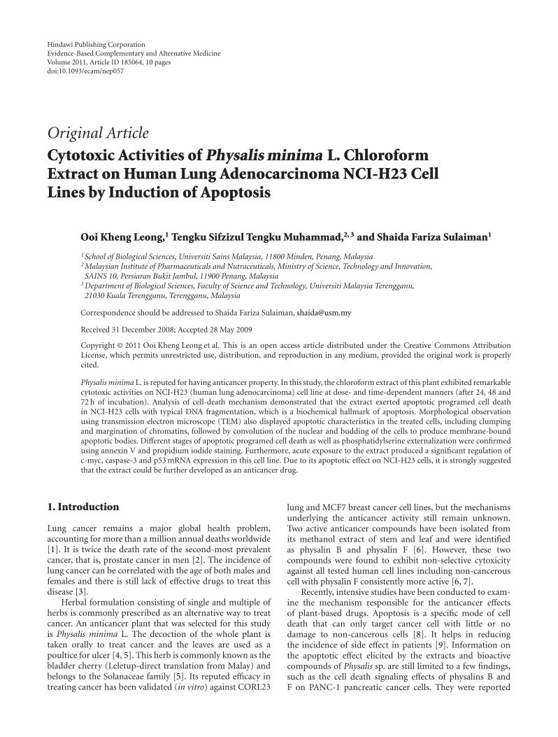
3.2. Effect of P. minima Chloroform Extract on Cell Death ofNCI-H23 Cells. In order to investigate whether apoptosismay play an important role in mediating the cell deathof NCl-H23 cells elicited by the chloroform extract of P.minima, the fragmented genomic DNA was detected usinga modified TUNEL assay. As shown in Figures 2(b), 2(c) and2(d), the extract-treated NCl-H23 cells produced dark brownstained nuclei with similar observation found in the positive
72 hours48 hours24 hours
Time of incubation
100 μg/mL50 μg/mL25 μg/mL12.5 μg/mL
6.25 μg/mL3.125 μg/mL1.563 μg/mL0.781 μg/mL
0
25
50
75
100
Gro
wth
inh
ibit
ion
(%)
∗∗∗∗
∗
∗∗∗∗∗
∗∗
∗∗∗∗∗
∗∗
Figure 1: Effect of P. minima chloroform extract on proliferationof NCI-H23 cells at 24, 48 and 72 h. Each value represented mean±SEM of six replicates (n = 6); ∗P < .05.
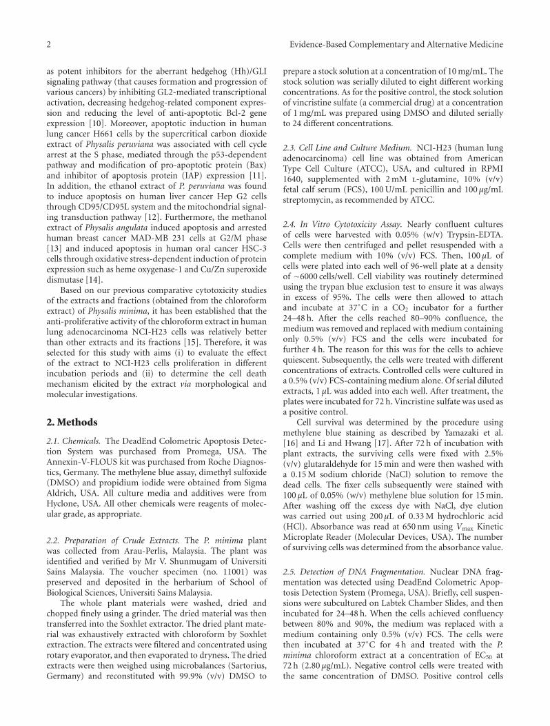
control cells treated with DNase I and vincristine sulfate.The nuclei were specifically stained and evenly distributed.Most of the positive stained nuclei were rounded or oblongin shape. But almost all nuclei of untreated negative controlcells were not stained with this assay (Figure 2(a)). Themean percentage of apoptotic index for the extract-treatedNCl-H23 cells was 49.89% and significantly different ascompared to the negative control (DMSO) (1.39%) (P <.001) (Figure 2(e). Comparatively, the percentage was lowerthan that in positive controls (68.41% and 56.40% for DNaseI and vincristine sulfate, resp.). This result strongly indicatedthat apoptosis was one of the possible type of cell death inNCl-H23 cells after 24 h exposure to the chloroform extract.The trypan blue exclusion assay showed that only a fewNCl-H23 cells were positively stained after treated with theextract for 24 and 72 h. The results strongly suggested thatthe surface of most treated cells was intact after incubationwith the extract at both time points (Figures 3(a) and 3(b).Therefore, the mode of cell death elicited by the chloroformextract of P. minima in NCI-H23 cells was unlikely vianecrotic mechanism in nature.
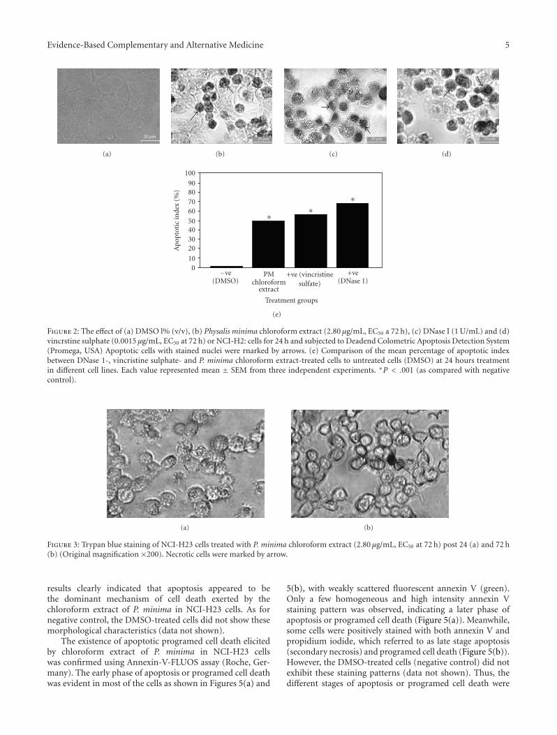
TEM analysis exhibited different morphological alter-ations in NCI-H23 cells after treatment with the chloroformextract of P. minima for 24 h. Characteristics of apoptoticcell death were observed in the majority of treated NCI-H23cells. There was clumping of the nuclear chromatin into moredensely packed material that becomes marginated against thenuclear membrane (Figure 4(a)). This was accompanied bya convolution of nuclear envelope and cytoplasmic mem-brane (Figure 4(b)). However, the cytoplasmic membraneand cell organelles remained intact. Budding of cells wasclearly demonstrated (Figure 4(c)), where the cytoplasmicmembrane formed extensions and separated. The plasmamembrane was then sealed to produce membrane-boundapoptotic bodies that contained cellular material of thecells in various combinations. These ultrastructural analysis
Evidence-Based Complementary and Alternative Medicine 5
20 μm
(a)
20 μm
(b)
20 μm
(c)
20 μm
(d)
+ve(DNase 1)
+ve (vincristinesulfate)
PMchloroform
extract
−ve(DMSO)
Treatment groups
010
20304050
60708090
100A
pop
toti
cin
dex
(%)
∗∗
∗
(e)
Figure 2: The effect of (a) DMSO l% (v/v), (b) Physalis minima chloroform extract (2.80 μg/mL, EC50 a 72 h), (c) DNase I (1 U/mL) and (d)vincrstine sulphate (0.0015 μg/mL, EC50 at 72 h) or NCI-H2: cells for 24 h and subjected to Deadend Colometric Apoptosis Detection System(Promega, USA) Apoptotic cells with stained nuclei were rnarked by arrows. (e) Comparison of the mean percentage of apoptotic indexbetween DNase 1-, vincristine sulphate- and P. minima chloroform extract-treated cells to untreated cells (DMSO) at 24 hours treatmentin different cell lines. Each value represented mean ± SEM from three independent experiments. ∗P < .001 (as compared with negativecontrol).
(a) (b)
Figure 3: Trypan blue staining of NCI-H23 cells treated with P. minima chloroform extract (2.80 μg/mL, EC50 at 72 h) post 24 (a) and 72 h(b) (Original magnification ×200). Necrotic cells were marked by arrow.
results clearly indicated that apoptosis appeared to bethe dominant mechanism of cell death exerted by thechloroform extract of P. minima in NCI-H23 cells. As fornegative control, the DMSO-treated cells did not show thesemorphological characteristics (data not shown).
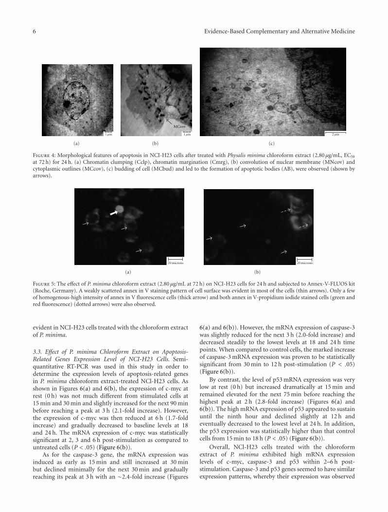
The existence of apoptotic programed cell death elicitedby chloroform extract of P. minima in NCI-H23 cellswas confirmed using Annexin-V-FLUOS assay (Roche, Ger-many). The early phase of apoptosis or programed cell deathwas evident in most of the cells as shown in Figures 5(a) and
5(b), with weakly scattered fluorescent annexin V (green).Only a few homogeneous and high intensity annexin Vstaining pattern was observed, indicating a later phase ofapoptosis or programed cell death (Figure 5(a)). Meanwhile,some cells were positively stained with both annexin V andpropidium iodide, which referred to as late stage apoptosis(secondary necrosis) and programed cell death (Figure 5(b)).However, the DMSO-treated cells (negative control) did notexhibit these staining patterns (data not shown). Thus, thedifferent stages of apoptosis or programed cell death were
6 Evidence-Based Complementary and Alternative Medicine
Cclp
Cmrg
Cmrg
1 μm
(a)
MNcov
MCcov
1 μm
(b)
AB
ABAB
AB
ABMCbud
2 μm
(c)
Figure 4: Morphological features of apoptosis in NCI-H23 cells after treated with Physalis minima chloroform extract (2.80 μg/mL, EC50
at 72 h) for 24 h. (a) Chromatin clumping (Cclp), chromatin margination (Cmrg), (b) convolution of nuclear membrane (MNcov) andcytoplasmic outlines (MCcov), (c) budding of cell (MCbud) and led to the formation of apoptotic bodies (AB), were observed (shown byarrows).
20 microns
(a)
20 microns
(b)
Figure 5: The effect of P. minima chloroform extract (2.80 μg/mL at 72 h) on NCI-H23 cells for 24 h and subjected to Annex-V-FLUOS kit(Roche, Germany). A weakly scattered annex in V staining pattern of cell surface was evident in most of the cells (thin arrows). Only a fewof homogenous-high intensity of annex in V fluorescence cells (thick arrow) and both annex in V-propidium iodide stained cells (green andred fluorescence) (dotted arrows) were also observed.
evident in NCI-H23 cells treated with the chloroform extractof P. minima.
3.3. Effect of P. minima Chloroform Extract on Apoptosis-Related Genes Expression Level of NCI-H23 Cells. Semi-quantitative RT-PCR was used in this study in order todetermine the expression levels of apoptosis-related genesin P. minima chloroform extract-treated NCl-H23 cells. Asshown in Figures 6(a) and 6(b), the expression of c-myc atrest (0 h) was not much different from stimulated cells at15 min and 30 min and slightly increased for the next 90 minbefore reaching a peak at 3 h (2.1-fold increase). However,the expression of c-myc was then reduced at 6 h (1.7-foldincrease) and gradually decreased to baseline levels at 18and 24 h. The mRNA expression of c-myc was statisticallysignificant at 2, 3 and 6 h post-stimulation as compared tountreated cells (P < .05) (Figure 6(b)).
As for the caspase-3 gene, the mRNA expression wasinduced as early as 15 min and still increased at 30 minbut declined minimally for the next 30 min and graduallyreaching its peak at 3 h with an ∼2.4-fold increase (Figures
6(a) and 6(b)). However, the mRNA expression of caspase-3was slightly reduced for the next 3 h (2.0-fold increase) anddecreased steadily to the lowest levels at 18 and 24 h timepoints. When compared to control cells, the marked increaseof caspase-3 mRNA expression was proven to be statisticallysignificant from 30 min to 12 h post-stimulation (P < .05)(Figure 6(b)).
By contrast, the level of p53 mRNA expression was verylow at rest (0 h) but increased dramatically at 15 min andremained elevated for the next 75 min before reaching thehighest peak at 2 h (2.8-fold increase) (Figures 6(a) and6(b)). The high mRNA expression of p53 appeared to sustainuntil the ninth hour and declined slightly at 12 h andeventually decreased to the lowest level at 24 h. In addition,the p53 expression was statistically higher than that controlcells from 15 min to 18 h (P < .05) (Figure 6(b)).
Overall, NCl-H23 cells treated with the chloroformextract of P. minima exhibited high mRNA expressionlevels of c-myc, caspase-3 and p53 within 2–6 h post-stimulation. Caspase-3 and p53 genes seemed to have similarexpression patterns, whereby their expression was observed

Evidence-Based Complementary and Alternative Medicine 7
241812963210.50.250
β-actin
hours
241812963210.50.250c-mychours
241812963210.50.250Caspase-3
hours
241812963210.50.250p53
hours
(a)
241812963210.50.25C
Time of incubation (hours)
0
1
2
3
4
5
6
p53
toβ
-act
inra
tio
241812963210.50.25C
Time of incubation (hours)
0
1
2
3
4
5
6
Cas
pase
-3toβ
-act
inra
tio
241812963210.50.25C
Time of incubation (hours)
0
1
2
3
4
5
6
c-m
yctoβ
-act
inra
tio
∗∗
∗
∗∗ ∗ ∗∗ ∗ ∗
∗ ∗∗ ∗ ∗
∗ ∗ ∗ ∗
(b)
Figure 6: (a) Time course mRNA expression of β-actin, c-myc,caspase-3 and p53 in NCI-H23 cells incubated in the absenceor presence of P. minima chloroform extract. β-actin was usedas an internal standard control for each PCR reation. (b) Semi-quantitative analysis of the c-myc, caspase-3 and p53 mRNA levelin NCI-H23 treated with P. minima chloroform extract usingdensitometric scanning. Each value represented mean ± SEM (n =3 at each point) of the ratio of RT-PCR product of the respectivegenes to β-actin, assigning the ratio in unstimulated cells as 1.∗P < .05 versus C (control).
at early post-stimulation. Moreover, the induction of p53 andcaspase-3 mRNA expression was slightly higher as comparedto that of c-myc gene. Thus, there was a clear indication thatthe cell death induced by the chloroform extract of P. minimain NCl-H23 cells was mediated via the activation of c-myc,caspase-3 and p53 gene expression.
4. Discussion
It has previously been shown that treatment using P. minimaextracts and compounds inhibited cell proliferation, but themechanism of cell death remained unclear [6]. In this study,we found that the chloroform extract was able to inhibit cellproliferation and induce apoptosis. These were confirmed bysix independent methods, namely the methylene blue assayfor cytotoxicity evaluation, DeadEnd Colometric System(Promega, USA) to label the apoptotic nuclei cells, trypanblue exclusion assay to detect the loss of plasma membraneintegrity of necrotic cell, TEM analysis to describe theultrastructural or micro morphology characteristics of theapoptotic cell, annexin V and propidium iodide stainingto detect the stages of apoptosis and RT-PCR analysis todetermine the mRNA expression level of apoptotic gene.
The extract was found to significantly abrogate thegrowth of NCI-H23 cells in a dose- and time-dependentmanner. Based on our chromatographic and spectroscopicanalyses, the extract was found to contain physalins B, Fand K [15]. These isolated physalins which are commonlyfound in genus Physalis were reported with a cytotoxiceffect against numerous human cell lines [7, 19, 20]. Theincrease in activity of the chloroform extract as comparedwith their fractions might be due to the synergistic effectof the various physalins in the extract. The occurrence ofapoptosis was initially detected based on the percentage ofapoptotic index of the extract-treated NCI-H23 cells thatwas significantly higher than that in untreated cells. DNAfragmentation exhibited in TUNEL assay-labeled nuclei cellsis commonly used as a biochemical index of apoptosis[21]. Analysis of plasma membrane permeability using thetrypan blue exclusion assay has ruled out necrosis as thecause of cell death in extract-treated NCI-H23 cells. Thecross-sections of apoptotic cells were also observed usingTEM analysis of the extract-treated NCI-H23 cells. It wasrecognized by stereotypical morphological changes such asshrinking and deformation of cells, blebbing of cytoplasmicmembrane, clumping, condensation and margination ofnuclear chromatin, followed by breaking up of the cellsinto small membrane-enclosed apoptotic bodies [22]. Theobservation of apoptotic-morphological features was clearlyestablished in various studies involving anticancer agentssuch as cisplatin and flavopiridol that induced apoptosisin C6 glioma cells [23] and A172 glioma cells, respectively[24].
The apoptotic programed cell death elicited by thechloroform extract was also confirmed using the Annexin-V-FLUOS assay (Roche, Germany). Annexin V is a Ca2+-dependent phospholipid-binding protein that detects thephosphatidylserine externalization of the plasma membrane[25]. With fluorescence microscopy observation, different
8 Evidence-Based Complementary and Alternative Medicine
Death receptor (extrinsic) pathway
p53
c-myc
Caspase-3
Mitochondrial(intrinsic) pathway
DNAfragmentation
Mitochondria
Apoptogenicfactors
Nucleus
Chromationmargination
Apoptoticbodies
Apoptotic morphology
Phosphatidylserineexteralization
Membranebelbbing
PS PSPS
Fragmented DNA
Physalis minimachloroform extract-EC50 = 2.8 μg/mL
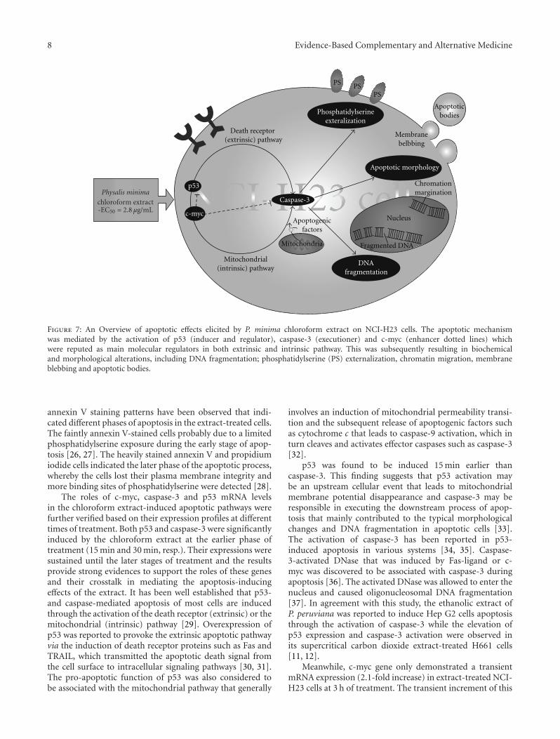
Figure 7: An Overview of apoptotic effects elicited by P. minima chloroform extract on NCI-H23 cells. The apoptotic mechanismwas mediated by the activation of p53 (inducer and regulator), caspase-3 (executioner) and c-myc (enhancer dotted lines) whichwere reputed as main molecular regulators in both extrinsic and intrinsic pathway. This was subsequently resulting in biochemicaland morphological alterations, including DNA fragmentation; phosphatidylserine (PS) externalization, chromatin migration, membraneblebbing and apoptotic bodies.
annexin V staining patterns have been observed that indi-cated different phases of apoptosis in the extract-treated cells.The faintly annexin V-stained cells probably due to a limitedphosphatidylserine exposure during the early stage of apop-tosis [26, 27]. The heavily stained annexin V and propidiumiodide cells indicated the later phase of the apoptotic process,whereby the cells lost their plasma membrane integrity andmore binding sites of phosphatidylserine were detected [28].
The roles of c-myc, caspase-3 and p53 mRNA levelsin the chloroform extract-induced apoptotic pathways werefurther verified based on their expression profiles at differenttimes of treatment. Both p53 and caspase-3 were significantlyinduced by the chloroform extract at the earlier phase oftreatment (15 min and 30 min, resp.). Their expressions weresustained until the later stages of treatment and the resultsprovide strong evidences to support the roles of these genesand their crosstalk in mediating the apoptosis-inducingeffects of the extract. It has been well established that p53-and caspase-mediated apoptosis of most cells are inducedthrough the activation of the death receptor (extrinsic) or themitochondrial (intrinsic) pathway [29]. Overexpression ofp53 was reported to provoke the extrinsic apoptotic pathwayvia the induction of death receptor proteins such as Fas andTRAIL, which transmitted the apoptotic death signal fromthe cell surface to intracellular signaling pathways [30, 31].The pro-apoptotic function of p53 was also considered tobe associated with the mitochondrial pathway that generally
involves an induction of mitochondrial permeability transi-tion and the subsequent release of apoptogenic factors suchas cytochrome c that leads to caspase-9 activation, which inturn cleaves and activates effector caspases such as caspase-3[32].
p53 was found to be induced 15 min earlier thancaspase-3. This finding suggests that p53 activation maybe an upstream cellular event that leads to mitochondrialmembrane potential disappearance and caspase-3 may beresponsible in executing the downstream process of apop-tosis that mainly contributed to the typical morphologicalchanges and DNA fragmentation in apoptotic cells [33].The activation of caspase-3 has been reported in p53-induced apoptosis in various systems [34, 35]. Caspase-3-activated DNase that was induced by Fas-ligand or c-myc was discovered to be associated with caspase-3 duringapoptosis [36]. The activated DNase was allowed to enter thenucleus and caused oligonucleosomal DNA fragmentation[37]. In agreement with this study, the ethanolic extract ofP. peruviana was reported to induce Hep G2 cells apoptosisthrough the activation of caspase-3 while the elevation ofp53 expression and caspase-3 activation were observed inits supercritical carbon dioxide extract-treated H661 cells[11, 12].
Meanwhile, c-myc gene only demonstrated a transientmRNA expression (2.1-fold increase) in extract-treated NCI-H23 cells at 3 h of treatment. The transient increment of this

Evidence-Based Complementary and Alternative Medicine 9
gene might enhance the apoptotic effect of p53 and caspase-3 with marked peak expression of these genes at 2-3 h oftreatment. This is supported by a finding that suggested c-myc gene as a strong inducer of cell death via inductionof several pro-apoptotic signal transduction mechanisms[38]. This gene has been described as a repressor to reduceubiquitination of p53 that leads to the accumulation of p53and subsequently triggers caspase-3 [31]. c-myc was reportedto induce apoptosis by destabilizing mitochondrial integrity[39]. The upregulation of c-myc was essential to enhance thepro-apoptotic proteins of Bcl-2 family and suppresses theiranti-apoptotic effects [38, 39].
In conclusion, this study could offer scientific basis forthe further in-depth evaluation of the chloroform extractof P. minima. It inhibited the proliferation of NCI-H23cells at dose- and time-dependent manner via an apoptoticprogramed cell death. The induction of apoptotic cell deathwas suggested to be mediated via p53-, caspase-3- andc-myc-dependent cell apoptotic pathways. Based on theresults obtained in this study, we constructed a mode ofaction as depicted in Figure 7. The model proposes thatthe chloroform extract induced the expression of p53 thatresulting in the release of apoptogenic factors that wouldfacilitate the cell apoptosis through the activation of caspasecascade.
Acknowledgments
The authors thank the Ministry of Science, Technology andInnovation (MOSTI) Malaysia and Universiti Sains Malaysiafor financing the project and providing the laboratoryfacilities.
References
[1] C.-H. Liang, L.-F. Liu, L.-Y. Shiu, Y.-S. Huang, L.-C. Chang,and K.-W. Kuo, “Action of solamargine on TNFs and cisplatin-resistant human lung cancer cells,” Biochemical and BiophysicalResearch Communications, vol. 322, no. 3, pp. 751–758, 2004.
[2] R. V. Ancuceanu and V. Istudor, “Pharmacologically activenatural compounds for lung cancer,” Alternative MedicineReview, vol. 9, no. 4, pp. 402–419, 2004.
[3] G. C. C. Lim and Y. Halimah, Second Report of the NationalCancer Registry. Cancer Incidence in Malaysia 2003, NationalCancer Registry, Kuala Lumpur, Malaysia, 2004.
[4] M. Zakaria and M. A. Mohamad, Traditional Malay MedicinalPlants, Fajar Bakti, Selangor, Malaysia, 1994.
[5] I. H. Burkill, A Dictionary of the Economic Products ofthe Malay Peninsula, vol. 1, Publication Unit Ministry ofAgriculture, 1966.
[6] C. C. Lee and P. Houghton, “Cytotoxicity of plants fromMalaysia and Thailand used traditionally to treat cancer,”Journal of Ethnopharmacology, vol. 100, no. 3, pp. 237–243,2005.
[7] H.-C. Chiang, S.-M. Jaw, and P.-M. Chen, “Inhibitory effectsof physalin B and physalin F on various human leukemia cellsin vitro,” Anticancer Research, vol. 12, no. 4, pp. 1155–1162,1992.
[8] M. S. Ricci and W.-X. Zong, “Chemotherapeutic approachesfor targeting cell death pathways,” Oncologist, vol. 11, no. 4,pp. 342–357, 2006.
[9] J. D. Benson, Y.-N. P. Chen, S. A. Cornell-Kennon et al.,“Validating cancer drug targets,” Nature, vol. 441, no. 7092,pp. 451–456, 2006.
[10] T. Hosoya, M. A. Arai, T. Koyano, T. Kowithayakorn, and M.Ishibashi, “Naturally occurring small-molecule inhibitors ofHedgehog/GLI-mediated transcription,” ChemBioChem, vol.9, no. 7, pp. 1082–1092, 2008.
[11] S.-J. Wu, S.-P. Chang, D.-L. Lin, S.-S. Wang, F.-F. Hou, andL.-T. Ng, “Supercritical carbon dioxide extract of Physalisperuviana induced cell cycle arrest and apoptosis in humanlung cancer H661 cells,” Food and Chemical Toxicology, vol. 47,no. 6, pp. 1132–1138, 2009.
[12] S.-J. Wu, L.-T. Ng, D.-L. Lin, S.-N. Huang, S.-S. Wang,and C.-C. Lin, “Physalis peruviana extract induces apoptosisin human Hep G2 cells through CD95/CD95L system andthe mitochondrial signaling transduction pathway,” CancerLetters, vol. 215, no. 2, pp. 199–208, 2004.
[13] W. T. Hsieh, K. Y. Huang, H. Y. Lin, and J. G. Chung, “Physalisangulata induced G2/M phase arrest in human breast cancercells,” Food and Chemical Toxicology, vol. 44, pp. 974–983,2006.
[14] H.-Z. Lee, W.-Z. Liu, W.-T. Hsieh, F.-Y. Tang, J.-G. Chung,and H. W.-C. Leung, “Oxidative stress involvement in Physalisangulata-induced apoptosis in human oral cancer cells,” Foodand Chemical Toxicology, vol. 47, no. 3, pp. 561–570, 2009.
[15] K. L. Ooi, Cytotoxicity and cell death mechanism elicitedby the Physalis minima chloroform extract and its fractionagainst different cancer cell lines, Ph.D. thesis, Universiti SainsMalaysia, Penang, Malaysia, 2009.
[16] S. Yamazaki, E. Onishi, K. Enami et al., “Proposal of standard-ized methods and reference for analyzing recombinant humantumor necrosis factor,” Japanese Journal of Medical Science &Biology, vol. 39, pp. 105–118, 1986.
[17] L. Lin and P. L. Hwang, “Antiproliferative effects of oxygenatedsterols: positive correlation with binding affinities for theantiestrogen-binding sites,” Biochimica et Biophysica Acta, vol.1082, no. 2, pp. 177–184, 1991.
[18] R. I. Geran, N. H. Greenberg, M. M. MacDonald, A. M.Schumacher, and B. J. Abbott, “Protocols for screeningchemical agents and natural products against animal tumoursand other biological systems,” Cancer Chemotherapy Reports,vol. 3, pp. 59–61, 1972.
[19] A. L. Perez-Castorena, M. Garcıa, M. Martınez, and E.Maldonado, “Physalins from Physalis solanaceus,” BiochemicalSystematics and Ecology, vol. 32, no. 12, pp. 1231–1234, 2004.
[20] H. I. Ferreira Magalhaes, M. L. Veras, M. Rocha Torres etal., “In-vitro and in-vivo antitumour activity of physalinsB and D from Physalis angulata,” Journal of Pharmacy andPharmacology, vol. 58, no. 2, pp. 235–241, 2006.
[21] E. Neuber, C. M. Luetjens, A. W. S Cahn, and G. P. Scahtten,“Analysis of DNA fragmentation of in vitro cultured bovineblastocysts using TUNEL,” Theriogenology, vol. 57, pp. 2193–2202, 2002.
[22] J. F. R. Kerr, C. M. Winterford, and B. V. Harmon, “Apoptosis:its significance in cancer and cancer therapy,” Cancer, vol. 73,no. 8, pp. 2013–2026, 1994.
[23] D. Krajcı, V. Mares, V. Lisa, A. Spanova, and J. Vorlıcek,“Ultrastructure of nuclei of cisplatin-treated C6 glioma cellsundergoing apoptosis,” European Journal of Cell Biology, vol.79, no. 5, pp. 365–376, 2000.
[24] M. Alonso, C. Tamasdan, D. C. Miller, and E. W. Newcomb,“Flavopiridol induces apoptosis in glioma cell lines indepen-dent of retinoblastoma and p53 tumor suppresor pathway
10 Evidence-Based Complementary and Alternative Medicine
alterations by a caspase-independent pathway,” MolecularCancer Therapeutics, vol. 2, pp. 139–150, 2003.
[25] M. K. Callahan, P. Williamson, and R. A. Schlegel, “Surfaceexpression of phosphatidylserine on macrophages is requiredfor phagocytosis of apoptotic thymocytes,” Cell Death andDifferentiation, vol. 7, no. 7, pp. 645–653, 2000.
[26] R. Overbeeke, H. Steffens-Nakken, I. Vermes, C. Reutel-ingsperger, and C. Haanen, “Early features of apoptosisdetected by four different flow cytometry assays,” Apoptosis,vol. 3, no. 2, pp. 115–121, 1998.
[27] H. C. Chiu, T. T. Chih, Y. M. Hsian, C. H. Tseng, M. J. Wu,and Y. C. Wu, “Bullatacin, a potent antitumor annonaceousacetogenin, induced apoptosis through a reduction of intra-cellular cAMP and cGMP levels in human hepatoma 2.2.15cells,” Biochemical Pharmacology, vol. 65, pp. 319–327, 2003.
[28] G. Koopman, C. P. M. Reutelingsperger, G. A. M. Kuijten, R.M. J. Keehnen, S. T. Pals, and M. H. J. van Oers, “Annexin V forflow cytometric detection of phosphatidylserine expression onB cells undergoing apoptosis,” Blood, vol. 84, no. 5, pp. 1415–1420, 1994.
[29] L. J. Hofseth, S. P. Hussain, and C. C. Harris, “p53: 25 yearsafter its discovery,” Trends in Pharmacological Sciences, vol. 25,no. 4, pp. 177–181, 2004.
[30] S. Haupt, M. Berger, Z. Goldberg, and Y. Haupt, “Apoptosis—the p53 network,” Journal of Cell Science, vol. 116, no. 20, pp.4077–4085, 2003.
[31] B. Hoffman and D. A. Liebermann, “Apoptotic signaling by c-MYC,” Oncogene, vol. 27, no. 50, pp. 6462–6472, 2008.
[32] Z. Jin and W. S. El-Deiry, “Overview of cell death signalingpathways,” Cancer Biology and Therapy, vol. 4, no. 2, pp. 139–163, 2005.
[33] G. S. Salvesen and S. J. Riedl, “Caspase mechanisms,” Advancesin Experimental Medicine and Biology, vol. 615, pp. 13–23,2008.
[34] C. Feng Gao, S. Ren, L. Zhang et al., “Caspase-dependentcytosolic release of cytochrome c and membrane translocationof Bax in p53-induced apoptosis,” Experimental Cell Research,vol. 265, no. 1, pp. 145–151, 2001.
[35] T. Chen, Y.-S. Wong, W. Zheng, and J. Liu, “Caspase- andp53-dependent apoptosis in breast carcinoma cells inducedby a synthetic selenadiazole derivative,” Chemico-BiologicalInteractions, vol. 180, no. 1, pp. 54–60, 2009.
[36] H. Sakahira, M. Enari, and S. Nagata, “Cleavage of CADinhibitor in CAD activation and DNA degradation duringapoptosis,” Nature, vol. 391, no. 6662, pp. 96–99, 1998.
[37] Q. Cui, J.-H. Yu, J.-N. Wu et al., “P53-mediated cell cycle arrestand apoptosis through a caspase-3- independent, but caspase-9-dependent pathway in oridonin-treated MCF-7 humanbreast cancer cells,” Acta Pharmacologica Sinica, vol. 28, no. 7,pp. 1057–1066, 2007.
[38] I. Petak, J. A. Houghton, and L. Kopper, “Molecular targetingof cell death signal transduction pathways in cancer,” CurrentSignal Transduction Therapy, vol. 1, pp. 113–331, 2006.
[39] P. Juin, A. Hunt, T. Littlewood et al., “c-Myc functionallycooperates with Bax to induce apoptosis,” Molecular andCellular Biology, vol. 22, no. 17, pp. 6158–6169, 2002.

Submit your manuscripts athttp://www.hindawi.com
Stem CellsInternational
Hindawi Publishing Corporationhttp://www.hindawi.com Volume 2014
Hindawi Publishing Corporationhttp://www.hindawi.com Volume 2014
MEDIATORSINFLAMMATION
of
Hindawi Publishing Corporationhttp://www.hindawi.com Volume 2014
Behavioural Neurology
EndocrinologyInternational Journal of
Hindawi Publishing Corporationhttp://www.hindawi.com Volume 2014
Hindawi Publishing Corporationhttp://www.hindawi.com Volume 2014
Disease Markers
Hindawi Publishing Corporationhttp://www.hindawi.com Volume 2014
BioMed Research International
OncologyJournal of
Hindawi Publishing Corporationhttp://www.hindawi.com Volume 2014
Hindawi Publishing Corporationhttp://www.hindawi.com Volume 2014
Oxidative Medicine and Cellular Longevity
Hindawi Publishing Corporationhttp://www.hindawi.com Volume 2014
PPAR Research
The Scientific World JournalHindawi Publishing Corporation http://www.hindawi.com Volume 2014
Immunology ResearchHindawi Publishing Corporationhttp://www.hindawi.com Volume 2014
Journal of
ObesityJournal of
Hindawi Publishing Corporationhttp://www.hindawi.com Volume 2014
Hindawi Publishing Corporationhttp://www.hindawi.com Volume 2014
Computational and Mathematical Methods in Medicine
OphthalmologyJournal of
Hindawi Publishing Corporationhttp://www.hindawi.com Volume 2014
Diabetes ResearchJournal of
Hindawi Publishing Corporationhttp://www.hindawi.com Volume 2014
Hindawi Publishing Corporationhttp://www.hindawi.com Volume 2014
Research and TreatmentAIDS
Hindawi Publishing Corporationhttp://www.hindawi.com Volume 2014
Gastroenterology Research and Practice
Hindawi Publishing Corporationhttp://www.hindawi.com Volume 2014
Parkinson’s Disease
Evidence-Based Complementary and Alternative Medicine
Volume 2014Hindawi Publishing Corporationhttp://www.hindawi.com