cytotoxic isovaleryl sucrose esters from ainsliaea
TRANSCRIPT

RSC Advances
PAPER
Ope
n A
cces
s A
rtic
le. P
ublis
hed
on 1
1 A
pril
2017
. Dow
nloa
ded
on 1
2/26
/202
1 5:
19:4
1 PM
. T
his
artic
le is
lice
nsed
und
er a
Cre
ativ
e C
omm
ons
Attr
ibut
ion-
Non
Com
mer
cial
3.0
Unp
orte
d L
icen
ce.
View Article OnlineView Journal | View Issue
Cytotoxic isovale
aDepartment of Phytochemistry, School o
University, Shanghai 200433, P. R. Chin
[email protected] University of Traditional ChinesecShanghai Institute of Pharmaceutical Indus
† Electronic supplementary informa10.1039/c7ra01986f
Cite this: RSC Adv., 2017, 7, 20865
Received 17th February 2017Accepted 4th April 2017
DOI: 10.1039/c7ra01986f
rsc.li/rsc-advances
This journal is © The Royal Society of C
ryl sucrose esters from Ainsliaeayunnanensis: reduction of mitochondrialmembrane potential and increase of reactiveoxygen species levels in A549 cells†
Xin Fang,ab Zhi-Guo Zhuo,a Xi-Ke Xu,a Ji Ye,a Hui-Liang Li,a Yun-Heng Shen *a
and Wei-Dong Zhang*abc
Eight isovaleryl sucrose esters, named ainslosides A–H (1–8), were isolated from Ainsliaea yunnanensis
Franch. Their structures, including the absolute configurations of the sugar residues, were elucidated by
extensive analysis of NMR spectra and acid hydrolysis. All compounds were tested in vitro for cytotoxicity
against four human tumour cell lines, A549, HCT116, MDA-MB-231, and BEL7404. Among the
compounds tested, ainsloside B (2) showed potent cytotoxicity against the A549 cell line with an IC50
value of 3.3 mM. Flow cytometry analysis showed that compound 2 can arrest the cell cycle at the G0/G1
phase and induce cell apoptosis in A549 cells. Further studies indicated that the apoptosis-inducing
effect of compound 2 may be involved in the reduction of mitochondrial membrane potential (MMP) and
increase of reactive oxygen species (ROS) level in A549 cells.
Introduction
The genus Ainsliaea of Asteraceae comprises about 70 species,which is mainly distributed in the southeast of Asia. ManyAinsliaea species have been long used for the treatment ofvarious diseases, such as rheumatism, traumatic injury, enter-itis dysentery, sore throat, and urological and gynecologicaldiseases.1 The chemical constituents and crude extracts fromAinsliaea species have been reported to have various biologicalactivities, such as anti-microbial,2 cytotoxic,3 antiviral,4 antiox-idant,5 and anti-inammation6–8 activities. Our previous inves-tigations on Ainsliaea species have reported the isolation ofa series of sesquiterpenoids, triterpenoids, sesquiterpenoidlactone dimers and trimers, of which the rst two guaianolidetrimers, ainsliatrimers A and B from A. fulvioides,9 and a newguaianolide dimer with an unusual carbon skeleton, ain-sliadimer A from A. macrocephala,10 showed potent cytotoxicityand anti-inammatory activities. Ainsliaea yunnanensis Franch.,a kind of perennial plant of the genus Ainsliaea, is distributedexclusively in China. In Chinese folk medicine, the whole plantsof A. yunnanensis have been used for the treatment of traumaticinjury and rheumatism pain.11 However, few studies of the
f Pharmacy, Second Military Medical
a. E-mail: [email protected];
Medicine, Shanghai 201203, P. R. China
try, Shanghai 200400, P. R. China
tion (ESI) available. See DOI:
hemistry 2017
chemical constituents and bioactivities of A. yunnanensis werereported except for the isolation of several sesquiterpenoids,triterpenoids, and some phenoloids.12,13 As part of ourcontinuing efforts to discover structurally interesting bioactivecompounds from Ainsliaea species, eight isovaleryl sucroseesters, ainslosides A–H (1–8), were isolated from the wholeplants of A. yunnanensis. Compounds 1–8 were in vitro tested forcytotoxicity against four human tumour cell lines A549,HCT116, MDA-MB-231, and BEL7404, and cell cycle arrestingand apoptosis induction of ainsloside B (2) in A549 cell line.Herein, we described the isolation, structural elucidation, andin vitro antitumor evaluation (Fig. 1).
Fig. 1 Structures of compounds 1–8.
RSC Adv., 2017, 7, 20865–20873 | 20865

Fig. 2 Key 1H–1H COSY, HMBC, and NOESY correlations forcompound 1.
RSC Advances Paper
Ope
n A
cces
s A
rtic
le. P
ublis
hed
on 1
1 A
pril
2017
. Dow
nloa
ded
on 1
2/26
/202
1 5:
19:4
1 PM
. T
his
artic
le is
lice
nsed
und
er a
Cre
ativ
e C
omm
ons
Attr
ibut
ion-
Non
Com
mer
cial
3.0
Unp
orte
d L
icen
ce.
View Article Online
Results and discussion
Ainsloside A (1) was obtained as colourless oil. Its molecularformula was determined to be C37H62O16 as deduced frompositive HR-ESI-MS (m/z 785.3942 [M + Na]+, calcd 785.3936),indicating seven degrees of unsaturation. The IR spectrum of 1showed characteristic absorption bands of hydroxyl group (3471cm�1) and carbonyl group (1743 cm�1). The 13C and DEPT NMRspectra (Table 1) displayed 37 carbon signals, in which 12oxygenated carbon resonances were observed in the region 60 <d < 105 ppm (dC 105.5, 90.1, 78.1, 77.5, 77.0, 72.4, 70.6, 70.0,68.8, 64.0, 63.6, 62.1), implying the presence of a disaccharideresidue. Characteristic signals from the NMR spectra, includingan anomeric proton at dH 5.64 (d, J ¼ 3.5 Hz) and the corre-sponding anomeric carbon at dC 90.1, a hydroxymethyl at dC
62.1, an hemiketal anomeric carbon at dC 105.5, and twohydroxymethyls at dC 63.6 and 64.0, exhibited quite similaritywith those of sucrose previously reported in literatures,14 indi-cating that the disaccharide moiety should be a sucrose residue.The 1H–1H COSY correlations of H-1/H-2/H-3/H-4/H-5/H-6 andH-30/H-40/H-50/H-60, in combination with the HMBC correlationfrom the anomeric proton (dH 5.64) of glucopyranosyl unit to thehemiketal anomeric carbon at dC 105.5, supported the above
Table 1 1H (500 MHz) and 13C (125 MHz) NMR data of anslosides A–D (
No.
1 2
dH dC dH dC
1 5.64 d (3.5) 90.1 5.55 d (3.5) 92.22 4.90 dd (10.0, 3.5) 72.4 3.66 m 72.33 4.05 m 70.0 3.88 t (9.5) 72.24 4.93 t (10.0) 70.6 4.87 t (9.5) 70.25 4.26 m 68.8 4.19 m 68.96 4.20 dd (12.0, 2.0) 62.1 4.15 m 62.1
4.15 dd (12.0, 5.5)10 3.66 m 63.6 4.32 m 63.0
20 — 105.5 — 104.330 4.34 t (7.0) 77.0 4.25 d (7.0) 77.240 5.17 t (7.0) 77.5 5.16 d (7.0) 77.450 4.12 m 78.1 4.08 m 78.060 4.30 m 64.0 4.31 m 64.1
C]O — 173.0 — 173.0172.9 172.9172.6 172.7172.5 172.6172.4 172.2
CH2 2.22–2.28 m 43.2 2.21–2.26 m 43.243.0 43.143.0 43.142.9 43.042.8 42.8
CH 2.06–2.14 m 25.7 2.06–2.14 m 25.725.7 25.625.6 25.625.5 25.625.4 25.4
CH3 0.93–0.98 m 22.3 � 10 0.92–0.98 m 22.3 � 10
20866 | RSC Adv., 2017, 7, 20865–20873
inference. In addition, the relative conguration of theanomeric proton of glucopyranosyl residue was determined tobe a-oriented based on the small coupling constant (J ¼ 3.5 Hz)between H-1 and H-2,15 while the relative conguration offructose was assigned to be b-orientation through the NOESYcorrelations of H-30 with H-10 and H-50 (Fig. 2). This conclusionalso can be conrmed by comparing their 13C NMR data withthose of a-glucose and b-fructose previously reported in litera-tures.16 Moreover, compound 1 was submitted alkaline hydro-lysis, and the hydrolysate was extracted with CHCl3 to afford
1–4) (in ppm) (CDCl3) (J in Hz)
3 4
dH dC dH dC
5.52 d (3.5) 91.9 5.57 d (3.5) 89.73.77 m 70.9 4.89 dd (10.0, 3.5) 69.85.25 t (9.5) 73.4 5.40 t (10.0) 72.25.07 t (10.0) 67.3 3.49 t (9.5) 69.44.29 m 68.8 4.22 m 70.94.18 dd (12.5, 2.0) 61.8 4.36 m 63.04.15 m3.76 m 64.1 3.58 q (14.5) 63.73.68 m— 105.0 — 105.34.31 m 78.0 4.34 m 76.85.20 t (6.5) 77.5 5.16 t (7.0) 77.34.12 m 78.6 4.08 m 78.44.34 m 64.3 4.43 m 64.2
4.27 dd (12.0, 3.5)— 174.1 — 174.1
172.6 173.5172.6 172.8172.5 172.7171.5 172.0
2.15–2.26 m 43.3 2.16–2.26 m 43.443.0 43.043.0 43.043.0 42.842.8 42.7
2.02–2.13 m 25.7 2.01–2.12 m 25.625.6 25.625.6 25.525.4 25.525.3 25.2
0.92–0.98 m 22.3 � 10 0.90–0.99 m 22.3 � 10
This journal is © The Royal Society of Chemistry 2017

Fig. 3 Key 1H–1H COSY, and HMBC correlations for compounds 2–4.
Paper RSC Advances
Ope
n A
cces
s A
rtic
le. P
ublis
hed
on 1
1 A
pril
2017
. Dow
nloa
ded
on 1
2/26
/202
1 5:
19:4
1 PM
. T
his
artic
le is
lice
nsed
und
er a
Cre
ativ
e C
omm
ons
Attr
ibut
ion-
Non
Com
mer
cial
3.0
Unp
orte
d L
icen
ce.
View Article Online
isovaleric acid. The subsequent acid hydrolysis yielded twosugar units, and the sugar units were identied as D-glucose andD-fructose through the HPLC analysis and comparing theircorresponding specic optical rotations with those of authenticsamples (glucose: [a]23D +53; fructose: [a]23D �149). In the higheld region of the 1H NMR spectrum, three groups of over-lapping signals, corresponding to methylene, methine, andmethyl protons, were observed occurring at dH 2.22–2.28 (10H,m), 2.06–2.14 (5H, m), and 0.93–0.98 (30H, m), respectively(Table 1). Combining with the 13C and DEPT NMR, empiricalcalculations indicated the presence of a total of tenmethyls, vemethines and ve methylenes. Also, the 13C NMR spectrum of 1exhibited ve ester carbonyl resonances at dC 173.0, 172.9,172.6, 172.5 and 172.4. Therefore, it may be proposed that thestructure of 1 composed of a sucrose moiety and ve isovalerylmoieties. Compared with sucrose,14 the 13C chemical shis of 1at C-2, C-4, C-6, C-40, and C-60, were shied to downeld, whiletheir adjacent carbons were shied to upeld, implying that theve isovaleryl moieties may be substituted at C-2, C-4, C-6, C-40,and C-60, respectively. This deduction was further evidenced onthe basis of analysis of the HMBC spectrum which showedcross-peaks between H-40 (dH 5.17), H-4 (dH 4.93), H-6 (dH 4.20,4.15), H-60 (dH 4.30), H-2 (dH 4.90) and the corresponding estercarbonyls at dC 173.0, 172.9, 172.6, 172.5, and 172.4, respectively(Fig. 2). Thus, compound 1 was deduced as 2,4,40,6,60-pentakis-isovaleryl-b-D-fructofuranosyl-a-D-glucopyranoside, and givena trivial name ainsloside A.
Ainslosides B–D (2–4) shared the same molecular formulaC37H62O16 as ainsloside A (1) by analysis of HR-ESI-MS andNMR spectroscopy, and their IR spectra suggested the presenceof hydroxyl group and ester carbonyl group based on charac-teristic absorption bands. The 13C and DEPT NMR spectra(Table 1) of all the compounds also showed twelve carbonsignals in the region 60 < d < 105 ppm which revealed the samesucrose moiety as compound 1. Deducting the sucrose moiety,in combination with the 1H NMR spectrum (Table 1), the le 25carbons were assigned as ten methyls, ve methylenes, vemethines, and ve ester carbonyls, which belonged to ve iso-valeryls. Comprehensive analyses of 1D and 2D NMR spectra ofainslosides B–D (2–4) suggested that these compounds wereisovaleryl substituted derivatives of sucrose similar tocompound 1 with different substitution pattern. The linkagepositions of the isovaleryls were assigned on the basis ofinterpretation of the HMBC spectrum. In the HMBC spectrumof compound 2, it could be observed the cross-peaks of H-40 (dH5.16), H-4 (dH 4.87), H-6 (dH 4.15), H-60 (dH 4.31), H-10 (dH 4.32)with the corresponding ester carbonyls of ve isovaleryls at dC173.0, 172.9, 172.7, 172.6, and 172.2, respectively. So,compound 2 was deduced as 10,4,40,6,60-pentakis-isovaleryl-b-D-fructofuranosyl-a-D-glucopyranoside, and named ainsloside B.In the same way, compound 3 was determined as 3,30,4,40,6-pentakis-isovaleryl-b-D-fructofuranosyl-a-D-glucopyranoside bythe long-range HMBC correlations between H-3 (dH 5.25), H-30
(dH 4.31), H-6 (dH 4.18), H-40 (dH 5.20), H-4 (dH 5.07) and thecorresponding ester carbonyls of ve isovaleryls at dC 174.1,172.6, 172.6, 172.5, and 171.5, respectively, and named ainslo-side C. Meanwhile, by the key HMBC correlations of H-3 (dH
This journal is © The Royal Society of Chemistry 2017
5.40), H-6 (dH 4.36), H-60 (dH 4.27), H-40 (dH 5.16), H-2 (dH 4.89)with the corresponding ester carbonyls of ve isovaleryls at dC174.1, 173.5, 172.8, 172.7, and 172.0, respectively, the structureof compound 4 was deduced as 2,3,40,6,60-pentakis-isovaleryl-b-D-fructofuranosyl-a-D-glucopyranoside, and named ainsloside D(Fig. 3).
Ainsloside E (5) was obtained as colourless oil. The molec-ular formula of 5 was inferred as C32H54O15 based on the HR-ESI-MS (m/z 701.3365 [M + Na]+, calcd 701.3360), implying theexistence of six degrees of unsaturation. The IR spectrum of 5showed characteristic absorption bands of hydroxyl group (3467cm�1), and ester carbonyl group (1739 cm�1). In the 13C andDEPT NMR spectra of 5 (Table 2), 12 carbon resonances in theregion 60 < d < 105 ppm showed characteristic signals of sucrose(dC 104.9, 90.1, 78.0, 77.3, 76.8, 72.0, 70.7, 70.7, 70.6, 64.4, 63.4,63.2), in agreement with those of previously reported for sucrosein literature,14 revealing the presence of sucrose moiety. The1H–1H COSY correlations of H-1/H-2/H-3/H-4/H-5/H-6 and H-30/H-40/H-50/H-60 further supported the above deduction. Similarto compounds 1–4, in the high eld region of the 1H NMRspectrum of 5 (Table 2), three groups of overlapping protonsignals for methylene, methine, and methyl signals wereobserved occurring in the region dH 2.20–2.29 (8H, m), 2.06–2.12(4H, m) and 0.93–0.97 (24H, m), respectively. Meanwhile, the13C and DEPT NMR spectra of 5 only exhibited four estercarbonyls at dC 173.9, 173.0, 172.8, and 172.6, eight methyls,four methylenes, and four methines. The information impliedthat there were only four isovaleryls in the structure of 5.Therefore, it could be concluded that compound 5was a sucrosederivative with four isovaleryl groups. The downeld shied C-2, C-6, C-40, and C-60 and their upeld shied adjacent carbons,disclosed that the four isovaleryl groups were attached to C-2, C-6, C-40, and C-60 positions, respectively. In the HMBC spectrum,the proton signals of the sucrose residue were observed thecorrelations from H-6 (dH 4.36) to the ester carbonyl at dC 173.9,from H-60 (dH 4.31) to the ester carbonyl at dC 173.0, from H-40
RSC Adv., 2017, 7, 20865–20873 | 20867

Table 2 1H (500 MHz) and 13C (125 MHz) NMR data of anslosides E-H (in ppm) (CDCl3) (J in Hz)
No.
5 6 7 8
dH dC dH dC dH dC dH dC
1 5.53 d (3.5) 90.1 5.54 d (3.5) 89.5 5.44 d (3.5) 91.7 5.67 d (3.5) 90.12 4.78 dd (10.0, 3.5) 72.0 4.87 dd (10.0, 4.0) 72.4 3.69 dd (9.5, 3.5) 72.2 4.87 dd (10.5, 4.0) 72.63 4.02 t (9.5) 70.6 4.04 dd (10.0, 4.0) 70.0 3.94 t (9.5) 71.8 4.05 t (10.0) 69.84 3.40 t (9.5) 70.7 4.94 t (9.5) 70.7 4.84 t (10.0) 70.1 4.95 t (10.0) 70.45 4.08 m 70.7 4.21 m 68.7 4.20 m 69.2 4.26 m 68.96 4.36 m 63.2 4.21 m 62.1 4.16 m 62.2 4.25 m 61.7
4.13 dd (13.0, 6.0) 4.14 dd (12.5, 5.0)10 3.58 q (12.0) 63.4 3.62 q (7.0) 64.2 3.76 d (12.5) 63.9 3.61 q (12.5) 64.0
3.61 d (13.0)20 — 104.9 — 104.5 — 104.7 — 105.530 4.08 m 78.0 4.18 m 78.3 4.25 d (7.0) 78.7 4.36 d (7.0) 76.940 5.16 t (7.0) 77.3 4.02 m 75.8 5.17 t (7.0) 78.0 5.26 t (6.5) 76.750 4.31 m 76.8 3.94 m 79.6 4.12 m 78.5 3.97 m 81.560 4.31 m 64.4 4.31 m 64.1 4.34 dd (12.0, 5.0) 64.7 3.84 dd (13.0, 3.0) 61.4
3.67 dd (13.0, 4.0)C]O — 173.9 — 173.3 — 173.1 — 173.1
173.0 172.9 172.8 172.9172.8 172.8 172.7 172.8172.6 172.4 172.6 172.7
CH2 � 4 2.20–2.29 m 43.1 2.23–2.29 m 43.2 2.18–2.27 m 43.2 2.23–2.30 m 43.243.0 43.0 43.1 43.142.9 43.0 43.0 42.942.9 42.9 42.8 42.8
CH � 4 2.06–2.12 m 25.7 2.06–2.14 m 25.7 2.03–2.14 m 25.7 2.06–2.13 m 25.725.7 25.7 25.7 25.725.6 25.6 25.6 25.725.4 25.5 25.4 25.4
CH3 � 8 0.93–0.97 m 22.3 � 8 0.94–0.98m 22.3 � 8 0.92–0.98 m 22.3 � 8 0.93–0.97 m 22.3 � 8
RSC Advances Paper
Ope
n A
cces
s A
rtic
le. P
ublis
hed
on 1
1 A
pril
2017
. Dow
nloa
ded
on 1
2/26
/202
1 5:
19:4
1 PM
. T
his
artic
le is
lice
nsed
und
er a
Cre
ativ
e C
omm
ons
Attr
ibut
ion-
Non
Com
mer
cial
3.0
Unp
orte
d L
icen
ce.
View Article Online
(dH 5.16) to the ester carbonyl at dC 172.8, and fromH-2 (dH 4.78)to ester carbonyl at dC 172.6, revealing that four isovaleroxylswere substituted at C-2, C-40, C-6, and C-60 positions of sucroseresidue, respectively (Fig. 4). Thus, compound 5 were deduced
Fig. 4 Key 1H–1H COSY, HMBC, and NOESY correlations forcompound 5–8.
20868 | RSC Adv., 2017, 7, 20865–20873
as 2,40,6,60-tetrakis-isovaleryl-b-D-fructofuranosyl-a-D-glucopyr-anoside, and named ainsloside E.
Ainslosides F–H (6–8) shared the same molecular formulaC32H54O15 as that of compound 5 due to analysis of their HR-ESI-MS spectra. The IR spectra indicated the presence ofhydroxyl and ester carbonyl based on their characteristicabsorption bands. The 1H and 13C NMR spectra (Table 2) of 6–8exhibited the same functionalities as ainsloside E (5), including12 carbon resonances for a sucrose in the region 60 < d <105 ppm and four ester carbonyls, eight methyls, four methy-lenes, and four methines in 13C NMR and DEPT spectra,together with three groups of overlapping proton signals formethylene, methine, and methyl in the 1H NMR spectrum(Table 1). The above evidences indicated that compounds 6–8were four isovaleryls substituted sucrose ester similar tocompound 5 with different substitution pattern.
The linkage positions of four isovaleryls were determined byHMBC experiment. In the HMBC spectrum of 6, the sucroseprotons at dH 4.94 (H-4), 4.31 (H-60), 4.21 (H-6), 4.87 (H-2) werecorrelated with the corresponding ester carbonyls of four iso-valeryls at dC 173.3, 172.9, 172.8, and 172.4, respectively,implying that four isovaleroyoxyls were linked to C-4, C-60, C-6,and C-2 positions of sucrose moiety, respectively. Therefore, thestructure of 6 was deduced as 2,4,6,60-tetrakis-isovaleryl-b-D-fructofuranosyl-a-D-glucopyranoside, and named ainsloside F.Similarly, the HMBC correlations from H-4 (dH 4.84) to the estercarbonyl at dC 173.1, from H-6 (dH 4.16) to the ester carbonyl at
This journal is © The Royal Society of Chemistry 2017

Fig. 5 Cell cycle assay of A549 cells exposed to different concen-trations of compound 2 were examined by flow cytometry (A). Theeffect of compound 2 on the cell cycle and the expression of cell cycleregulators in A549 cells (B). The results are represented as the mean �SEM for three independent experiments with similar results. *P < 0.05
Paper RSC Advances
Ope
n A
cces
s A
rtic
le. P
ublis
hed
on 1
1 A
pril
2017
. Dow
nloa
ded
on 1
2/26
/202
1 5:
19:4
1 PM
. T
his
artic
le is
lice
nsed
und
er a
Cre
ativ
e C
omm
ons
Attr
ibut
ion-
Non
Com
mer
cial
3.0
Unp
orte
d L
icen
ce.
View Article Online
dC 172.8, from H-40 (dH 5.17) to the ester carbonyl at dC 172.7,and from H-60 (dH 4.34) to the ester carbonyl at dC 172.6,proposed the structure of 7 as 4,40,6,60-tetrakis-isovaleryl-b-D-fructofuranosyl-a-D-glucopyranoside, given a trivial name ain-sloside G. The structure of 8 was identied as 2,4,40,6-tetrakis-isovaleryl-b-D-fructofuranosyl-a-D-glucopyranoside on the basisof the HMBC correlations between H-40 (dH 5.26) with the estercarbonyl at dC 173.1, between H-2 (dH 4.87) with the estercarbonyl at dC 172.9, between H-4 (dH4.95) with the estercarbonyl at dC 172.8, and between H-6 (dH 4.25) with the estercarbonyl at 172.7 (Fig. 4).
Sucrose esters are of interest in such applications as fatsubstitutes, bleaching boosters, and emulsiers in the food andcosmetic industries.17,18 Meanwhile, they also possessed bioac-tivities such as antimicrobial, antitumor, and insecticidalactivities.19 Their properties depend strongly on their compo-sitions in terms of degree of substitution and regiochemistry.20
Previous studies showed the hydroxyl group at OH-2 is veryreactive in water just as it is in organic solvents,21 while the esterat OH-6 is much more stable in synthesis.22 In our studies, thecontent of ainsloside A is much more than others', whichsuggests that the esters at OH-2, OH-4, OH-40, OH-6 and OH-60
might be more stable or important in plant, at least in thisspecies.
All isolates were evaluated for in vitro cytotoxic activityagainst four human tumour cell lines (A549, HCT116, MDA-MB-231, and BEL7404), using MTT assay with doxorubicin as posi-tive control (Table 3). Among these isolates, compounds 1, 2,and 5 showed strong inhibitory activity against A549 cell linewith IC50 values of 9.74, 3.30, and 10.75 mM, compounds 1, 2, 4,and 5 exhibited weak inhibitory activity against HCT116 cell linewith IC50 values of 10.77, 10.25, 16.56, and 16.10 mM, In addi-tion, compounds 1, 2, 4, and 5 also showed weak inhibitoryactivity against BEL7404 cell line with IC50 values of 11.45,11.23, 15.95, and 12.77 mM. Of all the isolates tested, compound2 displayed the strongest inhibitory activity against the prolif-eration of A549 cell line in a dose and time dependent manner(Fig. S84, see ESI†).
Cell cycle dysregulation contributes to the aberrant cellproliferation and development of cancer.23,24 Moreover, target-ing apoptosis is considered as one of the major strategies for
Table 3 In vitro cytotoxic activities of compounds 1–8 (mean � SD, n ¼
No.
IC50 (mM)
A549 HCT116
1 9.74 � 0.71 10.77 �2 3.30 � 0.22 10.25 �3 >100 68.51 �4 19.93 � 0.93 16.56 �5 10.75 � 1.07 16.10 �6 60.06 � 2.71 23.11 �7 46.19 � 3.21 23.65 �8 66.00 � 1.17 54.82 �Doxorubicin 0.034 � 0.002 0.045 �
This journal is © The Royal Society of Chemistry 2017
developing anticancer drugs and most clinically used anti-cancer medicines can induce cell apoptosis.25 Considering thestrong inhibition of compound 2 against the proliferation ofA549 cells, compound 2 were further studied for its cell cyclearresting and apoptosis induction in A549 cells.
To determine if compound 2 can arrest cell-cycle progres-sion, A549 cells were exposed to various concentrations ofcompound 2 for 24 hours, and the distribution of cells in thecycle was determined by ow-cytometric analysis. As shown inFig. 5, A549 cells treated with compound 2 exhibited a dosedependent increase in the proportion of cells in the G0/G1 phaseand a decrease of cells in the G2/M phase compared to theuntreated control. This indicated that the growth inhibitioninduced by 2 in a dose-dependent manner occurs through thearrest of A549 cells in G0/G1 phase.
In addition, we examined compound 2-induced apoptosis inA549 cells by using annexin V-FITC/PI double staining method.The X- and Y-axis represented annexin V-FITC and PI staining,respectively. The right upper and lower right quadrant of thegure represented late and early stages of cell apoptosis,respectively. As illustrated in Fig. 6, compound 2 induced
3)
MDA-MB-231 BEL7404
0.87 39.10 � 1.87 11.45 � 0.570.53 19.59 � 0.69 11.23 � 0.610.86 >100 95.48 � 4.470.67 38.23 � 1.23 15.95 � 1.311.12 25.54 � 2.13 12.77 � 0.781.73 95.19 � 3.57 29.86 � 1.752.01 >100 53.61 � 1.313.15 >100 80.03 � 2.160.003 0.11 � 0.01 0.12 � 0.01
vs. the control.
RSC Adv., 2017, 7, 20865–20873 | 20869

Fig. 6 Compound 2-induced apoptosis in A549 cells. (A) Apoptosiswas evaluated using an annexin V-FITC apoptosis detection kit andflow cytometry. The representative pictures are from A549 cellsincubated with different concentrations. (B) Compound 2 inducedapoptosis in A549 cells in a dose dependent manner. The data areexpressed as the means� SEM of three independent experiments withthe similar results. *P < 0.05; **P < 0.01 vs. the control.
Fig. 7 TheMMP of A549 cells treatedwith compound 2 (A). The loss ofthe MMP in A549 cells following treatment of compound 2 (B). Thedata are expressed as the means � SEM for three independentexperiments with the similar results. *P < 0.05 vs. the control.
RSC Advances Paper
Ope
n A
cces
s A
rtic
le. P
ublis
hed
on 1
1 A
pril
2017
. Dow
nloa
ded
on 1
2/26
/202
1 5:
19:4
1 PM
. T
his
artic
le is
lice
nsed
und
er a
Cre
ativ
e C
omm
ons
Attr
ibut
ion-
Non
Com
mer
cial
3.0
Unp
orte
d L
icen
ce.
View Article Online
a dose-dependent increase in A549 apoptosis when treated with10 mMof 2 for 24 h. These results conrmed that the cytotoxicityof compound 2 to A549 cells is associated with arrest of cell-cycle progression and induction of apoptosis.
Depolarization of mitochondrial membrane potential(MMP) is one of the characteristic event of apoptosis.26,27 Toinvestigate whether compound 2-induced apoptosis was asso-ciated with mitochondrial dysfunction, we analyzed MMPchanges of the A549 cells by staining with mitochondriasensitive Rhodamine 123, and the results showed the MMP ofthe A549 cells was decreased dose-dependently aer treatmentwith different concentration of compound 2 for 24 h. Theexperiment indicated that compound 2-induced A549 cellsapoptosis may be associated with the disruption of MMP(Fig. 7).
Previous studies showed that ROS (reactive oxygen species)can induce apoptotic cell death in various types of cancer cellaer treatment with anticancer drugs.28–30 So, the associationbetween compound 2-induced apoptosis with elevated levels ofROS in A549 cells was studied. When A549 cells were treatedwith compound 2 for 24 h, the DCFH-DA uorescence wasshied to a higher intensity, indicating an increase in ROSlevels compared to the untreated control (Fig. 8). These datasuggested that the level of intracellular ROS may be involved incompound 2-induced apoptosis in A549 cells.
Fig. 8 Effects of compound 2 on intracellular ROS accumulation inA549 cells (A). The results are presented as the percentage distribu-tions for specific phases (B). The data are expressed as the means �SEM for three independent experiments with the similar results. *P <0.05 vs. the control.
ExperimentalGeneral experimental procedures
1D and 2D NMR spectra were determined with a Bruker Avance-500 spectrometer in CDCl3 with TMS as internal standard. ESI-MS were acquired on an Agilent LC/MSD Trap XCT mass spec-trometer, whereas HR-ESI-MS were measured using a Waters Q-TOF micro mass spectrometer. Optical rotations were obtainedwith a JASCO P-2000 polarimeter. UV spectra were obtained on
20870 | RSC Adv., 2017, 7, 20865–20873
a Shimadzu UV-2550 spectrometer. IR spectra were recorded ona Bruker FTIR Vector 22 spectrometer using KBr pellets.Column chromatography (CC) was performed on silica gel (100–200, 200–300 mesh, Yantai, China), Sephadex LH-20 (GEHealthcare Bio-Sciences AB, Sweden) and YMC-Gel ODS-A (50mm; YMC, Milford, MA). Preparative TLC (0.4–0.5 mm) wasconducted with glass plates pre-coated with silica gel GF254(Yantai, China). A semi-preparative column (Agilent ZORBAXSB-C18, 5 mm, 9.4� 250 mm) was used for HPLC (Shimadzu LC-2010A HT), and an analytical column (YMC-Pack NH2, 5 mm,12 nm, 4.6 � 250 mm) was used for analysis of sugar units ona HPLC (Agilent 1100) instrument with an ELSD detector(SEDEX 85).
This journal is © The Royal Society of Chemistry 2017
Paper RSC Advances
Ope
n A
cces
s A
rtic
le. P
ublis
hed
on 1
1 A
pril
2017
. Dow
nloa
ded
on 1
2/26
/202
1 5:
19:4
1 PM
. T
his
artic
le is
lice
nsed
und
er a
Cre
ativ
e C
omm
ons
Attr
ibut
ion-
Non
Com
mer
cial
3.0
Unp
orte
d L
icen
ce.
View Article Online
HPLC grade acetonitrile was purchased from Merck (Darm-stadt, Germany). Water was puried by a Milli-Q system fromMillipore (Bedford, MA, USA). All other reagents were ofanalytical grade and obtained from conventional commercialsources.
Human cell lines A549 (lung adenocarcinoma cells), HCT116(colon carcinoma cells), MDA-MB-231 (breast cancer cells) andBEL7404 (hepatoma cancer cells) were obtained from the CellBank of Shanghai Institute of Biochemistry & Cell Biology,Shanghai Institute for Biological Sciences, Chinese Academy ofSciences.
Plant material
The whole plants of Ainsliaea yunnanensis were collected inBaoshan city, Yunnan province, in August 2013 and authenti-cated by Prof. Bao-Kang Huang in the Department of Pharma-cognosy, Second Military Medical University. A voucherspecimen (20130813001) was deposited at the Herbarium ofSchool of Pharmacy, Second Military Medical University,Shanghai, the People's Republic of China.
Extraction and isolation
The air-dried whole plants of A. yunnanensis (15.0 kg) werereuxed with 95% ethanol and 80% ethanol for each twice. Thecombined extract was chromatographed by a silica gel (100–200mesh) column eluted successively with petroleum ether (PE),CHCl3, EtOAc, and n-BuOH, respectively. The EtOAc extract (100g) was subjected to silica gel column (100–200 mesh, 200 g)chromatography (CC) eluted with gradient CHCl3/MeOH(100 : 1 to 1 : 1) to give 9 fractions (Fr.1–Fr.9) based on TLCanalysis. The Fr.1 (16.6 g) was subjected to column chroma-tography on reverse-phase silica gel (ODS) eluted successivelywith gradient MeOH–H2O to afford 8 fractions (Fr.1-1 to Fr.1-8).Subfraction Fr.1-2 (0.4 g) was subjected to semi-preparativeHPLC (MeOH/H2O, 70 : 30) to give ainsloside B (2, 26 mg) andainsloside C (3, 52 mg). Fr.1-3 (3.5 g) was subjected to a silica gelCC (f 2.5� 30 cm; 200–300 mesh, 50 g) eluted with gradient PE/EtOAc (20 : 1 to 1 : 1) to give ainsloside A (1, 610 mg). Fromsubfraction Fr.1-3-4 (0.9 g), ainsloside D (4, 28 mg) and ainslo-side E (5, 21 mg) were isolated aer CC over Sephadex LH-20 (f2.5 � 150 cm; MeOH) followed by semi-preparative HPLC(MeOH/H2O, 65 : 35). Fr.1-5 (3.3 g) was chromatographed onsilica gel (f 2.5 � 30 cm; 50 g, 200–300 mesh) eluted withgradient PE/EtOAc (20 : 1 to 1 : 1) followed by semi-preparativeHPLC (MeOH/H2O, 65 : 35) to give ainsloside F (6, 20 mg). Fr.1-7(3.0 g) was chromatographed on silica gel column (f 2.5 �30 cm; 40 g, 200–300 mesh) eluted with gradient PE/EtOAc(20 : 1 to 1 : 1) to give four subfractions (Fr.1-7a–Fr.1-7d). Fr.1-7b (0.2 g) was subjected to semi-preparative HPLC (MeOH/H2O, 60 : 40) to give ainsloside G (7, 23.0 mg). Fr.1-7d (0.3 g) wassubjected to semi-preparative HPLC (MeOH/H2O, 60 : 40) togive ainsloside H (8, 25 mg).
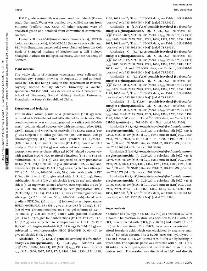
Ainsloside A (2,4,40,6,60-pentakis-isovaleryl-b-D-fructofur-anosyl-a-D-glucopyranoside, 1). C37H62O16; colorless oil;[a]20D +25 (c 0.068, MeOH); UV (MeOH) lmax 197.5 nm; IR (KBr)nmax 3471, 2960, 2927, 2873, 1743, 1468, 1369, 1296, 1254, 1188,
This journal is © The Royal Society of Chemistry 2017
1120, 1014 cm�1; 1H and 13C NMR data, see Table 1; HR-ESI-MS(positive) m/z 785.3939 [M + Na]+ (calcd 785.3936).
Ainsloside B (10,4,40,6,60-pentakis-isovaleryl-b-D-fructofur-anosyl-a-D-glucopyranoside, 2). C37H62O16; colorless oil;[a]20D +14 (c 0.077, MeOH); UV (MeOH) lmax 200.5 nm; IR (KBr)nmax 3446, 2960, 2929, 2871, 1741, 1468, 1371, 1296, 1252, 1188,1095, 1011 cm�1; 1H and 13C NMR data, see Table 1; HR-ESI-MS(positive) m/z 785.3952 [M + Na]+ (calcd 785.3936).
Ainsloside C (3,30,4,40,6-pentakis-isovaleryl-b-D-fructofur-anosyl-a-D-glucopyranoside, 3). C37H62O16; colorless oil;[a]20D +54 (c 0.112, MeOH); UV (MeOH) lmax 198.5 nm; IR (KBr)nmax 3485, 3259, 2960, 2873, 1741, 1468, 1369, 1296, 1188, 1115,1005 cm�1; 1H and 13C NMR data, see Table 1; HR-ESI-MS(positive) m/z 785.3946 [M + Na]+ (calcd 785.3936).
Ainsloside D (2,3,40,6,60-pentakis-isovaleryl-b-D-fructofur-anosyl-a-D-glucopyranoside, 4). C37H62O16; colorless oil;[a]20D +59 (c 0.054, MeOH); UV (MeOH) lmax 202.0 nm; IR (KBr)nmax 3477, 2960, 2933, 2873, 1741, 1468, 1369, 1296, 1254, 1188,1120, 1005 cm�1; 1H and 13C NMR data, see Table 1; HR-ESI-MS(positive) m/z 785.3942 [M + Na]+ (calcd 785.3936).
Ainsloside E (2,40,6,60- tetrakis-isovaleryl-b-D-fructofur-anosyl-a-D-glucopyranoside, 5). C32H54O15; colorless oil;[a]20D +39 (c 0.091, MeOH); UV (MeOH) lmax 198.0 nm; IR (KBr)nmax 3467, 2960, 2933, 2873, 1739, 1468, 1369, 1296, 1255, 1190,1120, 1063, 1005 cm�1; 1H and 13C NMR data, see Table 2; HR-ESI-MS (positive) m/z 701.3365 [M + Na]+ (calcd 701.3360).
Ainsloside F (2,4,6,60-tetrakis-isovaleryl-b-D-fructofuranosyl-a-D-glucopyranoside, 6). C32H54O15; colorless oil; [a]20D +30 (c0.073, MeOH); UV (MeOH) lmax 199.0 nm; IR (KBr) nmax 3460,2960, 2931, 2873, 1741, 1468, 1371, 1296, 1190, 1099, 1012cm�1; 1H and 13C NMR data, see Table 2; HR-ESI-MS (positive)m/z 701.3362 [M + Na]+ (calcd 701.3360).
Ainsloside G (4,40,6,60-tetrakis-isovaleryl-b-D-fructofuranosyl-a-D-glucopyranoside, 7). C32H54O15; colorless oil; [a]20D +28 (c0.089, MeOH); UV (MeOH) lmax 198.5 nm; IR (KBr) nmax 3408,2960, 2933, 2873, 1743, 1468, 1369, 1296, 1254, 1188, 1093, 1003cm�1; 1H and 13C NMR data, see Table 2; HR-ESI-MS (positive)m/z 701.3371 [M + Na]+ (calcd 701.3360).
Ainsloside H (2,4,40,6-tetrakis-isovaleryl-b-D-fructofuranosyl-a-D-glucopyranoside, 8). C32H54O15; colorless oil; [a]20D +45 (c0.100, MeOH); UV (MeOH) lmax 202.9 nm; IR (KBr) nmax 3456,2960, 2929, 2873, 1743, 1468, 1369, 1296, 1254, 1188, 1101,1065, 1012 cm�1; 1H and 13C NMR data, see Table 2; HR-ESI-MS(positive) m/z 701.3357 [M + Na]+ (calcd 701.3360).
Sugar analysis
A solution of 1 (35mg) in 5%KOH (5mL) was heated at 95 �C for3 hours. The reaction mixture was acidied to PH 4 with 1 MHCl, then extracted with CHCl3 (3 � 10 mL) and n-BuOH(3 � 10mL) each three times. The CHCl3 layer was concentrated toafford isovaleric acid, which was elucidated by extensive anal-ysis of 1D NMR spectra. The n-BuOH layer was hydrolyzed in2 M HCl–MeOH (1 : 1, v/v, 10 mL) at 90 �C for 2 h by heating inwater bath. The aqueous phase was extracted with n-BuOH (3 �10 mL) aer acid hydrolysis and concentrated to yield a col-ourless solid. The residue was dissolved in H2O and directly
RSC Adv., 2017, 7, 20865–20873 | 20871

RSC Advances Paper
Ope
n A
cces
s A
rtic
le. P
ublis
hed
on 1
1 A
pril
2017
. Dow
nloa
ded
on 1
2/26
/202
1 5:
19:4
1 PM
. T
his
artic
le is
lice
nsed
und
er a
Cre
ativ
e C
omm
ons
Attr
ibut
ion-
Non
Com
mer
cial
3.0
Unp
orte
d L
icen
ce.
View Article Online
analyzed by HPLC with an ELSD detector with reference stan-dards (MeCN–H2O, 75/25): D-glucose eluted at 16.308 min, andD-fructose at 13.116 min. Each of these eluates was individuallycollected, concentrated, and dissolved in H2O. The obtainedsugar residues were identied as D-glucose, [a]23D +53 (c 0.113,H2O), and D-fructose, [a]23D �149 (c 0.118, H2O), throughcomparisons of their specic rotations with those of the corre-sponding authentic samples.
Isovaleric acid1H NMR (in CDCl3, 500 MHz) d 2.23 (2H, d, J¼ 7.5 Hz), 2.12 (1H,m), 0.99 (6H, d, J ¼ 7.0 Hz); 13C NMR d 178.8 (–COOH), 43.0(CH2), 25.5 (CH), 23.3 (CH3), 22.3 (CH3).
Cytotoxic activity assays
The viability of cells was determined by MTT assay to detectfunctional mitochondria in living cells (Mosmann, 1983) usingdoxorubicin as positive control. Human cell lines A549 (lungadenocarcinoma cells), HCT116 (colon carcinoma cells), MDA-MB-231 (breast cancer cells) and BEL7404 (hepatoma cancercells) were incubated at 37 �C in a Dulbecco's minimumessential medium (DMEM) containing 10% FBS and penicillin–streptomycin solution. About 1� 104 cells per wells were seededinto 96-well microtiter plates. Aer twenty-four hours post-seeding, cells were treated with vehicle control or variousconcentrations of samples for 48 h. 20 mL of MTT solution (5 mgmL�1, Sigma Aldrich, St. Louis, MO, USA) was added to eachwell and the tumor cells were incubated at 37 �C in a humidiedatmosphere of 5% CO2 air for 4 h. Upon removal of MTT/medium, 150 mL of DMSO was added to each well and theplate was agitated at oscillator for 5 min to dissolve the MTT-formazan. The assay plate was read at a wavelength of 570 nmusing a microplate reader (Bio-Tek, Winooski, Vermont, USA).
Cell cycle arresting assay
A549 cells were incubated with compound 2 for 24 h, and thentrypsinized, washed with PBS, and xed with 70% of coldabsolute ethanol at 4 �C overnight. Aer washed twice with PBS,the cells were incubated with 100 mL of RNase A for 30 min andthen stained with 1 mL PI staining solution (3.8 mM sodiumcitrate, 50 mg mL�1 PI in PBS, Beyotime) for 10 min at roomtemperature in the dark. The DNA content of cells and cell cycledistribution were analyzed by ow cytometry (BD FACS Calibur).
Cell apoptosis assay
The apoptotic cells were quantied by using the annexin V-FITCand PI double staining kit (DOJINDO, Japan). Briey, A549 cellswere incubated with compound 2 for 24 h. Then the cells werecentrifuged, and washed with ice-cold PBS. The cultures weresuspended in 400 mL of binding buffer containing 5 mL annexinV-FITC (10 mg mL�1) for 15 min in the dark, and then incubatedwith 10 mL of PI (20 mg mL�1) for 5 min. The cells were imme-diately analyzed by ow cytometry (BD FACSC Calibur).
20872 | RSC Adv., 2017, 7, 20865–20873
Mitochondrial membrane potential measurement
The mitochondrial membrane potential (MMP) was measuredby ow cytometry using Rhodamine 123 (Rh-123) as uoro-chrome. A549 cells were treated with compound 2 for 24 h, andthen incubated with 5 mg mL�1 of Rh-123 for 30 min at roomtemperature in the dark. Aer centrifuged and washed twicewith PBS, the cells were resuspended in 1000 mL of PBS andanalyzed using ow cytometry with excitation and emissionwavelength of 488 and 530 nm, respectively.
Intracellular ROS production measurement
ROS levels were detected using a ow cytometer and a micro-plate spectrophotometer (Molecular Devices, Sunnyvale, CA,USA). Aer treatment, A549 cells were harvested and washedwith PBS and suspended in DMEM containing 10 mM 5(6)-car-boxy-20, 70-dichlorodihydrouorescein diacetate (carboxy-H2DCFDA; Invitrogen) at 37 �C for 20 min. The cells werewashed again, and ow cytometry analysis was performed.
Statistical analysis
Data were analyzed using GraphPad Prism soware (GraphPadsoware Inc., San Diego, CA, USA). The comparison betweentwo groups was analyzed by unpaired Student t-test, andmultiple comparisons were compared by one-way ANOVAanalysis of variance followed by Tukey post hoc test. Statisticalsignicance was determined as P < 0.05.
Conclusions
Isovaleryl sucrose esters are a kind of rare second metabolites,which have been sporadically isolated from some plants.31–33 Incurrent study, eight isovaleryl sucrose esters were obtained fromthe whole plants of A. yunnanensis. It is the rst report of polyisovaleryl sucrose derivatives from Ainsliaea species. Interest-ingly, ainsloside B (2), a poly isovaleryl sucrose derivative with10,4,40,6,60-pentakis-isovaleryl substitution, exhibited potentinhibition against the proliferation of A549 with IC50 value of3.3 mM, while other compounds showed weak inhibition orbeing inactive. It could be speculated that the quantity andsubstitution pattern of isovaleryl may affect cytotoxicities ofthese compounds. Meanwhile, ainsloside B (2) can arrest cellcycle of A549 at G0/G1 phase and induce A549 cell apoptosis indose-dependent manner. Further investigation indicated thatthe apoptosis-inducing effect of compound 2may be involved inreduction of mitochondrial membrane potential (MMP) andincrease of reactive oxygen species (ROS) levels in A549 cells.Therefore, polyisovaleryl sucrose esters could be a type ofimportant bioactive constituents of Ainsliaea plants.
Acknowledgements
The work was supported by Professor of Chang Jiang ScholarsProgram, NSFC (81573318, 81230090, 81520108030, 81373301,1302658), Shanghai Engineering Research Center for the Prep-aration of Bioactive Natural Products (10DZ2251300), theScientic Foundation of Shanghai China (12401900801,
This journal is © The Royal Society of Chemistry 2017

Paper RSC Advances
Ope
n A
cces
s A
rtic
le. P
ublis
hed
on 1
1 A
pril
2017
. Dow
nloa
ded
on 1
2/26
/202
1 5:
19:4
1 PM
. T
his
artic
le is
lice
nsed
und
er a
Cre
ativ
e C
omm
ons
Attr
ibut
ion-
Non
Com
mer
cial
3.0
Unp
orte
d L
icen
ce.
View Article Online
13401900101), National Major Project of China (2011ZX09307-002-03) and the National Key Technology R&D Program ofChina (2012BAI29B06).
Notes and references
1 R. Wang, Y. X. Tang and X. Y. Shang, J. Chin. Med. Mater.,2012, 35, 1171–1175.
2 R. Y. Qiu, J. Xu, W. Xu, J. Xiong, Y. H. Liu and B. Yuan, J. Chin.Pharm. Sci., 2009, 18, 13–14.
3 S. Z. Choi, M. C. Yang, S. U. Choi and K. R. Lee, Arch.Pharmacal Res., 2006, 29, 203–208.
4 M. M. Fan, S. L. Yang, Y. S. Wang, Y. Huang, H. Jian, Y. Rao,Q. P. Wu and X. Qian, IPC A61K36/28(2006.01)I, Patent101293003, 2008.
5 H. I. Moon, O. P. Zee and M. S. Shin, Han'guk NonghwaHakhoechi, 1999, 42, 162–165.
6 Z. J. Wu, X. K. Xu, H. W. Zeng, Y. H. Shen, J. M. Tian, J. Su,H. L. Li, L. Shan, R. H. Liu and W. D. Zhang, Planta Med.,2011, 77, 5517–5520.
7 H. Wang, T. Wu, M. Yan, G. Liu, P. Li, X. Q. Zhang, W. C. Yeand L. Y. Zhang, Chem. Pharm. Bull., 2009, 57, 597–599.
8 X. Chen, J. S. Miao, H. Wang, F. Zhao, J. Hu, P. Gao, Y. Wang,L. Y. Zhang andM. Yan, J. Ethnopharmacol., 2015, 170, 72–80.
9 Y. Wang, Y. H. Shen, H. Z. Jin, J. J. Fu, X. J. Hu, J. J. Qin,J. H. Liu, M. Chen, S. K. Yan and W. D. Zhang, Org. Lett.,2008, 10, 5517–5520.
10 Z. J. Wu, X. K. Xu, Y. H. Shen, J. Su, J. M. Tian, S. Liang,H. L. Li, R. H. Liu and W. D. Zhang, Org. Lett., 2008, 10,2397–2400.
11 R. Wang, Z. Sun, E. L. Wang, Z. Z. Yuan, J. J. Li andX. Y. Shang, J. Chin. Med. Mater., 2013, 36, 61–64.
12 J. J. Li, E. L. Wang, Z. Z. Yuan, C. Y. Wu, L. H. Yang andX. Y. Shang, China J. Chin. Mater. Med., 2013, 38, 3918–3922.
13 J. Yang, H. Y. Wu, Q. H. Li, P. Yi, Y. Min, W. Liu, X. D. Yangand L. Li, Adv. Mater. Res., 2012, 554–556, 1845–1848.
14 W. Li, Y. N. Sun, X. T. Yan, S. Y. Yang, S. B. Song, Y. M. Leeand Y. H. Kim, J. Agric. Food Chem., 2013, 61, 7081–7088.
15 J. Duus, C. H. Gotfredsen and K. Bock, Chem. Rev., 2000, 100,4589–4614.
This journal is © The Royal Society of Chemistry 2017
16 K. Bock and C. Pedersen, Adv. Carbohydr. Chem. Biochem.,1983, 41, 27–66.
17 C. C. Akoh, Food Sci. Technol., 2002, 117, 695–727.18 I. Janicot, A. Bouchu, G. Descotes and E. Wong, Tenside,
Surfactants, Deterg., 1996, 33, 290–296.19 M. Ferrera, J. Soliverib, F. J. Ploua, N. Lopez-Cortesa,
D. Reyes-Duartea, M. Christensenc, J. L. Copa-Patinob andA. Ballesterosa, Enzyme Microb. Technol., 2005, 36, 391–398.
20 Y. Queneau, S. Jarosz, B. Lewandowski and J. Fitremann,Adv. Carbohydr. Chem. Biochem., 2007, 61, 218–292.
21 S. Thevenet, A. Wernicke, S. Belniak, G. Descotes, A. Bouchuand Y. Queneau, Carbohydr. Res., 1999, 318, 52–66.
22 V. Molinier, K. Wisniewski, A. Bouchu, J. Fitremann andY. Queneau, J. Carbohydr. Chem., 2003, 22, 657–669.
23 D. F. Amanatullah, A. T. Reutens, B. T. Zafonte, M. Fu,S. Mani and R. G. Pestell, Front. Biosci., 2000, 5, 372–390.
24 Z. A. Stewart, M. D. Westfall and J. A. Pietenpol, TrendsPharmacol. Sci., 2003, 24, 139–145.
25 I. M. Ghobrial, T. E. Witzig and A. A. Adjei, Ca-Cancer J. Clin.,2005, 55, 178–194.
26 M. J. Wu, H. Zhang, J. H. Hu, Z. Y. Weng, C. Y. Li, H. Li,Y. Zhao, X. F. Mei, F. Ren and L. H. Li, PLoS One, 2013, 8,1–8.
27 G. W. Wang, C. Lv, Z. R. Shi, R. T. Zeng, X. Y. Dong,W. D. Zhang, R. H. Liu, L. Shan and Y. H. Shen, PLoS One,2014, 9, 1–19.
28 K. Gong and W. Li, Free Radical Biol. Med., 2011, 51, 2259–2271.
29 W. P. Tsang, S. P. Chau, S. K. Kong, K. P. Fung andT. T. Kwok, Life Sci., 2003, 73, 2047–2058.
30 Y. Tang, R. Chen, Y. Huang, G. Li, Y. Huang, J. Chen,L. Duan, B. T. Zhu, J. B. Thrasher, X. Zhang and B. Li, Mol.Cancer Ther., 2014, 13, 1526–1536.
31 A. T. Tchinda, P. Tane, J. F. Ayafor and J. D. Conolly,Phytochemistry, 2003, 63, 841–846.
32 A. P. Castorena, M. Luna, M. Martinez and E. Maldonado,Carbohydr. Res., 2012, 352, 211–214.
33 N. J. Toyang, M. A. Krause, R. M. Fairhurst, P. Tane, J. Bryantand R. Verpoorte, J. Ethnopharmacol., 2013, 147, 618–621.
RSC Adv., 2017, 7, 20865–20873 | 20873