cytotoxic effects of euterpe oleracea mart. in malignant cell lines
TRANSCRIPT

Silva et al. BMC Complementary and Alternative Medicine 2014, 14:175http://www.biomedcentral.com/1472-6882/14/175
RESEARCH ARTICLE Open Access
Cytotoxic effects of Euterpe oleracea Mart. inmalignant cell linesDulcelena Ferreira Silva1, Flávia Castello Branco Vidal1,2*, Debora Santos3, Maria Célia Pires Costa3,José Andrés Morgado-Díaz4, Maria do Desterro Soares Brandão Nascimento2,5 and Roberto Soares de Moura6
Abstract
Background: Euterpe oleracea Mart., a plant from the Amazon region, is commonly known as açaí or juçara; it hashigh nutritional value and elevated levels of lipids, proteins, and minerals. Açaí is an abundant and much consumedfruit by the Amazon local population, and studies have demonstrated that it is rich in phytochemicals withantioxidant, anti-inflammatory, and anticancer activities. Therefore, the aim of this study was to test this plant foranticancer activity in different human malignant cell lines.
Methods: Cell lines derived from breast and colorectal adenocarcinomas were treated with 10, 20, and 40 μg/mLof bark, seed, and total açaí fruit hydroalcoholic extracts for 24 and 48 h. After treatment, cell viability was measuredusing 3-(4,5-dimethylthiazol-2-yl)-2,5-diphenyltetrazolium bromide (MTT) assays, and cell morphological featureswere observed by light and transmission electron microscopy. The type of cell death was also evaluated. The datawere analyzed statistically by one-way analysis of variance (ANOVA), followed by Dunnett’s or Tukey’s post hoc tests,as appropriate.
Results: We observed that of all the cell lines tested, MCF-7 was the only line that responded to açaí treatment.The extracts caused significant reduction (p < 0.01) in cell viability and altered cell morphological features byinducing the appearance of autophagic vacuoles, as observed by transmission electron microscopy. Furthermore,increased expression of LC3BII, a protein marker of autophagosome formation, was observed by western blotting.Caspase Glo™ assays and morphologic observations by DAPI nuclear staining and transmission electron microscopydid not indicate any apoptotic events.
Conclusions: The present study demonstrated that açaí possesses antitumorigenic potential in the MCF-7 cell line.Further studies are needed to identify the compound (s) responsible for this cytotoxic activity and the moleculartarget in the cell. This discovery of the anticancer potential of açaí may help in the development of chemopreventivedrugs and may have therapeutic effects in the treatment of breast cancer.
Keywords: Anticancer, Euterpe oleracea mart., MCF-7, Phytochemicals, Chemopreventive
BackgroundEuterpe oleracea Mart. is an indigenous monocot plantfound in the estuary of the Amazon region and is com-monly known as juçara or acai. It is widely consumed bythe Amazonian population living on the shore of theAmazon River [1].
* Correspondence: [email protected] Department, Federal University of Maranhão, Rua Coelho Netonº 311, Centro, São Luís, Maranhão 65020-140, Brazil2Tumors and DNA Bank from Maranhão, Federal University of Maranhão,Maranhão, BrazilFull list of author information is available at the end of the article
© 2014 Silva et al.; licensee BioMed Central LtCommons Attribution License (http://creativecreproduction in any medium, provided the orDedication waiver (http://creativecommons.orunless otherwise stated.
Besides its high macronutrient content, açaí has beenshown to possess high levels of phytochemicals with anti-oxidant, anti-inflammatory, hypocholesterolemic, and an-ticancer activities [2-6]. Açaí was found to inhibit theproduction of reactive oxygen species and the activity ofcyclooxygenases 1 and 2 [7]. In rats, açaí extracts inducesendothelium-dependent vasodilation [8]. An in vivo studywith healthy volunteers demonstrated that açaí pulpcaused a significant increase in the antioxidant capacity ofplasma, which indicates the in vivo antioxidant potentialof E. oleracea Mart. [9].Additionally, a few studies have demonstrated the anti-
tumorigenic activity of açaí. One study showed that
d. This is an Open Access article distributed under the terms of the Creativeommons.org/licenses/by/2.0), which permits unrestricted use, distribution, andiginal work is properly credited. The Creative Commons Public Domaing/publicdomain/zero/1.0/) applies to the data made available in this article,

Silva et al. BMC Complementary and Alternative Medicine 2014, 14:175 Page 2 of 9http://www.biomedcentral.com/1472-6882/14/175
açaí’s polyphenolic, glycoside, and aglycone forms couldinduce apoptosis in HL-60 leukemia cells [10]. Anin vivo study reported that açaí intake could attenu-ate dimethylhydrazine-induced colon carcinogenesis inrats [11].Due to the wide usage of açaí by a local population of
the Amazon region and the antitumorigenic potential ofthis plant, we investigated whether açaí extracts fromthe fruit, bark, and seed possess anticancer activity inhuman malignant cell lines. MTT viability assays as wellas morphologic analysis of cells performed using lightand transmission electron microscopy were performed,and the type of cell death was also analyzed.
MethodsPlant material and preparation of açaí hydroalcoholicextractsThe specimen was collected at Juçara’s park, an environ-mental protection area located at São Luís County. Theplant material was authenticated by the Rosa MochelHerbarium from School of Biological Studies, State Uni-versity of Maranhão, where a voucher specimen was de-posited (reference number of 30).Fruits were harvested and stored at -20°C until use.
After defrosting at room temperature, fruits were sepa-rated into three portions: bark, seed and total fruit (bark +seed). The procedures used to prepared the hydroal-coholic extracts have been described previously [3].Briefly, about 360 g of each different portion werewashed in running water and boiled in distilled waterfor 5 minutes. After, they were macerated separately in400 ml of ethanol PA for 2 hours with intermittentshaking and then kept in dark bottles at 4°C for 10 d.The extracts were filtered thought Whatman no. 1 filterpaper, and the ethanol was evaporated under low pres-sure at 40°C. The extracts were then lyophilized (LIO-TOP model 202; Fisatom Equipamentos, São Paulo,Brazil) and frozen at -20°C until use.Determination of total polyphenols in açaí extracts
was performed by the Folin-Ciocalteau colorimetricmethod determined on a Spectrophotometer UV/VIS bymonitoring the absorbance at 700 nm using gallic acid asa reference standard (50, 100, 150, 250 and 1000 mg/mL).Values were evaluated as the mg equivalent of gallic acidper g of extract [12].
Cell culture and treatmentsThe cell lines Caco-2 (ATCC, # HTB-37, Rockville, MD,USA) and HT-29 (ATCC, # HTB-38), both derived fromhuman colon adenocarcinoma, and MCF-7 (ATCC, #HTB-22) and MDA-MB-468 (ATCC, # HTB-132, Rockville,MD, EUA), derived from human mammary adenocarcin-oma, were grown in Dulbecco’s modified Eagle’s medium(DMEM) (Invitrogen) supplemented with 10% fetal bovine
serum, penicillin G (60 mg/l), and streptomycin (100 mg/l)at 37°C in a humidified atmosphere of 5% CO2.The lyophilized extracts were dissolved in Milli-Q
water, and the solution was filtered through a 0.2-μmpore syringe filter and stored at -20°C until use. The cul-tured cells were treated with 10, 20, and 40 μg/ml of theextracts for 24 and 48 h.
MTT viability assayCells (1 × 10⁴ cell/ml) were cultured in 96-well plates inthe presence or absence of the extracts for 24 and 48 h.The supernatant was removed, and 10 μl of 3-(4,5-dimeth-ylthiazol-2-yl)-2,5-diphenyltetrazolium bromide (MTT) inDMEM medium was added to each well. Cells were incu-bated in a CO2 chamber for 3 h with protection fromlight. The absorbance at 538 nm was measured with aSpectra Max 190 spectrophotometer (Molecular Devices,Sunnyvale, CA, EUA).
Morphologic analysisCells were cultured in 96-well plates in the presence orabsence of the extracts for 24 and 48 h. After treatment,the morphological features of the cells were evaluated byphase-contrast microscopy using an inverted microscopeAxio Observer Z1, equipped with a camera, AxiocamHRcVer.3 (Carl Zeiss Inc., Germany).For transmission electron microscopy analysis, cells were
grown in culture flasks treated with açaí extracts for 24 h.Subsequently cells were washed with PBS and fixed for 1 hwith Karnovsky fixer (2.5% glutaraldehyde, 4% paraformal-dehyde, and 0.1 M cacodylate buffer). Post-fixation wascarried out with 1% osmium tetroxide in cacodylate buffercontaining 0.8% potassium ferrocyanide and 5 mM CaCl2for 45 min at 4°C. Subsequently, cells were dehydrated in agraded series of acetone and embedded in epoxy resin. Ul-trathin sections (60 nm) were stained with uranyl acetateand lead citrate and examined using a Zeiss EM 906 (Jena,Germany) transmission electron microscope.
Nuclear staining and Caspase-Glo® 3/7 luminescent assayThe nuclear and chromosome counterstain, DAPI (4′,6-diamidino-2-phenylindole), was used for nuclear mor-phologic observations of treated cells with açaí extracts.To evaluate possible apoptotic activity of the extracts,we used a luminescent kit that measures caspase-3/7 ac-tivity (Caspase-Glo™ 3/7, Promega, WI, USA). After treat-ment with the extracts, 10 μl of Caspase-Glo™ 3/7 reagentwas added to the culture, and after 90 min, the lumines-cence was analyzed in an illuminometer, Veritas TurnerBiosystems (Madison, USA).
Total cell lysate preparation and western blot analysisTotal cell lysates were obtained by incubating cells in lysisbuffer (1% Triton X-100, 0.5% sodium deoxycholate, 0.2%
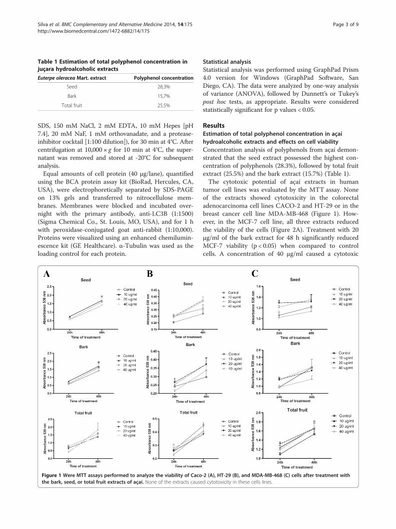
Table 1 Estimation of total polyphenol concentration injuçara hydroalcoholic extracts
Euterpe oleracea Mart. extract Polyphenol concentration
Seed 28,3%
Bark 15,7%
Total fruit 25,5%
Silva et al. BMC Complementary and Alternative Medicine 2014, 14:175 Page 3 of 9http://www.biomedcentral.com/1472-6882/14/175
SDS, 150 mM NaCl, 2 mM EDTA, 10 mM Hepes [pH7.4], 20 mM NaF, 1 mM orthovanadate, and a protease-inhibitor cocktail [1:100 dilution]), for 30 min at 4°C. Aftercentrifugation at 10,000 × g for 10 min at 4°C, the super-natant was removed and stored at -20°C for subsequentanalysis.Equal amounts of cell protein (40 μg/lane), quantified
using the BCA protein assay kit (BioRad, Hercules, CA,USA), were electrophoretically separated by SDS-PAGEon 13% gels and transferred to nitrocellulose mem-branes. Membranes were blocked and incubated over-night with the primary antibody, anti-LC3B (1:1500)(Sigma Chemical Co., St. Louis, MO, USA), and for 1 hwith peroxidase-conjugated goat anti-rabbit (1:10,000).Proteins were visualized using an enhanced chemilumin-escence kit (GE Healthcare). α-Tubulin was used as theloading control for each protein.
Figure 1 Were MTT assays performed to analyze the viability of Cacothe bark, seed, or total fruit extracts of açaí. None of the extracts cause
Statistical analysisStatistical analysis was performed using GraphPad Prism4.0 version for Windows (GraphPad Software, SanDiego, CA). The data were analyzed by one-way analysisof variance (ANOVA), followed by Dunnett’s or Tukey’spost hoc tests, as appropriate. Results were consideredstatistically significant for p values < 0.05.
ResultsEstimation of total polyphenol concentration in açaíhydroalcoholic extracts and effects on cell viabilityConcentration analysis of polyphenols from açaí demon-strated that the seed extract possessed the highest con-centration of polyphenols (28.3%), followed by total fruitextract (25.5%) and the bark extract (15.7%) (Table 1).The cytotoxic potential of açaí extracts in human
tumor cell lines was evaluated by the MTT assay. Noneof the extracts showed cytotoxicity in the colorectaladenocarcinoma cell lines CACO-2 and HT-29 or in thebreast cancer cell line MDA-MB-468 (Figure 1). How-ever, in the MCF-7 cell line, all three extracts reducedthe viability of the cells (Figure 2A). Treatment with 20μg/ml of the bark extract for 48 h significantly reducedMCF-7 viability (p < 0.05) when compared to controlcells. A concentration of 40 μg/ml caused a cytotoxic
-2 (A), HT-29 (B), and MDA-MB-468 (C) cells after treatment withd cytotoxicity in these cells lines.
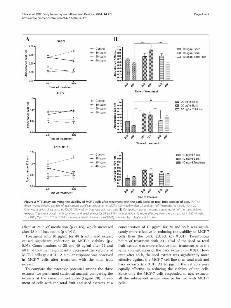
Figure 2 MTT assay analyzing the viability of MCF-7 cells after treatment with the bark, seed, or total fruit extracts of açaí. (A) Thethree hydroalcoholic extracts of açaí caused significant reduction in MCF-7 cell viability after 24 and 48 h of treatment. *p < 0.05, **p < 0.01.One-way analysis of variance (ANOVA) followed by Dunnett’s post hoc test. (B) Comparison using the same concentration of the three differentextracts. Treatment of cells with total fruit and seed extract for 24 and 48 h was significantly more effective than the bark extract in MCF-7 cells.*p < 0.05, **p < 0.01, ***p < 0.001. One-way analysis of variance (ANOVA), followed by Tukey’s post hoc test.
Silva et al. BMC Complementary and Alternative Medicine 2014, 14:175 Page 4 of 9http://www.biomedcentral.com/1472-6882/14/175
effect at 24 h of incubation (p < 0.05), which increasedafter 48 h of incubation (p < 0.01).Treatment with 10 μg/ml for 48 h with seed extract
caused significant reduction in MCF-7 viability (p <0.05). Concentrations of 20 and 40 μg/ml after 24 and48 h of treatment significantly decreased the viability ofMCF-7 cells (p < 0.01). A similar response was observedin MCF-7 cells after treatment with the total fruitextract.To compare the cytotoxic potential among the three
extracts, we performed statistical analysis comparing theextracts at the same concentration (Figure 2B). Treat-ment of cells with the total fruit and seed extracts at a
concentration of 10 μg/ml for 24 and 48 h was signifi-cantly more effective in reducing the viability of MCF-7cells than the bark extract (p < 0.001). Twenty-fourhours of treatment with 20 μg/ml of the seed or totalfruit extract was more effective than treatment with thesame concentration of the bark extract (p < 0.01). How-ever, after 48 h, the seed extract was significantly moreeffective against the MCF-7 cell line than total fruit andbark extracts (p < 0.01). At 40 μg/ml, the extracts wereequally effective in reducing the viability of the cells.Since only the MCF-7 cells responded to açaí extracts,all the subsequent assays were performed with MCF-7cells.

Silva et al. BMC Complementary and Alternative Medicine 2014, 14:175 Page 5 of 9http://www.biomedcentral.com/1472-6882/14/175
Açaí extracts caused morphologic alterations in MCF-7 cellsThe effects of açaí extracts on the morphological fea-tures of MCF-7 cells were investigated by phase-contrastmicroscopy and transmission electron microscopy (TEM).Phase-contrast microscopy showed a decrease in celldensity in the açaí extract-treated MCF-7 cells, as well ascell rounding and shrinking, membrane blebbing, and celllysis with apparent loss of cytoplasmic content (Figure 3).
Figure 3 Morphology analysis by phase-contrast microscopy of MCF-Bark extract treatment caused cell shrinking (arrow). The seed extract causeas cell shrinking (arrow), membrane blebbing (arrowhead), and cell lysis (astotal fruit extract.
The bark extract caused discrete cell rounding and shrink-ing in MCF-7 cells at concentrations of 20 and 40 μg/ml(Figure 3A). The morphologic alterations caused by seedand total fruit extracts from açaí were more severe thanthose caused by the bark extract. When used at 10 μg/ml,the seed extract caused a decrease in cell density and cellrounding. Treatment with 20 and 40 μg/ml of seed ex-tracts caused a more accentuated decrease in cell density,
7 cells treated with the bark (A), seed (B), or total fruit (C) extract.d more dramatic changes in MCF-7 cell morphological features, suchterisk). The same alterations were observed in cells treated with the
Silva et al. BMC Complementary and Alternative Medicine 2014, 14:175 Page 6 of 9http://www.biomedcentral.com/1472-6882/14/175
cell shrinking, membrane blebbing, and apparent cell lysis(Figure 3B). Similar alterations were observed in MCF-7cells treated with the total fruit extract (Figure 3C).Detailed morphologic analysis of MCF-7 intracellular
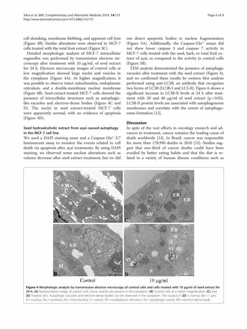
organelles was performed by transmission electron mi-croscopy after treatment with 10 μg/mL of seed extractfor 24 h. Electron microscopy images of control cells atlow magnification showed large nuclei and vesicles inthe cytoplasm (Figure 4A). At higher magnification, itwas possible to observe intact mitochondria, endoplasmicreticulum, and a double-membrane nuclear membrane(Figure 4B). Seed-extract-treated MCF-7 cells showed thepresence of intracellular structures such as autophagic-like vacuoles and electron-dense bodies (Figure 4C andD). The nuclei in seed extract-treated MCF-7 cellswere apparently normal, with no evidence of apoptosis(Figure 4D).
Seed hydroalcoholic extract from açaí caused autophagyin the MCF-7 cell lineWe used a DAPI staining assay and a Caspase-Glo® 3/7luminescent assay to monitor the events related to celldeath via apoptosis after açaí treatments. By using DAPIstaining, we observed some nuclear alterations such asvolume decrease after seed extract treatment, but we did
Figure 4 Morphologic analysis by transmission electron microscopy o24 h. (A) Representative image of control cells. Some vesicles are present i(D) Treated cells. Autophagic vacuoles and electron-dense bodies can be oN = nucleus, Nu = nucleolus, M =mitochondria, V = vesicle, ER = endoplasm
not detect apoptotic bodies or nuclear fragmentation(Figure 5A). Additionally, the Caspase-Glo™ assays didnot show lower caspase 3 and caspase 7 activity inMCF-7 cells treated with the seed, bark, or total fruit ex-tract of açaí, as compared to the activity in control cells(Figure 5B).TEM analysis demonstrated the presence of autophagic
vacuoles after treatment with the seed extract (Figure 4),and we confirmed these results by western blot analysisperformed using anti-LC3B, an antibody that recognizestwo forms of LC3B (LC3B-I and LC3-II). Figure 6 shows asignificant increase in LC3B-II levels at 24 h after treat-ment with 20 and 40 μg/ml of seed extract (p < 0.05).LC3B-II protein levels are associated with autophagosomemembranes and correlate with the extent of autophago-some formation [13].
DiscussionIn spite of the vast efforts in oncology research and ad-vances in treatment, cancer remains the leading cause ofdeath worldwide [14]. In Brazil, cancer was responsiblefor more than 178,990 deaths in 2010 [15]. Studies sug-gest that one-third of cancer deaths could have beenavoided by better eating habits and that the diet is re-lated to a variety of human disease conditions such as
f control cells and cells treated with 10 μg/ml of seed extract forn the cytoplasm. (B) Control cells at a higher magnification. (C) andbserved in the cytoplasm. The nucleus in (D) is normal. Bar = 1 μm.ic reticulum, AV = autophagic vesicle, EB = electron-dense body.

Figure 6 LC3B-II expression increased in cells treated with 20and 40 μg/ml seed extract. *p < 0.05. One-way analysis of variance(ANOVA) followed by Dunnett’s post hoc test.
Figure 5 The seed extract did not caused apoptosis on MCF-7 cells. (A) Nuclear morphology analysis by DAPI stain. The seed extract causeda few nuclear alterations such as nuclear shrinking (arrow). There was no evidence of apoptosis. (B) Caspase-Glo® 3/7 luminescent demonstratedthat none of the extracts increased the activity of caspase 3 and caspase 7 when compared to control cells after 24 h of treatment.
Silva et al. BMC Complementary and Alternative Medicine 2014, 14:175 Page 7 of 9http://www.biomedcentral.com/1472-6882/14/175
cancer, hypertension, diabetes, and chronic and acute in-flammation [16,17].Açaí, like other berries and drupes, is rich in polyphe-
nols, anthocyanin, anthocyanidins, and other flavonoids[7,10,18,19], and studies have shown that these sub-stances possess anti-cancer activity in the oral cavity,esophagus, and colon and against leukemia [2,10,11].However, the cellular mechanisms underlying these ac-tivities remain unclear. Since açaí is a frequently con-sumed plant in the Amazon region, we sought toelucidate the antitumorigenic potential of this drupe inthe case of two cancers, colon and breast cancer, with ahigh prevalence in Brazil.In the present study, we tested several cell lines and
found that the MCF-7 cell line was sensitive to açaí ex-tracts. MCF-7 cells differ from HT-29 and Caco-2 cellswith respect to the organ of origin and from MDA-MB-468 by the estrogen receptor status. Estrogen receptor ispresent in MCF-7 cells and absent in MDA-MB-468 al-though both are derived from breast cancer [20]. A pos-sible explanation for the specificity for MCF-7 cells isthat the cytotoxic effects are caused by substances likelignans present in açaí that may act as phytoestrogens[21]. The phytoestrogens can interact with estrogen re-ceptors and modulate a series of estrogenic and anties-trogenic effects [22,23]. It has been shown that equol, anatural estrogenic metabolite derived from soy isofla-vones, enhances tamoxifen’s antitumor activity in MCF-7breast cancer cells [24]. Whether these substances areresponsible for the antitumor activity of açaí remains to
Silva et al. BMC Complementary and Alternative Medicine 2014, 14:175 Page 8 of 9http://www.biomedcentral.com/1472-6882/14/175
be verified since the crude extract has not yet beenpurified.We verified that the hydroalcoholic extract from the
seed was most potent extract against MCF-7 cells. Ana-lysis of polyphenol content using the Folin-Ciocalteuprocedure showed that the seed extract contained thehighest concentration of these substances. Studies havedescribed an antitumorigenic effect of polyphenols inseveral cell lines, such as HeLa, SiHa, and HepG2 cells[25,26]. Therefore, it is likely that the efficacy of the seedextract observed in this study is due to the high concen-tration of polyphenols. We are currently characterizingthe chemical components of the seed extract in order toidentify the substance(s) responsible for the antitumori-genic effects.In order to investigate the type of cell death that oc-
curred in MCF-7 cells, we used DAPI for nuclearvisualization and TEM for morphologic analysis. We didnot observe nuclear alterations that would be indicativeof apoptosis, such as the presence of apoptotic bodies ornuclear chromatin condensation. The results of Caspase-Glo® 3/7 luminescent assays were consistent with these re-sults, in that no differences were seen between the caspase3 and caspase 7 activity of control and treated cells. How-ever, TEM images showed the presence of electron-densebodies and autophagic vacuoles in the cytoplasm of MCF-7 cells treated with the seed extract. To confirm autoph-agy, we performed western blot analysis using an LC3B-IIantibody, a well-known marker of autophagosome forma-tion. We observed an increase in LC3B-II expression incells treated with the seed extract. Other researchers alsoobserved autophagy caused by plant extracts treatmentson MCF-7 cells [27,28]. Ait-Mohamed et al. [27] observedthat acetonic extract of Buxus sempervirens induced au-tophagy in MCF-7 cells. They observed upregulation ofBeclin-1 and downregulation of Survivin and p21. In an-other study [28] the authors demonstrated that acetoneand ethyl acetate extracts from Eupatorium odoratumcaused autophagic cell death on MCF-7 cell line by a stillunknown mechanism. Tsuyuki et al. observed that antho-cyanidins caused autophagy but not apoptosis in HeLacells, a cervical cancer cell line [29]. The leaf water extractof Solanum nigrum Linn, rich in polyphenols and antho-cyanidins, has been shown to cause autophagy in theAU565 cell lineage [30]. Furthermore, the beneficial ef-fects of resveratrol, a polyphenolic compound found ingrapes, have been demonstrated in human subjects andhave been attributed to its capacity to promote autophagy[31]. In addition, it was also observed that other phenoliccompounds found in red wine were capable of stimulatingautophagy [31].The mechanism whereby cell death of MCF-7 was by
autophagy and not apoptosis in our work needs to bedetermined. Açaí extract may be causing activation of
autophagy by a still unknown mechanism. Some specula-tion based on other works may be done about the possibletarget of açaí extracts. Gavilán et al. [32] demonstratedthat proteasome inhibitors, used for the treatment of sometypes of cancer, inhibited GSK-3β enzyme regulating au-tophagy activation in the human breast cancer MCF7cells. We need to purified the extract and then researchthe possible target on the cell. It may be an important en-zyme like GSK-3β, which is involved in WNT signaling. Itis our future goal.
ConclusionWe conclude that açaí has antitumorigenic potential inthe MCF-7 cell line, causing cell viability reduction, mor-phological alterations, and autophagy induction. Furtherstudies are necessary in order to elucidate the phytochem-ical(s) responsible for this anticancer activity.
Competing interestsThe authors declare that they have no competing interests.
Authors’ contributionsMCPC and DS collected the plant and performed the extraction. DFS andFCBV performed the experiments under the supervision of JAMD, MDSBN,and RSM. All the authors analyzed and interpreted the data. FCBV wrote themanuscript draft, which was read and edited by all the authors. All authorsread and approved the final version of the manuscript.
AcknowledgmentsThis study was supported by grants from the Conselho Nacional deDesenvolvimento Cientifico e Tecnológico (CNPq), Ministério da Saúde, Brasil,and Fundação de Amparo à Pesquisa e ao Desenvolvimento Científico eTecnológico do Estado do Maranhão (FAPEMA). We are grateful to Dr. Marcelofor the preparation of açaí extracts and Simone Fernandes for sampleprocessing for TEM observations.
Author details1Morphology Department, Federal University of Maranhão, Rua Coelho Netonº 311, Centro, São Luís, Maranhão 65020-140, Brazil. 2Tumors and DNA Bankfrom Maranhão, Federal University of Maranhão, Maranhão, Brazil. 3Biologyand Chemistry Department, State University of Maranhão, Maranhão, Brazil.4Structural Biology Laboratory, Cell Biology Division, National Cancer InstituteJosé Alencar Gomes da Silva, Rio de Janeiro, Brazil. 5Pathology Department,Federal University of Maranhão, Maranhão, Brazil. 6Laboratory ofPharmacology and Psychobiology, Pharmacology Department, StateUniversity of Rio de Janeiro, Rio de Janeiro, Brazil.
Received: 13 November 2013 Accepted: 20 May 2014Published: 29 May 2014
References1. Lichtenthaler R, Rodrigues RB, Maia JG, Papagiannopoulos M, Fabricius H,
Marx F: Total oxidant scavenging capacities of Euterpe oleracea Mart.(Acai) fruits. Int J Food Sci Nutr 2005, 56:53–64.
2. Stoner GD: Foodstuffs for preventing cancer: the preclinical and clinicaldevelopment of berries. Cancer Prev Res 2009, 2:187–194.
3. da Costa CA, de Oliveira PR, de Bem GF, de Cavalho LC, Ognibene DT,da Silva AF, Dos Santos Valenca S, Pires KM, da Cunha Sousa PJ, de Moura RS,Resende AC: Euterpe oleracea Mart.-derived polyphenols preventendothelial dysfunction and vascular structural changes in renovascularhypertensive rats: role of oxidative stress. Naunyn Schmiedeberg’s ArchPharmacol 2012, 385:1199–1209.
4. de Souza MO, Souza ESL, de Brito Magalhaes CL, de Figueiredo BB, Costa DC,Silva ME, Pedrosa ML: The hypocholesterolemic activity of acai (Euterpeoleracea Mart.) is mediated by the enhanced expression of the

Silva et al. BMC Complementary and Alternative Medicine 2014, 14:175 Page 9 of 9http://www.biomedcentral.com/1472-6882/14/175
ATP-binding cassette, subfamily G transporters 5 and 8 and low-densitylipoprotein receptor genes in the rat. Nutr Res 2012, 32:976–984.
5. Moura RS, Ferreira TS, Lopes AA, Pires KM, Nesi RT, Resende AC, Souza PJ,Silva AJ, Borges RM, Porto LC, Valenca SS: Effects of Euterpe oleracea Mart.(ACAI) extract in acute lung inflammation induced by cigarette smoke inthe mouse. Phytomedicine 2012, 19:262–269.
6. Poulose SM, Fisher DR, Larson J, Bielinski DF, Rimando AM, Carey AN,Schauss AG, Shukitt-Hale B: Anthocyanin-rich acai (Euterpe oleracea Mart.)fruit pulp fractions attenuate inflammatory stress signaling in mousebrain BV-2 microglial cells. J Agric Food Chem 2012, 60:1084–1093.
7. Schauss AG, Wu X, Prior RL, Ou B, Huang D, Owens J, Agarwal A, Jensen GS,Hart AN, Shanbrom E: Antioxidant capacity and other bioactivities of thefreeze-dried Amazonian palm berry, Euterpe oleraceae mart. (acai).J Agric Food Chem 2006, 54:8604–8610.
8. Rocha AP, Carvalho LC, Sousa MA, Madeira SV, Sousa PJ, Tano T, Schini-Kerth VB,Resende AC, Soares de Moura R: Endothelium-dependent vasodilator effect ofEuterpe oleracea Mart. (Acai) extracts in mesenteric vascular bed of the rat.Vasc Pharmacol 2007, 46:97–104.
9. Mertens-Talcott SU, Rios J, Jilma-Stohlawetz P, Pacheco-Palencia LA,Meibohm B, Talcott ST, Derendorf H: Pharmacokinetics of anthocyaninsand antioxidant effects after the consumption of anthocyanin-rich acaijuice and pulp (Euterpe oleracea Mart.) in human healthy volunteers.J Agric Food Chem 2008, 56:7796–7802.
10. Del Pozo-Insfran D, Percival SS, Talcott ST: Acai (Euterpe oleracea Mart.)polyphenolics in their glycoside and aglycone forms induce apoptosis ofHL-60 leukemia cells. J Agric Food Chem 2006, 54:1222–1229.
11. Fragoso MF, Romualdo GR, Ribeiro DA, Barbisan LF: Acai (Euterpe oleraceaMart.) feeding attenuates dimethylhydrazine-induced rat coloncarcinogenesis. Food Chem Toxicol 2013, 58C:68–76.
12. Ainsworth EA, Gillespie KM: Estimation of total phenolic content andother oxidation substrates in plant tissues using Folin-Ciocalteu reagent.Nat Protoc 2007, 2:875–877.
13. de Albuquerque-Xavier AC, Bastos LG, de Freitas JC Jr, Leve F, de Souza WF,de Araujo WM, Wanderley JL, Tanaka MN, de Souza W, Morgado-Diaz JA:Blockade of irradiation-induced autophagosome formation impairsproliferation but does not enhance cell death in HCT-116 humancolorectal carcinoma cells. Int J Oncol 2012, 40:1267–1276.
14. Ferlay J, Shin HR, Bray F, Forman D, Mathers C, Parkin DM: Estimates ofworldwide burden of cancer in 2008: GLOBOCAN 2008. Int J Cancer 2008,2010(127):2893–2917.
15. Ministério da Saúde I: Estimativa 2012 – Incidência de Câncer no Brasil.In Edited by Casado L. Rio de Janeiro: Serviço de Edição e InformaçãoTécnico-Científica; 2011:118.
16. Bode AM, Dong Z: Cancer prevention research - then and now. Nat RevCancer 2009, 9:508–516.
17. Lee KW, Bode AM, Dong Z: Molecular targets of phytochemicals forcancer prevention. Nat Rev Cancer 2011, 11:211–218.
18. Neida S, Elba S: Characterization of the acai or manaca (Euterpe oleraceaMart.): a fruit of the Amazon. Arch Latinoam Nutr 2007, 57:94–98.
19. Mulabagal V, Keller WJ, Calderon AI: Quantitative analysis of anthocyaninsin Euterpe oleracea (acai) dietary supplement raw materials andcapsules by Q-TOF liquid chromatography/mass spectrometry.Pharm Biol 2012, 50:1289–1296.
20. Prabhakaran P, Hassiotou F, Blancafort P, Filgueira L: Cisplatin inducesdifferentiation of breast cancer cells. Front Oncol 2013, 3:134.
21. Heinrich M, Dhanji T, Casselman I: Açai (Euterpe oleracea Mart.) — aphytochemical and pharmacological assessment of the species’ healthclaims. Phytochem Lett 2011, 4:10–21.
22. Usui T: Pharmaceutical prospects of phytoestrogens. Endocr J 2006,53:7–20.
23. Branca F: Dietary phyto-oestrogens and bone health. Proc Nutr Soc 2003,62:877–887.
24. Charalambous C, Pitta CA, Constantinou AI: Equol enhances tamoxifen’santi-tumor activity by induction of caspase-mediated apoptosis in MCF-7breast cancer cells. BMC Cancer 2013, 13:238.
25. Singh M, Bhui K, Singh R, Shukla Y: Tea polyphenols enhance cisplatinchemosensitivity in cervical cancer cells via induction of apoptosis.Life Sci 2013, 1:7–16.
26. Apostolou A, Stagos D, Galitsiou E, Spyrou A, Haroutounian S, Portesis N,Trizoglou I, Wallace Hayes A, Tsatsakis AM, Kouretas D: Assessment ofpolyphenolic content, antioxidant activity, protection against ROS-
induced DNA damage and anticancer activity of Vitis vinifera stemextracts. Food Chem Toxicol 2013. in press.
27. Ait-Mohamed O, Battisti V, Joliot V, Fritsch L, Pontis J, Medjkane S, Redeuilh C,Lamouri A, Fahy C, Rholam M, Atmani D, Ait-Si-Ali S: Acetonic extract of Buxussempervirens induces cell cycle arrest, apoptosis and autophagy in breastcancer cells. PLoS One 2011, 6:1–11.
28. Harun FB, Jamalullail S, Yin K, Othman Z, Tilwari A, Balaram P: AutophagicCell Death Is Induced by Acetone and Ethyl Acetate Extracts fromEupatorium odoratum In Vitro: Effects on MCF-7 and Vero Cell Lines.Sci World J 2012, 2012:1–9.
29. Tsuyuki S, Fukui S, Watanabe A, Akune S, Tanabe M, Yoshida K: Delphinidininduces autolysosome as well as autophagosome formation anddelphinidin-induced autophagy exerts a cell protective role. J BiochemMol Toxicol 2012, 26:445–453.
30. Huang HC, Syu KY, Lin JK: Chemical composition of Solanum nigrum linnextract and induction of autophagy by leaf water extract and its majorflavonoids in AU565 breast cancer cells. J Agric Food Chem 2010,58:8699–8708.
31. Pietrocola F, Marino G, Lissa D, Vacchelli E, Malik SA, Niso-Santano M, Zamzami N,Galluzzi L, Maiuri MC, Kroemer G: Pro-autophagic polyphenols reduce theacetylation of cytoplasmic proteins. Cell Cycle 2012, 11:3851–3860.
32. Gavilán E, Sánchez-Aguayo I, Daza P, Ruano D: GSK-3β signaling determinesautophagy activation in the breast tumor cell line MCF7 and inclusionformation in the non-tumor cell line MCF10A in response to proteasomeinhibition. Cell Death Dis 2013, 4:1–11.
doi:10.1186/1472-6882-14-175Cite this article as: Silva et al.: Cytotoxic effects of Euterpe oleracea Mart.in malignant cell lines. BMC Complementary and Alternative Medicine2014 14:175.
Submit your next manuscript to BioMed Centraland take full advantage of:
• Convenient online submission
• Thorough peer review
• No space constraints or color figure charges
• Immediate publication on acceptance
• Inclusion in PubMed, CAS, Scopus and Google Scholar
• Research which is freely available for redistribution
Submit your manuscript at www.biomedcentral.com/submit