cytotoxic activity, dna damage, cellular uptake, apoptosis and western
DESCRIPTION
cytotoxic activity, DNA damageTRANSCRIPT
Journal of Inorganic Biochemistry 152 (2015) 1–9
Contents lists available at ScienceDirect
Journal of Inorganic Biochemistry
j ourna l homepage: www.e lsev ie r .com/ locate / j inorgb io
Cytotoxic activity, DNA damage, cellular uptake, apoptosis and westernblot analysis of ruthenium(II) polypyridyl complex against human lungdecarcinoma A549 cell
Shang-Hai Lai a, Guang-Bin Jiang a, Jun-Hua Yao b, Wei Li a, Bing-Jie Han a, Cheng Zhang a,Chuan-Chuan Zeng a, Yun-Jun Liu a,⁎a School of Pharmacy, Guangdong Pharmaceutical University, Guangzhou 510006, PR Chinab Instrumentation Analysis and Research Center, Sun Yat-Sen University, Guangzhou 510275, PR China
⁎ Corresponding author.E-mail address: [email protected] (Y.-J. Liu).
http://dx.doi.org/10.1016/j.jinorgbio.2015.08.0120162-0134/© 2015 Elsevier Inc. All rights reserved.
a b s t r a c t
a r t i c l e i n f oArticle history:Received 22 May 2015Received in revised form 30 July 2015Accepted 5 August 2015Available online 12 August 2015
Keywords:Ruthenium(II) complexApoptosisCell cycle arrestMitochondrial membrane potentialWestern blot analysis
A new ruthenium(II) polypyridyl complex [Ru(dmp)2(pddppn)](ClO4)2 Ru1was synthesized and characterized.The cytotoxic activity in vitro of the complex was evaluated by MTT method. Ru1 shows high effect on the inhi-bition of the cell growth against BEL-7402, HeLa, MG-63 and A549 cells with low IC50 values of 1.6 ± 0.4, 9.0 ±0.8, 1.5 ± 0.2 and 1.5 ± 0.3 μM, respectively. The cellular uptake indicates that Ru1 can enter into the cytoplasmand accumulate in the cell nuclei. Ru1 can induce apoptosis in A549 cells and enhance the levels of reactive ox-ygen species (ROS) and induce the decrease of mitochondrial membrane potential. In addition, Ru1 can down-regulate the levels of Bcl-2, Bcl-x, Bak, and Bim expression and up-regulate the expression of Bag-1 and Bad.The complex induces apoptosis of A549 cells through an intrinsic ROS-mediatedmitochondrial dysfunction path-way, which was accompanied by regulating the expression of caspases and Bcl-2 family proteins.
© 2015 Elsevier Inc. All rights reserved.
1. Introduction
Therapy with cytotoxic compounds or small molecule inhibitors is amajor strategy to treat human cancer at the disseminated stage. Howev-er, the occurrence of drug resistance and unwanted side-effects remainsa major obstacle for successful long-term treatment [1,2]. Cisplatin isone of the most widely used anticancer drugs. Significant side effectsand drug resistance have limited the clinical applications of cisplatin.In recent years, the anticancer activity of the ruthenium complexeshas been paid great attention. Up to now, two ruthenium complexesNAMI-A ([ImH][trans-RuCl4(DMSO)(Im)]) and KP1019 ([IndH][trans-RuCl4(Ind)2]) have entered clinical trials. NAMI-A has been used as anantimetastatic drug and KP1019 has been employed as colon carcino-mas drug [3,4]. A number of ruthenium(II) polypyridyl complexes dis-play unique antitumor properties [5–18]. [Ru(phpy)(bpy)(dppn)]+ is6 times more active than the platinum drug against HeLa cells, and itis able to disrupt the mitochondria membrane potential [19]. [(η6-hexamethylbenzene)Ru(dmp)(μ-Cl)2Sn(CH3)2Cl] exhibits significantlygood cytotoxicity on both HeLa (IC50 = 5.2 μM) and HepG-2 (IC50 =7.4 μM) cells [20]. [Ru(dip)2(1-Py-βC)]2+ shows high inhibition of cell
growth on HeLa with an low IC50 of 1.9 ± 0.2 μM [21], [Ru(dppz)2(CppH)]2+ specifically targeted mitochondria and showed a low IC50
value comparable to cisplatin in HeLa [22]. [Ru(bpy)2(addppn)]2+ ex-hibit very high cytotoxic effect and can effectively induce the apoptosisof BEL-7402 cells [23]. [Ru(Hdpa)2(dppz)]2+ exhibits a cytotoxicityagainst human cervical epidermoid carcinoma cell line (ME180) withpotency approximately 8 times more than cisplatin for 24 h incubation[24]. [Ru(bpy)2(2,9-dimethyl-dpq)]2+ has no cytotoxicity toward A549cells in the dark with an IC50 value of 250 (±5) μM, but on irradiationwith N450 nm light for 3 min, the complex shows high cytotoxicitywith an IC50 value of 1.2 (±0.1) μM [25]. [Ru(dmp)2(addpz)]2+
shows very high inhibitory effect on SK-BR-3 cell growth with a lowIC50 value of 2.2 ± 0.3 μM [26]. Complex [Ru(Hdpa)2(7-F-dppz)]2+
can effectively inhibit the growth in HeLa cells [27]. Schatzschneiderreported that the planarity of ligand of ruthenium complexes playsimportant role in antitumor activity [28]. In order to obtainmore insightinto anticancer activity of ruthenium complex, in this report, we de-signed a new ruthenium (II) complex [Ru(dmp)2(pddppn)](ClO4)2(Scheme1). The complex [Ru(dmp)2(pddppn)](ClO4)2was synthesizedand characterized by UV–vis spectra, elemental analysis, ESI-MS, 1HNMR and 13C NMR. The cytotoxicity in vitro of the complex againstBEL-7402, HeLa, MG-63 and A549 cells was assayed by MTT method.The morphological apoptosis and comet assay in A549 cells were inves-tigated. The cellular uptakewas observed underfluorescentmicroscopy.
Scheme 1. The synthetic route of complex Ru1.
2 S.-H. Lai et al. / Journal of Inorganic Biochemistry 152 (2015) 1–9
The cell cycle arrest was analyzed by flow cytometry. The reactive oxy-gen species, mitochondrial membrane potential and western blot werealso investigated in detail.
2. Experimental sections
2.1. Materials and method
All reagents and solvents were purchased commercially and usedwithout further purification unless otherwise noted. Ultrapure MilliQwater was used in all experiments. DMSO, phenanthrenequinone andRPMI 1640were purchased from Sigma. Cell lines of BEL-7402 (Hepato-cellular), HeLa (human cervical cancer cell line), A549 (human lungdenocarcinoma cell line), MG-63 (human osteosarcoma) were pur-chased from the American Type Culture Collection. RuCl3 · 3H2O waspurchased from the Kunming Institution of Precious Metals. 1,10-phenanthroline was obtained from the Guangzhou Chemical ReagentFactory.
Microanalyses (C, H, and N) were obtained with a Perkin-Elmer240Q elemental analyzer. Electrospray ionization mass spectra (ESI-MS)were recorded on a LCQ system (Finnigan MAT, USA) using methanolas mobile phase. The spray voltage, tube lens offset, capillary voltageand capillary temperature were set at 4.50 KV, 30.00 V, 23.00 V and200 °C, respectively, and the quoted m/z values are for the majorpeaks in the isotope distribution. 1H NMR and 13C NMR spectrawere recorded on a Varian-500 spectrometer with DMSO-d6 assolvent and tetramethylsilane (TMS) as an internal standard at500 MHz at room temperature.
2.2. Synthesis of complex
1,10-Phenanthroline-5,6-dione [29], liganddadppz [30] and [Ru(dmp)2(dadppz)](ClO4)2 [31] were prepared according to the methods in theliterature.
2.3. Synthesis of [Ru(dmp)2(pddppn)](ClO4)2 (Ru1)
0.104 g of phenanthraquinone (0.5 mmol) in 30 mL of glacial aceticacid was added to the solution of [Ru(dmp)2(dadppz)](ClO4)2 (0.515 g,0.5 mmol) in 10 mL of acetonitrile. The reaction mixture was refluxedunder argon for 6 h to give a clear red solution and the solvent was re-moved to about 10mLunder reduced pressure, and then cooled to room
temperature, a red precipitate was obtained by dropwise addition ofsaturated aqueous NaClO4 solution. The red precipitate was dried invacuo. The crude product was purified by column chromatography onneutral alumina with a mixture of CH3CN-toluene (3:1, v/v) as eluent.The red band was collected. The solvent was removed under reducedpressure and a red powder was obtained. Yield: 70%. Anal. Calc forC60H40N10Cl2O8Ru: C, 60.00; H, 3.36; N, 11.66%. Found: C, 60.22; H,3.28; N, 11.48%. λmax nm (relative intensity) for the complex (20 μM)in DMF: 263 nm (0.886), 301 nm (0.154), 445 nm (0.080), in CH3CN:269 nm (0.141), 304 nm (0.134), 440 nm (0.064), in ethanol: 270 nm(0.113), 311 nm (0.128) and 446 nm (0.074). ESI-MS (CH3CN, m/z):1001.3 [(M-2ClO4-H)]+, 501.4 [(M-2ClO4)]2+. 1H NMR (DMSO-d6):9.44 (d, 2H, J = 8.0 Hz), 9.39 (s, 2H), 9.33 (d, 2H, J = 7.5 Hz), 8.95(d, 2H, J = 8.5 Hz), 8.75 (d, 2H, J = 8.0 Hz), 8.51 (d, 2H, J = 8.5 Hz),8.46 (d, 2H, J = 8.5 Hz), 8.29 (d, 2H, J = 8.0 Hz), 8.00 (d, 2H, J =8.5 Hz), 7.91 (t, 2H, J = 7.5 Hz), 7.87 (t, 2H, J = 7.5 Hz), 7.64 (dd, 2H,J = 5.5, J = 5.5 Hz), 7.52 (d, 2H, J = 6.0 Hz), 7.48 (d, 2H, J = 8.0 Hz),1.95 (s, 6H), 1.93 (s, 6H). 13C NMR (DMSO-d6): 168.08, 166.85, 154.19,152.06, 148.79, 147.61, 144.74, 142.05, 140.59, 139.61, 138.33, 136.82,132.31, 129.89, 129.62, 128.99, 128.71, 128.30, 127.61, 127.20, 126.86,126.56, 123.75, 26.21, 24.57.
2.4. Cell culture
Cell was cultured in RPMI 1640 medium supplemented with heatinactivated fetal bovine serum (FBS, 10%), penicillin (100 μg/mL) andstreptomycin (100 μg/mL). Cells were maintained at 37 °C in a 5% CO2
incubator. Ru1 was dissolved in DMSO and the final concentration ofDMSO is 0.05%.
2.5. In vitro cytotoxicity assays
MTT assay procedures were used [32]. Cells were placed in 96-wellmicroassay culture plates (8 × 10⁴ cells per well) and grown overnightat 37 °C in a 5% CO2 incubator. The tested compoundwas then added tothe wells to achieve final concentrations ranging from 10−6 to 10−4 M.Control wells were prepared by addition of culture medium (100 μL).The plates were incubated at 37 °C in a 5% CO2 incubator for 48 h.Upon completion of the incubation, stock MTT dye solution (20 μL,5 mg/mL) was added to each well. After 4 h, buffer (100 μL) containingN,N-dimethylformamide (50%) and sodium dodecyl sulfate (20%) wasadded to solubilize the MTT formazan. The optical density of each well
3S.-H. Lai et al. / Journal of Inorganic Biochemistry 152 (2015) 1–9
was then measured with a microplate spectrophotometer at a wave-length of 490 nm. The IC50 values were calculated by plotting the per-centage viability versus concentration on a logarithmic graph andreading off the concentration at which 50% of cells remained viable rel-ative to the control. Each experiment was repeated at least three timesto obtain the mean values.
2.6. Apoptosis assay by AO/EB and Hoechst 33258 staining methods
A549 cells were seeded onto chamber slides in six-well plates at adensity of 2 × 10⁵ cells per well and incubated for 24 h. The cells werecultured in RPMI 1640 supplemented with 10% of FBS and incubatedat 37 °C in a 5% CO2. The medium was removed and replaced with me-dium (final DMSO concentration 0.05% v/v) containing Ru1 (0.5 μM or1.0 μM) for 24 h. The mediumwas removed and the cells were washedwith ice-cold PBS, and fixed with formalin (4%, w/v). Cell nuclei werecounterstained with AO/EB (100 μg/mL AO, 100 μg/mL EB) or Hoechst33258 (10 μg/mL in PBS) for 10min, Then the cellswere imaged by fluo-rescence microscope (Nikon, Yokohama, Japan) with excitation at350 nm and emission at 460 nm.
2.7. Comet assay
DNA damage was investigated by means of comet assay. A549 cellsin culture medium were incubated with 1.0, 2.5 and 5.0 μM of Ru1 at37 °C for 24 h. The cells were harvested by a trypsinization process at24 h. A total of 100 μL of 0.5% normal agarose in PBSwas dropped gentlyonto a fully frosted microslide, covered immediately with a coverslip,and then placed at 4 °C for 10 min. The coverslip was removed afterthe gel has been fixed. 50 μL of the cell suspension (200 cells/μL) wasmixed with 50 μL of 1% low melting agarose preserved at 37 °C. A totalof 100 μL of this mixture was applied quickly on top of the gel, coatedover the microslide, covered immediately with a coverslip, and thenplaced at 4 °C for 10 min. The coverslip was again removed after thegel has been fixed. A third coating of 50 μL of 0.5% low melting agarosewas placed on the gel and allowed to place at 4 °C for 15 min. After so-lidification of the agarose, the coverslips were removed, and the slideswere immersed in an ice-cold lysis solution (2.5 M NaCl, 100 mMEDTA, 10 mM Tris, 90 mM sodium sarcosinate, NaOH, pH 10, 1% TritonX-100 and 10% DMSO) and placed in a refrigerator at 4 °C for 2 h. Allof the above operations were performed under low lighting conditionsto avoid additional DNA damage. After the removal of the lysis solution,the slides were placed horizontally in an electrophoresis chamber. Thereservoirs were filled with an electrophoresis buffer (300 mM NaOH,1.2mMEDTA) until the slideswere just immersed in the buffer solution,and the DNA was allowed to unwind for 30 min in the electrophoresissolution. Then the electrophoresis was carried out at 25 V and 300 mAfor 20 min. After electrophoresis, the slides were removed, and washedthrice in a neutralization buffer (400mMTris, HCl, pH7.5). Nuclear DNAwas stained with 20 μL of EtBr (20 μg/mL) in the dark for 20 min. Theslides were washed in chilled distilled water for 10 min to neutralizethe excess alkali, air-dried and scored for comets by fluorescencemicroscopy.
Table 1IC50 values of Ru1 toward BEL-7402, HeLa, MG-63 and A549 cell lines.
Complex IC50 (μM)
BEL-7402 HeLa MG-63 A549
Ru1pddppn
1.6 ± 0.4N200
9.0 ± 0.8N200
1.5 ± 0.2N200
1.5 ± 0.3N200
Cisplatin 11.4 ± 1.8 7.3 ± 1.4 6.5 ± 0.5 6.6 ± 1.1
2.8. Cellular uptake
A549 cells were placed in 24-well microassay culture plates(4 × 10⁴ cells per well) and grown overnight at 37 °C in a 5% CO2 incu-bator. Different concentrations ofRu1were then added to thewells. Theplateswere incubated at 37 °C in a 5% CO2 incubator for 24 h. Upon com-pletion of the incubation, the wells were washed three times with PBS.After discarding the culture medium, the cells were visualized by fluo-rescent microscopy.
2.9. Reactive oxygen species (ROS) levels studies
A549 cells were seeded into six-well plates (Costar, Corning Corp,New York) at a density of 1 × 10⁶ cells per well and incubated for 24 h.The cells were cultured in RPMI 1640 medium supplemented with 10%of FBS and incubated at 37 °C and 5% CO2. The medium was removedand replaced with medium (final DMSO concentration 0.05% v/v) con-taining different concentrations of Ru1 for 24 h. The medium was re-moved again. The fluorescent dye 2′,7′-dichlorodihydrofluoresceindiacetate (DCFH-DA) was added to the medium with a final concentra-tion of 10 μM to cover the cells. The treated cells were then washedwith cold PBS-EDTA twice, collected by trypsinization and centrifugationat 1500 rpm for 5 min, and resuspended in PBS-EDTA. Fluorescence wasimaged with fluorescent microscope at an excitation wavelength of488 nm and emission at 525 nm.
2.10. Mitochondrial membrane potentials assay
A549 cellswere treatedwith Ru1 for 24 h in 12-well plates andwerethen washed three times with cold PBS. The cells were then detachedwith trypsin-EDTA solution. The collected cells were incubated for20 min with 1 μg/mL of JC-1 in culture medium at 37 °C in the dark.The cells were immediately centrifuged to remove the supernatant.Cell pellets were suspended in PBS and then imaged under fluorescencemicroscope.
2.11. Apoptosis assay by flow cytometry
After chemical treatment, 1 × 10⁶ cells were harvested, washedwithPBS, fixed with 70% ethanol, and finally maintained at 4 °C for at least12 h. The pelletswere stainedwith afluorescent probe solution contain-ing 50 μg/mL propidium iodide and 1mg/mL Annexin V in PBS on ice inthe dark for 15min. Thefluorescence emissionwasmeasured at 530 nmusing 488 nm excitation with a FACS Calibur flow cytometry (BeckmanDickinson & Co., Franklin Lakes, NJ). A minimum of 10,000 cells wereanalyzed per sample.
2.12. Cell cycle arrest by flow cytometry
A549 or BEL-7402 or MG-63 cells were seeded into six-well plates(Costar, Corning Corp, New York) at a density of 1 × 10⁶ cells per welland incubated for 24 h. The cells were cultured in RPMI 1640 supple-mentedwith 10% of FBS and incubated at 37 °C and 5%CO2. Themediumwas removed and replaced with medium (final DMSO concentration0.05% v/v) containing Ru1 (1.0 μM). After incubation for 24 h, the celllayer was trypsinized and washed with cold PBS and fixed with 70%ethanol. Twenty microliters of RNAse (0.2 mg/mL) and 20 μL ofpropidium iodide (0.02 mg/mL) were added to the cell suspensionsand the mixtures were incubated at 37 °C for 30 min. The sampleswere then analyzed with a FACS Calibur flow cytometry. The numberof cells analyzed for each sample was 10,000 [33].
2.13. Western blot analysis
A549 cells were seeded in 3.5-cm dishes for 24 h and incubatedwithdifferent concentrations of complex in thepresence of 10% FBS. Then thecells were harvested in lysis buffer. After sonication, the samples were
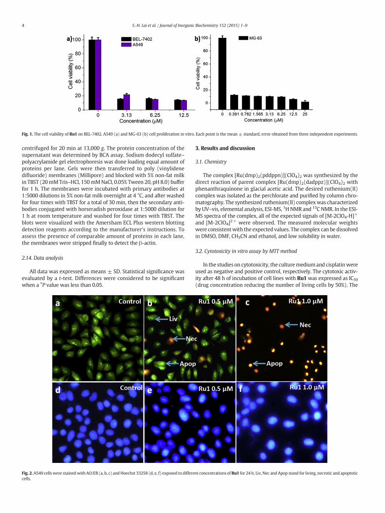
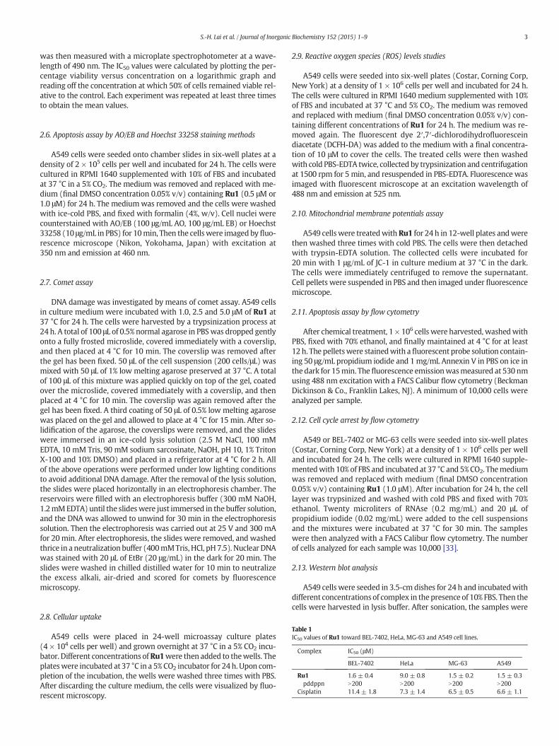
Fig. 1. The cell viability of Ru1 on BEL-7402, A549 (a) and MG-63 (b) cell proliferation in vitro. Each point is the mean ± standard, error obtained from three independent experiments.
4 S.-H. Lai et al. / Journal of Inorganic Biochemistry 152 (2015) 1–9
centrifuged for 20 min at 13,000 g. The protein concentration of thesupernatant was determined by BCA assay. Sodium dodecyl sulfate–polyacrylamide gel electrophoresis was done loading equal amount ofproteins per lane. Gels were then transferred to poly (vinylidenedifluoride) membranes (Millipore) and blocked with 5% non-fat milkin TBST (20mMTris–HCl, 150mMNaCl, 0.05% Tween 20, pH 8.0) bufferfor 1 h. The membranes were incubated with primary antibodies at1:5000 dilutions in 5% non-fat milk overnight at 4 °C, and after washedfor four times with TBST for a total of 30 min, then the secondary anti-bodies conjugated with horseradish peroxidase at 1:5000 dilution for1 h at room temperature and washed for four times with TBST. Theblots were visualized with the Amersham ECL Plus western blottingdetection reagents according to the manufacturer's instructions. Toassess the presence of comparable amount of proteins in each lane,the membranes were stripped finally to detect the β-actin.
2.14. Data analysis
All data was expressed as means ± SD. Statistical significance wasevaluated by a t-test. Differences were considered to be significantwhen a ⁎P value was less than 0.05.
Fig. 2.A549 cells were stainedwith AO/EB (a, b, c) andHoechst 33258 (d, e, f) exposed to differecells.
3. Results and discussion
3.1. Chemistry
The complex [Ru(dmp)2(pddppn)](ClO4)2 was synthesized by thedirect reaction of parent complex [Ru(dmp)2(dadppz)](ClO4)2 withphenanthraquinone in glacial acetic acid. The desired ruthenium(II)complex was isolated as the perchlorate and purified by column chro-matography. The synthesized ruthenium(II) complexwas characterizedby UV–vis, elemental analysis, ESI-MS, 1H NMR and 13C NMR. In the ESI-MS spectra of the complex, all of the expected signals of [M-2ClO4-H]+
and [M-2ClO4]2+ were observed. The measured molecular weightswere consistentwith the expected values. The complex can be dissolvedin DMSO, DMF, CH3CN and ethanol, and low solubility in water.
3.2. Cytotoxicity in vitro assay by MTT method
In the studies on cytotoxicity, the culturemediumand cisplatinwereused as negative and positive control, respectively. The cytotoxic activ-ity after 48 h of incubation of cell lines with Ru1 was expressed as IC50(drug concentration reducing the number of living cells by 50%). The
nt concentrations ofRu1 for 24 h. Liv, Nec andApop stand for living, necrotic and apoptotic

Fig. 3. Images of A549 cell exposure to 2.5 μM of Ru1 and DAPI-stained at 37 °C for 24 h.
5S.-H. Lai et al. / Journal of Inorganic Biochemistry 152 (2015) 1–9
IC50 values of Ru1 against BEL-7402, HeLa, MG-63 and A549 cells arelisted in Table 1, and the cell viability of BEL-7402, A549 and MG-63 isdepicted in Fig. 1. As expectation, ligand pddppn is found to exhibit nocytotoxic activity against the above cell lines. Ru1 shows high cytotoxicactivity with low IC50 values of 1.6 ± 0.4, 9.0 ± 0.8, 1.5 ± 0.2 and 1.5 ±0.3 μM toward BEL-7402, HeLa, MG-63 and A549 cells. Comparing theIC50 values of Ru1 with cisplatin, Ru1 is found to show higher cytotox-icity than cisplatin and its parent complex [Ru(dmp)2(dadppz)]2+
(IC50 values are 16.4 ± 1.5, 11.0 ± 1.1 and 25.7 ± 2.4 μM) [31] towardBEL-7402, MG-63 and A549 cells under the same conditions. Addition-ally, Fig. 1 indicates that the cell viability is concentration-dependent,and the cell viability decreases with increasing concentrations of Ru1.Also, Table 1 shows when ligand pddppn bonded metal to form com-plex, the cytotoxic activity is greatly enhanced. Since the complexdisplays the same cytotoxic effect on MG-63 and A549 cells, in this re-port, we selected A549 cell line for further investigation of the underly-ing mechanisms accounting for the action of ruthenium complex.
3.3. Apoptosis assay by AO/EB staining method
Apoptosis was investigated with AO/EB staining method. Accordingto the difference in membrane integrity between necrotic and apopto-sis, AO can pass through cell membrane, but EB cannot. After the treat-ment of A549 cells with 0.5 and 1.0 μM of Ru1 for 24 h, the apoptoticeffect is shown in Fig. 2a. In the control, the living cells of A549 werestained bright green in spots. A549 cell exposure to 0.5 and 1.0 μM ofRu1 (Fig. 2b and c) for 24 h, green or orange apoptotic cells containingapoptotic bodies, as well as red necrotic cells, were observed. Addition-ally, A549 cells (Fig. 2d) were treated with 0.5 (Fig. 2e) and 1.0 μM(Fig. 2f) of Ru1 for 24 h and stained with Hoechst 33258, apoptotic fea-tures such as nuclear shrinkage and chromatin condensation were also
Fig. 4. (A) Intracellular ROSwas detected in A549 cell (a) exposure to Rosup (b) and 1.0 (c) andexposed to Rosup and 1.0 and 5.0 μM of Ru1 for 24 h. Rosup was used as a positive control.
observed. These results suggest that Ru1 can induce apoptosis in A549cells.
3.4. Cellular uptake studies
The cellular uptake characteristics of a small molecule are critical toits application as a therapeutic or diagnostic agent [34]. In order to tes-tify whether or not Ru1 can be transported into the cellular interior, thecellular uptake and localizationwas investigated using fluorescencemi-croscope. After A549 cells exposed to 2.5 μM of Ru1 for 24 h, the cellswere stained with DAPI. As shown in Fig. 3, the blue channel showsDAPI-stained nuclei, the red channel displays the luminescence of Ru1with an excitation wavelength of 460 nm, and the overlay representscellular association of the complex. The complete overlay of blue chan-nel and red channel suggests that Ru1 can be successfully taken up byA549 cells and the complex can be transported into the cellular interiorand accumulates in the cell nuclei.
3.5. The detection of ROS levels
Reactive oxygen species (ROS) play an important role in cancer celldeath and apoptosis. In the assay of ROS, 2′,7′-dichlorodihydrofluoresceindiacetate (DCFH-DA) was used as fluorescence probe. DCFH-DA iscleaved by intracellular esterases into its non-fluorescent form (DCFH).Then the non-fluorescent substrate is oxidized by intracellular free radi-cals to produce a fluorescent product DCF [35,36]. The changes of thelevels of ROS generation can be evaluated according to the fluorescentintensity of DCF. The fluorescent intensity of DCF is proportional to theamount of peroxide (ROS) produced by the cells [37]. As shown inFig. 4A, in the control, little fluorescent image was observed. After thetreatment of A549 cells with Rosup (positive control, b) and 1.0 (c) and
5.0 (d) μMof Ru1 for 24 h. (B) DCF fluorescence intensity on ROS generation in A549 cells
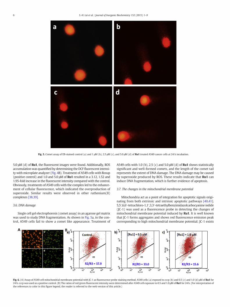
Fig. 5. Comet assay of EB-stained control (a) and 1 μM (b), 2.5 μM (c), and 5.0 μM (d) of Ru1 treated A549 cancer cells at 24 h incubation.
6 S.-H. Lai et al. / Journal of Inorganic Biochemistry 152 (2015) 1–9
5.0 μM (d) of Ru1, the fluorescent images were found. Additionally, ROSaccumulationwas quantified by determining theDCFfluorescent intensi-tywithmicroplate analyzer (Fig. 4B). Treatment of A549 cells with Rosup(positive control) and 1.0 and 5.0 μM of Ru1 resulted in a 3.12, 1.52 and1.95-fold increase in the fluorescent intensity comparedwith the control.Obviously, treatments of A549 cells with the complex led to the enhance-ment of cellular fluorescence, which indicated the overproduction ofsuperoxide. Similar results were observed in other ruthenium(II)complexes [38,39].
3.6. DNA damage
Single cell gel electrophoresis (comet assay) in an agarose gel matrixwas used to study DNA fragmentation. As shown in Fig. 5a, in the con-trol, A549 cells fail to show a comet like appearance. Treatment of
Fig. 6. (A) Assay of A549 cell mitochondrial membrane potential with JC-1 as fluorescence prob24 h. cccpwas used as a positive control. (B) The ratios of red/green fluorescent intensity were dthe references to color in this figure legend, the reader is referred to the web version of this ar
A549 cells with 1.0 (b), 2.5 (c) and 5.0 μM (d) of Ru1 shows statisticallysignificant and well-formed comets, and the length of the comet tailrepresents the extent of DNA damage. The DNA damage may be causedby superoxide produced by ROS. These results indicate that Ru1 caninduce DNA fragmentation, which is further evidence of apoptosis.
3.7. The changes in the mitochondrial membrane potential
Mitochondria act as a point of integration for apoptotic signals origi-nating from both extrinsic and intrinsic apoptotic pathways [40,41].5,5′,6,6′-tetrachloro-1,1′,3,3′-tetraethylbenzimidazolcarbocyanine iodide(JC-1) was used as a fluorescence probe in detecting the changes ofmitochondrial membrane potential induced by Ru1. It is well knownthat JC-1 forms aggregates and shows red fluorescence emission peakcorresponding to high mitochondrial membrane potential; JC-1 exists
e stainingmethod. A549 cells (a) exposed to cccp (b) and 0.5 (c) and 1.0 (d) μMof Ru1 foretermined after A549 cell exposure to 0.5 and 1.0 μMofRu1 for 24 h. (For interpretation ofticle.)
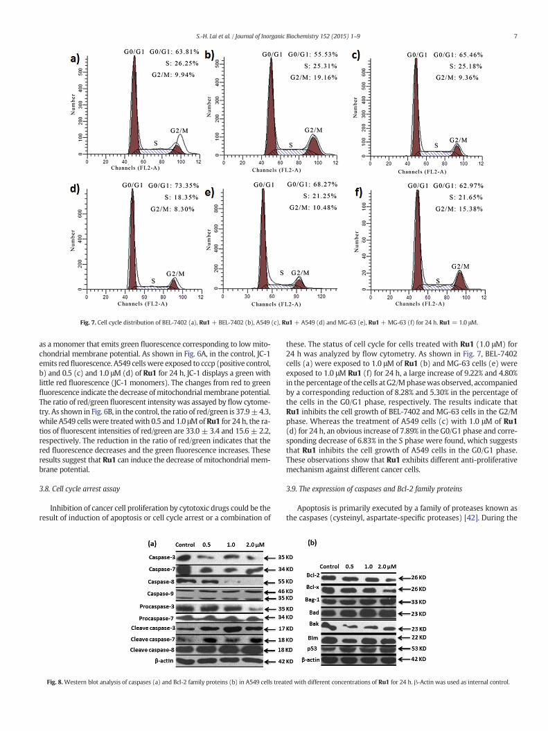
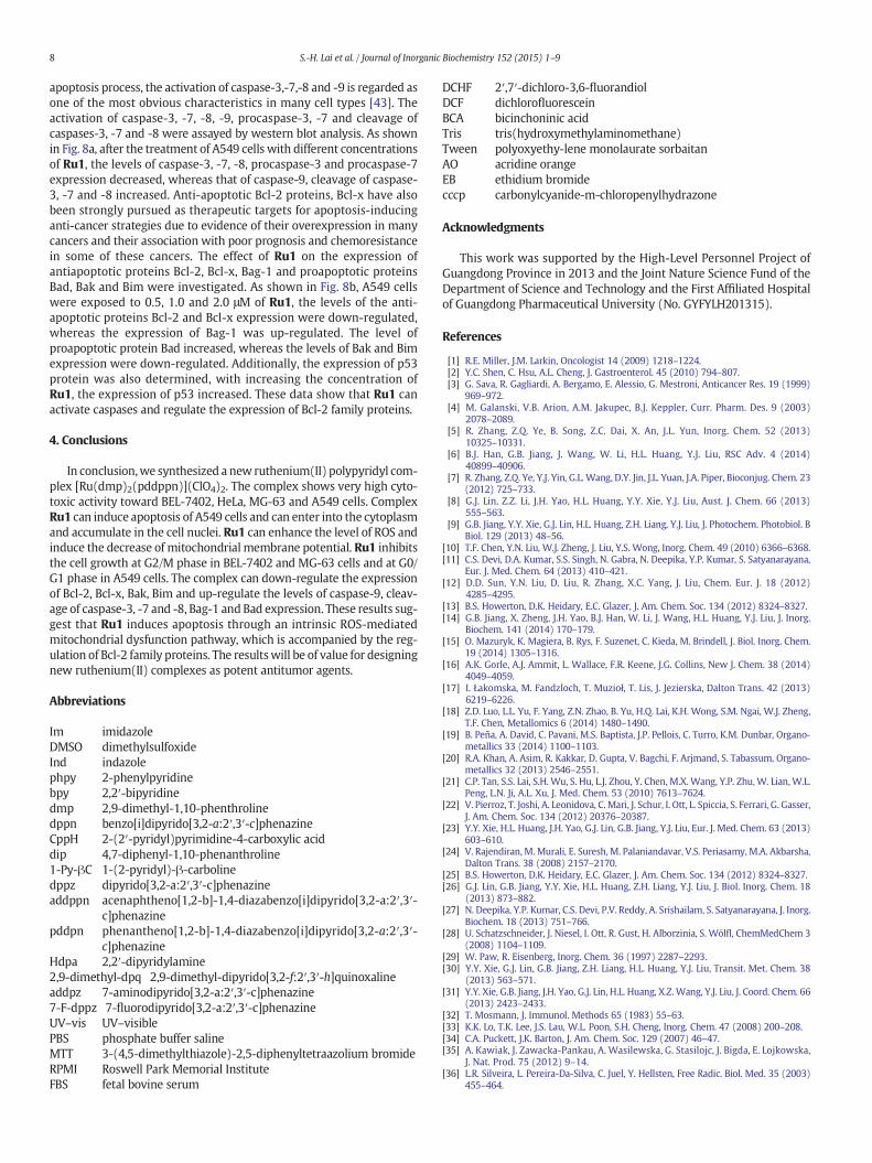
Fig. 7. Cell cycle distribution of BEL-7402 (a), Ru1 + BEL-7402 (b), A549 (c), Ru1 + A549 (d) and MG-63 (e), Ru1 + MG-63 (f) for 24 h. Ru1 = 1.0 μM.
7S.-H. Lai et al. / Journal of Inorganic Biochemistry 152 (2015) 1–9
as a monomer that emits green fluorescence corresponding to lowmito-chondrial membrane potential. As shown in Fig. 6A, in the control, JC-1emits redfluorescence. A549 cellswere exposed to cccp (positive control,b) and 0.5 (c) and 1.0 μM (d) of Ru1 for 24 h, JC-1 displays a green withlittle red fluorescence (JC-1 monomers). The changes from red to greenfluorescence indicate the decrease ofmitochondrialmembrane potential.The ratio of red/green fluorescent intensity was assayed by flow cytome-try. As shown in Fig. 6B, in the control, the ratio of red/green is 37.9±4.3,while A549 cells were treatedwith 0.5 and 1.0 μMof Ru1 for 24 h, the ra-tios of fluorescent intensities of red/green are 33.0 ± 3.4 and 15.6 ± 2.2,respectively. The reduction in the ratio of red/green indicates that thered fluorescence decreases and the green fluorescence increases. Theseresults suggest that Ru1 can induce the decrease of mitochondrial mem-brane potential.
3.8. Cell cycle arrest assay
Inhibition of cancer cell proliferation by cytotoxic drugs could be theresult of induction of apoptosis or cell cycle arrest or a combination of
Fig. 8.Western blot analysis of caspases (a) and Bcl-2 family proteins (b) in A549 cells trea
these. The status of cell cycle for cells treated with Ru1 (1.0 μM) for24 h was analyzed by flow cytometry. As shown in Fig. 7, BEL-7402cells (a) were exposed to 1.0 μM of Ru1 (b) and MG-63 cells (e) wereexposed to 1.0 μM Ru1 (f) for 24 h, a large increase of 9.22% and 4.80%in the percentage of the cells at G2/Mphasewas observed, accompaniedby a corresponding reduction of 8.28% and 5.30% in the percentage ofthe cells in the G0/G1 phase, respectively. The results indicate thatRu1 inhibits the cell growth of BEL-7402 and MG-63 cells in the G2/Mphase. Whereas the treatment of A549 cells (c) with 1.0 μM of Ru1(d) for 24 h, an obvious increase of 7.89% in the G0/G1 phase and corre-sponding decrease of 6.83% in the S phase were found, which suggeststhat Ru1 inhibits the cell growth of A549 cells in the G0/G1 phase.These observations show that Ru1 exhibits different anti-proliferativemechanism against different cancer cells.
3.9. The expression of caspases and Bcl-2 family proteins
Apoptosis is primarily executed by a family of proteases known asthe caspases (cysteinyl, aspartate-specific proteases) [42]. During the
ted with different concentrations of Ru1 for 24 h. β-Actin was used as internal control.
8 S.-H. Lai et al. / Journal of Inorganic Biochemistry 152 (2015) 1–9
apoptosis process, the activation of caspase-3,-7,-8 and -9 is regarded asone of the most obvious characteristics in many cell types [43]. Theactivation of caspase-3, -7, -8, -9, procaspase-3, -7 and cleavage ofcaspases-3, -7 and -8 were assayed by western blot analysis. As shownin Fig. 8a, after the treatment of A549 cells with different concentrationsof Ru1, the levels of caspase-3, -7, -8, procaspase-3 and procaspase-7expression decreased, whereas that of caspase-9, cleavage of caspase-3, -7 and -8 increased. Anti-apoptotic Bcl-2 proteins, Bcl-x have alsobeen strongly pursued as therapeutic targets for apoptosis-inducinganti-cancer strategies due to evidence of their overexpression in manycancers and their association with poor prognosis and chemoresistancein some of these cancers. The effect of Ru1 on the expression ofantiapoptotic proteins Bcl-2, Bcl-x, Bag-1 and proapoptotic proteinsBad, Bak and Bim were investigated. As shown in Fig. 8b, A549 cellswere exposed to 0.5, 1.0 and 2.0 μM of Ru1, the levels of the anti-apoptotic proteins Bcl-2 and Bcl-x expression were down-regulated,whereas the expression of Bag-1 was up-regulated. The level ofproapoptotic protein Bad increased, whereas the levels of Bak and Bimexpression were down-regulated. Additionally, the expression of p53protein was also determined, with increasing the concentration ofRu1, the expression of p53 increased. These data show that Ru1 canactivate caspases and regulate the expression of Bcl-2 family proteins.
4. Conclusions
In conclusion,we synthesized a new ruthenium(II) polypyridyl com-plex [Ru(dmp)2(pddppn)](ClO4)2. The complex shows very high cyto-toxic activity toward BEL-7402, HeLa, MG-63 and A549 cells. ComplexRu1 can induce apoptosis of A549 cells and can enter into the cytoplasmand accumulate in the cell nuclei. Ru1 can enhance the level of ROS andinduce the decrease of mitochondrial membrane potential. Ru1 inhibitsthe cell growth at G2/M phase in BEL-7402 and MG-63 cells and at G0/G1 phase in A549 cells. The complex can down-regulate the expressionof Bcl-2, Bcl-x, Bak, Bim and up-regulate the levels of caspase-9, cleav-age of caspase-3, -7 and -8, Bag-1 and Bad expression. These results sug-gest that Ru1 induces apoptosis through an intrinsic ROS-mediatedmitochondrial dysfunction pathway, which is accompanied by the reg-ulation of Bcl-2 family proteins. The resultswill be of value for designingnew ruthenium(II) complexes as potent antitumor agents.
Abbreviations
Im imidazoleDMSO dimethylsulfoxideInd indazolephpy 2-phenylpyridinebpy 2,2′-bipyridinedmp 2,9-dimethyl-1,10-phenthrolinedppn benzo[i]dipyrido[3,2-a:2′,3′-c]phenazineCppH 2-(2′-pyridyl)pyrimidine-4-carboxylic aciddip 4,7-diphenyl-1,10-phenanthroline1-Py-βC 1-(2-pyridyl)-β-carbolinedppz dipyrido[3,2-a:2′,3′-c]phenazineaddppn acenaphtheno[1,2-b]-1,4-diazabenzo[i]dipyrido[3,2-a:2′,3′-
c]phenazinepddpn phenantheno[1,2-b]-1,4-diazabenzo[i]dipyrido[3,2-a:2′,3′-
c]phenazineHdpa 2,2′-dipyridylamine2,9-dimethyl-dpq 2,9-dimethyl-dipyrido[3,2-f:2′,3′-h]quinoxalineaddpz 7-aminodipyrido[3,2-a:2′,3′-c]phenazine7-F-dppz 7-fluorodipyrido[3,2-a:2′,3′-c]phenazineUV–vis UV–visiblePBS phosphate buffer salineMTT 3-(4,5-dimethylthiazole)-2,5-diphenyltetraazolium bromideRPMI Roswell Park Memorial InstituteFBS fetal bovine serum
DCHF 2′,7′-dichloro-3,6-fluorandiolDCF dichlorofluoresceinBCA bicinchoninic acidTris tris(hydroxymethylaminomethane)Tween polyoxyethy-lene monolaurate sorbaitanAO acridine orangeEB ethidium bromidecccp carbonylcyanide-m-chloropenylhydrazone
Acknowledgments
This work was supported by the High-Level Personnel Project ofGuangdong Province in 2013 and the Joint Nature Science Fund of theDepartment of Science and Technology and the First Affiliated Hospitalof Guangdong Pharmaceutical University (No. GYFYLH201315).
References
[1] R.E. Miller, J.M. Larkin, Oncologist 14 (2009) 1218–1224.[2] Y.C. Shen, C. Hsu, A.L. Cheng, J. Gastroenterol. 45 (2010) 794–807.[3] G. Sava, R. Gagliardi, A. Bergamo, E. Alessio, G. Mestroni, Anticancer Res. 19 (1999)
969–972.[4] M. Galanski, V.B. Arion, A.M. Jakupec, B.J. Keppler, Curr. Pharm. Des. 9 (2003)
2078–2089.[5] R. Zhang, Z.Q. Ye, B. Song, Z.C. Dai, X. An, J.L. Yun, Inorg. Chem. 52 (2013)
10325–10331.[6] B.J. Han, G.B. Jiang, J. Wang, W. Li, H.L. Huang, Y.J. Liu, RSC Adv. 4 (2014)
40899–40906.[7] R. Zhang, Z.Q. Ye, Y.J. Yin, G.L. Wang, D.Y. Jin, J.L. Yuan, J.A. Piper, Bioconjug. Chem. 23
(2012) 725–733.[8] G.J. Lin, Z.Z. Li, J.H. Yao, H.L. Huang, Y.Y. Xie, Y.J. Liu, Aust. J. Chem. 66 (2013)
555–563.[9] G.B. Jiang, Y.Y. Xie, G.J. Lin, H.L. Huang, Z.H. Liang, Y.J. Liu, J. Photochem. Photobiol. B
Biol. 129 (2013) 48–56.[10] T.F. Chen, Y.N. Liu, W.J. Zheng, J. Liu, Y.S. Wong, Inorg. Chem. 49 (2010) 6366–6368.[11] C.S. Devi, D.A. Kumar, S.S. Singh, N. Gabra, N. Deepika, Y.P. Kumar, S. Satyanarayana,
Eur. J. Med. Chem. 64 (2013) 410–421.[12] D.D. Sun, Y.N. Liu, D. Liu, R. Zhang, X.C. Yang, J. Liu, Chem. Eur. J. 18 (2012)
4285–4295.[13] B.S. Howerton, D.K. Heidary, E.C. Glazer, J. Am. Chem. Soc. 134 (2012) 8324–8327.[14] G.B. Jiang, X. Zheng, J.H. Yao, B.J. Han, W. Li, J. Wang, H.L. Huang, Y.J. Liu, J. Inorg.
Biochem. 141 (2014) 170–179.[15] O. Mazuryk, K. Magiera, B. Rys, F. Suzenet, C. Kieda, M. Brindell, J. Biol. Inorg. Chem.
19 (2014) 1305–1316.[16] A.K. Gorle, A.J. Ammit, L. Wallace, F.R. Keene, J.G. Collins, New J. Chem. 38 (2014)
4049–4059.[17] I. Łakomska, M. Fandzloch, T. Muzioł, T. Lis, J. Jezierska, Dalton Trans. 42 (2013)
6219–6226.[18] Z.D. Luo, L.L. Yu, F. Yang, Z.N. Zhao, B. Yu, H.Q. Lai, K.H. Wong, S.M. Ngai, W.J. Zheng,
T.F. Chen, Metallomics 6 (2014) 1480–1490.[19] B. Peña, A. David, C. Pavani, M.S. Baptista, J.P. Pellois, C. Turro, K.M. Dunbar, Organo-
metallics 33 (2014) 1100–1103.[20] R.A. Khan, A. Asim, R. Kakkar, D. Gupta, V. Bagchi, F. Arjmand, S. Tabassum, Organo-
metallics 32 (2013) 2546–2551.[21] C.P. Tan, S.S. Lai, S.H. Wu, S. Hu, L.J. Zhou, Y. Chen, M.X. Wang, Y.P. Zhu, W. Lian, W.L.
Peng, L.N. Ji, A.L. Xu, J. Med. Chem. 53 (2010) 7613–7624.[22] V. Pierroz, T. Joshi, A. Leonidova, C. Mari, J. Schur, I. Ott, L. Spiccia, S. Ferrari, G. Gasser,
J. Am. Chem. Soc. 134 (2012) 20376–20387.[23] Y.Y. Xie, H.L. Huang, J.H. Yao, G.J. Lin, G.B. Jiang, Y.J. Liu, Eur. J. Med. Chem. 63 (2013)
603–610.[24] V. Rajendiran, M.Murali, E. Suresh, M. Palaniandavar, V.S. Periasamy, M.A. Akbarsha,
Dalton Trans. 38 (2008) 2157–2170.[25] B.S. Howerton, D.K. Heidary, E.C. Glazer, J. Am. Chem. Soc. 134 (2012) 8324–8327.[26] G.J. Lin, G.B. Jiang, Y.Y. Xie, H.L. Huang, Z.H. Liang, Y.J. Liu, J. Biol. Inorg. Chem. 18
(2013) 873–882.[27] N. Deepika, Y.P. Kumar, C.S. Devi, P.V. Reddy, A. Srishailam, S. Satyanarayana, J. Inorg.
Biochem. 18 (2013) 751–766.[28] U. Schatzschneider, J. Niesel, I. Ott, R. Gust, H. Alborzinia, S. Wölfl, ChemMedChem 3
(2008) 1104–1109.[29] W. Paw, R. Eisenberg, Inorg. Chem. 36 (1997) 2287–2293.[30] Y.Y. Xie, G.J. Lin, G.B. Jiang, Z.H. Liang, H.L. Huang, Y.J. Liu, Transit. Met. Chem. 38
(2013) 563–571.[31] Y.Y. Xie, G.B. Jiang, J.H. Yao, G.J. Lin, H.L. Huang, X.Z.Wang, Y.J. Liu, J. Coord. Chem. 66
(2013) 2423–2433.[32] T. Mosmann, J. Immunol. Methods 65 (1983) 55–63.[33] K.K. Lo, T.K. Lee, J.S. Lau, W.L. Poon, S.H. Cheng, Inorg. Chem. 47 (2008) 200–208.[34] C.A. Puckett, J.K. Barton, J. Am. Chem. Soc. 129 (2007) 46–47.[35] A. Kawiak, J. Zawacka-Pankau, A. Wasilewska, G. Stasilojc, J. Bigda, E. Lojkowska,
J. Nat. Prod. 75 (2012) 9–14.[36] L.R. Silveira, L. Pereira-Da-Silva, C. Juel, Y. Hellsten, Free Radic. Biol. Med. 35 (2003)
455–464.

9S.-H. Lai et al. / Journal of Inorganic Biochemistry 152 (2015) 1–9
[37] S.N. Hajare, M. Subramanian, S. Gautam, A. Sharma, Biochimie 95 (2013)1722–1731.
[38] Z.D. Luo, L.L. Yu, F. Yang, Z.N. Zhao, B. Yu, H.Q. Lai, K.H. Wong, S.M. Ngai, W.J. Zheng,T.F. Chen, Metallomics 6 (2014) 1480–1490.
[39] L.C. Yang, J.N. Zhang, C. Wang, X.Y. Qin, Q.Q. Yu, Y.H. Zhou, J. Liu, Metallomics 6(2014) 518–531.
[40] T.F. Chen, Y.S. Wong, Int. J. Biochem. Cell Biol. 41 (2009) 666–676.[41] T.F. Chen, Y.S. Wong, Cell. Mol. Life Sci. 65 (2008) 2763–2775.[42] J. Li, J. Yuan, Oncogene 27 (2008) 6194–6206.[43] R.U. Janicke, M.L. Sprengart, M.R. Wati, A.G. Porter, J. Biol. Chem. 273 (1998)
9357–9360.