cytosolic sensing of bacterial flagellin: a tale of two
TRANSCRIPT

Cytosolic Sensing of Bacterial Flagellin: A Tale of Two Proteins
by
Karla Louise Lightfield
A dissertation submitted in partial satisfaction of the
requirements for the degree of
Doctor of Philosophy
in
Infectious Disease and Immunity
in the
Graduate Division
of the
University of California, Berkeley
Committee in charge:
Professor Russell E. Vance, Chair Professor Suzanne M. Fleiszig
Professor David H. Raulet
Spring 2010

Cytosolic Sensing of Bacterial Flagellin: A Tale of Two Proteins
© 2010
by Karla Louise Lightfield

1
Abstract
Cytosolic Sensing of Bacterial Flagellin: A Tale of Two Proteins
by
Karla Louise Lightfield
Doctor of Philosophy in Infectious Disease and Immunity
University of California, Berkeley
Professor Russell E Vance, Chair
The innate immune system can detect the presence of microbial infection by employing germline-encoded receptors specific for conserved microbial ligands. For example, the extracellular presence of the bacterial protein flagellin is sensed by Toll-like receptor 5, leading to activation of the transcription factor NF-κB, cytokine expression, and immune responses. In addition, certain flagellated bacterial pathogens, also appear to be sensed by an inflammasome. The inflammasome is a cytosolic molecular complex that contains a host Nod-like protein, Ipaf, and triggers caspase-1 and macrophage death. We used the intracellular pathogen Legionella pneumophila as a model intracellular bacterium to study the activation of the Ipaf inflammasome. Macrophage resistance to Legionella growth is entirely dependent upon the host expression of Ipaf and the bacterial expression of flagellin making it a useful tool to probe this pathway.
Using a retroviral expression vector, we found that the cytosolic expression of bacterial flagellin, in the absence of other bacterial contaminants and virulence factors was sufficient to trigger macrophage cell death. Through the use of genetic knock-out macrophages we found that this cell death is dependent upon caspase-1 and Ipaf. Taken together these results indicate that flagellin expressed in the cytosol is sufficient to activate the Ipaf inflammasome. We then mapped the region of flagellin required to activate Ipaf to the highly conserved C-terminus of flagellin. This finding indicates that the region of flagellin recognized by Ipaf is distinct from the region recognized by Toll-like receptor 5. Indeed, when the residues recognized by Toll-like receptor 5 are mutated the flagellin is still capable of activating Ipaf. The minimal region of flagellin that is required for sensing by Ipaf is the C-terminal 35 amino acids including three conserved C-terminal leucine residues that are critcal for sensing. Interestingly, activation of the inflammasome in response to this minimal peptide also requires the host protein Naip5, a unique member of the Nod-like and inhibitor of apoptosis (IAP) gene families.

2
Consistent with this result, the restriction of L. pneumophila growth within macrophages is dependent upon both Naip5 and Ipaf. However, Naip5 was dispensable for the response to other Ipaf-dependent stimuli including retroviral transduction of full-length flagellin. Activation of caspase-1 in response to S. typhimurium infection is also only partially dependent on Naip5. By expressing Salmonella flagellin in L. pneumophila we were able to show that it is not the type of flagellin, but rather the mode of delivery that results in a Naip5-dependent or -independent response. Indeed, Salmonella expresses an Ipaf activator, prgJ, that is Naip5 independent, explaining why some of the inflammasome activation in response to Salmonella is Naip5 independent. We have also determined that the N-terminus of flagellin plays a critical role in determining the differential roles of Naip5 and Ipaf in flagellin-recognition. Our results provide a molecular framework for understanding the cytosolic recognition of flagellin by host cells.

i
Dedication
For my parents Nancy and Thomas Lightfield
For my grandparents Roy Lightfield, Marjorie Lightfield, William Holl and Louise Holl

ii
Table of Contents
Dedication................................................................................................................... i
Table of Contents ...................................................................................................... ii
List of Figures ...........................................................................................................iv
List of Tables.............................................................................................................ix
Acknowledgements.....................................................................................................x
Chapter 1....................................................................................................................1
Introduction ...............................................................................................................1
1.1 Basics of Innate Immune Sensing .....................................................................1
1.2 Nod-like Receptors ............................................................................................2
1.3 Inflammasomes..................................................................................................3
1.4 Flagellin .............................................................................................................4
1.5 Legionella as a Model........................................................................................6
1.6 Previous Studies ................................................................................................7
1.7 Thesis .................................................................................................................7
Chapter 2....................................................................................................................9
A Conserved C-terminal Peptide of Bacterial Flagellin is Sensed within the
Cytosol ........................................................................................................................9
2.1 Introduction.......................................................................................................9
2.2 Experimental Procedures................................................................................10
2.3 Results..............................................................................................................12
2.3.1 Flagellin itself triggers Ipaf and Caspase-1. ................................................12

iii
2.3.2 The C-terminus of flagellin is necessary and sufficient to trigger Ipaf-
dependent pyroptosis..............................................................................................15
2.3.3 Ipaf and TLR5 sense distinct regions of flagellin. .......................................20
2.3.4 Conserved and essential amino acids in flagellin are sensed in the cytosol. .21
2.4 Discussion ........................................................................................................29
2.5 Acknowledgements..........................................................................................30
Chapter 3..................................................................................................................31
Differential requirements for Naip5 and Ipaf in sensing cytosolic flagellin ..........31
3.1 Introduction.....................................................................................................31
3.2 Experimental Procedures................................................................................32
3.3 Results and Discussion ....................................................................................34
3.3.1 Naip5 and Ipaf interact in vitro ...................................................................34
3.3.2 Naip5 is required for sensing GFP-C35 but not full-length flagellin............35
3.3.3 Role for the N-terminus of flagellin ............................................................37
3.3.4 Sensing of full-length flagellin is generally Naip5 dependent except when
expressed via a retrovirus .......................................................................................41
3.3.5 Salmonella PrgJ activates Ipaf independent of Naip5..................................43
3.3.6 Conclusions ................................................................................................47
3.4 Acknowledgements..........................................................................................48
Chapter 4..................................................................................................................49
Conclusions and Future Directions .........................................................................49
References ................................................................................................................52

iv
List of Figures Figure 1.1 Diagram of various Nod-like receptor proteins. LRR, leucine rich
repeat; CARD, caspase recruitment domin; PYD, pyrin domain; BIR, baculovirus inhibitor of apoptosis repeat; TIR, Toll/Il-1 receptor domain; CC Coiled Coil. .....2
Figure 1.2 Diagram of Ipaf activation pathway. Activation of Ipaf within the macrophage cytosol via bacterial products, including bacterial flagellin and certain secretion system components (15, 16) leads to the activation of caspase-1 and the eventual cleavage of the proinflammatory cytokines Il-1β and Il-18. Additionally, active caspase-1 results in a rapid (less than 4 hours) cell death. As discussed in this thesis, the rapid cell death is critical for the restriction of bacterial growth within the mammalian macrophage. .....................................................................4
Figure 1.3 Diagram of the flagellin monomer within the filament. D0, purple; D1, blue; D2, grey; D3, green......................................................................................5
Figure 1.4 Legionella lifecycle. Legionella is an infectious motile bacterium that is taken up into the host macrophage by phagocytosis. Once within the macrophage Legionella utilizes a type four secretion system (T4SS) to secrete effector molecules within the host cytosol. Within 5 minutes the phagosome begins to mature, but does not fuse with lysosomes and instead begins to associate with ER-derived vesicles. After approximately 4 hours the Legionella begin replicating. The bacteria then turn on virulence factors such as motility so that once they are released from the cell they are capable of infecting new cells. Legionella that lack a T4SS are taken up by phagocytosis, but enter a phagosome that fuses with lysosomes and are not capable of replicating within this vacuole. .........................6
Figure 2.1 Schematic of Retroviral Transduction Strategy. We developed a retroviral system to express flagellin directly in the host cell cytosol in the absence of all other bacterial products. This retroviral construct contains the flagellin gene followed by an internal ribosomal entry site and gfp used as a marker of transduction. The experimental set up is as follows: First, bone marrow stem cells are transduced with the retrovirus of interest, then these cells are differentiated into macrophages and analyzed for GFP expression using microscopy and flow cytometry. We expect that if the construct is not capable of inducing cell death that we will see GFP positive cells, however we expect that if the construct is capable of inducing cell death we will not see GFP positive cells, because the positively transduced cells will be killed upon the expression of that construct. ..13
Figure 2.2 Retroviral transduction of Legionella flagellin into macrophages. B6, B6.Caspase-1-/- and B6.Ipaf-/- macrophages were transduced with retroviruses expressing Legionella flagellin (FlaA) or a control gene (Irgb10) followed by an internal ribosomal entry site (IRES) and GFP. Transductants were analyzed using either fluorescence microscopy (a) or flow cytometry (b) Note that the highly efficient transduction with control retrovirus in this experiment was not a consistent finding and was not seen in other experiments (e.g., Figure 2.3).........15
Figure 2.3 The C-terminus, but not N-terminus of flagellin is required for activation of Ipaf. (a) B6 macrophages were transduced with retroviruses expressing a control protein, full length FlaA or flagellin lacking the C-terminal 2,

v
4 or 48 amino acids (FlaAC∆2, FlaAC∆4, FlaAC∆48), or (b) flagellin lacking the N-terminal 309 amino acids (FlaAN∆309). The high transduction efficiency of the FlaN∆309 construct is likely due to the small size of the retroviral construct and a resulting increase in its packaging efficiency. (c) Retroviruses expressing the indicating constructs were transduced into B6 or B6.Ipaf-/- macrophages. (d) A retrovirus expressing the C-terminal 35 amino acids of Legionella flagellin was transduced into B6 and B6.Ipaf-/- macrophages. Transduction efficiency was measured by flow cytometry...............................................................................17
Figure 2.4 Cytosolic expression of flagellin leads to loss of GFP positive cells in an Ipaf dependent manner. Bone marrow cells were transduced with retroviral constructs expressing the GFP-C35 fusion, as in Figure 2c, and were analyzed at the indicated timepoints after transduction for GFP expression. ..........................18
Figure 2.5 Cell death in response to cytosolic flagellin is not dependent on host expression of Caspase-3. B6-backcrossed Caspase-3-/- macrophages (gift of Craig Roy) were transduced with retroviral constructs expressing the cytotoxic GFP-C35 or noncytotoxic GFP-C20 fusions. Unlike Capsase-1-/- bone marrow cells, Caspase-3-/- cells were still susceptible to flagellin-dependent pyroptotic cell death. .................................................................................................................19
Figure 2.6 Restriction of Legionella growth is partially dependent upon host macrophage expression of Caspase-7. B6, B6.Ipaf-/- and Caspase-7-/- macrophages were infected with wildtype Legionella at an MOI of .01. .............19
Figure 2.7 The region of flagellin sensed by Ipaf is distinct from the region sensed by TLR5. (a) Model of the Salmonella flagellin monomer as it appears within the assembled flagellar filament [43]. Isoleucine 411 is critical for sensing by TLR5 [42], and corresponds to I391 in Legionella flagellin. (b) B6 macrophages infected with indicated Legionella strains (MOI=1). Cell death was measured as release of lactate dehydrogenase (LDH). ............................................................................20
Figure 2.8 Conserved C-terminal 35 amino acids of flagellin are sufficient to induce Ipaf dependent cytotoxicity. (a) Alignment of the C-terminal 35 amino acids of flagellin. (b) Retroviral delivery of full length flagellin from Legionella (Lp FlaA), Salmonella (St FliC), Pseudomonas (Pa FliC), or Shigella (Sf FliC); (c) Retroviral expression of the C-terminal 35 amino acids (C35) of Legionella (Lp) or Salmonella (St) flagellin, fused to GFP. (d) Retroviral delivery of full length flagellin from Legionella (GFP LpFlaA) and Escherichia coli (GFP EcFliC)...............................................................................................................22
Figure 2.9 Leucine residues in the C-terminus of Legionella flagellin are critical for induction of cell death via Ipaf during retroviral transduction. (a) B6 and B6.Ipaf-/- macrophages were transduced with GFP-C35 harboring the mutations I470A (GFP-C35A), I472A and I473A (GFP-C35AA), or I470A, I472A and I473A (GFP-C35AAA) and the transduction efficiency was determined by flow cytometry. (b) Ipaf-/- macrophages expressing the constructs in (a) were western-blotted with anti-GFP. ........................................................................................23
Figure 2.10 Not all conserved amino acid residues within the C-terminal 35 amino acids of flagellin are required for cytotoxicity. B6 and B6.Ipaf-/- macrophages were transduced with FlaAN∆309 and retroviruses harboring the mutations

vi
L459A and Q461A and the transduction efficiency was determined by flow cytometry. ..........................................................................................................24
Figure 2.11 Large mutations in the C-terminal 35 amino acids result in altered stability and cytotoxicity. B6 and B6.Ipaf-/- macrophages were transduced with GFP-C35 harboring mutations changing the first 5 amino acids of this peptide to alanines (GFP-5A+30). Macrophages were assayed for GFP expression by flow cytometry. B6.Ipaf-/- macrophages expressing these constructs (GFP-C35 and GFP-5A-C30) in were western-blotted with anti-GFP.........................................25
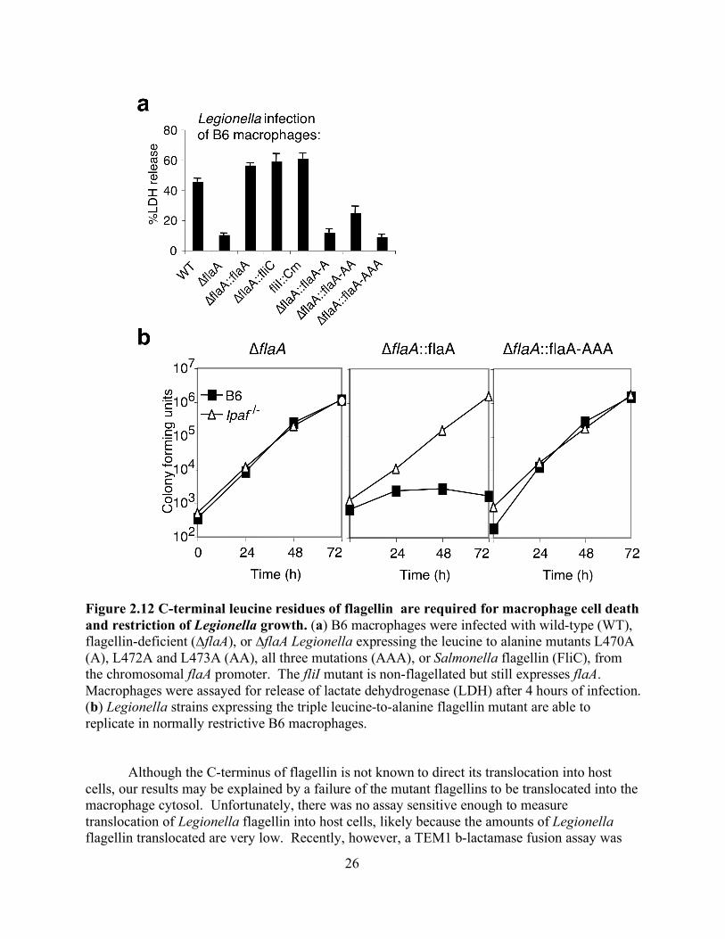
Figure 2.12 C-terminal leucine residues of flagellin are required for macrophage cell death and restriction of Legionella growth. (a) B6 macrophages were infected with wild-type (WT), flagellin-deficient (∆flaA), or ∆flaA Legionella expressing the leucine to alanine mutants L470A (A), L472A and L473A (AA), all three mutations (AAA), or Salmonella flagellin (FliC), from the chromosomal flaA promoter. The fliI mutant is non-flagellated but still expresses flaA. Macrophages were assayed for release of lactate dehydrogenase (LDH) after 4 hours of infection. (b) Legionella strains expressing the triple leucine-to-alanine flagellin mutant are able to replicate in normally restrictive B6 macrophages. ....26
Figure 2.13 C-terminal leucine residues of flagellin are required for macrophage cell death and restriction of Legionella growth. Wildtype (IR715), or flagellin-deficient (fliCfljB), Salmonella were transformed with IPTG-inducible plasmids for expressing wildtype Salmonella flagellin, or flagellin in which the three C-terminal leucines are mutated to alanines (FliC-AAA). Infections were performed as described by Sun et al [38] and LDH release was measured after 4 hours. An asterisk (*) indicates p<0.05 (Student’s T-test) as compared to the fliCfljB pFliC sample. ...............................................................................................................27
Figure 2.14 C-terminal leucines in Salmonella flagellin are required for cytotoxicity but not for translocation into host cells. Translocation of FliC fused to the TEM1 b-lactamase was measured as in Sun et al [38]. Cells in which the TEM1 fusion is translocated into the cytosol appear blue. A SPI-1 mutant strain (invA) was used as a control to demonstrate that translocation of flagellin is SPI-1-dependent, as previously reported by Sun et al [38]. The percentage of blue cells is indicated (at least 100 cells counted per sample). Expression of the FliC-beta lactamase fusion proteins in IR715 were assessed by western blotting using an anti-beta lactamase antibody (QED Bioscience Inc. Cat#15720) (top panel) or rabbit serum against Salmonella H antigen (bottom panel). An arrow indicates flagellin expressed from the chromosome. Both original and mutated Salmonella flagellin-beta lactamase fusions (indicated with an asterisk) were expressed at a similar level........................................................................................................28
Figure 2.15 Model of cytosolic sensing of flagellin within the macrophage. Legionella with a competent T4SS grows within a phagosome. Flagellin leaks through the T4SS into the macrophage cytosol. Once within the cytosol the flagellin is detected by Ipaf leads to the activation of Caspase-1 via proteolytic cleavage. Active caspase-1 leads to rapid macrophage death..............................30
Figure 3.1 Naip5 and Ipaf can interact in vitro. a) HEK293T cells were transfected as follows: 1) Ipaf-HA, Naip5-Myc, GFP-FlaA 2) Ipaf-HA, GFP-FlaA 3) Ipaf-HA, Naip5∆LRR-Myc, GFP-FlaA and were immunoprecipitated as indicated with

vii
either HA or Myc antibodies and immunoblotted using a polyclonal Ipaf antibody, bands shown are 100kD b) HEK293T cells were transfected with Ipaf-HA, Naip5-Myc and GFP-FlaA. Cells were lysed and immunoprecipitated as indicated with either HA or Myc antibodies and immunoblotted for GFP, bands shown are 75kD. .................................................................................................................35
Figure 3.2 Sensing of GFP-C35 requires Naip5. Retroviral expression of the C-terminal 35 amino acids of either Legionella (GFP-LpC35) or Salmonella (GFP-StC35) flagellin fused to GFP.............................................................................36
Figure 3.3 Sensing of retrovirally expressed full-length flagellin is Naip5 independent. Retroviral expression of full-length Legionella flagellin in B6, Ipaf-/- or Naip5-/- macrophages. ................................................................................37
Figure 3.4 The N-terminus of flagellin is required for Naip-5 independent sensing. a) Diagram of retroviral constructs, FlaA, GFP-C65 and N65-GFP-C65. b) Flow cytometry of wild-type, Naip5 deficient and Ipaf deficient macrophages transduced with GFP fused to either the C-terminal 20 amino acids (GFP-C20), N-terminal 65 amino acids (N65-GFP), C-terminal 65 amino acids (GFP-C65), the N-terminal and C-terminal 65 amino acids (N65-GFP-C65) or full length flagellin (GFP-FlaA) from Legionella flagellin. c) or a series of N-terminal deletion mutants of Legionella flagellin, lacking 65, 85, 100 or 125 amino acids as indicated.............................................................................................................39
Figure 3.5 Coiled coil interactions between the N and C terminus of flagellin are not required for Naip5 independence. a) Wild-type, Naip5 deficient or Naip5 and Ipaf deficient macrophages were transduced with GFP-C20, N65-GFP-C65, or N65-GFP-C65 containing the amino acid mutations of leucines 12 and 32 to alanines. b) or with the retroviral construct N65-GFP-C65 containing amino acid substitutions of isoleucine 5, leucine 9 and valine 12 to alanines.........................40
Figure 3.6 Expression of the N and C terminus of Legionella flagellin on separate molecules. The N-terminal 65 amino acids of flagellin fused to GFP and the C-terminal 65 amino acids of flagellin to MCherry were transduced simultaneously into wild-type, Naip5 deficient, or Ipaf deficient macrophages. ..........................41
Figure 3.7 Expression of Legionella flagellin via Listeria is Naip5 independent. Death of wild-type, Naip5 deficient or Ipaf deficient macrophages were infected with Listeria expressing Legionella flagellin fused to the secretion signal of ActA under an IPTG inducible promoter induced with IPTG at the given concentration for 4 hours prior to infection assessed as release of LDH five hours post infection............................................................................................................................42
Figure 3.8 Cytosolic delivery of the N-terminus of anthrax lethal factor (Lfn) fused to the full-length Legionella flagellin. a) Cell death measured from the delivery of various concentrations of Lfn-FlaA to the cytosol of wild-type, Naip5 deficient or Naip5 and Ipaf deficient macrophages via the protective antigen pore. b) Flow cytometry of wild-type, Naip5 deficient or Naip5 and Ipaf deficient macrophages transduced with a retrovirus expressing the same Lfn-FlaA construct.............................................................................................................43
Figure 3.9 Sensing of retrovirally expressed full-length flagellin is Naip5 independent. a) Cell death in wild-type, Naip5 deficient, or Naip5 and Ipaf deficient macrophages infected with either wild-type Legionella (Lp02) or

viii
flagellin deficient Legionella (∆FlaA) and assessed by lactate dehydrogenase release. b) or wild-type Salmonella (LT2) or flagellin deficient Salmonella (dFliC). c) or Legionella expressing Salmonella FliC complemented on the Legionella chromosome. d) Growth of Legionella lacking flagellin or Legionella expressing Salmonella FliC within wild-type, Naip5 deficient and Ipaf deficient macrophages.......................................................................................................46
Figure 3.10 Sensing of Salmonella PrgJ is Naip5 independent. Flow cytometry of wild-type, Naip5 deficient or Naip5 and Ipaf deficient macrophages transduced with N65-GFP, GFP-C65 or Salmonella PrgJ. ....................................................47
Figure 4.1 In vivo infection of Naip5 and Ipaf deficient mice with Legionella pneumophila. Age matched mice were infected intranasally with 2.5x10⁶ LP02 and analyzed 24 hours post infection for a) the number of cells in the broncoalveolar lavage fluid (BALF), n=6 mice per genotype b) IL-1b levels in the BALF were measured by ELISA using recombinant IL-1b was used to determine a standard curve. n=3 mice per genotype c) colony forming units per gram of lung, n=3 mice per genotype or d) colony forming units per mL of BAL, n=3 mice WT, Naip5-/-, n=2 mice Naip5-/-/Ipaf-/-.....................................................................50

ix
List of Tables Table 1.1 Retroviral constructs used in this study.................................................11

x
Acknowledgements I thank my advisor, Russell Vance, for being a supportive and inspiring mentor. I could not have asked for a better place to spend the last four years. I thank my committee members, Suzanne Fleiszig and David Raulet for their support and constructive advice. To the members of the Vance lab, thanks for being so awesome! I am inspired by all of you daily. You are an amazing group of people and very good-looking! To my previous scientific mentors, Laura Parmentier, Brock Spencer and Michael Bottum, thanks for always encouraging me in my interest in science and teaching. Jenny and Sky, I love and miss you both. Janelle, thanks for making this past year fun. I think I would have gone crazy without you. MORE crazy. Eric and Norver, best undergrads imaginable! I thank Sara and Andy for critical comments on my dissertation. To my parents, thanks for always pushing me and teaching me never to make excuses. To all of my family, parents, uncles, aunts, grandparents, cousins and brothers thank you for your patience and support throughout my entire life and throughout my career in graduate school. Your advice and wisdom is invaluable. To my friends, thank you for your understanding about my crazy schedule and your support throughout this process. I thank Cully’s family for making me feel welcome and showing me such great kindness. Jack and Max, thanks for making me smile every day! Cully, thank you for the endless kindness and love you show me, Jack and Max. Thank you for making me feel at home.
1
Chapter 1
Introduction
This thesis describes experiments focused on determining how bacterial flagellin is sensed within the cytosol of infected host cells. In Chapter 1, background on intracellular sensing of bacteria and an overview of the model intracellular pathogen Legionella pneumophila is presented. Chapter 2 describes experiments that show that bacterial flagellin is sensed within the host cell cytosol and experiments that map the domain of flagellin required for intracellular sensing. In Chapter 3, experiments exploring the role of two host proteins, Naip5 and Ipaf, in sensing cytosolic bacterial flagellin are explored in detail. Chapter 4 offers some concluding perspectives.
1.1 Basics of Innate Immune Sensing The immune system works to detect pathogens encountered by the body. The adaptive immune response is capable of detecting an infinite number of antigens by employing millions of cell-surface receptors that are generated in lymphoid cells via complex gene rearrangements. In contrast, the innate immune system contains germ-line encoded receptors that detect microbe-associated molecules (also called pathogen associated molecular patterns or PAMPs). This mechanism of defense is evolutionarily conserved, and is present in many different multicellular organisms[1]. The innate immune system detects a wide variety of PAMPs, including cell wall components, viral nucleic acids and flagellin, from bacteria, viruses, protozoa and fungi that distinguish these foreign organisms from host cells. In addition to protecting from foreign organisms, improper activation of the innate immune system has been linked to autoimmune and autoinflammatory diseases[2], highlighting the importance of regulation in these pathways.
The discovery of mammalian Toll-like receptors (TLRs) in the 1990s made the study of these receptors a major focus of research. TLRs are transmembrane receptors that are localized in the plasma membrane, such as TLR4 and TLR5, or in intracelllular compartments, such as TLR3 and TLR9. TLRs respond to a variety of microbial structures including bacterial lipoproteins, lipopolysaccharide (LPS), flagellin and bacterial or viral nucleic acids[3]. Because of their subcellular localization. TLRs respond to extracellular PAMPs, or PAMPs that reside within phagosomes. Signaling downstream of TLR stimulation results in the activation of NF-κB and the induction of the expression of proinflammatory cytokines, chemokines and other antimicrobial defenses including antimicrobial peptides[4]. In addition to the membrane bound TLRs there are several types of receptors that lack transmembrane domains and function to sense disturbances within the cytosol[5,6]. For example, the cytosolic presence of RNA is detected by the MDA5/RIG-I family of RNA helicases[7]. Additionally, a large family of receptors, the nucleotide binding and oligomerization domain (Nod)-like receptors (NLRs) function to sense cytosolic perturbation[8]

2
or a large variety of pathogen-derived products. Two of these NLRs, Naip5 and Ipaf, are discussed in this thesis.
1.2 Nod-like Receptors Nod-like receptors (NLRs also referred to as NBD-LRR, NACHT-LRR or CATERPILLAR proteins) are cytosolic sensors of conserved microbial components or cytosolic perturbations. NLRs lack the transmembrane domains found in the membrane bound TLRs [9,10,11]. In addition, NLRs are characterized by the presence of a central nucleotide binding domain (NBD) that functions in oligomerization of the proteins. These proteins also contain C-terminal leucine rich repeats (LRRs), thought to function in sensing. For instance, LRRs in plants are required for pathogen detection[12] and the LRRs of NOD1 and NOD2 are required to sense bacterial cell wall components[13]. The NLRs contain variety of N-terminal effector domains. These effector domains are required for downstream signaling [14]. Some of these proteins are diagrammed in Figure 1. The structure of mammalian NLRs resemble that of the disease resistance (R-proteins) in plants as well as Apaf-1 from insects and Ced4 in C. elegans [15]. The function of these proteins is thought to be dependent upon ligand binding to the LRR motif that leads to downstream signaling via the N-terminal protein binding domains, however there is limited evidence for this mechanism in mammalian NLRs.
Figure 1.1 Diagram of various Nod-like receptor proteins. LRR, leucine rich repeat; CARD, caspase recruitment domin; PYD, pyrin domain; BIR, baculovirus inhibitor of apoptosis repeat; TIR, Toll/Il-1 receptor domain; CC Coiled Coil.

3
The mechanism of NLR signaling is presumed to be similar to that described for the plant R-proteins[6]. Based on this mechanism, the LRRs are normally folded back onto the central part of the protein resulting in an auto-repressed inactive protein as the LRRs block the oligomerization mediated by the NBDs. For instance, in Nod1 and Nod2 Leucine Rich Repeat (LRR) domains that act as a ligand sensor and self-inhibitor[13,14]. In Nod1/2, the LRR domain has been shown to inhibit signal transduction in the absence of its ligand but when ligand is present, the LRR domain binds to the ligand and is no longer able to function in inhibition. [16]Without this inhibition, Nod1/2 activate a signal cascade and when the LRR domain is removed the protein is constitutively active and no longer sensitive to ligand[13]. The oligomerization of the NBD precedes downstream signal transduction by the N-terminal effector domain, which can be BIR, CARD or Pyrin domains. In the case of Nod1/2 activation of these proteins by specific fragments of bacterial peptidoglycan results in the downstream activation of mitogen activated protein kinases (MAPKs) and NF-κB[6]. This activation takes place upon binding of the CARD effector domain of Nod1/2 with RIP2[17]. Eventually the activation of these proteins leads to the secretion of proinflammatory cytokines including tumor necrosis factor (TNF), IL-6 and IL-12[18]. In contrast, the activation of other NLRs, including Ipaf and Nalp1-3 leads to the activation of Caspase-1 through the action of the inflammasome. In the case of Ipaf, its N-terminal CARD domain has been shown to bind specifically to caspase-1 and not to other caspases[19,20].
1.3 Inflammasomes The inflammasome is a multiprotein complex that is required for the cleavage and activation of the cystiene protease caspase-1 that normally exists as an inactive pro-protein. When an inflammasome NLR encounters its specific activating molecule the caspase-1 zymogens are brought into close proximity via their CARD domains for autocatalytic activation[21]. Once active, caspase-1 cleaves the proinflammatory cytokines pro-IL-1β and pro-IL-18 that are also present in the cytosol as inactive pro-proteins. Upon cleavage, the cytokines are secreted from the host cell by a still poorly understood mechanism[22]. The transcription and translation of pro-IL-1β and pro-IL-18 is activated upon TLR signaling. Thus the secretion of these proinflammatory cytokines is dependent upon two signals, a TLR signal followed by the action of an inflammasome. In addition to the cleavage of these proinflammatory cytokines, active caspase-1 can also lead to a rapid form of cell death termed “pyroptosis”[23]. Similar to apoptosis, pyroptosis is a programmed form of cell suicide. However, unlike apoptosis, pyroptosis is thought to be a highly inflammatory form of cell death, resulting in the loss of membrane integrity[24]. The substrates downstream of caspase-1 that are cleaved, leading to pyroptosis, are unknown. A basic diagram of the Ipaf inflammasome pathway that will be discussed in detail in this thesis is show in figure 2.

4
Figure 1.2 Diagram of Ipaf activation pathway. Activation of Ipaf within the macrophage cytosol via bacterial products, including bacterial flagellin and certain secretion system components (15, 16) leads to the activation of caspase-1 and the eventual cleavage of the proinflammatory cytokines Il-1β and Il-18. Additionally, active caspase-1 results in a rapid (less than 4 hours) cell death. As discussed in this thesis, the rapid cell death is critical for the restriction of bacterial growth within the mammalian macrophage.
There are several distinct inflammasomes and many, but not all, contain a different Nod-like protein and respond to distinct stimuli. The Nalp1 inflammasome responds to the presence of lethal toxin (LT) from Bacillus anthracis [25] and muramyl dipeptide[26]. The pore-forming protective antigen (PA) of LT allows delivery of lethal factor (LF) directly into the host cell cytosol. It is within the cell cytosol that the presence of LF is detected by Nalp1 resulting in caspase-1 activation and cell death. The Nalp3 inflammasome can be activated in response to a variety of stimuli including particulates (alum, asbestos and others) and a wide array of bacterial, viral and fungal pathogens among many others[27]. Because of the wide variety of stimuli it is hypothesized that Nalp3 might respond to a secondary factor common to these stimuli, however, the exact sequence of events leading to Nalp3 activation is not yet well understood. Current research suggests a role for reactive oxygen species[28] and/or phagosomal or lysosomal disruption[29,30]. Recently, AIM2, unique in that it is not an NLR but rather a HIN200 containing protein, has been shown to activate caspase-1 in response to dsDNA[31,32,33]. In this thesis I describe our understanding of how the Ipaf inflammasome responds to the presence of cytosolic bacterial flagellin. In some cases this response is also dependent upon the presence of another NLR, Naip5[34,35,36]. The Ipaf inflammasome can also be activated by components of the bacterial type three secretion system (TTSS) including PrgJ from Salmonella [37]. An intact bacterial secretion system is required for the activation of Ipaf in all cases. Experiments presented in this thesis clarify the molecular basis of intracellular flagellin sensing via Naip5 and Ipaf.
1.4 Flagellin As mentioned previously, the Ipaf inflammasome responds to the presence of cytosolic
flagellin. Flagellin is interesting in that it is one of only a few protein bacterial components that is recognized by the innate immune system. In addition to sensing by Ipaf, bacterial flagellin is sensed extracellularly by TLR5 at the cell surface. Flagellin seems to be a rather common

5
immune target, as the adaptive immune system generates antibodies against flagellin[24]. The detection of a single microbial product by multiple sensors is not unique to bacterial flagellin. Viral RNA is detected by TLR3, 7 and 8 within phagasomes in addition to being sensed by RIG-I and MDA5 in the cytosol.
Flagellin is an important bacterial product and is highly expressed. It is conserved amongst many bacterial pathogens and is vital for the survival of many bacteria, making it an ideal immune target. Flagellin is a monomer that polymerizes to make up the bacterial flagellum and acts to provide bacterial motility. Flagellar gene expression is highly regulated and the expression of the flagellin monomer takes place after the flagellar hook and basal body (the anchor for the flagella within the bacterial cell wall) are assembled[38]. Flagellin is exported through a flagellar secretion system that resembles the TTSS that secrete virulence factors[39]. Flagellin is exported via an ATP dependent process through this structure, that spans the periplasm, inner membrane and outer membrane. Once secreted up to 30,000 flagellin monomers assemble at the end of the hook[24]. The structure of the Salmonella typhimurium monomer has been studied, revealing a protein with four distinct domains, two of which D0 and D1 form the stem that is contained within the filament when the monomers are polymerized monomers are polymerized (Figure 1.3). The D0 and D1 domains are comprised of α helices that are highly conserved within a variety of bacterial species. The other two domains, D2 and D3 are more variable and are exposed on the surface within the flagellar filament. The adaptive immune responses to flagellin are typically generated against these more variable domains. By contrast, the known innate immune responses to flagellin are against more conserved regions. TLR5 recognizes an amino acid residue within the conserved D1 portion of flagellin that is required for flagellar stability and motility[40]. Additionally, the plant flagellin sensor, FLS2, recognizes a conserved peptide within the N-terminus of flagellin[41].
Figure 1.3 Diagram of the flagellin monomer within the filament. D0, purple; D1, blue; D2, grey; D3, green.

6
1.5 Legionella as a Model Legionella pneumophila is the causative agent of the severe pneumonia called Legionnaires’ disease. Legionella is a gram-negative bacterium that is ubiquitous in the natural environment. It is not a natural pathogen of humans; rather, it has evolved as an aquatic bacterium living as a parasite of fresh water amoebae[42]. Although it has evolved to survive and replicate within amoebae, Legionella is capable of infecting and replicating within human alveolar macrophages after the inhalation of Legionella containing droplets. The ability of Legionella to grow within macrophages is key to its virulence, as many avirulent mutants of the organism have been found to be defective in intracellular growth[43]. Legionella’s ability to grow intracellularly is dependent upon the Dot/Icm type IV secretion apparatus that is used to direct the host cell to create a unique vacuole that associates with the endoplasmic reticulum. The Legionella lifecycle is detailed in Figure 1.3.
Figure 1.4 Legionella lifecycle. Legionella is an infectious motile bacterium that is taken up into the host macrophage by phagocytosis. Once within the macrophage Legionella utilizes a type four secretion system (T4SS) to secrete effector molecules within the host cytosol. Within 5 minutes the phagosome begins to mature, but does not fuse with lysosomes and instead begins to associate with ER-derived vesicles. After approximately 4 hours the Legionella begin replicating. The bacteria then turn on virulence factors such as motility so that once they are released from the cell they are capable of infecting new cells. Legionella that lack a T4SS are taken up by phagocytosis, but enter a phagosome that fuses with lysosomes and are not capable of replicating within this vacuole.
Legionella provides us a unique model with which to study the innate immune responses.
First, Legionella is not capable of transmission from person to person and thus has not evolved within humans. Therefore Legionella is not likely to be as immune evasive as better adapted

7
pathogens and immune responses generated against Legionella must be against conserved molecules or pathogenesis mechanisms. Specifically, Legionella offers an important tool to study the flagellin dependent activation of Ipaf and Naip5 because it uses a type IV secretion system for pathogensis rather than the TTSS employed by other activators of the Ipaf inflammasome, including Salmonella and Pseudomonas. This is important given the recent discovery that inner rod proteins from the TTSS can activate the Ipaf inflammasome[37]. Other important pathogens that are less well studied, but also express a type IV secretion system include Brucella abortus and Coxiella burnettii.
1.6 Previous Studies Prior to the work described in this thesis it was demonstrated that murine macrophage restriction of Legionella growth was dependent upon the presence of functional Naip5 and Ipaf genes[44]. This restriction was also found to be dependent upon the bacterial expression of flagellin[36,45]. Caspase-1 activation downstream of Legionella infection of macrophages was shown to be dependent upon the actions of Naip5, Ipaf and the expression of bacterial flagellin. These data suggest that bacterial flagellin plays an important roll in the activation of the Naip5/Ipaf mediated macrophage resistance responses and the eventual restriction of Legionella growth. However, many questions remained unanswered about this pathway. First, Legionella lacking flagellin have altered motility, infectivity and adhesion- any of which might be involved evading inflammasome activation. Secondly, it is known that there are non-flagellated bacteria, including Shigella, that activate caspase-1 via Ipaf [46]. In addition, the type IV secretion system of Legionella creates pores within the phagasome that might also be involved in Ipaf dependent sensing. The type IV secretion system of Legionella and the TTSS of Salmonella are required to activate Ipaf during infection. Thus, while flagellin seemed necessary to activate Ipaf, it remained unclear whether it was sufficient. The role of flagellin in inflammasome activation in the absence of a bacterial secretion system, lipofection reagents or other co-purifying bacterial contaminants had not previously been explored. Because there is a second flagellin sensor, TLR5, it is possible that flagellin is sensed by TLR5 and a second signal sensed by Ipaf resulting in cell death and restriction of bacterial growth. Experiments supporting the hypothesis that flagellin is leaked into the macrophage cytosol via the bacterial secretion systems where it then activates caspase-1 via Naip5 and Ipaf are presented in this thesis.
1.7 Thesis This thesis describes experiments aimed at understanding how bacterial flagellin is sensed within the mammalian host cell cytosol. We have demonstrated that bacterial flagellin expressed within macrophages is sufficient to induce macrophage cell death via activation of the Nod-like protein Ipaf. Prior to the work described in this thesis the role of flagellin in activating the inflammasome was unclear as the flagellin used was either expressed by bacteria or purified from bacteria, leaving the possibility that other bacterial ligands were required for activation. To circumvent these potential problems, we used a retroviral expression system to express bacterial flagellin directly in the host cell cytosol. We were able to show that flagellin itself in the absence of bacterial contaminants or secretions systems is sufficient to activate the Ipaf inflammasome. Chapter 2 of this thesis describes how we used this approach to narrow down the region of flagellin required for inflammasome activation.

8
The remainder of the thesis describes experiments aimed at understanding the role of the host Nod-like proteins, Naip5 and Ipaf, in the sensing of intracellular bacterial flagellin. Naip5 is not required for all activities of Ipaf, such as activation of the inflammasome in response to retroviral expression of full-length flagellin. However, in Chapter 3, we present results that support a role for Naip5 in sensing flagellin during infection with the intracellular bacterium Legionella pneumophila. In addition we show that Naip5 is also required for sensing of the minimal flagellin domain required for activation of the Ipaf inflammasome as defined in Chapter 2. Our results confirm that flagellin is capable of activating the inflammasome in a manner dependent on Ipaf and Naip5.

9
Chapter 2
A Conserved C-terminal Peptide of Bacterial Flagellin is Sensed within the Cytosol
This chapter describes experiments that demonstrate that bacterial flagellin is sensed in the host cell cytosol via the Ipaf inflammasome. A paper describing some of these experiments has been published in Nature Immunology [35]. Inflammasomes are cytosolic multiprotein complexes that sense microbial infection and trigger cytokine production and cell death. However, the molecular components of inflammasomes, and what they sense, remain poorly defined. Here we demonstrate that 35 amino acids from the C-terminus of flagellin triggered inflammasome activation in the absence of bacterial contaminants or secretion systems. These results begin to define the molecular basis for cytosolic sensing of bacterial flagellin by elucidating key amino acid residues in flagellin required for sensing via the Ipaf inflammasome.
2.1 Introduction Inflammasomes are cytosolic multiprotein complexes that are critical regulators of
inflammation, and are required for proteolytic activation of the cysteine protease caspase-1 [47,48,49]. Caspase-1 is itself required for the proteolytic processing and release of inflammatory cytokines such as IL-1β and IL-18, as well as for induction of a necrotic-like cell death called pyroptosis [23,50,51]. The molecular components and structures of inflammasomes remain poorly defined. It is believed that multiple distinct inflammasomes may exist, each containing a key scaffold protein, many from the NLR (Nucleotide-binding domain, Leucine-rich Repeat) superfamily, that confers specificity for particular microbial products. For example, NLR proteins of the Nlrp1 family (also called Nalp1) appear to activate the inflammasome in response to anthrax lethal toxin [25] and bacterial muramyl dipeptide [26]. In contrast, the NLR protein Nlrp3 (also called Nalp3 or Cryopyrin) has been proposed to sense a wide range of microbial products including bacterial RNA [52], viral DNA [53], uric acid crystals [54], muramyl dipeptide [55,56], nigericin [8] alpha toxin from Staphylococcus aureus [57], asbestos [28], and Candida albicans [58], among others . There is at present no explanation for how a single NLR protein can sense all these microbial products and the precise molecular nature of what is sensed by any inflammasome remains undefined. In constrast to these NLR containing inflammasomes, the AIM2 inflammasome contains a HIN-200 family member protein that activates caspase-1 via interaction with apoptosis-associated speck like protein (ASC) through its pyrin domain [59]. ASC is a bipartite adapter molecule required for the activation of caspase-1 in a variety of inflammasomes, including the Nalp3 inflammasome[27].
The inflammasome containing the NLR protein Ipaf (also called Nlrc4) is one of the best characterized inflammasomes, and has been proposed by several groups to sense the cytosolic presence of flagellin[60,61,62]. Ipaf can activate cell death via interaction with caspase-1 in a manner that is independent upon the presence of the adapter ASC, however, secretion of IL-

10
1β initiated by the Ipaf inflammasome is dependent upon ASC[63]. Flagellin-deficient mutants of Salmonella typhimurium and Legionella pneumophila are defective in Ipaf-dependent inflammasome activation, and flagellin, purified from or expressed in bacteria, triggers Ipaf-dependent caspase-1 activation when delivered to the cytosol of macrophages by use of a pore-forming toxin (listeriolysin O (LLO)) or transfection reagents [36,45,60,61,62]. It was proposed that during natural infections, flagellin triggers inflammasome activation upon secretion into the host cytosol via bacterial type III/IV secretion systems[36,45,60,61,62]. However, doubts were expressed as to whether flagellin is indeed sensed cytosolically [15,47] since none of the existing studies eliminated the possibility that bacterial secretion systems, LLO, transfection reagents, and/or copurifying bacterial contaminants, contributed to activation of caspase-1. Moreover, the role of Ipaf in sensing flagellin remains unclear. It has been reported that Ipaf-deficient macrophages also fail to activate caspase-1 in response to non-flagellated pathogens, including flagellin-deficient Pseudomonas aeruginosa [64] and Shigella flexneri [46]. In addition, Ipaf appears to play a key role in caspase-1 activation in response to aerolysin, a pore-forming toxin [65]. Thus it has been proposed that Ipaf may not in fact sense flagellin, but may instead respond to bacterial contaminants, membrane pores, or type III/IV secretion systems [15,47], which appear to be required for inflammasome activation by all bacterial pathogens tested.
Even less clear is the role of Naip5, an NLR protein that hetero-oligomerizes with Ipaf [66,67], and has also been proposed to be involved in cytosolic sensing of flagellin [36,45]. Although we now know that Naip5 is required for inflammasome activation in some contexts, prior to the work described in this thesis, the role of Naip5 in inflammasome activation was unclear [34,68]. The role of Naip5 in sensing bacterial flagellin will be discussed in Chapter 3. In this chapter I describe experiments that clarify the role of flagellin in activating the Ipaf inflammasome. For the first time, we define a short peptide domain that appears sufficient to activate the inflammasome in the absence of bacterial contaminants, pores or secretion systems.
2.2 Experimental Procedures Mice.
Wildtype C57BL/6 (B6) mice and B6 mice harboring A/J chromosome 13 (B6.A-Chr13) were obtained from the Jackson Labs (Bar Harbor, ME). B6.Ipaf-/- mice [69] were obtained from S. Mariathasan and V. Dixit (Genentech). B6.Caspase-1-/- [51] were the gift of A. Van der Velden and M. Starnbach. Bacterial Strains.
LP02 is a streptomycin-resistant thymidine auxotroph derived from L. pneumophila LP01. An unmarked deletion of flaA was described previously [36]. The ∆flaA strain was complemented by pBBR1-MCS2 plasmids expressing Legionella or Salmonella flagellin (or point mutants thereof). The ∆flaA strain was also complemented by reintroducing a copy of flagellin (or point mutants thereof) onto the chromosome under the control of the endogenous Legionella flaA promoter. FlaA and its promoter were cloned into the suicide vector pSR47S and introduced onto the chromosome by a single crossover and selection with kanamycin. Salmonella typhimurium LT2 and isogenic mutants were the gift of A. Van der Velden and M. Starnbach. Pseudomonas aeruginosa strain PAK was the gift of T. Machen. Salmonella were grown overnight in Luria-Bertani broth and reinoculated at a 1:100 dilution and grown to mid-
11
exponential phase (3h) to induce SPI-1. Overnight Pseudomonas cultures were diluted 1:10 and grown 3h.
Growth Curves.
Growth curves were performed as previously described [44]. Briefly, macrophages were plated in 96 or 24 well tissue culture plates at 5 x 10⁵/mL, allowed to adhere, and then infected with stationary phase Legionella at an MOI of 0.01. Growth of luminescent Legionella strains was assessed in a LmaxII plate-reading luminometer (Molecular Devices). Growth of non-luminescent strains was assessed by plating for colony forming units on BCYE plates. Cytotoxicity Assays.
Cytotoxicity was measured by evaluating the activity of lactate dehydrogenase (LDH) released from cells [70]. Overnight cultures of Legionella in stationary phase were added at an MOI of 1 to a confluent monolayer of macrophages, and plates were spun at 400 xg for 10 minutes to ensure comparable infectivity of motile and non-motile strains. Culture supernatants were assayed for LDH activity after 4h. Salmonella strains (LT2 background) were added at an MOI of 2, and Pseudomonas at an MOI of 10. Plates were spun as described above, and gentamicin (100ug/ml) was added 30 min post infection (PI) to kill extracellular bacteria. LDH release was measured after 4h and 2h PI, respectively. Specific lysis was calculated as a percentage of detergent lysed macrophages. The cytotoxicity experiments in (Figure with ST-AAA) were performed as previously described [71]. Retroviral Constructs and Production.
Genes encoding flagellin or control protein were cloned into a replication defective mouse stem-cell retroviral construct (pMSCV2.2). Retroviral particles were generated using Phoenix-eco packaging cells, and were used to transduce bone marrow cells after 48h and 72h culture in MCSF. Cells were typically analyzed 3-4 days after the first transduction. Western blotting.
106 bone marrow-derived macrophages were seeded in a 6-well plate and infected at an MOI of 1. At 1h post infection, the bacterial suspension was replaced with fresh media without serum, and infection was allowed to continue for 2 hours. Subsequently, supernatants were harvested and precipitated with 10% trichloroacetic acid (TCA). Precipitated proteins were separated on 12% gels (Invitrogen), blotted onto Immobilon-P transfer membrane (Millipore) and probed with rabbit polyclonal anti-caspase-1 p10 antibody (Santa Cruz Biotechnology; antibody sc-514). GFP was detected with monoclonal antibody JL-8 (Clontech).
Table 1.1 Retroviral constructs used in this study. Name Mscv 2.2 Empty vector: IRES-GFP Control Irgb10-IRES-GFP Mscv FlaA Legionella flagellin (flaA)-IRES-GFP Mscv StFliC Salmonella flagellin (fliC)-IRES-GFP Mscv FlaAC∆2 Legionella flaA lacking C-terminal 2 amino acids-IRES-GFP Mscv FlaAC∆4 Legionella flaA lacking C-terminal 4 amino acids-IRES-GFP Mscv Legionella flaA lacking C-terminal 48 amino acids-IRES-GFP

12
FlaAC∆48 Mscv FlaAN∆309
Legionella flaA lacking the N-terminal 309 amino acids-IRES-GFP (this results in 166 amino acids from the C-terminus).
Mscv GFP-C166
GFP fused directly to the C-terminal 166 amino acids of Legionella flaA
Mscv GFP-C35 GFP fused directly to the C-terminal 35 amino acids of Legionella flaA Mscv GFP-C20 GFP fused directly to the C-terminal 20 amino acids of Legionella flaA Mscv GFP-IRES-Nco-Sal-Cla
Mscv GFP-IRES backbone used to generate GFP-IRES-Flagellin constructs
Mscv GFP Lp FlaA
GFP-IRES-Full Length flaA from Legionella
Mscv GFP St FliC
GFP-IRES-Full Length fliC from Salmonella
Mscv GFP Sf FliC
GFP-IRES-Full length fliC from Shigella
Mscv GFP Pa FliC
GFP-IRES-Full length fliC from Pseudomonas
Mscv GFP Ec FliC
GFP-IRES-Full length fliC from Escherichia
Mscv GFP-St35 GFP fused directly to the C-terminal 35 amino acids of Salmonella fliC
Mscv GFP-C35A
GFP fused directly to the C-terminal 35 amino acids of Legionella flaA with Leucine 470 mutated to alanine
Mscv GFP-C35AA
GFP fused directly to the C-terminal 35 amino acids of Legionella flaA with leucines 472 and 473 mutated to alanine
Mscv GFP-C35AAA
GFP fused directly to the C-terminal 35 amino acids of Legionella flaA with leucines 470, 472 and 473 mutated to alanine
Mscv L459A Mscv FlaAN∆309 with leucine 459 mutated to alanine Mscv Q461A Mscv FlaAN∆309 with glutamine 461 mutated to alanine Mscv GFP-5A+30
GFP fused directly to 5 alanine residues preceding the C-terminal 30 amino acids of Legionella flaA
Mscv C35-IRES-GFP
C-terminal 35 amino acids of Legionella FlaA-IRES-GFP
2.3 Results
2.3.1 Flagellin itself triggers Ipaf and Caspase-1. Other studies describing intracellular sensing of Salmonella flagellin [61,62] or
Legionella flagellin [45], demonstrated that delivery of ‘purified’ flagellin to the cytosol (via transfection or LLO-mediated delivery) was sufficient to trigger rapid macrophage death. However, these studies did not address the possibility that a bacterial contaminant could be

13
required or responsible for the apparent cytotoxicity of the transfected flagellin preparation. It has also been questioned whether membrane damage associated with the transfection procedures used in these studies might sensitize macrophages to rapid cell death [72]. To determine whether flagellin itself, in the absence of bacterial modifications, contaminants, secretion systems or transfection reagents, is sufficient to trigger Ipaf and macrophage death, we used retroviral transduction to express L. pneumophila flagellin (flaA) directly in the macrophage cytosol from a eukaryotic promoter. The retroviral expression constructs also contained an internal ribosomal entry site (IRES) and a green fluorescent protein (GFP) gene to permit identification of positively transduced cells. A schematic detailing this strategy is detailed in Figure 2.1. Briefly, primary bone marrow cells were transduced, differentiated into macrophages in the presence of MCSF, and resulting macrophages were analyzed for GFP expression four days after transduction.
Figure 2.1 Schematic of Retroviral Transduction Strategy. We developed a retroviral system to express flagellin directly in the host cell cytosol in the absence of all other bacterial products. This retroviral construct contains the flagellin gene followed by an internal ribosomal entry site and GFP used as a marker of transduction. The experimental set up is as follows: First, bone marrow stem cells are transduced with the retrovirus of interest, then these cells are differentiated into macrophages and analyzed for GFP expression using microscopy and flow cytometry. We expect that if the construct is not capable of inducing cell death that we will see GFP positive cells, however we expect that if the construct is capable of inducing cell death we will not see GFP positive cells, because the positively transduced cells will be killed upon the expression of that construct. Although wild-type B6 macrophages were efficiently transduced with a control retrovirus, we were unable to recover B6 macrophages transduced with a flaA-expressing retrovirus when analyzed four days after transduction by visualizing GFP expression (Figure 2.2a). By contrast, B6.Caspase-1-/- and B6.Ipaf-/- macrophages were transduced with flaA-expressing retrovirus

14
(Figure 2.2a and b). The percentage of transduced macrophages expressing GFP was assessed using flow cytometry (Figure 2.2b). Thus, the inability to transduce B6 macrophages with flaA is due to an activity of Ipaf and caspase-1 (presumably induction of pyroptotic cell death, see below) and the presence of the flagellin gene as opposed to the control protein. Interestingly, B6.caspase-1-/- macrophages were never transduced to the same levels as B6.Ipaf-/- macrophages, indicating that there is potentially a caspase-1 independent, but Ipaf dependent form of cell death also contributing to this process. In agreement with these results, B6.caspase-1-/- macrophages are not as permissive to wild-type Legionella growth as B6.Ipaf-/- macrophages (unpublished observations). We conclude that flagellin itself is sensed in the cytosol in the absence of other bacterially derived signals and also in the absence of potentially damaging transfection reagents.

15
Figure 2.2 Retroviral transduction of Legionella flagellin into macrophages. B6,
B6.Caspase-1-/- and B6.Ipaf-/- macrophages were transduced with retroviruses expressing Legionella flagellin (flaA) or a control gene (Irgb10) followed by an internal ribosomal entry site (IRES) and GFP. Transductants were analyzed using either fluorescence microscopy (a) or flow cytometry (b) Note that the highly efficient transduction with control retrovirus in this experiment was not a consistent finding and was not seen in other experiments (e.g., Figure 2.3).
2.3.2 The C-terminus of flagellin is necessary and sufficient to trigger Ipaf-dependent pyroptosis. In order to identify the region of flagellin that is sensed cytosolically, we transduced macrophages with retroviruses expressing a series of C- and N-terminal deletion mutants of Legionella flagellin. Flagellins lacking 4 or 48 C-terminal amino acids (FlaAC∆4, FlaAC∆48) were not cytotoxic and efficiently transduced wildtype B6 macrophages (Figure 2.3a) that are generally intolerant of transduction of full length flagellin. In contrast, a deletion mutant lacking the N-terminal two-thirds of flagellin (FlaAN∆309), resulting in a protein that contains only the C-terminal 166 amino acids of flagellin, still triggered Ipaf-dependent cell death (Figure 2.3b) similar to the results seen with the full length flagellin protein. These results implied that the C-terminal third (166 amino acids) of flagellin is necessary and sufficient to trigger Ipaf. In order to further narrow down the region of flagellin detected by Ipaf, and in order to define a minimal region required for activation, we made a series of small C-terminal flagellin peptides fused directly to GFP. Macrophages were transduced with retroviruses that express GFP fused to the C-terminal 20 (GFP-C20), 35 (GFP-C35) or 166 (GFP-C166) amino acids of flagellin. Although

16
transduction of B6 macrophages with the GFP-C20 construct was not cytotoxic and resulted in significant green fluorescence, the GFP-C35 and GFP-C166 expression constructs failed to transduce wildtype, but not B6.Ipaf-/-, macrophages (Figure 2.3c). Thus, we conclude that the C-terminal 35 amino acids of flagellin, fused to GFP, is sufficient to be sensed by Ipaf in the cytosol. Interestingly, the C-terminal 35 amino acids of flagellin (Mscv C35-IRES-GFP), when expressed in the retrovirus without fusion to GFP did not induce Ipaf dependent macrophage death (Figure 2.3d) presumably due to instability of this small peptide.

17
Figure 2.3 The C-terminus, but not N-terminus of flagellin is required for activation of Ipaf. (a) B6 macrophages were transduced with retroviruses expressing a control protein, full length FlaA or flagellin lacking the C-terminal 2, 4 or 48 amino acids (FlaAC∆2, FlaAC∆4, FlaAC∆48), or (b) flagellin lacking the N-terminal 309 amino acids (FlaAN∆309). The high transduction efficiency of the FlaN∆309 construct is likely due to the small size of the retroviral construct and a resulting increase in its packaging efficiency. (c) Retroviruses expressing the indicating constructs were transduced into B6 or B6.Ipaf-/- macrophages. (d) A retrovirus expressing the C-terminal 35 amino acids of Legionella flagellin was transduced into B6 and B6.Ipaf-/- macrophages. Transduction efficiency was measured by flow cytometry.
Although the simplest interpretation of our data is that cytosolic expression of flagellin triggers an Ipaf- and Capsase-1-dependent pyroptotic cell death of B6 macrophages, we considered the alternative possibility that flagellin-expressing retrovirus selectively failed to transduce B6 macrophages. Both possibilities are formally consistent with the lack of GFP+ macrophages four days after transduction. To demonstrate that B6 macrophages were first being transduced by flagellin-expressing retrovirus, and then dying, we analyzed bone marrow cells at early timepoints after transduction. At 6 hours after transduction, we were able to detect both B6 and B6.Ipaf-/- bone marrow cells expressing the GFP-C35 flagellin construct. We found that B6 cells transduced with the GFP-C35 flagellin construct gradually disappeared from the culture, whereas B6.Ipaf-/- cells were maintained (Figure 2.4). These data are supported by experiments done by Jakob von Moltke that showed that doxycyclin-inducible expression of flagellin results in flagellin expression followed by pyroptosis (unpublished observation).

18
Figure 2.4 Cytosolic expression of flagellin leads to loss of GFP positive cells in an Ipaf dependent manner. Bone marrow cells were transduced with retroviral constructs expressing the GFP-C35 fusion, as in Figure 2c, and were analyzed at the indicated timepoints after transduction for GFP expression.
During certain infection conditions (when using high MOIs or infection of permissive macrophages) Legionella results in the activation of capsase-3, an effector caspase within the apoptotic pathway [73]. In order to rule out a role for caspase-3 in the cell death pathway that results from the transduction of wildtype macrophages with flagellin we transduced caspase-3-/- macrophages with a flagellin expressing retrovirus. We found that caspase-3-/- macrophages are still susceptible to flagellin dependent cell death (Figure 2.5). A lack of dependence on caspase-3 is consistent only with pyroptosis. In addition, caspase-7 has been shown to be cleaved by active caspase-1, and has been implicated in the restriction of Legionella growth [74]. In Figure 2.6 we show that restriction of wildtype Legionella growth is at least partially dependent upon macrophage expression of caspase-7. Importantly, these B6.Caspase-7-/- contain the B6 allele of Naip5. Thus, restriction of Legionella growth by the activation of caspase-1 may be at least partially due to its role in activating caspase-7. We conclude that cytosolic expression of the C-terminal 35 amino acids of flagellin, fused to GFP, induces an Ipaf- and capsase-1-inflammasome-dependent pyroptotic cell death.

19
Figure 2.5 Cell death in response to cytosolic flagellin is not dependent on host expression
of Caspase-3. B6-backcrossed Caspase-3-/- macrophages (gift of Craig Roy) were transduced with retroviral constructs expressing the cytotoxic GFP-C35 or noncytotoxic GFP-C20 fusions. Unlike Capsase-1-/- bone marrow cells, Caspase-3-/- cells were still susceptible to flagellin-dependent pyroptotic cell death.
Figure 2.6 Restriction of Legionella growth is partially dependent upon host macrophage expression of Caspase-7. B6, B6.Ipaf-/- and B6.Caspase-7-/- macrophages were infected with wildtype Legionella at an MOI of .01.

20
2.3.3 Ipaf and TLR5 sense distinct regions of flagellin.
Previous studies demonstrated that sensing of flagellin by TLR5 requires amino acids in the
D1 region of flagellin (e.g., isoleucine 411) [40], whereas the above studies indicate it is the C-terminal D0 region that is sensed cytosolically (Figure 2.7a). When expressed from Salmonella, an I411A flagellin mutant also reportedly failed to activate caspase-1, but this may have resulted from a failure of the I411A flagellin to be translocated into host cells by Salmonella [61]. We circumvented this difficulty by expressing Salmonella flagellin (FliC) in flagellin-deficient (∆flaA) Legionella. As we previously showed, Salmonella flagellin (FliC) is able to restore cytotoxicity to the ∆flaA Legionella mutant [36]. Importantly, both wildtype and I411A FliC were equally able to complement the cytotoxicity defect of the ∆flaA mutant (Figure 2.7b), implying that amino acids required for TLR5 sensing are not required for inflammasome activation. Our results therefore establish that there are two distinct innate immune pathways for detecting flagellin: a cell-surface TLR5-dependent pathway that senses the D1 region of flagellin and triggers NF-kB activation, and a cytosolic Ipaf-dependent pathway that senses the C-terminal D0 region and activates the inflammasome.
Figure 2.7 The region of flagellin sensed by Ipaf is distinct from the region sensed by
TLR5. (a) Model of the Salmonella flagellin monomer as it appears within the assembled flagellar filament [75]. Isoleucine 411 is critical for sensing by TLR5 [40], and corresponds to I391 in Legionella flagellin. (b) B6 macrophages infected with indicated Legionella strains (MOI=1). Cell death was measured as release of lactate dehydrogenase (LDH).

21
2.3.4 Conserved and essential amino acids in flagellin are sensed in the cytosol. The C-terminal 35 amino acids of flagellin are essential for flagellum filament assembly [75] and are highly conserved (Figure 2.8a). Because of this conservation, we predicted that cytosolic expression of flagellins from Salmonella, Pseudomonas, Shigella (Figure 2.8 b) and Escherichia (Figure 2.8d) should be sufficient to trigger Ipaf, as was indeed observed. The C-terminal 35 amino acids of Salmonella flagellin, fused to GFP, were also sufficient to trigger Ipaf (Figure 2.8c). Thus, the minimal motif within flagellin sensed by Ipaf appears to be conserved among bacterial species. However, flagellin is not necessarily involved in inflammasome activation for all these bacterial species [46,64]; indeed, many bacterial species may evade detection by failing to express or secrete potentially cytotoxic flagellins into the host cytosol. For instance, Shigella is a non-motile non-flagellagated bacteria [46]. It contains a flagellin gene within its genome, but does not express the protein to detectable amounts. In addition, it does not contain the flagellar machinery required to produce a functional flagellum. However, Ipaf is required for the restriction of Shigella growth within macrophages and thus Ipaf potentially senses an additional bacterial product [46], presumably an inner rod protein from Shigella’s TTSS, MxiI [37].

22
Figure 2.8 Conserved C-terminal 35 amino acids of flagellin are sufficient to induce Ipaf
dependent cytotoxicity. (a) Alignment of the C-terminal 35 amino acids of flagellin. (b) Retroviral delivery of full length flagellin from Legionella (Lp FlaA), Salmonella (St FliC), Pseudomonas (Pa FliC), or Shigella (Sf FliC); (c) Retroviral expression of the C-terminal 35 amino acids (C35) of Legionella (Lp) or Salmonella (St) flagellin, fused to GFP. (d) Retroviral delivery of full length flagellin from Legionella (GFP LpFlaA) and Escherichia coli (GFP EcFliC).
We further refined the region of flagellin sensed by Ipaf by mutating several conserved C-terminal leucines to alanines. Mutation of L470 slightly reduced the cytotoxicity of flagellin, whereas mutation of L472 and L473 had a greater effect, and mutation of all three leucines abolished the ability of GFP-C35 flagellin fusion to trigger Ipaf-dependent cell death (Figure 2.9a). Importantly, western blot analysis suggested that the mutations did not adversely affect the abundance or stability of the GFP-C35 fusion (Figure 2.9b).

23
Figure 2.9 Leucine residues in the C-terminus of Legionella flagellin are critical for
induction of cell death via Ipaf during retroviral transduction. (a) B6 and B6.Ipaf-/- macrophages were transduced with GFP-C35 harboring the mutations I470A (GFP-C35A), I472A and I473A (GFP-C35AA), or I470A, I472A and I473A (GFP-C35AAA) and the transduction efficiency was determined by flow cytometry. (b) Ipaf-/- macrophages expressing the constructs in (a) were western-blotted with anti-GFP. Not all conserved amino acid residues within the C-terminal 35 amino acids of flagellin are
required for activation of Ipaf mediated macrophage death. We mutated leucine 459 and glutamine 461 to alanine within the FlaAN∆309 retrovirus. Using these mutants to transduce both B6 and B6.Ipaf-/- macrophages we found that they were still capable of inducing Ipaf mediated cell death (Figure 2.10). In order to address the question of whether the specific residues within the N-terminal portion of GFP-C35 are important for Ipaf sensing, or if they simply act to physically extend the C-terminal peptide so that it is available for recognition we mutated the first five amino acids of this construct to alanines. This construct was tolerated by B6 macrophages, unlike GFP-C35, and although these mutations did not negatively impact the expression of the peptide, it seems to be slightly less stable than the parental GFP-C35 (Figure 2.11). Thus it is still unclear whether these N-terminal residues are required for sensing by Ipaf, or if they are simply required for the stability of the peptide.

24
Figure 2.10 Not all conserved amino acid residues within the C-terminal 35 amino acids of flagellin are required for cytotoxicity. B6 and B6.Ipaf-/- macrophages were transduced with FlaAN∆309 and retroviruses harboring the mutations L459A and Q461A and the transduction efficiency was determined by flow cytometry.

25
Figure 2.11 Large mutations in the C-terminal 35 amino acids result in altered stability and cytotoxicity. B6 and B6.Ipaf-/- macrophages were transduced with GFP-C35 harboring mutations changing the first 5 amino acids of this peptide to alanines (GFP-5A+30). Macrophages were assayed for GFP expression by flow cytometry. B6.Ipaf-/- macrophages expressing these constructs (GFP-C35 and GFP-5A-C30) in were western-blotted with anti-GFP.
Introduction of the same point mutations shown to abrogate sensing by Ipaf (in Figure
2.9) during retroviral transduction into a copy of flagellin on the Legionella chromosome abolished motility of Legionella (data not shown), indicating that the amino acids sensed by Ipaf are critical for flagellar function. As expected, the point mutations abolished the ability of Legionella to trigger macrophage pyroptosis (Figure 2.12a), or and also allowed Legionella to evade Ipaf-mediated growth restriction and replicate in B6 macrophages (Figure 2.12b). Flagellin deficient Legionella grow robustly within wildtype B6 macrophages but when complemented with flagellin the growth is restricted within wildtype macrophages but not in Ipaf deficient macrophages. When the flagellin mutant is complemented with flagellin containing the mutations from Figure 2.9 the Legionella are still able to grow within wildtype macrophages.
26
Figure 2.12 C-terminal leucine residues of flagellin are required for macrophage cell death and restriction of Legionella growth. (a) B6 macrophages were infected with wild-type (WT), flagellin-deficient (∆flaA), or ∆flaA Legionella expressing the leucine to alanine mutants L470A (A), L472A and L473A (AA), all three mutations (AAA), or Salmonella flagellin (FliC), from the chromosomal flaA promoter. The fliI mutant is non-flagellated but still expresses flaA. Macrophages were assayed for release of lactate dehydrogenase (LDH) after 4 hours of infection. (b) Legionella strains expressing the triple leucine-to-alanine flagellin mutant are able to replicate in normally restrictive B6 macrophages.
Although the C-terminus of flagellin is not known to direct its translocation into host cells, our results may be explained by a failure of the mutant flagellins to be translocated into the macrophage cytosol. Unfortunately, there was no assay sensitive enough to measure translocation of Legionella flagellin into host cells, likely because the amounts of Legionella flagellin translocated are very low. Recently, however, a TEM1 b-lactamase fusion assay was

27
successfully employed to measure type III-dependent translocation of Salmonella flagellin (FliC) into host cells [71]. We therefore decided to use the Salmonella system to ascertain whether the conserved C-terminal leucines were required for cytotoxicity and translocation of Salmonella flagellin. We found that a Salmonella strain expressing a mutant FliC (in which the three C-terminal leucines were changed to alanine) was much less cytotoxic to macrophages than a Salmonella strain expressing wildtype FliC (Figure 2.14). Salmonella expressing these mutant flagellins were again nonmotile (data not shown) much like the Legionella expressing flagellin with these mutations.
Figure 2.13 C-terminal leucine residues of flagellin are required for macrophage cell death
and restriction of Legionella growth. Wildtype (IR715), or flagellin-deficient (fliCfljB), Salmonella were transformed with IPTG-inducible plasmids for expressing wildtype Salmonella flagellin, or flagellin in which the three C-terminal leucines are mutated to alanines (FliC-AAA). Infections were performed as described by Sun et al [71] and LDH release was measured after 4 hours. An asterisk (*) indicates p<0.05 (Student’s T-test) as compared to the fliCfljB pFliC sample.
Importantly, translocation of the mutant flagellin into macrophages was not impaired by the leucine-to-alanine mutations, as measured by the TEM1 b-lactamase fusion assay (Figure 2.15). These data establish that conserved amino acids at the C-terminus of flagellin are required for triggering the Ipaf inflammasome, independent of the requirements for translocation into host

28
cells. The Salmonella data also strongly validate our retroviral transduction approach for mapping the regions within flagellin that trigger inflammasome activation.
Figure 2.14 C-terminal leucines in Salmonella flagellin are required for cytotoxicity but not
for translocation into host cells. Translocation of FliC fused to the TEM1 β-lactamase was measured as in Sun et al [71]. Cells in which the TEM1 fusion is translocated into the cytosol appear blue. A SPI-1 mutant strain (invA) was used as a control to demonstrate that translocation of flagellin is SPI-1-dependent, as previously reported by Sun et al [71]. The percentage of blue cells is indicated (at least 100 cells counted per sample). Expression of the FliC-beta lactamase fusion proteins in IR715 were assessed by western blotting using an anti-beta lactamase antibody (QED Bioscience Inc. Cat#15720) (top panel) or rabbit serum against Salmonella H antigen (bottom panel). An arrow indicates flagellin expressed from the chromosome. Both original and mutated Salmonella flagellin-beta lactamase fusions (indicated with an asterisk) were expressed at a similar level.
A recent report suggested that amino acids at the N-terminus of Pseudomonas flagellin may contribute to Ipaf activation [76]. This study did not distinguish whether the N-terminal mutations in flagellin affected recognition by Ipaf or translocation of flagellin into host cells. Future studies will therefore be required to reconcile these results with our findings. It should also be emphasized that in addition to the C-terminal leucines we identified, there may be other regions within the minimal 35 amino acids that are required for Ipaf activation. For example, the GFP-C20 construct that contains the C-terminal leucines is not cytotoxic, implying that amino acids outside the C-terminal 20 amino acids but within the C-terminal 35 amino acids may also play a role. Future studies will be required to determine whether these amino acids (i.e., 21-35 from the C-terminus) are directly sensed or whether they merely physically extend the C-

29
terminus so that it is available to be recognized. Preliminary data from our experiment with the GFP-5A-C30 flagellin retrovirus would suggest that these amino acids do more than just physically extend the flagellin peptide from GFP.
2.4 Discussion The specific molecular determinants capable of activating inflammasomes are in general exceedingly poorly defined. Here we have specified particular amino acids at the C-terminus of flagellin that are required to trigger a cytosolic inflammasome comprised of Ipaf. To our knowledge, this is the first description of a highly molecularly defined trigger of the inflammasome in living cells. Since the region of flagellin that triggers the Ipaf inflammasome appears to be a relatively short peptide, our findings may be valuable in the design of peptide mimetic compounds that could have therapeutic application. For example, small Smac-mimetic peptides that activate apoptosis have shown promise as anti-tumor drugs [77]. A major outstanding question is whether inflammasomes are triggered directly by microbial products, or alternatively, whether microbial products have indirect effects on host cells that ultimately result in inflammasome activation [78]. Our results do not settle this question. The fact that C-terminal leucine-to-alanine point mutations can abolish the ability of flagellin to trigger the inflammasome is strongly suggestive of a direct physical interaction between flagellin and a host sensor. However, it remains possible that the flagellin peptide indirectly triggers the inflammasome. For example, the flagellin peptide might adversely interfere with some host cellular process, and this physiological disruption might be what is sensed by the inflammasome. These questions will be settled in future studies aimed at determining whether a host protein, possibly Naip5 or Ipaf, directly binds the region of flagellin we have identified. These studies are likely to be challenging given that direct binding between a microbial ligand and a cytosolic sensor has yet to be unequivocally demonstrated.
Our data also indicate that the Ipaf inflammasome responds to a region of flagellin that is distinct from that which is sensed by TLR5 at the cell surface. This raises the interesting question as to why two independent innate immune pathways have convergently evolved to recognize the same microbial molecule. Ipaf and TLR5 are localized to distinct subcellular compartments and trigger markedly different signalling outcomes – inflammasome activation and cell death (Figure 2.16) versus NF-κB activation. Thus, it seems likely that the innate immune system is not simply geared to detect the mere presence or absence of microbial ligands. Instead, pathogen sensors are distributed to distinct subcellular compartments in order to discriminate and respond appropriately to different classes of microbes, e.g., those that access the cytosol from those that do not. The importance of subcellular localization of pathogen sensors is important in other contexts as well [79], and is likely a feature of general importance for appropriate immune responses.

30
Figure 2.15 Model of cytosolic sensing of flagellin within the macrophage. Legionella with a
competent T4SS grows within a phagosome. Flagellin leaks through the T4SS into the macrophage cytosol. Once within the cytosol the flagellin is detected by Ipaf leads to the activation of Caspase-1 via proteolytic cleavage. Active caspase-1 leads to rapid macrophage death. The Naip5 protein (not shown here) is also involved in flagellin sensing, and its role is discussed in Chapter 3.
2.5 Acknowledgements We thank V. Dixit and S. Mariathasan for Ipaf knockouts, K. Fitzgerald and D. Golenbock for immortalized B6 macrophages, Craig Roy and Chris Case for B6.Caspase-3-/- femurs, and D. Raulet, G. Barton, and the Barton and Vance labs for discussions.
31
Chapter 3
Differential requirements for Naip5 and Ipaf in sensing cytosolic flagellin
Naip5 and Ipaf are members of the Nod-like receptor family of proteins that act as cytosolic sensors of conserved microbial components. In this chapter the differential role that these two NLRs play in sensing cytosolic flagellin are discussed in detail. The restriction of Legionella growth within macrophages is dependent on both the macrophage expression of Naip5 and Ipaf and the bacterial expression of flagellin. As discussed previously flagellin itself is sensed via Ipaf in the host cell cytosol. Macrophage cell death in response to the minimal flagellin peptide required to activate Ipaf, C-35, is also dependent upon host expression of Naip5. However, Ipaf activation is not always dependent upon Naip5 as full-length flagellin expressed via a retrovirus activates Ipaf independent of Naip5. Ipaf can also be activated independently of Naip5 by inner rod components of the TTSS. These data clarify the requirements for Naip5 and Ipaf in response to cytosolic flagellin.
3.1 Introduction
Legionella pneumophila is a motile gram-negative bacterium that is ubiquitous in the natural environment. It is not a natural pathogen of humans; rather, it is believed to have evolved as an aquatic bacterium living as a parasite of fresh water amoebae[80]. Although it has evolved to survive and replicate within amoebae, Legionella is capable of infecting and replicating within human alveolar macrophages after the inhalation of Legionella containing droplets, presumably due to similarities between macrophages and amoebae[42]. Once inhaled, Legionella is capable of causing a severe pneumonia called Legionnaires disease[81]. The ability of Legionella to replicate within macrophages is key to its virulence, as many avirulent mutants of the organism have been found to be defective in intracellular growth[43]. Legionella’s ability to grow intracellularly is dependent upon the expression of the Dot/Icm type IV secretion apparatus, which is used to direct the host cell to create a unique vacuole that associate with the endoplasmic reticulum by secreting effectors into the host cell cytosol[82,83]. This apparatus allows the vacuole to avoid lysosomal fusion, thereby allowing for the growth and replication of Legionella. Because of its unique intracellular lifecycle Legionella has become a valuable model for the study of immune sensing pathways.
Legionella activates a variety of immune sensing pathways upon infection of macrophages. Legionella activates membrane bound receptors at the cell surface, including TLR2 [84,85] and TLR5[86], leading to the induction of proinflammatory cytokines. In addition infection with Legionella activates a variety of cytosolic receptors, including RIG-I and MDA-5 that leads to induction of type-1 interferons within macrophages[87]. One of the best-studied immune sensing pathways that detects Legionella infection involves activation of the inflammasome requiring the host proteins Naip5 and Ipaf and the expression of bacterial

32
flagellin[34,35,45]. Activation of this pathway within macrophages results in the cleavage of caspase-1, rapid pyroptotic macrophage death and the restriction of bacterial replication. Legionella is unique among well-studied activators of the Ipaf inflammasome, in that it is motile, expresses flagellin and utilizes a type IV secretion system for intracellular replication.
Inflammasomes are cytosolic multiprotein molecular complexes that act as a scaffold for the proteolytic cleavage of pro-caspase-1 into its active form[47,48,49]. Once active caspase-1 is in turn capable of cleaving the cytokines Il-1β and Il-18 that are then secreted from the cell. In addition, active caspase-1 leads to a rapid, inflammatory cell death that is important for the restriction of bacterial growth[23,50,51]. There appear to be a variety of inflammasomes each containing a key scaffold protein, often, but not always, from the family of Nod-like receptors (NLRs). For example, the NLR Nalp3 (also called NLRP3 or cryopryin) is the central protein in an inflammasome that seems to respond to a wide variety of stimuli[52,53,54,55,56] where Nalp1 (NLRP1) activates the inflammasome in response to anthrax lethal toxin[25] or bacterial muramyl dipeptide[26]. The Ipaf inflammasome is one of the most well characterized inflammasomes and has been shown to respond to cytosolic flagellin[35,60,61,62] and components of the type III secretion system[37]. Interestingly, in some instances, such as infection with Legionella, activation of the Ipaf inflammasome requires the presence of a second NLR, Naip5[35,36,45].
Naip5 is an NLR protein that hetero-oligomerizes with Ipaf[66,67], and has also been proposed to be involved in cytosolic sensing of flagellin. However, the role of Naip5 in inflammasome activation remains unclear. Like Ipaf, Naip5 contains a nucleotide binding domain and leucine rich repeats, but Naip5 lacks a caspase recruitment domain (CARD) that is found in the N-terminus of Ipaf. Instead, Naip5 contains baculovirus inhibitor of apoptosis repeats (BIRs), and is thus classified as a member of the inhibitor of apoptosis (IAP) protein family. Indeed, it was reported that the BIRs from human NAIP are capable of binding and inhibiting the apoptotic caspases, caspase-3, -7 and -9 though these findings have been questioned[88,89]. Although Naip5 is required for the activation of the Ipaf inflammasome in response to Legionella flagellin and for the restriction of Legionella growth during infection of macrophages, it does not seem to be required for the activation of Ipaf in all cases[35,46]. In this chapter we describe the differential requirements for Naip5 and Ipaf in sensing of bacterial flagellin. The sensing of the minimal C-terminal 35 amino acid peptide expressed via a retrovirus is dependent upon host expression of both Naip5 and Ipaf. In contrast, sensing of full-length flagellin expressed from a retrovirus is dependent upon Ipaf, but not Naip5. While caspase-1 mediated cell death in response to Legionella infection is entirely dependent upon Naip5 and Ipaf, caspase-1 mediated macrophage death in response to infection with Salmonella is only partially Naip5 dependent, while it remains entirely Ipaf dependent. Our results indicate that this disparity may be due to the expression of a Naip5 independent, but Ipaf dependent molecule contained in Salmonella but not Legionella, a component of Salmonella’s TTSS, PrgJ. Additionally, we show that full length flagellin delivered to the host cytosol via an antrax lethal toxin mediate pore and full length flagellin delivered via expression by Listeria during infection both cause a Naip5 and Ipaf mediated cell death. These results imply that in general, flagellin sensing within the cytosol is dependent on Naip5 and Ipaf.
3.2 Experimental Procedures
33
Mice Wildtype C57BL/6 (B6) mice and B6 mice harboring A/J chromosome 13 (B6.A-Chr13)
were obtained from the Jackson Labs (Bar Harbor, ME). B6.Ipaf-/- mice [69] were obtained from S. Mariathasan and V. Dixit (Genentech). B6.Caspase-1-/- were the gift of A. Van der Velden and M. Starnbach. Naip5 knockouts were generated by gene targeting in Bruce4 C57BL/6-derived ES cells and maintained on a pure B6 background. Bacterial Strains.
LP02 is a streptomycin-resistant thymidine auxotroph derived from L. pneumophila LP01. An unmarked deletion of flaA was described previously. The ∆flaA strain was complemented by pBBR1-MCS2 plasmids expressing Legionella or Salmonella flagellin (or point mutants thereof). As described previously, the ∆flaA strain was also complemented by reintroducing a copy of flagellin (or point mutants thereof) onto the chromosome under the control of the endogenous Legionella flaA promoter. Salmonella typhimurium LT2 and isogenic mutants were the gift of A. Van der Velden and M. Starnbach. Salmonella were grown overnight in Luria-Bertani broth and reinoculated at a 1:100 dilution and grown to mid-exponential phase (3h) to induce SPI-I. Listeria strains expressing Legionella flagellin under an IPTG inducible plasmid, pLIV2, were a gift from J. Sauer and D. Portnoy. Cell culture Macrophages were derived from bone marrow cells cultured for seven days in RMPI containing 10%FBS, 2mM L-glutamine, 100uM streptomycin and 100U/mL penicillin, and 10% supernatant from 3T3-CSF cells with feeding on the fifth day of growth. B6, Ipaf-/- and Naip5-/- immortalized macrophages were grown in RPBI containing 10% FBS, 2mM L-glutamine, 100uM streptomycin and 100U/mL penicillin. HEK293T cells were cultured in DMEM supplemented with 10% FBS, 2mM L-glutamine, 100uM streptomycin and 100U/mL penicillin. Cytoxicity assays Cytotoxicity was determined by measuring lactate dehydrogenase release as previously described. Macrophages were plated at a density of 5x10⁶ to 1x10⁵ in a 96 well plate. Macrophages were infected with Salmonella at mid-exponential phase and Legionella at stationary phase at the MOIs indicated. Listeria was grown at 37C without shaking overnight. Plates were spun at 400xg for 10 minutes at room temperature to assure equal infectivity among strains. Gentamicin (100ug/ml) was added 30 min post infection After 4 hours post infection the cell cultures were harvested for analysis. Specific lysis was calculated as a percentage of detergent lysed cells. Retroviral Constructs and Production.
Genes encoding flagellin or control protein were cloned into a replication defective mouse stem-cell retroviral construct (pMSCV2.2). Retroviral particles were generated using Phoenix-eco packaging cells, and were used to transduce bone marrow cells after 48h and 72h culture in MCSF. Cells were typically analyzed 3-4 days after the first transduction. Western blotting and Co-IP HEK293T cells were plated at a density of 5x10⁵ in a 6 well plate. They were transiently transfected with 1uG each of the plasmids indicated. 48 hours after transfection they were lysed

34
with NP-40 lysis buffer and immunoprecipitated overnight with either anti-c-Myc or anti-HA. Immunoprecipitated proteins were separated by electrophoresis through 12% gels (invitrogen), immunoblotted onto Immobilon-P transfer membranes (Millipore) and were probed with anti-GFP monoclonal antibody JL-8 (clontech) or rabbit polyclonal anti-Ipaf antibodies.
3.3 Results and Discussion
3.3.1 Naip5 and Ipaf interact in vitro
Naip5 and Ipaf have been shown previously to be required for the restriction of Legionella growth within macrophages. They are also required for the rapid caspase-1 mediated cell death in addition to the release of IL-1β in response to Legionella infection. The activation of these pathways is dependent upon Legionella expressing both flagellin and its type IV secretion system[35,36,45]. Naip5 and Ipaf are both members of the NLR family of proteins and Ipaf is a known scaffold protein mediating inflammasome activation in response to cytosolic bacterial flagellin and other bacterial factors within the cytosol[37,61,62,76]. Activation of the inflammasome is presumed to require the oligomerization of scaffold proteins via their nucleotide binding domains, similar to the mechanism seen in the plant R-proteins[6] and with the NLRs Nod-1 and Nod-2 and the closely related protein Apaf-1[13,90]. It has been reported that Naip5 and Ipaf can weakly heteroligomerize[66,67], however flagellin binding to either of these proteins has not been shown. We first sought to confirm the ability of these proteins to heteroligomerize and to begin to analyze the domains required for this interaction. HEK293Ts were transiently transfected with expression plasmids encoding full-length HA tagged Ipaf, full-length Myc tagged Naip5 and GFP tagged flagellin. When co-expressed these proteins weakly interacted (Figure 3.1a). Leucine rich repeats (LRRs) tend act as autoinhibitory domains within these proteins, folding back to inhibit oligomerization until activated. Thus, we would expect the Naip5 ∆LRR would interact more strongly with Ipaf, however this was not observed (Figure 3.1a). It is possible that Ipaf’s LRRs play a stronger role in regulation. Further studies mapping the domains required for interaction between the two proteins and activation in response to stimulation are required. During the course of our experiments we were unable to observe direct binding between flagellin and either Naip5 or Ipaf using coimmunoprecipitation of overexpressed proteins (Figure 3.1b), however this was not entirely surprising as to our knowledge no direct binding has been unequivocally shown between a cytosolic sensor and a bacterial ligand.

35
Figure 3.1 Naip5 and Ipaf can interact in vitro. a) HEK293T cells were transfected as follows: 1) Ipaf-HA, Naip5-Myc, GFP-FlaA 2) Ipaf-HA, GFP-FlaA 3) Ipaf-HA, Naip5∆LRR-Myc, GFP-FlaA and were immunoprecipitated as indicated with either HA or Myc antibodies and immunoblotted using a polyclonal Ipaf antibody, bands shown are 100kD b) HEK293T cells were transfected with Ipaf-HA, Naip5-Myc and GFP-FlaA. Cells were lysed and immunoprecipitated as indicated with either HA or Myc antibodies and immunoblotted for GFP, bands shown are 75kD.
3.3.2 Naip5 is required for sensing GFP-C35 but not full-length flagellin Naip5’s role in inflammasome activation has been controversial. However, previous
studies have relied on the use of the defective A/J allele of Naip5, which may not be a null allele[36,67,91]. Naip5-/- mice recently generated in our lab allowed us to more directly address the role of Naip5 in inflammasome activation. These mice were shown to be defective in caspase-1 activation, IL-1β release, and pyroptotic cell death in response to Legionella infection and are highly permissive to Legionella replication showing that Naip5 is indeed a critical component of the inflammasome that sensing Legionella infection[35]. Using macrophages derived from these mice, we sought to determine whether Naip5 is required for inflammasome activation in response to the same 35-amino acid flagellin-derived peptide that is sensed by Ipaf[35]. Macrophages were transduced with retroviral constructs that express the C-terminal 35 amino acid peptide from Legionella and Salmonella flagellin fused to GFP. These constructs were cytotoxic to B6 macrophages, but were transduced as efficiently into B6.Naip5-/- macrophages as into B6.Ipaf-/- macrophages (Figure 3.2). Thus, Naip5 is required for inflammasome activation in response to the same region of flagellin that activates Ipaf. These data do not imply that flagellin binds directly to Naip5 or Ipaf; in fact, we and others have been unable to observe such an interaction. It therefore remains possible that flagellin is sensed indirectly by the Naip5/Ipaf inflammasome. Importantly, GFP-C35 was cytotoxic to B6 macrophages containing the A/J allele of Naip5 on chromosome 13 (B6.A-Chr13) indicating that this allele might not be a functional null.

36
Figure 3.2 Sensing of GFP-C35 requires Naip5. Retroviral expression of the C-terminal 35 amino acids of either Legionella (GFP-LpC35) or Salmonella (GFP-StC35) flagellin fused to GFP. Although our results demonstrate an unequivocal requirement for Naip5 in inflammasome activation in response to Legionella, we were interested to determine if Naip5 was required for all known functions of Ipaf. For instance, we hypothesized that closely related Naip paralogs (e.g., Naip6, 95% amino acid identity to Naip5) might circumvent the requirement for Naip5 in particular instances. We observed that retroviral-mediated overexpression of full-length Legionella flagellin was cytotoxic to Naip5-/- but not Ipaf-/- macrophages (Figure 3.3). Since the 35-amino acid C-terminal region of flagellin is sensed in a strictly Naip5-dependent manner (Figure 3.2), these results may suggest that regions of flagellin outside of the C-terminal 35 amino acids can contribute to Ipaf activation in the absence of Naip5. Regardless of how other regions of flagellin might contribute to sensing in overexpression scenarios, it remains clear that flagellin-dependent activation of the inflammasome upon natural infection with Legionella is strictly Naip5-dependent. Given that Ipaf is also required for inflammasome activation in response to certain non-flagellated bacteria[46,64], we favor a model in which Ipaf provides a flexible scaffold for the assembly of multiple distinct inflammasomes, only a subset of which require Naip5. Naip5 may thus function as a regulator of Ipaf rather than as a direct sensor of flagellin.

37
Figure 3.3 Sensing of retrovirally expressed full-length flagellin is Naip5 independent. Retroviral expression of full-length Legionella flagellin in B6, Ipaf-/- or Naip5-/- macrophages.
3.3.3 Role for the N-terminus of flagellin
We then wanted to determine what regions of flagellin are required for making flagellin a Naip5 independent stimulus when expressed from a retrovirus. The C-terminal peptide that is cytotoxic in a Naip5 and Ipaf dependent manner (Figure 3.2) interacts closely with the N-terminus of flagellin when the flagellin monomer is present within the polymer (Figure 3.4a). Flagellin containing both the N and C terminus activates Ipaf in a Naip5 independent manner when expressed from a retrovirus, in contrast with the GFP-C35 protein. In order to isolate the role of the N-terminal portion of flagellin we fused the N-terminal 65 amino acids of Legionella flagellin to GFP fused to the C-terminal 65 amino acids of flagellin (N65-GFP-C65) as diagramed in Figure 3.4a. The N65-GFP-C65 construct was cytotoxic in a manner requiring Ipaf, but independent of the presence or absence of Naip5. Thus indicating that the N-terminus of flagellin, when fused to GFP is sufficient to transform GFP-C65 into a Naip5 independent stimulus (Figure 3.4b). Importantly, fusion to GFP does not alter the cytotoxicity of full-length flagellin as cytotoxicity of GFP-FlaA is dependent on Ipaf and independent of Naip5, similar to full-length flagellin not fused to GFP. The N-terminal 65 amino acids of flagellin, when fused to GFP alone, are not cytotoxic in wild-type macrophages (Figure 3.4b). Removing the N-terminal amino acids from flagellin is sufficient to transform full-length flagellin (not fused to GFP) from a Naip5-independent stimuli to a Naip5-dependent stimuli (Figure 3.4c). Deleting as few as 65 amino acids from the N-terminus of flagellin is sufficient to result in this alteration of sensing. These results confirm the role of the N-terminus of flagellin in modulating Naip5 dependence of inflammasome education.

38

39
Figure 3.4 The N-terminus of flagellin is required for Naip-5 independent sensing. a) Diagram of retroviral constructs, FlaA, GFP-C65 and N65-GFP-C65. b) Flow cytometry of wild-type, Naip5 deficient and Ipaf deficient macrophages transduced with GFP fused to either the C-terminal 20 amino acids (GFP-C20), N-terminal 65 amino acids (N65-GFP), C-terminal 65 amino acids (GFP-C65), the N-terminal and C-terminal 65 amino acids (N65-GFP-C65) or full length flagellin (GFP-FlaA) from Legionella flagellin. c) or a series of N-terminal deletion mutants of Legionella flagellin, lacking 65, 85, 100 or 125 amino acids as indicated. The N and C-terminus of flagellin interact via a weak coiled coil interaction when present in the flagellar filament[75]. We mutated the N-terminus of flagellin in order to disrupt these interactions in order to determine if interaction between the N and C-terminal helices are required for Naip5 independent sensing. In Figure 3.5a two leucines in the N-terminus of flagellin, 12 and 32, were mutated to alanines, which should result in a slight disruption of the coiled coil interactions. In Figure 3.5b a series of highly disruptive mutations were made to amino acids core to coiled coil interactions, isoleucine 5, leucine 9 and valine 12 were mutated to alanines. In both cases neither of these mutations were sufficient to transform this Naip5 independent stimulus (N65-GFP-C65) to a Naip5 dependent one. These results imply that the N-terminus of flagellin does not have to interact directly with the C-terminus of flagellin in order to transform the C-terminus into a Naip5 independent stimulus.

40
Figure 3.5 Coiled coil interactions between the N and C terminus of flagellin are not required for Naip5 independence. a) Wild-type, Naip5 deficient or Naip5 and Ipaf deficient macrophages were transduced with GFP-C20, N65-GFP-C65, or N65-GFP-C65 containing the amino acid mutations of leucines 12 and 32 to alanines. b) or with the retroviral construct N65-GFP-C65 containing amino acid substitutions of isoleucine 5, leucine 9 and valine 12 to alanines. Because coiled-coil interactions may not be required for Naip5 independent sensing we then wanted to determine if the N and C-terminal portions of flagellin needed to be expressed on the same molecule or if they could act in trans to transform the C-terminal peptide into a Naip5

41
independent stimulus. In order to address this question we expressed the N-terminus 65 amino acids of flagellin fused to GFP and the C-terminal 65 amino acids of flagellin fused to mCherry were expressed simultaneously in wild-type, Naip5 deficient and Naip5 and Ipaf deficient macrophages via retroviral transduction. MCherry-C65 is cytotoxic to wild-type macrophages, but not Naip5-/- or Ipaf-/- macrophages. While a robust population of brightly double positive macrophages was detected within Ipaf-/- macrophages within Naip5-/- macrophages only a smaller, less bring population of double positive macrophages were detected (Figure 3.6). These results indicate that it is possible, at high expression levels, that the N-terminus of flagellin can in trans convert the C-terminus of flagellin into a Naip5-independent stimuli.
Figure 3.6 Expression of the N and C terminus of Legionella flagellin on separate molecules. The N-terminal 65 amino acids of flagellin fused to GFP and the C-terminal 65 amino acids of flagellin to MCherry were transduced simultaneously into wild-type, Naip5 deficient, or Ipaf deficient macrophages.
3.3.4 Sensing of full-length flagellin is generally Naip5 dependent except when expressed via a retrovirus
In an effort to further understand the cytosolic flagellin sensing pathways, we wanted to determine if full-length flagellin delivered to the cytosol via methods other than retroviral expression and distinct from Legionella infection resulted in a Naip5 dependent or independent cell death. We used Listeria containing the plasmid pLiv2 that expresses flagellin fused to the secretion signal from ActA under and IPTG inducible promoter to express Legionella flagellin directly into the macrophage cytosol. After infection Listeria escapes from the phagasome via action of Listeriolysin O (LLO) thus allowing cytosolic access for flagellin. Listeria expressing these constructs induced overnight with various amounts of IPTG resulted in cell death of wild-

42
type macrophages that was dependent on both Naip5 and Ipaf regardless of the level of induction (Figure3.7).
Figure 3.7 Expression of Legionella flagellin via Listeria is Naip5 independent. Death of wild-type, Naip5 deficient or Ipaf deficient macrophages were infected with Listeria expressing Legionella flagellin fused to the secretion signal of ActA under an IPTG inducible promoter induced with IPTG at the given concentration for 4 hours prior to infection assessed as release of LDH five hours post infection. We next attempted to titrate the levels of flagellin translocated into the macrophage cytosol by making use of the protective antigen (PA) pore from Anthrax toxin. The first 263 amino acids of anthrax lethal factor (LF) are sufficient to allow transfer through the PA pore[92]. PA is taken up into the host cell and cleaved into the active form by a host protease where it is endocytosed. Upon acidification of the endosome PA inserts into the membrane where it can translocate LF into the cell cytosol[92]. LFn was fused to full-length FlaA and used to translocate flagellin into the host cytosol in a variety of concentrations. LFn-FlaA translocated at any concentration above 1ng/mL was capable of causing high levels of macrophage death within wild-type macrophages. This cell death was always dependent upon both Naip5 and Ipaf regardless of the concentration of flagellin (Figure 3.8a). We then found that when LFn-FlaA was delivered to the host cell cytosol via retroviral transduction instead of via the PA pore this cell death was dependent upon host expression of Ipaf, but not Naip5 (Figure 3.8b). This could be due to the differential method of delivery, through a pore vs. direct transcription within the cytosol. It could also be due to the differential levels of flagellin delivered via both of these systems. Even with the highest dose of flagellin in Figure 3.8b, 10,000ng/mL which causes approximately 80% cell lysis, more flagellin was detected via western blot in the macrophages transduced with the LF-FlaA retrovirus, despite the low (4%) transduction rate (Figure 3.8c). This indicates that perhaps the reason LF-FlaA is Naip5 independent when expressed via a retrovirus is because of high expression levels, allowing the N and C-terminus to interact in trans such as in Figure 3.6.

43
Figure 3.8 Cytosolic delivery of the N-terminus of anthrax lethal factor (Lfn) fused to the full-length Legionella flagellin. a) Cell death measured from the delivery of various concentrations of Lfn-FlaA to the cytosol of wild-type, Naip5 deficient or Naip5 and Ipaf deficient macrophages via the protective antigen pore. b) Flow cytometry of wild-type, Naip5 deficient or Naip5 and Ipaf deficient macrophages transduced with a retrovirus expressing the same Lfn-FlaA construct.
3.3.5 Salmonella PrgJ activates Ipaf independent of Naip5

44
Infection of macrophages with Legionella causes macrophage cell death in a manner dependent upon both host expression of Naip5 and Ipaf and bacterial expression of flagellin as shown previously and in Figure 3.9a. This is true at a range of MOIs, from .3 to 10. In contrast, Salmonella is capable of causing a partially Naip5 independent and partially flagellin independent cell death at higher MOIs, 2.5-10, but a fully Naip5 dependent and flagellin dependent death at lower MOIs (Figure 3.9b). These results are consistent with previous results that show Salmonella inducing a flagellin independent cell death at higher MOIs[37]. We determined that the Naip5 independent death initiated by Salmonella is not due to an intrinsic difference between Legionella and Salmonella flagellin as expression of Salmonella FliC by Legionella results in a fully Naip5 dependent form of cell death (Figure 3.9c). In addition, complementation of flagellin deficient Legionella with Salmonella FliC restored the flagellin dependent growth restriction of Legionella within macrophages (Figure 3.9d).

45

46
Figure 3.9 Sensing of retrovirally expressed full-length flagellin is Naip5 independent. a) Cell death in wild-type, Naip5 deficient, or Naip5 and Ipaf deficient macrophages infected with either wild-type Legionella (Lp02) or flagellin deficient Legionella (∆FlaA) and assessed by lactate dehydrogenase release. b) or wild-type Salmonella (LT2) or flagellin deficient Salmonella (dFliC). c) or Legionella expressing Salmonella FliC complemented on the Legionella chromosome. d) Growth of Legionella lacking flagellin or Legionella expressing Salmonella FliC within wild-type, Naip5 deficient and Ipaf deficient macrophages.
Recently, it has been reported that Salmonella PrgJ activates an Ipaf dependent macrophage death through the action of caspase-1[37]. PrgJ is an essential inner rod component of the SPI-1 TT33 that bears a resemblence to FliC, particularly the D0 region shown to activate Ipaf, including sharing some of the critical C-terminal residues. Expression of this protein via retroviral transduction within macrophages results in a Naip5 independent, Ipaf dependent cell death. These results help to explain why Legionella activation of the inflammasome is dependent entirely upon Naip5, where Salmonella activation of the inflammasome is only partially dependent upon Naip5. Legionella does not express a TTSS, but rather utilizes a Type IV secretion system, components of which are not known to activate the inflammasome[37].

47
Figure 3.10 Sensing of Salmonella PrgJ is Naip5 independent. Flow cytometry of wild-type, Naip5 deficient or Naip5 and Ipaf deficient macrophages transduced with N65-GFP, GFP-C65 or Salmonella PrgJ.
3.3.6 Conclusions
The activation of the inflammasome in response to various bacterial products and toxins is an important part of the innate immune response and in regulating inflammation. Inflammasome activation results in the proteolytic cleavage of Caspase-1 into its active form allowing for the cleavage and secretion of IL-1β and rapid pyroptotic cell death. Mammals encode a variety of NLRs capable of activating the inflammasome allowing them to respond to a wide variety of molecular signals. Infection of murine macrophages with a variety of bacteria, both flagellated and nonflagellated results in the activation of the Ipaf inflammasome[61,62,76,93]. In the case of Legionella, activation of Caspase-1 and rapid macrophage death is also dependent upon an additional NLR, Naip5[34,94,95]. In this study we sought to clarify the role of Naip5 in activation of the inflammasome and further our understanding of how Naip5 and Ipaf interact to sense bacterial infection.
Our data demonstrate that Naip5 and Ipaf are both required to respond to the C-terminal 35 amino acids of flagellin (Figure 3.2) delivered via retrovirus, the region shown in Chapter 2 to be the minimal domain of flagellin required for Ipaf activation. This result indicates that Naip5 is indeed involved in recognition of bacterial flagellin within the host cell cytosol. This result is consistent with previous reports that macrophages restrict Legionella growth in a manner that is dependent upon both macrophage expression of Naip5 and Legionella expression of flagellin. Our results demonstrate an unexpected requirement for an inhibitor of apoptosis (IAP) gene, Naip5, in activation of the inflammasome. Other IAP family members, such as XIAP and cIAP-1/2, were previously described to function as apoptosis inhibitors[96]. Our data, and other recent data[77,97,98], suggest that IAPs may have broader functions within cells than previously assumed. Surprisingly, activation of the inflammasome in response to the retroviral delivery of
48
full-length flagellin is dependent upon Ipaf but fully independent of Naip5 (Figure 3.3). Indicating that although Naip5 is required for some of the functions of Ipaf it is not absolutely required in all cases.
The requirement for Naip5 in sensing retrovirally-expressed flagellin seems to be dependent upon the N-terminus of flagellin. The N-terminus and C-terminus must be expressed (on the same or separate molecules) within the same macrophage for activation of the Ipaf inflammasome to be Naip5 independent. However, expression of full-length flagellin from Legionella is always a Naip5 dependent activator of the Ipaf inflammasome. As discussed, this difference is potentially due to the high levels of expression of flagellin from the retrovirus that may allow the N and C-termini to interact in a way that is not possible given lower (undetectable by western blot) levels of flagellin secreted from Legionella during infection. This does not explain the differential requirement for Naip5 in inflammasome activation from infection with Salmonella, which is only partially dependent upon Naip5 and Legionella, which is fully dependent upon Naip5. This difference may be accounted for by the fact that Salmonella expresses PrgJ a Naip5 independent, Ipaf dependent stimuli. Further studies are required to clarify the role of Naip5 in sensing flagellin expressed from other bacteria.
3.4 Acknowledgements
I thank the Vance and Barton labs for helpful discussions and to JD Sauer, the Portnoy and Krantz labs for reagents. I am grateful for help with flow cytometry from Hector Nolla. I also thank Jenny Persson, Jakob von Moltke and Norver Trinidad for help with experiments.
49
Chapter 4
Conclusions and Future Directions
The results presented in this thesis advance our understanding of the sensing of bacterial flagellin within the cytosol. In particular, our findings define a minimal motif of flagellin sufficient for inflammasome activation and clarify the role of the NLRs Naip5 and Ipaf in flagellin sensing. There are, of course, new and lingering questions related to our work. In this chapter, our findings are summarized and recommendations are given for future areas of investigation
In Chapter 2, we were able to show that flagellin itself is capable of activating the Ipaf inflammasome in the absence of bacterial contaminants, secretion systems and transfection reagents. We were also able to map the domain of flagellin required for activation of the inflammasome to the C-terminal 35 amino acids, including three C-terminal leucine residues required for sensing. This domain is distinct from the domain recognized by the surface flagellin sensor, TLR-5. In Chapter 3 we were able to show that Naip5 plays a definitive role in the activation of the inflammasome in response to cytosolic bacterial flagellin. Naip5 is required for activation of the inflammasome in response to the minimal domain required to activate Ipaf. Naip5 is also required for activation of the inflammasome in response to Legionella infection. However, Naip5 is not required for all actions of Ipaf, including the response to full-length flagellin expressed from a retrovirus. While the experiments in Chapter 3 begin to define the requirements of Naip5 in inflammasome activation, further studies are required to understand exactly what role Naip5 and Ipaf play in flagellin sensing.
We began some preliminary in vivo studies investigating the role Naip5 and Ipaf during intranasal infections. Mice lacking caspase-1 show increased susceptibility to oral infection with Salmonella[99] and higher bacterial burdens upon infection with Francisella [100]. In addition, mice lacking Ipaf have previously been shown to be slightly permissive to bacterial growth within the lung after infection with Legionella [93]. Although our results are very preliminary they suggest a role for Naip5 and Ipaf during in vivo infection with Legionella. In the case of B6.Naip5-/-/Ipaf-/- mice fewer cells infiltrate into the lungs following intranasal infection with Legionella (Figure 4.1). Although this result is not yet statistically significant the phenotype has shown a consistent trend throughout several experiments. Mice lacking Nalp3 show decreased recruitment of inflammatory cells to the lung following asbestos inhalation[101], giving precedence for inflammasome involvement in cellular recruitment to the lung. There are only minor differences in the levels of IL-1β and bacterial CFUs in the 24 hours post infection however in previous studies the largest differences in bacterial growth were noted after 48-72hrs[93], thus it is likely that further experiments extending a matter of days will yield more conclusive results. Future studies should characterize the cells recruited to the lungs and in addition follow Legionella CFUs within the lung to describe the affect of inflammasome activation on Legionella replication in vivo. It would also be interesting to study the affect of Naip5 and Ipaf deficiency using a more adapted mammalian pathogen. Because Legionella

50
cannot spread from person to person (or animal to animal) it is unlikely that Naip5 and Ipaf have evolved to recognize this particular pathogen. Our results suggest a role for both Naip5 and Ipaf in sensing Salmonella flagellin during macrophage infection. It would be informative to study the role of Naip5 and Ipaf during an in vivo Salmonella infection. Naip5 and Ipaf are expressed within the mouse digestive tract (Russell Vance and Janelle Ayres unpublished results) thus it is possible that the Naip5/Ipaf inflammasome would play a role in gastrointestinal Salmonella infection.
Figure 4.1 In vivo infection of Naip5 and Ipaf deficient mice with Legionella pneumophila. Age matched mice were infected intranasally with 2.5x10⁶ LP02 and analyzed 24 hours post infection for the number of cells in the broncoalveolar lavage fluid (BALF), n=6 mice per genotype.
A major outstanding question from this thesis is whether the Naip5/Ipaf inflammasome is triggered directly by flagellin, or alternatively, whether the sensing is indirect. In addition, it is unclear how Naip5 and Ipaf interact with each other to sense flagellin. The fact that specific residues within the C-terminus of flagellin are required to trigger the inflammasome suggests that flagellin directly interacts with a host sensor, however results presented in this thesis do not confirm this interaction. GFP-C35 offers a very specific tool to probe the activation of the inflammasome. Further studies should look for the ability of this peptide to bind host proteins. In addition, this activating peptide could be used to probe what domains of Ipaf and Naip5 are required for sensing. In addition, the fact that Salmonella PrgJ activates Ipaf in a Naip5 independent manner could offer an important clue in how this inflammasome is activated. PrgJ and the C-35 peptide could be used to further dissect the requirements for Naip5 in the activation of the Ipaf inflammasome. Given how little is known about how inflammasomes are activated, it is exciting that we now know of two small bacterial proteins that activate the Ipaf inflammasome in a differential manner. It might be possible to mutate these proteins to understand what makes one a Naip5 independent stimulus while the other is Naip5 dependent. In addition, it should be

51
possible to generate a Legionella strain that expresses PrgJ to see if this strain is capable of causing Naip5 independent cell death, in a manner similar to that seen with Salmonella infection. This result would further support the hypothesis that flagellin is generally sensed in a Naip5 dependent manner and the conclusion that the Naip5 independent cell death seen during Salmonella infection is dependent upon PrgJ, not flagellin. In conclusion, I believe the results presented in this thesis present a starting point to further understand the molecular basis of inflammasome activation and are particularly valuable given that they define a very specific activator of the inflammasome.

52
References
1. Janeway CA, Jr., Medzhitov R (2002) Innate immune recognition. Annu Rev Immunol 20:
197-216. 2. Karin M, Lawrence T, Nizet V (2006) Innate immunity gone awry: linking microbial
infections to chronic inflammation and cancer. Cell 124: 823-835. 3. Medzhitov R, Preston-Hurlburt P, Janeway CA, Jr. (1997) A human homologue of the
Drosophila Toll protein signals activation of adaptive immunity. Nature 388: 394-397. 4. Akira S, Takeda K (2004) Toll-like receptor signalling. Nat Rev Immunol 4: 499-511. 5. Kawai T, Akira S (2006) Innate immune recognition of viral infection. Nat Immunol 7: 131-
137. 6. Meylan E, Tschopp J, Karin M (2006) Intracellular pattern recognition receptors in the host
response. Nature 442: 39-44. 7. Yoneyama M, Kikuchi M, Natsukawa T, Shinobu N, Imaizumi T, et al. (2004) The RNA
helicase RIG-I has an essential function in double-stranded RNA-induced innate antiviral responses. Nat Immunol 5: 730-737.
8. Mariathasan S, Weiss DS, Newton K, McBride J, O'Rourke K, et al. (2006) Cryopyrin activates the inflammasome in response to toxins and ATP. Nature 440: 228-232.
9. Inohara, Chamaillard, McDonald C, Nunez G (2005) NOD-LRR proteins: role in host-microbial interactions and inflammatory disease. Annu Rev Biochem 74: 355-383.
10. Martinon F, Tschopp J (2005) NLRs join TLRs as innate sensors of pathogens. Trends Immunol 26: 447-454.
11. Ting JP, Davis BK (2005) CATERPILLER: a novel gene family important in immunity, cell death, and diseases. Annu Rev Immunol 23: 387-414.
12. Zou H, Henzel WJ, Liu X, Lutschg A, Wang X (1997) Apaf-1, a human protein homologous to C. elegans CED-4, participates in cytochrome c-dependent activation of caspase-3. Cell 90: 405-413.
13. Inohara N, Nunez G (2001) The NOD: a signaling module that regulates apoptosis and host defense against pathogens. Oncogene 20: 6473-6481.
14. Inohara N, Nunez G (2003) NODs: intracellular proteins involved in inflammation and apoptosis. Nat Rev Immunol 3: 371-382.
15. Sutterwala FS, Ogura Y, Flavell RA (2007) The inflammasome in pathogen recognition and inflammation. J Leukoc Biol 82: 259-264.
16. Proell M, Riedl SJ, Fritz JH, Rojas AM, Schwarzenbacher R (2008) The Nod-like receptor (NLR) family: a tale of similarities and differences. PLoS One 3: e2119.
17. Bertin J, Nir WJ, Fischer CM, Tayber OV, Errada PR, et al. (1999) Human CARD4 protein is a novel CED-4/Apaf-1 cell death family member that activates NF-kappaB. J Biol Chem 274: 12955-12958.
18. Kobayashi KS, Chamaillard M, Ogura Y, Henegariu O, Inohara N, et al. (2005) Nod2-dependent regulation of innate and adaptive immunity in the intestinal tract. Science 307: 731-734.

53
19. Wang L, Guo Y, Huang WJ, Ke X, Poyet JL, et al. (2001) Card10 is a novel caspase recruitment domain/membrane-associated guanylate kinase family member that interacts with BCL10 and activates NF-kappa B. J Biol Chem 276: 21405-21409.
20. Poyet JL, Srinivasula SM, Tnani M, Razmara M, Fernandes-Alnemri T, et al. (2001) Identification of Ipaf, a human caspase-1-activating protein related to Apaf-1. J Biol Chem 276: 28309-28313.
21. Lamkanfi M, Dixit VM (2009) The inflammasomes. PLoS Pathog 5: e1000510. 22. Franchi L, Park JH, Shaw MH, Marina-Garcia N, Chen G, et al. (2008) Intracellular NOD-
like receptors in innate immunity, infection and disease. Cell Microbiol 10: 1-8. 23. Fink SL, Cookson BT (2005) Apoptosis, pyroptosis, and necrosis: mechanistic description of
dead and dying eukaryotic cells. Infect Immun 73: 1907-1916. 24. Miao EA, Andersen-Nissen E, Warren SE, Aderem A (2007) TLR5 and Ipaf: dual sensors of
bacterial flagellin in the innate immune system. Semin Immunopathol 29: 275-288. 25. Boyden ED, Dietrich WF (2006) Nalp1b controls mouse macrophage susceptibility to
anthrax lethal toxin. Nat Genet 38: 240-244. 26. Faustin B, Lartigue L, Bruey JM, Luciano F, Sergienko E, et al. (2007) Reconstituted
NALP1 inflammasome reveals two-step mechanism of caspase-1 activation. Mol Cell 25: 713-724.
27. Latz E The inflammasomes: mechanisms of activation and function. Curr Opin Immunol 22: 28-33.
28. Cassel SL, Eisenbarth SC, Iyer SS, Sadler JJ, Colegio OR, et al. (2008) The Nalp3 inflammasome is essential for the development of silicosis. Proc Natl Acad Sci U S A 105: 9035-9040.
29. Halle A, Hornung V, Petzold GC, Stewart CR, Monks BG, et al. (2008) The NALP3 inflammasome is involved in the innate immune response to amyloid-beta. Nat Immunol 9: 857-865.
30. Hornung V, Bauernfeind F, Halle A, Samstad EO, Kono H, et al. (2008) Silica crystals and aluminum salts activate the NALP3 inflammasome through phagosomal destabilization. Nat Immunol 9: 847-856.
31. Burckstummer T, Baumann C, Bluml S, Dixit E, Durnberger G, et al. (2009) An orthogonal proteomic-genomic screen identifies AIM2 as a cytoplasmic DNA sensor for the inflammasome. Nat Immunol 10: 266-272.
32. Fernandes-Alnemri T, Yu JW, Datta P, Wu J, Alnemri ES (2009) AIM2 activates the inflammasome and cell death in response to cytoplasmic DNA. Nature 458: 509-513.
33. Hornung V, Ablasser A, Charrel-Dennis M, Bauernfeind F, Horvath G, et al. (2009) AIM2 recognizes cytosolic dsDNA and forms a caspase-1-activating inflammasome with ASC. Nature 458: 514-518.
34. Lamkanfi M, Amer A, Kanneganti TD, Munoz-Planillo R, Chen G, et al. (2007) The Nod-like receptor family member Naip5/Birc1e restricts Legionella pneumophila growth independently of caspase-1 activation. J Immunol 178: 8022-8027.
35. Lightfield KL, Persson J, Brubaker SW, Witte CE, von Moltke J, et al. (2008) Critical function for Naip5 in inflammasome activation by a conserved carboxy-terminal domain of flagellin. Nat Immunol 9: 1171-1178.
36. Ren T, Zamboni DS, Roy CR, Dietrich WF, Vance RE (2006) Flagellin-deficient Legionella mutants evade caspase-1- and Naip5-mediated macrophage immunity. PLoS Pathog 2: e18.

54
37. Miao EA, Mao DP, Yudkovsky N, Bonneau R, Lorang CG, et al. Innate immune detection of the type III secretion apparatus through the NLRC4 inflammasome. Proc Natl Acad Sci U S A 107: 3076-3080.
38. Aldridge P, Hughes KT (2002) Regulation of flagellar assembly. Curr Opin Microbiol 5: 160-165.
39. Hueck CJ (1998) Type III protein secretion systems in bacterial pathogens of animals and plants. Microbiol Mol Biol Rev 62: 379-433.
40. Smith KD, Andersen-Nissen E, Hayashi F, Strobe K, Bergman MA, et al. (2003) Toll-like receptor 5 recognizes a conserved site on flagellin required for protofilament formation and bacterial motility. Nat Immunol 4: 1247-1253.
41. Chinchilla D, Bauer Z, Regenass M, Boller T, Felix G (2006) The Arabidopsis receptor kinase FLS2 binds flg22 and determines the specificity of flagellin perception. Plant Cell 18: 465-476.
42. Chien M, Morozova I, Shi S, Sheng H, Chen J, et al. (2004) The genomic sequence of the accidental pathogen Legionella pneumophila. Science 305: 1966-1968.
43. Vogel JP, Isberg RR (1999) Cell biology of Legionella pneumophila. Curr Opin Microbiol 2: 30-34.
44. Coers J, Vance RE, Fontana MF, Dietrich WF (2007) Restriction of Legionella pneumophila growth in macrophages requires the concerted action of cytokine and Naip5/Ipaf signalling pathways. Cell Microbiol 9: 2344-2357.
45. Molofsky AB, Byrne BG, Whitfield NN, Madigan CA, Fuse ET, et al. (2006) Cytosolic recognition of flagellin by mouse macrophages restricts Legionella pneumophila infection. J Exp Med 203: 1093-1104.
46. Suzuki T, Franchi L, Toma C, Ashida H, Ogawa M, et al. (2007) Differential regulation of caspase-1 activation, pyroptosis, and autophagy via Ipaf and ASC in Shigella-infected macrophages. PLoS Pathog 3: e111.
47. Mariathasan S, Monack DM (2007) Inflammasome adaptors and sensors: intracellular regulators of infection and inflammation. Nat Rev Immunol 7: 31-40.
48. Ogura Y, Sutterwala FS, Flavell RA (2006) The inflammasome: first line of the immune response to cell stress. Cell 126: 659-662.
49. Petrilli V, Papin S, Tschopp J (2005) The inflammasome. Curr Biol 15: R581. 50. Kuida K, Lippke JA, Ku G, Harding MW, Livingston DJ, et al. (1995) Altered cytokine
export and apoptosis in mice deficient in interleukin-1 beta converting enzyme. Science 267: 2000-2003.
51. Li P, Allen H, Banerjee S, Franklin S, Herzog L, et al. (1995) Mice deficient in IL-1 beta-converting enzyme are defective in production of mature IL-1 beta and resistant to endotoxic shock. Cell 80: 401-411.
52. Kanneganti TD, Ozoren N, Body-Malapel M, Amer A, Park JH, et al. (2006) Bacterial RNA and small antiviral compounds activate caspase-1 through cryopyrin/Nalp3. Nature 440: 233-236.
53. Muruve DA, Petrilli V, Zaiss AK, White LR, Clark SA, et al. (2008) The inflammasome recognizes cytosolic microbial and host DNA and triggers an innate immune response. Nature 452: 103-107.
54. Martinon F, Petrilli V, Mayor A, Tardivel A, Tschopp J (2006) Gout-associated uric acid crystals activate the NALP3 inflammasome. Nature 440: 237-241.

55
55. Marina-Garcia N, Franchi L, Kim YG, Miller D, McDonald C, et al. (2008) Pannexin-1-mediated intracellular delivery of muramyl dipeptide induces caspase-1 activation via cryopyrin/NLRP3 independently of Nod2. J Immunol 180: 4050-4057.
56. Martinon F, Agostini L, Meylan E, Tschopp J (2004) Identification of bacterial muramyl dipeptide as activator of the NALP3/cryopyrin inflammasome. Curr Biol 14: 1929-1934.
57. Craven RR, Gao X, Allen IC, Gris D, Bubeck Wardenburg J, et al. (2009) Staphylococcus aureus alpha-hemolysin activates the NLRP3-inflammasome in human and mouse monocytic cells. PLoS One 4: e7446.
58. Gross O, Poeck H, Bscheider M, Dostert C, Hannesschlager N, et al. (2009) Syk kinase signalling couples to the Nlrp3 inflammasome for anti-fungal host defence. Nature 459: 433-436.
59. Rathinam VA, Jiang Z, Waggoner SN, Sharma S, Cole LE, et al. The AIM2 inflammasome is essential for host defense against cytosolic bacteria and DNA viruses. Nat Immunol.
60. Amer A, Franchi L, Kanneganti TD, Body-Malapel M, Ozoren N, et al. (2006) Regulation of Legionella phagosome maturation and infection through flagellin and host Ipaf. J Biol Chem 281: 35217-35223.
61. Franchi L, Amer A, Body-Malapel M, Kanneganti TD, Ozoren N, et al. (2006) Cytosolic flagellin requires Ipaf for activation of caspase-1 and interleukin 1beta in salmonella-infected macrophages. Nat Immunol 7: 576-582.
62. Miao EA, Alpuche-Aranda CM, Dors M, Clark AE, Bader MW, et al. (2006) Cytoplasmic flagellin activates caspase-1 and secretion of interleukin 1beta via Ipaf. Nat Immunol 7: 569-575.
63. Sutterwala FS, Flavell RA (2009) NLRC4/IPAF: a CARD carrying member of the NLR family. Clin Immunol 130: 2-6.
64. Sutterwala FS, Mijares LA, Li L, Ogura Y, Kazmierczak BI, et al. (2007) Immune recognition of Pseudomonas aeruginosa mediated by the IPAF/NLRC4 inflammasome. J Exp Med 204: 3235-3245.
65. Gurcel L, Abrami L, Girardin S, Tschopp J, van der Goot FG (2006) Caspase-1 activation of lipid metabolic pathways in response to bacterial pore-forming toxins promotes cell survival. Cell 126: 1135-1145.
66. Damiano JS, Oliveira V, Welsh K, Reed JC (2004) Heterotypic interactions among NACHT domains: implications for regulation of innate immune responses. Biochem J 381: 213-219.
67. Zamboni DS, Kobayashi KS, Kohlsdorf T, Ogura Y, Long EM, et al. (2006) The Birc1e cytosolic pattern-recognition receptor contributes to the detection and control of Legionella pneumophila infection. Nat Immunol 7: 318-325.
68. Miao EA, Ernst RK, Dors M, Mao DP, Aderem A (2008) Pseudomonas aeruginosa activates caspase 1 through Ipaf. Proc Natl Acad Sci U S A 105: 2562-2567.
69. Mariathasan S, Newton K, Monack DM, Vucic D, French DM, et al. (2004) Differential activation of the inflammasome by caspase-1 adaptors ASC and Ipaf. Nature 430: 213-218.
70. Decker T, Lohmann-Matthes ML (1988) A quick and simple method for the quantitation of lactate dehydrogenase release in measurements of cellular cytotoxicity and tumor necrosis factor (TNF) activity. J Immunol Methods 115: 61-69.
71. Sun YH, Rolan HG, Tsolis RM (2007) Injection of flagellin into the host cell cytosol by Salmonella enterica serotype Typhimurium. J Biol Chem 282: 33897-33901.

56
72. Roy CR, Zamboni DS (2006) Cytosolic detection of flagellin: a deadly twist. Nat Immunol 7: 549-551.
73. Amer AO Modulation of caspases and their non-apoptotic functions by Legionella pneumophila. Cell Microbiol 12: 140-147.
74. Akhter A, Gavrilin MA, Frantz L, Washington S, Ditty C, et al. (2009) Caspase-7 activation by the Nlrc4/Ipaf inflammasome restricts Legionella pneumophila infection. PLoS Pathog 5: e1000361.
75. Yonekura K, Maki-Yonekura S, Namba K (2003) Complete atomic model of the bacterial flagellar filament by electron cryomicroscopy. Nature 424: 643-650.
76. Franchi L, Stoolman J, Kanneganti TD, Verma A, Ramphal R, et al. (2007) Critical role for Ipaf in Pseudomonas aeruginosa-induced caspase-1 activation. Eur J Immunol 37: 3030-3039.
77. Wu H, Tschopp J, Lin SC (2007) Smac mimetics and TNFalpha: a dangerous liaison? Cell 131: 655-658.
78. Petrilli V, Dostert C, Muruve DA, Tschopp J (2007) The inflammasome: a danger sensing complex triggering innate immunity. Curr Opin Immunol 19: 615-622.
79. Barton GM, Kagan JC, Medzhitov R (2006) Intracellular localization of Toll-like receptor 9 prevents recognition of self DNA but facilitates access to viral DNA. Nat Immunol 7: 49-56.
80. Fields BS (1996) The molecular ecology of legionellae. Trends Microbiol 4: 286-290. 81. Fields BS, Benson RF, Besser RE (2002) Legionella and Legionnaires' disease: 25 years of
investigation. Clin Microbiol Rev 15: 506-526. 82. Horwitz MA (1983) Formation of a novel phagosome by the Legionnaires' disease bacterium
(Legionella pneumophila) in human monocytes. J Exp Med 158: 1319-1331. 83. Marra A, Shuman HA (1992) Genetics of Legionella pneumophila virulence. Annu Rev
Genet 26: 51-69. 84. Archer KA, Roy CR (2006) MyD88-dependent responses involving toll-like receptor 2 are
important for protection and clearance of Legionella pneumophila in a mouse model of Legionnaires' disease. Infect Immun 74: 3325-3333.
85. Hawn TR, Smith KD, Aderem A, Skerrett SJ (2006) Myeloid differentiation primary response gene (88)- and toll-like receptor 2-deficient mice are susceptible to infection with aerosolized Legionella pneumophila. J Infect Dis 193: 1693-1702.
86. Hawn TR, Berrington WR, Smith IA, Uematsu S, Akira S, et al. (2007) Altered inflammatory responses in TLR5-deficient mice infected with Legionella pneumophila. J Immunol 179: 6981-6987.
87. Monroe KM, McWhirter SM, Vance RE (2009) Identification of host cytosolic sensors and bacterial factors regulating the type I interferon response to Legionella pneumophila. PLoS Pathog 5: e1000665.
88. Davoodi J, Lin L, Kelly J, Liston P, MacKenzie AE (2004) Neuronal apoptosis-inhibitory protein does not interact with Smac and requires ATP to bind caspase-9. J Biol Chem 279: 40622-40628.
89. Maier JK, Lahoua Z, Gendron NH, Fetni R, Johnston A, et al. (2002) The neuronal apoptosis inhibitory protein is a direct inhibitor of caspases 3 and 7. J Neurosci 22: 2035-2043.
90. Inohara N, Koseki T, Lin J, del Peso L, Lucas PC, et al. (2000) An induced proximity model for NF-kappa B activation in the Nod1/RICK and RIP signaling pathways. J Biol Chem 275: 27823-27831.

57
91. Wright EK, Goodart SA, Growney JD, Hadinoto V, Endrizzi MG, et al. (2003) Naip5 affects host susceptibility to the intracellular pathogen Legionella pneumophila. Curr Biol 13: 27-36.
92. Kintzer AF, Thoren KL, Sterling HJ, Dong KC, Feld GK, et al. (2009) The protective antigen component of anthrax toxin forms functional octameric complexes. J Mol Biol 392: 614-629.
93. Case CL, Shin S, Roy CR (2009) Asc and Ipaf Inflammasomes direct distinct pathways for caspase-1 activation in response to Legionella pneumophila. Infect Immun 77: 1981-1991.
94. Diez E, Lee SH, Gauthier S, Yaraghi Z, Tremblay M, et al. (2003) Birc1e is the gene within the Lgn1 locus associated with resistance to Legionella pneumophila. Nat Genet 33: 55-60.
95. Fortier A, de Chastellier C, Balor S, Gros P (2007) Birc1e/Naip5 rapidly antagonizes modulation of phagosome maturation by Legionella pneumophila. Cell Microbiol 9: 910-923.
96. Liston P, Fong WG, Korneluk RG (2003) The inhibitors of apoptosis: there is more to life than Bcl2. Oncogene 22: 8568-8580.
97. Varfolomeev E, Blankenship JW, Wayson SM, Fedorova AV, Kayagaki N, et al. (2007) IAP antagonists induce autoubiquitination of c-IAPs, NF-kappaB activation, and TNFalpha-dependent apoptosis. Cell 131: 669-681.
98. Vince JE, Wong WW, Khan N, Feltham R, Chau D, et al. (2007) IAP antagonists target cIAP1 to induce TNFalpha-dependent apoptosis. Cell 131: 682-693.
99. Lara-Tejero M, Sutterwala FS, Ogura Y, Grant EP, Bertin J, et al. (2006) Role of the caspase-1 inflammasome in Salmonella typhimurium pathogenesis. J Exp Med 203: 1407-1412.
100. Mariathasan S, Weiss DS, Dixit VM, Monack DM (2005) Innate immunity against Francisella tularensis is dependent on the ASC/caspase-1 axis. J Exp Med 202: 1043-1049.
101. Dostert C, Petrilli V, Van Bruggen R, Steele C, Mossman BT, et al. (2008) Innate immune activation through Nalp3 inflammasome sensing of asbestos and silica. Science 320: 674-677.